deer-grove ems protocolsinternal.deergroveems.com/documents/2012protocolsodtupdate815.… ·...
TRANSCRIPT
DEER-GROVE EMS
PROTOCOLS


2
Table of Contents-Protocols
Preliminary Information Overview .................................................................................................... 6
Acknowledgements.................................................................................... 7 Authorization ............................................................................................. 8 General Principles for Medical Care .......................................................... 9 Medical Transport Destination .................................................................. 12 Physician On Scene................................................................................... 13 Patient Care During Transport .................................................................. 14 Patient Care Standards During Interfacility Transport............................... 15 Radio Report Format ................................................................................ 16 Transfer of Care at Hospitals .................................................................... 18 Authorized Pharmaceuticals ..................................................................... 19 Scope of Practice – EMT Basic ……………………………………………. 20 Scope of Practice – EMT Intermediate Technician……………………… 21 Scope of Practice – EMT Intermediate ……………………………………. 22 Scope of Practice – EMT Paramedic ………………………………………. 23
Adult Protocols General Approach to All Adult Patients ..................................................... 24
Abdominal Pain/GI Bleeding ...................................................................... 26 Airway Emergencies:
Adult Dyspnea ................................................................................. 27 Adult Airway Management .............................................................. 30 Rapid Sequence Airway .................................................................. 33 Failed Airway................................................................................. . 37
Allergic Reaction ........................................................................................ 38 Altered Mental Status............................................................................... . 40 Behavioral Emergencies/Excited Delirium ................................................. 41 Bites and Envenomations ........................................................................... 42 Cardiac Arrest:
General Approach ........................................................................... 43 Asystole............................................................................................ 44 Pulseless Electrical Activity (PEA) .................................................. 45 Ventricular Fibrillation/Pulseless Ventricular Tachycardia ............... 46 Post-Resuscitation Care ................................................................... 47 Hypothermia: Therapeutic/Induced .................................................. 48
Termination of Resuscitation ....................................................................... 50 No Resuscitation Indicated .......................................................................... 51 Cardiac Arrhythmias:
Atrial Fibrillation or Flutter ................................................................. 52 Bradycardia ....................................................................................... 53 Supraventricular Tachycardia............................................................ 55 Wide-Complex Tachycardia .............................................................. 56 Polymorphous Ventricular Tachycardia (Torsades de Pointes)........ 58
Chest Pain .................................................................................................... 59
3
Hazardous Material Exposures:
Basic Approach ............................................................................. 61 Cyanide Toxicity and Smoke Inhalation ........................................ 63 Nerve Agent/WMD ........................................................................ 64
Hypertensive Emergencies ...................................................................... 65 Hyperthermia ........................................................................................... 66 Hypothermia ............................................................................................ 67 Obstetrics/Gynecology:
Perinatal Emergencies .................................................................. 68 Vaginal Bleeding ……………………………………………………. 69 Childbirth ....................................................................................... 71
Overdose and Poisonings: General Approach ......................................................................... 72 Antidepressants ............................................................................. 73 Tricyclic and Tetracyclic Antidepressant Overdose ...................... 74 Cholinergic Poisoning/Organophosphates .................................... 75 Antipsychotics/Acute Dystonic Reaction ....................................... 76 Beta Blocker Toxicity..................................................................... 77 Calcium Channel Blockers ............................................................ 78 Carbon Monoxide .......................................................................... 79 Cocaine and Sympathomimetic Overdose .................................... 80
Pain Management—Adult ........................................................................ 81 Policy Custody
Patient Care Standards ................................................................. 82 Taser ............................................................................................. 83
Refusal of Medical Care........................................................................... 84 Refusal of Transport After Treatment Given:
Bronchospasm Resolved After Nebulizer Treatment .................... 86 Induced Hypoglycemia—Resolved ............................................... 87
Sedation/Sedative Agent Use .................................................................. 88 Seizure...................................................................................................... 89 Shock (Non-Trauma) ............................................................................... 91 Stroke—Suspected .................................................................................. 92 Syncope ................................................................................................... 94 Trauma:
General Approach to All Patients .................................................. 95 Burns—Thermal ............................................................................ 97 Chest Injuries ................................................................................ 98 Head Injuries ................................................................................. 99 Eye Injuries ................................................................................... 101 Extremity ....................................................................................... 102 Traumatic Amputations ................................................................. 103 Indications for Withholding Resuscitation in Traumatic Cardiopulmonary Arrest............................................ 104 Sexual Assault .............................................................................. 105
Spinal Immobilization—Indications: Blunt Trauma................................................................................. 106 Penetrating Trauma ...................................................................... 109
4
Pediatric Protocols General Approach to All Pediatric Patients ............................................ 111
Airway Emergencies: Pediatric Dyspnea ....................................................................... 113 Pediatric Airway Management...................................................... 115
Allergic Reactions—Pediatric ................................................................. 118 Altered Mental Status—Pediatric ........................................................... 119 Apparent Life-Threatening Event (ALTE) ............................................... 120 Cardiac Arrest:
General—Pediatric ...................................................................... 121 Asystole and PEA—Pediatric ...................................................... 122 VF/VT—Pediatric ........................................................................ 123
Cardiac Arrhythmia: Pediatric Bradycardia .................................................................. 124 Pediatric Tachycardia .................................................................. 127
Newborn Resuscitation .......................................................................... 130 Overdose, Poisoning, or Ingestion—Pediatric ....................................... 132 Pain Management—Pediatric ................................................................ 134 Seizure—Pediatric ................................................................................. 136 Trauma:
Pediatric General ........................................................................ 138 Pediatric Burns ............................................................................ 139 Pediatric Head Injuries ................................................................ 140
5
Overview
The Deer-Grove EMS Protocols contained within this document are
intended to provide and ensure uniform treatment for all patients who
receive care.
While attempts have been made to address all patient care scenarios,
unforeseen circumstances and patient care needs will arise. For these
instances medical personnel should follow the “General Approach”
protocols (or other appropriate protocol), exercise their own judgment,
and contact On-line Medical Control (OLMC) for additional physician
orders as needed. The patient’s best interest should be the final
determinant for all decisions.
6
Acknowledgements
The hard-work and dedication of the following agencies and individuals contributed
greatly to the development of these protocols.
Contributing Agencies and Individuals:
Baraboo District Ambulance Service Dane County Advanced Life Support System Deputy Chief Lisa Antoniewicz, DGEMS Lieutenant Jeremy McMullen, DGEMS Captain Jennifer Hanson, BDAS Thomas Brazelton III, MD Lee Faucher, MD Peter Stier, MD Christian Zuver, MD
7
Authorization
In accordance with Wisconsin Statute 256 and Chapter 110 of the Wisconsin
Administrative Code, effective 05/01/2012 the following medical treatment protocols are authorized by the Medical Director for use by the Deer-Grove EMS District. Changes to these protocols can be made only with authorization of the Medical Director.
Peter Stier, MD Medical Director
Deer-Grove EMS District
8
General Principles for Medical Care
The following measures shall be applied to help promote prompt and efficient
emergency medical care to the sick, ill, injured, or infirmed. They shall be utilized by EMS personnel in the field, in the Emergency Department, and when dealing with On-line Medical Control Physicians.
1. The safety of EMS personnel is paramount. Each scene must be
properly evaluated for crew safety and hazards upon arrival and throughout patient care. Assess the need for additional public safety resources as soon as possible after arrival.
2. Proper Personal Protective Equipment and Body Substance Isolation
must be utilized according to agency and industry standards.
3. A patient is any person who is requesting and/or in need of medical
attention or medical assistance of any kind.
4. A patient care encounter shall be considered any event when subjective
or objective signs or symptoms, or a patient complaint, results in evaluation or treatment.
5. All patients in the care of EMS shall be offered transport by ambulance to
the nearest appropriate hospital, regardless of the nature of the complaint. In the event a patient for whom EMS has responded refuses transport to the hospital, a properly executed refusal process must be completed.
6. In accordance with system guidelines, the only appropriate transport
destination for EMS patients transported by ambulance is an Emergency Department. Exceptions to this are outlined within the specific protocols. Additional details concerning hospital destination based on clinical criteria are outlined in specific protocols.
7. For all 911 calls, upon initial patient contact, be prepared for immediate
medical intervention appropriate for the call level (defibrillation, airway management, drug therapy etc.).
8. Upon arrival at a scene where an initial EMS crew is rendering patient care, all subsequent arriving EMS crews should immediately engage the on-scene crew. The goal is to determine the status of assessment and seamlessly assist in patient care.
9. Prior to the transfer of care between crews, the provider rendering initial
care should directly interface with the provider assuming care, to ensure all pertinent information is conveyed.
9
10. For all patients in cardiac arrest, call into dispatch “patient contact
time” at the time of initial patient contact and “first shock time” at the time of initial defibrillation.
11. Try to always obtain verbal consent prior to treatment. Respect the
patient’s right to privacy and dignity. Courtesy, concern and common sense will ensure the patient of the best possible care.
12. The paramedic should generally be able to decide within 3 minutes
after patient contact if advanced life support (ALS) measures will be needed. If identified, they should be instituted simultaneously with the initial assessment. A comprehensive exam is appropriate after the patient has been stabilized.
13. Generally, initial assessment and therapy should be completed within
10 minutes after patient contact. Except for extensive extrication, or atypical situations, trauma patients should be en route to the receiving facility within 10 minutes and medical patients should be en route to the receiving facility within 20 minutes. Additional therapy, if indicated, should be continued during transport.
14. For all 911 calls where EMTs and paramedics are in attendance, the
paramedic shall make final patient care decisions.
15. Prior to administration of medication, assess for the possibility of
medication allergies. I f any questions arise in reference to medication allergies, contact On-Line Medical Control (OLMC) prior to giving any medications.
16. When caring for pediatric patients, use the Broslow-Luten® weight/
length based system to determine medication dosages and equipment sizes.
17. An EMS Patient Care Report will be generated at the conclusion of
each patient encounter. Patient care reports should be transmitted to the receiving hospital in accordance with state requirements.
18. For cases that do not exactly fit into a treatment category, refer to the
general illness protocol and contact OLMC as needed.
19. Following training and successful competency assessment by the
Medical Director, EMT’s in this system are authorized to apply pulse oximetry and capnography monitoring devices, perform blood glucose evaluations, perform bag-valve-mask ventilation, perform Laryngeal Tube Airway (LTA) insertion and ventilation and perform bag-valve-mask ventilation of paramedic placed LMA or endotracheal tubes.
10
20. To perform as an EMT/Paramedic, personnel must be knowledgeable and proficient in the scope of practice described and taught in the Department of Transportation National Standardized Curriculum, and maintain active state certificates.
21. Perform all procedures as per the Deer-Grove EMS
System Procedures Manual. If a procedure that is not addressed in this manual is deemed necessary, contact OLMC or the receiving hospital physician for orders prior to proceeding.
22. If OLMC gives orders for performance of a procedure
that is not covered in the Dane County ALS System
Procedures Manual, but is within the scope of practice of an EMT/Paramedic, follow the Department of Transportation National Standard Curriculum.
23. For all cases where patients require administration of
narcotics or sedative agents, continuous cardiac, oxygen saturation, and ETCO2 monitoring shall be performed.
24. The Regional Poison Control Center (RPCC) should be
contacted when handling calls involving poisonous or hazardous material exposures, overdoses or suspected envenomation. I n the event that the RPCC gives recommendations or orders that are not contained within these protocols, EMS providers are authorized to carry out the RPCC’s instructions. The RPCC can be reached at 1.800.222.1222.
25. All defibrillators used in the Deer-Grove EMS system
must be able to deliver biphasic energy.
26. When using supplemental oxygen in accordance with
adult or pediatric treatment protocols, adhere to the
following:
A. In patients who are non-critical, and have no evidence of respiratory distress, use only the concentration of oxygen needed to achieve oxygen saturations over 95%. Typically this may be accomplished by the use of a nasal cannula.
B. For patients with serious respiratory symptoms,
persistent hypoxia or where otherwise specified in
protocol, use 100% supplemental oxygen via non-re-
breather mask or BVM.
11
Medical Transport Destination
All patients should be transported to the hospital of their choice (when operationally
feasible) unless the patient is unstable.
All patients whose condition is judged to be unstable will be transported to the
closest appropriate receiving facility.
If several hospitals are within the same approximate distance from the scene, allow
the patient, and/or patients’ family to select the receiving facility of their choice.
For transport destination of STROKE, STEMI, TRAUMA or OB (>20 weeks)
patients, refer to the appropriate protocol.
Contact Medical Control with any concerns or questions
12
Physician on Scene
The control of the scene of an emergency should be the responsibility of the
individual in attendance who is the most appropriately trained in providing pre- hospital stabilization and transport. As a representative of the Medical Director of an EMS system, the Paramedic/EMT fulfills that role.
Occasions will arise when a physician on the scene will desire to direct pre-hospital
care. A standardized scheme for dealing with these contingencies will optimize the care given to the patient.
The physician desiring to assume care of the patient must:
Provide documentation of his/her status as a physician (MD or DO) to include
a current copy of his/her license to practice medicine in Wisconsin.
Assume care of the patient and allow documentation of his/her assumption of
care on the patient care report.
Agree to accompany the patient during transport to the hospital.
Contact with OLMC must be established as soon as possible. The OLMC Physician
must agree and relinquish the responsibility of patient care to the physician on scene in order for care to be transferred.
Orders provided by the physician assuming responsibility for the patient should be followed as long as they do not, in the judgment of the paramedic, endanger patient well-being. The paramedic may request the physician attend to the patient during transport if the suggested treatment varies significantly from standing orders.
If the physician’s care is judged by the paramedic to be potentially harmful to the
patient, the paramedic should:
Politely voice his/her objection
Immediately place the on-scene physician in contact with the OLMC Physician
When conflicts arise between the physician on scene and the OLMC Physician,
EMS personnel should:
Follow the directives of the OLMC Physician
Offer no assistance in carrying out the order in question; offer no resistance to
the physician performing this care.
If the physician on scene continues to carry out the order in question, offer no
resistance and enlist the aid of law enforcement.
All interactions with physicians on the scene must be completely documented in the
Patient Care Report, including the name and license number of the on scene physician.
13
Patient Care During Transport
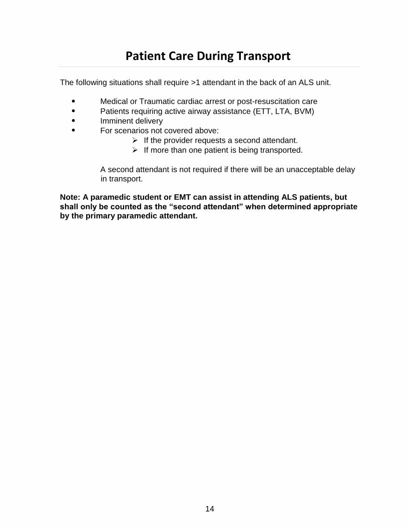
The following situations shall require >1 attendant in the back of an ALS unit.
Medical or Traumatic cardiac arrest or post-resuscitation care
Patients requiring active airway assistance (ETT, LTA, BVM)
Imminent delivery
For scenarios not covered above:
If the provider requests a second attendant.
If more than one patient is being transported.
A second attendant is not required if there will be an unacceptable delay in transport.
Note: A paramedic student or EMT can assist in attending ALS patients, but
shall only be counted as the “second attendant” when determined appropriate by the primary paramedic attendant.
14
Patient Care Standards During Interfacility Transport
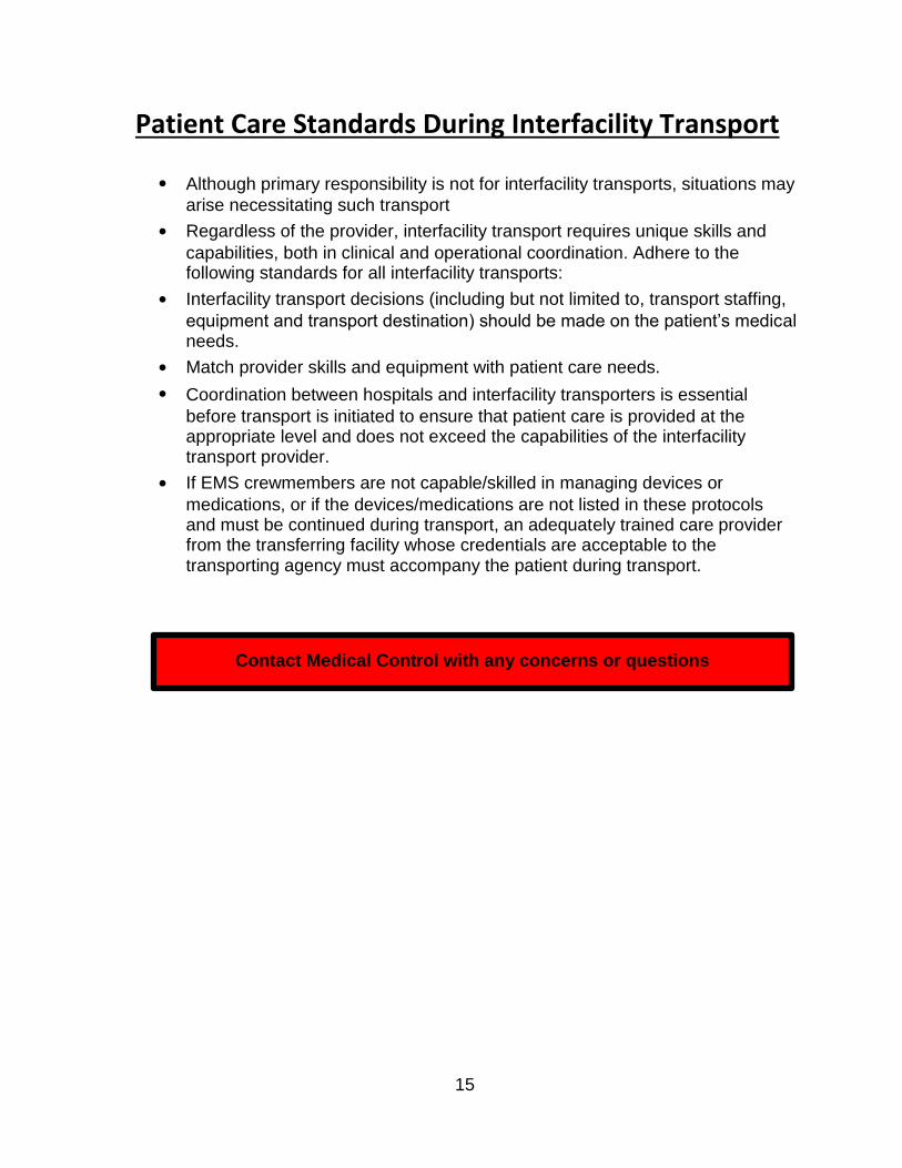
Although primary responsibility is not for interfacility transports, situations may
arise necessitating such transport
Regardless of the provider, interfacility transport requires unique skills and
capabilities, both in clinical and operational coordination. Adhere to the following standards for all interfacility transports:
Interfacility transport decisions (including but not limited to, transport staffing,
equipment and transport destination) should be made on the patient’s medical needs.
Match provider skills and equipment with patient care needs.
Coordination between hospitals and interfacility transporters is essential
before transport is initiated to ensure that patient care is provided at the appropriate level and does not exceed the capabilities of the interfacility transport provider.
If EMS crewmembers are not capable/skilled in managing devices or
medications, or if the devices/medications are not listed in these protocols and must be continued during transport, an adequately trained care provider from the transferring facility whose credentials are acceptable to the transporting agency must accompany the patient during transport.
Contact Medical Control with any concerns or questions
15
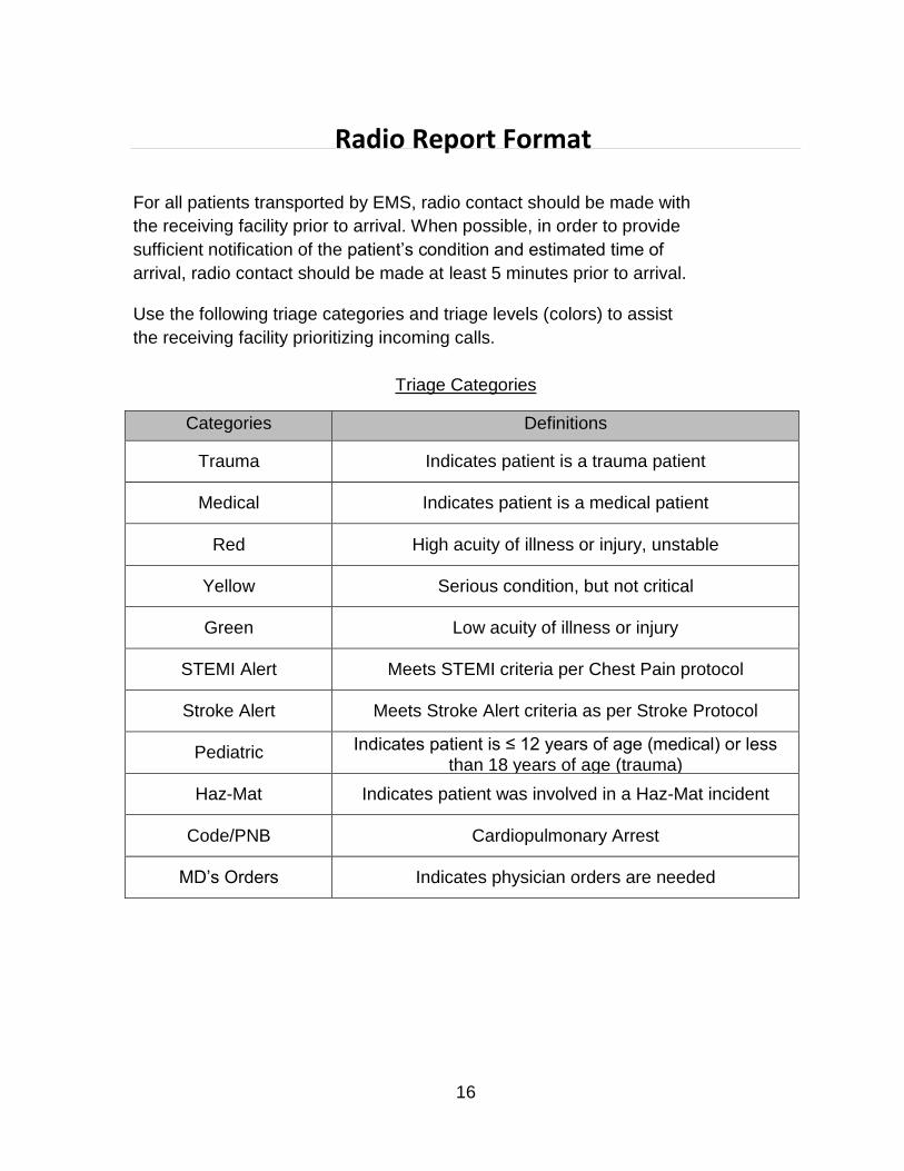
Categories Definitions
Trauma Indicates patient is a trauma patient
Medical Indicates patient is a medical patient
Red High acuity of illness or injury, unstable
Yellow Serious condition, but not critical
Green Low acuity of illness or injury
STEMI Alert Meets STEMI criteria per Chest Pain protocol
Stroke Alert Meets Stroke Alert criteria as per Stroke Protocol
Pediatric Indicates patient is ≤ 12 years of age (medical) or less than 18 years of age (trauma)
Haz-Mat Indicates patient was involved in a Haz-Mat incident
Code/PNB Cardiopulmonary Arrest
MD’s Orders Indicates physician orders are needed
Radio Report Format
For all patients transported by EMS, radio contact should be made with
the receiving facility prior to arrival. When possible, in order to provide
sufficient notification of the patient’s condition and estimated time of
arrival, radio contact should be made at least 5 minutes prior to arrival.
Use the following triage categories and triage levels (colors) to assist
the receiving facility prioritizing incoming calls.
Triage Categories
16
Radio Report Format
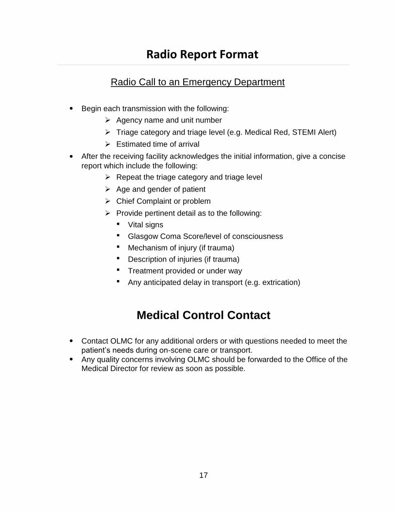
Radio Call to an Emergency Department
Begin each transmission with the following:
Agency name and unit number
Triage category and triage level (e.g. Medical Red, STEMI Alert)
Estimated time of arrival
After the receiving facility acknowledges the initial information, give a concise
report which include the following:
Repeat the triage category and triage level
Age and gender of patient
Chief Complaint or problem
Provide pertinent detail as to the following:
Vital signs
Glasgow Coma Score/level of consciousness
Mechanism of injury (if trauma)
Description of injuries (if trauma)
Treatment provided or under way
Any anticipated delay in transport (e.g. extrication)
Medical Control Contact
Contact OLMC for any additional orders or with questions needed to meet the
patient’s needs during on-scene care or transport.
Any quality concerns involving OLMC should be forwarded to the Office of the Medical Director for review as soon as possible.
17
Transfer of Care at Hospitals
Once on hospital property, the receiving facility assumes responsibility for all further
medical care delivered to EMS transported patients. Deer-Grove EMS personnel are not authorized to follow pre-hospital protocols after arrival at an ED and DGEMS Medical Control should not be contacted for orders.
Exceptions to this should occur only in the following circumstances:
Life-threatening situations such as cardiac arrest, airway emergencies or
imminent delivery of a newborn.
Continuation of treatment started prior to arrival (i.e. nebulizers, CPAP, IV fluids)
When specifically instructed to continue care by the ED physician (when possible document the physician’s name and the time the verbal order was given)
To assure all known pertinent information is conveyed to the hospital staff, crews
should interface with nursing staff within 2 minutes of arrival to give a verbal report. Transporting personnel shall provide to the receiving facility all known pertinent incident; patient identification and patient care information at the time the patient is transferred. In addition turn over all pre-hospital 12 lead EKGs to the ED staff. Patient care reports may be transmitted by physical (paper) means or electronic means.
18
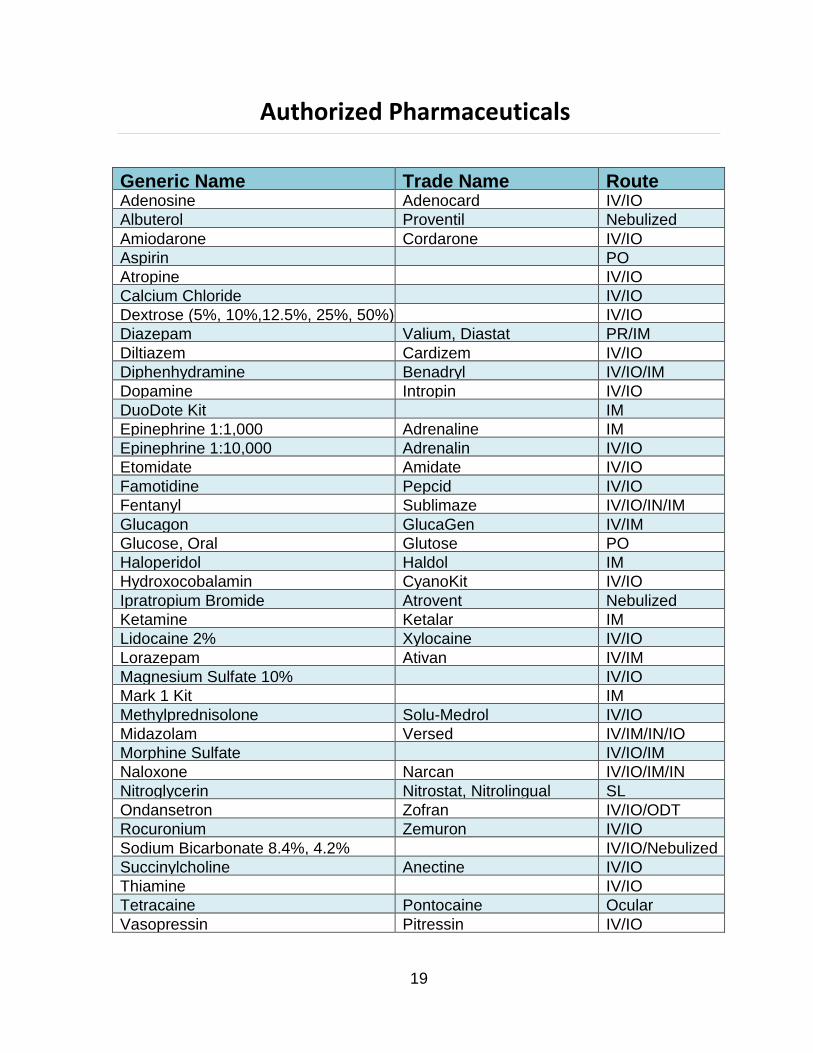
Generic Name Trade Name Route Adenosine Adenocard IV/IO
Albuterol Proventil Nebulized
Amiodarone Cordarone IV/IO
Aspirin PO
Atropine IV/IO
Calcium Chloride IV/IO
Dextrose (5%, 10%,12.5%, 25%, 50%) IV/IO
Diazepam Valium, Diastat PR/IM
Diltiazem Cardizem IV/IO
Diphenhydramine Benadryl IV/IO/IM
Dopamine Intropin IV/IO
DuoDote Kit IM
Epinephrine 1:1,000 Adrenaline IM
Epinephrine 1:10,000 Adrenalin IV/IO
Etomidate Amidate IV/IO
Famotidine Pepcid IV/IO
Fentanyl Sublimaze IV/IO/IN/IM
Glucagon GlucaGen IV/IM
Glucose, Oral Glutose PO
Haloperidol Haldol IM
Hydroxocobalamin CyanoKit IV/IO
Ipratropium Bromide Atrovent Nebulized
Ketamine Ketalar IM
Lidocaine 2% Xylocaine IV/IO
Lorazepam Ativan IV/IM
Magnesium Sulfate 10% IV/IO
Mark 1 Kit IM
Methylprednisolone Solu-Medrol IV/IO
Midazolam Versed IV/IM/IN/IO
Morphine Sulfate IV/IO/IM
Naloxone Narcan IV/IO/IM/IN
Nitroglycerin Nitrostat, Nitrolingual SL
Ondansetron Zofran IV/IO/ODT
Rocuronium Zemuron IV/IO
Sodium Bicarbonate 8.4%, 4.2% IV/IO/Nebulized
Succinylcholine Anectine IV/IO
Thiamine IV/IO
Tetracaine Pontocaine Ocular
Vasopressin Pitressin IV/IO
Authorized Pharmaceuticals
19
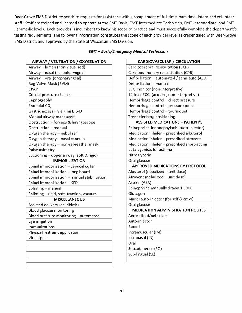
Deer-Grove EMS District responds to requests for assistance with a complement of full-time, part-time, intern and volunteer
staff. Staff are trained and licensed to operate at the EMT-Basic, EMT-Intermediate Technician, EMT-Intermediate, and EMT-
Paramedic levels. Each provider is incumbent to know his scope of practice and must successfully complete the department’s
testing requirements. The following information constitutes the scope of each provider level as credentialed with Deer-Grove
EMS District, and approved by the State of Wisconsin EMS Division.
EMT – Basic/Emergency Medical Technician
AIRWAY / VENTILATION / OXYGENATION
Airway – lumen (non-visualized)
Airway – nasal (nasopharyngeal)
Airway – oral (oropharyngeal)
Bag-Valve-Mask (BVM)
CPAP
Cricoid pressure (Sellick)
Capnography
End tidal CO2
Gastric access – via King LTS-D
Manual airway maneuvers
Obstruction – forceps & laryngoscope
Obstruction – manual
Oxygen therapy – nebulizer
Oxygen therapy – nasal cannula
Oxygen therapy – non-rebreather mask
Pulse oximetry
Suctioning – upper airway (soft & rigid)
IMMOBILIZATION
Spinal immobilization – cervical collar
Spinal immobilization – long board
Spinal immobilization – manual stabilization
Spinal immobilization – KED
Splinting – manual
Splinting – rigid, soft, traction, vacuum
MISCELLANEOUS
Assisted delivery (childbirth)
Blood glucose monitoring
Blood pressure monitoring – automated
Eye irrigation
Immunizations
Physical restraint application
Vital signs
CARDIOVASCULAR / CIRCULATION
Cardiocerebral resuscitation (CCR)
Cardiopulmonary resuscitation (CPR)
Defibrillation – automated / semi-auto (AED)
Defibrillation – manual
ECG monitor (non-interpretive)
12-lead ECG (acquire, non-interpretive)
Hemorrhage control – direct pressure
Hemorrhage control – pressure point
Hemorrhage control – tourniquet
Trendelenberg positioning
ASSISTED MEDICATIONS – PATIENT’S
Epinephrine for anaphylaxis (auto-injector)
Medication inhaler – prescribed albuterol
Medication inhaler – prescribed atrovent
Medication inhaler – prescribed short-acting beta agonists for asthma
Nitroglycerin
Oral glucose
APPROVED MEDICATIONS BY PROTOCOL
Albuterol (nebulized – unit dose)
Atrovent (nebulized – unit dose)
Aspirin (ASA)
Epinephrine manually drawn 1:1000
Glucagon
Mark I auto-injector (for self & crew)
Oral glucose
MEDICATION ADMINISTRATION ROUTES
Aerosolized/nebulizer
Auto-injector
Buccal
Intramuscular (IM)
Intranasal (IN)
Oral
Subcutaneous (SQ)
Sub-lingual (SL)
20
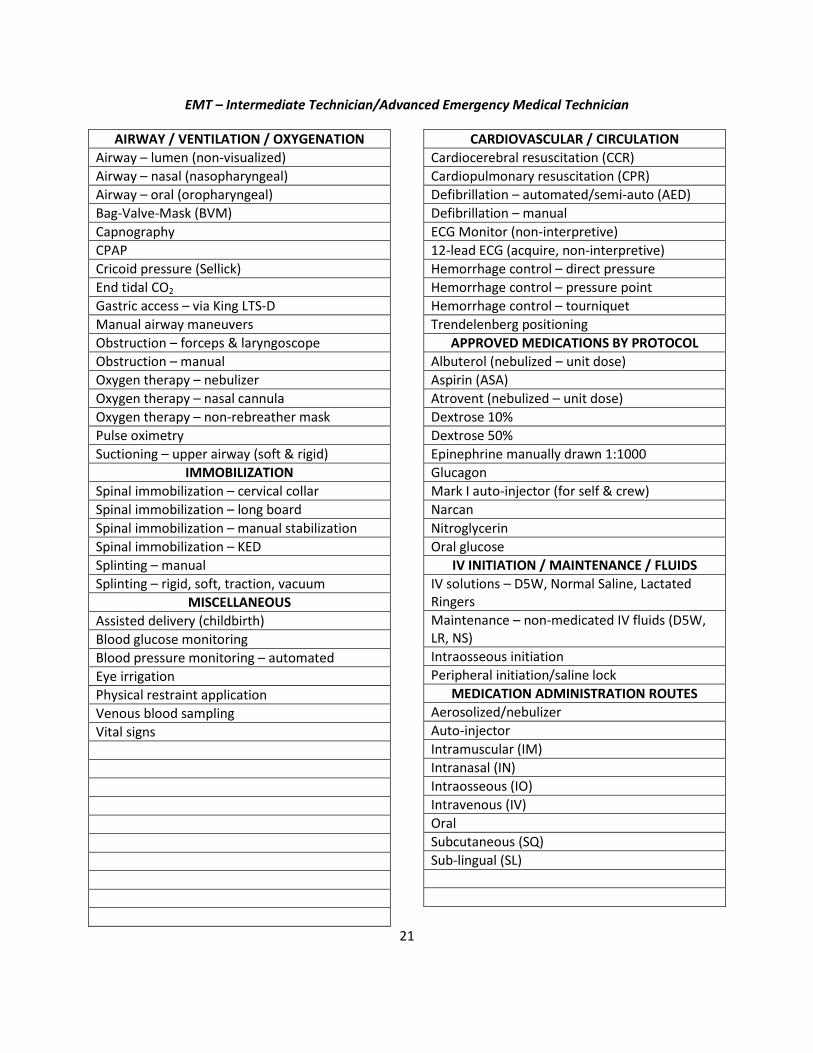
EMT – Intermediate Technician/Advanced Emergency Medical Technician
AIRWAY / VENTILATION / OXYGENATION
Airway – lumen (non-visualized)
Airway – nasal (nasopharyngeal)
Airway – oral (oropharyngeal)
Bag-Valve-Mask (BVM)
Capnography
CPAP
Cricoid pressure (Sellick)
End tidal CO2
Gastric access – via King LTS-D
Manual airway maneuvers
Obstruction – forceps & laryngoscope
Obstruction – manual
Oxygen therapy – nebulizer
Oxygen therapy – nasal cannula
Oxygen therapy – non-rebreather mask
Pulse oximetry
Suctioning – upper airway (soft & rigid)
IMMOBILIZATION
Spinal immobilization – cervical collar
Spinal immobilization – long board
Spinal immobilization – manual stabilization
Spinal immobilization – KED
Splinting – manual
Splinting – rigid, soft, traction, vacuum
MISCELLANEOUS
Assisted delivery (childbirth)
Blood glucose monitoring
Blood pressure monitoring – automated
Eye irrigation
Physical restraint application
Venous blood sampling
Vital signs
CARDIOVASCULAR / CIRCULATION
Cardiocerebral resuscitation (CCR)
Cardiopulmonary resuscitation (CPR)
Defibrillation – automated/semi-auto (AED)
Defibrillation – manual
ECG Monitor (non-interpretive)
12-lead ECG (acquire, non-interpretive)
Hemorrhage control – direct pressure
Hemorrhage control – pressure point
Hemorrhage control – tourniquet
Trendelenberg positioning
APPROVED MEDICATIONS BY PROTOCOL
Albuterol (nebulized – unit dose)
Aspirin (ASA)
Atrovent (nebulized – unit dose)
Dextrose 10%
Dextrose 50%
Epinephrine manually drawn 1:1000
Glucagon
Mark I auto-injector (for self & crew)
Narcan
Nitroglycerin
Oral glucose
IV INITIATION / MAINTENANCE / FLUIDS
IV solutions – D5W, Normal Saline, Lactated Ringers
Maintenance – non-medicated IV fluids (D5W, LR, NS)
Intraosseous initiation
Peripheral initiation/saline lock
MEDICATION ADMINISTRATION ROUTES
Aerosolized/nebulizer
Auto-injector
Intramuscular (IM)
Intranasal (IN)
Intraosseous (IO)
Intravenous (IV)
Oral
Subcutaneous (SQ)
Sub-lingual (SL)
21
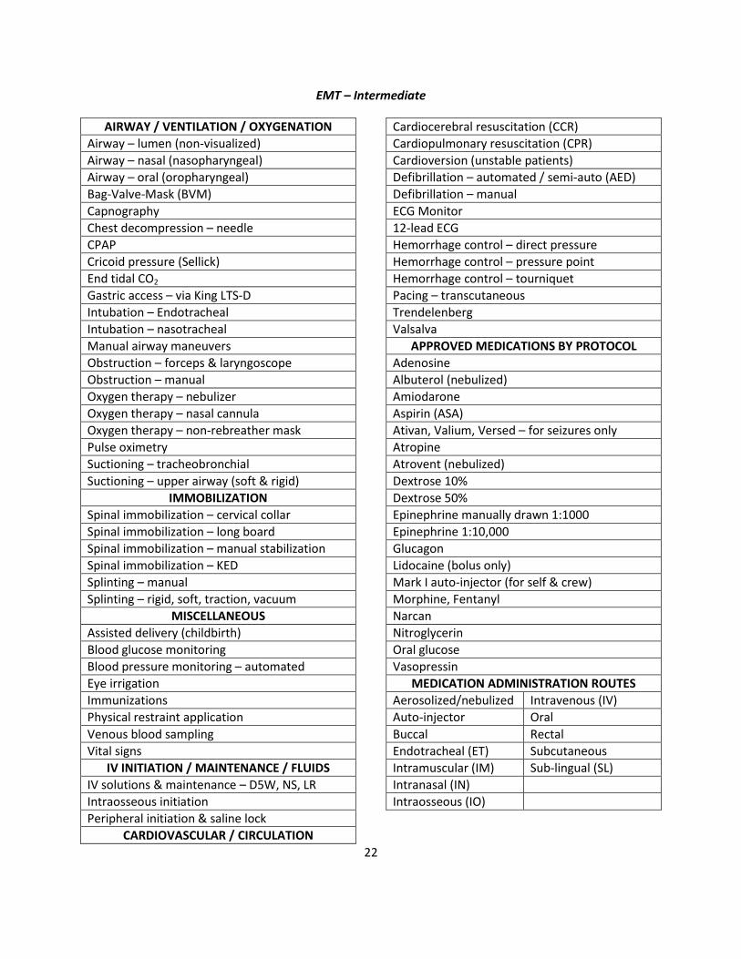
EMT – Intermediate
AIRWAY / VENTILATION / OXYGENATION
Airway – lumen (non-visualized)
Airway – nasal (nasopharyngeal)
Airway – oral (oropharyngeal)
Bag-Valve-Mask (BVM)
Capnography
Chest decompression – needle
CPAP
Cricoid pressure (Sellick)
End tidal CO2
Gastric access – via King LTS-D
Intubation – Endotracheal
Intubation – nasotracheal
Manual airway maneuvers
Obstruction – forceps & laryngoscope
Obstruction – manual
Oxygen therapy – nebulizer
Oxygen therapy – nasal cannula
Oxygen therapy – non-rebreather mask
Pulse oximetry
Suctioning – tracheobronchial
Suctioning – upper airway (soft & rigid)
IMMOBILIZATION
Spinal immobilization – cervical collar
Spinal immobilization – long board
Spinal immobilization – manual stabilization
Spinal immobilization – KED
Splinting – manual
Splinting – rigid, soft, traction, vacuum
MISCELLANEOUS
Assisted delivery (childbirth)
Blood glucose monitoring
Blood pressure monitoring – automated
Eye irrigation
Immunizations
Physical restraint application
Venous blood sampling
Vital signs
IV INITIATION / MAINTENANCE / FLUIDS
IV solutions & maintenance – D5W, NS, LR
Intraosseous initiation
Peripheral initiation & saline lock
CARDIOVASCULAR / CIRCULATION
Cardiocerebral resuscitation (CCR)
Cardiopulmonary resuscitation (CPR)
Cardioversion (unstable patients)
Defibrillation – automated / semi-auto (AED)
Defibrillation – manual
ECG Monitor
12-lead ECG
Hemorrhage control – direct pressure
Hemorrhage control – pressure point
Hemorrhage control – tourniquet
Pacing – transcutaneous
Trendelenberg
Valsalva
APPROVED MEDICATIONS BY PROTOCOL
Adenosine
Albuterol (nebulized)
Amiodarone
Aspirin (ASA)
Ativan, Valium, Versed – for seizures only
Atropine
Atrovent (nebulized)
Dextrose 10%
Dextrose 50%
Epinephrine manually drawn 1:1000
Epinephrine 1:10,000
Glucagon
Lidocaine (bolus only)
Mark I auto-injector (for self & crew)
Morphine, Fentanyl
Narcan
Nitroglycerin
Oral glucose
Vasopressin
MEDICATION ADMINISTRATION ROUTES
Aerosolized/nebulized Intravenous (IV)
Auto-injector Oral
Buccal Rectal
Endotracheal (ET) Subcutaneous
Intramuscular (IM) Sub-lingual (SL)
Intranasal (IN)
Intraosseous (IO)
22
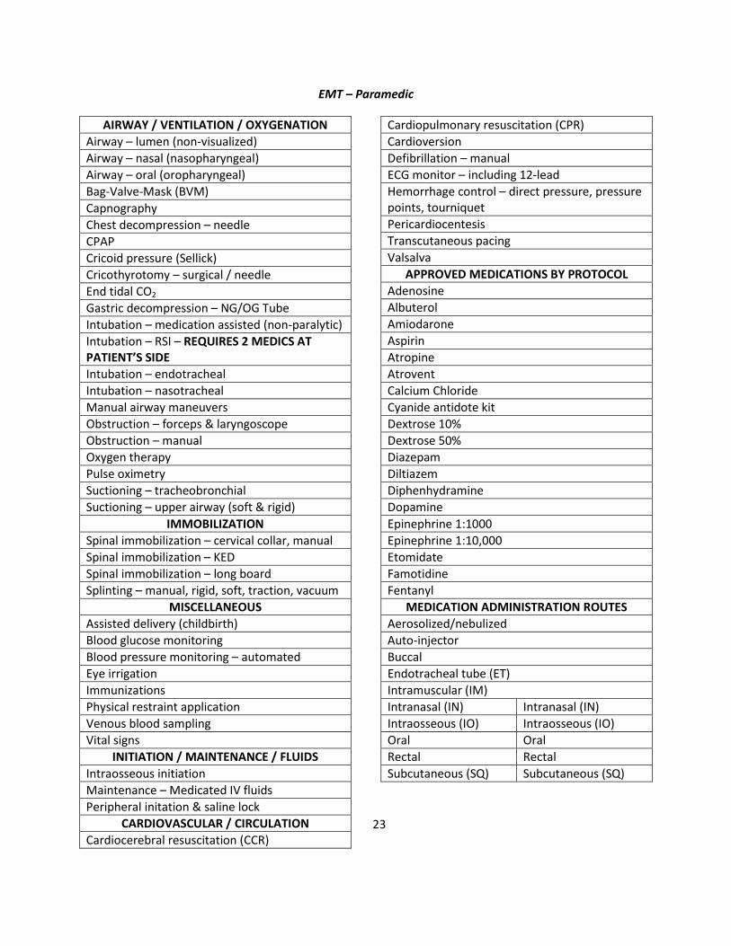
EMT – Paramedic
AIRWAY / VENTILATION / OXYGENATION
Airway – lumen (non-visualized)
Airway – nasal (nasopharyngeal)
Airway – oral (oropharyngeal)
Bag-Valve-Mask (BVM)
Capnography
Chest decompression – needle
CPAP
Cricoid pressure (Sellick)
Cricothyrotomy – surgical / needle
End tidal CO2
Gastric decompression – NG/OG Tube
Intubation – medication assisted (non-paralytic)
Intubation – RSI – REQUIRES 2 MEDICS AT PATIENT’S SIDE
Intubation – endotracheal
Intubation – nasotracheal
Manual airway maneuvers
Obstruction – forceps & laryngoscope
Obstruction – manual
Oxygen therapy
Pulse oximetry
Suctioning – tracheobronchial
Suctioning – upper airway (soft & rigid)
IMMOBILIZATION
Spinal immobilization – cervical collar, manual
Spinal immobilization – KED
Spinal immobilization – long board
Splinting – manual, rigid, soft, traction, vacuum
MISCELLANEOUS
Assisted delivery (childbirth)
Blood glucose monitoring
Blood pressure monitoring – automated
Eye irrigation
Immunizations
Physical restraint application
Venous blood sampling
Vital signs
INITIATION / MAINTENANCE / FLUIDS
Intraosseous initiation
Maintenance – Medicated IV fluids
Peripheral initation & saline lock
CARDIOVASCULAR / CIRCULATION
Cardiocerebral resuscitation (CCR)
Cardiopulmonary resuscitation (CPR)
Cardioversion
Defibrillation – manual
ECG monitor – including 12-lead
Hemorrhage control – direct pressure, pressure points, tourniquet
Pericardiocentesis
Transcutaneous pacing
Valsalva
APPROVED MEDICATIONS BY PROTOCOL
Adenosine
Albuterol
Amiodarone
Aspirin
Atropine
Atrovent
Calcium Chloride
Cyanide antidote kit
Dextrose 10%
Dextrose 50%
Diazepam
Diltiazem
Diphenhydramine
Dopamine
Epinephrine 1:1000
Epinephrine 1:10,000
Etomidate
Famotidine
Fentanyl
MEDICATION ADMINISTRATION ROUTES
Aerosolized/nebulized
Auto-injector
Buccal
Endotracheal tube (ET)
Intramuscular (IM)
Intranasal (IN) Intranasal (IN)
Intraosseous (IO) Intraosseous (IO)
Oral Oral
Rectal Rectal
Subcutaneous (SQ) Subcutaneous (SQ)
23
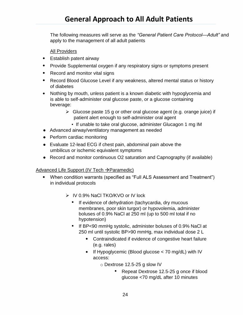
General Approach to All Adult Patients
The following measures will serve as the “General Patient Care Protocol—Adult” and
apply to the management of all adult patients
All Providers
Establish patent airway
Provide Supplemental oxygen if any respiratory signs or symptoms present
Record and monitor vital signs
Record Blood Glucose Level if any weakness, altered mental status or history
of diabetes
Nothing by mouth, unless patient is a known diabetic with hypoglycemia and
is able to self-administer oral glucose paste, or a glucose containing beverage:
Glucose paste 15 g or other oral glucose agent (e.g. orange juice) if
patient alert enough to self-administer oral agent
▪ If unable to take oral glucose, administer Glucagon 1 mg IM
● Advanced airway/ventilatory management as needed
● Perform cardiac monitoring
● Evaluate 12-lead ECG if chest pain, abdominal pain above the
umbilicus or ischemic equivalent symptoms
● Record and monitor continuous O2 saturation and Capnography (if available)
Advanced Life Support (IV Tech Paramedic)
When condition warrants (specified as “Full ALS Assessment and Treatment”)
in individual protocols
IV 0.9% NaCl TKO/KVO or IV lock
If evidence of dehydration (tachycardia, dry mucous
membranes, poor skin turgor) or hypovolemia, administer boluses of 0.9% NaCl at 250 ml (up to 500 ml total if no hypotension)
If BP<90 mmHg systolic, administer boluses of 0.9% NaCl at
250 ml until systolic BP>90 mmHg, max individual dose 2 L
Contraindicated if evidence of congestive heart failure
(e.g. rales)
If Hypoglycemic (Blood glucose < 70 mg/dL) with IV
access:
o Dextrose 12.5-25 g slow IV
Repeat Dextrose 12.5-25 g once if blood
glucose <70 mg/dL after 10 minutes
24
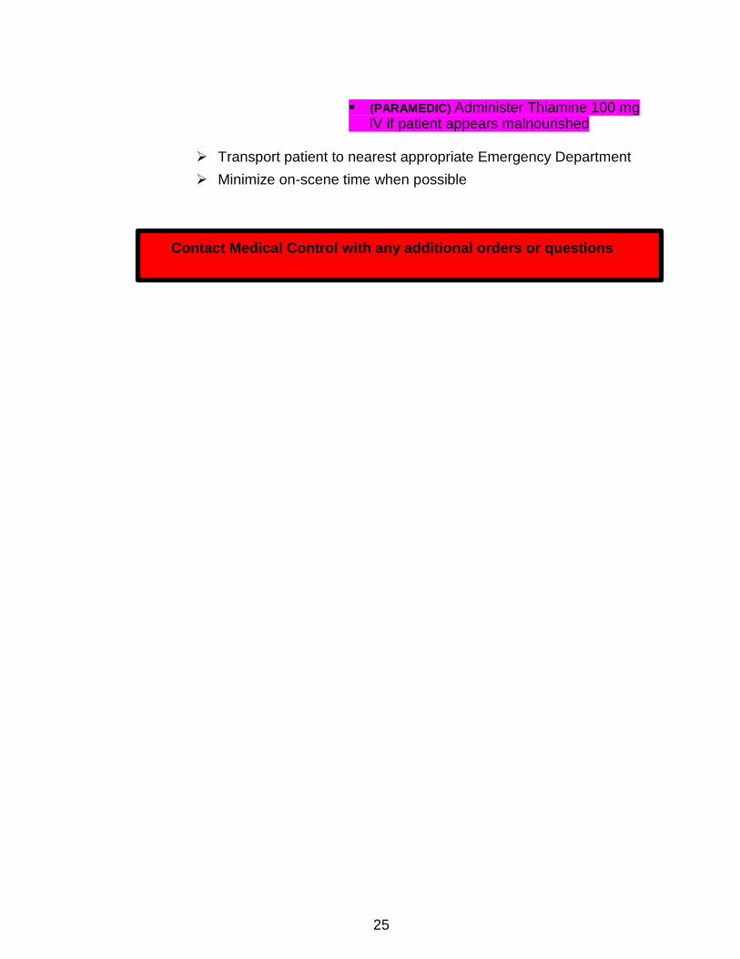
(PARAMEDIC) Administer Thiamine 100 mg IV if patient appears malnourished
Transport patient to nearest appropriate Emergency Department
Minimize on-scene time when possible
Contact Medical Control with any additional orders or questions
25
Abdominal Pain/GI Bleeding
All Providers
General Patient Care Protocol—Adult
Nothing by mouth
If pain is above the umbilicus, perform cardiac monitoring and 12-lead EKG, refer to Chest Pain Protocol if indicated
Advanced Life Support (IV Tech Paramedic)
Full ALS Assessment and Treatment
For Patients with severe nausea or vomiting:
(PARAMEDIC) Ondansetron (Zofran), 4 mg IV/IM/Oral Disintegrating Tablet (ODT)
Refer to Pain Management Protocol if indicated
Contact Medical Control for any additional orders or questions
26
Airway Emergencies: Adult Dyspnea
All Providers
General Patient Care Protocol—Adult
Supplemental oxygen to maintain SPO2 > 92%
Albuterol (Proventil) 2.5 mg/3 ml and Ipratropium Bromide 0.02%
(Atrovent) 0.5 mg/2.5 ml via nebulizer, if wheezing or history of Asthma/COPD. May repeat PRN for continued wheezing.
Perform obstructed airway procedures per BLS standards. Consider CPAP if symptoms moderate to severe.
Advanced Life Support (IV Tech Paramedic)
Full ALS Assessment and Treatment
Observe for signs of impending respiratory failure: Refer to Airway
Management Protocol if indicated:
Hypoxia (O2 sat < 90%) not improved with 100% O2
Poor ventilatory effort
Altered mental status/decreased level of consciousness
Inability to maintain patent airway
Acute Bronchospasm (wheezing with or without history of Asthma or COPD)
Mild Symptoms:
Albuterol (Proventil) 2.5 mg/3 ml and Ipratropium Bromide 0.02%
(Atrovent) 0.5 mg/2.5 ml via nebulizer if not already given
May repeat Albuterol (Proventil) PRN for continued wheezing
Moderate Symptoms:
As for mild symptoms, additionally:
(PARAMEDIC) Methylprednisolone (Solumedrol) 125 mg IV if wheezing
persists after 1st nebulizer treatment
Consider CPAP if symptoms moderate to severe
Use settings specified in procedure manual for Asthma/COPD
Severe Symptoms (not speaking, little or no air movement):
As above, additionally:
Epinephrine 0.3 mg 1:1000 IM
OLMC if age >50, HR>150 or history of CAD
(PARAMEDIC) Magnesium Sulfate 2 g IV in 100 ml D5W over 10 min
Do not use if CHF or history of Renal Failure
27
Acute Pulmonary Edema Suspected
(History of CHF, peripheral edema elevated SBP)
Nitroglycerin 0.4 mg SL every 3 min:
Contraindicated if SBP <90 mmHg
Contraindicated if use of a Phosphodiesterase-5 (PDE-5) inhibitor
within last 24 hours (Viagra, Levitra); 48 hours (Cialis)
Aspirin 324 mg PO
Consider CPAP if symptoms moderate/severe:
Use settings specified in procedure manual for CHF/Pulmonary Edema
For Hypotension (systolic BP <90 mmHg):
(PARAMEDIC) Consider Dopamine infusion 5-20 mcg/kg/min titrate to maintain
SBP >90 mmHg
If severe nausea or vomiting:
(PARAMEDIC) Ondansetron (Zofran) 4 mg IV/IO/ODT
For bronchospasm (wheezing) associated with Acute Pulmonary Edema:
Albuterol (Proventil) 2.5 mg/3 ml and Ipratropium Bromide 0.02%
(Atrovent) 0.5 mg/2.5 ml via nebulizer
May repeat Albuterol (Proventil) PRN for continued wheezing
Consider Airway Management Protocol
Carbon Monoxide Poisoning
Refer to Overdose and Poisoning Protocol
Foreign Body Obstruction Suspected
Perform Obstructed Airway Procedures per BLS Standard
Attempt suction
Attempt removal with Magill forceps under direct visualization with
laryngoscope
Observe for signs of impending respiratory failure
Refer to Airway Management Protocol if indicated
28
Drowning/Near Drowning
Full ALS Assessment and Treatment
Spinal Immobilization if indicated
Protect from heat loss
Patients may develop delayed onset respiratory symptoms:
Consider CPAP for patients with significant dyspnea or hypoxia
Airway Protocol as needed
Refer to appropriate protocol if cardiac arrest present
Contact Medical Control for any additional orders or questions
Acute Bronchospasm: Contact Medical Control prior to Epinephrine administration if:
Age > 50 years
Heart Rate >150
History of Coronary Artery Disease
29
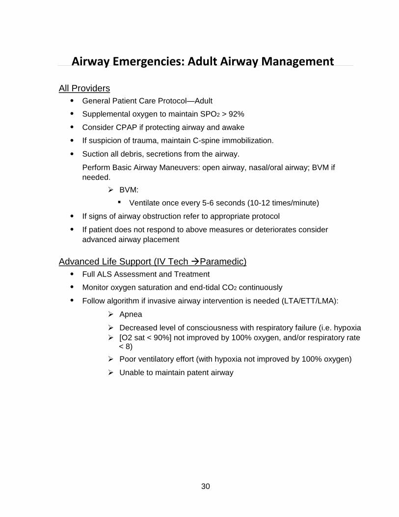
Airway Emergencies: Adult Airway Management
All Providers
General Patient Care Protocol—Adult
Supplemental oxygen to maintain SPO2 > 92%
Consider CPAP if protecting airway and awake
If suspicion of trauma, maintain C-spine immobilization.
Suction all debris, secretions from the airway.
Perform Basic Airway Maneuvers: open airway, nasal/oral airway; BVM if
needed.
BVM:
Ventilate once every 5-6 seconds (10-12 times/minute)
If signs of airway obstruction refer to appropriate protocol
If patient does not respond to above measures or deteriorates consider
advanced airway placement
Advanced Life Support (IV Tech Paramedic)
Full ALS Assessment and Treatment
Monitor oxygen saturation and end-tidal CO2 continuously
Follow algorithm if invasive airway intervention is needed (LTA/ETT/LMA):
Apnea
Decreased level of consciousness with respiratory failure (i.e. hypoxia
[O2 sat < 90%] not improved by 100% oxygen, and/or respiratory rate < 8)
Poor ventilatory effort (with hypoxia not improved by 100% oxygen)
Unable to maintain patent airway
30
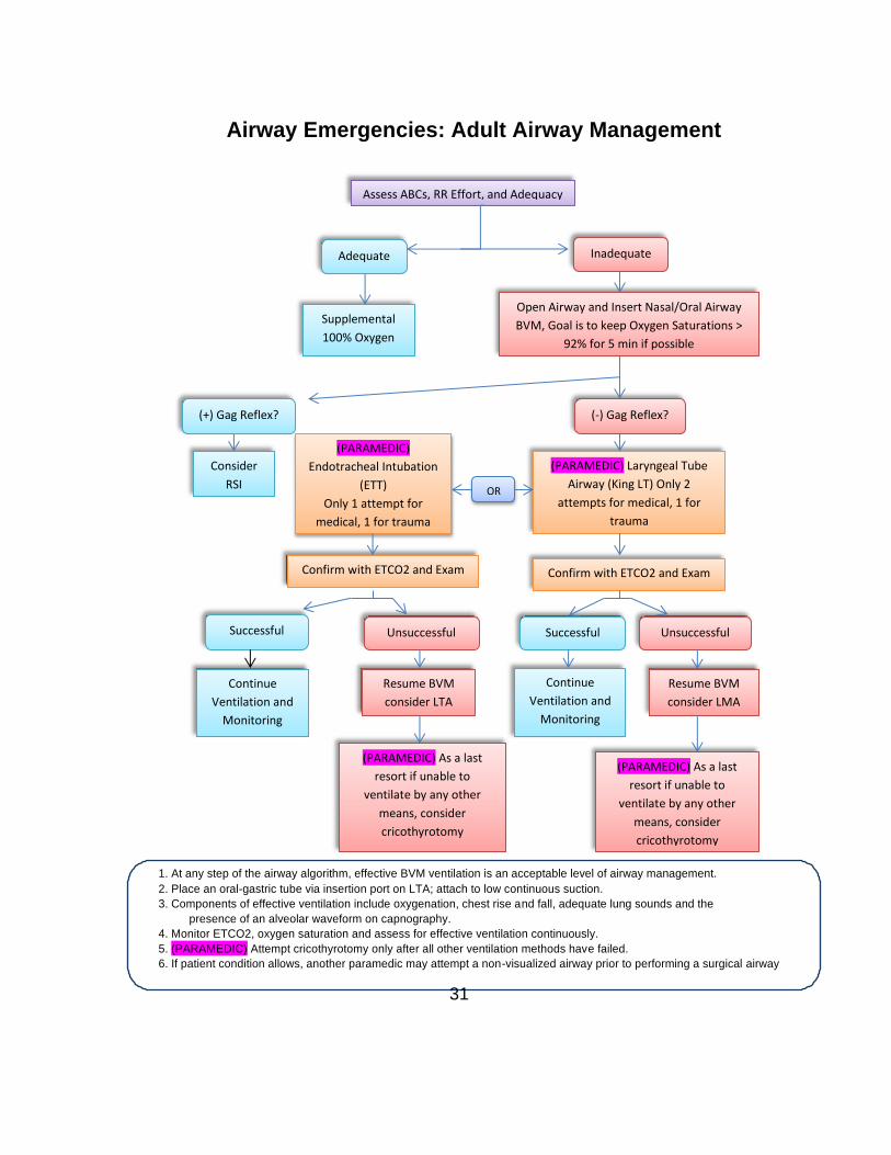
Airway Emergencies: Adult Airway Management
As a last resort if unable to
ventilate by any other
means, consider
cricothyrotomy5
1. At any step of the airway algorithm, effective BVM ventilation is an acceptable level of airway management.
2. Place an oral-gastric tube via insertion port on LTA; attach to low continuous suction.
3. Components of effective ventilation include oxygenation, chest rise and fall, adequate lung sounds and the
presence of an alveolar waveform on capnography.
4. Monitor ETCO2, oxygen saturation and assess for effective ventilation continuously.
5. (PARAMEDIC) Attempt cricothyrotomy only after all other ventilation methods have failed.
6. If patient condition allows, another paramedic may attempt a non-visualized airway prior to performing a surgical airway
31
Assess ABCs, RR Effort, and Adequacy
Adequate
Supplemental
100% Oxygen
(+) Gag Reflex?
Consider
RSI
Inadequate
Open Airway and Insert Nasal/Oral Airway
BVM, Goal is to keep Oxygen Saturations >
92% for 5 min if possible
(-) Gag Reflex?
(PARAMEDIC) Laryngeal Tube
Airway (King LT) Only 2
attempts for medical, 1 for
trauma
Confirm with ETCO2 and Exam
OR
(PARAMEDIC)
Endotracheal Intubation
(ETT)
Only 1 attempt for
medical, 1 for trauma
Confirm with ETCO2 and Exam
Successful Unsuccessful Successful Unsuccessful
Continue
Ventilation and
Monitoring
Resume BVM
consider LTA
(PARAMEDIC) As a last
resort if unable to
ventilate by any other
means, consider
cricothyrotomy
Continue
Ventilation and
Monitoring
Resume BVM
consider LMA
(PARAMEDIC) As a last
resort if unable to
ventilate by any other
means, consider
cricothyrotomy
Following placement of the ETT/LTA/LMA confirm proper placement:
Assess for absence of epigastric sounds, presence of breath sounds,
and chest rise and fall
Observe for presence of alveolar waveform on capnography
Record tube depth and secure in place using a commercial holder if
applicable
Utilize head restraint devices (i.e. “head-blocks”) or rigid cervical collar
and long spine board as needed to help secure airway device in place
Capnography/ETCO2 Monitoring
Digital capnography (waveform) is the system standard for ETCO2 monitoring.
With the exception of on-scene equipment failure, patients should not be
routinely switched from digital capnography (monitor) to a colorimetric device for monitoring end-tidal CO2.
In the event digital capnography is not available due to on-scene equipment
failure, continuous colorimetric monitoring of ETCO2 is an acceptable alternative.
Continuous ETCO2 monitoring is a MANDATORY component of invasive
airway management.
If ETCO2 monitoring cannot be accomplished by either of the above
methods, the invasive device MUST be REMOVED, and the airway managed non-invasively.
If an alveolar waveform is not present with capnography (i.e. flat line),
briefly check the filter line coupling to assure it is securely in place then remove the ETT/LMA or LTA and proceed to the next step in the algorithm.
32
Airway Emergencies: Rapid Sequence Airway
Under no circumstances should transport be delayed for RSA if the additional
time to perform the procedure is greater than the transport time.
2 PARAMEDICS ARE REQUIRED AT ALL TIMES
All Providers
General Patient Care Protocol—Adult
Preoxygenate with 100% oxygen
Basic Airway maneuvers: open airway, nasal/oral airway; BVM
Advanced Life Support (PARAMEDICS)
Full ALS assessment and treatment
Simultaneously contact OLMC
Assess for Indications:
Age ≥18 unless specific permission given prior to procedure by
medical control
Need for invasive airway management in the setting of an intact gag
reflex or inadequate sedation to perform non pharmacologically assisted airway management:
Apnea
Decreased level of consciousness with respiratory failure (i.e.
hypoxia [O2 sat < 90%] not improved by 100% oxygen, and/or respiratory rate < 8)
Poor ventilatory effort (with hypoxia not improved by 100%
oxygen)
Unable to maintain patent airway by other means
Burns with suspected significant inhalation injury
Preoxygenate 100% oxygen via NRB for at least 5 min or 8 Vital Capacity
(deep) breaths with 100% O2
Only assist ventilations with BVM if patient’s ventilations are
inadequate or if hypoxemic (O2 Saturation < 92% on supplemental oxygen)
Assisted ventilations increase risk of aspiration during laryngoscopy
Patients cannot have any contraindications to succinylcholine or other RSA
drugs:
Inability to ventilate via BVM
Suspected Hyperkalemia
33
Myopathy or neuromuscular disease
History of Malignant Hyperthermia
Recent crush injury or major burn (>48 hours after injury)
End Stage Renal Disease
Recent Spinal Cord Injury (72 hours-6 months)
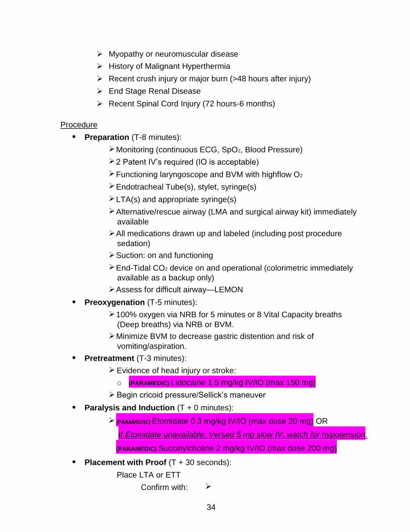
Procedure
Preparation (T-8 minutes):
Monitoring (continuous ECG, SpO2, Blood Pressure)
2 Patent IV’s required (IO is acceptable)
Functioning laryngoscope and BVM with highflow O2
Endotracheal Tube(s), stylet, syringe(s)
LTA(s) and appropriate syringe(s)
Alternative/rescue airway (LMA and surgical airway kit) immediately
available
All medications drawn up and labeled (including post procedure
sedation)
Suction: on and functioning
End-Tidal CO2 device on and operational (colorimetric immediately
available as a backup only)
Assess for difficult airway—LEMON
Preoxygenation (T-5 minutes):
100% oxygen via NRB for 5 minutes or 8 Vital Capacity breaths
(Deep breaths) via NRB or BVM.
Minimize BVM to decrease gastric distention and risk of
vomiting/aspiration.
Pretreatment (T-3 minutes):
Evidence of head injury or stroke:
o (PARAMEDIC) Lidocaine 1.5 mg/kg IV/IO (max 150 mg)
Begin cricoid pressure/Sellick’s maneuver
Paralysis and Induction (T + 0 minutes):
(PARAMEDIC) Etomidate 0.3 mg/kg IV/IO (max dose 20 mg) OR
If Etomidate unavailable, Versed 5 mg slow IV, watch for hypotension.
(PARAMEDIC) Succinylcholine 2 mg/kg IV/IO (max dose 200 mg)
Placement with Proof (T + 30 seconds):
Place LTA or ETT
Confirm with:
34
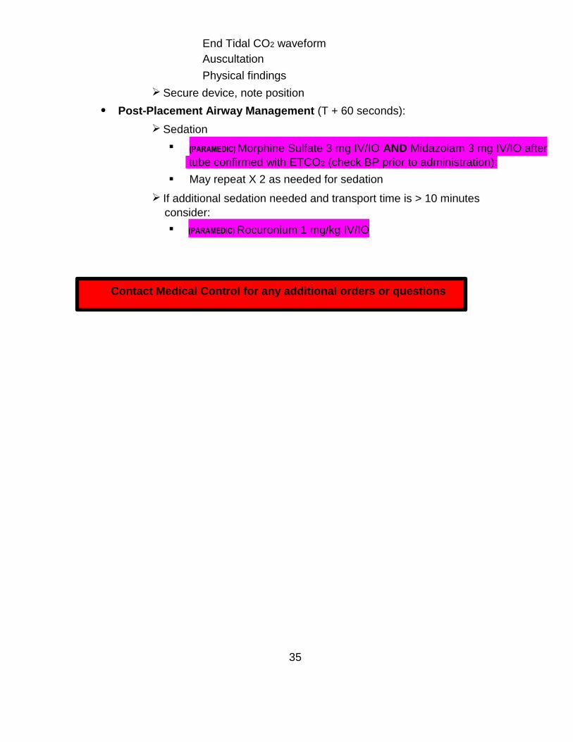
End Tidal CO2 waveform
Auscultation
Physical findings
Secure device, note position
Post-Placement Airway Management (T + 60 seconds):
Sedation
(PARAMEDIC) Morphine Sulfate 3 mg IV/IO AND Midazolam 3 mg IV/IO after
tube confirmed with ETCO2 (check BP prior to administration):
May repeat X 2 as needed for sedation
If additional sedation needed and transport time is > 10 minutes
consider:
(PARAMEDIC) Rocuronium 1 mg/kg IV/IO
Contact Medical Control for any additional orders or questions
35
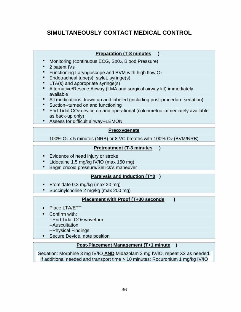
SIMULTANEOUSLY CONTACT MEDICAL CONTROL
Preparation (T-8 minutes )
Monitoring (continuous ECG, Sp02, Blood Pressure)
2 patent IVs Functioning Laryngoscope and BVM with high flow O2
Endotracheal tube(s), stylet, syringe(s) LTA(s) and appropriate syringe(s) Alternative/Rescue Airway (LMA and surgical airway kit) immediately available All medications drawn up and labeled (including post-procedure sedation) Suction--turned on and functioning End Tidal CO2 device on and operational (colorimetric immediately available as back-up only) Assess for difficult airway--LEMON
Preoxygenate
100% O2 x 5 minutes (NRB) or 8 VC breaths with 100% O2 (BVM/NRB)
Pretreatment (T-3 minutes )
Evidence of head injury or stroke
Lidocaine 1.5 mg/kg IV/IO (max 150 mg) Begin cricoid pressure/Sellick’s maneuver
Paralysis and Induction (T=0 )
Etomidate 0.3 mg/kg (max 20 mg)
Succinylcholine 2 mg/kg (max 200 mg)
Placement with Proof (T+30 seconds )
Place LTA/ETT
Confirm with:
--End Tidal CO2 waveform --Auscultation --Physical Findings
Secure Device, note position
Post-Placement Management (T+1 minute )
Sedation: Morphine 3 mg IV/IO AND Midazolam 3 mg IV/IO, repeat X2 as needed.
If additional needed and transport time > 10 minutes: Rocuronium 1 mg/kg IV/IO
36
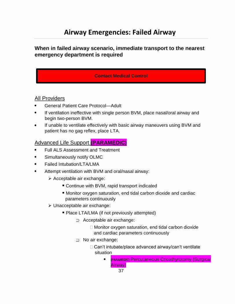
Airway Emergencies: Failed Airway
When in failed airway scenario, immediate transport to the nearest
emergency department is required
Contact Medical Control
All Providers
General Patient Care Protocol—Adult
If ventilation ineffective with single person BVM, place nasal/oral airway and
begin two-person BVM.
If unable to ventilate effectively with basic airway maneuvers using BVM and
patient has no gag reflex, place LTA.
Advanced Life Support (PARAMEDIC)
Full ALS Assessment and Treatment
Simultaneously notify OLMC
Failed Intubation/LTA/LMA
Attempt ventilation with BVM and oral/nasal airway:
Acceptable air exchange:
Continue with BVM, rapid transport indicated
Monitor oxygen saturation, end tidal carbon dioxide and cardiac
parameters continuously
Unacceptable air exchange:
Place LTA/LMA (if not previously attempted)
Acceptable air exchange:
Monitor oxygen saturation, end tidal carbon dioxide
and cardiac parameters continuously
No air exchange:
Can’t intubate/place advanced airway/can’t ventilate
situation
(PARAMEDIC) Percutaneous Cricothyrotomy (Surgical
Airway)
37
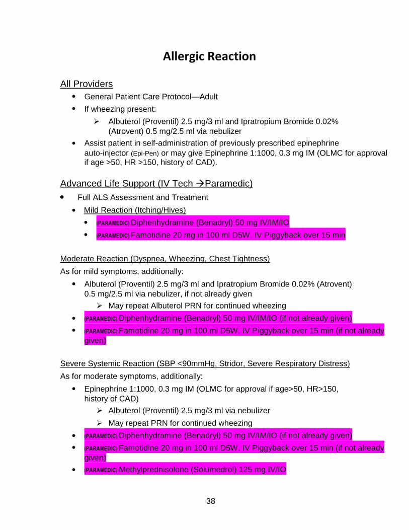
Allergic Reaction
All Providers
General Patient Care Protocol—Adult
If wheezing present:
Albuterol (Proventil) 2.5 mg/3 ml and Ipratropium Bromide 0.02%
(Atrovent) 0.5 mg/2.5 ml via nebulizer
Assist patient in self-administration of previously prescribed epinephrine
auto-injector (Epi-Pen) or may give Epinephrine 1:1000, 0.3 mg IM (OLMC for approval if age >50, HR >150, history of CAD).
Advanced Life Support (IV Tech Paramedic)
Full ALS Assessment and Treatment
Mild Reaction (Itching/Hives)
(PARAMEDIC) Diphenhydramine (Benadryl) 50 mg IV/IM/IO
(PARAMEDIC) Famotidine 20 mg in 100 ml D5W, IV Piggyback over 15 min
Moderate Reaction (Dyspnea, Wheezing, Chest Tightness)
As for mild symptoms, additionally:
Albuterol (Proventil) 2.5 mg/3 ml and Ipratropium Bromide 0.02% (Atrovent)
0.5 mg/2.5 ml via nebulizer, if not already given
May repeat Albuterol PRN for continued wheezing
(PARAMEDIC) Diphenhydramine (Benadryl) 50 mg IV/IM/IO (if not already given)
(PARAMEDIC) Famotidine 20 mg in 100 ml D5W, IV Piggyback over 15 min (if not already
given)
Severe Systemic Reaction (SBP <90mmHg, Stridor, Severe Respiratory Distress)
As for moderate symptoms, additionally:
Epinephrine 1:1000, 0.3 mg IM (OLMC for approval if age>50, HR>150,
history of CAD)
Albuterol (Proventil) 2.5 mg/3 ml via nebulizer
May repeat PRN for continued wheezing
(PARAMEDIC) Diphenhydramine (Benadryl) 50 mg IV/IM/IO (if not already given)
(PARAMEDIC) Famotidine 20 mg in 100 ml D5W, IV Piggyback over 15 min (if not already
given)
(PARAMEDIC) Methylprednisolone (Solumedrol) 125 mg IV/IO
38
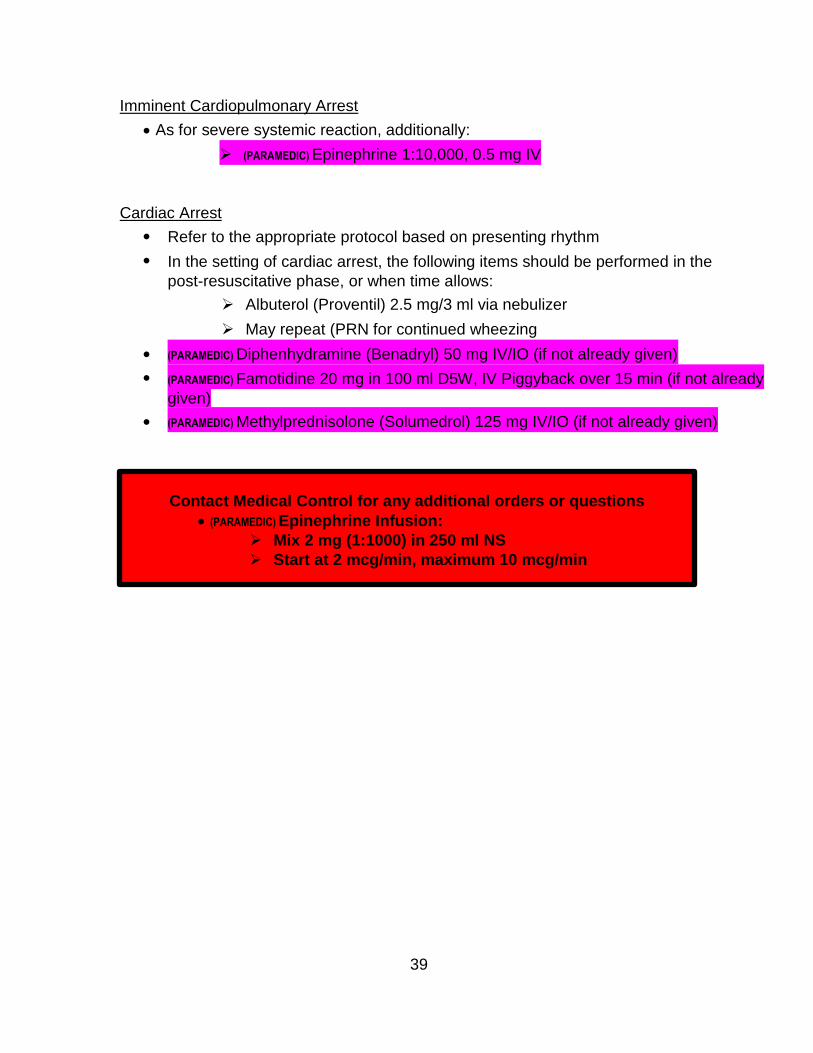
Imminent Cardiopulmonary Arrest
As for severe systemic reaction, additionally:
(PARAMEDIC) Epinephrine 1:10,000, 0.5 mg IV
Cardiac Arrest
Refer to the appropriate protocol based on presenting rhythm
In the setting of cardiac arrest, the following items should be performed in the
post-resuscitative phase, or when time allows:
Albuterol (Proventil) 2.5 mg/3 ml via nebulizer
May repeat (PRN for continued wheezing
(PARAMEDIC) Diphenhydramine (Benadryl) 50 mg IV/IO (if not already given)
(PARAMEDIC) Famotidine 20 mg in 100 ml D5W, IV Piggyback over 15 min (if not already
given)
(PARAMEDIC) Methylprednisolone (Solumedrol) 125 mg IV/IO (if not already given)
Contact Medical Control for any additional orders or questions
(PARAMEDIC) Epinephrine Infusion:
Mix 2 mg (1:1000) in 250 ml NS
Start at 2 mcg/min, maximum 10 mcg/min
39
40
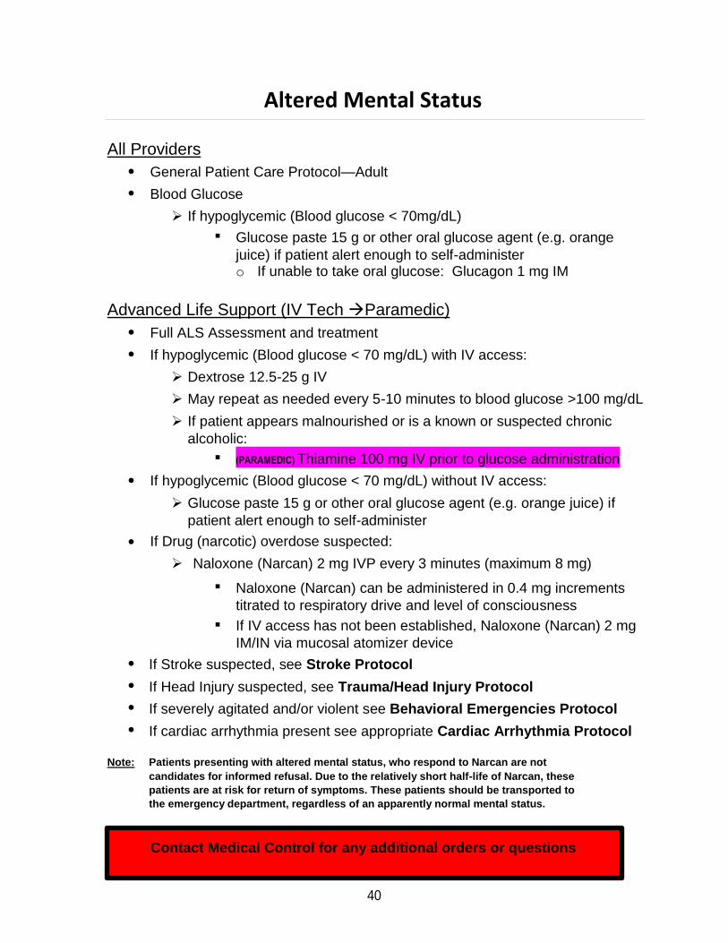
Altered Mental Status
All Providers
General Patient Care Protocol—Adult
Blood Glucose
If hypoglycemic (Blood glucose < 70mg/dL)
Glucose paste 15 g or other oral glucose agent (e.g. orange
juice) if patient alert enough to self-administer o If unable to take oral glucose: Glucagon 1 mg IM
Advanced Life Support (IV Tech Paramedic)
Full ALS Assessment and treatment
If hypoglycemic (Blood glucose < 70 mg/dL) with IV access:
Dextrose 12.5-25 g IV
May repeat as needed every 5-10 minutes to blood glucose >100 mg/dL
If patient appears malnourished or is a known or suspected chronic
alcoholic:
(PARAMEDIC) Thiamine 100 mg IV prior to glucose administration
If hypoglycemic (Blood glucose < 70 mg/dL) without IV access:
Glucose paste 15 g or other oral glucose agent (e.g. orange juice) if
patient alert enough to self-administer
If Drug (narcotic) overdose suspected:
Naloxone (Narcan) 2 mg IVP every 3 minutes (maximum 8 mg)
Naloxone (Narcan) can be administered in 0.4 mg increments
titrated to respiratory drive and level of consciousness
If IV access has not been established, Naloxone (Narcan) 2 mg
IM/IN via mucosal atomizer device
Note:
If Stroke suspected, see Stroke Protocol
If Head Injury suspected, see Trauma/Head Injury Protocol
If severely agitated and/or violent see Behavioral Emergencies Protocol
If cardiac arrhythmia present see appropriate Cardiac Arrhythmia Protocol Patients presenting with altered mental status, who respond to Narcan are not
candidates for informed refusal. Due to the relatively short half-life of Narcan, these
patients are at risk for return of symptoms. These patients should be transported to
the emergency department, regardless of an apparently normal mental status.
Contact Medical Control for any additional orders or questions
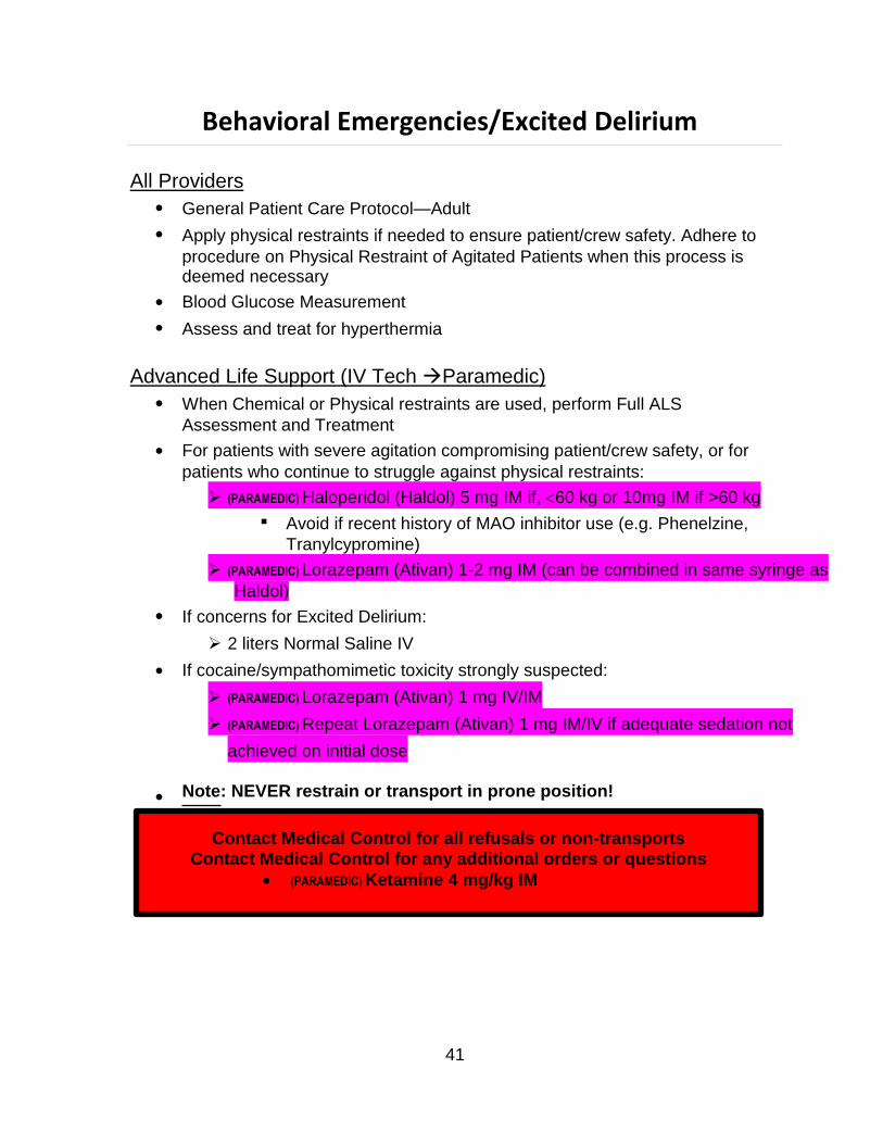
Behavioral Emergencies/Excited Delirium
All Providers
General Patient Care Protocol—Adult
Apply physical restraints if needed to ensure patient/crew safety. Adhere to
procedure on Physical Restraint of Agitated Patients when this process is deemed necessary
Blood Glucose Measurement
Assess and treat for hyperthermia
Advanced Life Support (IV Tech Paramedic)
When Chemical or Physical restraints are used, perform Full ALS
Assessment and Treatment
For patients with severe agitation compromising patient/crew safety, or for
patients who continue to struggle against physical restraints:
(PARAMEDIC) Haloperidol (Haldol) 5 mg IM if,60 kg or 10mg IM if >60 kg
Avoid if recent history of MAO inhibitor use (e.g. Phenelzine,
Tranylcypromine)
(PARAMEDIC) Lorazepam (Ativan) 1-2 mg IM (can be combined in same syringe as
Haldol)
If concerns for Excited Delirium:
2 liters Normal Saline IV
If cocaine/sympathomimetic toxicity strongly suspected:
(PARAMEDIC) Lorazepam (Ativan) 1 mg IV/IM
(PARAMEDIC) Repeat Lorazepam (Ativan) 1 mg IM/IV if adequate sedation not
achieved on initial dose
Note: NEVER restrain or transport in prone position!
Contact Medical Control for all refusals or non-transports
Contact Medical Control for any additional orders or questions
(PARAMEDIC) Ketamine 4 mg/kg IM
41
Bites and Envenomations
All Providers
General Patient Care Protocol—Adult
Irrigate/Cleanse wound with 0.9% NaCl (remove any large debris)
Remove stinger if wasp/bee (if easily removed)
Mark edematous area with pen and note time
Immobilize affected part and remove distal jewelry
Attempt to identify what caused bite and bring to Emergency Department if
dead (use caution when handling dead snakes as envenomation has occurred secondary to reflex motor movement)
Advanced Life Support (IV Tech Paramedic)
Full ALS Assessment and Treatment
For hypotension (SBP<90 mmHg) not improved with fluid boluses up to 2L,
0.9% NaCl, or when fluid boluses are contraindicated:
(PARAMEDIC) Dopamine infusion 5-20 mcg/kg/min titrate to maintain SBP>90mmHg
For Black Widow spider envenomation with severe muscle spasms:
(PARAMEDIC) Lorazepam (Ativan) 1 mg IV/IM
Consider Allergic Reaction Protocol
Transport to closest facility
Contact Medical Control for any additional orders or questions
42
Cardiac Arrest: General Approach
General
CCR is indicated in ADULT patients that have suffered cardiac arrest of a
presumed cardiac nature. It is not indicated in those situations where other etiologies are probable (overdose, drowning, hanging etc.). In these instances CPR is indicated.
CCR is not to be used on individuals less than 18 years of age.
Successful resuscitation requires planning and clear role definition.
In the event a patient suffers cardiac arrest in the presence of EMS (EMS
witnessed Cardiac Arrest), the absolute highest priority is to apply the AED/Defibrillator and deliver a shock immediately if indicated.
Reassess airway frequently and with every patient move.
DO NOT INTERRUPT CHEST COMPRESSIONS!
Designate a “code leader” to coordinate transitions, defibrillation and
pharmacological interventions. “Code Leader” should not have any procedural tasks. If the “code leader” is needed for a specific task, a new leader must be designated.
All Providers
General Patient Care Protocol
Check responsiveness and check for a carotid pulse
Call “patient contact” to dispatch when you arrive at the patient’s side
If adequate bystander compressions ongoing, continue compressions until
monitor pads in place and monitor charged. Stop compressions for rhythm analysis (< 5 sec)
If VT or VF (or AED Advises Shock), defibrillate
Call “first shock” time to dispatch
If PEA/Asystole, go to appropriate protocol and resume compressions
Immediately after defibrillation, resume chest compressions with a different
operator compressing. Do not pause for post-shock rhythm analysis. Stop compressions only for signs of life (patient movement) or rhythm visible through compressions on monitor or pre-defibrillation rhythm analysis every 2 minutes
If compressions are not being performed upon arrival or if compressions are
not deemed adequate, immediately perform compressions at a rate of 100 compressions per minute for 2 minutes.
43
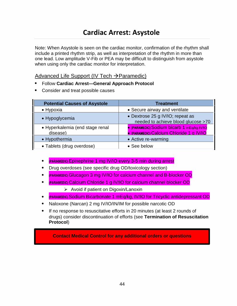
Potential Causes of Asystole Treatment
Hypoxia Secure airway and ventilate
Hypoglycemia Dextrose 25 g IV/IO; repeat as
needed to achieve blood glucose >70
Hyperkalemia (end stage renal disease)
(PARAMEDIC) Sodium bicarb 1 mEq/kg IV/IO
(PARAMEDIC) Calcium Chloride 1 g IV/IO
Hypothermia Active re-warming
Tablets (drug overdose) See below
Cardiac Arrest: Asystole
Note: When Asystole is seen on the cardiac monitor, confirmation of the rhythm shall
include a printed rhythm strip, as well as interpretation of the rhythm in more than one lead. Low amplitude V-Fib or PEA may be difficult to distinguish from asystole when using only the cardiac monitor for interpretation.
Advanced Life Support (IV Tech Paramedic)
Follow Cardiac Arrest—General Approach Protocol
Consider and treat possible causes
(PARAMEDIC) Epinephrine 1 mg IV/IO every 3-5 min during arrest
Drug overdoses (see specific drug OD/toxicology section)
(PARAMEDIC) Glucagon 3 mg IV/IO for calcium channel and B-blocker OD
(PARAMEDIC) Calcium Chloride 1 g IV/IO for calcium channel blocker OD
Avoid if patient on Digoxin/Lanoxin
(PARAMEDIC) Sodium Bicarbonate 1 mEq/kg, IV/IO for Tricyclic antidepressant OD
Naloxone (Narcan) 2 mg IV/IO/IN/IM for possible narcotic OD
If no response to resuscitative efforts in 20 minutes (at least 2 rounds of
drugs) consider discontinuation of efforts (see Termination of Resuscitation Protocol)
Contact Medical Control for any additional orders or questions
44
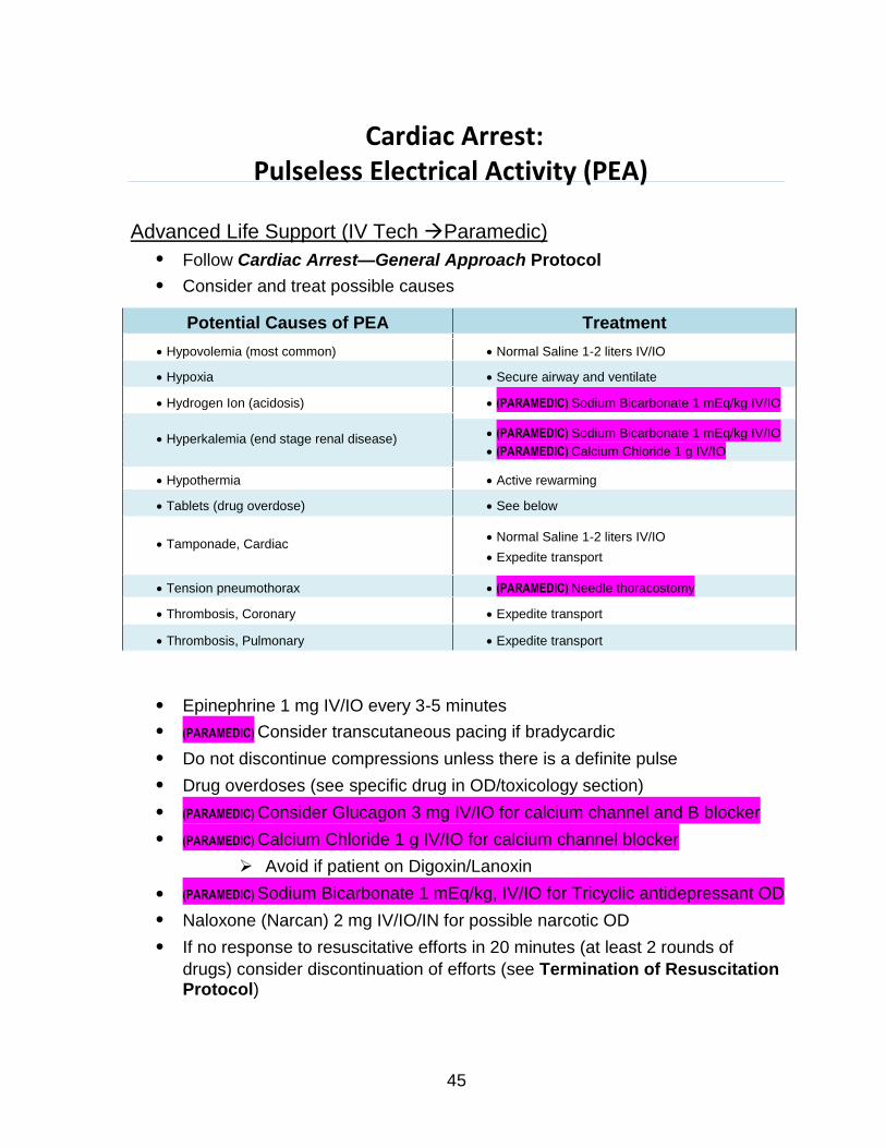
Potential Causes of PEA Treatment
Hypovolemia (most common) Normal Saline 1-2 liters IV/IO
Hypoxia Secure airway and ventilate
Hydrogen Ion (acidosis) (PARAMEDIC) Sodium Bicarbonate 1 mEq/kg IV/IO
Hyperkalemia (end stage renal disease) (PARAMEDIC) Sodium Bicarbonate 1 mEq/kg IV/IO
(PARAMEDIC) Calcium Chloride 1 g IV/IO
Hypothermia Active rewarming
Tablets (drug overdose) See below
Tamponade, Cardiac Normal Saline 1-2 liters IV/IO
Expedite transport
Tension pneumothorax (PARAMEDIC) Needle thoracostomy
Thrombosis, Coronary Expedite transport
Thrombosis, Pulmonary Expedite transport
Cardiac Arrest: Pulseless Electrical Activity (PEA)
Advanced Life Support (IV Tech Paramedic)
Follow Cardiac Arrest—General Approach Protocol
Consider and treat possible causes
Epinephrine 1 mg IV/IO every 3-5 minutes
(PARAMEDIC) Consider transcutaneous pacing if bradycardic
Do not discontinue compressions unless there is a definite pulse
Drug overdoses (see specific drug in OD/toxicology section)
(PARAMEDIC) Consider Glucagon 3 mg IV/IO for calcium channel and B blocker
(PARAMEDIC) Calcium Chloride 1 g IV/IO for calcium channel blocker
Avoid if patient on Digoxin/Lanoxin
(PARAMEDIC) Sodium Bicarbonate 1 mEq/kg, IV/IO for Tricyclic antidepressant OD
Naloxone (Narcan) 2 mg IV/IO/IN for possible narcotic OD
If no response to resuscitative efforts in 20 minutes (at least 2 rounds of
drugs) consider discontinuation of efforts (see Termination of Resuscitation Protocol)
45
Cardiac Arrest: Ventricular Fibrillation/ Pulseless Ventricular Tachycardia
Advanced Life Support (IV Tech Paramedic)
Follow Cardiac Arrest—General Approach Protocol
Defibrillate for persistent VF/VT
200 J for initial biphasic shock, 360 J for subsequent shocks (if available)
Continue Chest Compressions immediately after shock (do not stop for
pulse or rhythm check)
Call first defibrillation time to dispatch
Analyze rhythm after 2 minutes of good CPR; If VF/VT persists:
Defibrillate at 200 J (360 J if available)
Continue compressions immediately after shock (do not stop for pulse
or rhythm check)
(PARAMEDIC) Epinephrine 1 mg IV/IO every 3-5 min during arrest; alternate with Amiodarone
(PARAMEDIC) Vasopressin 40 Units IV/IO with 1st Epinephrine dose only
Analyze rhythm after 2 minutes of good CPR; If VF/VT persists:
Defibrillate at 200 J (360 J if available)
Continue Chest Compressions immediately after shock (do not stop for
pulse or rhythm check)
(PARAMEDIC) Amiodarone 300 mg IV/IO bolus
(PARAMEDIC) For persistent VT/VF give Amiodarone 150 mg IV/IO bolus on second
round.
Continue cycle of Compressions & Drug Rhythm CheckCompressions
ShockCompressions & Drug Rhythm CheckCompressions
Shock as needed
Additional interventions to consider in special circumstances
(PARAMEDIC) Magnesium Sulfate 2 g IV/IO push over 1-2 minutes only if suspected
Polymorphous VT (torsades de pointes) or hypomagnesemic state (chronic alcohol or diuretic use)
(PARAMEDIC) Sodium Bicarbonate 1 mEq/kg, IV/IO if suspected hyperkalemia
(dialysis patient) or tricyclic antidepressant OD
(PARAMEDIC) Calcium Chloride 1 g IV/IO if suspected hyperkalemia (dialysis patient)
Contact Medical Control for any additional orders or questions 46
Cardiac Arrest: Post Resuscitation Care
All Providers
General Patient Care Protocol—Adult
Maintain assisted ventilation as needed
Supplemental 100% oxygen
Advanced Life Support (IV Tech Paramedic)
Full ALS Assessment and Treatment
Monitor ETCO2, goal is 40, DO NOT HYPERVENTILATE
Consider Therapeutic/Induced Hypothermia Protocol
1-2 Liters fluid bolus if no evidence of pulmonary edema
For hypotension (systolic BP <90 mmHg) not improved by fluid boluses, or
when fluid administration is contraindicated:
(PARAMEDIC) Dopamine infusion at 5-20 mcg/kg/min titrated to maintain systolic BP
>90 mmHg
If VF/pulseless VT occurred during arrest AND Amiodarone was
administered, no additional anti-arrhythmic is required unless arrhythmia recurs.
If VF/VT reoccurs after previous conversion with Amiodarone 300 mg:
Defibrillate
(PARAMEDIC) and administer Amiodarone 150 mg IV/IO
If patient becomes combative, administer:
(PARAMEDIC) Lorazepam (Ativan) 1-2 mg slow IV/IO may repeat X 1 (maximum dose
4 mg)
Transport to nearest appropriate facility
Contact Medical Control for any additional orders or questions
47
Cardiac Arrest: Hypothermia Therapeutic/Induced
Most patients suffering from cardiac arrest with return of spontaneous circulation
(ROSC) die with anoxic brain injury. Therapeutic hypothermia serves to improve the chance of a good neurologic outcome.
Criteria for inclusion:
Witnessed cardiac arrest with ROSC
Not pregnant
Age 18 years
No evidence of trauma or intracranial hemorrhage
Significant altered level of consciousness
Not following commands
No purposeful movement
Incomprehensible speech
No known surgery within the preceding 2 weeks
No history of bleeding disorder
Warfarin/Coumadin and Heparin are NOT contraindications
Patient must have airway secured (LTA/ETT)
Advanced Life Support (PARAMEDIC)
Full ALS Assessment and Treatment
12-Lead EKG
Ensure all inclusion/exclusion criteria are met.
If airway not secured and it will not delay transport to the appropriate
receiving facility, perform RSA, refer to Invasive Airway Management Protocol as needed.
Assess neurological status prior to intubation.
Once airway secured/sedated, expose patient and apply ice packs to axilla,
groin and neck.
Administer Midazolam 1-2 mg every 3-5 minutes IV/IO to a max of 10 mg.
Administer 30 ml/kg of cool saline (4C) to a max of 2 liters IV.
If shivering, administer Rocuronium 1 mg/kg IV/IO.
If systolic blood pressure < 90 mmHg, initiate Dopamine infusion at 5-20
mcg/kg/min, titrate to SBP > 90 mmHg.
48
Closely monitor ventilation, target ETCO2 to 40 mmHg, do not hyperventilate.
If at any time there is loss of spontaneous circulation, discontinue cooling and
go to the appropriate protocol.
Contact Medical Control for any additional orders or questions
49
Cardiac Arrest: Termination of Resuscitation
Note: When asystole is seen on the cardiac monitor, confirmation of the rhythm shall
include a PRINTED rhythm strip, as well as interpretation of the rhythm strip in more than one lead. Low amplitude V-Fib or PEA may be difficult to distinguish from asystole when using only the cardiac monitor display for interpretation.
On-line Medical Control NOT Required
The paramedic may terminate resuscitative efforts in non-hypothermic adults
provided all 6 of the following criteria exist:
1) Initial rhythm is asystole, confirmed in two leads on a printed strip
2) Terminal rhythm is asystole confirmed in two leads on a printed strip
3) Secure airway confirmed by digital capnography (ETT/LTA/LMA)
4) At least two doses of Epinephrine have been administered
5) Cardiac Arrest refractory to at least 20 minutes of ACLS
6) Quantitative EtCO2 value < 10mmHg with effective CPR and 20 minutes of ACLS
The paramedic has the discretion to continue resuscitation efforts despite the above
criteria being met if scene safety, location, patient’s age, time of arrest, or bystander input compels this decision
DO NOT TERMINATE RESUSCITATION
IF PATIENT HAS BEEN MOVED TO THE AMBULANCE OR IF
TRANSPORT HAS BEEN INITIATED
Contact Medical Control for any additional orders or questions
50
Cardiac Arrest: No Resuscitation Indicated
Resuscitation can be withheld in Medical Cardiopulmonary Arrest under the
following circumstances:
Adult patient ≥ 18 years of age AND
Pulseless, Apenic and no other signs of life present AND
Asystole verified in two (2) leads AND
Not exposed to an environment likely to promote hypothermia AND
The presence of one or more of the following:
Rigor mortis
Decomposition of body tissues
Dependent lividity OR
When the patient has a valid State of Wisconsin DNR bracelet/wristband
If unknown DNR status or questions regarding validity of DNR status, initiate
resuscitation and contact OLMC
Contact Medical Control for any additional orders or questions
51
Cardiac Arrhythmias: Atrial Fibrillation or Flutter
All Providers
General Patient Care Protocol-Adult
Supplemental oxygen
Advanced Life Support (PARAMEDIC)
Full ALS Assessment and Treatment
Do not delay treatment if patient is unstable by obtaining 12-lead ECG unless
diagnosis is in question
Stable or borderline – Systolic BP >90 mmHg and mild symptoms (chest pain,
SOB or lightheadedness)
No history of WPW:
Diltiazem 0.25 mg/kg IV over 5 min (Max 20 mg per dose)
If unsuccessful after 10 min and SBP >100 mmHg
Diltiazem 0.35 mg/kg IV (max 20mg)
History of WPW
Amiodarone 150 mg IV in 100 ml D5W over 10 min
If unsuccessful and SBP>100 mmHg, may repeat one time
Unstable (serious signs and symptoms-pulmonary edema, BP<90 mmHg
systolic, altered consciousness) AND atrial fibrillation at a rate >150 beats/minute
Sedation if patient condition and time allows (hold if SBP <90mmHg):
Fentanyl 25-50 mcg and Midazolam 1-2 mg IV/IO/IN
Titrate to maximum total dose of Fentanyl 200 mcg and
Midazolam 4 mg
Synchronized Cardioversion
1st energy level:
If no response:
If no response:
If no response:
120 Joules
200 J
200 J (300 J if available)
200 J (360 J if available)
Contact Medical Control for any additional orders or questions
52
Cardiac Arrhythmias: Bradycardia
All Providers
General Patient Care Protocol-Adult
Supplemental oxygen
Advanced Life Support (PARAMEDIC)
Full ALS Assessment and Treatment
Do not delay transport if patient is unstable by obtaining a 12 lead ECG
unless diagnosis is in question
Note: The following therapies are indicated only when serious signs and
symptoms are present. If symptoms are mild, provide supportive care and expedite transport.
Symptomatic (SBP<90mmHg, altered mental status or severe chest pain)
Atropine 0.5 mg IVP, Repeat every 3 minutes as needed (Maximum dose
0.04 mg/kg)
If symptoms persist after Atropine or any delay in establishing IV:
Initiate transcutaneous pacing using demand mode
Start at lowest MA’s; increase until electrical capture with pulses
achieved
Start rate at 70 or default and increase rate to achieve systolic BP
>90mmHg (Maximum 100 beats/minute)
Sedation if patient condition and time allows (hold if SBP<90 mmHg):
Fentanyl 25-50 mcg and Midazolam 1-2 mg IV/IO/IN
Titrate to maximum total dose of Fentanyl 200 mcg and
Midazolam 4 mg
For hypotension (systolic BP <90 mmHg) not improved by above
Dopamine infusion at 5-20 mcg/kg/min titrated to maintain SBP
>90 mmHg
If above unsuccessful:
Epinephrine infusion at 2-10 mcg/min titrated to maintain SBP
>90 mmHg
53
If drug induced, treat for specific drug overdose
Calcium Chloride 1g IV/IO for calcium channel blocker OD
Contraindicated if patient on Digoxin/Lanoxin
Consider Glucagon 3mg IV/IO for calcium channel blocker
OD if no response to Calcium Chloride
Consider Glucagon 3mg IV/IO for Beta Blocker OD
Naloxone (Narcan) can be given in 0.4 mg increments titrated to
level of consciousness and respiratory drive
If IV access has not been established, Naloxone
(Narcan) 2 mg IM or via Mucosal Atomizer Device
Sodium Bicarbonate 1 mEq/kg IV/IO for Tricyclic antidepressant OD
Contact Medical Control for any additional orders or questions
54
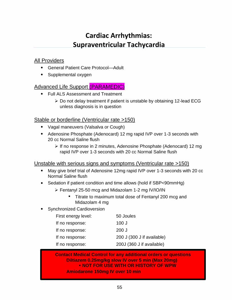
Cardiac Arrhythmias: Supraventricular Tachycardia
All Providers
General Patient Care Protocol—Adult
Supplemental oxygen
Advanced Life Support (PARAMEDIC)
Full ALS Assessment and Treatment
Do not delay treatment if patient is unstable by obtaining 12-lead ECG
unless diagnosis is in question
Stable or borderline (Ventricular rate >150)
Vagal maneuvers (Valsalva or Cough)
Adenosine Phosphate (Adenocard) 12 mg rapid IVP over 1-3 seconds with
20 cc Normal Saline flush
If no response in 2 minutes, Adenosine Phosphate (Adenocard) 12 mg
rapid IVP over 1-3 seconds with 20 cc Normal Saline flush
Unstable with serious signs and symptoms (Ventricular rate >150)
May give brief trial of Adenosine 12mg rapid IVP over 1-3 seconds with 20 cc
Normal Saline flush
Sedation if patient condition and time allows (hold if SBP<90mmHg)
Fentanyl 25-50 mcg and Midazolam 1-2 mg IV/IO/IN
Titrate to maximum total dose of Fentanyl 200 mcg and
Midazolam 4 mg
Synchronized Cardioversion
First energy level:
If no response:
If no response:
If no response:
If no response:
50 Joules
100 J
200 J
200 J (300 J if available)
200J (360 J if available)
Contact Medical Control for any additional orders or questions
Diltiazem 0.25mg/kg slow IV over 5 min (Max 20mg) NOT FOR USE WITH OR HISTORY OF WPW
Amiodarone 150mg IV over 10 min
55
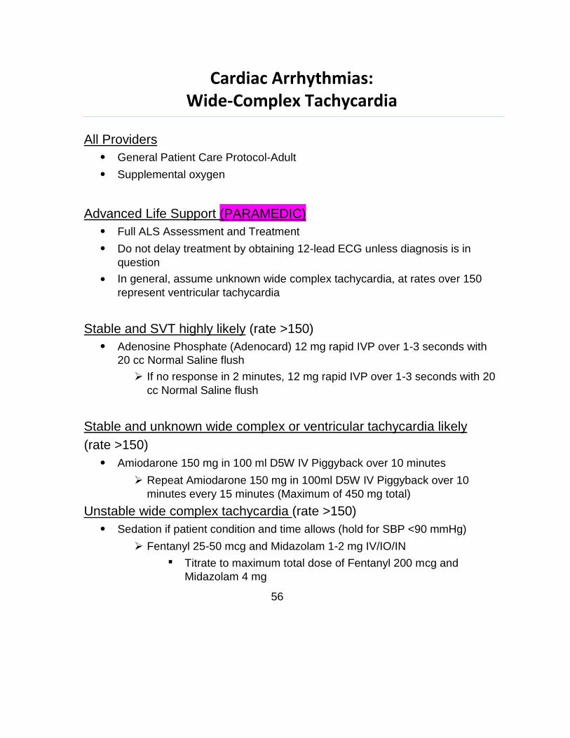
Cardiac Arrhythmias: Wide-Complex Tachycardia
All Providers
General Patient Care Protocol-Adult
Supplemental oxygen
Advanced Life Support (PARAMEDIC)
Full ALS Assessment and Treatment
Do not delay treatment by obtaining 12-lead ECG unless diagnosis is in
question
In general, assume unknown wide complex tachycardia, at rates over 150
represent ventricular tachycardia
Stable and SVT highly likely (rate >150)
Adenosine Phosphate (Adenocard) 12 mg rapid IVP over 1-3 seconds with
20 cc Normal Saline flush
If no response in 2 minutes, 12 mg rapid IVP over 1-3 seconds with 20
cc Normal Saline flush
Stable and unknown wide complex or ventricular tachycardia likely
(rate >150)
Amiodarone 150 mg in 100 ml D5W IV Piggyback over 10 minutes
Repeat Amiodarone 150 mg in 100ml D5W IV Piggyback over 10
minutes every 15 minutes (Maximum of 450 mg total)
Unstable wide complex tachycardia (rate >150)
Sedation if patient condition and time allows (hold for SBP <90 mmHg)
Fentanyl 25-50 mcg and Midazolam 1-2 mg IV/IO/IN
Titrate to maximum total dose of Fentanyl 200 mcg and
Midazolam 4 mg
56
Attempt unsynchronized shock (defibrillation) at 200 Joules.
If wide complex tachycardia reoccurs following shock:
Amiodarone 150 mg in 100 ml D5W IV Piggyback, over 10 minutes,
every 15 minutes (maximum 450 mg cumulative total dose)
If hyperkalemia suspected in any wide complex tachycardia (e.g. renal failure
patient) administer the following medications:
Calcium Chloride 1 g IV/IO
Contraindicated if patient on Digoxin/Lanoxin
Sodium Bicarbonate 1 mEq/kg IV/IO
Contact Medical Control for any additional orders or questions
57
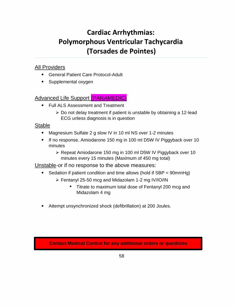
Cardiac Arrhythmias: Polymorphous Ventricular Tachycardia
(Torsades de Pointes)
All Providers
General Patient Care Protocol-Adult
Supplemental oxygen
Advanced Life Support (PARAMEDIC)
Full ALS Assessment and Treatment
Do not delay treatment if patient is unstable by obtaining a 12-lead
ECG unless diagnosis is in question
Stable
Magnesium Sulfate 2 g slow IV in 10 ml NS over 1-2 minutes
If no response, Amiodarone 150 mg in 100 ml D5W IV Piggyback over 10
minutes
Repeat Amiodarone 150 mg in 100 ml D5W IV Piggyback over 10
minutes every 15 minutes (Maximum of 450 mg total)
Unstable-or if no response to the above measures:
Sedation if patient condition and time allows (hold if SBP < 90mmHg)
Fentanyl 25-50 mcg and Midazolam 1-2 mg IV/IO/IN
Titrate to maximum total dose of Fentanyl 200 mcg and
Midazolam 4 mg
Attempt unsynchronized shock (defibrillation) at 200 Joules.
Contact Medical Control for any additional orders or questions
58
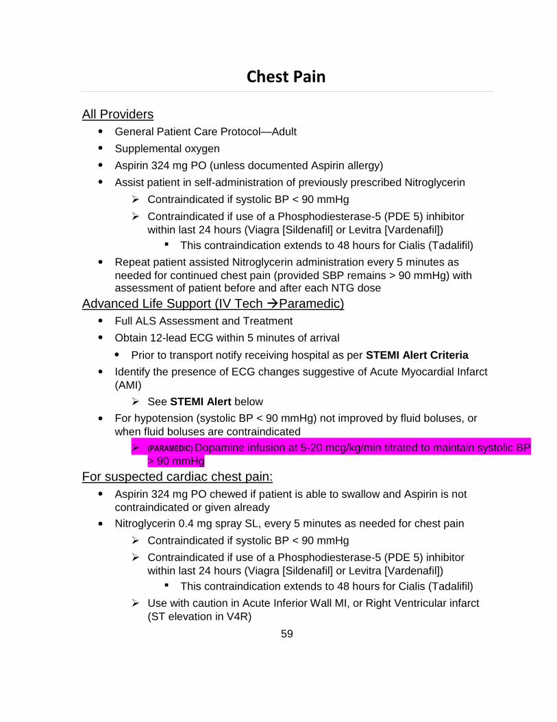
Chest Pain
All Providers
General Patient Care Protocol—Adult
Supplemental oxygen
Aspirin 324 mg PO (unless documented Aspirin allergy)
Assist patient in self-administration of previously prescribed Nitroglycerin
Contraindicated if systolic BP < 90 mmHg
Contraindicated if use of a Phosphodiesterase-5 (PDE 5) inhibitor
within last 24 hours (Viagra [Sildenafil] or Levitra [Vardenafil])
This contraindication extends to 48 hours for Cialis (Tadalifil)
Repeat patient assisted Nitroglycerin administration every 5 minutes as
needed for continued chest pain (provided SBP remains > 90 mmHg) with assessment of patient before and after each NTG dose
Advanced Life Support (IV Tech Paramedic)
Full ALS Assessment and Treatment
Obtain 12-lead ECG within 5 minutes of arrival
Prior to transport notify receiving hospital as per STEMI Alert Criteria
Identify the presence of ECG changes suggestive of Acute Myocardial Infarct
(AMI)
See STEMI Alert below
For hypotension (systolic BP < 90 mmHg) not improved by fluid boluses, or
when fluid boluses are contraindicated
(PARAMEDIC) Dopamine infusion at 5-20 mcg/kg/min titrated to maintain systolic BP
> 90 mmHg
For suspected cardiac chest pain:
Aspirin 324 mg PO chewed if patient is able to swallow and Aspirin is not
contraindicated or given already
Nitroglycerin 0.4 mg spray SL, every 5 minutes as needed for chest pain
Contraindicated if systolic BP < 90 mmHg
Contraindicated if use of a Phosphodiesterase-5 (PDE 5) inhibitor
within last 24 hours (Viagra [Sildenafil] or Levitra [Vardenafil])
This contraindication extends to 48 hours for Cialis (Tadalifil)
Use with caution in Acute Inferior Wall MI, or Right Ventricular infarct
(ST elevation in V4R)
59
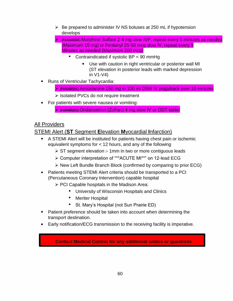
Be prepared to administer IV NS boluses at 250 mL if hypotension
develops
(PARAMEDIC) Morphine Sulfate 2-4 mg slow IVP; repeat every 5 minutes as needed
(Maximum 15 mg) or Fentanyl 25-50 mcg slow IV; repeat every 5 Minutes as needed (Maximum 200 mcg)
Contraindicated if systolic BP < 90 mmHg
Use with caution in right ventricular or posterior wall MI
(ST elevation in posterior leads with marked depression in V1-V4)
Runs of Ventricular Tachycardia:
(PARAMEDIC) Amiodarone 150 mg in 100 ml D5W IV piggyback over 10 minutes
Isolated PVCs do not require treatment
For patients with severe nausea or vomiting:
(PARAMEDIC) Ondansetron (Zofran) 4 mg slow IV or ODT tablet
All Providers
STEMI Alert (ST Segment Elevation Myocardial Infarction)
A STEMI Alert will be instituted for patients having chest pain or ischemic
equivalent symptoms for < 12 hours, and any of the following
ST segment elevation 1mm in two or more contiguous leads
Computer interpretation of “**ACUTE MI**” on 12-lead ECG
New Left Bundle Branch Block (confirmed by comparing to prior ECG)
Patients meeting STEMI Alert criteria should be transported to a PCI
(Percutaneous Coronary Intervention) capable hospital
PCI Capable hospitals in the Madison Area:
University of Wisconsin Hospitals and Clinics
Meriter Hospital
St. Mary’s Hospital (not Sun Prairie ED)
Patient preference should be taken into account when determining the
transport destination.
Early notification/ECG transmission to the receiving facility is imperative.
Contact Medical Control for any additional orders or questions
60
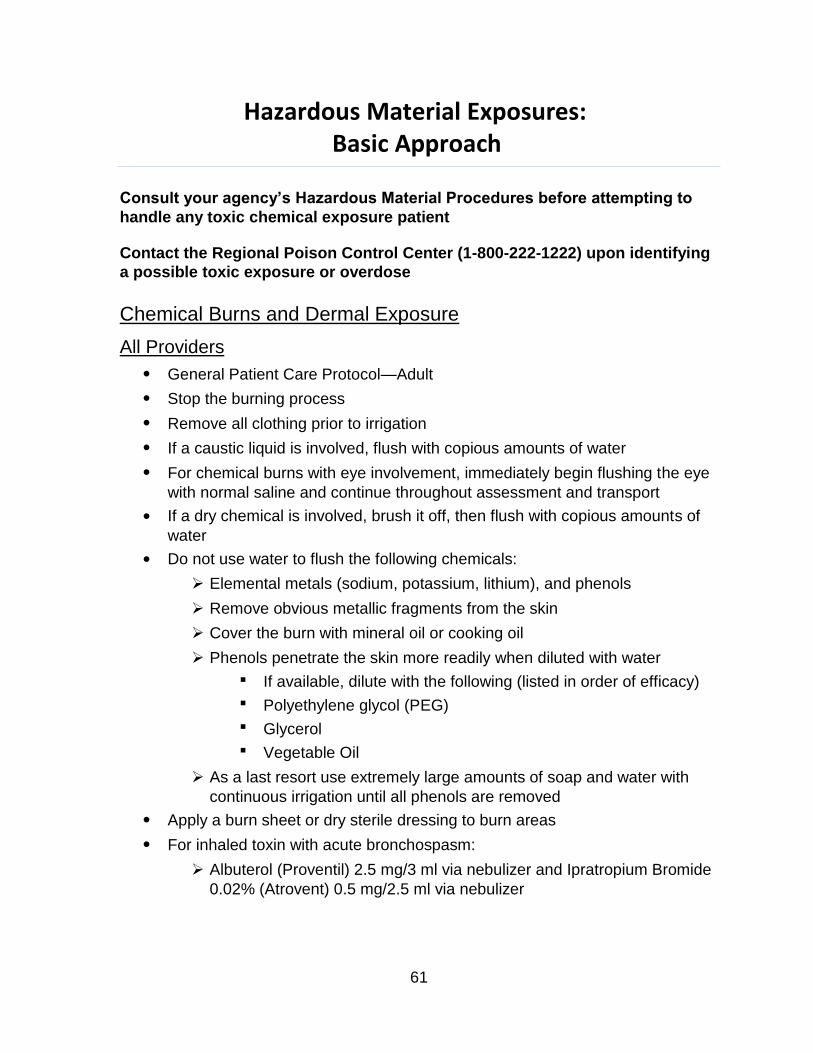
Hazardous Material Exposures: Basic Approach
Consult your agency’s Hazardous Material Procedures before attempting to
handle any toxic chemical exposure patient
Contact the Regional Poison Control Center (1-800-222-1222) upon identifying
a possible toxic exposure or overdose
Chemical Burns and Dermal Exposure
All Providers
General Patient Care Protocol—Adult
Stop the burning process
Remove all clothing prior to irrigation
If a caustic liquid is involved, flush with copious amounts of water
For chemical burns with eye involvement, immediately begin flushing the eye
with normal saline and continue throughout assessment and transport
If a dry chemical is involved, brush it off, then flush with copious amounts of
water
Do not use water to flush the following chemicals:
Elemental metals (sodium, potassium, lithium), and phenols
Remove obvious metallic fragments from the skin
Cover the burn with mineral oil or cooking oil
Phenols penetrate the skin more readily when diluted with water
If available, dilute with the following (listed in order of efficacy)
Polyethylene glycol (PEG)
Glycerol
Vegetable Oil
As a last resort use extremely large amounts of soap and water with
continuous irrigation until all phenols are removed
Apply a burn sheet or dry sterile dressing to burn areas
For inhaled toxin with acute bronchospasm:
Albuterol (Proventil) 2.5 mg/3 ml via nebulizer and Ipratropium Bromide
0.02% (Atrovent) 0.5 mg/2.5 ml via nebulizer
61
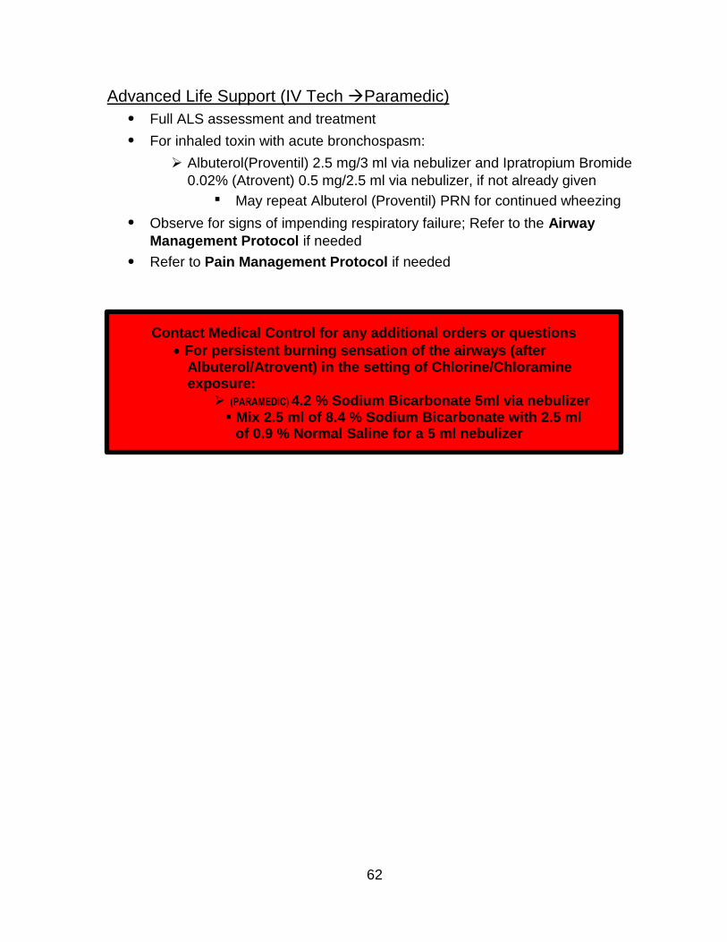
Advanced Life Support (IV Tech Paramedic)
Full ALS assessment and treatment
For inhaled toxin with acute bronchospasm:
Albuterol(Proventil) 2.5 mg/3 ml via nebulizer and Ipratropium Bromide
0.02% (Atrovent) 0.5 mg/2.5 ml via nebulizer, if not already given
May repeat Albuterol (Proventil) PRN for continued wheezing
Observe for signs of impending respiratory failure; Refer to the Airway
Management Protocol if needed
Refer to Pain Management Protocol if needed
Contact Medical Control for any additional orders or questions
For persistent burning sensation of the airways (after Albuterol/Atrovent) in the setting of Chlorine/Chloramine exposure:
(PARAMEDIC) 4.2 % Sodium Bicarbonate 5ml via nebulizer Mix 2.5 ml of 8.4 % Sodium Bicarbonate with 2.5 ml
of 0.9 % Normal Saline for a 5 ml nebulizer
62
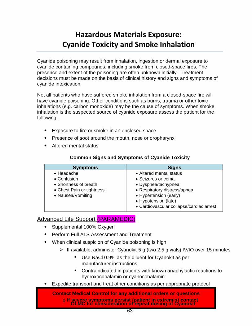
Contact Medical Control for any additional orders or questions
If severe symptoms persist (patient in extremis) contact
Symptoms Signs Headache
Confusion
Shortness of breath
Chest Pain or tightness
Nausea/Vomiting
Altered mental status
Seizures or coma
Dyspnea/tachypnea
Respiratory distress/apnea
Hypertension (early)
Hypotension (late)
Cardiovascular collapse/cardiac arrest
Hazardous Materials Exposure: Cyanide Toxicity and Smoke Inhalation
Cyanide poisoning may result from inhalation, ingestion or dermal exposure to cyanide containing compounds, including smoke from closed-space fires. The presence and extent of the poisoning are often unknown initially. Treatment decisions must be made on the basis of clinical history and signs and symptoms of cyanide intoxication.
Not all patients who have suffered smoke inhalation from a closed-space fire will
have cyanide poisoning. Other conditions such as burns, trauma or other toxic inhalations (e.g. carbon monoxide) may be the cause of symptoms. When smoke inhalation is the suspected source of cyanide exposure assess the patient for the following:
Exposure to fire or smoke in an enclosed space
Presence of soot around the mouth, nose or oropharynx
Altered mental status
Common Signs and Symptoms of Cyanide Toxicity
Advanced Life Support (PARAMEDIC)
Supplemental 100% Oxygen
Perform Full ALS Assessment and Treatment
When clinical suspicion of Cyanide poisoning is high
If available, administer Cyanokit5 g (two 2.5 g vials) IV/IO over 15 minutes
Use NaCl 0.9% as the diluent for Cyanokit as per
manufacturer instructions
Contraindicated in patients with known anaphylactic reactions to
hydroxocobalamin or cyanocobalamin
Expedite transport and treat other conditions as per appropriate protocol
OLMC for consideration of repeat dosing of Cyanokit
63
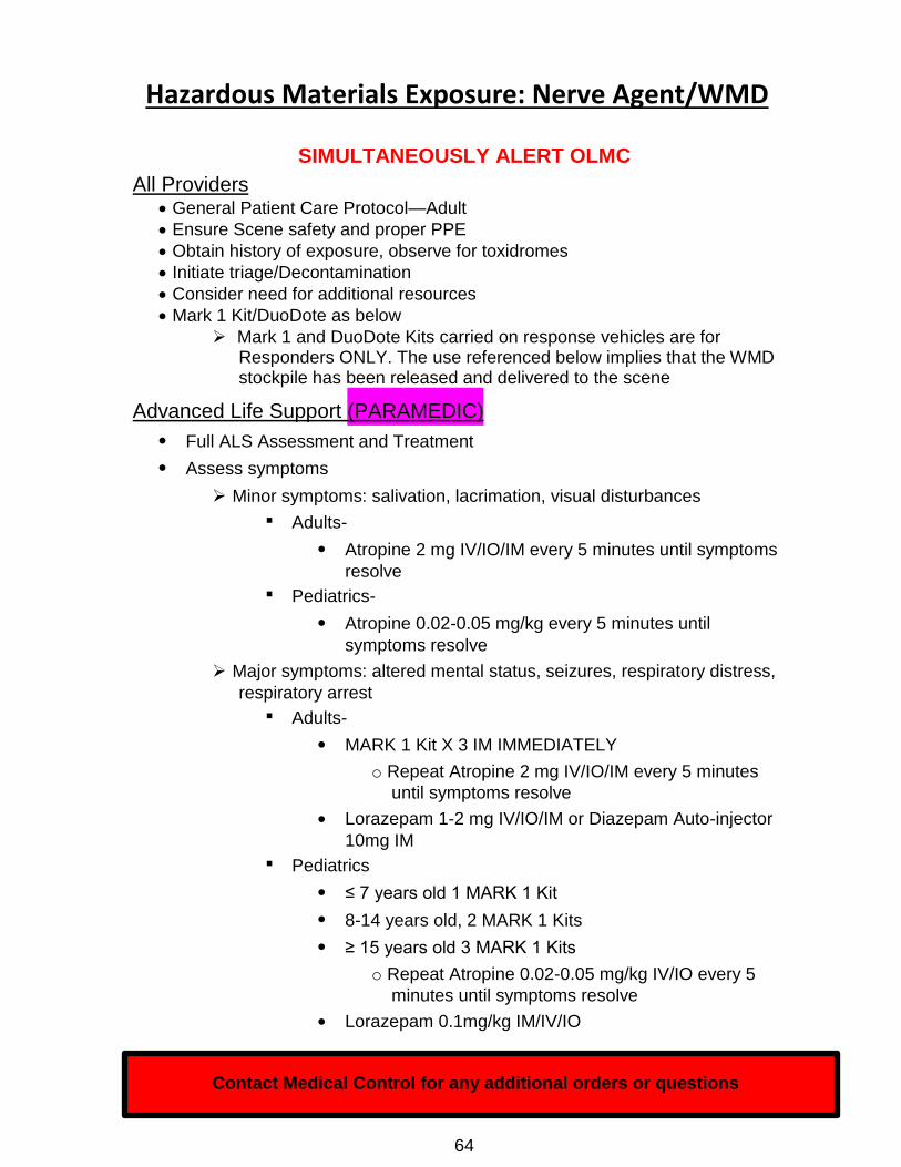
Hazardous Materials Exposure: Nerve Agent/WMD
SIMULTANEOUSLY ALERT OLMC
All Providers General Patient Care Protocol—Adult
Ensure Scene safety and proper PPE
Obtain history of exposure, observe for toxidromes
Initiate triage/Decontamination
Consider need for additional resources
Mark 1 Kit/DuoDote as below
Mark 1 and DuoDote Kits carried on response vehicles are for Responders ONLY. The use referenced below implies that the WMD stockpile has been released and delivered to the scene
Advanced Life Support (PARAMEDIC)
Full ALS Assessment and Treatment
Assess symptoms
Minor symptoms: salivation, lacrimation, visual disturbances
Adults-
Atropine 2 mg IV/IO/IM every 5 minutes until symptoms
resolve
Pediatrics-
Atropine 0.02-0.05 mg/kg every 5 minutes until
symptoms resolve
Major symptoms: altered mental status, seizures, respiratory distress,
respiratory arrest
Adults-
MARK 1 Kit X 3 IM IMMEDIATELY
o Repeat Atropine 2 mg IV/IO/IM every 5 minutes
until symptoms resolve
Lorazepam 1-2 mg IV/IO/IM or Diazepam Auto-injector
10mg IM
Pediatrics
≤ 7 years old 1 MARK 1 Kit
8-14 years old, 2 MARK 1 Kits
≥ 15 years old 3 MARK 1 Kits
o Repeat Atropine 0.02-0.05 mg/kg IV/IO every 5
minutes until symptoms resolve
Lorazepam 0.1mg/kg IM/IV/IO
Contact Medical Control for any additional orders or questions
64
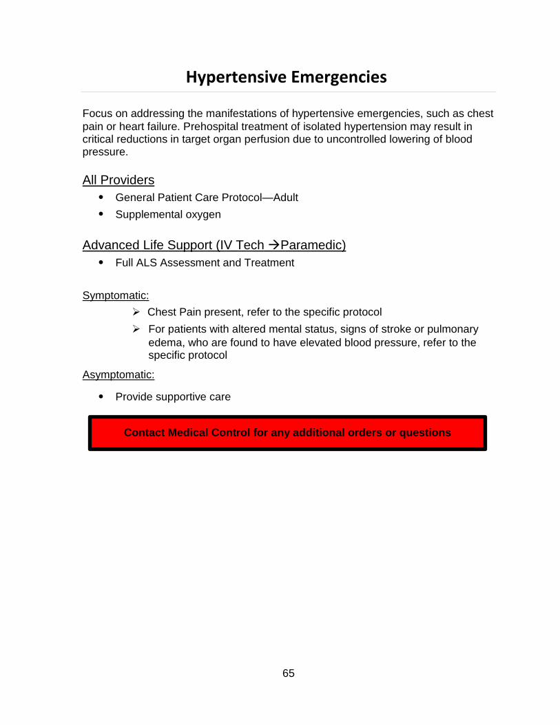
Hypertensive Emergencies
Focus on addressing the manifestations of hypertensive emergencies, such as chest
pain or heart failure. Prehospital treatment of isolated hypertension may result in critical reductions in target organ perfusion due to uncontrolled lowering of blood pressure.
All Providers
General Patient Care Protocol—Adult
Supplemental oxygen
Advanced Life Support (IV Tech Paramedic)
Full ALS Assessment and Treatment
Symptomatic:
Chest Pain present, refer to the specific protocol
For patients with altered mental status, signs of stroke or pulmonary
edema, who are found to have elevated blood pressure, refer to the specific protocol
Asymptomatic:
Provide supportive care
Contact Medical Control for any additional orders or questions
65
Hyperthermia
All Providers
General Patient Care Protocol—Adult
Move patient to cooler environment
Heat Cramps (Painful spasms of the extremities or abdominal muscles, normal
mental status and vital signs)
Oral fluids as tolerated
Sponge with cool water
Heat Exhaustion (Dizziness, light-headedness, headache, irritability, normal or
slightly decreased LOC, normal or decreased BP [hypovolemia], tachycardia, normal or slightly elevated temperature)
Keep patient supine
Supplemental 100% oxygen
Remove clothing
Sponge with cool water and fan
Heat Stroke (Marked alteration in LOC, extremely high temperature [often > 104]
may be sweating or have red/hot/dry skin)
Semi-reclining with head elevated 15-30
Supplemental 100% oxygen
Rapid cooling (prevent shivering as it increases body temperature)
Cold packs, sponge with cool water, fan
Advanced Life Support (IV Tech Paramedic)
If symptoms are moderate to severe, perform Full ALS Assessment and
Treatment
Hyperthermia may result from cocaine or sympathomimetic toxicity
If cocaine/sympathomimetic toxicity strongly suspected, refer to the
Cocaine/Sympathomimetic Protocol
Expedite Transport
Contact Medical Control for any additional orders or questions
66
Hypothermia
All Providers
General Patient Care Protocol—Adult
Remove wet clothing
Measure core temperature, If < 95F (35C), handle gently
Warm blankets/warm temperature
Advanced Life Support (IV Tech Paramedic)
Full ALS Assessment and Treatment
If available, and no contraindications, administer warmed 0.9% Normal saline
If Cardiac Arrest occurs with a core temp > 88F
Refer to appropriate protocol
Prolong interval between drugs to 5 minutes
If defibrillation is necessary, limit to one shock
Continue CPR
If cardiac arrest with core temp < 88F
Contact OLMC
Contact Medical Control for any additional orders or questions
67
Obstetrics / Gynecology Perinatal Emergencies
Pregnancy-induced hypertension, pre-eclampsia and eclampsia are conditions typically encountered in late 2nd or 3rd trimester pregnancy, and less commonly in the postpartum period. Clinical manifestations may include elevated blood pressure (SBP > 160 mmHg), headache, confusion or agitation.
All Providers General Patient Care Protocol-Adult
Blood glucose measurement
Advanced Life Support (PARAMEDIC)
Full ALS Assessment and Treatment
Administer Magnesium Sulfate 4 g in 100 ml D5W IV over 10 minutes for
either of the following:
Systolic BP > 160 mm Hg on two readings
Seizure activity
For active seizures, in addition to Magnesium Sulfate choose one of the
following options:
Lorazepam (Ativan) 1-2 mg, slow IV/IO
-OR-
Midazolam (Versed), 1-2 mg, slow IV/IO
If no IV access, Midazolam (Versed), 5 mg IM/IN
For seizure not controlled by the above, or if seizure reoccurs after initial
control, choose one of the following:
Lorazepam (Ativan) 1-2 mg, slow IV/IO
-OR-
Midazolam (Versed) 1-2 mg, slow IV/IO
If no IV access, Midazolam (Versed) 5 mg IM/IN
Blood Glucose measurement 68
Vaginal Bleeding
1st or 2nd trimester or unknown pregnancy status
Position of comfort, consider lateral recumbent position if hypotensive
3rd Trimester Bleeding (>26 weeks)
Lateral recumbent position
Do not place finger or hand inside birth canal during assessment
Advanced Life Support (IV Tech Paramedic)
If bleeding moderate or heavy, perform Full ALS Assessment and Treatment
If gestational age known to be < 20 weeks, transport to closest hospital
If gestational age known or possibly ≥ 20 weeks, transport to nearest OB
receiving facility
If BP<90 mmHg systolic, administer boluses of 0.9%NaCl at 250 ml until
systolic BP>90 mmHg
Obstetrical Transport Destination
Patient known to be < 20 weeks gestation
1st day of last menstrual period < 20 weeks ago
Available information verifying gestational age < 20 weeks (e.g., known due
date)
Refer to Medical Transport Destination for more information
Does not have to be an OB receiving facility
Patient known or possibly ≥ 20 weeks gestation
Imminent delivery or medically unstable mother:
Transport to nearest ED (not nearest OB receiving facility)
Non-traumatic abdominal, pelvic or back complaints, vaginal bleeding,
spotting or any vaginal fluid leak or discharge:
Transport to closest appropriate obstetric receiving facility
Contact appropriate obstetric facility ED for radio report and any additional
direction/assistance
69
Whenever possible, transport to patient’s requested obstetric receiving
facility (e.g. high risk pregnancy with pre-selected obstetrical destination) if patient not having imminent delivery.
Appropriate Obstetric Receiving Facilities: -Meriter Hospital -St. Mary’s Hospital – Madison Campus -Fort Atkinson Hospital
Contact Medical Control for any additional orders or
questions
70
Obstetrics / Gynecology: Childbirth
All Providers
General Patient Care Protocol-Adult
Supplemental oxygen
Do not place fingers or hand inside the birth canal for assessment
If presenting part is not the head (i.e., foot-, arm-, or buttock-first),
immediately begin transport to the nearest OB receiving facility while further care continues
Delivery
Slow, controlled delivery of head; apply gentle perineal pressure
Observe for meconium staining
If present, suction oral pharynx and nose as soon as head is delivered
Following delivery, follow newborn resuscitation protocol
Double clamp cord 10-12 inches from abdomen
Cut cord between clamps
Maintain body temperature
Allow spontaneous delivery of placenta; do not apply traction to umbilical cord
for placental delivery
If placental delivery occurs, package in bio-hazardous waste bag and
hand over to hospital staff upon arrival
Postpartum
For neonate, see Newborn Resuscitation Protocol
Assess for postpartum hemorrhage
Gently massage abdominal wall overlying the uterine fundus until firm
Advanced Life Support (IV Tech Paramedic)
Transport to nearest OB receiving facility
See newborn resuscitation for care of the neonate
Contact Medical Control for any additional orders or questions
71
Overdose and Poisonings: General Approach
For any overdose or poisoning, contact should be made with the Regional Poison
Control Center (RPCC) 1-800-222-1222. Whenever possible, determine the agent(s) involved, the time of the ingestion/exposure, and the amount ingested. Bring empty pill bottles, etc. to the receiving facility.
All Providers
General Patient Care Protocol-Adults
Nothing by mouth
Advanced Life Support (IV Tech Paramedic)
Refer to the specific protocol when an agent has been identified or is strongly suspected
Contact Medical Control for any additional orders or questions
72
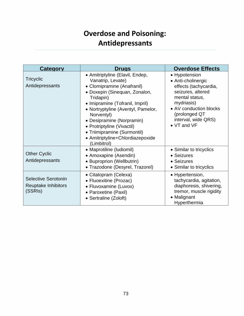
Category Drugs Overdose Effects
Tricyclic
Antidepressants
Amitriptyline (Elavil, Endep, Vanatrip, Levate)
Clomipramine (Anafranil)
Doxepin (Sinequan, Zonalon, Tridapin)
Imipramine (Tofranil, Impril)
Nortryptyline (Aventyl, Pamelor, Norventyl)
Desipramine (Norpramin)
Protriptyline (Vivactil)
Triimipramine (Surmontil)
Amitriptyline+Chlordiazepoxide (Limbitrol)
Hypotension
Anti-cholinergic effects (tachycardia, seizures, altered mental status, mydriasis)
AV conduction blocks (prolonged QT interval, wide QRS)
VT and VF
Other Cyclic
Antidepressants
Maprotiline (ludiomil)
Amoxapine (Asendin)
Buproprion (Wellbutrin)
Trazodone (Desyrel, Trazorel)
Similar to tricyclics
Seizures
Seizures
Similar to tricyclics
Selective Serotonin
Reuptake Inhibitors (SSRIs)
Citalopram (Celexa)
Fluoexitine (Prozac)
Fluvoxamine (Luvox)
Paroxetine (Paxil)
Sertraline (Zoloft)
Hypertension,
tachycardia, agitation, diaphoresis, shivering, tremor, muscle rigidity
Malignant Hyperthermia
Overdose and Poisoning: Antidepressants
73
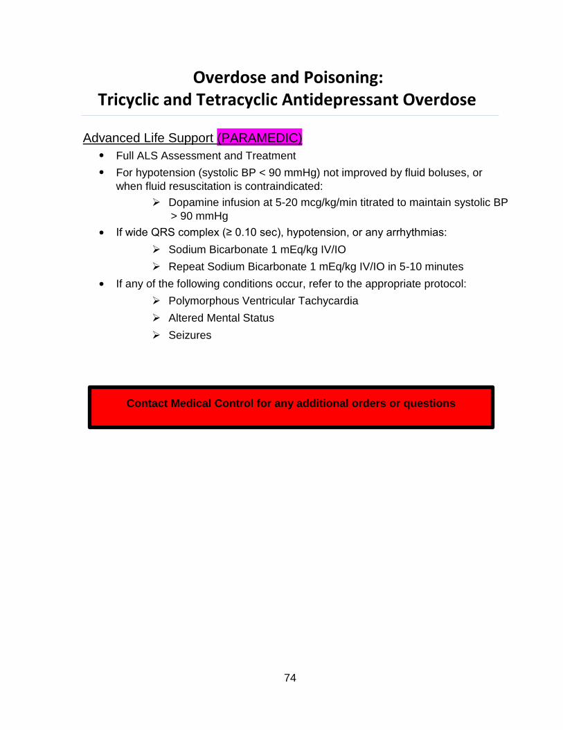
Overdose and Poisoning: Tricyclic and Tetracyclic Antidepressant Overdose
Advanced Life Support (PARAMEDIC)
Full ALS Assessment and Treatment
For hypotension (systolic BP < 90 mmHg) not improved by fluid boluses, or
when fluid resuscitation is contraindicated:
Dopamine infusion at 5-20 mcg/kg/min titrated to maintain systolic BP
> 90 mmHg
If wide QRS complex (≥ 0.10 sec), hypotension, or any arrhythmias:
Sodium Bicarbonate 1 mEq/kg IV/IO
Repeat Sodium Bicarbonate 1 mEq/kg IV/IO in 5-10 minutes
If any of the following conditions occur, refer to the appropriate protocol:
Polymorphous Ventricular Tachycardia
Altered Mental Status
Seizures
Contact Medical Control for any additional orders or questions
74
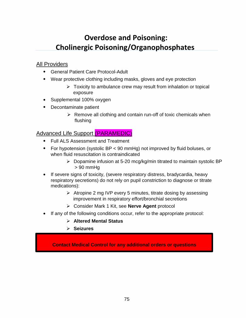
Overdose and Poisoning: Cholinergic Poisoning/Organophosphates
All Providers
General Patient Care Protocol-Adult
Wear protective clothing including masks, gloves and eye protection
Toxicity to ambulance crew may result from inhalation or topical
exposure
Supplemental 100% oxygen
Decontaminate patient
Remove all clothing and contain run-off of toxic chemicals when
flushing
Advanced Life Support (PARAMEDIC)
Full ALS Assessment and Treatment
For hypotension (systolic BP < 90 mmHg) not improved by fluid boluses, or
when fluid resuscitation is contraindicated
Dopamine infusion at 5-20 mcg/kg/min titrated to maintain systolic BP
> 90 mmHg
If severe signs of toxicity, (severe respiratory distress, bradycardia, heavy
respiratory secretions) do not rely on pupil constriction to diagnose or titrate medications):
Atropine 2 mg IVP every 5 minutes, titrate dosing by assessing
improvement in respiratory effort/bronchial secretions
Consider Mark 1 Kit, see Nerve Agent protocol
If any of the following conditions occur, refer to the appropriate protocol:
Altered Mental Status
Seizures
Contact Medical Control for any additional orders or questions
75
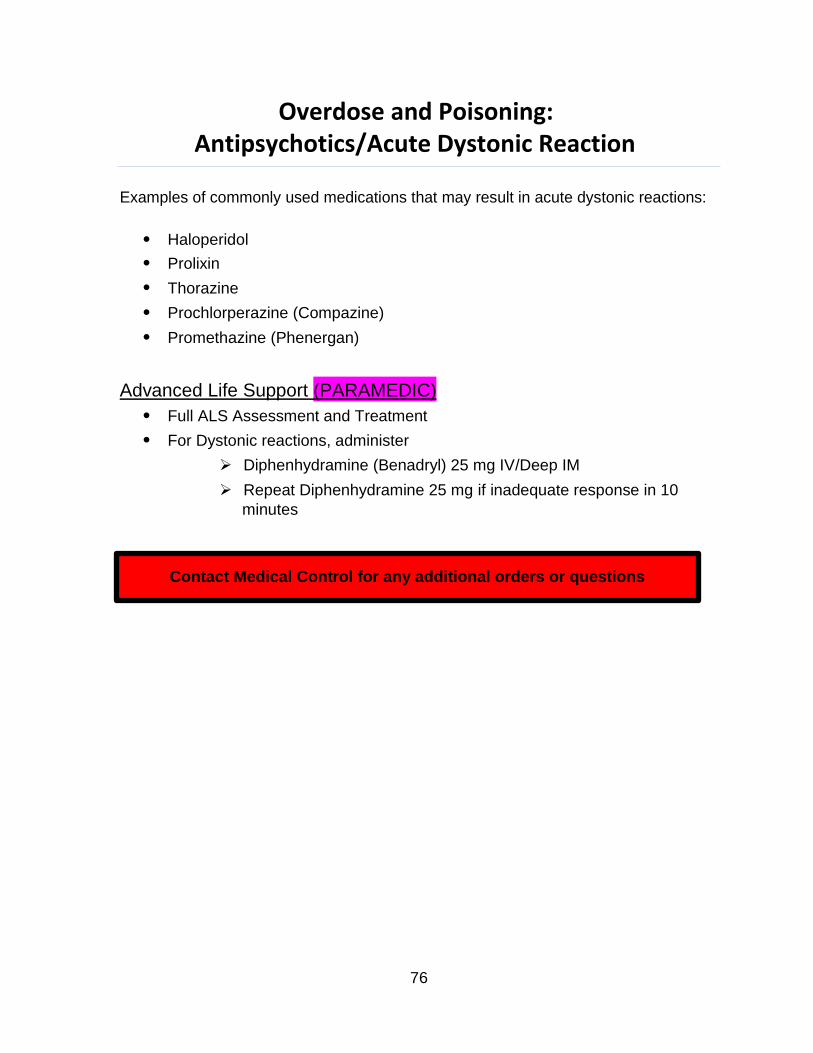
Overdose and Poisoning: Antipsychotics/Acute Dystonic Reaction
Examples of commonly used medications that may result in acute dystonic reactions:
Haloperidol
Prolixin
Thorazine
Prochlorperazine (Compazine)
Promethazine (Phenergan)
Advanced Life Support (PARAMEDIC)
Full ALS Assessment and Treatment
For Dystonic reactions, administer
Diphenhydramine (Benadryl) 25 mg IV/Deep IM
Repeat Diphenhydramine 25 mg if inadequate response in 10
minutes
Contact Medical Control for any additional orders or questions
76
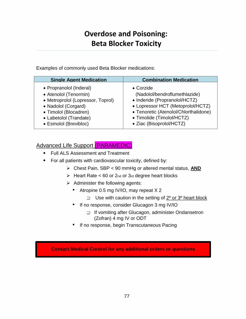
Single Agent Medication Combination Medication
Propranolol (Inderal)
Atenolol (Tenormin)
Metroprolol (Lopressor, Toprol)
Nadolol (Corgard)
Timolol (Blocadren)
Labetolol (Trandate)
Esmolol (Brevibloc)
Corzide
(Nadolol/bendroflumethlazide)
Inderide (Propranolol/HCTZ)
Lopressor HCT (Metoprolol/HCTZ)
Tenoretic (Atenolol/Chlorthalidone)
Timolide (Timolol/HCTZ)
Ziac (Bisoprolol/HCTZ)
Overdose and Poisoning: Beta Blocker Toxicity
Examples of commonly used Beta Blocker medications:
Advanced Life Support (PARAMEDIC)
Full ALS Assessment and Treatment
For all patients with cardiovascular toxicity, defined by:
Chest Pain, SBP < 90 mmHg or altered mental status, AND
Heart Rate < 60 or 2nd or 3rd degree heart blocks
Administer the following agents:
Atropine 0.5 mg IV/IO, may repeat X 2
Use with caution in the setting of 2º or 3º heart block
If no response, consider Glucagon 3 mg IV/IO
If vomiting after Glucagon, administer Ondansetron
(Zofran) 4 mg IV or ODT
If no response, begin Transcutaneous Pacing
Contact Medical Control for any additional orders or questions
77
Overdose and Poisonings: Calcium Channel Blockers
Examples of commonly used Calcium Channel Blocker medication:
Amlodipine (Norvasc)
Felodipine (Plendil, Renedil)
Isradipine (DynaCirc)
Nicardipine (Cardene)
Nifedipine (Procardia, Adalat)
Verapamil (Calan)
Diltiazem (Cardizem)
Advanced Life Support (PARAMEDIC)
Full ALS Assessment and Treatment
For all patients with cardiovascular toxicity, defined by:
Chest Pain, SBP < 90 mmHg or altered mental status, AND
Heart Rate < 60 or 2nd or 3rd degree heart blocks
Administer the following agents
Atropine 0.5 mg IV/IO, may repeat X 2
Use with caution in the setting of 2º or 3º heart block
If no response, administer Calcium Chloride 1 g IV/IO
Contraindicated if patient taking Digoxin (Lanoxin)
If no response, may repeat Calcium Chloride 1 g IV/IO
If no response, consider Glucagon 3 mg IV/IO
If vomiting after Glucagon administer Ondansetron (Zofran) 4
mg IV or ODT
If no response, begin Transcutaneous Pacing
78
Overdose and Poisonings: Carbon Monoxide
All Providers
General Patient Care Protocol—Adult
Wear appropriate PPE (SCBA) as indicated
Remove the patient from the contaminated source
Supplemental 100% oxygen, document time oxygen started
Advanced Life Support (IV Tech Paramedic)
Full ALS Assessment and Treatment
For smoke inhalation patients also consider Cyanide poisoning (See Hazardous
Materials—Basic Approach Protocol)
Contact Medical Control for any additional orders or questions
79
Overdose and Poisoning: Cocaine and Sympathomimetic Overdose
Advanced Life Support (PARAMEDIC)
Full ALS Assessment and Treatment
For patients with Sympathomimetic toxidrome (e.g. hypertension, tachycardia,
agitation):
Lorazepam (Ativan) 1-2 mg, slow IV
If no IV access, Midazolam (Versed)
5 mg IM/IN if < 60 years old
2.5 mg IM/IN if > 60 years old
Repeat either medication once in 5-10 minutes if signs and symptoms
continue
If seizures occur, refer to Seizure Protocol
Consider Excited Delirium, refer to protocol
Contact Medical Control for any additional orders or questions
80
Pain Management—Adult
All Providers
General Patient Care Protocol
Assess baseline pain level (0-10 scale: 0=no pain, 10=worst pain)
Advanced Life Support (PARAMEDIC)
Analgesic agents may be administered under standing orders for patients
experiencing moderate/severe pain (typically ≥ 6/10)
Common complaints:
Trauma/isolated extremity injury
Burns (without airway, breathing or circulation compromise)
Sickle crisis
Acute Chest Pain, in accordance with the Chest Pain protocol
Kidney stone highly suspected, in accordance with the Abdominal Pain
protocol
Agents for pain control
Morphine Sulfate 2-4 mg slow IVP every 2 minutes until pain relief achieved
(Maximum 15 mg)
-OR-
Fentanyl 25-50 mcg slow IVP/IN every 2 minutes until pain relief achieved
(Maximum 200 mcg)
Both are contraindicated if SBP 90 mmHg
In addition, consider Lorazepam 1 mg slow IVP for acute musculoskeletal
back pain/spasms
After each drug dosage administration
Reassess and document the patient’s pain level (0-10 scale)
Note adequacy of ventilation and perfusion
Assess vital signs
Continuously monitor oxygen saturation and end tidal CO2
For severe nausea or vomiting, Ondansetron (Zofran) 4 mg slow IV or ODT
Contact Medical Control for any additional orders or questions
81
Police Custody: Patient Care Standards
When called to a scene to assess a person in police custody, perform all
assessment and treatment consistent with the standards set for the typical, non- detained patient. EMS personnel are not equipped to perform formal medical clearance for patients in police custody prior to jail transport.
After assessing the patient and treating any obvious conditions, transport to the ED should be offered in a manner consistent with the Deer-Grove EMS General Guidelines.
If the detained patient refuses transport, execute a standard refusal process
as detailed in protocol.
Advise the Law Enforcement Officer (LEO) of the patient’s decision, and if all
criteria are met, release the patient to the LEO.
If the patient does not meet refusal criteria, advise the LEO that
transport is indicated and coordinate a safe transport of the detained patient in accordance with Agency SOPs.
If the LEO requires EMS transport in a scenario where the patient has
refused, comply with the LEO’s request and transport the patient to the nearest appropriate ED.
In scenarios where a LEO is unwilling to allow transport of a detained patient after
EMS personnel have determined transport is indicated (i.e. requested transport, is not a candidate for refusal, or obvious medical necessity) adhere to the following:
Assure that the LEO understands transport is indicated and that medical
clearance prior to incarceration is not a procedure performed by EMS.
Contact On-Line Medical Control for further input and assistance as needed.
If these actions fail to resolve the issue, defer to the officer’s legal authority to
retain custody of the patient.
Document the interaction well, including the law enforcement agency and
officer involved.
82

Taser
For patients who have been controlled by law enforcement via a Taser device, follow
this protocol in conjunction with any protocol that applies to underlying conditions (e.g. behavioral emergencies, cocaine/sympathomimetic toxicity, agitated delirium)
All Providers
Confirm scene safety with law enforcement
Turn patient supine if found in a prone position
Secure the taser prongs in place if not removed by law enforcement
Do not remove the prongs if lodged in the patient and left in place by
law enforcement unless there is interference with important patient care measures
Advanced Life Support (PARAMEDIC)
If the patient requires Chemical or Physical restraints, perform Full ALS
Assessment and Treatment
For patients with severe agitation resulting in interference with patient care or
patient/crew safety, or for patients who continue to struggle against restraints
refer to Behavioral Emergencies Protocol
Transport patient supine or lateral recumbent position only
Patient transport in the prone position is not authorized!
83
Contact Medical Control for any additional orders or questions
Refusal of Medical Care
General guidelines for patient refusal of treatment and/or transport:
A patient is any person who is requesting and/or is in need of medical
attention or medical assistance of any kind.
All patients shall be assessed and offered transport by ambulance to the
nearest appropriate emergency department, regardless of the nature of the complaint.
In the event a patient, or his/her guardian, refuses transport to the hospital, a
properly executed refusal process must be completed.
Three-Step Process for EMS personnel when accepting a refusal of care:
Step 1: Determine if the patient is legally recognized as an
informed decision maker.
Step 2: Determine if the patient’s decision making capacity
appears to be intact.
Step 3: Document the interaction well.
Step 1
To undergo the informed refusal of medical care process, the patient should be one
of the following:
A person 18 years of age
A court-emancipated minor
A legally married person of any age
An unwed pregnant female < 18 years of age only when the medical concern
relates to her pregnancy
A parent (of any age)/ or legal guardian on behalf of their child when the
refusal of care does not place the child at risk
Involve OLMC for any refusal involving a minor when the parent/legal
guardian cannot be contacted
Step 2
To undergo the informed refusal of medical care process, the patient or his/her
guardian’s decision-making process cannot be impaired by medical or psychiatric conditions:
84
Contact Medical Control for any additional orders or questions
All of the following must be present:
Awake, alert and oriented to person, place, time and situation (A+OX4)
Is not experiencing a medical condition which may interfere with
informed decision making capacity (e.g. hypoxia, hypoglycemia, head injury, sepsis etc.)
Does not appear clinically intoxicated or under the influence of
substances which may impair decision making and judgment
Does not express suicidal or homicidal ideations, and does not
otherwise pose an obvious threat to themselves or others
Is not experiencing hallucination or other apparent thought disorder
Step 3
The following items should be documented for every refusal:
A mental status examination as detailed in Step 2 above
A physical examination (including vital signs)
Perform blood glucose level and oxygen saturation when appropriate
Pediatric Refusals:
The following scenarios require OLMC contact prior to completing the refusal
process:
Refusals involving patients less than 1 year old
Pediatric refusals where significant vital sign/ or physical exam
abnormalities are present
In the event a parent or guardian refuses medical care for a minor when there
is reasonable concern that this decision poses a threat to the well-being of the minor:
Contact the OLMC Physician for input
Enlist the aid of law enforcement personnel for patient and crew safety
If an immediately life threatening condition exists, transport the patient
to the nearest appropriate emergency department.
Refusal of Transport After ALS Initiated:
Contact OLMC for refusal situations that arise after advanced life support
measures have been initiated
Exceptions to this requirement are:
Bronchospasm, resolved after nebulizer treatment (see protocol)
Insulin-induced hypoglycemia, resolved after glucose administration
(see protocol)
85
Refusal of Transport After Treatment Given
Bronchospasm Resolved After Nebulizer Treatment
After treatment of bronchospasm, and return to an asymptomatic state, some
patients will refuse transport to the hospital. The following items should be accounted for and included in the assessment and documentation:
The presentation is consistent with a mild exacerbation of asthma
No severe dyspnea at onset
No pain, sputum, fever or hemoptysis
Not clinically hypoxic (oxygen saturation > 92%)
Significant improvement after a single nebulizer treatment
Complete resolution of symptoms
Vital signs within normal limits after treatment (BP, pulse, respiratory rate and
oxygenation)
Additional patient safety measures that should be considered:
A family member or caregiver should be available to stay with the patient and
assist if a relapse occurs
Assure the patient understands transport has been offered and subsequently
refused
Inform the patient to follow-up with their physician as soon as possible and/or
to re-contact 911 if symptoms reoccur
If the above are accounted for, a properly executed refusal of medical care can
be accepted from the patient or custodian without contacting Medical Control.
Contact Medical Control for any additional orders or questions
86
Refusal of Transport After Treatment Given
Insulin-Induced Hypoglycemia—Resolved
This protocol applies only to insulin dependent diabetic patients refusing transport
after the resolution of insulin-induced hypoglycemia by the administration of intravenous glucose. This protocol cannot be used if the patient takes any oral diabetes medications. After treatment of insulin-induced hypoglycemia and return to an asymptomatic state, some patients will refuse transport to the hospital. The following items should be accounted for and included in the assessment and documentation.
The patient is on Insulin only (does not take any oral diabetes medications)
The presentation is consistent with hypoglycemia
Rapid improvement, and complete resolution of symptoms after glucose
Vital signs within normal limits after glucose given (BP, pulse, respiratory rate,
oxygenation and blood sugar > 70)
There is no indication of an intentional overdose or dosing error
Additional patient safety measures that should be considered:
A family member or caregiver should be available to stay with the patient and
assist if a relapse occurs
Assure the patient understands transport has been offered and subsequently
refused
Inform the patient to follow-up with their physician as soon as possible and/or
to re-contact 9-1-1 if symptoms reoccur
If the above are accounted for, a properly executed refusal of medical care can
be accepted from the patient or custodian without contacting Medical Control.
Contact Medical Control for any additional orders or questions
87
Sedation/Sedative Agent Use
Because sedation is a continuum, it is not always possible to predict how an
individual patient receiving an agent with sedative properties will respond. This protocol is to be used in conjunction with any protocol that involves the use of medication given by any route, which may result in sedation. Examples of medications that may result in sedation are narcotics, benzodiazepines, haloperidol, Diphenhydramine, and ketamine.
Minimal Sedation (anxiolysis): A drug induced state in which patients respond
normally to verbal commands. Although cognitive function and coordination may be impaired, ventilatory and cardiovascular functions are unaffected.
Moderate sedation (“conscious sedation”): A drug-induced depression of
consciousness during which patients respond purposefully to verbal commands, either alone or accompanied by light tactile stimulation. Airway patency, spontaneous ventilations, gag reflex, and cardiovascular function are maintained.
Deep Sedation: A drug induced depression of consciousness, during which patients
cannot be easily aroused but respond purposefully after repeated or painful stimulation. The ability to independently maintain ventilatory function may be impaired. Patients may require assistance in maintaining a patent airway and spontaneous ventilations may be inadequate.
The goal of sedative agent use is to produce the minimal degree of sedation
that achieves the desired clinical effect. Sedative agent use should ideally result in minimal or moderate sedation only.
Advanced Life Support (PARAMEDIC)
Full ALS Assessment and Treatment
Continuously monitor the following:
Patency of airway
Vital signs
Oxygen saturation and microstream capnography
Cardiac rhythm
Level of consciousness and ability to follow commands
Assure that appropriate equipment and personnel are immediately available
for care and resuscitation if problems arise
Document the indications for sedation
Contact Medical Control for any additional orders or questions
88
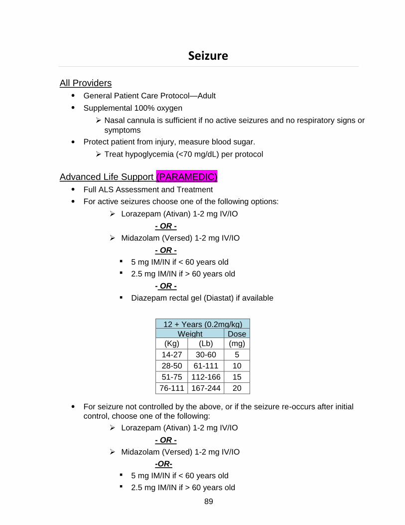
12 + Years (0.2mg/kg)
Weight Dose
(Kg) (Lb) (mg)
14-27 30-60 5
28-50 61-111 10
51-75 112-166 15
76-111 167-244 20
Seizure
All Providers
General Patient Care Protocol—Adult
Supplemental 100% oxygen
Nasal cannula is sufficient if no active seizures and no respiratory signs or
symptoms
Protect patient from injury, measure blood sugar.
Treat hypoglycemia (<70 mg/dL) per protocol
Advanced Life Support (PARAMEDIC)
Full ALS Assessment and Treatment
For active seizures choose one of the following options:
Lorazepam (Ativan) 1-2 mg IV/IO
- OR -
Midazolam (Versed) 1-2 mg IV/IO
- OR -
5 mg IM/IN if < 60 years old
2.5 mg IM/IN if > 60 years old
- OR -
Diazepam rectal gel (Diastat) if available
For seizure not controlled by the above, or if the seizure re-occurs after initial
control, choose one of the following:
Lorazepam (Ativan) 1-2 mg IV/IO
- OR -
Midazolam (Versed) 1-2 mg IV/IO
-OR-
5 mg IM/IN if < 60 years old
2.5 mg IM/IN if > 60 years old
89
Maximum dose of Lorazepam is 4 mg
Maximum dose of Midazolam is 10 mg
If hypoxic seizures, drug induced seizures, seizures from head trauma, stroke
or eclampsia suspected:
Treat as above and refer to appropriate protocol for further care
Contact Medical Control for any additional orders or questions
Additional Benzodiazepines
90
Shock (Non-Trauma)
Shock is defined as a state of inadequate organ perfusion and tissue oxygenation.
It is evidenced by the presence of any of the following signs and symptoms:
Hypotension
Narrow pulse pressure Tachypnea Tachycardia Delayed capillary refill
Mottled skin appearance
Diaphoresis Cool clammy skin Pallor Altered mental status
Signs and symptoms vary depending upon the stage of shock, which may be
compensated (normal perfusion maintained) or decompensated (unable to maintain normal perfusion).
Categories of shock
Obstructive shock: Caused by an obstruction that interferes with return of
blood to the heart (e.g. tension pneumothorax, cardiac tamponade, massive pulmonary embolus)
Hypovolemic shock: Caused by decreased blood or water volume.
Hypovolemic shock may be hemorrhagic or non-hemorrhagic
Distributive shock: Caused by abnormal distribution of blood resulting from
vasodilaton, vasopermeability or both. Distributive shock may result from anaphylactic reactions, sepsis, or spinal cord injury
Cardiogenic shock: Caused as a result of cardiac pump failure, usually
secondary to severe Left Ventricular failure. May result from massive MI
Perform the following in conjunction with protocols that apply to the specific etiology
of the shock state (e.g. allergic reactions, STEMI, etc.):
Advanced Life Support (IV Tech Paramedic)
Full ALS Assessment and Treatment
Do not delay transport for IV insertion
IV 0.9% NaCl en route (if not contraindicated):
Administer 250 ml boluses until systolic BP > 90 mmHg
Total amount of IVF should not exceed 2 L
Boluses may be given in rapid succession if systolic remains < 90
mmHg
If systolic BP remains < 90 mmHg after 4th bolus (1000ml):
(PARAMEDIC) Consider Dopamine 5 mcg/kg/min, titrate infusion to maintain SBP >
90 mmHg (Maximum dose 20 mcg/kg/min)
Contact Medical Control for any additional orders or questions 91
Stroke—Suspected
Early recognition and transport of stroke is essential to good patient outcomes. Any
patient presenting with a normal blood glucose (> 70 mg/dL), a positive Cincinnati Pre-Hospital Stroke Screen and onset of symptoms (when last seen normal) less
than 24 hours should have early notification of the stroke center (STROKE ALERT) and rapid transport.
All Providers
General Patient Care Protocol-Adult
Supplemental oxygen via nasal cannula only if O2 saturation < 95%
Keep head of stretcher at 30-45 elevation (unless clinical condition will not
allow)
Spinal Immobilization if indicated; elevate head of backboard 15-30
Check blood glucose
Give nothing by mouth (oral glucose is permitted if patient is able to self
administer)
Cincinnati Pre-Hospital Stroke Screen
Advanced Life Support (IV Tech Paramedic)
Full ALS Assessment and Treatment
For hypotension (systolic BP < 90 mmHg) not improved by fluid boluses or
when fluid resuscitation is contraindicated
(PARAMEDIC) Dopamine infusion 5-20 mcg/kg/min titrate to maintain systolic BP >
90 mmHg
If hypoglycemic (<70 mg/dL) with IV access
Dextrose 12.5 g IV
May repeat as needed every 5-10 minutes to blood glucose >
70 mg/dL
(PARAMEDIC) Note: If patient appears malnourished or is a known or suspected
chronic alcoholic administer Thiamine 100 mg IV prior to glucose
administration
If hypoglycemic (< 70 mg/dL) without IV access
Glucose paste 15 g or other oral glucose agent (e.g., orange juice) if
patient alert enough to self-administer oral agent
- OR -
Glucagon 1 mg IM
92
If patient blood glucose is ≥ 70 mg/dl, Cincinnati Pre-Hospital Stroke Screen
is positive and onset of symptoms (when last seen normal) is < 24 hours,
immediately notify ED (STROKE ALERT) and commence rapid transport.
DO NOT DELAY TRANSPORT TO OBTAIN IV ACCESS
Contact Medical Control for any additional orders or questions
93
Syncope
All Providers
General Patient Care Protocol-Adult
Advanced Life Support (IV Tech Paramedic)
Full ALS Assessment and Treatment
For hypotension (systolic BP < 90 mmHg) not improved by fluid boluses or
when fluid resuscitation is contraindicated
(PARAMEDIC) Dopamine infusion 5-20 mcg/kg/min titrate to maintain systolic BP >
90 mmHg
If hypoglycemic (< 70mg/dL), treat per Altered Mental Status/Hypoglycemia
Protocol
If ECG rhythm is bradycardia, heart block or dysrhythmia see specific protocol
If Altered Mental Status persists, or if Acute Stroke suspected, refer to
appropriate protocol.
Contact Medical Control for any additional orders or questions
94
Trauma: General Approach to All Trauma Patients
Assess all patients for major trauma criteria. Major trauma patients should have
transport initiated within 10 minutes of arrival on scene whenever possible. In the setting of major trauma, DO NOT prolong scene time to perform procedures unless immediately necessary to stabilize patient (e.g. hemorrhage control). Initiate all other procedures en-route to the trauma center.
All Providers
General Patient Care Protocol—Adult
Secure airway/spinal immobilization if indicated
Supplemental 100% oxygen if any respiratory symptoms
Examine patient for obvious bleeding
Control active bleeding with direct pressure
Assess disability-neurologic status/record Glasgow coma score
Head to toe examination to assess for injuries
Restrain as needed
Advanced Life Support (IV Tech Paramedic)
When conditions warrant (specified as “Full ALS Assessment and Treatment”
in individual protocol)
Advanced airway/ventilatory management as needed
Perform cardiac monitoring
Record and monitor O2 saturation
Microstream capnography if any acute respiratory symptoms
IV 0.9% NaCl TKO/KVO or IV lock
If SBP < 90 mmHg, administer boluses of 0.9%NaCl/Lactated Ringers
at 250 ml until SBP > 90 mmHg
Assess for Tension Pneumothorax
Tension pneumothorax should be suspected in patients who
exhibit
Severe respiratory distress with hypoxia
Unilateral decreased or absent lung sounds (may see
tracheal deviation away from collapsed lung field)
Evidence of hemodynamic compromise (e.g. shock,
hypotension, tachycardia, altered mental status)
95
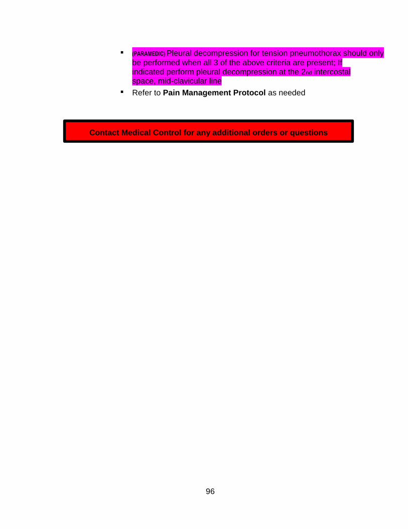
(PARAMEDIC) Pleural decompression for tension pneumothorax should only
be performed when all 3 of the above criteria are present; If indicated perform pleural decompression at the 2nd intercostal space, mid-clavicular line
Refer to Pain Management Protocol as needed
Contact Medical Control for any additional orders or questions
96
Trauma: Burns—Thermal
All Providers
General Patient Care Protocol—Adult
Remove or cool heat source if present (e.g. clothing, tar)
Keep patient warm and dry with a sheet on burns:
2 burns greater than 15% of body surface area
3 burns
Electrical and chemical burns
Spinal immobilization if high voltage electrical injuries
Observe for signs of impending loss of airway; Refer to the Airway
Management Protocol as needed:
Hypoxia
Poor ventilatory effort
Altered mental status/decreased level of consciousness
Inability to maintain patent airway
Advanced Life Support (IV Tech Paramedic)
If moderate to severe pain, see Pain Management Protocol
Estimate Total Body Surface Area (TBSA) burned
If > 20% 2nd and 3rd degree TBSA burned, initiate volume resuscitation
with up to 2 L Normal Saline/Lactated Ringers IV/IO
Contact Medical Control for any additional orders or questions
97
Trauma: Chest Injuries
All Providers
General Patient Care Protocol—Adult
Assess breath sounds frequently
Assess for ventilatory compromise and assist with BVM as needed
For open/sucking chest wounds, apply occlusive dressing sealed on three (3) sides or commercially available chest seal
Remove temporarily to vent air if respiratory status worsens
Advanced Life Support (IV Tech Paramedic) Full ALS Assessment and Treatment
Assess for flail segment
Observe for signs of impending respiratory failure; Refer to the Airway Management Protocol as needed:
Hypoxia Poor ventilatory effort Altered mental status/decreased level of consciousness Inability to maintain patent airway
Contact Medical Control for any additional orders or questions
98
Trauma: Head Injuries
All Providers
General Patient Care Protocol-Adult
Supplemental oxygen
Restrain as needed
If normotensive or hypertensive
Elevate head of backboard 15 - 30
Advanced Life Support (IV Paramedic)
Full ALS Assessment and Treatment
Advanced airway/ventilatory management as needed
Note: Airway interventions can be detrimental to patients with head injury
by raising intracranial pressure, worsening hypoxia (and secondary brain injury) and increasing risk of aspiration. Whenever possible these patients should be managed in the least invasive manner to maintain O2 saturation > 90% (i.e. NRB, BVM with 100% O2)
Observe for signs of impending respiratory failure; Refer to the Airway
Management Protocol if needed:
Hypoxia
Poor ventilatory effort
Altered mental status/decreases level of consciousness
Inability to maintain patent airway
For patients with assisted ventilation
Administer eucapneic (normal rate 12-15/min) ventilations
Titrate to target an ETCO2 of 40 mmHg
Acute herniation should be suspected when the following signs are present:
Acute unilateral dilated and non-reactive pupil
Abrupt deterioration in mental status
Abrupt onset of motor posturing
Abrupt increase in blood pressure
Abrupt decrease in heart rate
Hyperventilation (ventilatory rate of 20) is a temporizing measure which is
only indicated in the event of acute herniation
If signs of herniation develop, increase ventilatory rate to 20/minute and target
an ETCO2 of 30-35 mmHg
99
If severely agitated/combative and unable to de-escalate by any other means, consider:
(PARAMEDIC) Lorazepam 1-2 mg IV/IO/IM
Contact Medical Control for any additional orders or questions
Additional sedation for combative patients
100
Trauma: Eye Injuries
All Providers
General Patient Care Protocol—Adult
Measure visual acuity
If injury is secondary to a chemical exposure:
Remove patient from source if safe to do so
Remove contact lenses if appropriate; transport with patient
Irrigate the eyes with 0.9 % Normal Saline for a minimum of 20
minutes
(PARAMEDIC) For significant pain, administer 2 drops of Tetracaine HCL to affected
eye(s)
Determine chemical involved, bring MSDS sheet if available
If eye injury is due to trauma:
Do not irrigate or apply Tetracaine HCL
Stabilize any penetrating objects
Do not remove any impaled object
Protective metal shield unless impaled object precludes
Prevent patient bending or standing
If blood observed in anterior chamber, transport with head elevated 60
Contact Medical Control for any additional orders or questions
101
Trauma: Extremity
All Providers
General Patient Care Protocol—Adult
Remove or cut away clothing to expose area of injury
Check distal pulses, capillary refill, sensation/movement prior to splinting
If pulse present, splint in position found if possible
If pulse absent, attempt to place the injury into anatomical position
Check distal pulses, capillary refill and sensation after splinting
Control active bleeding
For uncontrollable hemorrhage (heavy bleeding despite aggressive
direct pressure):
-Apply tourniquet device as proximal on extremity as possible
-Tourniquet must be at least 2 inches wide
-Tighten tourniquet until bright red bleeding has stopped
-Secure in place and expedite transport to Level 1 Trauma
Center
-Document time placed on chart and on device (if possible)
-Notify receiving center of presence, time placed, and
location of tourniquet
Open wounds/fractures should be covered with sterile dressings and
immobilized in the presenting position
Dislocations should be immobilized to prevent any further movement of
the joint
Advanced Life Support (IV Tech Paramedic)
Full ALS Assessment and Treatment
Refer to Spinal Immobilization protocol as needed
For isolated extremity trauma:
Stabilize BP (SBP 90 mmHg)
Refer to the Pain Management protocol as needed
Contact Medical Control for any additional orders or questions
102
Trauma: Traumatic Amputations
All Providers
General Patient Care Protocol—Adult
For uncontrollable hemorrhage (heavy bleeding despite aggressive direct
pressure):
Apply tourniquet device as proximal on extremity as possible
Tourniquet must be at least 2 inches wide
Tighten tourniquet until bright red bleeding has stopped
Secure in place and expedite transport to Level 1 Trauma Center
Document time placed on chart and on device (if possible)
Notify receiving center of presence, time placed, and location of
tourniquet
If amputation incomplete:
Attempt to stabilize with bulky pressure dressing
Splint inline
If amputation complete:
Wrap in sterile dressing moistened in sterile saline
Place in plastic bag if possible
Attempt to cool with cool pack during transport
Advanced Life Support (IV Tech Paramedic)
For isolated extremity trauma:
Stabilize BP (SBP ≥90 mmHg)
Refer to Pain Management protocol as needed
For uncontrollable hemorrhage (heavy bleeding despite aggressive direct
pressure):
Apply tourniquet device as proximal to injury site on extremity as possible
Tourniquet must be at least 2 inches wide
Tighten tourniquet until bright red bleeding has stopped
Secure in place and expedite transport to Level 1 Trauma Center
Document time placed on chart and on device (if possible)
Notify receiving center of presence, time placed and location of tourniquet
Contact Medical Control for any additional orders or questions
103
Trauma: Indications for Withholding Resuscitation in Traumatic Cardiopulmonary Arrest
CPR can be withheld in Traumatic Cardiopulmonary Arrest under the following circumstances:
Pulseless, apenic, and no other signs of life present AND
The presence of one or more of the following:
Rigor Mortis
Decomposition of body tissues
Dependent Lividity
Injuries incompatible with life (e.g. incineration, decapitation,
hemicorporectomy)
Evidence of significant time lapse since pulselessness
- OR -
Patients who present pulseless after blunt trauma or penetrating trauma
provided that all other signs of life are absent:
Pulseless and Apenic
Lack of pupillary reflexes and spontaneous movement
Asystole or agonal rhythm < 20 on cardiac monitor
- OR -
Patients who become pulseless after severe traumatic injury when transport
to the NEAREST ED cannot be accomplished within 15 minutes (i.e., prolonged extrications), provided that all other signs of life are absent and transport has not been initiated:
Pulseless and apneic
Lack of pupillary reflexes and spontaneous movement
Asystole or agonal rhythm < 20 on cardiac monitor
This criterion does not apply in the following scenarios:
When the Cardiac Arrest is inconsistent with Cardiac Arrest due to trauma
Lightning or other high voltage injuries
Drowning
Suspected hypothermia
Transport has been initiated
Contact Medical Control for any additional orders or questions
104
Trauma: Sexual Assault
General Patient Care Protocol
For victims of sexual assault who meet major trauma criteria, transport to
Level 1 Trauma Center
For all other cases, transport to nearest appropriate Emergency Department
Provide supportive care as indicated by patient’s condition
Preserve evidence
Contact Medical Control for any additional orders or questions
105
Spinal Immobilization: Indications
Determining the need for spinal immobilization requires a careful assessment of the
injury, the patient’s complaints and overall condition, and the patient’s ability to recognize and convey the presence of spinal injury symptoms. It is not feasible to “clear” the spine in the prehospital setting. Spinal immobilization should always be applied when any doubt exists as to the possibility of spinal trauma. The following algorithms (Blunt and Penetrating Trauma) can be used to assist paramedics in making the most appropriate decision about the need for spinal immobilization.
Blunt Trauma
Assess for concerning mechanism of injury:
Any mechanism the produces a violent impact on the head, neck, torso or
pelvis
Incidents that produce sudden acceleration or deceleration, including lateral bending forces
Any fall, especially in the elderly
Ejection or fall from a moving mode of transportation
Shallow-water drowning or diving injuries
High-voltage electrical injuries
Assess patient’s ability to clearly communicate and/or comprehend the
nature of their injuries:
Altered level of consciousness
GCS < 15
Evidence of significant intoxication
Dementia
Speech or hearing impairment
Age (young children)
Language barrier
Assess for physical signs or symptoms of spinal trauma:
Spinal pain or tenderness, including paraspinal muscles
Neurologic deficit or complaint, including parasthesia, paralysis, or weakness
Anatomical deformity of the spine
Assess for presence of distracting injuries, e.g.:
Long bone fractures
Joint dislocations
106
Abdominal or thoracic pain, or obvious visceral injury
Large lacerations, degloving injuries or crush injuries
Any injury producing acute functional impairment
Craniofacial injuries
IF ANY OF THE ABOVE MENTIONED FEATURES ARE PRESENT,
OR IF ANY QUESTION, IMMOBILIZE!
Contact Medical Control for any additional orders or questions
107
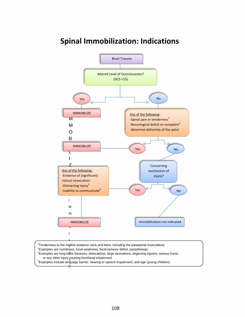
1Tenderness to the midline posterior neck and back, including the paraspinal musculature
2Examples are numbness, focal weakness, focal sensory deficit, parasthesias
3Examples are long bone fractures, dislocations, large lacerations, degloving injuries, serious burns
or any other injury causing functional impairment 4Examples include language barrier, hearing or speech impairment, and age (young children)
Spinal Immobilization: Indications
I
M
M
O
B
I
L
I
Z
E
I
M
M
O
B
I
L
I
Z
1
E
Blunt Trauma
Altered Level of Consciousness?
(GCS <15)
Yes No
Any of the following:
-Spinal pain or tenderness1
-Neurological deficit or complaint2
-Abnormal deformity of the spine
No
Concerning
mechanism of
injury?
IMMOBILIZE
Yes
Any of the following:
-Evidence of (significant)
clinical intoxication
-Distracting injury3
-Inability to communicate4
Immobilization not indicated
No
IMMOBILIZE
IMMOBILIZE
Yes
108
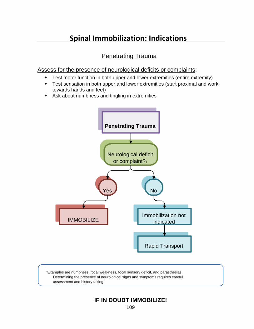
Spinal Immobilization: Indications
Penetrating Trauma
Assess for the presence of neurological deficits or complaints:
Test motor function in both upper and lower extremities (entire extremity)
Test sensation in both upper and lower extremities (start proximal and work towards hands and feet)
Ask about numbness and tingling in extremities
Penetrating Trauma
Neurological deficit
or complaint?1
IMMOBILIZE
Yes
No Immobilization not
indicated
Rapid Transport
1
Examples are numbness, focal weakness, focal sensory deficit, and parasthesias.
Determining the presence of neurological signs and symptoms requires careful
assessment and history taking.
IF IN DOUBT IMMOBILIZE!
109
Immobilize all patients with the following conditions:
High voltage electrical injuries (does not include Taser use)
Shallow water drowning or diving injuries
If spinal immobilization is indicated but refused by the patient:
Advise the patient of the indication for immobilization, and the risks of refusing
the intervention.
If the patient allows, apply the cervical collar even if backboard is refused.
Maintain spinal alignment as best as can be achieved during transport.
Clearly document refusal of immobilization.
For patients who cannot tolerate supine position due to clinical condition:
Apply all elements of spinal immobilization that the patient will tolerate.
Maintain spinal alignment as best as can be achieved during transport.
Clearly document the clinical condition that interfered with full immobilization.
Contact Medical Control for any additional orders or questions
110

General Approach to All Pediatric Patients
The following measures will apply to the management of all pediatric patients:
A Child shall be defined as:
Age 12 years of age or weight 40 kg (if age unknown)
No signs of puberty if age/weight not able to be determined
For PALS resuscitation 8 years
For Major Trauma < 18 years
All Providers
Ensure scene safety.
Scene survey to determine environmental conditions, mechanism of injury or
illness and potential for hazardous conditions.
Form general impression of patient’s condition.
Establish patient responsiveness.
Immobilize spine if cervical or other spine injury suspected.
Assess airway and breathing.
Supplemental 100% oxygen if any respiratory signs or symptoms
Assess circulation and perfusion by measuring heart rate, and observing skin
color, temperature, capillary refill and the quality of central/peripheral pulses
For children with absent pulses, initiate cardiopulmonary resuscitation
Control hemorrhage using direct pressure or a pressure dressing
Measure BP in children older than 3
Evaluate mental status including pupil reaction, motor function and sensation
For mental status, use the AVPU scale:
A-The patient is alert and orientated (age appropriate)
V-The patient is responsive to verbal stimulus
P-The patient is responsive to painful stimulus
U-The patient is unresponsive to any stimulus
Expose the child only as necessary to perform further assessments
Maintain the child’s body temperature throughout the assessment
Utilize the Broslow-Luten system for estimating patient weight
111
Advanced Life Support (IV Tech Paramedic)
When condition warrants (specified as “Full Pediatric ALS Assessment and
Treatment” in individual protocols):
Advanced airway/ventilatory management as needed
Perform cardiac monitoring
Record and monitor O2 saturation
Record and monitor End-tidal CO2
If symptoms severe or for medication access IV 0.9% NaCl TKO/KVO or
IV Lock
If signs of shock administer boluses of 0.9% NaCl at 20 ml/kg until
signs of shock resolve or 60 ml/kg total
If signs of severe cardiopulmonary compromise (poor systemic perfusion,
hypotension, altered consciousness and/or respiratory distress/failure) and IV attempts unsuccessful in a child consider intraosseous access
If a child’s condition is critical or unstable, initiate transport without delay
Perform procedures, history and detailed physical exam en route
to the hospital
All medication dosages and equipment sizes should be calculated using the
Broselow-Luten system
Reassess the patient frequently
112
Contact Medical Control for any additional orders or questions
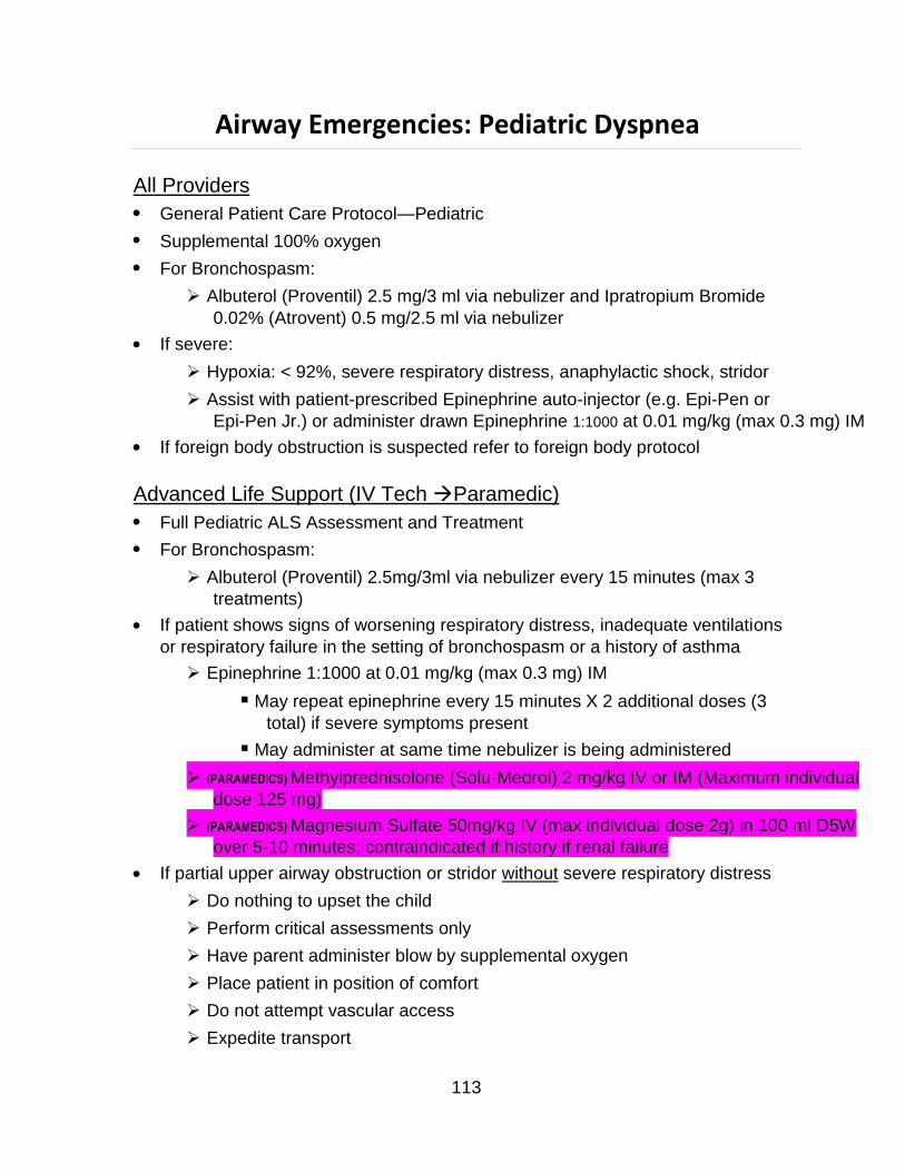
Airway Emergencies: Pediatric Dyspnea
All Providers
General Patient Care Protocol—Pediatric
Supplemental 100% oxygen
For Bronchospasm:
Albuterol (Proventil) 2.5 mg/3 ml via nebulizer and Ipratropium Bromide
0.02% (Atrovent) 0.5 mg/2.5 ml via nebulizer
If severe:
Hypoxia: < 92%, severe respiratory distress, anaphylactic shock, stridor
Assist with patient-prescribed Epinephrine auto-injector (e.g. Epi-Pen or
Epi-Pen Jr.) or administer drawn Epinephrine 1:1000 at 0.01 mg/kg (max 0.3 mg) IM
If foreign body obstruction is suspected refer to foreign body protocol
Advanced Life Support (IV Tech Paramedic)
Full Pediatric ALS Assessment and Treatment
For Bronchospasm:
Albuterol (Proventil) 2.5mg/3ml via nebulizer every 15 minutes (max 3
treatments)
If patient shows signs of worsening respiratory distress, inadequate ventilations
or respiratory failure in the setting of bronchospasm or a history of asthma
Epinephrine 1:1000 at 0.01 mg/kg (max 0.3 mg) IM
May repeat epinephrine every 15 minutes X 2 additional doses (3
total) if severe symptoms present
May administer at same time nebulizer is being administered
(PARAMEDICS) Methylprednisolone (Solu-Medrol) 2 mg/kg IV or IM (Maximum individual
dose 125 mg)
(PARAMEDICS) Magnesium Sulfate 50mg/kg IV (max individual dose 2g) in 100 ml D5W
over 5-10 minutes; contraindicated if history if renal failure
If partial upper airway obstruction or stridor without severe respiratory distress
Do nothing to upset the child
Perform critical assessments only
Have parent administer blow by supplemental oxygen
Place patient in position of comfort
Do not attempt vascular access
Expedite transport
113
If complete airway obstruction or severe respiratory distress, failure or arrest
Advanced airway/ventilatory management as needed
Drowning/Near Drowning
Advanced ventilatory management/100% O2 as indicated
Spinal Immobilization if indicated
Protect from heat loss
Patients may develop delayed onset respiratory symptoms
Refer to appropriate protocol if cardiac arrest present
114
Contact Medical Control for any additional orders or questions
▪ (PARAMEDIC) CONSIDER RACEMIC EPINEPHRINE
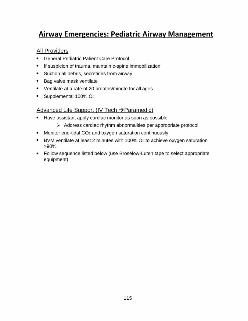
Airway Emergencies: Pediatric Airway Management
All Providers
General Pediatric Patient Care Protocol
If suspicion of trauma, maintain c-spine immobilization
Suction all debris, secretions from airway
Bag valve mask ventilate
Ventilate at a rate of 20 breaths/minute for all ages
Supplemental 100% O2
Advanced Life Support (IV Tech Paramedic)
Have assistant apply cardiac monitor as soon as possible
Address cardiac rhythm abnormalities per appropriate protocol
Monitor end-tidal CO2 and oxygen saturation continuously
BVM ventilate at least 2 minutes with 100% O2 to achieve oxygen saturation
>90%
Follow sequence listed below (use Broselow-Luten tape to select appropriate
equipment)
115
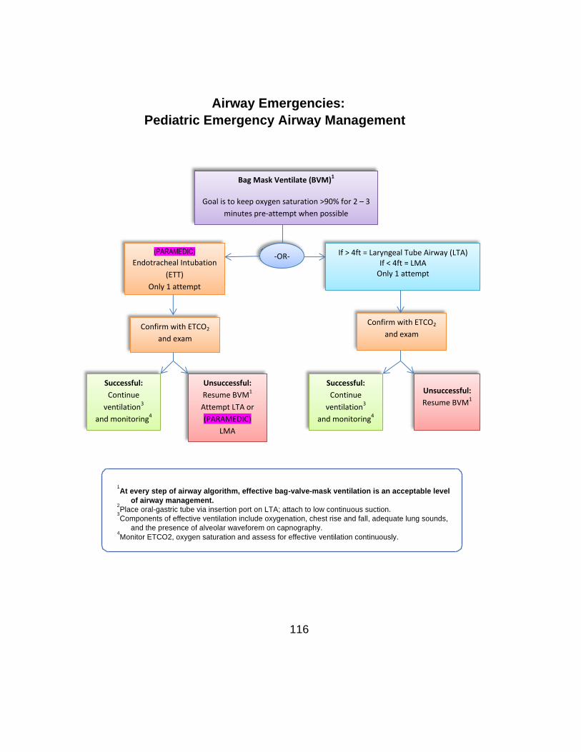
Airway Emergencies:
Pediatric Emergency Airway Management
1
2
3
4
At every step of airway algorithm, effective bag-valve-mask ventilation is an acceptable level
of airway management.
Place oral-gastric tube via insertion port on LTA; attach to low continuous suction.
Components of effective ventilation include oxygenation, chest rise and fall, adequate lung sounds,
and the presence of alveolar waveforem on capnography.
Monitor ETCO2, oxygen saturation and assess for effective ventilation continuously.
116
Bag Mask Ventilate (BVM)1
Goal is to keep oxygen saturation >90% for 2 – 3
minutes pre-attempt when possible
-OR- (PARAMEDIC)
Endotracheal Intubation
(ETT)
Only 1 attempt
Confirm with ETCO2
and exam
If > 4ft = Laryngeal Tube Airway (LTA) If < 4ft = LMA
Only 1 attempt
Confirm with ETCO2
and exam
Successful:
Continue
ventilation3
and monitoring4
Unsuccessful:
Resume BVM1
Attempt LTA or
(PARAMEDIC)
LMA
Successful:
Continue
ventilation3
and monitoring4
Unsuccessful:
Resume BVM1
Following placement or ETT, LTA, or LMA confirm proper placement
Assess epigastric sounds, breath sounds and chest rise and fall
Observe for presence of alveolar waveform on capnography
Record tube depth and secure in place using a commercial tube holder (if
available)
Utilize head restraint device (i.e. “head blocks”) or rigid cervical collar and
long spine board as needed to help secure airway in place
Capnography/ETCO2 Monitoring
Digital capnography (waveform) is the system STANDARD for ETCO2 monitoring
With the exception of on-scene equipment failure, patients should not be
switched from digital capnography (Zoll/Phillips/Lifepack) to a colorimetric device for monitoring end-tidal CO2
In the event digital capnography is not possible due to on-scene equipment
failure, continuous colorimetric monitoring of ETCO2 is an acceptable alternative
Continuous ETCO2 monitoring is a mandatory component of invasive airway
management
If ETCO2 monitoring cannot be accomplished by either of the above
methods, the invasive device must be removed, and the airway managed non-invasively
If an alveolar waveform is not present with capnography (i.e., flat line),
remove the device, and proceed to the next step in the algorithm
Foreign Body Airway Obstruction
If unresponsive open airway using a head tilt/chin lift (if no trauma)
If < 1 year old, administer up to 5 back blows and 5 chest thrusts
If 1 to 8 years, administer compressions and attempts at ventilation until
the foreign body is dislodged
If ventilation is unsuccessful (O2 saturations cannot be kept > 92 %) perform in
the following order:
Reposition airway and attempt bag valve mask assisted ventilation again
If unsuccessful, establish direct view of object with laryngoscope and
attempt to remove it with Magill forceps
(PARAMEDIC) If unable to visualize a foreign body using laryngoscope, and vocal cords
are clearly seen, attempt intubation only once
If unsuccessful, re-attempt BVM ventilation; If oxygen saturation > 90 with
BVM proceed no further and expedite transport
Expedite transport to nearest emergency department
Contact Medical Control for any additional orders or questions 117
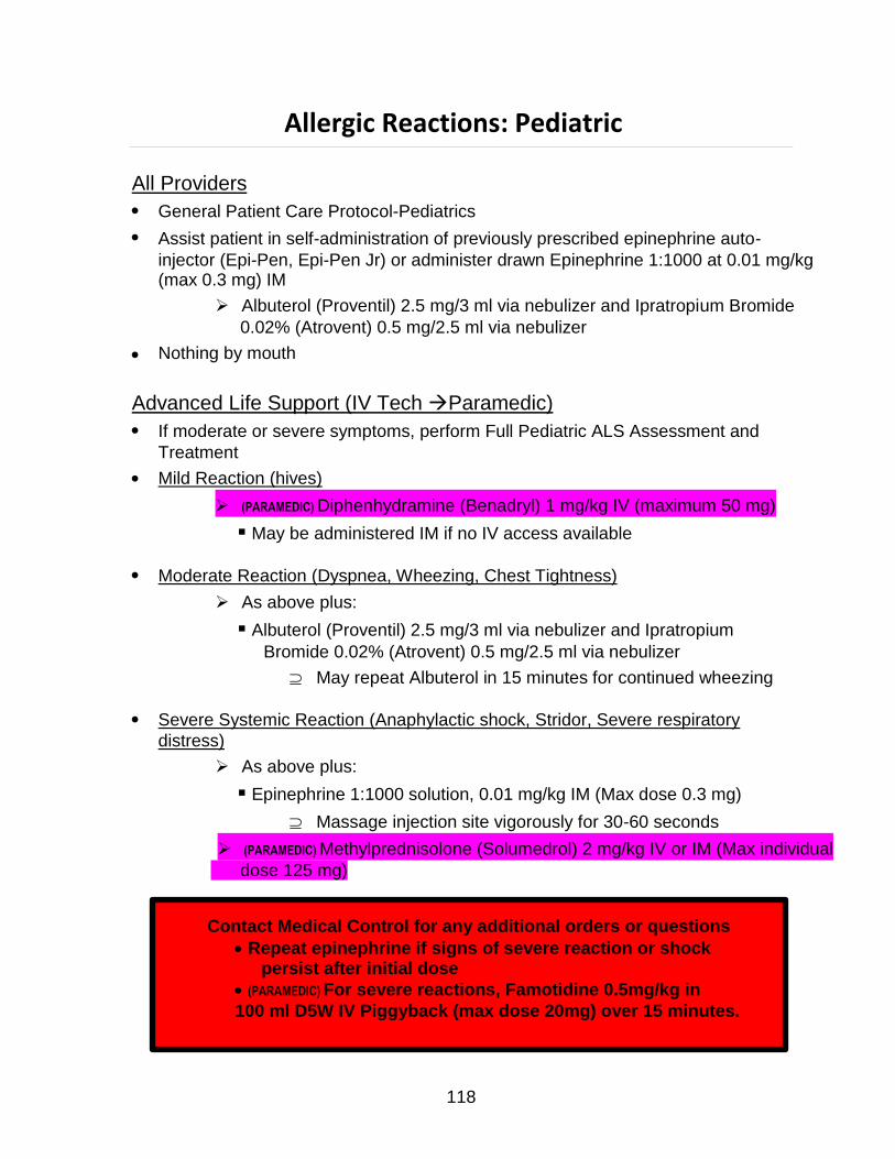
Allergic Reactions: Pediatric
All Providers
General Patient Care Protocol-Pediatrics
Assist patient in self-administration of previously prescribed epinephrine auto-
injector (Epi-Pen, Epi-Pen Jr) or administer drawn Epinephrine 1:1000 at 0.01 mg/kg (max 0.3 mg) IM
Albuterol (Proventil) 2.5 mg/3 ml via nebulizer and Ipratropium Bromide
0.02% (Atrovent) 0.5 mg/2.5 ml via nebulizer
Nothing by mouth
Advanced Life Support (IV Tech Paramedic)
If moderate or severe symptoms, perform Full Pediatric ALS Assessment and
Treatment
Mild Reaction (hives)
(PARAMEDIC) Diphenhydramine (Benadryl) 1 mg/kg IV (maximum 50 mg)
May be administered IM if no IV access available
Moderate Reaction (Dyspnea, Wheezing, Chest Tightness)
As above plus:
Albuterol (Proventil) 2.5 mg/3 ml via nebulizer and Ipratropium
Bromide 0.02% (Atrovent) 0.5 mg/2.5 ml via nebulizer
May repeat Albuterol in 15 minutes for continued wheezing
Severe Systemic Reaction (Anaphylactic shock, Stridor, Severe respiratory
distress)
As above plus:
Epinephrine 1:1000 solution, 0.01 mg/kg IM (Max dose 0.3 mg)
Massage injection site vigorously for 30-60 seconds
(PARAMEDIC) Methylprednisolone (Solumedrol) 2 mg/kg IV or IM (Max individual
dose 125 mg)
Contact Medical Control for any additional orders or questions
Repeat epinephrine if signs of severe reaction or shock persist after initial dose
(PARAMEDIC) For severe reactions, Famotidine 0.5mg/kg in
100 ml D5W IV Piggyback (max dose 20mg) over 15 minutes.
118
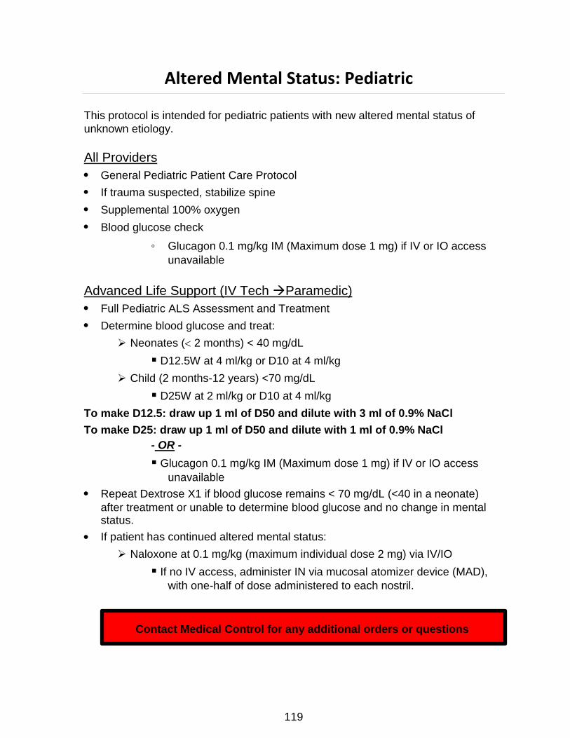
Altered Mental Status: Pediatric
This protocol is intended for pediatric patients with new altered mental status of
unknown etiology.
All Providers
General Pediatric Patient Care Protocol
If trauma suspected, stabilize spine
Supplemental 100% oxygen
Blood glucose check
◦ Glucagon 0.1 mg/kg IM (Maximum dose 1 mg) if IV or IO access
unavailable
Advanced Life Support (IV Tech Paramedic)
Full Pediatric ALS Assessment and Treatment
Determine blood glucose and treat:
Neonates ( 2 months) < 40 mg/dL
D12.5W at 4 ml/kg or D10 at 4 ml/kg
Child (2 months-12 years) <70 mg/dL
D25W at 2 ml/kg or D10 at 4 ml/kg
To make D12.5: draw up 1 ml of D50 and dilute with 3 ml of 0.9% NaCl
To make D25: draw up 1 ml of D50 and dilute with 1 ml of 0.9% NaCl
- OR -
Glucagon 0.1 mg/kg IM (Maximum dose 1 mg) if IV or IO access
unavailable
Repeat Dextrose X1 if blood glucose remains < 70 mg/dL (<40 in a neonate)
after treatment or unable to determine blood glucose and no change in mental status.
If patient has continued altered mental status:
Naloxone at 0.1 mg/kg (maximum individual dose 2 mg) via IV/IO
If no IV access, administer IN via mucosal atomizer device (MAD),
with one-half of dose administered to each nostril.
Contact Medical Control for any additional orders or questions
119
Apparent Life-Threatening Event (ALTE)
An Apparent Life Threatening Event (ALTE), often referred to as a “near miss SIDS”,
is an episode that is frightening to the observer/caregiver, and involves some combination of the following:
Apnea (central or obstructive)
Color change (cyanosis, pallor, erythema, plethora)
Marked change in muscle tone (e.g. limpness/rigid)
Choking or gagging
All Providers
Assume the history given is accurate and reliable
Determine the severity, nature and duration of the episode
Obtain a medical history:
Known chronic diseases
History of preterm delivery
Evidence of seizure activity
Current or recent infection
Gastroesophageal reflux
Inappropriate mixture of formula
Recent trauma
Perform a thorough physical assessment that includes the general appearance,
skin color, level of interaction with environment and evidence of trauma and blood glucose check
Transport to the nearest appropriate receiving facility
For patients < 1 year of age
If the parent/guardian is refusing medical care and/or EMS transport, OLMC
must be contacted prior to accepting a refusal.
Contact Medical Control for any additional orders,
questions, or refusal in a patient <1 year of age
120
Cardiac Arrest, General: Pediatric
Airway management by BVM is sufficient in the pediatric arrest patient.
A single attempt at intubation can be made only if time allows. Do not prolong transport or scene time to attempt intubation.
All Providers
General Pediatric Patient Care Protocol
Establish responsiveness
If trauma suspected, stabilize spine
Confirm apnea and pulselessness and administer CPR
Apply AED/monitor as soon as available for 8 years old
For children 1-8 years old use pediatric AED cables/electrodes if available
As a last resort in a child 1-8 years old, apply AED with available
cables/electrodes
Do not apply AED to children < 1 year old
Advanced Life Support (IV Tech Paramedic)
Full Pediatric ALS Assessment and Treatment
Determine cardiac rhythm and refer to appropriate protocol for further
management actions
Determine blood glucose and treat:
Neonates ( 2 months) < 40 mg/dL
D12.5W at 4 ml/kg or D10 at 4 ml/kg
Child (2 months-12 years) <70 mg/dL
D25W at 2 ml/kg or D10 at 4 ml/kg
To make D12.5: draw up 1 ml of D50 and dilute with 3 ml of 0.9% NaCl
To make D25: draw up 1 ml of D50 and dilute with 1 ml of 0.9% NaCl
- OR -
Glucagon 0.1 mg/kg IM (Maximum dose 1 mg) if IV or IO access
unavailable
Repeat Dextrose X1 if blood glucose remains < 70 mg/dL (<40 in a neonate)
after treatment or unable to determine blood glucose and no change in mental status.
Due to the child’s critical condition, initiate transport without delay.
Contact Medical Control for any additional orders or questions
121
Cardiac Asystole and PEA: Pediatric
Asystole and Pulseless Electrical Activity
Advanced Life Support (IV Tech Paramedic)
Follow Cardiac Arrest/Non-Traumatic—Pediatric Protocol
Confirm the presence of asystole in 2 leads
Minimize any interruptions in compressions
Using the most readily available route, administer (during CPR)
(PARAMEDIC) Epinephrine 1:10,000 at 0.01 mg/kg IV/IO (max dose 1mg)
(PARAMEDIC) Repeat Epinephrine as above every 3-5 minutes
Flush medication with 10-20 ml of normal saline after each dose
Treat any suspected contributing factors:
If hypovolemic, administer 0.9% NaCl at 20 ml/kg IV/IO bolus, may repeat
twice (to a maximum of 60 ml/kg)
If hypoxic, secure airway and assist ventilation
If hypothermic, rewarm
If hyperkalemia suspected (history of renal failure/dialysis)
(PARAMEDIC) Calcium Chloride (10%), 20 mg/kg IV/IO (max 1 g)
(PARAMEDIC) Sodium Bicarbonate 1 mEq/kg IV/IO (max dose 50 mEq)
If narcotic suspected, Naloxone 0.1 mg/kg (max dose 2 mg) IM/IV/IO/IN
If toxic ingestion, see specific toxin
Assess for tension pneumothorax
Unilateral decreased or absent lung sounds (may see tracheal
deviation away from collapsed lung)
Evidence of hemodynamic compromise
(PARAMEDIC) If tension pneumothorax suspected due to history or condition,
perform pleural decompression at 2nd intercostal space, mid- clavicular line
Contact Medical Control for any additional orders or questions
122
Cardiac Arrest: VF/VT: Pediatric
Ventricular Fibrillation or Pulseless Ventricular Tachycardia
Advanced Life Support
Follow Cardiac Arrest/Non-traumatic-Pediatric Protocol
Confirm the presence of ventricular fibrillation/pulseless ventricular tachycardia
Defibrillate at 2 J/kg (maximum of 200J)
Continue compressions while defibrillator charges
Immediately resume CPR after shock
Check rhythm after 2 minutes of CPR
Using the most readily available route (give drug during CPR)
(PARAMEDIC) Epinephrine 1:10,000 at 0.01 mg/kg IV/IO
(PARAMEDIC) Repeat Epinephrine as above every 3-5 minutes
Flush medication with 10-20 ml of normal saline after each dose
If shockable rhythm persists, Defibrillate at 4 J/kg
Continue compressions while defibrillator charges
Immediately resume CPR after shock
Check rhythm after 2 minutes of CPR
(PARAMEDIC) Amiodarone 5 mg/kg IV/IO bolus (give during CPR) (max dose 300mg)
(PARAMEDIC) Magnesium 50 mg/kg IV/IO for suspected torsades de pointes (max dose
2g)
If shockable rhythm persists, Defibrillate at 4 J/kg
Continue compressions while defibrillator charges
Immediately resume CPR after shock
Check rhythm after 2 minutes of CPR
Continue cycle
Contact Medical Control for any additional orders or questions
123
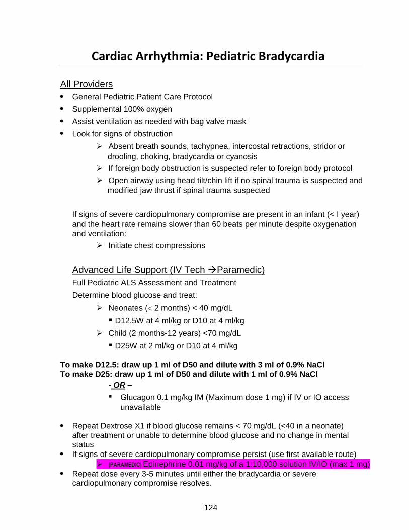
Cardiac Arrhythmia: Pediatric Bradycardia
All Providers
General Pediatric Patient Care Protocol
Supplemental 100% oxygen
Assist ventilation as needed with bag valve mask
Look for signs of obstruction
Absent breath sounds, tachypnea, intercostal retractions, stridor or
drooling, choking, bradycardia or cyanosis
If foreign body obstruction is suspected refer to foreign body protocol
Open airway using head tilt/chin lift if no spinal trauma is suspected and
modified jaw thrust if spinal trauma suspected
If signs of severe cardiopulmonary compromise are present in an infant (< I year)
and the heart rate remains slower than 60 beats per minute despite oxygenation and ventilation:
Initiate chest compressions
Advanced Life Support (IV Tech Paramedic)
Full Pediatric ALS Assessment and Treatment
Determine blood glucose and treat:
Neonates ( 2 months) < 40 mg/dL
D12.5W at 4 ml/kg or D10 at 4 ml/kg
Child (2 months-12 years) <70 mg/dL
D25W at 2 ml/kg or D10 at 4 ml/kg
To make D12.5: draw up 1 ml of D50 and dilute with 3 ml of 0.9% NaCl
To make D25: draw up 1 ml of D50 and dilute with 1 ml of 0.9% NaCl
- OR –
Glucagon 0.1 mg/kg IM (Maximum dose 1 mg) if IV or IO access
unavailable
Repeat Dextrose X1 if blood glucose remains < 70 mg/dL (<40 in a neonate)
after treatment or unable to determine blood glucose and no change in mental status
If signs of severe cardiopulmonary compromise persist (use first available route)
(PARAMEDIC) Epinephrine 0.01 mg/kg of a 1:10,000 solution IV/IO (max 1 mg)
Repeat dose every 3-5 minutes until either the bradycardia or severe cardiopulmonary compromise resolves.
124
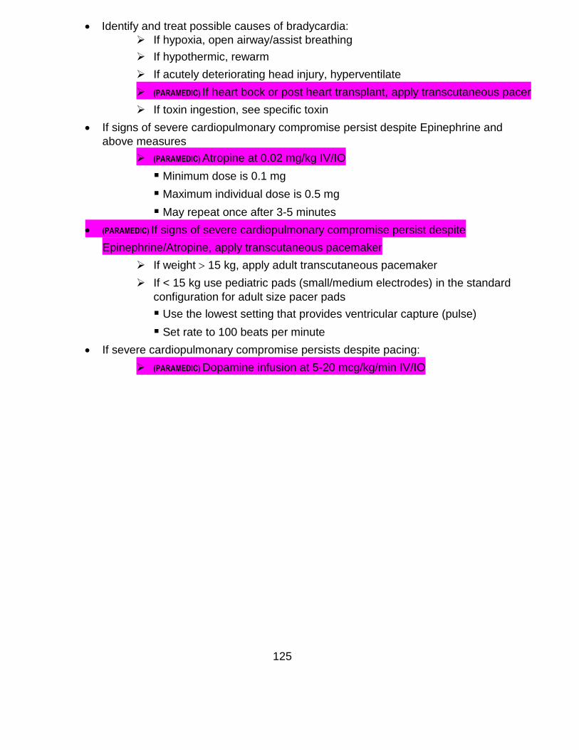
Identify and treat possible causes of bradycardia:
If hypoxia, open airway/assist breathing
If hypothermic, rewarm
If acutely deteriorating head injury, hyperventilate
(PARAMEDIC) If heart bock or post heart transplant, apply transcutaneous pacer
If toxin ingestion, see specific toxin
If signs of severe cardiopulmonary compromise persist despite Epinephrine and
above measures
(PARAMEDIC) Atropine at 0.02 mg/kg IV/IO
Minimum dose is 0.1 mg
Maximum individual dose is 0.5 mg
May repeat once after 3-5 minutes
(PARAMEDIC) If signs of severe cardiopulmonary compromise persist despite
Epinephrine/Atropine, apply transcutaneous pacemaker
If weight 15 kg, apply adult transcutaneous pacemaker
If < 15 kg use pediatric pads (small/medium electrodes) in the standard
configuration for adult size pacer pads
Use the lowest setting that provides ventricular capture (pulse)
Set rate to 100 beats per minute
If severe cardiopulmonary compromise persists despite pacing:
(PARAMEDIC) Dopamine infusion at 5-20 mcg/kg/min IV/IO
125
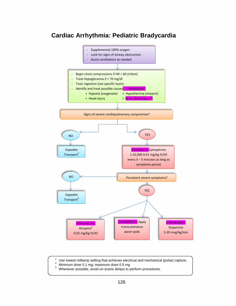
Use lowest milliamp setting that achieves electrical and mechanical (pulse) capture.
Minimum dose 0.1 mg; maximum dose 0.5 mg
Whenever possible, avoid on-scene delays to perform procedures.
Cardiac Arrhythmia: Pediatric Bradycardia
1
2
3
126
- Supplemental 100% oxygen
- Look for signs of airway obstruction
- Assist ventilations as needed
- Begin chest compressions if HR < 60 (infant)
- Treat Hypoglycemia if < 70 mg/dl
- Toxic ingestion (see specific toxin)
- Identify and treat possible causes: (PARAMEDIC)
+ Hypoxia (oxygenate) + Hypothermia (rewarm)
+ Head injury + Heart Block (pace1)
Signs of severe cardiopulmonary compromise?
NO
NO
Expedite
Transport3
Expedite
Transport3
YES
(PARAMEDIC) Epinephrine
1:10,000 0.01 mg/kg IV/IO
every 3 – 5 minutes as long as
symptoms persist
Persistent severe symptoms?
YES
(PARAMEDIC)
Atropine2
0.02 mg/kg IV/IO
(PARAMEDIC) Apply
transcutaneous
pacer pads
(PARAMEDIC)
Dopamine
5-20 mcg/kg/min
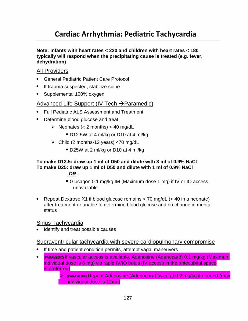
Cardiac Arrhythmia: Pediatric Tachycardia
Note: Infants with heart rates < 220 and children with heart rates < 180
typically will respond when the precipitating cause is treated (e.g. fever, dehydration)
All Providers
General Pediatric Patient Care Protocol
If trauma suspected, stabilize spine
Supplemental 100% oxygen
Advanced Life Support (IV Tech Paramedic)
Full Pediatric ALS Assessment and Treatment
Determine blood glucose and treat:
Neonates ( 2 months) < 40 mg/dL
D12.5W at 4 ml/kg or D10 at 4 ml/kg
Child (2 months-12 years) <70 mg/dL
D25W at 2 ml/kg or D10 at 4 ml/kg
To make D12.5: draw up 1 ml of D50 and dilute with 3 ml of 0.9% NaCl
To make D25: draw up 1 ml of D50 and dilute with 1 ml of 0.9% NaCl
- OR -
Glucagon 0.1 mg/kg IM (Maximum dose 1 mg) if IV or IO access
unavailable
Repeat Dextrose X1 if blood glucose remains < 70 mg/dL (< 40 in a neonate)
after treatment or unable to determine blood glucose and no change in mental status
Sinus Tachycardia Identify and treat possible causes
Supraventricular tachycardia with severe cardiopulmonary compromise
If time and patient condition permits, attempt vagal maneuvers
(PARAMEDIC) If vascular access is available, Adenosine (Adenocard) 0.1 mg/kg (Maximum
individual dose is 6 mg) via rapid IV/IO bolus (IV access in the antecubital space is preferred)
(PARAMEDIC) Repeat Adenosine (Adenocard) twice at 0.2 mg/kg if needed (max
individual dose is 12mg)
127
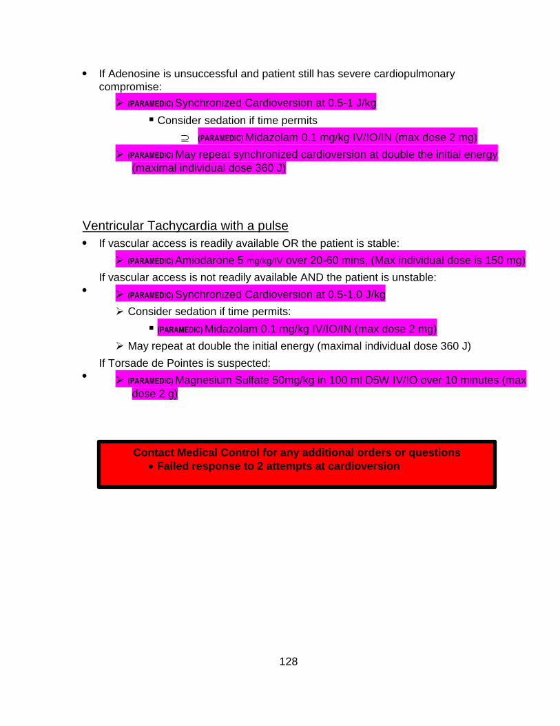
If Adenosine is unsuccessful and patient still has severe cardiopulmonary
compromise:
(PARAMEDIC) Synchronized Cardioversion at 0.5-1 J/kg
Consider sedation if time permits
(PARAMEDIC) Midazolam 0.1 mg/kg IV/IO/IN (max dose 2 mg)
(PARAMEDIC) May repeat synchronized cardioversion at double the initial energy
(maximal individual dose 360 J)
Ventricular Tachycardia with a pulse
If vascular access is readily available OR the patient is stable:
(PARAMEDIC) Amiodarone 5 mg/kg/IV over 20-60 mins, (Max individual dose is 150 mg)
If vascular access is not readily available AND the patient is unstable:
(PARAMEDIC) Synchronized Cardioversion at 0.5-1.0 J/kg
Consider sedation if time permits:
(PARAMEDIC) Midazolam 0.1 mg/kg IV/IO/IN (max dose 2 mg)
May repeat at double the initial energy (maximal individual dose 360 J)
If Torsade de Pointes is suspected:
(PARAMEDIC) Magnesium Sulfate 50mg/kg in 100 ml D5W IV/IO over 10 minutes (max
dose 2 g)
Contact Medical Control for any additional orders or questions
Failed response to 2 attempts at cardioversion
128

1If Torsades de Pointes suspected, give Magnesium Sulfate 50 mg/kg IV over 10 minutes
(maximum individual dose: 2 g). (PARAMEDIC)
Cardiac Arrhythmia: Pediatric Tachycardia
1
2
5
Sinus Tachycardia
YES NO
Identify/treat
causes
Expedite transport
Supraventricular
tachycardia
Ventricular
tachycardia
with a pulse
Severe
symptoms? Stable Unstable
or no
IV access
(PARAMEDIC)
Synchronized
cardioversion at 0.5
– 1.0 J/kg if
unsuccessful, repeat
at double the dose
(max 360 J)
(PARAMEDIC)
Amiodarone
5mg/kg IV/IO
OVER 20 – 60
min1
No response with
severe
cardiopulmonary
compromise
(PARAMEDIC)
Synchronized
cardioversion
at 0.5 – 1.0 J/kg.
If unsuccessful, repeat
at double the dose
(max 360 J)
YES NO
Expedite
transport
(PARAMEDIC)
Adenosine 0.1
mg/kg IV/IO
Repeat x2 at
0.2 mg/kg
No response with
severe
cardiopulmonary
compromise
(PARAMEDIC)
Synchronized
cardioversion
at 0.5 – 1.0 J/kg
If unsuccessful,
repeat at double the
dose (max 360 J)
129
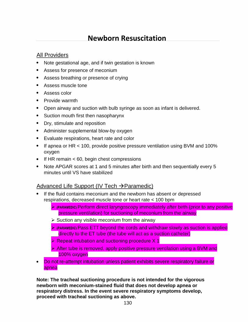
Newborn Resuscitation
All Providers
Note gestational age, and if twin gestation is known
Assess for presence of meconium
Assess breathing or presence of crying
Assess muscle tone
Assess color
Provide warmth
Open airway and suction with bulb syringe as soon as infant is delivered.
Suction mouth first then nasopharynx
Dry, stimulate and reposition
Administer supplemental blow-by oxygen
Evaluate respirations, heart rate and color
If apnea or HR < 100, provide positive pressure ventilation using BVM and 100%
oxygen
If HR remain < 60, begin chest compressions
Note APGAR scores at 1 and 5 minutes after birth and then sequentially every 5
minutes until VS have stabilized
Advanced Life Support (IV Tech Paramedic)
If the fluid contains meconium and the newborn has absent or depressed
respirations, decreased muscle tone or heart rate < 100 bpm
(PARAMEDIC) Perform direct laryngoscopy immediately after birth (prior to any positive
pressure ventilation) for suctioning of meconium from the airway
Suction any visible meconium from the airway
(PARAMEDIC) Pass ETT beyond the cords and withdraw slowly as suction is applied
directly to the ET tube (the tube will act as a suction catheter)
Repeat intubation and suctioning procedure X 1
After tube is removed, apply positive pressure ventilation using a BVM and
100% oxygen
Do not re-attempt intubation unless patient exhibits severe respiratory failure or
apnea
Note: The tracheal suctioning procedure is not intended for the vigorous
newborn with meconium-stained fluid that does not develop apnea or respiratory distress. In the event severe respiratory symptoms develop, proceed with tracheal suctioning as above.
130
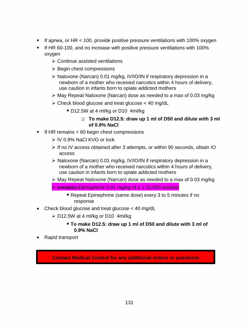
If apnea, or HR < 100, provide positive pressure ventilations with 100% oxygen
If HR 60-100, and no increase with positive pressure ventilations with 100%
oxygen
Continue assisted ventilations
Begin chest compressions
Naloxone (Narcan) 0.01 mg/kg, IV/IO/IN if respiratory depression in a
newborn of a mother who received narcotics within 4 hours of delivery, use caution in infants born to opiate addicted mothers
May Repeat Naloxone (Narcan) dose as needed to a max of 0.03 mg/kg
Check blood glucose and treat glucose < 40 mg/dL
D12.5W at 4 ml/kg or D10 4ml/kg
To make D12.5: draw up 1 ml of D50 and dilute with 3 ml
of 0.9% NaCl
If HR remains < 60 begin chest compressions
IV 0.9% NaCl KVO or lock
If no IV access obtained after 3 attempts, or within 90 seconds, obtain IO
access
Naloxone (Narcan) 0.01 mg/kg, IV/IO/IN if respiratory depression in a
newborn of a mother who received narcotics within 4 hours of delivery, use caution in infants born to opiate addicted mothers
May Repeat Naloxone (Narcan) dose as needed to a max of 0.03 mg/kg
(PARAMEDIC) Epinephrine 0.01 mg/kg of a 1:10,000 solution
Repeat Epinephrine (same dose) every 3 to 5 minutes if no
response
Check blood glucose and treat glucose < 40 mg/dL
D12.5W at 4 ml/kg or D10 4ml/kg
To make D12.5: draw up 1 ml of D50 and dilute with 3 ml of
0.9% NaCl
Rapid transport
Contact Medical Control for any additional orders or questions
131
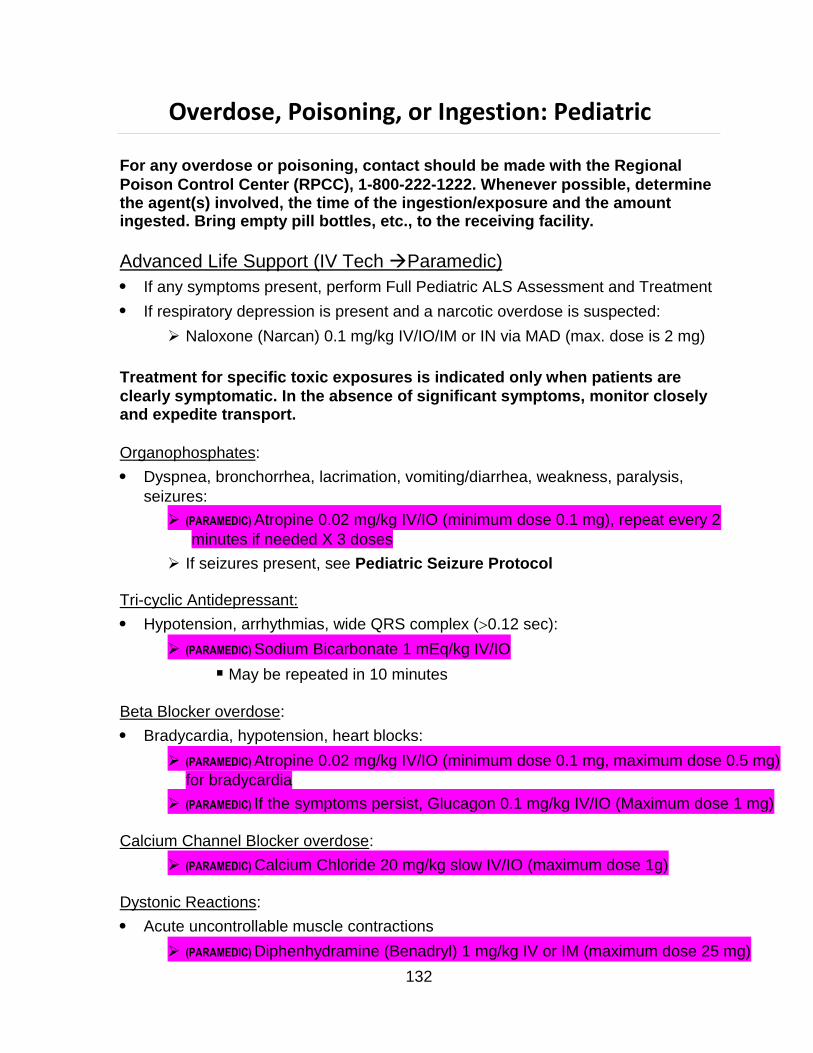
Overdose, Poisoning, or Ingestion: Pediatric
For any overdose or poisoning, contact should be made with the Regional
Poison Control Center (RPCC), 1-800-222-1222. Whenever possible, determine the agent(s) involved, the time of the ingestion/exposure and the amount ingested. Bring empty pill bottles, etc., to the receiving facility.
Advanced Life Support (IV Tech Paramedic)
If any symptoms present, perform Full Pediatric ALS Assessment and Treatment
If respiratory depression is present and a narcotic overdose is suspected:
Naloxone (Narcan) 0.1 mg/kg IV/IO/IM or IN via MAD (max. dose is 2 mg)
Treatment for specific toxic exposures is indicated only when patients are
clearly symptomatic. In the absence of significant symptoms, monitor closely and expedite transport.
Organophosphates:
Dyspnea, bronchorrhea, lacrimation, vomiting/diarrhea, weakness, paralysis,
seizures:
(PARAMEDIC) Atropine 0.02 mg/kg IV/IO (minimum dose 0.1 mg), repeat every 2
minutes if needed X 3 doses
If seizures present, see Pediatric Seizure Protocol
Tri-cyclic Antidepressant:
Hypotension, arrhythmias, wide QRS complex (0.12 sec):
(PARAMEDIC) Sodium Bicarbonate 1 mEq/kg IV/IO
May be repeated in 10 minutes
Beta Blocker overdose:
Bradycardia, hypotension, heart blocks:
(PARAMEDIC) Atropine 0.02 mg/kg IV/IO (minimum dose 0.1 mg, maximum dose 0.5 mg)
for bradycardia
(PARAMEDIC) If the symptoms persist, Glucagon 0.1 mg/kg IV/IO (Maximum dose 1 mg)
Calcium Channel Blocker overdose:
(PARAMEDIC) Calcium Chloride 20 mg/kg slow IV/IO (maximum dose 1g)
Dystonic Reactions:
Acute uncontrollable muscle contractions
(PARAMEDIC) Diphenhydramine (Benadryl) 1 mg/kg IV or IM (maximum dose 25 mg)
132
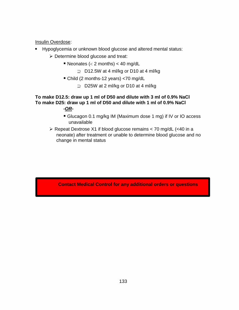
Insulin Overdose:
Hypoglycemia or unknown blood glucose and altered mental status:
Determine blood glucose and treat:
Neonates ( 2 months) < 40 mg/dL
D12.5W at 4 ml/kg or D10 at 4 ml/kg
Child (2 months-12 years) <70 mg/dL
D25W at 2 ml/kg or D10 at 4 ml/kg
To make D12.5: draw up 1 ml of D50 and dilute with 3 ml of 0.9% NaCl
To make D25: draw up 1 ml of D50 and dilute with 1 ml of 0.9% NaCl
-OR-
Glucagon 0.1 mg/kg IM (Maximum dose 1 mg) if IV or IO access
unavailable
Repeat Dextrose X1 if blood glucose remains < 70 mg/dL (<40 in a
neonate) after treatment or unable to determine blood glucose and no change in mental status
133
Contact Medical Control for any additional orders or questions
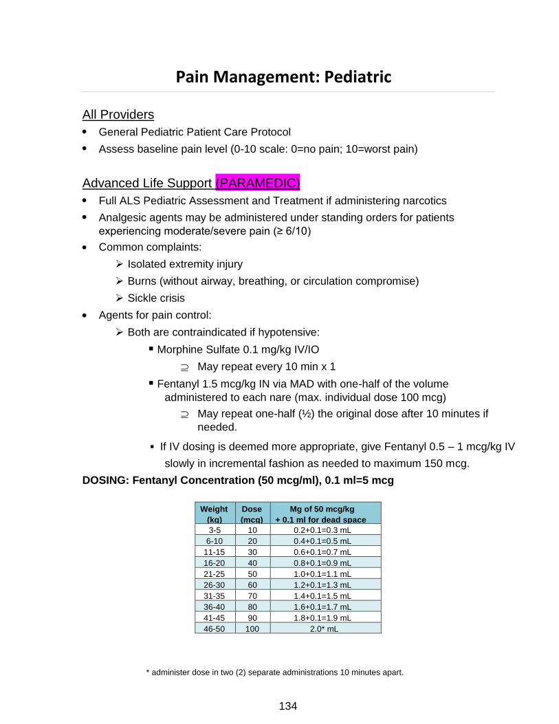
Weight
(kg)
Dose
(mcg)
Mg of 50 mcg/kg
+ 0.1 ml for dead space
3-5 10 0.2+0.1=0.3 mL
6-10 20 0.4+0.1=0.5 mL
11-15 30 0.6+0.1=0.7 mL
16-20 40 0.8+0.1=0.9 mL
21-25 50 1.0+0.1=1.1 mL
26-30 60 1.2+0.1=1.3 mL
31-35 70 1.4+0.1=1.5 mL
36-40 80 1.6+0.1=1.7 mL
41-45 90 1.8+0.1=1.9 mL
46-50 100 2.0* mL
Pain Management: Pediatric
All Providers
General Pediatric Patient Care Protocol
Assess baseline pain level (0-10 scale: 0=no pain; 10=worst pain)
Advanced Life Support (PARAMEDIC)
Full ALS Pediatric Assessment and Treatment if administering narcotics
Analgesic agents may be administered under standing orders for patients
experiencing moderate/severe pain (≥ 6/10)
Common complaints:
Isolated extremity injury
Burns (without airway, breathing, or circulation compromise)
Sickle crisis
Agents for pain control:
Both are contraindicated if hypotensive:
Morphine Sulfate 0.1 mg/kg IV/IO
May repeat every 10 min x 1
Fentanyl 1.5 mcg/kg IN via MAD with one-half of the volume
administered to each nare (max. individual dose 100 mcg)
May repeat one-half (½) the original dose after 10 minutes if
needed.
▪ If IV dosing is deemed more appropriate, give Fentanyl 0.5 – 1 mcg/kg IV
slowly in incremental fashion as needed to maximum 150 mcg.
DOSING: Fentanyl Concentration (50 mcg/ml), 0.1 ml=5 mcg
* administer dose in two (2) separate administrations 10 minutes apart.
134

After each drug dosage administration:
Reassess the patient’s pain level
Note adequacy of ventilation and perfusion
Assess vital signs
Continuously monitor oxygen saturation and end tidal CO2
135
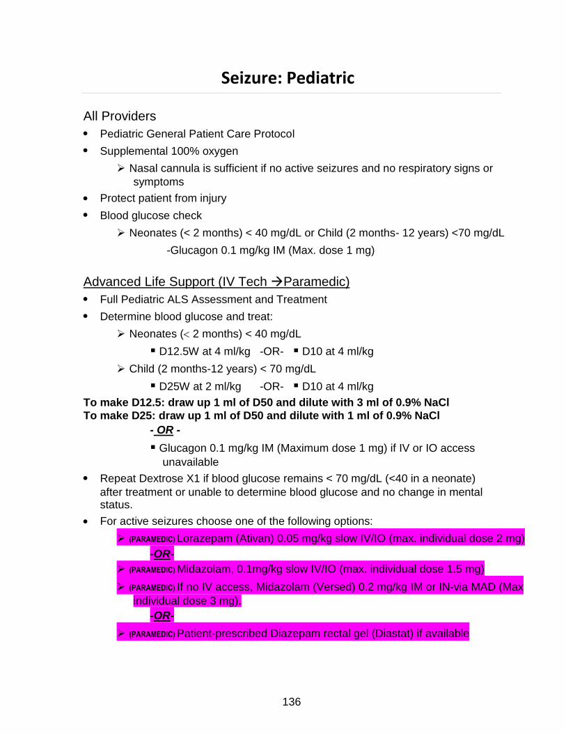
Seizure: Pediatric
All Providers
Pediatric General Patient Care Protocol
Supplemental 100% oxygen
Nasal cannula is sufficient if no active seizures and no respiratory signs or
symptoms
Protect patient from injury
Blood glucose check
Neonates (< 2 months) < 40 mg/dL or Child (2 months- 12 years) <70 mg/dL
-Glucagon 0.1 mg/kg IM (Max. dose 1 mg)
Advanced Life Support (IV Tech Paramedic)
Full Pediatric ALS Assessment and Treatment
Determine blood glucose and treat:
Neonates ( 2 months) < 40 mg/dL
D12.5W at 4 ml/kg -OR- D10 at 4 ml/kg
Child (2 months-12 years) < 70 mg/dL
D25W at 2 ml/kg -OR- D10 at 4 ml/kg
To make D12.5: draw up 1 ml of D50 and dilute with 3 ml of 0.9% NaCl
To make D25: draw up 1 ml of D50 and dilute with 1 ml of 0.9% NaCl
- OR -
Glucagon 0.1 mg/kg IM (Maximum dose 1 mg) if IV or IO access
unavailable
Repeat Dextrose X1 if blood glucose remains < 70 mg/dL (<40 in a neonate)
after treatment or unable to determine blood glucose and no change in mental status.
For active seizures choose one of the following options:
(PARAMEDIC) Lorazepam (Ativan) 0.05 mg/kg slow IV/IO (max. individual dose 2 mg)
-OR-
(PARAMEDIC) Midazolam, 0.1mg/kg slow IV/IO (max. individual dose 1.5 mg)
(PARAMEDIC) If no IV access, Midazolam (Versed) 0.2 mg/kg IM or IN-via MAD (Max
individual dose 3 mg).
-OR-
(PARAMEDIC) Patient-prescribed Diazepam rectal gel (Diastat) if available
136
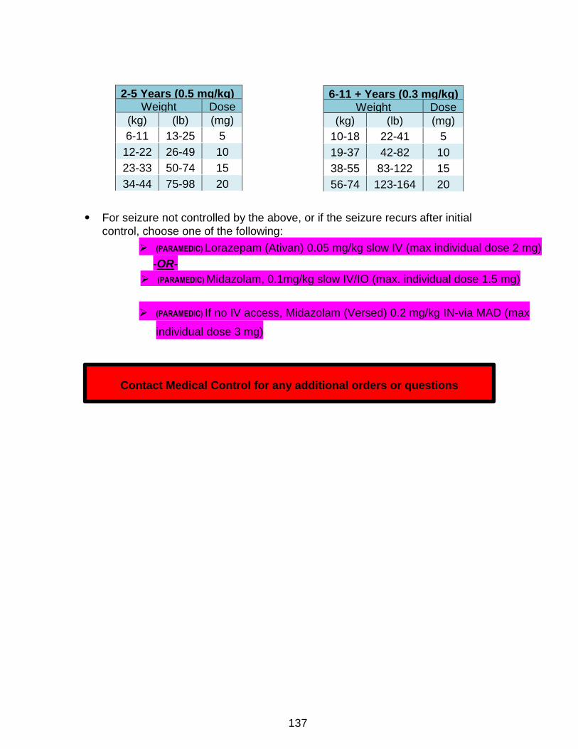
6-11 + Years (0.3 mg/kg) Weight Dose
(kg) (lb) (mg)
10-18 22-41 5
19-37 42-82 10
38-55 83-122 15
56-74 123-164 20
For seizure not controlled by the above, or if the seizure recurs after initial
control, choose one of the following:
(PARAMEDIC) Lorazepam (Ativan) 0.05 mg/kg slow IV (max individual dose 2 mg)
-OR-
(PARAMEDIC) Midazolam, 0.1mg/kg slow IV/IO (max. individual dose 1.5 mg)
(PARAMEDIC) If no IV access, Midazolam (Versed) 0.2 mg/kg IN-via MAD (max
individual dose 3 mg)
Contact Medical Control for any additional orders or questions
137
2-5 Years (0.5 mg/kg) Weight Dose
(kg) (lb) (mg)
6-11 13-25 5
12-22 26-49 10
23-33 50-74 15
34-44 75-98 20
Trauma: Pediatric
For major trauma criteria, a pediatric patient is anyone < 18 years of age
All Providers
Pediatric Patient Care Protocol
Stabilize spine
Use modified jaw thrust if airway obstructed
Supplemental 100% oxygen
Control hemorrhage using direct pressure or pressure dressing
Perform head-to-toe survey to identify injuries
Splint obvious fractures of long bones
Attempt to preserve body temperature
Advanced Life Support (IV Tech Paramedic)
If moderate or severe injuries present, perform Full Pediatric ALS Assessment
and Treatment
Assess for Pediatric Trauma Triage Criteria and initiate transport to Pediatric
Trauma Center
Assess for Tension Pneumothorax
Severe respiratory distress with hypoxia
Unilateral decreased or absent lung sounds (may see tracheal deviation
away from collapsed lung field)
Evidence of hemodynamic compromise (shock, hypotension, altered
mental status)
Pleural decompression for tension pneumothorax should only be performed
when all 3 of the above criteria are present!
(PARAMEDIC) If indicated, perform pleural decompression at 2nd intercostal space, mid-
clavicular line
Initiate transport to an appropriate trauma facility within 10 minutes of arrival on
the scene, unless extenuating circumstances (extrication)
Perform procedures, history and detailed physical examination en route to the
hospital
If moderate to severe pain, treat per Pediatric Pain Management Protocol
Reassess frequently
Contact Medical Control for any additional orders or questions
138
Trauma: Pediatric Burns
All Providers
General Pediatric Patient Care Protocol
Remove or cool heat source if present (e.g., clothing, tar)
Keep patient warm and dry with a sheet on all burns:
2 burns greater than 15% of body surface area
3 burns
Electrical and chemical burns
Spinal immobilization if high voltage electrical injuries
Observe for signs of impending loss of airway; Refer to the Airway Management
Protocol as needed:
Hypoxia
Poor ventilatory effort
Altered mental status/decreased level of consciousness
Inability to maintain patent airway
Advanced Life Support (IV Tech Paramedic)
Estimate Total Body Surface Area (TBSA) burned
If > 20% 2nd and 3rd degree TBSA burned, initiate volume resuscitation
with 0.9 % Normal Saline/LR, 20 ml/kg, repeat twice up to 60 ml/kg IV/IO
If moderate to severe pain, treat per Pediatric Pain Management Protocol
Contact Medical Control for any additional orders or questions
139
Trauma: Pediatric Head Injuries
All Providers
Supplemental oxygen
Stabilize spine
Check blood glucose
Apply physical restraints if needed to ensure patient/crew safety. Adhere to
procedure on Physical Restraint of Agitated Patients when this process is deemed necessary
If normotensive or hypertensive
Elevate head of backboard 15-30
Advanced Life Support (IV Tech Paramedic)
Full Pediatric ALS Assessment and Treatment
Advanced airway/ventilatory management as needed
Note: Airway interventions can be detrimental to patients with head injury by
raising intercranial pressure, worsening hypoxia (and secondary brain injury) and increasing risk of aspiration. Whenever possible these patients should be managed in the least invasive manner to maintain O2 saturation > 90% (i.e., NRB, BVM with 100% O2)
Observe for signs of impending respiratory failure; Refer to the Airway
Management Protocol if needed:
Hypoxia
Poor ventilatory effort
Altered mental status/decreases level of consciousness
Inability to maintain patent airway
For patients with assisted ventilation :
Administer eucapneic (normal rate 12-15/min) ventilations
Acute herniation should be suspected when the following signs are present:
Abrupt increase in blood pressure
Abrupt decrease in heart rate
Acute unilateral dilated and non-reactive pupil
Abrupt deterioration in mental status
Abrupt onset of motor posturing
Hyperventilation is a temporizing measure which is only indicated in the event of
acute herniation
140
If signs of herniation develop, begin hyperventilation
If < 1 year old, 35/minute
If > 1 year old, 25/minute
Target an ETCO2 of 30-35 mmHg
If severely agitated/combative and unable to deescalate by any other means,
consider:
(PARAMEDIC) Lorazepam 0.05 mg/kg IV/IO/IM
Contact Medical Control for any additional orders or questions
Any additional sedation
141
MEDICAL TREATMENT
PROCEDURES
D-1
Table of Contents--Procedures
12-Lead ECG ............................................................................................................... P-3
Airway Orotracheal Intubation...................................................................................... P-4 Airway Suctioning – Basic............................................................................................ P-7 Airway Suctioning – Advanced .................................................................................... P-8 Blood Glucose Analysis ............................................................................................... P-9 Cardioversion............................................................................................................. P-10 Cardio-Cerebral Resuscitation (CCR) ........................................................................ P-11 Chest Decompression................................................................................................ P-18 Childbirth Complications ............................................................................................ P-19 Childbirth Procedure .................................................................................................. P-20 Combat Application Tourniquet (CAT) ....................................................................... P-21 Continuous Positive Airway Pressure (CPAP) ........................................................... P-25 Cardiopulmonary Resuscitation (CPR) ...................................................................... P-27 Cricothyrotomy Guidelines: ........................................................................................ P-28 Cricothyrotomy Guidelines: Open Surgical ................................................................ P-29 Decontamination ........................................................................................................ P-30 Defibrillation—Automated .......................................................................................... P-31 Defibrillation – Manual ............................................................................................... P-32 Endotracheal Tube Introducer (Bougie) ..................................................................... P-33 External Cardiac Pacing ............................................................................................ P-35 Eye Irrigation.............................................................................................................. P-36 General Airway Management .................................................................................... P-37 Laryngeal Mask Airway (LMA) ................................................................................... P-39 Mucosal Atomizer Device®......................................................................................... P-44 Orogastric Tube Insertion .......................................................................................... P-45 Laryngeal Tube Airway (LTA) (King LTS-D) .............................................................. P-46 Pulse Oximetry........................................................................................................... P-52 Restraints................................................................................................................... P-53 Spinal Immobilization of Football Players .................................................................. P-54 Splinting ..................................................................................................................... P-57 Temperature Measurement ....................................................................................... P-58 Guidelines for Transport ............................................................................................ P-59 Venous Access – Existing Catheters ......................................................................... P-61 Venous Access – Extremity ....................................................................................... P-62 Venous Access – Intraosseous.................................................................................. P-64 Wound Care............................................................................................................... P-66
P-2
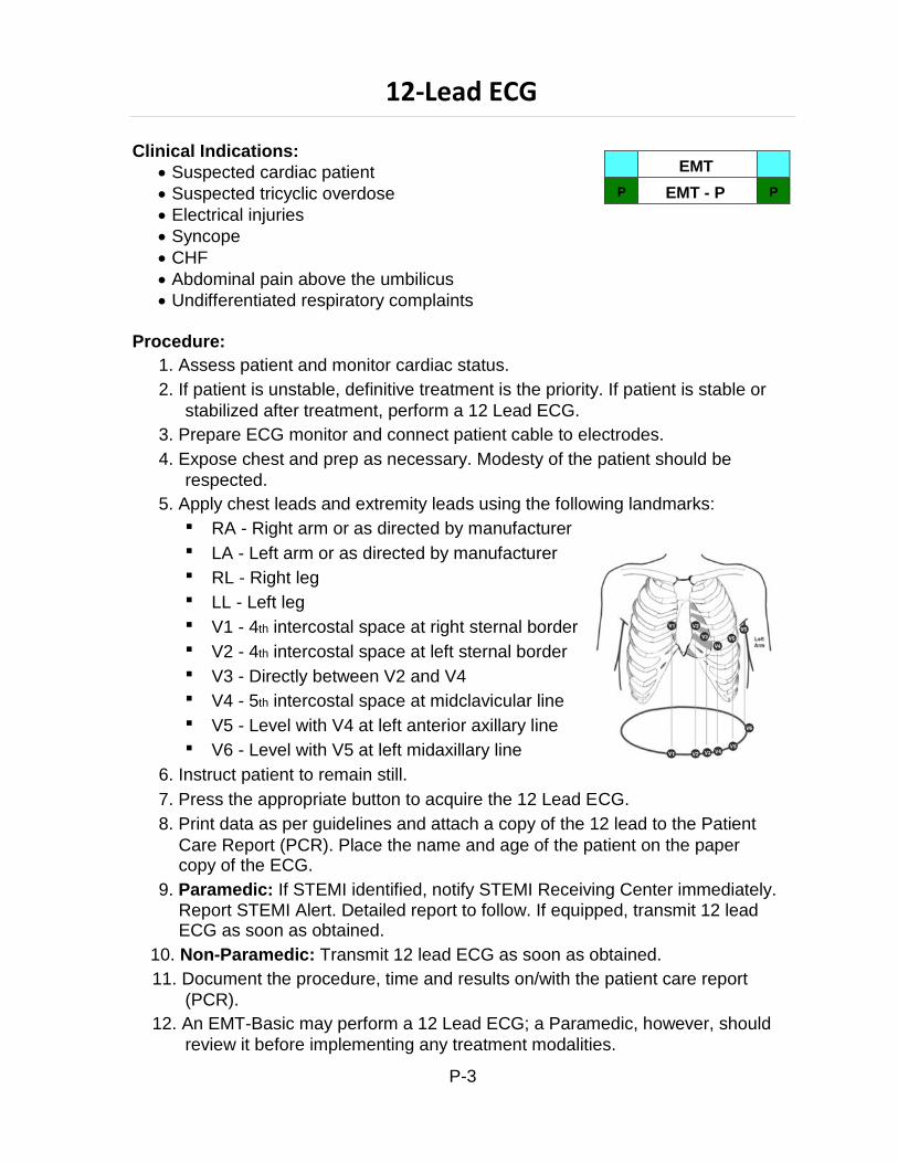
EMT
P EMT - P P
12-Lead ECG
Clinical Indications:
Suspected cardiac patient
Suspected tricyclic overdose
Electrical injuries
Syncope
CHF
Abdominal pain above the umbilicus
Undifferentiated respiratory complaints
Procedure:
1. Assess patient and monitor cardiac status.
2. If patient is unstable, definitive treatment is the priority. If patient is stable or
stabilized after treatment, perform a 12 Lead ECG.
3. Prepare ECG monitor and connect patient cable to electrodes.
4. Expose chest and prep as necessary. Modesty of the patient should be
respected.
5. Apply chest leads and extremity leads using the following landmarks:
RA - Right arm or as directed by manufacturer
LA - Left arm or as directed by manufacturer
RL - Right leg
LL - Left leg
V1 - 4th intercostal space at right sternal border
V2 - 4th intercostal space at left sternal border
V3 - Directly between V2 and V4
V4 - 5th intercostal space at midclavicular line
V5 - Level with V4 at left anterior axillary line
V6 - Level with V5 at left midaxillary line
6. Instruct patient to remain still.
7. Press the appropriate button to acquire the 12 Lead ECG.
8. Print data as per guidelines and attach a copy of the 12 lead to the Patient
Care Report (PCR). Place the name and age of the patient on the paper copy of the ECG.
9. Paramedic: If STEMI identified, notify STEMI Receiving Center immediately. Report STEMI Alert. Detailed report to follow. If equipped, transmit 12 lead ECG as soon as obtained.
10. Non-Paramedic: Transmit 12 lead ECG as soon as obtained.
11. Document the procedure, time and results on/with the patient care report
(PCR).
12. An EMT-Basic may perform a 12 Lead ECG; a Paramedic, however, should
review it before implementing any treatment modalities.
P-3
P P EMT - P
Airway Orotracheal Intubation
Clinical Indications:
Respiratory or cardiac arrest
Inadequate ventilation with bag valve mask
Impending respiratory failure:
Decreased level of consciousness with, hypoxia unresponsive to 100%
oxygen, apnea, and/or respiratory rate < 8
OR poor ventilatory effort (with hypoxia unresponsive to 100% oxygen)
OR unable to maintain patent airway
Airway obstruction or potential for airway obstruction (airway burns)
Equipment:
Laryngoscope handle with appropriate size blade.
Proper size endotracheal tube (ETT) plus back up ETT 0.5 – 1.0 mm smaller
Water-soluble lubrication gel, (lubricate distal end of tube at cuff).
10 cc syringe (larger syringe if low pressure cuff)
Stylet, (insert into ET tube and do not let stylet extend beyond tip of ET tube).
Tape or ETT securing device.
Proper size oral pharyngeal airway.
BVM or automatic ventilator
Oxygen source
Suction device.
Stethoscope
ETCO2 detection device (Capnography)
Oxygen saturation monitor
Procedure:
Patient/equipment preparation:
Maintain cervical alignment and immobilization as appropriate
Attach proper blade to laryngoscope handle and check light
Check endotracheal tube cuff
Raise gurney so that patient’s nose is at intubator’s xiphoid (if possible)
Confirm patient attached to cardiac monitor and oxygen saturation monitor
Ready ETC02 detection device
P-4
Intubation – Endotracheal – Adult, continued
Specify personnel to:
Apply cricoid pressure
Keep neck immobile during procedure
Watch cardiac and oxygen saturation monitors
Intubation:
Preoxygenate patient with 100% Oxygen (BVM or NRB) before intubation
attempt to achieve O2 saturation > 92% for 5 minutes. Have assistant apply cricoid pressure (Sellick’s maneuver) during entire procedure.
Remove all foreign objects, such as dentures, oral pharyngeal airways,
etc. and suction the patient's airway if needed. (Do not remove an esophageal located ETT if in place from prior attempt)
Grasp laryngoscope handle in left hand.
Grasp ET tube in right hand.
Insert the blade into the right side of the patient's mouth sweeping the
tongue to the left side.
Visualize the vocal cords while avoiding any pressure on the teeth.
Insert the endotracheal tube until the cuff passes the vocal cords. (Insert
far enough so that balloon port tubing is even with lips.)
Typical depth = tube size (ID) X 3 (example would be tube depth of 24 for
a 8.0mm tube)
Remove the laryngoscope blade.
Inflate the endotracheal cuff with the syringe with 5 - 10 cc of air (low
pressure cuff may require larger volume) and r remove the syringe from inflation valve
Confirm tube placement
Ventilate with BVM and:
Observe immediate (within 6 breaths) ETC02 waveform and number
with capnography
Watch for chest rise AND
Listen to abdomen to ensure tube is not esophageal
Then, listen for bilateral breath sounds
Observe oxygen saturation
P-5
Intubation – Endotracheal – Adult, continued
Note: Regardless of the apparent presence of lung sounds, tube misting and chest rise, or lack of gastric sounds, if ETCO2 does not indicate proper tube location (alveolar waveform), ETT must be removed.
If unilateral right-sided breath sounds are heard consider:
Right mainstem intubation
If present, deflate the cuff and withdraw tube 1-2 cm.
Re-listen for breath sounds.
If bowel sounds heard with bagging or ETCO2 device does not indicate proper
ETT placement, deflate cuff, remove tube and ventilate with bag valve mask for 2 minutes
If intubation attempt unsuccessful, refer to the next step in the Airway
Management Protocol
If successful tube placement:
Secure tube using an endotracheal securing device
Document depth of tube.
Reassess lung sounds and patient clinical status
Insert oral pharyngeal airway, or use ET tube holder with built in bite block (if available)
Ensure c-spine is still immobilized
Continue ventilations
Document ETC02 waveform and reading continuously- at time of each patient movement, including waveform and reading at time of transfer of care at the emergency department (ED).
P-6
EMT
P EMT - P P
Airway Suctioning – Basic
Clinical Indications:
Obstruction of the airway (secondary to secretions, blood, or any other
substance) in a patient who cannot maintain or keep the airway clear.
Procedure:
1.
2.
3.
4.
5.
6.
7.
8.
9.
Ensure suction device is in proper working order with suction tip in place.
Preoxygenate the patient.
Explain the procedure to the patient if they are coherent.
Examine the oropharynx and remove any potential foreign bodies or material that
may occlude the airway if dislodged by the suction device.
If applicable, remove ventilation devices (e.g., bag-valve mask) from the airway.
Use the suction device to remove any secretions, blood, or other substance.
The alert patient may assist with this procedure.
Reattach ventilation device (e.g., bag-valve mask) and ventilate or assist the
patient.
Record the time and result of the suctioning in the patient care report (PCR).
P-7
Airway Suctioning – Advanced
Clinical Indications:
P
EMT - P
P
Obstruction of the airway (secondary to secretions, blood, or any other
substance) in a patient currently being assisted with an airway adjunct such as a LTA/LMA, endotracheal tube, tracheostomy tube, or a cricothyrotomy tube.
Procedure:
1. Ensure suction device is in proper working order.
2. Preoxygenate the patient.
3. Attach suction catheter to suction device, keeping sterile plastic covering over
catheter.
4. For all devices, use the suprasternal notch as the end of the airway. Measure the
depth desired for the catheter (judgment must be used regarding the depth of suctioning with cricothyrotomy and tracheostomy tubes).
5. If applicable, remove ventilation devices (e.g., bag-valve mask) from the airway.
6. With the thumb port of the catheter uncovered, insert the catheter through the
airway device.
7. Once the desired depth (measured in #4 above) has been reached, occlude the
thumb port and remove the suction catheter slowly.
8. Small volume (< 10 ml) of normal saline lavage may be used as needed.
9. Reattach ventilation device (e.g., bag-valve-mask) and ventilate the patient.
10. Document time and result in the patient care report (PCR).
P-8
Blood Glucose Analysis
Clinical Indications:
P
EMT
EMT - P
P
Patients with suspected hypoglycemia (diabetic emergencies, altered/change
in mental status, bizarre behavior, etc.)
Procedure:
1. Gather and prepare equipment.
2. Blood samples for performing glucose analysis should be obtained
simultaneously with intravenous access when possible.
3. Place correct amount of blood on reagent strip or site on glucometer per the
manufacturer’s instructions.
4. Time the analysis as instructed by the manufacturer.
5. Document the glucometer reading and treat the patient as indicated by the
analysis and protocol.
6. Repeat glucose analysis as indicated for reassessment after treatment and as
per protocol.
P-9
Cardioversion
Clinical Indications:
P
EMT - P
P
Unstable patient with a tachydysrhythmia (rapid atrial fibrillation, supraventricular tachycardia, ventricular tachycardia)
Patient is not pulseless (the pulseless patient requires unsynchronized cardioversion, i.e., defibrillation)
Procedure:
1. Ensure the patient is attached properly to a monitor/defibrillator capable of
synchronized cardioversion.
2. Have all equipment prepared for unsynchronized cardioversion/defibrillation if
the patient fails synchronized cardioversion and the condition worsens.
3. Consider the use of pain medication or sedatives per protocol.
4. Set energy selection to the appropriate setting.
5. Set monitor/defibrillator to synchronized cardioversion mode.
6. Make certain all personnel are clear of patient.
7. Press and hold the shock button to cardiovert. Stay clear of the patient until
you are certain the energy has been delivered. NOTE: It may take the monitor/defibrillator several cardiac cycles to “synchronize”, so there may be a delay between activating the cardioversion and the actual delivery of energy.
8. Note patient response and perform immediate unsynchronized
cardioversion/defibrillation if the patient’s rhythm has deteriorated into pulseless ventricular tachycardia/ventricular fibrillation. Follow the procedure for Defibrillation-Manual.
9. If the patient’s condition is unchanged, repeat steps 2 to 8 above, using
escalating energy settings per protocol.
10. Repeat per protocol until maximum setting or until efforts succeed.
11. Note procedure, response, and times in the patient care report (PCR).
P-10
EMT
P EMT - P P
Cardio-Cerebral Resuscitation (CCR)
Clinical Indications:
Age ≥ 18 years of age (CCR causes worse outcomes in the pediatric
population)
Suspected cardiac cause of arrest (not respiratory-OD, drowning etc.)
It occurs with Ventricular Fibrillation or Pulseless Ventricular Tachycardia, with PEA and with Asystole. You must assume there is no blood perfusing the brain and heart…which is bad for survival. Your patient needs both a pump (chest compressions) and diastolic pressure support (to perfuse the coronary arteries). Time spent doing other things (such as prolonged airway management) at the expense of not delivering pump support is not good for the patient. Determine as close as possible when the patient collapsed, and document this. Also, look for any signs of patient gasping prior to and/or during resuscitation, and document this. If gasping is present, note pupil reaction and document also. It is also important to remember that not all pulseless patients are the result of cardiac related events. Other mechanisms such as trauma, drowning, hypothermia, choking and other respiratory problems, etc. must be considered as a possible cause for the arrest and should be addressed with immediate, appropriate airway intervention.
Key points in the Cardio Cerebral Resuscitation (CCR) approach:
Survival is determined by a functional recovery of two organs: the heart and
the brain.
Without adequate blood flow neither organ will survive.
That makes properly performed chest compressions (CC) the single most important determinant of survival.
Anything that interrupts or otherwise decreases the quality of CC
contributes to the death of your patient.
This concept – continuous maximal quality CC – must become the
foundation of all you think and do during resuscitation!
All patients are treated the same during the first 2 minutes of the code.
They get uninterrupted continuous CC (CCC) while other interventions are
performed.
The cardiac rhythm is irrelevant during this period.
The cardiac rhythm determines subsequent management.
It is analyzed (using manual interpretation) briefly AFTER each set of 200
CCC.
It is either shockable or non-shockable – don’t make it more complicated
than that.
P-11

CCR, continued
CCCs are to be resumed immediately following a rhythm assessment
shock.
The rhythm observed after a shock is not – meaning NOT – to be treated!
Otherwise deadly pauses in CCC will be introduced in an attempt to
gather information that is irrelevant to survival!
The initial rhythm (after 200 CCC) determines subsequent treatments:
When to initiate invasive airway insertion and positive pressure ventilation
Need for anti-arrhythmic medications
How long to remain on scene
Success depends on:
Leadership
Delegation of a limited set of specific tasks
Timely focused completion of these specific tasks by rescuers
Interventions that are critical to survival MUST whenever possible be
performed by two persons solely dedicated to that task
One to perform it and a second person to assure quality performance
This applies especially to chest compressions and it is also important in
the management of an invasive airway + ventilations.
Code Commander:
Someone must assume the role of code commander. This person is
responsible for delegating tasks, is the only person interpreting the rhythm, and is responsible for monitoring/critiquing the overall performance of the team.
Other members must work as a team and take direction from the code
commander. They must focus on their assigned tasks and let the code commander manage the overall response (in other words, keep their noses out of other rescuers business)
Critical first tasks: (delegated and performed in first two minutes if at all possible)
MCMAID – a prioritized sequence consisting of:
M = Metronome (100/min)
C = Chest compressions (focus on rate, recoil and depth)
M = Monitor (turn on in defib mode, pads on, joules set at maximum)
A = Airway (OPA, ensure patency, NRB @ 15/lpm)
I = Intravenous or Intraosseous access
D = Drugs (Epi, Vasopressin, Amiodarone)(be ready to administer when needed and monitor timing for repeat doses)
P-12
CCR, continued
Chest Compressions: MCMAID
Metronome should be turned on to assure a rate of 100/minute.
CCC should be started ASAP after arrival.
A two-person task if at all possible.
Switch compressors rapidly/frequently (every 1 or 2 minutes).
The non-compressor continuously monitors the quality of CCC: rate,
depth, and recoil.
CCC should be continuous = not interrupted.
The only valid reasons for interrupting compressions are for analyzing the
rhythm and shocking.
All other requests to pause CCC must be cleared by the code
commander, and the reason and duration documented in the run report.
Monitor/Defibrillator: MCMAID
Initial:
Turn unit on when compressions are started and set mode to defib.
Ensure joules are set to maximum allowed. (200 for Zoll/360 Physio)
Place pads in sternum/apex position without interruption of chest
compressions.
Defibrillation process:
Charge defibrillator during the last 10 seconds of 200 CCC.
Ensure all rescuers will be clear if a shock is needed.
Pause a few seconds only for analysis – determine if it is shockable or not.
If indicated, immediately deliver a single (not stacked) shock at
maximum joules.
If no shock is indicated, dump the charge by either decreasing the
energy level and immediately returning back to maximum energy setting (200 J or 360J), OR switching the mode to monitor and then quickly back to defib mode.
Immediately resume CCC after analysis shock.
The pause from stopping CCC to resumption of CCC should be less
than 5 seconds.
P-13
CCR, continued
Airway: MCMAID
Initially:
Insert OPA, apply NRB @ 15/lpm (look for misting), ensure patency (listen for exhausted air with compressions. If unsure, give one single breath with BVM, looking for chest rise and feeling for compliance)
When to insert invasive airway depends on the initial rhythm:
If non-shockable, initiate immediately after first rhythm analysis
If shockable – ONLY after three cycles (2 min. of CCC + analysisshock). NOT earlier, even if second rhythm is non-shockable
Once the invasive airway is in place, the airway persons sole task is to perform/monitor that task and no other
Invasive airway monitoring includes attention to:
Proper placement
Apply Capnography and verify waveform/presence of ETCO2
Avoidance of any interruption of CCC
Ventilation rate of 6 per minute. Each breath must be timed – aim for 10 seconds between each breath. Excessive ventilation rates are deadly!
Volume should be ~ 500 cc
Delivery of breath should be over one second.
(PARAMEDIC) Use a LTA if placing an endotracheal tube is met with any problems or delays
(PARAMEDIC) If the initial rhythm is shockable, seriously consider
using the LTA instead of an ETT because these patients cannot
tolerate even brief periods of less than optimal CCC
Assure that oxygen is attached
Intravenous/Intraosseous access: MCMAID
Consider intraosseous route whenever there are any delays in IV insertion
Drugs: MCMAID
The individual assigned to the Drug “task”:
Initially ensures medications are available and ready to
administer.
Is responsible for:
The rapid administration when indicated
Re-dosing at appropriate intervals
P-14
CCR, continued
Detecting when V-Fib is persistent or recurrent, and therefore indicating
the use of Amiodarone
Accurate timing of when meds are to be given (to the second – using
elapsed time since defibrillator was turned on)
Vasopressors should be given ASAP after analysis shock, so their effect will
be seen after the next 200 chest compressions
(PARAMEDIC) Epinephrine first!
**The one exception is the patient who you suspect may get return of
spontaneous circulation with the first shock. Such patients may include those with short down times or those who have had excellent chest compression generated perfusion. A clue to this is the presence of regular agonal respirations (gasping). In these patients consider giving Vasopressin initially, and reserving Epinephrine until the code commander observes persistent pulseless V-fib/Tach after the first shock – or until another 200 CCC cycle has been completed.
Epi dose is 1mg IV/IO. Endotracheal administration is not to be utilized -
start an IO instead
If repeating doses, administer every other cycle of 200 compressions.
(equivalent to every 4 min)
(PARAMEDIC) Vasopressin: administer with first Epi dose as per protocol.
Dose is 40 units (two vials of 20 U each)
Same dose is used for IV, intraosseous (IO) routes, or as a last resort
endotracheal If resuscitation process is expected to be prolonged, a second dose of 40
units of Vasopressin may be given 10 minutes or more after the first dose
(PARAMEDIC) Amiodarone: administered for persistent or recurrent pulseless V-fib/Tach.
This should be administered immediately during the next 200 chest compression cycle if a second shock was indicated and delivered at time of analysis. The code commander may visualize return of fibrillation during the 200 CCC and as such may order Amiodarone earlier since it has recurred.
Dose is 300 mg IV/IO
Repeat doses are 150 mg IV/IO
(PARAMEDIC) Additional treatments to consider:
Consider possible renal failure (hyperkalemia) or suspected Tricyclic
antidepressant overdose. If suspected, administer Sodium Bicarbonate 1mEq/Kg. If renal failure is suspected, also administer Calcium Chloride 1g IVP.
P-15
CCR, continued
If rhythm is persistent shockable V-Fib or Pulseless V-Tach, consider the
possible use of Magnesium Sulfate 2g IVP.
If the patient is successfully converted from V-Fib/Pulseless V-Tach to a
perfusing sustainable rhythm, consider post resuscitation Amiodarone boluses.
Amiodarone bolus: 150mg
If rhythm is a non-shockable Asystole or PEA, seek out and treat any possible
contributing factors.
Also consider external pacing: for PEA. Apply the pads in (or move them to)
the anterior/posterior position, attach the 4-lead cable, and set the pacer at
the maximum milliamps at a rate of 80/min. Do NOT interrupt compressions while attempting pacing.
When to stop CCC:
If the patient shows signs of cerebral activity and the rhythm is non-
shockable.
Use end-tidal CO2 as a marker for possible ROSC. Look for a dramatic
increase.
Pulse checks are ONLY performed during brief rhythm analysis with location
of carotid pulse ascertained during chest compressions.
This may be modified by the code commander if cerebral function signs of
life appear.
The code commander is the only individual who can order a pulse check
other than that done during rhythm analysis.
The code commander must ensure the pulse checker is clear if a shock is
indicated.
When to move patient:
Remember that moving a patient inevitably results in compromised quality of
compressions. If crew safety is compromised or inadequate resuscitation space is available, patient should be quickly moved to a safe or larger area. This should be done initially and not after resuscitation efforts have begun.
Initially shockable patients will live or die in the field!
Move is allowed after 3 cycles are completed and a non-shockable rhythm
is identified at the 3rd analysis.
If 3rd analysis is still shockable, continue resuscitation at the scene until
a non-shockable rhythm is encountered.
P-16
Initially non-shockable rhythms
Medical control must make this determination, but these patients may
deserve at least 3 cycles of treatment with optimal quality compressions.
Avoid excessive pauses:
Rhythm analysis - Only the Code commander pays attention to the rhythm
(not everyone).
Resume CCC immediately after analysis shock. The code commander
must assure this happens.
Charging - perform during last 10 seconds of 200 chest compressions.
During Intubation - It is the responsibility of both the code commander and the
second airway person to avoid pauses in CCC here. This MUST be able to be performed without any interruption of compressions! Consider using the LTA if unable to intubate effectively.
Pulse checks - only performed during the rhythm analysis pause; must be
correlated with rhythm.
P-17
Chest Decompression
Clinical Indications:
P
EMT - P
P
Tension pneumothorax should be suspected in patients who exhibit:
Severe respiratory distress with hypoxia
Unilateral decreased or absent lung sounds
Evidence of hemodynamic compromise (Shock, hypotension, tachycardia,
altered mental status)
Tracheal deviation away from the collapsed lung field (less reliable than
the above)
Pleural decompression for tension pneumothorax should only be performed
when at least 3 of the above criteria are present
Equipment:
14 gauge 2 inch - 2.5 inch over the needle catheter.
Tape
Sterile gauze pads
Antiseptic swabs
Occlusive dressing
Procedure:
Locate decompression site.
Identify the 2nd intercostal space in the mid-clavicular line on the
same side as the pneumothorax.
Prepare the site with an antiseptic swab:
Firmly introduce catheter immediately above distal rib of selected site
Insert the catheter into the thorax until air exits.
Advance catheter and remove needle.
Secure the catheter taking care not to allow it to kink.
Reassess lung sounds and patient condition.
Dress area with Occlusive dressing then cover with sterile gauze pad. Assess breath sounds and respiratory status
P-18
EMT
P EMT - P P
Childbirth Complications
Shoulder Dystocia:
Place mother in knee-chest position and reattempt delivery.
If delivery fails, support child’s airway, provide supplemental oxygen.
Breech Birth:
Do not attempt to pull infant by trunk or legs.
Place mother in knee-chest position.
If head does not deliver, push baby’s mouth and nose away from vaginal wall
with two gloved fingers. Provide supplemental oxygen to infant.
Prolapsed Cord:
Place mother in knee-chest position.
Do not push cord back into birth canal.
Insert gloved fingers into birth canal and keep pressure off prolapsed cord.
Cover exposed cord with warm moist dressing.
Meconium-Stained Amniotic Fluid:
Suction mouth and nose after delivery
If baby is vigorous (normal respiratory effort, muscle tone, and heart rate >
100), provide supportive care.
(PARAMEDIC) If baby is not vigorous (depressed respirations, poor muscle tone, or heart rate
<100), perform endotracheal intubation and suction trachea while removing ET
tube, may repeat one additional time.
Support ventilation and re-intubate with a clean tube as per DCALS Protocols.
P-19
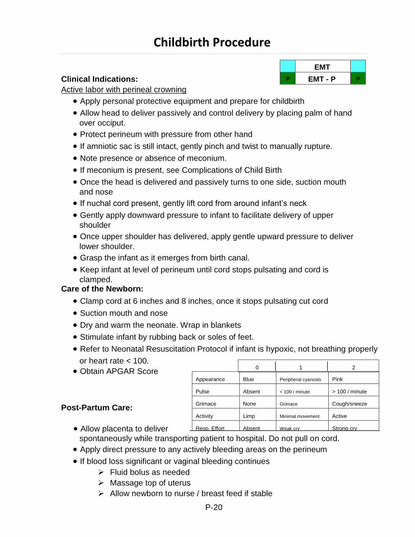
Appearance Blue Peripheral cyanosis Pink
Pulse Absent < 100 / minute > 100 / minute
Grimace None Grimace Cough/sneeze
Activity Limp Minimal movement Active
Resp. Effort Absent Weak cry Strong cry
EMT
P EMT - P P
Allow placenta to deliver
Childbirth Procedure
Clinical Indications:
Active labor with perineal crowning
Apply personal protective equipment and prepare for childbirth
Allow head to deliver passively and control delivery by placing palm of hand
over occiput.
Protect perineum with pressure from other hand
If amniotic sac is still intact, gently pinch and twist to manually rupture.
Note presence or absence of meconium.
If meconium is present, see Complications of Child Birth
Once the head is delivered and passively turns to one side, suction mouth
and nose
If nuchal cord present, gently lift cord from around infant’s neck
Gently apply downward pressure to infant to facilitate delivery of upper
shoulder
Once upper shoulder has delivered, apply gentle upward pressure to deliver
lower shoulder.
Grasp the infant as it emerges from birth canal.
Keep infant at level of perineum until cord stops pulsating and cord is
clamped. Care of the Newborn:
Clamp cord at 6 inches and 8 inches, once it stops pulsating cut cord
Suction mouth and nose
Dry and warm the neonate. Wrap in blankets
Stimulate infant by rubbing back or soles of feet.
Refer to Neonatal Resuscitation Protocol if infant is hypoxic, not breathing properly
or heart rate < 100.
Obtain APGAR Score
Post-Partum Care:
spontaneously while transporting patient to hospital. Do not pull on cord.
Apply direct pressure to any actively bleeding areas on the perineum
If blood loss significant or vaginal bleeding continues
Fluid bolus as needed
Massage top of uterus
Allow newborn to nurse / breast feed if stable
P-20
0 1 2
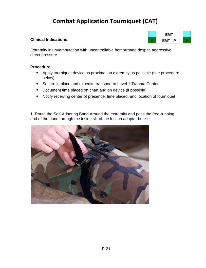
EMT
P EMT - P P
Combat Application Tourniquet (CAT)
Clinical Indications:
Extremity injury/amputation with uncontrollable hemorrhage despite aggressive
direct pressure.
Procedure:
Apply tourniquet device as proximal on extremity as possible (see procedure
below)
Secure in place and expedite transport to Level 1 Trauma Center
Document time placed on chart and on device (if possible)
Notify receiving center of presence, time placed, and location of tourniquet
1. Route the Self-Adhering Band Around the extremity and pass the free-running
end of the band through the inside slit of the friction adaptor buckle.
P-21
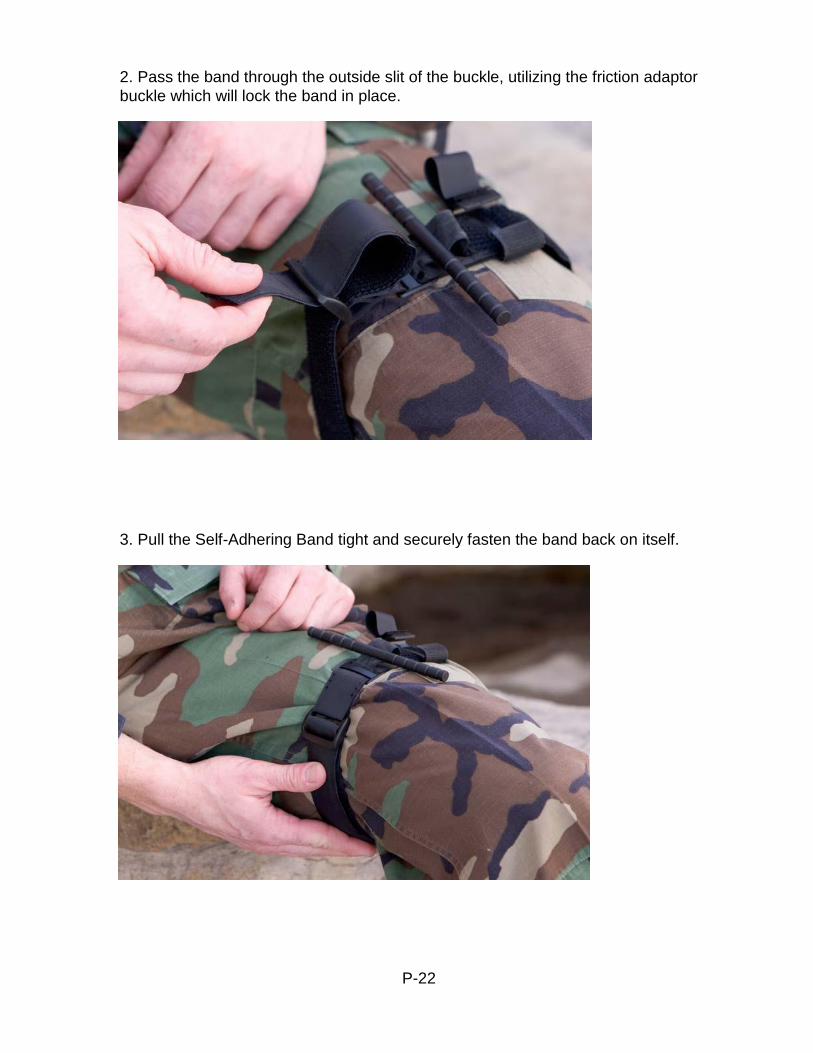
2. Pass the band through the outside slit of the buckle, utilizing the friction adaptor
buckle which will lock the band in place.
3. Pull the Self-Adhering Band tight and securely fasten the band back on itself.
P-22
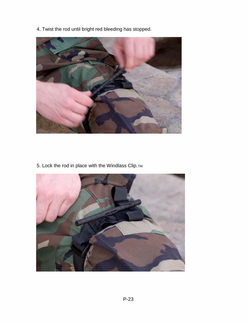
4. Twist the rod until bright red bleeding has stopped.
5. Lock the rod in place with the Windlass Clip.TM
P-23
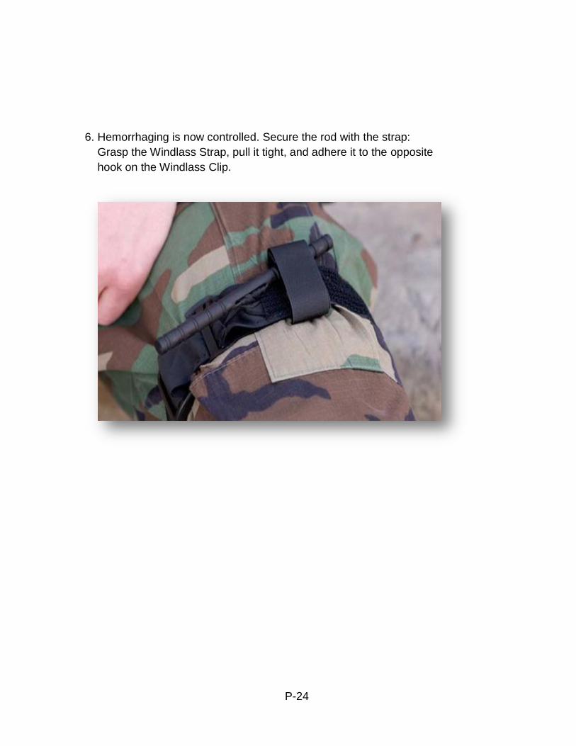
6. Hemorrhaging is now controlled. Secure the rod with the strap:
Grasp the Windlass Strap, pull it tight, and adhere it to the opposite
hook on the Windlass Clip.
P-24
EMT
P EMT - P P
Continuous Positive Airway Pressure (CPAP)
Clinical Indications:
For patients with Acute Bronchospastic Disorders (acute or chronic bronchitis,
emphysema, or asthma) or Acute Pulmonary Edema, who have hypoxemia and/or respiratory distress that does not quickly improve with pharmaceutical treatment.
Consider CPAP protocol if 2 or more present:
Retraction of intercostals or accessory muscles
Bronchospasm
Rales
Respiratory rate > 25 per minute.
Oxygen saturation <92% on high flow oxygen
Contraindications:
Respiratory arrest
Agonal respirations
Unconsciousness or obtundation
Shock associated with cardiac insufficiency
Trauma
Persistent nausea and vomiting
Facial anomalies
Inability to cooperate with the procedure
Equipment:
Medical Director approved Continuous Positive Airway Pressure (CPAP)
device
Procedure:
Perform primary and secondary surveys
Attach cardiac monitor, +/- capnography, and pulse oximetry
Verbally instruct patient (this is a critical item)
Patient requires verbal sedation to use this device effectively
P-25
CPAP, continued
Setup CPAP device as per manufacturer’s instructions
Instruct patient to slowly breathe in through the nose and exhale through
the mouth (exhalation phase should be about 4 seconds)
For CHF/ACPE use a CPAP setting of 10cm H20
For COPD use a CPAP setting of 5cm H20
Continue treatment throughout transport to the ED
Record and monitor vital signs, ETCO2, and O2 saturation as
needed/available.
In the event of progressive respiratory failure:
-Offer reassurance
-Stop treatment if necessary
-Institute BLS and ALS care per DCALS protocols
-Document adverse reactions, and reasons why CPAP was discontinued,
in patient care report
The following items should also be documented:
-CPAP level used
-FiO2 level used
P-26
Age Location Depth Rate
Infant
Over sternum,
between nipples (inter-mammary line), 2-3 fingers
1.5 inches (1/3 the
anterior-posterior chest dimension
At least 100/minute
Child Over sternum, between nipples, heel of one hand
2 inches (1/3 the anterior-posterior chest dimension)
At least 100/minute
Adult
Over sternum, just
above the xyphoid process, hands with interlocked fingers
At least 2 inches (1/3 the anterior- posterior chest dimension)
At least 100/minute
Cardiopulmonary Resuscitation (CPR)
Clinical Indications:
P
EMT
EMT - P
P
Pediatric arrest
Suspected non-cardiac arrest/respiratory arrest in adult patients (i.e.
overdose, drowning)
Procedure:
Assess the patient’s level of responsiveness (shake and shout).
If no response, open the patient’s airway with the head-tilt, chin-lift. Look,
listen, and feel for respiratory effort. If the patient may have sustained C- spine trauma, use the modified jaw thrust while maintaining immobilization of the C-Spine. For infants, positioning the head in the sniffing position is the most effective method for opening the airway.
If patient is an adult, go to step 4. If no respiratory effort in the pediatric
patient, give two ventilations. If air moves successfully, go to step 4. If air movement fails, proceed per AHA obstructed airway guidelines.
Check for pulse (carotid for adults and older children, brachial or femoral for
infants) for at least 10 seconds. If no pulse, begin chest compressions as directed below:
Go to Cardiac Arrest procedure.
Chest compressions should be provided in an uninterrupted manner. Only
brief interruptions are allowed for rhythm analysis and defibrillation.
Document the time and procedure in the Patient Care Report (PCR).
P-27
Cricothyrotomy Guidelines: Pertrach
CONTACT MEDICAL CONTROL IF POSSIBLE
P
EMT - P
P
BEFORE PROCEEDING!
Clinical Indications:
When all airway management measures have failed and the patient needs an
advanced airway immediately, consider performing cricothyrotomy. The percutaneous approach is preferred.
If in the paramedics’ judgment, the time necessary to contact medical control will
compromise the patient's chance of survival AND it is not possible to ventilate the patient with a bag-valve-mask during transport, cricothyrotomy may be performed without Medical Control.
Relative Contraindications:
Ability to ventilate patient with an oral-pharyngeal/nasal-pharyngeal airway, bag-
valve-mask, LTA/LMA, or endotracheal tube.
Procedure:
1. Cleanse anterior neck.
2. Identify and mark cricothyroid membrane.
3. Fill a 10 cc syringe with 5 cc’s of 0.9% Normal Saline
4. Remove dilator from the package and s heath and advance into the tracheostomy
tube.
5. Penetrate the skin and cricothyroid membrane with the splitting needle perpendicular
to the skin while gently aspirating with the syringe. Air aspiration as evidenced by bubbles into syringe should flow easily confirming tracheal airspace. Incline needle more than 45 degrees toward carina and complete insertion of needle aspirating to ensure continued proper placement.
6. Disconnect needle from syringe and advance tip of dilator into the hub of the splitting
needle until resistance met.
7. Squeeze wings of needle together then, open them out completely to split the
needle. Remove needle, continuing to pull it apart in opposite directions, while leaving the dilator in the trachea.
8. Place thumb on dilator knob while first and second fingers are curved under flange of
trachea tube. By exerting pressure, advanced dilator and tracheostomy tube into position until flange is against skin.
9. Remove dilator and inflate cuff until you have control of the airway (max 5 cc's).
Attach EtCO2 and BVM. Secure tube around patient’s neck with twill tape.
10. Confirm placement with gentle ventilation via bag-valve-mask, continuous
Capnography and physical means. B e sure air movement is fluid with bilateral symmetric chest rise and that no visible neck soft-tissue distortion is noted.
11. If tracheal placement is unclear, remove and convert to open surgical cricothyrotomy procedure.
12. Secure tube and consider sedation per protocol.
13. If not previously done, immediately notify medical control physician.
P-28
Cricothyrotomy Guidelines: Open Surgical
CONTACT MEDICAL CONTROL IF POSSIBLE BEFORE PROCEEDING!
P
EMT - P
P
Clinical Indications:
When all airway management measures have failed and the patient needs an
advanced airway immediately, consider performing cricothyrotomy. The percutaneous approach is preferred.
If in the paramedics’ judgment, the time necessary to contact medical control will
compromise the patient's chance of survival AND it is not possible to ventilate the patient with a bag-valve-mask during transport, cricothyrotomy may be performed without Medical Control.
Relative Contraindications:
Ability to ventilate patient with oral-pharyngeal/nasal-pharyngeal airway, bag-valve-
mask, LTA/LMA, or endotracheal tube.
Procedure:
1. Cleanse anterior neck. 2. Identify and stabilize thyroid cartilage with non-dominant thumb and 3 rd finger and
identify the cricothyroid membrane with your non-dominant index finger. Do not release hand until tracheal hook is stabilizing cartilage.
3. Make a vertical incision 3 centimeters long above the cricothyroid membrane and re- identify cricothyroid membrane with index finger.
4. Complete incision through pretracheal fascia to the level of the cricoid and thyroid cartilage with a second scalpel blade pass.
5. Re-identify cricothyroid membrane with index finger and incise membrane with single horizontal stab.
6. Use tracheal hook to secure thyroid cartilage. Once thyroid cartilage stabilized/secure, release tracheal cartilage with non-dominant hand. Then hold tracheal hook with gentle upward/proximal traction (towards chin).
7. Use scalpel blade handle to dilate cricothyroid membrane incision (dominant index finger may be used as well).
8. Insert cuffed 6.0 ETT through incised cricothyroid membrane. If unable to pass, attempt 5.5, 5.0, 4.5 ETT sequentially. Use the largest size able to be passed.
9. Confirm with continuous Capnography as well as physical signs, watching carefully for any signs of air filtration into the neck. If any question remains regarding tracheal placement, remove immediately and repeat procedure beginning at Step #6.
10. Secure ETT and trim length of ETT to 2 cm above pilot balloon cuff. Take off and reinsert adaptor to shortened tube and bag with bag-valve-mask.
11. Secure tube and consider sedation per protocol. 12. If not previously done, immediately notify Medical Control.
Pearls:
Open surgical cricothyrotomy is a blind, bloody procedure that should be performed
only as a last resort life-saving intervention.
Use extreme caution with scalpel as operator biohazard exposure from scalpel is
extremely common.
P-29
Decontamination
Clinical Indications:
P
EMT
EMT - P
P
Any patient who may have been exposed to significant hazardous materials,
including chemical, biological, or radiological weapons.
Procedure:
1. In coordination with HazMat and other Emergency Management personnel, establish hot, warm, and cold zones of operation.
2. Ensure that personnel assigned to operate within each zone have proper
personal protective equipment.
3. In coordination with other public safety personnel, assure each patient from
the hot zone undergoes appropriate initial decontamination. This is specific to each incident; such decontamination may include:
Removal of patients from Hot Zone
Simple removal of clothing
Irrigation of eyes
Passage through high-volume water bath (e.g., between two fire
apparatus) for patients contaminated with liquids or certain solids. Patients exposed to gases, vapors, and powders often will not require this stop as it may unnecessarily delay treatment and/or increase dermal absorption of the agent(s).
4. Initial triage of patients should occur after step #3. Immediate life threats
should be addressed prior to technical decontamination.
5. Assist patients with technical decontamination (unless contraindicated based
on #3 above). This may include removal of all clothing and gentle cleansing with soap and water. All body areas should be thoroughly cleansed, although overly harsh scrubbing which could break the skin should be avoided.
6. Place triage identification on each patient. Match triage information with each
patient’s personal belongings, which were removed during technical decontamination. Preserve these personnel affects for law enforcement.
7. Monitor all patients for environmental illness.
8. Transport patients per local protocol.
P-30
EMT
P EMT - P P
Defibrillation—Automated
Clinical Indications:
Patients in cardiac arrest (pulseless, non-breathing).
Age <8 years, use Pediatric Pads if available.
Contraindications:
Pediatric patients whose body habitus is such that the pads cannot be placed
without touching one another.
Procedure:
1. If multiple rescuers available, one rescuer should provide uninterrupted
chest compressions while the AED is being prepared for use.
2. Apply defibrillator pads per manufacturer recommendations. Use alternate
placement when implanted devices (pacemakers, AICD’s) occupy preferred pad positions.
3. Remove any medication patches on the chest and wipe off any residue.
4. If necessary, connect defibrillator leads: white to the anterior chest pad and
the red to the posterior pad.
5. Activate AED for analysis of rhythm.
6. Stop CPR and clear the patient for rhythm analysis. Keep interruption in
CPR as brief as possible.
7. Defibrillate if appropriate by depressing the “shock” button. Assertively state
“CLEAR” and visualize that no one, including yourself, is in contact with the patient prior to defibrillation. The sequence of defibrillation charges is preprogrammed for monophasic defibrillators. Biphasic defibrillators will determine the correct joules accordingly.
8. Begin CPR (chest compressions and ventilations) immediately after the
delivery of the defibrillation.
9. After 2 minutes of CPR, analyze rhythm and defibrillate if indicated. Repeat
this step every 2 minutes.
10. If “no shock advised” appears, perform CPR for two minutes and then
reanalyze.
11. Transport and continue treatment as indicated.
12. Keep interruption of CPR compressions as brief as possible. Adequate
CPR is a key to successful resuscitation.
If pulse returns: See Post-Resuscitation protocol.
P-31
EMT
P EMT - P P
Defibrillation – Manual
Clinical Indications:
Cardiac arrest with ventricular fibrillation or pulseless ventricular tachycardia.
Procedure:
1. Ensure chest compressions are adequate and interrupted only when
necessary.
2. Clinically confirm the diagnosis of cardiac arrest and identify the need for
defibrillation.
3. Apply hands-free pads to the patient’s chest in the proper position.
4. Charge the defibrillator to the maximum energy level. Continue chest
compressions while the defibrillator is charging.
5. Pause compressions, assertively state, “CLEAR” and visualize that no one,
including yourself, is in contact with the patient.
6. Deliver the countershock by depressing the shock button for hands-free
operation.
7. Immediately resume chest compressions and ventilations for 2 minutes. After
2 minutes of CPR, analyze rhythm and check for pulse only if appropriate for rhythm.
8. Repeat the procedure every two minutes as indicated by patient response
and ECG rhythm.
9. Keep interruption of compressions as brief as possible. Adequate
compressions are the key to successful resuscitation.
P-32
Endotracheal Tube Introducer (Bougie)
Clinical Indications:
P
EMT - P
P
Patients meet clinical indications for oral intubation.
Initial intubation attempt(s) unsuccessful.
Predicted difficult intubation.
Contraindications:
Two attempts at orotracheal intubation (utilize failed airway protocol).
ETT size less than 6.5 mm.
Procedure:
1. Prepare, position, and oxygenate the patient with 100% oxygen.
2. Select proper ET tube without stylette, test cuff and prepare suction.
3. Lubricate the distal end and cuff of the endotracheal tube (ETT) and the distal
½ of the Endotracheal Tube Introducer (Bougie) (note: Failure to lubricate the Bougie and the ETT may result in being unable to pass the ETT).
4. Using laryngoscopic techniques, visualize the vocal cords if possible using
Sellick’s/BURP as needed.
5. Introduce the Bougie with curved tip anteriorly and visualize the tip passing
the vocal cords or above the arytenoids if the cords cannot be visualized.
6. Once inserted, gently advance the Bougie until you meet resistance or “hold-
up” (if you do not meet resistance you have a probable esophageal intubation and insertion should be re-attempted or the failed airway protocol implemented as indicated.
7. Withdraw the Bougie ONLY to a depth sufficient to allow loading of the ETT
while maintaining proximal control of the Bougie.
8. Gently advance the Bougie and loaded ET tube until you have hold-up again,
thereby assuring tracheal placement and minimizing the risk of accidental displacement of the Bougie.
9. While maintaining a firm grasp on the proximal Bougie, introduce the ET tube
over the Bougie passing the tube to its appropriate depth.
10. If you are unable to advance the ETT into the trachea and the Bougie and
ETT are adequately lubricated, withdraw the ETT slightly and rotate the ETT 90 degrees COUNTER clockwise to turn the bevel of the ETT posteriorly. If this technique fails, to facilitate passing of the ETT you may attempt direct
P-33
Endotracheal Tube Introducer, continued
laryngoscopy while advancing the ETT (this will require an assistant to maintain the position of the Bougie and, if so desired, advance the ETT).
11. Once the ETT is correctly placed, hold the ET tube securely and remove the Bougie.
12. Confirm tracheal placement with capnography according to the intubation protocol. Inflate the cuff, auscultate for equal breath sounds and reposition accordingly.
13. When final position is determined secure the ET tube, continuously record and monitor capnography, reassess breath sounds and monitor patient to assure continued tracheal intubation.
14. If there is any question regarding placement of ETT (Esophageal vs. Tracheal) remove immediately and ventilate with bag-valve-mask.
P-34
External Cardiac Pacing
Clinical Indications:
P
EMT - P
P
Monitored heart rate less than 60 per minute with signs and symptoms of
inadequate cerebral or cardiac perfusion such as:
Severe chest pain
Hypotension
Pulmonary edema
ALOC, disorientation, confusion, etc.
Procedure:
1. Attach standard four-lead monitor.
2. Apply defibrillation/pacing pads to chest and back:
One pad to left mid chest next to sternum, one pad to left mid posterior
chest next to spine.
3. Rotate selector switch to pacing option.
4. Adjust heart rate to 70 BPM for an adult, 100 BPM for pediatric patients.
5. Note pacer spikes on ECG screen.
6. Slowly increase output until capture of electrical rhythm on the monitor.
7. If unable to capture while at maximum current output, stop pacing
immediately.
8. If capture observed on monitor, check for corresponding pulse and assess
vital signs.
9. Mechanical capture occurs when paced electrical spikes on monitor
correspond with palpable pulse.
10. Consider the use of sedation or analgesia if patient is uncomfortable.
11. Document the dysrhythmia and the response to external pacing with ECG
strips in the Patient Care Report (PCR).
P-35
EMT
P EMT - P P
Eye Irrigation
Clinical Indications:
Concern for ocular chemical exposure by either history or physical exam.
Procedure:
1.
2.
3.
4.
Identify agent that victim was exposed to.
Tilt head forward and brush eyelids/lashes if dry chemical present.
Remove contact lenses if present.
(PARAMEDIC) Instill 1 to 2 drops of Tetracaine into each affected eye; consider repeated
instillation of Tetracaine PRN.
5. Prime irrigation tubing after hanging a 1000 ml bag of NSS. Close tubing
clamp.
6. Start irrigation with normal saline and continue en route to hospital.
P-36
EMT
P EMT - P P
General Airway Management
Clinical Indications:
Inadequate oxygenation or ventilation
Unable to remove/relieve obstructing foreign body
Contraindications:
See specific technique for contraindications to specific procedures
Equipment:
Bag (appropriate for patient size)
Bag valve mask or automatic ventilator
Oxygen source
Suction device with Yankee
Oral and Nasal-pharyngeal airways
King LT airway
Laryngeal mask airways
Endotracheal tube (prepare estimated correct size and one size smaller as
backup)
Stylet for endotracheal tube
Oxygen saturation monitor
End-tidal C02 detection device
Capnography (waveform)
Colorimetric device (back-up device)
Procedure:
1. Select and ready equipment, tube sizes, ETC02, suction, non-visualized
devices (LTA, [PARAMEDIC] LMA).
2. If suspicion of trauma, maintain c-spine immobilization
3. Have assistant apply cricoid pressure
4. Suction all debris, secretions from airway
5. Bag valve mask or spontaneous ventilations for 2-5 minutes with 100%
oxygen attempting to keep the oxygen saturation > 92%
P-37
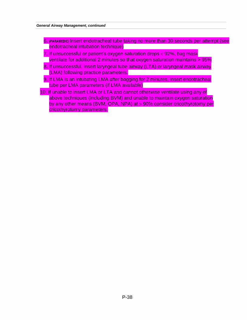
General Airway Management, continued
6. (PARAMEDIC) Insert endotracheal tube taking no more than 30 seconds per attempt (see
endotracheal intubation technique)
7. If unsuccessful or patient’s oxygen saturation drops 92%, bag mask
ventilate for additional 2 minutes so that oxygen saturation maintains > 95%
8. If unsuccessful, insert laryngeal tube airway (LTA) or laryngeal mask airway
(LMA) following practice parameters.
9. If LMA is an intubating LMA after bagging for 2 minutes, insert endotracheal
tube per LMA parameters (if LMA available)
10. If unable to insert LMA or LTA and cannot otherwise ventilate using any of
above techniques (including BVM) and unable to maintain oxygen saturation
by any other means (BVM, OPA, NPA) at 90% consider cricothyrotomy per cricothyrotomy parameters.
P-38
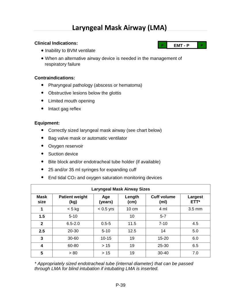
Laryngeal Mask Airway Sizes
Mask
size
Patient weight
(kg)
Age
(years)
Length
(cm)
Cuff volume
(ml)
Largest
ETT*
1 < 5 kg < 0.5 yrs 10 cm 4 ml 3.5 mm
1.5 5-10 10 5-7
2 6.5-2.0 0.5-5 11.5 7-10 4.5
2.5 20-30 5-10 12.5 14 5.0
3 30-60 10-15 19 15-20 6.0
4 60-80 > 15 19 25-30 6.5
5 > 80 > 15 19 30-40 7.0
Laryngeal Mask Airway (LMA)
Clinical Indications:
Inability to BVM ventilate
P
EMT - P
P
When an alternative airway device is needed in the management of
respiratory failure
Contraindications:
Pharyngeal pathology (abscess or hematoma)
Obstructive lesions below the glottis
Limited mouth opening
Intact gag reflex
Equipment:
Correctly sized laryngeal mask airway (see chart below)
Bag valve mask or automatic ventilator
Oxygen reservoir
Suction device
Bite block and/or endotracheal tube holder (if available)
25 and/or 35 ml syringes for expanding cuff
End tidal CO2 and oxygen saturation monitoring devices
* Appropriately sized endotracheal tube (internal diameter) that can be passed through LMA for blind intubation if intubating LMA is inserted.
P-39
LMA, continued
Procedure – Laryngeal Mask Airway Placement:
Pre-oxygenate patient with 100% oxygen via bag valve mask to achieve O2
saturation of >92 % if possible
Remove the red tag from the balloon port
Check the integrity of the cuff and pilot balloon
Tightly deflate the cuff with the syringe
The deflated cuff should appear BOAT shaped
Lubricate the posterior surface
Place patient in neutral sniffing position (if no C-spine/spinal injury suspected)
For patients with suspected C-spine injury, perform two-person insertion
technique:
One person maintains manual in-line cervical spine stabilization while
the other person proceeds with procedure as below:
Pull mandible down to open mouth
Insert uninflated LMA into oral cavity with cuff facing away from hard palate
Guide LMA around curvature of the posterior pharynx into the hypopharynx
until resistance is felt. Resistance is due to the tip of the LMA stopping at the upper esophageal sphincter
If uninflated LMA insertion is difficult:
If the curvature of the posterior/hypopharynx is too acute, perform a jaw
thrust, pulling the tongue forward. Alternately, a laryngoscope may be used to lift the jaw/mandible to facilitate insertion.
A slight inflation of the cuff to 1/3 – ½ of typical inflation volume may also
increase ease of insertion
Insert LMA with cuff facing hard palate, then rotate 180 degrees into the
proper position after the angle around the posterior aspect of the tongue has been cleared.
Inflate cuff without holding the tube
Ensure that the black line running the length of the LMA shaft is in the midline
of the upper lip and between the two central incisors (this will help maintain a seal)
Administer gentle positive pressure ventilation
Obtain end-tidal CO 2,(waveform), listen for breath sounds bilaterally, look for
chest excursion, and check oxygen saturation.
Secure in the midline to help maintain a good seal over the Larynx
P-40
LMA, continued
Place bite block, gauze or endotracheal tube holder (if available) between
teeth to prevent biting tube.
Ensure C-spine is still immobilized
If repeated attempts are made, oxygenate with 100% O2 for 2 minutes
between attempts
Endotracheal intubation using LMA:
Select correct size for LMA
Insert endotracheal tube into oropharynx at 90 angle (from corner of mouth)
During insertion and passage through the LMA rotate ET tube 90 degrees so
that the tip of the ET tube will pass through the bars that traverse the distal opening of the LMA
Confirm placement as per endotracheal intubation procedural section
(continued on next page)
P-41
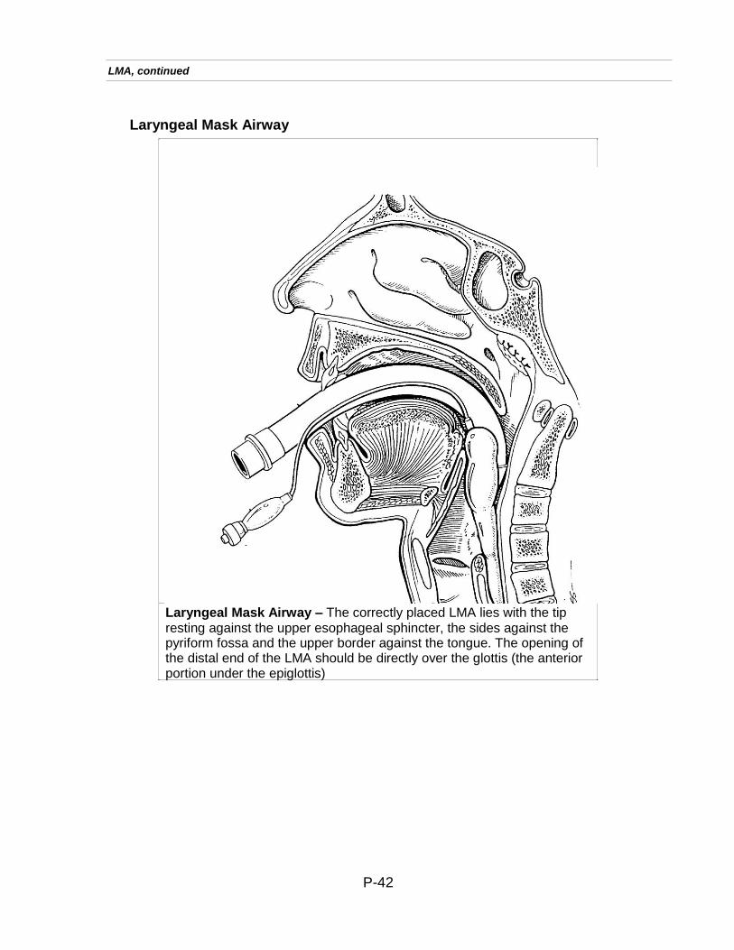
LMA, continued
Laryngeal Mask Airway
Laryngeal Mask Airway – The correctly placed LMA lies with the tip resting against the upper esophageal sphincter, the sides against the pyriform fossa and the upper border against the tongue. The opening of the distal end of the LMA should be directly over the glottis (the anterior portion under the epiglottis)
P-42
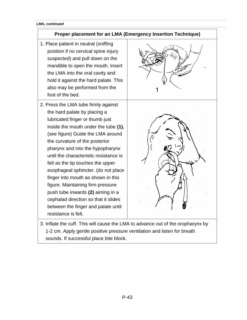
Proper placement for an LMA (Emergency Insertion Technique)
1. Place patient in neutral (sniffing
position if no cervical spine injury
suspected) and pull down on the
mandible to open the mouth. Insert
the LMA into the oral cavity and
hold it against the hard palate. This
also may be performed from the
foot of the bed.
2. Press the LMA tube firmly against
the hard palate by placing a
lubricated finger or thumb just
inside the mouth under the tube (1).
(see figure) Guide the LMA around
the curvature of the posterior
pharynx and into the hypopharynx
until the characteristic resistance is
felt as the tip touches the upper
esophageal sphincter. (do not place
finger into mouth as shown in this
figure. Maintaining firm pressure
push tube inwards (2) aiming in a
cephalad direction so that it slides
between the finger and palate until
resistance is felt.
3. Inflate the cuff. This will cause the LMA to advance out of the oropharynx by
1-2 cm. Apply gentle positive pressure ventilation and listen for breath
sounds. If successful place bite block.
LMA, continued
P-43
EMT
P EMT - P P
Mucosal Atomizer Device®
Purpose:
Administration of medications via a non-invasive route
Clinical Indications:
Altered Mental Status, presumed or possible opiate overdose
Seizures
Pain management
Contraindications:
DO NOT use on a patient if:
Severe nasal/facial trauma
Active nasal bleeding or discharge
Procedure for medication administration via the MAD®:
1. Put on protective eyewear, mask, and gloves.
2. Determine appropriate dose of medication per DCALS protocols.
3. Draw medication into syringe and dispose of the sharps (add an additional 0.1
ml of medication due to dead space), do not administer more than 1 ml per nostril.
4. Attach Mucosal Atomizer Device (MAD) to syringe.
5. With one hand, control the patient’s head.
6. Gently introduce MAD into nare, stop when resistance is met.
7. Aim slightly upwards and toward the ear on the same side.
8. Briskly compress the syringe to administer one-half of the medication, repeat
the procedure with the remaining medication on the other nare.
9. Document the results in the patient care report.
P-44
EMT
P EMT - P P
Orogastric Tube Insertion
Clinical Indications:
Gastric decompression in adult intubated patients
After successful placement of the LTA
Contraindications:
History of alkali ingestion, or esophageal disease (e.g. stricture or cancer)
Comatose state with unprotected airway (as procedure will induce vomiting)
Penetrating cervical injuries in the awake trauma patient.
Procedure:
1. Estimate insertion length by superimposing the tube over the body from the
mouth to the stomach.
2. Lubricate the tube with a water based lubricant prior to insertion.
3. Insert lubricated tube through the gastric port of the LTA or lift tongue/jaw
anteriorly while passing tip lateral to endotracheal tube.
4. Continue to advance the tube gently until the appropriate distance is reached.
5. Confirm placement by injecting 20cc of air and auscultate for the swish or
bubbling of the air over the stomach. If any doubt about placement, remove and repeat the insertion.
6. Secure the tube.
7. Decompress the stomach of air and food by connecting the tube to low
continuous suction.
8. Document the procedure, time, and result (success) on/with the patient care
report (PCR).
P-45

King LTS-D Airway Sizes
Airway
Size
Connector
Color
Patient
Height OD/ID(mm) Cuff Volume
(ml)
Gastric
Tube(Fr.) 3 Yellow 4-5 feet 18/10mm 45-60 ml Up to 18
4 Red 5-6 feet 18/10 60-90 Up to 18
5 Purple > 6 feet 18/10 70-90 Up to 18
EMT
P EMT - P P
Laryngeal Tube Airway: LTA (KING LTS-D)
Purpose: To establish control of the patient's airway and to facilitate ventilation for the listed indications.
Indications:
When an alternative airway device is needed in the management of
respiratory failure in patients 4 feet tall or greater.
Contraindications:
Intact gag reflex
Patients with known esophageal disease
Patients who have ingested caustic substances
Patients with known tracheal obstruction
Patients with a tracheostomy or laryngectomy
Patients less than 4 feet tall
Equipment:
Correctly sized LTA (see chart below)
Bag valve mask
Oxygen reservoir
Suction device
Bite block and/or endotracheal tube holder (if available)
Appropriately sized syringes for expanding cuff
End tidal CO2 and oxygen saturation monitoring devices
P-46
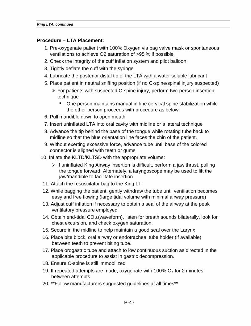
King LTA, continued
Procedure – LTA Placement:
1. Pre-oxygenate patient with 100% Oxygen via bag valve mask or spontaneous
ventilations to achieve O2 saturation of >95 % if possible
2. Check the integrity of the cuff inflation system and pilot balloon
3. Tightly deflate the cuff with the syringe
4. Lubricate the posterior distal tip of the LTA with a water soluble lubricant
5. Place patient in neutral sniffing position (if no C-spine/spinal injury suspected)
For patients with suspected C-spine injury, perform two-person insertion
technique
One person maintains manual in-line cervical spine stabilization while
the other person proceeds with procedure as below:
6. Pull mandible down to open mouth
7. Insert uninflated LTA into oral cavity with midline or a lateral technique
8. Advance the tip behind the base of the tongue while rotating tube back to
midline so that the blue orientation line faces the chin of the patient.
9. Without exerting excessive force, advance tube until base of the colored
connector is aligned with teeth or gums
10. Inflate the KLTD/KLTSD with the appropriate volume:
If uninflated King Airway insertion is difficult, perform a jaw thrust, pulling
the tongue forward. Alternately, a laryngoscope may be used to lift the jaw/mandible to facilitate insertion
11. Attach the resuscitator bag to the King LT.
12. While bagging the patient, gently withdraw the tube until ventilation becomes
easy and free flowing (large tidal volume with minimal airway pressure)
13. Adjust cuff inflation if necessary to obtain a seal of the airway at the peak
ventilatory pressure employed
14. Obtain end-tidal CO 2,(waveform), listen for breath sounds bilaterally, look for
chest excursion, and check oxygen saturation.
15. Secure in the midline to help maintain a good seal over the Larynx
16. Place bite block, oral airway or endotracheal tube holder (if available)
between teeth to prevent biting tube.
17. Place orogastric tube and attach to low continuous suction as directed in the
applicable procedure to assist in gastric decompression.
18. Ensure C-spine is still immobilized
19. If repeated attempts are made, oxygenate with 100% O2 for 2 minutes
between attempts
20. **Follow manufacturers suggested guidelines at all times**
P-47
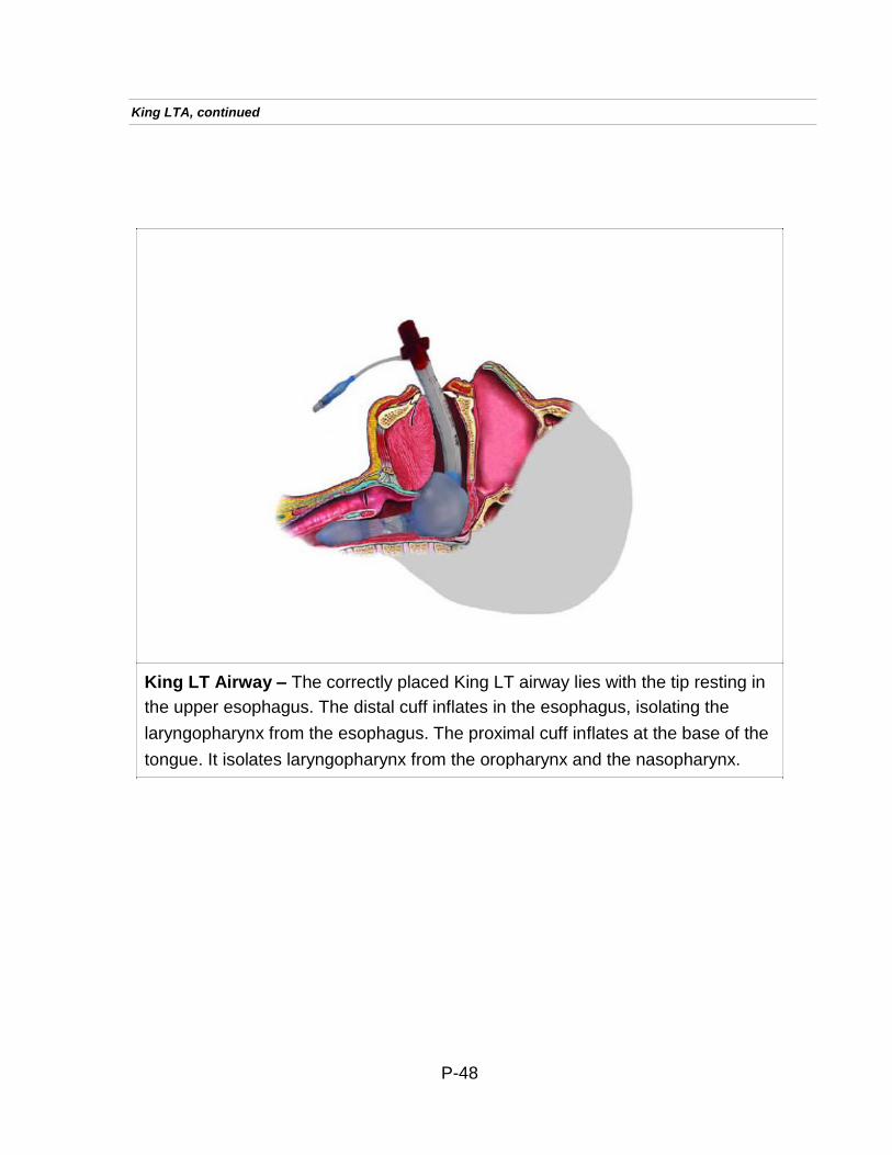
King LTA, continued
King LT Airway – The correctly placed King LT airway lies with the tip resting in
the upper esophagus. The distal cuff inflates in the esophagus, isolating the
laryngopharynx from the esophagus. The proximal cuff inflates at the base of the
tongue. It isolates laryngopharynx from the oropharynx and the nasopharynx.
P-48
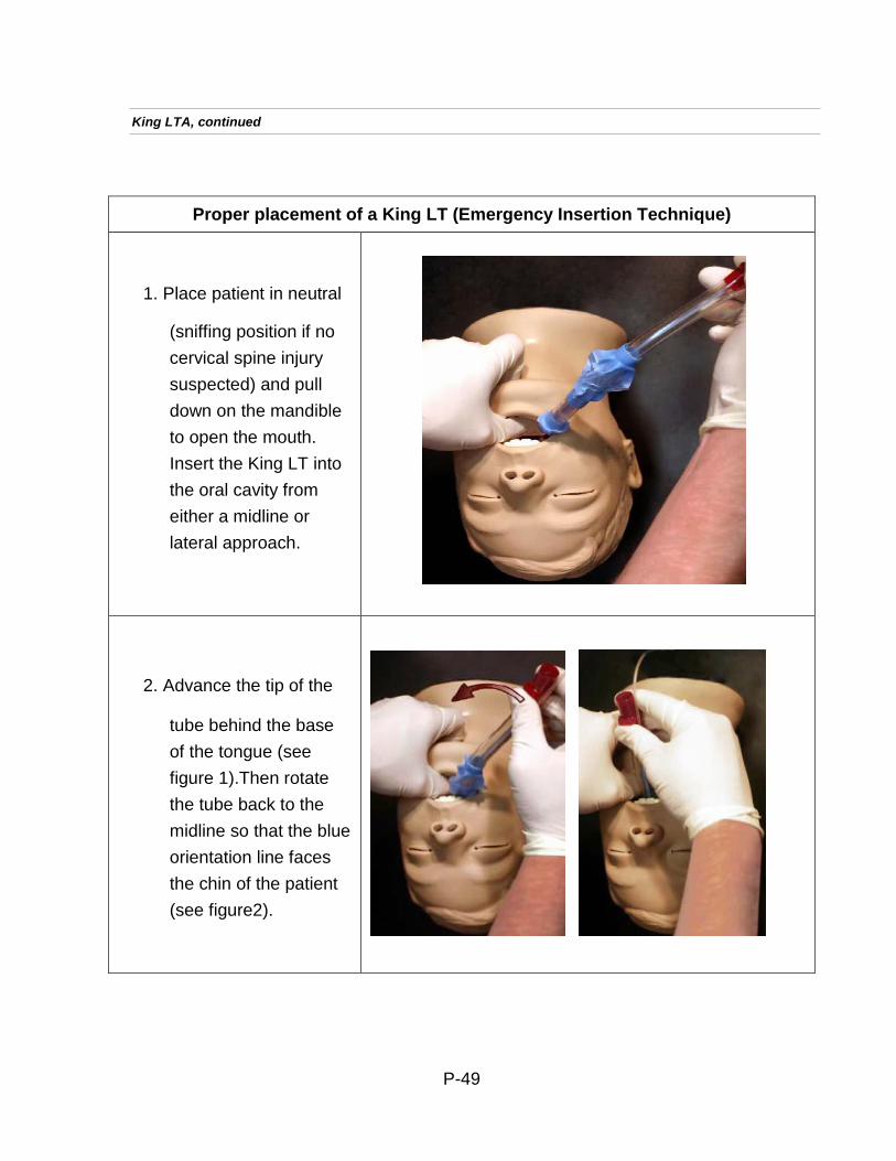
Proper placement of a King LT (Emergency Insertion Technique)
1. Place patient in neutral
(sniffing position if no
cervical spine injury
suspected) and pull
down on the mandible
to open the mouth.
Insert the King LT into
the oral cavity from
either a midline or
lateral approach.
2. Advance the tip of the
tube behind the base
of the tongue (see
figure 1).Then rotate
the tube back to the
midline so that the blue
orientation line faces
the chin of the patient
(see figure2).
King LTA, continued
P-49
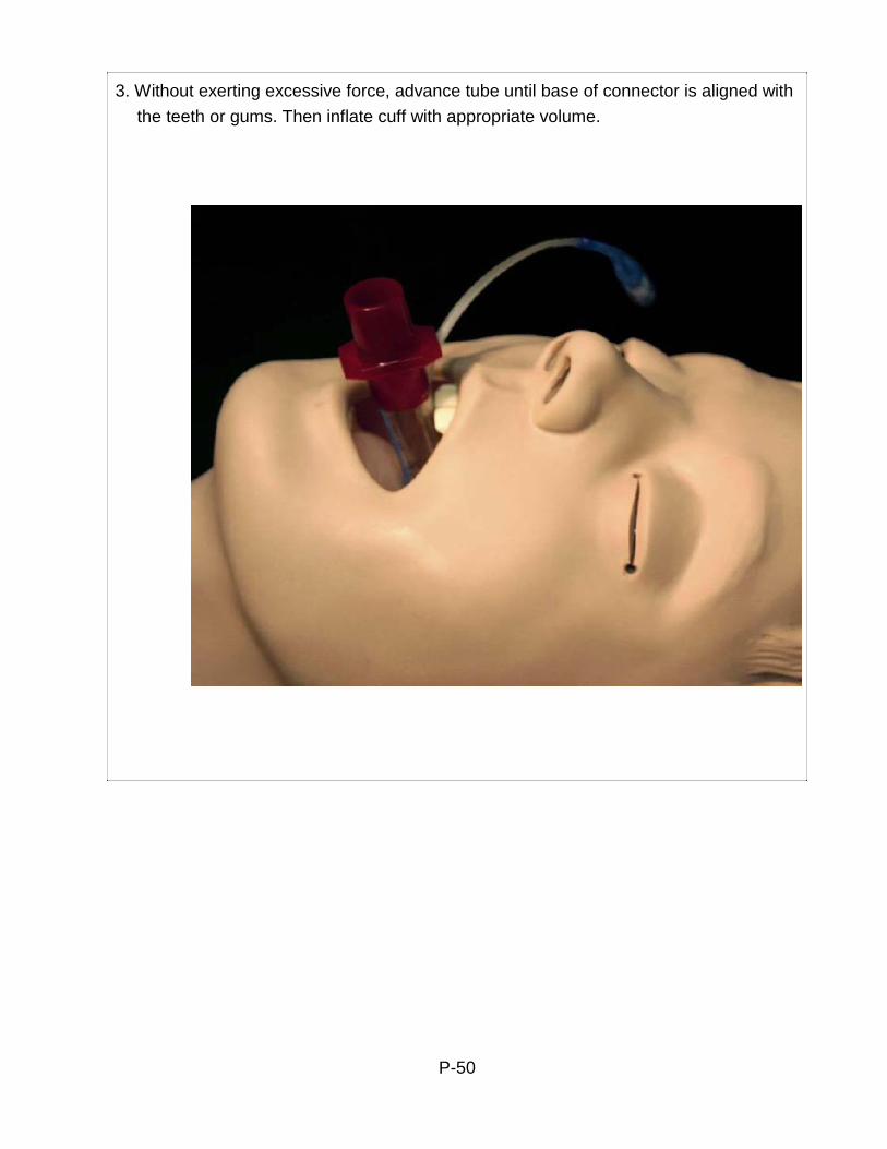
3. Without exerting excessive force, advance tube until base of connector is aligned with
the teeth or gums. Then inflate cuff with appropriate volume.
P-50
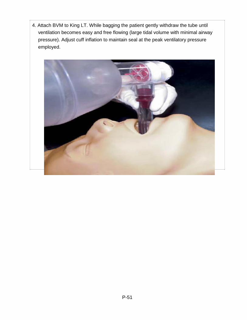
4. Attach BVM to King LT. While bagging the patient gently withdraw the tube until
ventilation becomes easy and free flowing (large tidal volume with minimal airway
pressure). Adjust cuff inflation to maintain seal at the peak ventilatory pressure
employed.
P-51
Pulse Oximetry
Clinical Indications:
P
EMT
EMT - P
P
Patients with suspected hypoxemia, altered level of consciousness or
respiratory issues
Procedure:
1. Apply probe to patient’s finger or any other digit as recommended by the
device manufacturer.
2. Allow machine to register saturation level.
3. Record time and initial saturation percent on room air if possible on/with the
patient care report (PCR).
4. Verify pulse rate on machine with actual pulse of the patient.
5. Monitor critical patients continuously until arrival at the hospital. If recording a
one-time reading, monitor patients for a few minutes as oxygen saturation can vary.
6. Document percent of oxygen saturation every time vital signs are recorded
and in response to therapy to correct hypoxemia.
7. In general, normal saturation is 97-99%. Below 93%, suspect a respiratory
compromise.
8. Use the pulse oximetry as an added tool for patient evaluation. Treat the
patient, not the data provided by the device.
9. The pulse oximeter reading should never be used to withhold oxygen from a
patient in respiratory distress or when it is the standard of care to apply oxygen despite good pulse oximetry readings, such as chest pain.
10. Factors which may reduce the reliability of the pulse oximetry reading include:
Poor peripheral circulation (blood volume, hypotension, hypothermia)
Excessive pulse oximeter sensor motion
Fingernail polish (may be removed with acetone pad)
Carbon monoxide bound to hemoglobin
Irregular heart rhythms (atrial fibrillation, SVT, etc.)
Jaundice
Placement of BP cuff on same extremity as pulse ox probe.
P-52

EMT
P EMT - P P
Restraints
Clinical Indications:
Any patient who may harm himself, herself, or others may be gently
restrained to prevent injury to the patient or crew. This restraint must be in a humane manner and used only as a last resort. Other means to prevent injury to the patient or crew must be attempted first. These efforts could include reality orientation, distraction techniques, or other less restrictive therapeutic means. Physical or chemical restraint should be a last resort technique.
Procedure:
1. Attempt less restrictive means of managing the patient.
2. Ensure that there are sufficient personnel available to physically restrain the
patient safely.
3. Restrain the patient in a lateral or supine position. No devices such as
backboards, splints, or other devices will be on top of the patient.
4. The patient will never be restrained in the prone position.
5. The patient must be under constant observation by the EMS crew at all times.
This includes direct visualization of the patient as well as cardiac, pulse oximetry and capnography monitoring as indicated.
6. The extremities that are restrained will have a circulation check at least every
15 minutes. The first of these checks should occur as soon after placement of the restraints as possible. This MUST be documented on the PCR.
7. If the above actions are unsuccessful, or if the patient is resisting the
restraints, consider chemical restraint per protocol.
8. If a patient is restrained by law enforcement personnel with handcuffs or other
devices EMS personnel cannot remove, a law enforcement officer must accompany the patient to the hospital in the transporting EMS vehicle.
9. Consider excited delirium protocol.
10. Restraining a patient in the prone position is never authorized.
P-53
Spinal Immobilization of Football Players
EMS providers must use extreme caution when evaluating
and treating an injured football player, especially when the
extent of the injury remains unknown. Suspect any unconscious
football player has an accompanying spinal injury until proven
otherwise. If the football player isn't breathing or the possibility of
respiratory arrest exists, it's essential that certified athletic trainers and
EMS providers work quickly and effectively to remove the face mask
and administer care. In most situations, the helmet should not be
removed in the field. Proper management of head and neck injuries
includes leaving the helmet and shoulder pads in place whenever
possible, removing only the face mask from the helmet and developing
a plan to manage head- and neck-injured football players using well-
trained sports medicine and EMS providers.
Guidelines and Recommendations
The following guidelines and recommendations were developed by the
Inter-Association Task Force for the Appropriate Care of the Spine-
Injured Athlete:
General Guidelines for Care Prior to the Arrival of EMS
The Emergency Medical Services system should be activated.
Any athlete suspected of having a spinal injury should not be moved and should be managed as though a spinal injury exists.
- The athlete's airway, breathing and circulation, neurological status and level of consciousness should be assessed.
The athlete should not be moved unless absolutely essential to maintain airway, breathing and circulation. If the athlete must be moved to maintain airway, breathing and circulation, the athlete should be placed in a supine position
while maintaining spinal immobilization. When moving a suspected spine injured athlete, the head and
trunk should be moved as a unit. One accepted technique is to manually splint the head to the trunk.
Face Mask Removal
The face mask should be removed prior to transport,
regardless of current respiratory status. (See Fig. 1)
Those involved in the pre-hospital care of injured
football players must have the tools for face mask
removal readily available.
P-54
EMT
P EMT - P P
Spinal Immobilization, continued
Indications for Football Helmet Removal:
The athletic helmet and chin strap should only be removed if:
The helmet and chin strap do not hold the head securely, such that
immobilization of the helmet does not also immobilize the head.
The design of the helmet and chin strap is such that even after removal of
the face mask the airway cannot be controlled, or ventilation be provided.
The face mask cannot be removed after a reasonable period of time.
The helmet prevents immobilization for transportation in an appropriate
position.
Helmet Removal:
If it becomes absolutely necessary, spinal immobilization must be
maintained while removing the helmet
Helmet removal should be frequently practiced under proper supervision
by an EMS supervisor or Field Training Officer.
Due to the varying types of helmets encountered, the helmet should be
removed with close oversight by the team athletic trainers and/or sports medicine staff.
In most circumstances, it may be helpful to remove cheek padding and/or
deflate air padding prior to helmet removal.
Spinal Alignment:
Appropriate spinal alignment must be maintained during care and
transport using backboard, straps, tape, head blocks or other necessary equipment.
Be aware that the helmet and shoulder pads elevate an athlete's trunk
when in the supine position.
Should either be removed, or if only one is present, appropriate spinal
alignment must be maintained.
The front of the shoulder pads can be opened to allow access for CPR
and defibrillation.
P-55

Figure 1
P-56
Splinting
Clinical Indications:
P
EMT
EMT - P
P
Immobilization of an extremity for transport, either due to suspected fracture,
sprain, or injury.
Immobilization of an extremity for transport to secure medically necessary
devices such as intravenous catheters.
Procedure:
1. Assess and document pulses, sensation, and motor function prior to placement of the splint. If no pulses are present and a fracture is suspected, consider reduction of the fracture prior to placement of the splint.
2. Remove all clothing from the extremity.
3. Select a site to secure the splint both proximal and distal to the area of
suspected injury, or the area where the medical device will be placed.
4. Do not secure the splint directly over the injury or device.
5. Place the splint and secure with Velcro, straps, or bandage material (e.g.,
kling, kerlex, cloth bandage, etc.) depending on the splint manufacture and design.
6. Document pulses, sensation, and motor function after placement of the splint.
If there has been a deterioration in any of these 3 parameters, reposition the splint and reassess. If no improvement, remove splint.
7. If a femur fracture is suspected and there is no evidence of pelvic fracture or
instability, place traction splint.
8. Consider pain management per protocol
9. Document the time, type of splint, and the pre and post assessment of pulse,
sensation, and motor function in the patient care report (PCR).
P-57
Temperature Measurement
Clinical Indications:
P
EMT
EMT - P
P
Monitoring body temperature in a patient with suspected infection,
hypothermia, hyperthermia, or to assist in evaluating resuscitation.
Procedure:
1. If clinically appropriate, allow the patient to reach equilibrium with the
surrounding environment.
2. To obtain an oral temperature, ensure the patient has no oral trauma and
place the device under the tongue.
3. Leave the device in place until there is indication an accurate temperature
has been recorded (per the “beep” or other indicator specific to the device).
4. Record time, temperature, method (tympanic or oral), and scale (C or F) in
Patient Care Report (PCR).
P-58

Guidelines for Transport of Major Trauma Patients to UW ED
Patients with major trauma (one or more of the following) should be
transported to UW Hospital:
Patient unresponsive to voice and /or GCS ≤12
Adult Unstable Vital Signs (BP < 90 mm Hg, HR > 120 or < 60,
Respirations <10 or > 30)
Pediatric Unstable Vital Signs
Less than 1 year
1 to 5 years
5 to 12 years
Heart Rate
< 90 or > 205
< 70 or > 140
< 60 or > 140
Systolic BP
< 60
< 70
< 80
Respirations
< 20 or > 80
< 16 or > 30
< 12 or > 30
** Crying will elevate vital signs. The above values are resting or “calm” guidelines
Clinical signs of shock
Penetrating injuries to head, neck, torso, groin or extremity with signs of distal
vascular compromise.
Flail chest or pelvic fracture
Burns > 15% TBSA and/or airway involvement
Two or more proximal long bone fractures (humerus, femur)
Signs of spinal cord injury
Amputation injuries proximal to the wrist or ankle
Significant mechanism of injury in a pregnant patient
CONSIDER transport to UW ER for patients with the following mechanisms of
injury and medical conditions:
Ejection from an automobile during a motor vehicle crash
Death of another patient in the same auto
Extrication time of greater than 20 minutes
Falls:
Children > 10 feet (2-3x’s patient height)
Adults > 20 feet
P-59
Guidelines for Transport of Major Trauma Patients, continued
Victim of a high speed auto crash (impact speed of greater than 40 mph,
major auto deformity, intrusion of auto damage into the passenger compartment)
Auto-pedestrian or auto-bicycle injury with significant impact (> 20 mph)
Pedestrian thrown or run over
Motorcycle crash of greater than 20 mph, or separation of rider from bike
Age of less than 5 or greater than 55 years old
Patient with cardiac or respiratory disease
Major trauma patient with immune system problems
Major trauma patient with bleeding disorder, or currently taking an
anticoagulant medication
Note: Above reference is from the Wisconsin Trauma Field Triage Protocol.
P-60
Venous Access – Existing Catheters
Clinical Indications:
P
EMT - P
P
Access of an existing venous catheter for medication or fluid administration in
a life threatening situation when no other access is available.
Central venous access in a patient in cardiac arrest.
Contraindications:
Non-externalized ports (subcutaneous or tunneled ports)
Procedure:
1. Clean the port of the catheter with alcohol wipe.
2. Using sterile technique, withdraw 5 -10 cc of blood and place syringe in sharps
box.
3. Using 5cc of normal saline, access the port with sterile technique and gently
attempt to flush the saline.
4. If there is no resistance, no evidence of infiltration (e.g., no subcutaneous,
collection of fluid), and no pain experienced by the patient, then proceed to step 5. If there is resistance, evidence of infiltration, pain experienced by the patient, or any concern that the catheter may be clotted or dislodged, do not use the catheter.
5. Begin administration of medications or IV fluids slowly. Observe for any signs
of infiltration. If difficulties are encountered, stop the infusion and reassess.
6. Record procedure, any complications, and fluids/medications administered in
the Patient Care Report (PCR).
P-61
EMT IV
P EMT - P P
Venous Access – Extremity
Clinical Indications:
Patients requiring IV medications or fluids
Patients with any potential for deterioration (e.g. seizures, altered mentation,
trauma, chest pain, difficulty breathing)
Contraindications:
Child with partial airway obstruction (e.g., suspected epiglottitis) – when
agitation from performing procedure may worsen respiratory difficulty
Equipment:
Appropriate tubing or IV lock
#14 - #24 catheter over the needle, or butterfly needle
Venous tourniquet
Antiseptic swab
Gauze pad or adhesive bandage
Tape or other securing device
Procedure
1. Saline locks may be used as an alternative to IV Tubing and fluid at the
discretion of the paramedic.
2. Paramedics can use intraosseous access where threat to life exists as
provided for in the Venous Access – Intraosseous procedure.
3. Use the largest catheter bore necessary based upon the patient’s condition
and size of veins.
4. Fluid and setup choice is preferably:
Normal Saline/LR with a macro drip (10gtt/cc) for medical/trauma conditions.
Normal Saline with a micro drip (60gtt/cc) for medication infusions or for
patients where fluid overload is of concern.
5. Assemble IV solution and tubing:
Open IV bag and check for clarity, expiration date, etc.
Verify correct solution
Open IV tubing.
Assemble IV tubing according to manufacturer’s guidelines
P-62
Venous Access—Extremity, continued
6. Insertion
Explain to the patient that an IV is going to be started.
Place the tourniquet around the patient's arm proximal to the IV site, if
appropriate
Palpate veins for resilience.
Clean the skin with the antiseptic swab in an increasing sized concentric
circle and follow it with an alcohol swab.
Stabilize the vein distally with the Paramedic's thumb/fingers.
Enter the skin with the bevel of the needle facing upward.
Enter the vein, obtain a flash, and advance the catheter off of the catheter
over the needle and remove the needle while compressing the proximal tip of the catheter to minimize blood loss.
Remove the tourniquet.
Connect IV tubing to the catheter, or secure the IV lock to the catheter and
flush with appropriate solution (normal saline)
Open the IV clamp to assure free flow.
Set IV infusion rate.
7. Secure the IV:
Secure the IV catheter and tubing.
Recheck IV drip rate to make sure it is flowing at appropriate rate.
Troubleshooting the IV, (if the IV is not working well):
Make sure the tourniquet is off.
Check the IV insertion site for swelling.
Check the IV tubing clamp to make sure it is open.
Check the drip chamber to make sure it is half full.
Lower the IV bag below IV site and watch for blood to return into the
tubing.
P-63
EMT IV
P EMT - P P
Venous Access – Intraosseous
Clinical Indications:
Patients where rapid, regular IV access is unavailable with any of the following:
Cardiac Arrest
CCR – IO is preferable
Multisystem trauma with severe hypovolemia
Severe dehydration with vascular collapse and/or loss of consciousness
Respiratory failure/Respiratory arrest
Contraindications:
Fracture proximal to proposed intraosseous site.
History of Osteogenesis Imperfecta.
Current or prior infection at proposed intraosseous site.
Previous intraosseous insertion or joint replacement at the selected
Procedure:
1. Wear personal protective equipment (gloves, eye protection, etc.)
2. Identify anteromedial aspect of the proximal tibia (bony prominence below the
knee cap). The insertion location will be 1-2 cm (2 finger widths) below this.
3. Cleanse the site.
4. For manual pediatric devices, hold the intraosseous needle at a 60 to 90
degree angle, aimed away from the nearby joint and epiphyseal plate, twist the needle handle with a rotating grinding motion applying controlled downward force until a “pop” or “give” is felt indicating loss of resistance. Do not advance the needle any further.
5. For the EZ-IO intraosseous device, hold the intraosseous needle at a 60 to 90
degree angle, aimed away from the nearby joint and epiphyseal plate, power the driver until a “pop” or “give” is felt indicating loss of resistance. Do not advance the needle any further.
6. Remove the stylette and place in an approved sharps container.
7. Attach a 12 cc syringe filled with 5 cc NS; aspirate bone marrow to verify
correct placement, then inject 5 cc of NS to clear the lumen of the needle.
8. Attach the IV line. Use a pressure bag.
9. Stabilize and secure the needle with dressings and tape.
~continued~
P-64
Venous Access—Intraosseous, continued
10. (PARAMEDIC) Administer 10 to 20 mg (1 to 2 cc) of 2% Lidocaine in adult patients
who experience infusion-related pain.
11. Following the administration of any IO medications, flush the IO line with 10
cc of IV fluid to expedite medication absorption.
12. Document the procedure, time, and result (success) on/with the patient care
report (PCR).
P-65
EMT
P EMT - P P
Wound Care
Clinical Indications:
Protection and care for open wounds prior to and during transport.
Procedure:
1. Use personal protective equipment, including gloves, gown, and mask as
indicated.
2. If active bleeding, elevate the affected area if possible and hold direct
pressure. Do not rely on “compression” bandage to control bleeding. Direct pressure is much more effective.
3. Once bleeding is controlled, irrigate contaminated wounds with saline as
appropriate (this may have to be avoided if bleeding was difficult to control). Consider analgesia per protocol prior to irrigation.
4. Cover wounds with sterile gauze/dressings. Check distal pulses, sensation,
and motor function to ensure the bandage is not too tight.
5. Monitor wounds and/or dressings throughout transport for bleeding.
6. Consider tourniquet use as indicated in DCALS protocols.
7. Document the wound and assessment and care in the patient care report
(PCR).
P-66
AUTHORIZED
PHARMACEUTICALS
D-1
Table of Contents: Authorized Pharmaceuticals
Overview ...................................................................................................... D-3
Adenosine (Adenocard®) ............................................................................. D-4
Albuterol Sulfate (Proventil, Ventolin) ........................................................ D-5 Amiodarone (Cordarone®) ........................................................................... D-6 Acetylsalicylic Acid (Aspirin®)....................................................................... D-8 Atropine Sulfate (As a Cardiac Agent) ......................................................... D-9 Atropine Sulfate (As an Antidote for Poisonings) ......................................... D-11 Calcium Chloride.......................................................................................... D-13 Dextrose ...................................................................................................... D-14 Diazepam..................................................................................................... D-15 Diltiazem (Cardizem®).................................................................................. D-16 Diphenhydramine Hydrochloride (Benadryl®) .............................................. D-18 Dopamine Hydrochloride (Intropin ®)............................................................ D-20 DuoDote Kit (see Mark 1 Kit) ....................................................................... D-43 Epinephrine Hydrochloride (1:1,000) ........................................................... D-23 Epinephrine Hydrochloride (1:10,000) ....................................................... D-25 Adult Epinephrine Dosing Summary ............................................................ D-27 Pediatric Epinephrine Dosing Summary ...................................................... D-27 Etomidate..................................................................................................... D-28 Famotidine (Pepcid®) ................................................................................... D-29 Fentanyl ....................................................................................................... D-30 Glucagon ..................................................................................................... D-32 Glucose (Oral).............................................................................................. D-33 Haloperidol (Haldol ®)................................................................................... D-34 Hydroxocobalamin (Cyanokit®) ................................................................... D-36 Ipratropium bromide (Atrovent®) .................................................................. D-37 Ketamine...................................................................................................... D-38 Lidocaine Hydrochloride (Xylocaine®) .......................................................... D-39 Lorazepam (Ativan ®) ................................................................................... D-40 Magnesium Sulfate ...................................................................................... D-41 Mark 1 Kit..................................................................................................... D-43 Methylprednisolone (Solumedrol®)............................................................... D-44 Midazolam (Versed®) ................................................................................... D-45 Morphine Sulfate.......................................................................................... D-47 Naloxone (Narcan ®) .................................................................................... D-49 Nitroglycerin (Nitrostat ®) .............................................................................. D-51 Ondansetron Hydrochloride (Zofran®).......................................................... D-53 Rocuronium ................................................................................................. D-54 Sodium Bicarbonate..................................................................................... D-55 Succinylcholine ............................................................................................ D-56 Thiamine Hydrochloride ............................................................................... D-57 Tetracaine .................................................................................................... D-58 Vasopressin ................................................................................................. D-59
D-2
Overview
The purpose of this document is to serve as a drug information
supplement and to provide a brief description of the prehospital drugs
used. This document in no way represents the comprehensive drug
knowledge required for use of these medications by paramedic
practitioners. The comprehensive information about use of these
medications by practicing paramedics, requires reference to other
sources, including, but not limited to, pharmacological textbooks, the
DOT curriculum, the Physician’s Desk Reference, paramedic text book
(e.g. Prehospital Emergency Care, Paramedic Care: Principles and
Practice, BTLS, PHTLS), American Heart Association publications
(e.g., ACLS, PALS, NALS), etc.
Drugs are listed alphabetically, based on their generic names (brand
names are shown in parenthesis).
Peter Stier, MD
Medical Director Deer-Grove EMS District
D-3
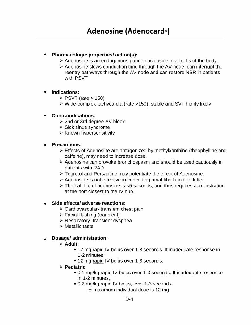
Adenosine (Adenocard®)
Pharmacologic properties/ action(s):
Adenosine is an endogenous purine nucleoside in all cells of the body.
Adenosine slows conduction time through the AV node, can interrupt the reentry pathways through the AV node and can restore NSR in patients with PSVT
Indications:
PSVT (rate > 150) Wide-complex tachycardia (rate >150), stable and SVT highly likely
Contraindications:
2nd or 3rd degree AV block Sick sinus syndrome Known hypersensitivity
Precautions:
Effects of Adenosine are antagonized by methylxanthine (theophylline and caffeine), may need to increase dose.
Adenosine can provoke bronchospasm and should be used cautiously in patients with RAD
Tegretol and Persantine may potentiate the effect of Adenosine.
Adenosine is not effective in converting atrial fibrillation or flutter.
The half-life of adenosine is <5 seconds, and thus requires administration at the port closest to the IV hub.
Side effects/ adverse reactions:
Cardiovascular- transient chest pain Facial flushing (transient) Respiratory- transient dyspnea Metallic taste
Dosage/ administration:
Adult 12 mg rapid IV bolus over 1-3 seconds. If inadequate response in
1-2 minutes, 12 mg rapid IV bolus over 1-3 seconds.
Pediatric 0.1 mg/kg rapid IV bolus over 1-3 seconds. If inadequate response
in 1-2 minutes, 0.2 mg/kg rapid IV bolus, over 1-3 seconds.
maximum individual dose is 12 mg
D-4
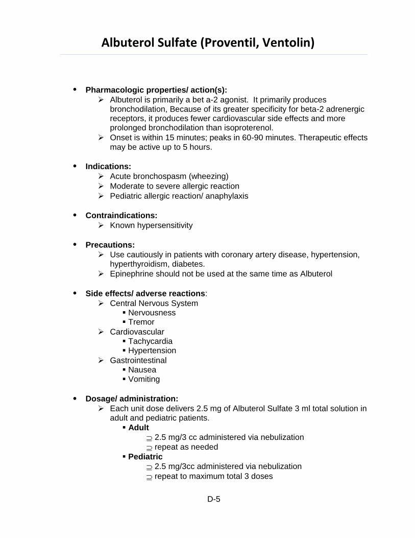
Albuterol Sulfate (Proventil, Ventolin)
Pharmacologic properties/ action(s):
Albuterol is primarily a bet a-2 agonist. It primarily produces bronchodilation, Because of its greater specificity for beta-2 adrenergic receptors, it produces fewer cardiovascular side effects and more prolonged bronchodilation than isoproterenol.
Onset is within 15 minutes; peaks in 60-90 minutes. Therapeutic effects may be active up to 5 hours.
Indications:
Acute bronchospasm (wheezing)
Moderate to severe allergic reaction
Pediatric allergic reaction/ anaphylaxis
Contraindications:
Known hypersensitivity
Precautions:
Use cautiously in patients with coronary artery disease, hypertension, hyperthyroidism, diabetes.
Epinephrine should not be used at the same time as Albuterol
Side effects/ adverse reactions:
Central Nervous System Nervousness Tremor
Cardiovascular Tachycardia Hypertension
Gastrointestinal Nausea Vomiting
Dosage/ administration:
Each unit dose delivers 2.5 mg of Albuterol Sulfate 3 ml total solution in adult and pediatric patients.
Adult
2.5 mg/3 cc administered via nebulization
repeat as needed
Pediatric
2.5 mg/3cc administered via nebulization
repeat to maximum total 3 doses
D-5
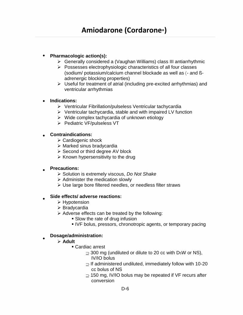
Amiodarone (Cordarone®)
Pharmacologic action(s):
Generally considered a (Vaughan Williams) class III antiarrhythmic Possesses electrophysiologic characteristics of all four classes
(sodium/ potassium/calcium channel blockade as well as- and ß- adrenergic blocking properties)
Useful for treatment of atrial (including pre-excited arrhythmias) and ventricular arrhythmias
Indications:
Ventricular Fibrillation/pulseless Ventricular tachycardia Ventricular tachycardia, stable and with impaired LV function Wide complex tachycardia of unknown etiology Pediatric VF/pulseless VT
Contraindications:
Cardiogenic shock Marked sinus bradycardia Second or third degree AV block Known hypersensitivity to the drug
Precautions:
Solution is extremely viscous, Do Not Shake Administer the medication slowly Use large bore filtered needles, or needless filter straws
Side effects/ adverse reactions:
Hypotension Bradycardia Adverse effects can be treated by the following:
Slow the rate of drug infusion IVF bolus, pressors, chronotropic agents, or temporary pacing
Dosage/administration:
Adult Cardiac arrest
300 mg (undiluted or dilute to 20 cc with D5W or NS), IV/IO bolus
If administered undiluted, immediately follow with 10-20 cc bolus of NS
150 mg, IV/IO bolus may be repeated if VF recurs after conversion
D-6
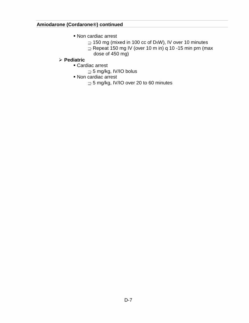
Amiodarone (Cordarone®) continued
Non cardiac arrest
150 mg (mixed in 100 cc of D5W), IV over 10 minutes
Repeat 150 mg IV (over 10 m in) q 10 -15 min prn (max dose of 450 mg)
Pediatric Cardiac arrest
5 mg/kg, IV/IO bolus Non cardiac arrest
5 mg/kg, IV/IO over 20 to 60 minutes
D-7
Acetylsalicylic Acid (Aspirin®)
Pharmacologic properties:
ASA interferes with platelet aggregation and is known as an antiplatelet medication.
Indications:
Acute Myocardial Infarction Chest Pain/Suspected Myocardial Ischemia/Unstable Angina
Contraindications:
Known hypersensitivity to this medication Active ulcer disease Pregnant (especially third trimester) or a nursing mother Taking medication for anticoagulation (e.g. coumadin)
Side effects:
Anaphylaxis (if history of hypersensitivity) Abdominal discomfort Gastrointestinal bleeding (if previous condition exists)
Dosage/ administration:
324 mg PO
D-8
Atropine Sulfate (As a Cardiac Agent)
Pharmacologic properties/ action(s):
Atropine is a potent parasympatholytic anticholinergic. It reduces vagal tone, increases automaticity of the SA node and
increases AV conductions, thus increasing heart rate.
Indications:
Bradydysrhythmias (rate <60) accompanied by hemodynamic compromise, i.e. hypotension (systolic less than 90 mmHg), shock, pulmonary edema, altered level of consciousness.
Pediatric Bradycardia (HR <60) despite adequate oxygenation, ventilation, chest compressions, and refractory to epinephrine.
Contraindications:
Atropine has no effect in patients with transplanted hearts 3rd degree AV block in the setting of an acute anterior wall MI.
Precautions:
Too small a dose (<0.5 mg adults or <0.1 mg pediatrics) or if normal dose pushed too slowly, may initially cause the heart rate to decrease.
Atropine is potentiated by antihistamines and antidepressants.
A maximum dose of 0.4 mg/kg should not be exceeded.
Cautious use in Type II AV block and 3rd degree block with wide QRS complexes.
Side effects/ adverse reactions:
Central Nervous System Restlessness Agitation Confusion Psychotic reaction Pupil dilation Blurred vision Headache
Cardiovascular Increase heart rate May worsen ischemia or increase area of infarction. Ventricular fibrillation
Gastrointestinal Dry mouth Difficulty swallowing
Genitourinary Urinary retention
D-9
Atropine Sulfate (As a Cardiac Agent) continued
Dosage/ administration:
Adult Bradydysrhythmias:
0.5 mg IV bolus, repeat every 3 minutes until improved or up to a total of 0.04 mg/kg.
Shorter dosing intervals may be used in more severe clinical conditions.
Pediatric Pediatric Symptomatic Bradycardia:
0.02 mg/kg IV, IO. This dose may be repeated once in 5 minutes.
Minimum dose is 0.1 mg and maximum single dose is 0.5 mg
D-10

Atropine Sulfate (As an Antidote for Poisonings)
Pharmacologic properties/ action(s):
Atropine is a parasympatholytic.
Atropine binds to acetylcholine receptors and diminishes the actions of acetylcholine.
Indications:
Organophosphate Poisoning (i.e. parathion, malathion, rid-a-bug) and carbamate (Baygon, sevin, and many common roach and ant sprays).
Poisoning Signs
"SLUDGE”
Salivation
Lacrimation
Urination
Defecation
GI hypermotility (Emesis, cramping, diarrhea)
Excessive sweating and bronchorrhea
Additional Signs
Pinpoint pupils
Bradycardia
Contraindications:
None when used in the management of severe organophosphate poisoning.
Precautions:
It is important that the patient be adequately oxygenated and ventilated prior to using atropine, as atropine may precipitate ventricular fibrillation in a poorly oxygenated patient.
Do not rely upon pupillary constriction to discontinue or to titrate medications
Side effects/ adverse reactions:
Victims of organophosphate poisoning can tolerate large doses (1000 mg) of atropine.
Signs of atropinization are likely to occur.
Flushing, pupil dilation, dry mouth, tachycardia.
Dosage/ administration:
Adult 2 mg IV. May repeat 2 mg every 5 minutes
Titrate until respiratory secretions/ distress begins to decrease.
D-11

Atropine Sulfate (As an Antidote for Poisonings) continued
Pediatric 0.02 mg/kg repeat q 5 minutes as necessary
Titrate until respiratory secretions/ distress begins to decrease.
D-12
Calcium Chloride
Pharmacologic properties/ action(s):
Calcium is a cation. Calcium ions increase the force of myocardial contraction and thus act to
increase cardiac output. Calcium has a stabilizing effect on cardiac cell membranes when dangerously high potassium levels make the heart at risk for fibrillation.
Indications:
Hyperkalemia associated with ECG disturbances Hypocalcemia (known) Calcium channel blocker toxicity with hemodynamic compromise Magnesium (MgS04) toxicity
Contraindications:
Cardiac arrest not associated with one of the above
Precautions:
Cautious use in patients receiving digoxin Do not mix with Na bicarbonate, it will precipitate.
Side effects/ adverse reactions:
Bradycardia, usually caused by rapid administration. Arrhythmias especially in patients on digoxin. Sclerosis of veins (if IV infiltrates)
Dosage/ administration:
Adult 1 amp=1 g (10 cc of a 10% sol’n), IV bolus May repeat X 1 in ten minutes if no response in the setting of a calcium channel blocker overdose.
Pediatric 20 mg/kg slow IV over 2 minutes. Maximum dose 1 g
D-13
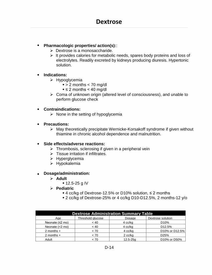
Dextrose Administration Summary Table Age Threshold glucose Dosage Dextrose solution
Neonate (≤2 mo) < 40 4 cc/kg D10%
Neonate (<2 mo) < 40 4 cc/kg D12.5%
2 months + < 70 4 cc/kg D10% or D12.5%
2 months + < 70 2 cc/kg D25%
Adult < 70 12.5-25g D10% or D50%
Dextrose
Pharmacologic properties/ action(s)::
Dextrose is a monosaccharide. It provides calories for metabolic needs, spares body proteins and loss of
electrolytes. Readily excreted by kidneys producing diuresis. Hypertonic solution.
Indications:
Hypoglycemia > 2 months < 70 mg/dl ≤ 2 months < 40 mg/dl
Coma of unknown origin (altered level of consciousness), and unable to perform glucose check
Contraindications:
None in the setting of hypoglycemia
Precautions:
May theoretically precipitate Wernicke-Korsakoff syndrome if given without thiamine in chronic alcohol dependence and malnutrition.
Side effects/adverse reactions:
Thrombosis, sclerosing if given in a peripheral vein Tissue irritation if infiltrates. Hyperglycemia Hypokalemia
Dosage/administration:
Adult 12.5-25 g IV
Pediatric 4 cc/kg of Dextrose-12.5% or D10% solution, ≤ 2 months 2 cc/kg of Dextrose-25% or 4 cc/kg D10-D12.5%, 2 months-12 y/o
D-14
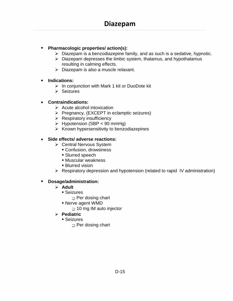
Diazepam
Pharmacologic properties/ action(s):
Diazepam is a benzodiazepine family, and as such is a sedative, hypnotic. Diazepam depresses the limbic system, thalamus, and hypothalamus
resulting in calming effects.
Diazepam is also a muscle relaxant.
Indications:
In conjunction with Mark 1 kit or DuoDote kit Seizures
Contraindications:
Acute alcohol intoxication Pregnancy, (EXCEPT in eclamptic seizures) Respiratory insufficiency Hypotension (SBP < 90 mmHg) Known hypersensitivity to benzodiazepines
Side effects/ adverse reactions:
Central Nervous System Confusion, drowsiness Slurred speech Muscular weakness Blurred vision
Respiratory depression and hypotension (related to rapid IV administration)
Dosage/administration:
Adult Seizures
Per dosing chart Nerve agent WMD
10 mg IM auto injector
Pediatric Seizures
Per dosing chart
D-15
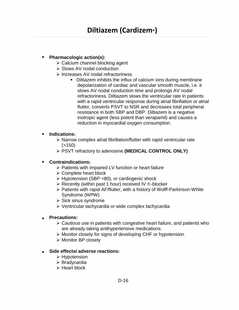
Diltiazem (Cardizem®)
Pharmacologic action(s):
Calcium channel blocking agent
Slows AV nodal conduction
Increases AV nodal refractoriness Diltiazem inhibits the influx of calcium ions during membrane
depolarization of cardiac and vascular smooth muscle, i.e. it slows AV nodal conduction time and prolongs AV nodal refractoriness. Diltiazem slows the ventricular rate in patients with a rapid ventricular response during atrial fibrillation or atrial flutter, converts PSVT to NSR and decreases total peripheral resistance in both SBP and DBP. Diltiazem is a negative inotropic agent (less potent than verapamil) and causes a reduction in myocardial oxygen consumption.
Indications:
Narrow complex atrial fibrillation/flutter with rapid ventricular rate (>150)
PSVT refractory to adenosine (MEDICAL CONTROL ONLY)
Contraindications:
Patients with impaired LV function or heart failure Complete heart block Hypotension (SBP <80), or cardiogenic shock Recently (within past 1 hour) received IV-blocker Patients with rapid AF/flutter, with a history of Wolff-Parkinson-White
Syndrome (WPW)
Sick sinus syndrome
Ventricular tachycardia or wide complex tachycardia
Precautions:
Cautious use in patients with congestive heart failure, and patients who are already taking antihypertensive medications.
Monitor closely for signs of developing CHF or hypotension
Monitor BP closely
Side effects/ adverse reactions:
Hypotension Bradycardia Heart block
D-16

Diltiazem (Cardizem®) continued
Dosage/administration:
0.25 mg/kg (20 mg), over 5 minutes
Repeat 0.35 mg/kg (25 mg), slow IV bolus if no response within 10
minutes and SBP > 100 mmHg.
D-17
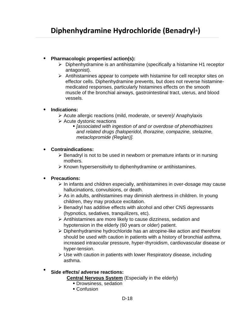
Diphenhydramine Hydrochloride (Benadryl®)
Pharmacologic properties/ action(s):
Diphenhydramine is an antihistamine (specifically a histamine H1 receptor antagonist).
Antihistamines appear to compete with histamine for cell receptor sites on effector cells. Diphenhydramine prevents, but does not reverse histamine- medicated responses, particularly histamines effects on the smooth muscle of the bronchial airways, gastrointestinal tract, uterus, and blood vessels.
Indications:
Acute allergic reactions (mild, moderate, or severe)/ Anaphylaxis Acute dystonic reactions
[associated with ingestion of and or overdose of phenothiazines and related drugs (haloperidol, thorazine, compazine, stelazine, metaclopromide (Reglan)].
Contraindications:
Benadryl is not to be used in newborn or premature infants or in nursing mothers.
Known hypersensitivity to diphenhydramine or antihistamines.
Precautions:
In infants and children especially, antihistamines in over-dosage may cause hallucinations, convulsions, or death.
As in adults, antihistamines may diminish alertness in children. In young
children, they may produce excitation.
Benadryl has additive effects with alcohol and other CNS depressants
(hypnotics, sedatives, tranquilizers, etc).
Antihistamines are more likely to cause dizziness, sedation and
hypotension in the elderly (60 years or older) patient.
Diphenhydramine hydrochloride has an atropine-like action and therefore
should be used with caution in patients with a history of bronchial asthma,
increased intraocular pressure, hyper-thyroidism, cardiovascular disease or
hyper-tension.
Use with caution in patients with lower Respiratory disease, including
asthma.
Side effects/ adverse reactions:
Central Nervous System (Especially in the elderly) Drowsiness, sedation Confusion
D-18
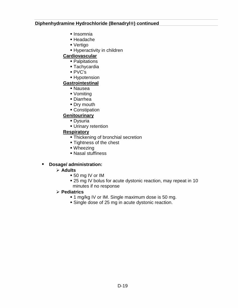
Diphenhydramine Hydrochloride (Benadryl®) continued
Insomnia
Headache Vertigo Hyperactivity in children
Cardiovascular Palpitations Tachycardia PVC's Hypotension
Gastrointestinal Nausea Vomiting Diarrhea Dry mouth Constipation
Genitourinary Dysuria Urinary retention
Respiratory Thickening of bronchial secretion Tightness of the chest Wheezing Nasal stuffiness
Dosage/ administration:
Adults 50 mg IV or IM 25 mg IV bolus for acute dystonic reaction, may repeat in 10 minutes if no response
Pediatrics 1 mg/kg IV or IM. Single maximum dose is 50 mg. Single dose of 25 mg in acute dystonic reaction.
D-19
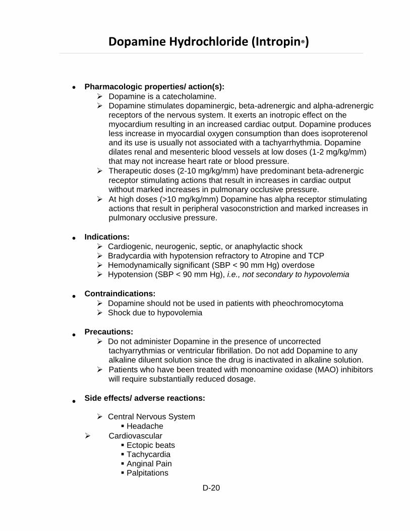
Dopamine Hydrochloride (Intropin®)
Pharmacologic properties/ action(s):
Dopamine is a catecholamine. Dopamine stimulates dopaminergic, beta-adrenergic and alpha-adrenergic
receptors of the nervous system. It exerts an inotropic effect on the myocardium resulting in an increased cardiac output. Dopamine produces less increase in myocardial oxygen consumption than does isoproterenol and its use is usually not associated with a tachyarrhythmia. Dopamine dilates renal and mesenteric blood vessels at low doses (1-2 mg/kg/mm) that may not increase heart rate or blood pressure.
Therapeutic doses (2-10 mg/kg/mm) have predominant beta-adrenergic
receptor stimulating actions that result in increases in cardiac output without marked increases in pulmonary occlusive pressure.
At high doses (>10 mg/kg/mm) Dopamine has alpha receptor stimulating actions that result in peripheral vasoconstriction and marked increases in pulmonary occlusive pressure.
Indications:
Cardiogenic, neurogenic, septic, or anaphylactic shock Bradycardia with hypotension refractory to Atropine and TCP Hemodynamically significant (SBP < 90 mm Hg) overdose Hypotension (SBP < 90 mm Hg), i.e., not secondary to hypovolemia
Contraindications:
Dopamine should not be used in patients with pheochromocytoma Shock due to hypovolemia
Precautions:
Do not administer Dopamine in the presence of uncorrected tachyarrythmias or ventricular fibrillation. Do not add Dopamine to any alkaline diluent solution since the drug is inactivated in alkaline solution.
Patients who have been treated with monoamine oxidase (MAO) inhibitors will require substantially reduced dosage.
Side effects/ adverse reactions:
Central Nervous System
Headache
Cardiovascular Ectopic beats Tachycardia Anginal Pain Palpitations
D-20
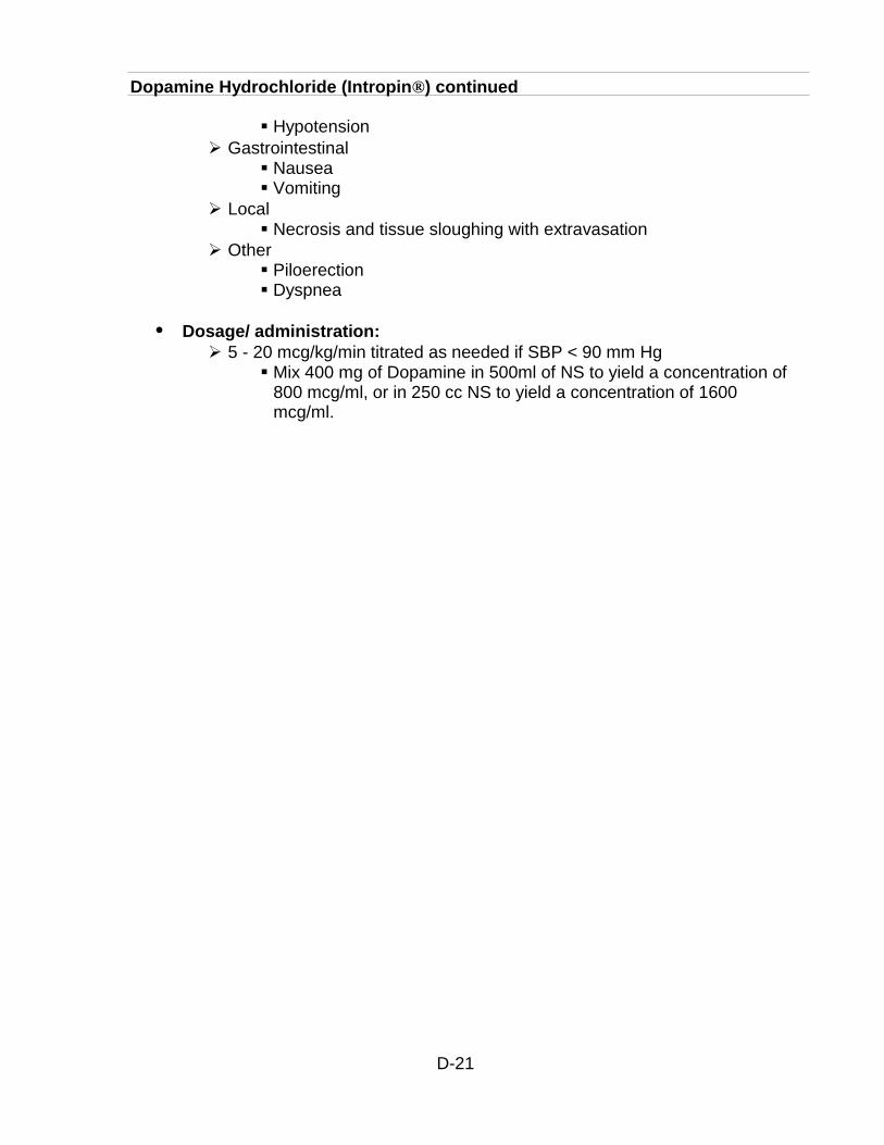
Dopamine Hydrochloride (Intropin®) continued
Hypotension
Gastrointestinal Nausea Vomiting
Local Necrosis and tissue sloughing with extravasation
Other Piloerection Dyspnea
Dosage/ administration:
5 - 20 mcg/kg/min titrated as needed if SBP < 90 mm Hg Mix 400 mg of Dopamine in 500ml of NS to yield a concentration of
800 mcg/ml, or in 250 cc NS to yield a concentration of 1600 mcg/ml.
D-21

DuoDote Kit
See Mark 1 Kit
D-22
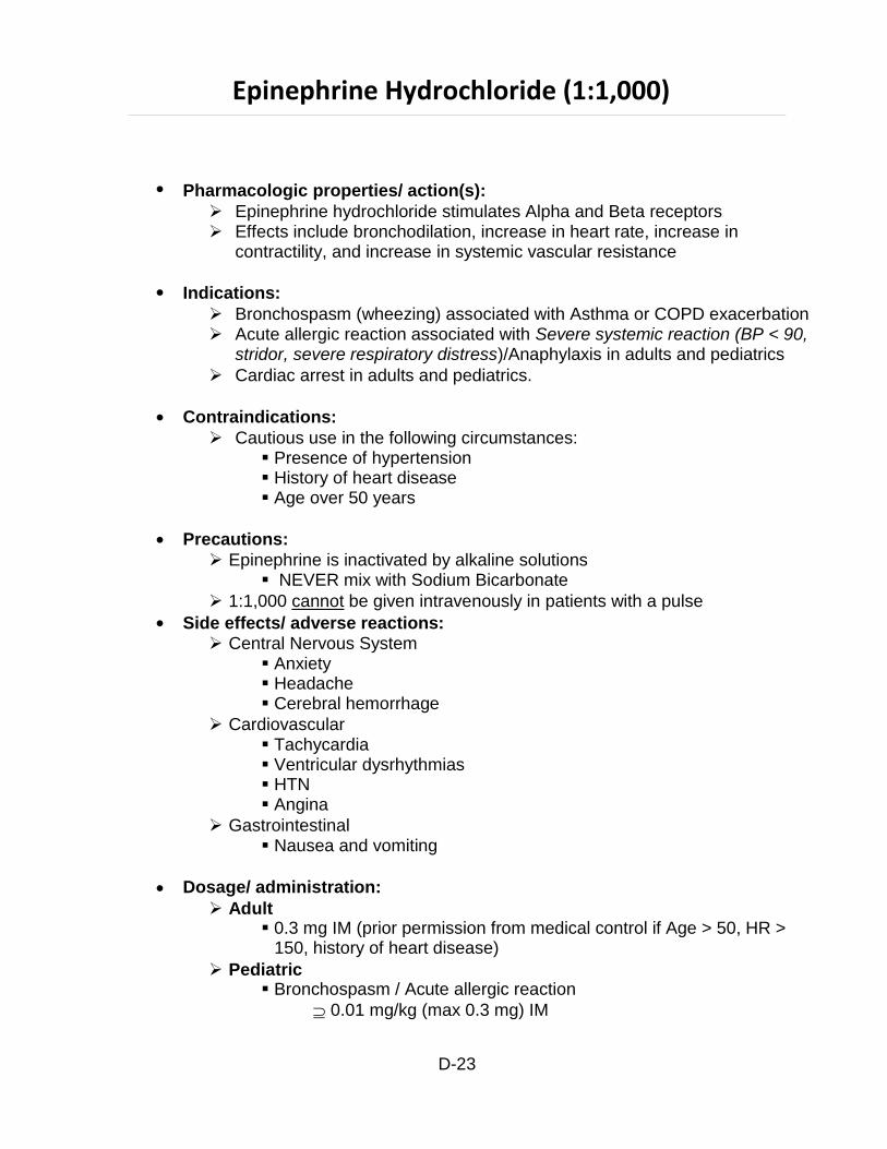
Epinephrine Hydrochloride (1:1,000)
Pharmacologic properties/ action(s):
Epinephrine hydrochloride stimulates Alpha and Beta receptors Effects include bronchodilation, increase in heart rate, increase in
contractility, and increase in systemic vascular resistance
Indications:
Bronchospasm (wheezing) associated with Asthma or COPD exacerbation Acute allergic reaction associated with Severe systemic reaction (BP < 90,
stridor, severe respiratory distress)/Anaphylaxis in adults and pediatrics
Cardiac arrest in adults and pediatrics.
Contraindications:
Cautious use in the following circumstances: Presence of hypertension History of heart disease Age over 50 years
Precautions:
Epinephrine is inactivated by alkaline solutions NEVER mix with Sodium Bicarbonate
1:1,000 cannot be given intravenously in patients with a pulse
Side effects/ adverse reactions: Central Nervous System
Anxiety Headache Cerebral hemorrhage
Cardiovascular Tachycardia Ventricular dysrhythmias HTN Angina
Gastrointestinal Nausea and vomiting
Dosage/ administration:
Adult 0.3 mg IM (prior permission from medical control if Age > 50, HR >
150, history of heart disease)
Pediatric Bronchospasm / Acute allergic reaction
0.01 mg/kg (max 0.3 mg) IM
D-23
Epinephrine Hydrochloride (1:1,000) continued
May repeat every 15 minutes as needed X 2 additional doses (3 total)
May administer at same time nebulizer is being administered
D-24
Epinephrine Hydrochloride (1:10,000)
Pharmacologic properties/ action(s):
Epinephrine is a sympathomimetic which stimulates both Alpha and Beta receptors
Its effects are to increase systemic vascular resistance, arterial blood pressure, coronary and cerebral blood flow, heart rate and contractility, and myocardial oxygen requirements
The alpha-adrenergic effect is to increase SVR and coronary blood flow (makes the fibrillating myocardium more susceptible to counter-shock)
Indications:
Ventricular fibrillation or pulseless ventricular tachycardia Asystole Pulseless electrical activity (PEA) Anaphylactic shock Newborn resuscitation/Neonatal asystole or bradycardia Pediatric bradycardia/Pulseless cardiac arrest/VF/pulseless VT Bradycardia with hypotension refractory to Atropine and TCP
Contraindications:
NONE in the cardiac arrest situation
Precautions:
Epinephrine is inactivated by alkaline solutions. NEVER mix with Sodium Bicarbonate
Side effects/ adverse reactions:
Central Nervous System Anxiety Headache Cerebral hemorrhage
Cardiovascular Tachycardia Ventricular dysrhythmias HTN Angina
Gastrointestinal Nausea Vomiting
D-25
Epinephrine Hydrochloride (1:10,000) continued
Dosage/ administration:
Adult Cardiac arrest
IV/IO: 1 mg (10 cc)
Repeat every 3-5 minutes at the same dose Bradycardia (with serious signs and symptoms)
Continuous IV infusion: set rate at 2-10 mcg/min, titrate to affect
Allergic reaction (Cardiac Arrest or Respiratory arrest Imminent)
0.5 mg (5cc of 1:10,000) IVP
Pediatric Pulseless cardiac arrest / Ventricular fibrillation/pulseless VT
0.01 mg/kg, (max 1.0 mg) IV or IO
Repeat every 3-5 minutes at the same dose Newborn resuscitation
0.01 mg/kg IV of a 1:10,000 solution
repeat every 3 to 5 minutes as indicated Bradycardia with cardiopulmonary compromise
0.01 mg/kg (max 1 mg) via IV/IO
Use first route available
Repeat dose every 3-5 minutes until either the
bradycardia or severe cardiopulmonary compromise resolves
**********ALSO SEE EPINEPHRINE DOSING SUMMARY **********
D-26
Adult Epinephrine Dosing Summary
Cardiac arrest: (for V-fib, pulseless V-tach, asystole, or PEA only)
Standard: 1 mg (1:10,000) IV/IO bolus every 3-5 minutes
For Anaphylactic Shock:
0.5 mg (1:10,000) IV/IO bolus.
For bradycardia with hypotension refractory to Atropine and TCP:
Continuous IV infusion: mix 2 mg (1:1,000) in 250 cc NS (8 mcg/cc) and run at 2-10 mcg/min
Every 15 gtts/min = 2 mcg/min.
Pediatric Epinephrine Dosing Summary
Newborn resuscitation:
0.01 mg/kg IV/IO of a 1:10,000 solution
repeat every 3 to 5 minutes as indicated
Bradycardia with cardiopulmonary compromise:
0.01 mg/kg IV/IO (1:10,000) max 1 mg
Use first route available
Repeat dose every 3-5 minutes until either the bradycardia or severe
cardiopulmonary compromise resolves
Pulseless cardiac arrest / Ventricular fibrillation/pulseless VT:
0.01 mg/kg IV/IO (1:10,000) max 1 mg
Use first route available
Repeat every 3-5 minutes at the same dose
D-27
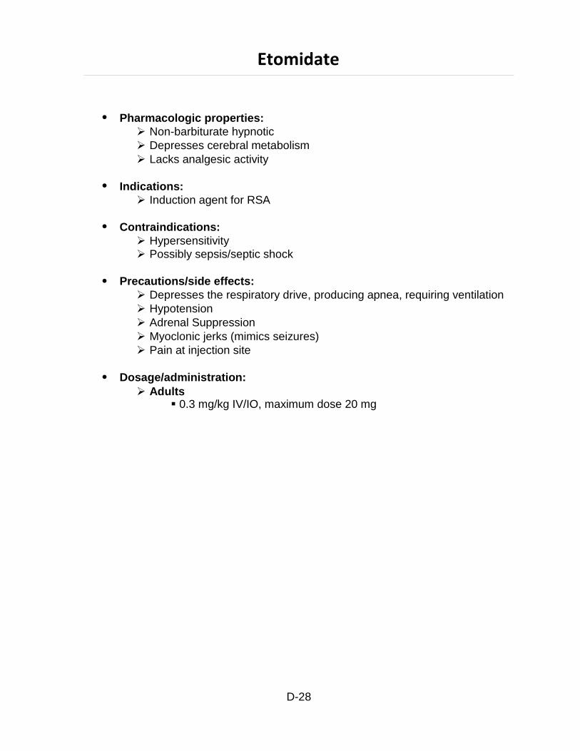
Etomidate
Pharmacologic properties:
Non-barbiturate hypnotic
Depresses cerebral metabolism
Lacks analgesic activity
Indications:
Induction agent for RSA
Contraindications:
Hypersensitivity
Possibly sepsis/septic shock
Precautions/side effects:
Depresses the respiratory drive, producing apnea, requiring ventilation
Hypotension
Adrenal Suppression
Myoclonic jerks (mimics seizures)
Pain at injection site
Dosage/administration:
Adults 0.3 mg/kg IV/IO, maximum dose 20 mg
D-28
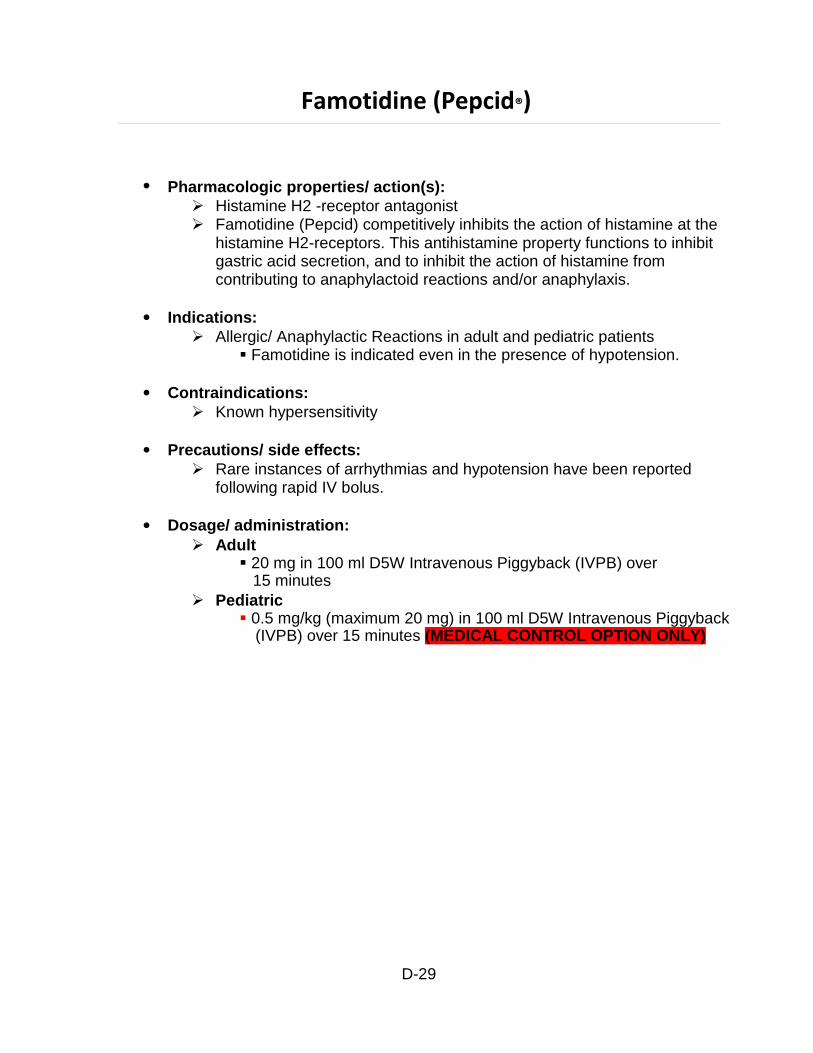
Famotidine (Pepcid®)
Pharmacologic properties/ action(s):
Histamine H2 -receptor antagonist Famotidine (Pepcid) competitively inhibits the action of histamine at the
histamine H2-receptors. This antihistamine property functions to inhibit gastric acid secretion, and to inhibit the action of histamine from contributing to anaphylactoid reactions and/or anaphylaxis.
Indications:
Allergic/ Anaphylactic Reactions in adult and pediatric patients Famotidine is indicated even in the presence of hypotension.
Contraindications:
Known hypersensitivity
Precautions/ side effects:
Rare instances of arrhythmias and hypotension have been reported following rapid IV bolus.
Dosage/ administration:
Adult 20 mg in 100 ml D5W Intravenous Piggyback (IVPB) over 15 minutes
Pediatric 0.5 mg/kg (maximum 20 mg) in 100 ml D5W Intravenous Piggyback (IVPB) over 15 minutes (MEDICAL CONTROL OPTION ONLY)
D-29
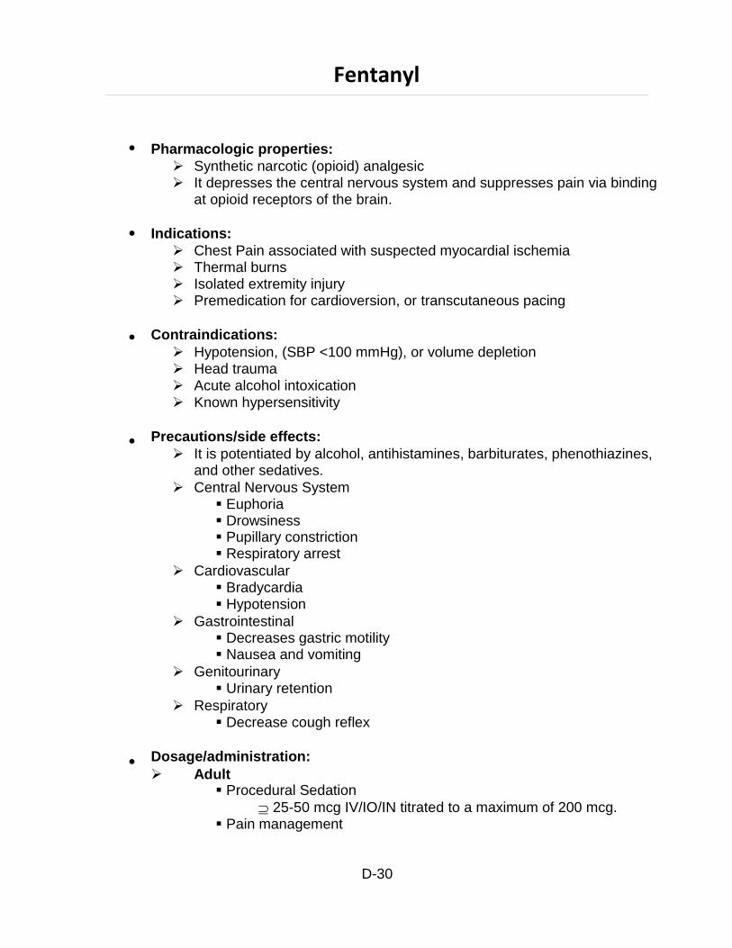
Fentanyl
Pharmacologic properties:
Synthetic narcotic (opioid) analgesic It depresses the central nervous system and suppresses pain via binding
at opioid receptors of the brain.
Indications:
Chest Pain associated with suspected myocardial ischemia Thermal burns Isolated extremity injury Premedication for cardioversion, or transcutaneous pacing
Contraindications:
Hypotension, (SBP <100 mmHg), or volume depletion Head trauma Acute alcohol intoxication Known hypersensitivity
Precautions/side effects:
It is potentiated by alcohol, antihistamines, barbiturates, phenothiazines, and other sedatives.
Central Nervous System Euphoria Drowsiness Pupillary constriction Respiratory arrest
Cardiovascular Bradycardia Hypotension
Gastrointestinal Decreases gastric motility Nausea and vomiting
Genitourinary Urinary retention
Respiratory Decrease cough reflex
Dosage/administration:
Adult Procedural Sedation
25-50 mcg IV/IO/IN titrated to a maximum of 200 mcg. Pain management
D-30
Fentanyl, continued
25-50 mcg IV/IO/IN every 5 minutes, titrated to a maximum dose of 200 mcg
Chest Pain (Cardiac)
25-50 mcg IV/IO/IN every 2 minutes, titrated to a maximum dose of 200 mcg.
Pediatric Pain management
1.5 mcg/kg IN, maximum dose 100 mcg.
D-31
Glucagon
Pharmacologic properties/ action(s):
Glucagon is a hormone. Glucagon, produced in the pancreas by the Alpha cells of the Islets of
Langerhans, causes an increase in blood glucose concentrations. It is effective in small doses and no evidence of toxicity has been reported with its use. Glucagon acts only on liver glycogen, converting it to glucose.
Glucagon effectively restores inotropy and chronotropy, via a cyclic-AMP mechanism, in patients with cardio-vascular toxicity secondary to beta- blockers, and to a more variable degree, to calcium channel blockers.
Indications:
Hypoglycemia (where IV access cannot be obtained). Beta-blocker and calcium channel blocker overdoses.
Contradictions:
Since glucagon is a protein, hypersensitivity is a possibility.
Precautions:
Glucagon should be administered with caution in patients with a history of insulinoma and/or pheochromocytoma.
Side effects/ adverse reactions:
Gastrointestinal Occasional nausea and vomiting
Dosage/ administration:
Adult Hypoglycemia
1.0 unit (1.0 mg) of Glucagon, IM. Beta-blocker (or calcium channel blocker)
3.0 mg IV bolus.
Pediatric 0.1 mg/kg (max 1.0 mg), IV/IM
.
D-32

Glucose (Oral)
Pharmacologic properties:
Glucose is a monosaccharide.
It provides calories for metabolic needs, spares body proteins and loss of electrolytes.
Indications:
Hypoglycemia
Contraindications:
Inability to self-administer due to altered level of consciousness
Precautions/side effects:
Aspiration risk in patients with altered level of consciousness
Dosage/administration:
Adult If Hypoglycemic (Blood glucose < 70 mg/dL) without IV access.
Glucose paste 15 g or other oral glucose containing agent (e.g., orange juice) if patient alert enough to self-administer oral agent
D-33
Haloperidol (Haldol ®)
Pharmacologic properties/ action(s):
Antipsychotic agent or major tranquilizer (of the butyrophenone class). May block dopamine receptors in CNS, but exact mechanism has not been
clearly established. It acts to have a profound calming effect on patients.
Indications:
Behavioral emergencies for patients with severe agitation or aggressive behavior resulting in interference with patient care or patient/crew safety.
Contraindications:
Not to be administered to patients on MAO inhibitors Known hypersensitivity
Precautions:
Haldol should be administered cautiously to patients with a history of cardiovascular disorders, seizures, anticoagulant therapy.
If hypotension occurs, do not use epinephrine as its vasopressor action is blocked and blood pressure may drop even further.
If acute laryngospasm occurs, immediately administer diphenhydramine (benadryl) 50 mg IV bolus.
Side effects/ adverse reactions:
Central Nervous System Parkinson-like symptoms, i.e. Extrapyramidal symptoms characterized by dystonic reactions.
Cardiovascular Tachycardia Hypotension Hypertension EKG changes
Respiratory Laryngospasm Bronchospasm
Respiratory depression
Gastrointestinal Nausea and vomiting Anorexia
Dosage/ administration:
Adult 5 mg if < 60 kg IM (standing)
D-34
Haloperidol (Haldol®) continued
10 mg if > 60 kg IM (standing)
If inadequate response, call medical control for repeat dosing
Pediatric Safety and effectiveness in children has not been conducted.
D-35
Hydroxocobalamin (Cyanokit®)
Pharmacologic properties:
Hydroxocobalamin is a precursor element. The action of hydroxocobalamin in the treatment of Cyanide poisoning
is based on its ability to bind cyanide ions. Each hydroxocobalamin molecule can bind one cyanide ion by substituting if for the hydroxo ligand linked to the trivalent cobalt ion, to form cyanocobalamin, which is then excreted in the urine.
Indications:
Treatment for known or suspected cyanide poisoning
Contraindications:
None in the setting of known or suspected cyanide poisoning
Precautions/side effects:
Increased blood pressure Allergic reaction Skin and subcutaneous tissue disorders: urticaria, pruritus Vascular disorders: hot, flush
Dosage/administration:
5 g IV/IO over 15 minutes
D-36
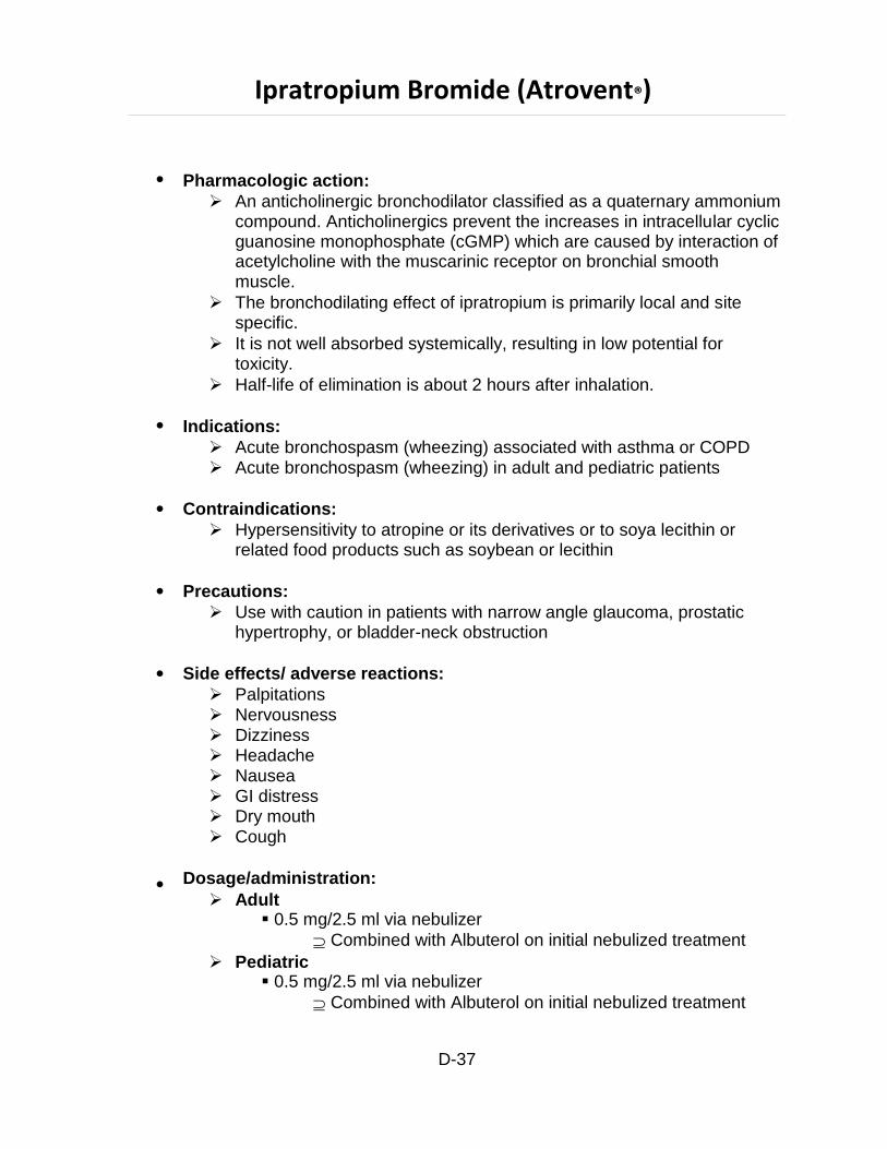
Ipratropium Bromide (Atrovent®)
Pharmacologic action:
An anticholinergic bronchodilator classified as a quaternary ammonium compound. Anticholinergics prevent the increases in intracellular cyclic guanosine monophosphate (cGMP) which are caused by interaction of acetylcholine with the muscarinic receptor on bronchial smooth muscle.
The bronchodilating effect of ipratropium is primarily local and site specific.
It is not well absorbed systemically, resulting in low potential for toxicity.
Half-life of elimination is about 2 hours after inhalation.
Indications:
Acute bronchospasm (wheezing) associated with asthma or COPD Acute bronchospasm (wheezing) in adult and pediatric patients
Contraindications:
Hypersensitivity to atropine or its derivatives or to soya lecithin or related food products such as soybean or lecithin
Precautions:
Use with caution in patients with narrow angle glaucoma, prostatic hypertrophy, or bladder-neck obstruction
Side effects/ adverse reactions:
Palpitations Nervousness Dizziness Headache Nausea GI distress Dry mouth Cough
Dosage/administration:
Adult 0.5 mg/2.5 ml via nebulizer
Combined with Albuterol on initial nebulized treatment
Pediatric 0.5 mg/2.5 ml via nebulizer
Combined with Albuterol on initial nebulized treatment
D-37
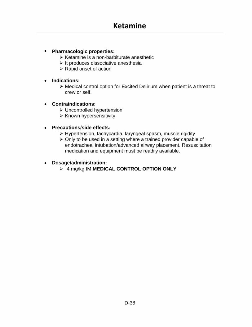
Ketamine
Pharmacologic properties:
Ketamine is a non-barbiturate anesthetic It produces dissociative anesthesia Rapid onset of action
Indications:
Medical control option for Excited Delirium when patient is a threat to crew or self.
Contraindications:
Uncontrolled hypertension Known hypersensitivity
Precautions/side effects:
Hypertension, tachycardia, laryngeal spasm, muscle rigidity Only to be used in a setting where a trained provider capable of
endotracheal intubation/advanced airway placement. Resuscitation medication and equipment must be readily available.
Dosage/administration:
4 mg/kg IM MEDICAL CONTROL OPTION ONLY
D-38
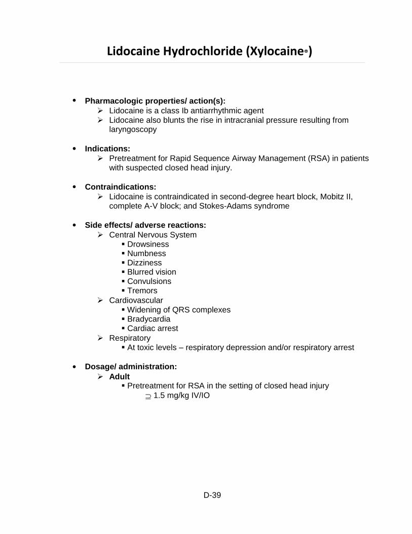
Lidocaine Hydrochloride (Xylocaine®)
Pharmacologic properties/ action(s):
Lidocaine is a class Ib antiarrhythmic agent Lidocaine also blunts the rise in intracranial pressure resulting from
laryngoscopy
Indications:
Pretreatment for Rapid Sequence Airway Management (RSA) in patients with suspected closed head injury.
Contraindications:
Lidocaine is contraindicated in second-degree heart block, Mobitz II, complete A-V block; and Stokes-Adams syndrome
Side effects/ adverse reactions:
Central Nervous System Drowsiness Numbness Dizziness Blurred vision Convulsions Tremors
Cardiovascular Widening of QRS complexes Bradycardia Cardiac arrest
Respiratory At toxic levels – respiratory depression and/or respiratory arrest
Dosage/ administration:
Adult Pretreatment for RSA in the setting of closed head injury
1.5 mg/kg IV/IO
D-39
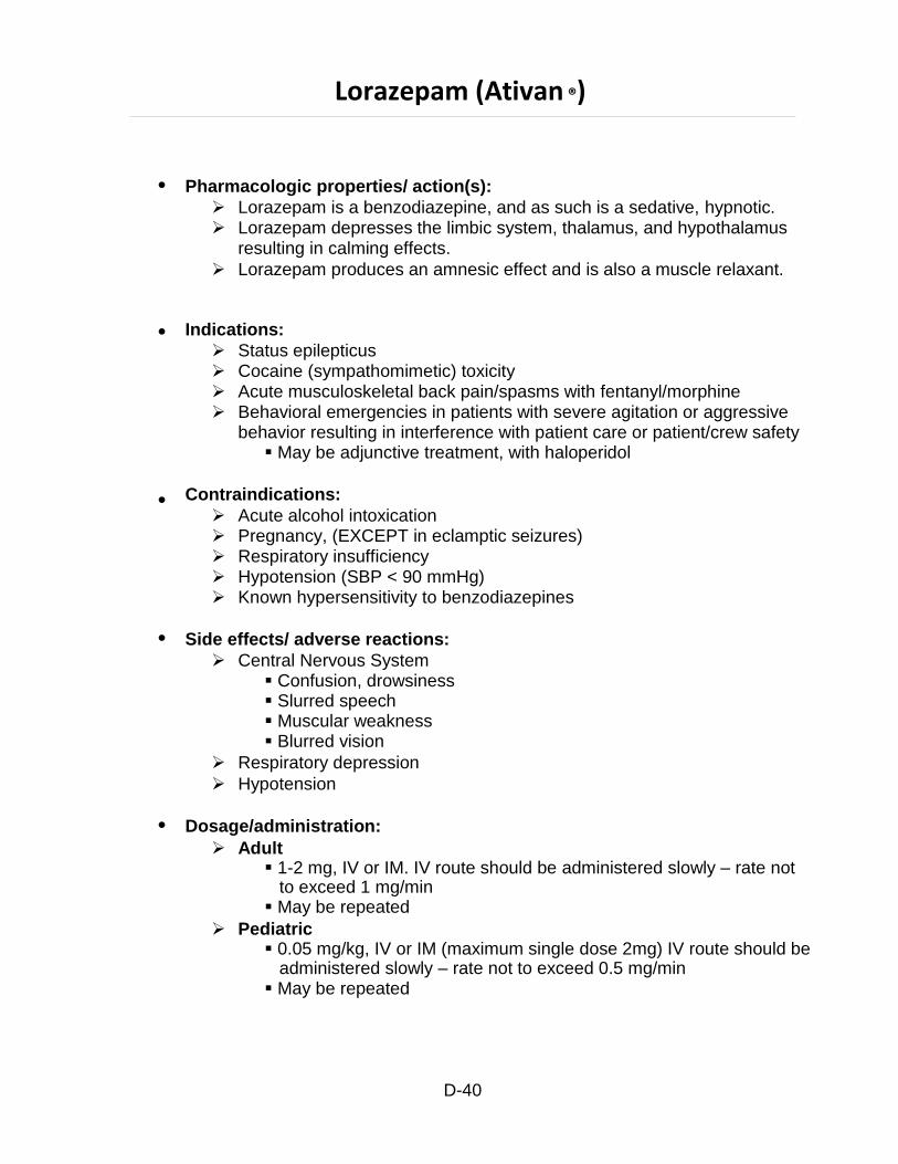
Lorazepam (Ativan ®)
Pharmacologic properties/ action(s):
Lorazepam is a benzodiazepine, and as such is a sedative, hypnotic. Lorazepam depresses the limbic system, thalamus, and hypothalamus
resulting in calming effects.
Lorazepam produces an amnesic effect and is also a muscle relaxant.
Indications:
Status epilepticus Cocaine (sympathomimetic) toxicity Acute musculoskeletal back pain/spasms with fentanyl/morphine Behavioral emergencies in patients with severe agitation or aggressive
behavior resulting in interference with patient care or patient/crew safety May be adjunctive treatment, with haloperidol
Contraindications:
Acute alcohol intoxication Pregnancy, (EXCEPT in eclamptic seizures) Respiratory insufficiency Hypotension (SBP < 90 mmHg) Known hypersensitivity to benzodiazepines
Side effects/ adverse reactions:
Central Nervous System Confusion, drowsiness Slurred speech Muscular weakness Blurred vision
Respiratory depression
Hypotension
Dosage/administration:
Adult 1-2 mg, IV or IM. IV route should be administered slowly – rate not to exceed 1 mg/min May be repeated
Pediatric 0.05 mg/kg, IV or IM (maximum single dose 2mg) IV route should be administered slowly – rate not to exceed 0.5 mg/min May be repeated
D-40
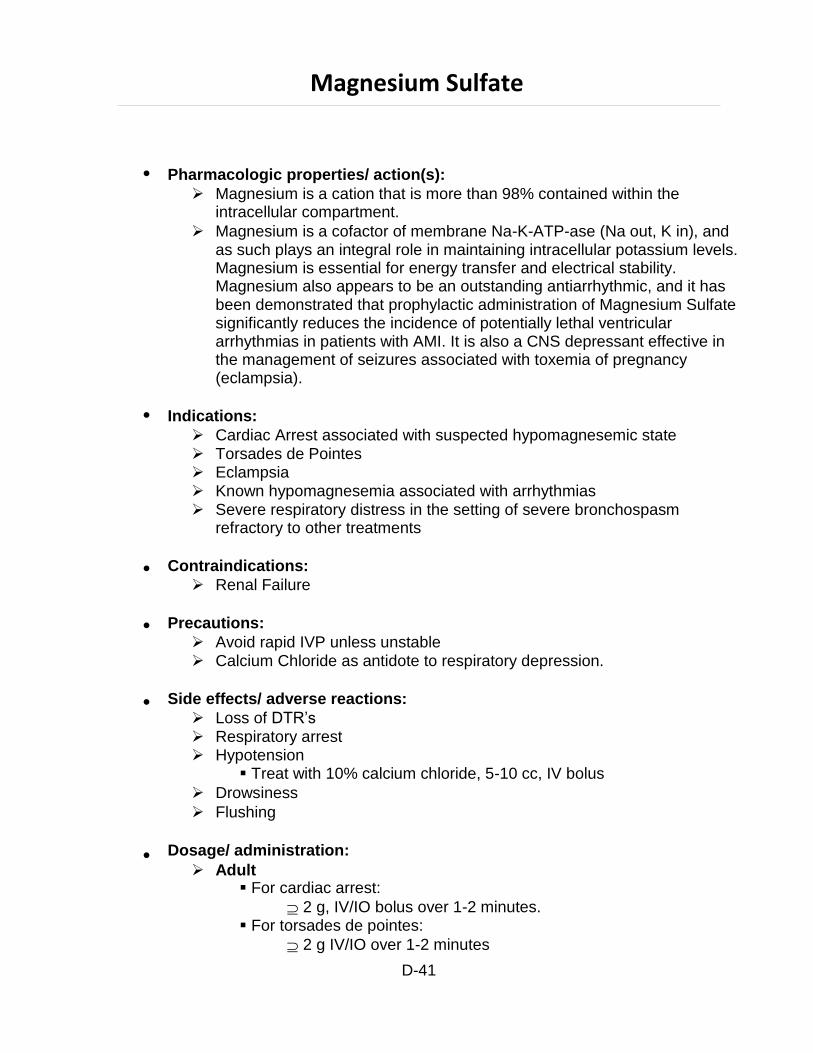
Magnesium Sulfate
Pharmacologic properties/ action(s):
Magnesium is a cation that is more than 98% contained within the intracellular compartment.
Magnesium is a cofactor of membrane Na-K-ATP-ase (Na out, K in), and as such plays an integral role in maintaining intracellular potassium levels. Magnesium is essential for energy transfer and electrical stability. Magnesium also appears to be an outstanding antiarrhythmic, and it has been demonstrated that prophylactic administration of Magnesium Sulfate significantly reduces the incidence of potentially lethal ventricular arrhythmias in patients with AMI. It is also a CNS depressant effective in the management of seizures associated with toxemia of pregnancy (eclampsia).
Indications:
Cardiac Arrest associated with suspected hypomagnesemic state Torsades de Pointes Eclampsia Known hypomagnesemia associated with arrhythmias Severe respiratory distress in the setting of severe bronchospasm
refractory to other treatments
Contraindications:
Renal Failure
Precautions:
Avoid rapid IVP unless unstable Calcium Chloride as antidote to respiratory depression.
Side effects/ adverse reactions:
Loss of DTR’s Respiratory arrest Hypotension
Treat with 10% calcium chloride, 5-10 cc, IV bolus
Drowsiness
Flushing
Dosage/ administration:
Adult For cardiac arrest:
2 g, IV/IO bolus over 1-2 minutes. For torsades de pointes:
2 g IV/IO over 1-2 minutes
D-41
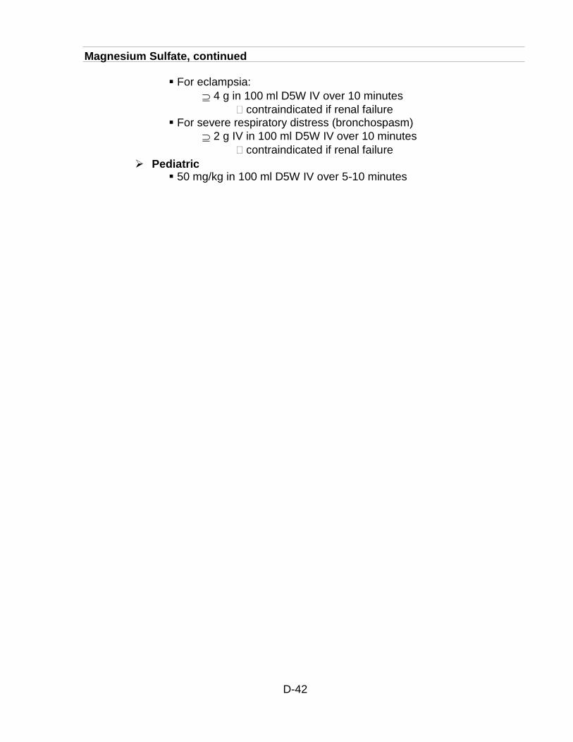
Magnesium Sulfate, continued
For eclampsia:
4 g in 100 ml D5W IV over 10 minutes
contraindicated if renal failure For severe respiratory distress (bronchospasm)
2 g IV in 100 ml D5W IV over 10 minutes
contraindicated if renal failure
Pediatric 50 mg/kg in 100 ml D5W IV over 5-10 minutes
D-42
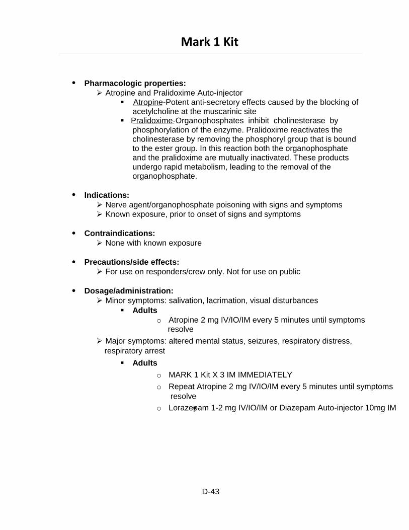
Mark 1 Kit
Pharmacologic properties:
Atropine and Pralidoxime Auto-injector Atropine-Potent anti-secretory effects caused by the blocking of
acetylcholine at the muscarinic site Pralidoxime-Organophosphates inhibit cholinesterase by
phosphorylation of the enzyme. Pralidoxime reactivates the cholinesterase by removing the phosphoryl group that is bound to the ester group. In this reaction both the organophosphate and the pralidoxime are mutually inactivated. These products undergo rapid metabolism, leading to the removal of the organophosphate.
Indications:
Nerve agent/organophosphate poisoning with signs and symptoms Known exposure, prior to onset of signs and symptoms
Contraindications:
None with known exposure
Precautions/side effects:
For use on responders/crew only. Not for use on public
Dosage/administration:
Minor symptoms: salivation, lacrimation, visual disturbances
Adults
o Atropine 2 mg IV/IO/IM every 5 minutes until symptoms resolve
Major symptoms: altered mental status, seizures, respiratory distress,
respiratory arrest
Adults
o MARK 1 Kit X 3 IM IMMEDIATELY
o Repeat Atropine 2 mg IV/IO/IM every 5 minutes until symptoms
resolve
o Lorazepam 1-2 mg IV/IO/IM or Diazepam Auto-injector 10mg IM
D-43
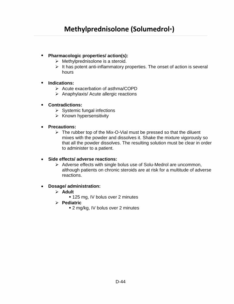
Methylprednisolone (Solumedrol®)
Pharmacologic properties/ action(s):
Methylprednisolone is a steroid.
It has potent anti-inflammatory properties. The onset of action is several hours
Indications:
Acute exacerbation of asthma/COPD Anaphylaxis/ Acute allergic reactions
Contradictions:
Systemic fungal infections Known hypersensitivity
Precautions:
The rubber top of the Mix-O-Vial must be pressed so that the diluent mixes with the powder and dissolves it. Shake the mixture vigorously so that all the powder dissolves. The resulting solution must be clear in order to administer to a patient.
Side effects/ adverse reactions:
Adverse effects with single bolus use of Solu-Medrol are uncommon, although patients on chronic steroids are at risk for a multitude of adverse reactions.
Dosage/ administration:
Adult 125 mg, IV bolus over 2 minutes
Pediatric 2 mg/kg, IV bolus over 2 minutes
D-44
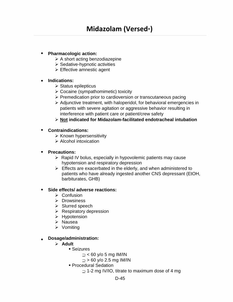
Midazolam (Versed®)
Pharmacologic action:
A short acting benzodiazepine Sedative-hypnotic activities Effective amnestic agent
Indications:
Status epilepticus
Cocaine (sympathomimetic) toxicity
Premedication prior to cardioversion or transcutaneous pacing
Adjunctive treatment, with haloperidol, for behavioral emergencies in
patients with severe agitation or aggressive behavior resulting in
interference with patient care or patient/crew safety
Not indicated for Midazolam-facilitated endotracheal intubation
Contraindications:
Known hypersensitivity Alcohol intoxication
Precautions:
Rapid IV bolus, especially in hypovolemic patients may cause hypotension and respiratory depression
Effects are exacerbated in the elderly, and when administered to patients who have already ingested another CNS depressant (EtOH, barbiturates, GHB)
Side effects/ adverse reactions:
Confusion Drowsiness Slurred speech Respiratory depression Hypotension Nausea Vomiting
Dosage/administration:
Adult Seizures
< 60 y/o 5 mg IM/IN
> 60 y/o 2.5 mg IM/IN Procedural Sedation
1-2 mg IV/IO, titrate to maximum dose of 4 mg
D-45
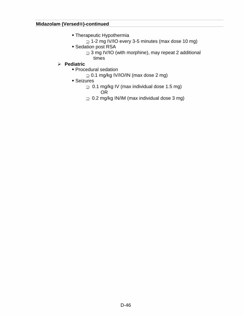
Midazolam (Versed®)-continued
Therapeutic Hypothermia
1-2 mg IV/IO every 3-5 minutes (max dose 10 mg) Sedation post RSA
3 mg IV/IO (with morphine), may repeat 2 additional times
Pediatric Procedural sedation
0.1 mg/kg IV/IO/IN (max dose 2 mg) Seizures
0.1 mg/kg IV (max individual dose 1.5 mg)
OR
0.2 mg/kg IN/IM (max individual dose 3 mg)
D-46
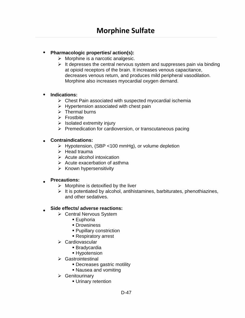
Morphine Sulfate
Pharmacologic properties/ action(s):
Morphine is a narcotic analgesic.
It depresses the central nervous system and suppresses pain via binding at opioid receptors of the brain. It increases venous capacitance, decreases venous return, and produces mild peripheral vasodilation. Morphine also increases myocardial oxygen demand.
Indications:
Chest Pain associated with suspected myocardial ischemia Hypertension associated with chest pain Thermal burns Frostbite Isolated extremity injury Premedication for cardioversion, or transcutaneous pacing
Contraindications:
Hypotension, (SBP <100 mmHg), or volume depletion Head trauma Acute alcohol intoxication Acute exacerbation of asthma Known hypersensitivity
Precautions:
Morphine is detoxified by the liver It is potentiated by alcohol, antihistamines, barbiturates, phenothiazines,
and other sedatives.
Side effects/ adverse reactions:
Central Nervous System Euphoria Drowsiness Pupillary constriction Respiratory arrest
Cardiovascular Bradycardia Hypotension
Gastrointestinal Decreases gastric motility Nausea and vomiting
Genitourinary Urinary retention
D-47
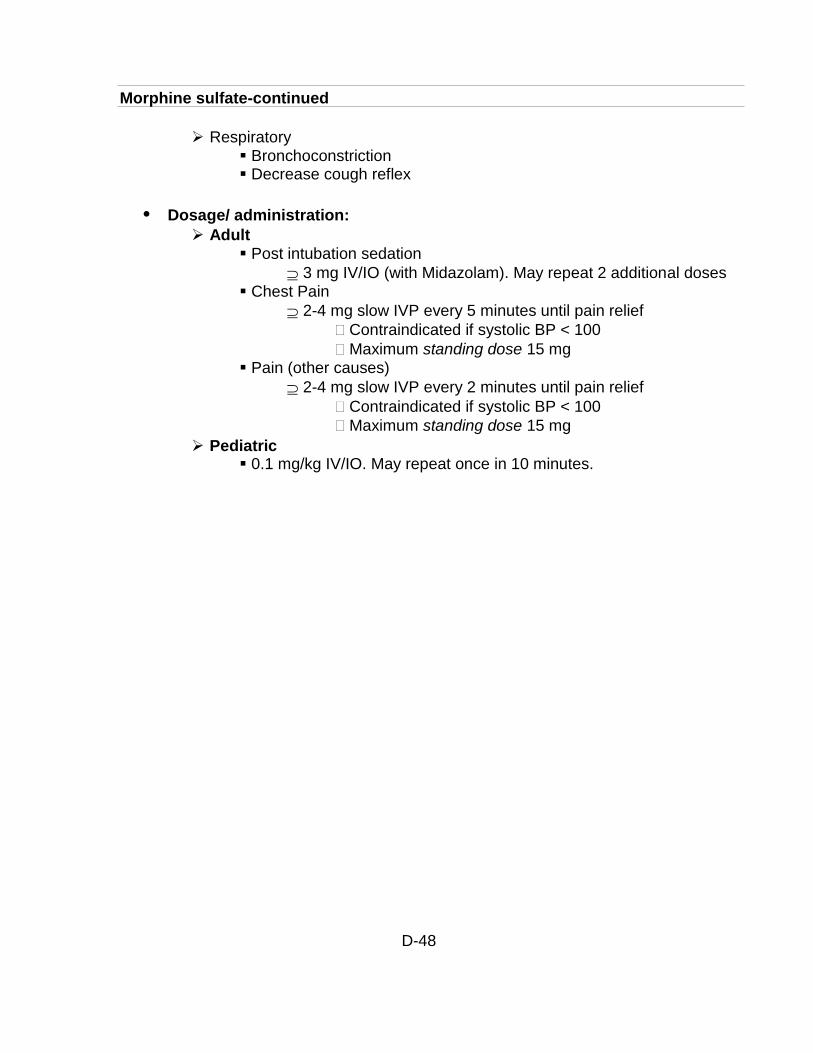
Morphine sulfate-continued
Respiratory Bronchoconstriction Decrease cough reflex
Dosage/ administration:
Adult Post intubation sedation
3 mg IV/IO (with Midazolam). May repeat 2 additional doses Chest Pain
2-4 mg slow IVP every 5 minutes until pain relief
Contraindicated if systolic BP < 100
Maximum standing dose 15 mg Pain (other causes)
2-4 mg slow IVP every 2 minutes until pain relief
Contraindicated if systolic BP < 100
Maximum standing dose 15 mg
Pediatric 0.1 mg/kg IV/IO. May repeat once in 10 minutes.
D-48
Naloxone (Narcan ®)
Pharmacologic properties/ action(s):
Naloxone is a mu receptor (narcotic) antagonist. The mechanism of action is not fully understood. It does appear that
Narcan antagonizes the effects of opiates by competing at the same receptor sites.
When given IV, the action is apparent within two (2) minutes. IM or SC administration is slightly less rapid.
Indications:
Altered mental status
Naloxone is indicated for the complete or partial reversal of narcotic depression and respiratory depression secondary to narcotics or related drugs (examples include, but are not limited to the following):
Heroin, or any opiate derivative Meperidine (Demerol), Morphine, Methadone Codeine Lomotil Hydromorphone (Dilaudid) Pentazocin (Talwin) Propoxyphene (Darvon, Darvocet) Oxycodone (Percodan, Percocet, Tylox) Hydrocodone (Vicodin, Anexia, Lortab)
Contraindications:
Narcan is contraindicated in patients known to be hypersensitive to it.
Precautions:
Narcan should be administered cautiously to persons including newborns of mothers who are known or suspected to be physically dependent on opiates – may precipitate an acute abstinence syndrome.
May need to repeat Narcan since duration of action of some narcotics may exceed that of Narcan.
Narcan is not effective against a respiratory depression due to non-opioid drugs.
Use caution during administration as patient may become violent as level of consciousness increases
Patients who become responsive secondary to naloxone administration are NOT authorized to refuse medical care and transportation
Transport all such patients as medically incapacitated Utilize the assistance of LEO if necessary
D-49
Naloxone (Narcan ®) continued
Side effects/ adverse reactions:
Central Nervous System (occurs in narcotic dependent patients) Tremor Agitation Belligerence Pupillary dilation Seizures Increases tear production Sweating
Cardiovascular
Hypertension Hypotension Ventricular tachycardia Pulmonary edema Ventricular fibrillation
Gastrointestinal Nausea Vomiting
Dosage/ administration:
Adult With respiratory depression
Naloxone (Narcan) 2 mg IVP every 3 min as needed (Maximum 8 mg)
Naloxone (Narcan) 2 mg IN/IM if no IV access. Without respiratory depression
Naloxone (Narcan) 0.4 mg IVP every 3 min as needed (Maximum 2 mg)
Pediatric 0.1 mg/kg IV/IO/IM/IN (Maximum individual dose 2.0 mg)
D-50
Nitroglycerin (Nitrostat ®)
Pharmacologic properties/ action(s):
Nitroglycerin causes relaxation of vascular smooth muscle.
Nitroglycerin is a direct vasodilator which acts primarily on the venous system, although it also produces direct coronary artery vasodilation as a result. There is a decrease in venous return which decreases the workload on the heart and thus, decreases myocardial oxygen demand. Sublingual Nitroglycerin spray is preferred as it is more reliably absorbed and bio-available.
Indications:
Angina Pectoris/Suspected Acute MI Cardiac alert Hypertension associated with pulmonary edema
Contraindications:
Hypertension associated with acute stroke (stroke alert), or severe brain Injury
Systolic BP < 90 mmHg Systolic BP <110 mmHg without IV if IMI, with/without RV infarct Known hypersensitivity
Precautions:
Because of an easily developed tolerance, patients on chronic nitrate therapy may require larger doses of nitroglycerine during acute anginal episodes. Nitro tablets are inactivated by light, air and moisture. Must be kept in amber glass containers with tight-fitting lids. Do not leave cotton in container. Once opened, nitroglycerine has a shelf life of 3 months. Nitro spray has a shelf life of 1 to 2 years. Alcohol will accentuate vasodilating and hypotensive effects.
Open mouth and bring the canister as close as possible, press button firmly with forefinger to release spray onto or under tongue. Advise patient not to inhale spray; or place tablet directly under tongue and advise patient not to swallow. Note the expiration date on all nitroglycerin self-administered by the patient prior to your arrival.
Dosage/ administration:
Adult 0.4 mg spray sublingually, every 3 minutes as needed for chest
pain, if no contraindication develops
contraindicated if systolic BP < 90 mm Hg or Viagra/Levitra use in past 24 hours and Cialis use in past 48 hours.
D-51

Nitroglycerin (Nitrostat®) continued
use with caution in inferior wall MI or acute right ventricular
infarct
ensure IV line started, SBP 110 and be prepared to administer IV NS boluses at 250 cc if hypotension develops
Pediatric Not indicated
D-52
Ondansetron Hydrochloride (Zofran®)
Pharmacologic action:
Ondansetron hydrochloride (HCl) is a selective blocking agent of the serotonin 5-HT 3 receptor type.
Indications:
Severe, persistent nausea/vomiting
Contraindications:
Known hypersensitivity
Precautions:
Routine prophylaxis is not recommended for patients in whom there is little expectation that nausea and/or vomiting will occur
Side effects/ adverse reactions:
Pregnancy Category B Reproduction studies have been performed in pregnant rats and
rabbits at IV doses up to 4 mg/kg per day and have revealed no evidence of impaired fertility or harm to the fetus due to ondansetron.
Little information is available about dosage in pediatric patients under 2 years of age
Dosage/administration:
Adult 4 mg IV/IO/IM/ODT
Pediatric Medical Control Option
D-53
Rocuronium
Pharmacologic properties:
Intermediate acting non-depolarizing neuromuscular blocker Binds cholinergic receptors at post synaptic motor end plate preventing
acetylcholine mediated neurotransmission
Complete paralysis occurs within 2 minutes and may last greater than 60 minutes
Indications:
Paralysis a part of the RSA procedure
Contraindications:
Unable to effectively ventilate with a bag-valve-mask Hypersensitivity
Precautions/side effects:
Only to be used in a setting where a trained provider capable of endotracheal intubation/advanced airway placement.
Causes apnea, oxygen and resuscitation medication and equipment must be readily available
Dosage/administration:
1 mg/kg IV/IO
D-54
Sodium Bicarbonate
Pharmacologic properties/ action(s):
Sodium bicarbonate is an endogenous anion that reacts with hydrogen ions to form water and carbon dioxide to buffer metabolic acidosis.
An alkalizing agent used to buffer acids present in the body during and after severe hypoxia. It's effect is to raise the serum pH. This effect is favorable in the treatment of pre-existing metabolic acidosis, hyperkalemia, tricyclic anti-depressant/salicylate (aspirin)/or phenobarbital overdose. Sodium bicarbonate is effective only when administered with adequate ventilation and oxygenation.
Indications:
Bicarbonate responsive metabolic acidosis precipitating cardiac arrest Hyperkalemia Tricyclic antidepressant overdose
Contraindications:
Congestive heart failure Alkalotic states Hypoxic Lactic Acidosis
Precautions:
Excessive bicarbonate therapy inhibits the release of oxygen, includes hyperosmolarity and hypernatremia, and produces paradoxical acidosis in myocardial and cerebral cells.
Bicarbonate does not improve the ability to defibrillate.
May inactivate simultaneously administered catecholamines.
Will precipitate if mixed with calcium chloride.
Side effects/ adverse reactions:
Metabolic alkalosis Hypernatremia/Hyperosmolality Cerebral acidosis (paradoxical effect) Sodium and H20 retention which can cause CHF
Dosage/ administration:
Adult 1 meq/kg IV. Repeat once with 1.0 meq/kg in 5-10 minutes.
Pediatric 1 meq/kg IV. Maximum dose is 50 mEq
Infant 0.5 meq/kg IV (diluted) slowly. May repeat in 10 minutes.
D-55
Succinylcholine
Pharmacologic properties:
Short acting depolarizing neuromuscular blocker Binds cholinergic receptors at motor end plate causing depolarization,
inhibiting further neurotransmission
Complete paralysis occurs within 60-90 seconds and lasts for 5-6 minutes
Indications:
To facilitate endotracheal intubation/advanced airway placement in RSA
Contraindications:
Hypersensitivity Hyperkalemia Myopathy or neuromuscular disease History of Malignant Hyperthermia Recent history of major burn or crush injury (> 48 hours after the injury) End Stage Renal Disease History of Plasma Cholinesterase deficiency Unable to effectively ventilate with a bag-valve-mask Open globe eye injuries Glaucoma Increased intracranial pressure
Precautions/side effects:
Only to be used in a setting where a trained provider capable of endotracheal intubation/advanced airway placement.
Causes apnea, oxygen and resuscitation medication and equipment must be readily available
Apnea, wheezing, fasciculations, increased ocular and intracranial pressure, bradycardia, hypotension
Dosage/administration:
2 mg/kg IV/IO (maximum individual dose 200 mg)
D-56
Thiamine Hydrochloride
Pharmacologic properties/ action(s):
Thiamine is vitamin B1.
A cofactor needed for the utilization of glucose. Deficiency of Thiamine can lead to Wernicke’s encephalopathy
(opthalmoplegia, ataxia, and confusion) or Korsakoff’s syndrome (amnesia, confabulation, and impaired memory).
Chronic ETOH abuse interferes with intake, absorption, and utilization of Thiamine.
Indications:
Altered mental status (especially in alcoholic patient) Wernicke's or Korsakoff’s Delirium Tremens (DT’s)
Contraindications:
Known hypersensitivity
Precautions:
Should be given prior to the administration of D50 because administration of glucose may precipitate acute symptoms of thiamine deficiency in marginally nourished subjects.
Administer Thiamine prior to D50 in all patients with a history of ethanol abuse.
Side effects/ adverse reactions:
Avoid rapid IVP
Dosage/ administration:
100 mg IV or IM
D-57
Tetracaine
Pharmacologic properties:
Ophthalmic anesthetic
Ester compound
Produces effects by interfering with nerve permeability to sodium
Indications:
Ophthalmic anesthesia for eye irrigation
Contraindications:
Known hypersensitivity
Precautions/side effects:
Produces eye anesthesia and the inability to detect eye foreign bodies, therefore the eye must be covered after completion of irrigation
Dosage/administration:
1-2 gtts to affected eye prior to irrigation
D-58
Vasopressin
Pharmacologic properties:
The naturally occurring antidiuretic hormone. Acts as a non-adrenergic peripheral vasoconstrictor. Acts by direct stimulation of smooth muscle V1 receptors (causes pallor
of the skin, nausea, intestinal cramps, bronchial constriction, uterine contractions, and vasoconstriction).
Half-life is 10 – 20 minutes
Increases coronary perfusion pressure, vital organ blood flow, and cerebral oxygen delivery
V1 receptor interaction during CPR causes intense peripheral vasoconstriction, with less coronary and renal vasoconstriction, and cerebral vasodilatation
Indications:
Ventricular fibrillation/pulseless V Tach
Contraindications:
None in a ventricular fibrillation cardiac arrest
Precautions/side effects:
May precipitate angina pectoris in conscious patients
Dosage/administration:
40 U, IV bolus (one time only dose)
D-59
