deep learning for accelerated ultrasound imaging … · · 2017-10-31deep learning for...
TRANSCRIPT
DEEP LEARNING FOR ACCELERATED ULTRASOUND IMAGING
Yeo Hun Yoon and Jong Chul Ye ∗
Bio Imaging & Signal Processing Lab., Dept. of Bio and Brain Engineering, KAIST291 Daehak-ro, Yuseong-gu, Daejeon, Republic of Korea
ABSTRACT
In portable, 3-D, or ultra-fast ultrasound (US) imaging sys-tems, there is an increasing demand to reconstruct high qual-ity images from limited number of data. However, the ex-isting solutions require either hardware changes or computa-tionally expansive algorithms. To overcome these limitations,here we propose a novel deep learning approach that interpo-lates the missing RF data by utilizing the sparsity of the RFdata in the Fourier domain. Extensive experimental resultsfrom sub-sampled RF data from a real US system confirmedthat the proposed method can effectively reduce the data ratewithout sacrificing the image quality.
Index Terms— Deep learning, ultrasound imaging, low-rank Hankel matrix
1. INTRODUCTION
Due to the the excellent temporal resolution with reasonableimage quality and minimal invasiveness, ultrasound imaginghas been adopted as a golden-standard for many disease diag-nosis in heart, liver, etc. Accordingly, there have been manyresearch efforts to extend the US imaging to new applicationssuch as portable imaging in emergency care, 3-D imaging,ultra-fast imaging, etc. However, the common technical hur-dle in these applications is to reconstruct high resolution im-ages from significantly reduced radio-frequency (RF) data ac-quired with an US transducer.
While the compressed sensing approach has been inves-tigated to address this issue [1], accurate modeling of wavepropagation is usually required. Recently, Wagner et al [2]models a scan line profile as finite rate of innovation, andproposed a specially-designed hardware architecture that en-ables high resolution scan line reconstruction using sparsitydriven recovery algorithm [2]. Another recent proposal is touse a low rank Hankel matrix completion approach [3]. Inparticular, thanks to the strong correlation between scan linesand adjacent temporal frames, Jin et al [4] showed that a rankdeficient Hankel structured matrix can be obtained from thereordered RF measurement data. Accordingly, the missing
∗This work is supported by National Research Foundation of Korea(NRF-2016R1A2B3008104).
data can be accurately reconstructed using a low rank Han-kel matrix completion algorithm [3]. However, the algorithmis computationally very expensive, which is not suitable forroutine diagnostic applications.
One of the most important contributions of this paper istherefore to show that the low-rank interpolations problem ofRF data can be solved using deep convolutional neural net-work (CNN), which enables very fast run-time reconstruction.In particular, inspired by recent finding that a CNN can beinterpreted as a deep convolutional framelets obtained froma decomposition of Hankel matrix [5], we construct a CNNthat performs direct interpolation of RF data. Thus, the finalreconstruction can be done using the standard delay-and-sum(DAS) beamformer without changing any hardware/softwarestructures. Compared to an image domain CNN that attemptsto learn acquisition geometry-dependent artifacts, one of themost important advantages of the proposed RF domain CNNis its universality. Specifically, although an image domaindeep learning requires many data set from various acquisitiongeometry and body parts [6], our CNN can be trained usinga RF data measured by, for example, the linear array trans-ducer for a particular organ, which can be then used for othertype of transducers and/or different body parts. Therefore, theproposed systems is very practical in real applications.
Extensive experimental results using RF data from a realUS system confirmed that the proposed method has signifi-cant potential for accelerated US systems.
2. THEORY
2.1. Sub-sampled dynamic aperture US system
As a proof-of-concept, we consider a sub-sampled dynamicaperture US system [4]. Specifically, a B-mode ultrasoundscans the body and displays the 2-D image as shown in Fig.1.In Fig.1, SC, Rx and DAS denote scan line for each ultra-sound beam, receivers of the transducer, and delay-and-sum(DAS) beamformer, respectively. After a focused ultrasoundbeam is illuminated along the scan line as shown in Fig.1(a),ultrasound beam is reflected at some tissue boundaries andthe reflected US data are recorded by receivers as RF data(see Fig.1(b)). Thus, for each scan line (SC), a Depth-Rx co-ordinate RF data is obtained, and this is repeated for each scan
arX
iv:1
710.
1000
6v1
[cs
.CV
] 2
7 O
ct 2
017

(a) Beam emission (b) RFs record
(c) conventional data acquisitions
Fig. 1. B-mode ultrasound imaging flow.
line to obtain a 3-D cube of RF data in Depth-Rx-SC coordi-nates. Then, a DAS beamformer uses the 3-D stacked data togenerate one temporal frame as shown in Fig.1(c). Becauseof huge dat set, a hardware beamformer is usually incorpo-rated to a transducer and the generated image is transferredto an image workstation. On the other hand, recent software-based US systems directly transfer RF data to the image work-station, where a software-based beamformer reconstructs im-ages. However, for many portable or high spatio/temporalapplications, online reconstruction or huge data transfer maynot be a viable option.
In this paper, we thus consider a randomly sub-sampleddynamic aperture US system that acquires only partial dataas shown in Fig.3. Because hardware is not changed, thismethod can be easily used in any conventional ultrasound sys-tem. Also, it can relieve the frame rate limitation caused bypulse repetition frequency.
2.2. Redundancy of RF data and low-rank Hankel matrix
Recall that B-mode ultrasound measurements are acquired bypoint-by-point scanning for each scan line. Since the scan lineonly changes incrementally, the acquired Rx data along de-tectors do not change rapidly for each scan line. This impliesthat there exists some level of skewed redundancy in Rx-SCcoordinate data, which can be also observed in Fig. 2.
This type of redundancy can be easily seen as a sparsityin the Fourier domain as demonstrated in Fig. 2. Therefore,
Fig. 2. Extraction of Rx-SC data for each depth direction.
when the Fourier component of the Rx-SC image is given byf , then we can find an annihilating function h in the spectraldomain such that their multiplication becomes zero:
f h = 0 (1)
This is equivalent to the convolutional relationship in the Rx-SC domain:
f ~ h = 0, (2)
which can be represented in a matrix form :
H(f)h = 0, (3)
where H(f) is a Hankel structured matrix constructed fromf . This implies that the Hankel matrix constructed from re-ordered raw data is rank-deficient. Furthermore, its rank isdetermined by sparsity level as theoretically proven in [3]. Infact, Jin et al [4] utilized this idea to interpolate missing RFdata using low-rank Hankel matrix completion. However, themain limitation of [4] is its computational complexity, whichis prohibitive in real applications.
2.3. Deep Convolutional Framelets
Recently, it was shown that CNN is closely related to Hankelmatrix decomposition [5]. Specifically, for a given Hankelmatrix H(f) ∈ Rn×d, let Φ and Φ ∈ Rn×m (resp. Ψ andΨ ∈ Rd×q) are frame and its dual frame, respectively, satis-fying the frame condition: ΦΦ> = I,ΨΨ> = I such that itsatisfies the perfect reconstruction condition:
H(f) = ΦΦ>H(f)ΨΨ>. (4)
One of the most important discoveries in [5] is to show that(4) can be equivalently represented in the signal domain usingso-called deep convolutional framelets expansion, where thedecoder-part convolution is given by
f =(
ΦC)~ τ(Ψ) (5)
where C is the framelet coefficient matrix obtained from theencoder part of convolution:
C = Φ>(f ~ Ψ
). (6)

Fig. 3. Network architecture for B-mode ultrasound reconstruction
Here, the local filters for the encoder and decoder part convo-lution are given by
Ψ :=[ψ1 · · · ψq
]∈ Rd×q , τ(Ψ) :=
1
d
ψ1
...ψq
∈ Rdq .
where v represents a flipped version of a vector v. Moreover,for a given matrix input Z ∈ Rn×p, we have the followingconvolutional framelet expansion [5]:
Z = (ΦC) ~ τ(Ψ) (7)
where the framelet coefficients C is given by
C = Φ>(Z ~ Ψ
)(8)
Here, the encoder and decoder filters are defined by
Ψ :=
ψ1
1 · · · ψ1
q...
. . ....
ψp
1 · · · ψp
q
∈ Rdp×q (9)
τ(Ψ) :=1
d
ψ11 · · · ψp
1...
. . ....
ψ1q · · · ψp
q
∈ Rdq×p (10)
The simple convolutional framelet expansion using (5), (7),(6) and (8) is so powerful that a CNN with the encoder-decoder architecture emerges from them by inserting the pair(8) and (7) between the pair (6) and (5). Moreover, a deepCNN training can be interpreted to learn the basis matrix Ψfor a given basis Φ such that maximal energy compaction canbe achieved. For more detail, see [5].
In brief, due to the redundancy in Rx-SC domain data,the associated Hankel matrix is low-ranked, which makes theconvolutional framelet coefficients sparse. This implies thata directly interpolation of Rx-SC domain RF data is feasibleusing deep CNN.
3. METHOD
US data in the RF domain were acquired by E-CUBE 12R USsystem (Alpinion Co., Korea). Real RF data were acquired bya linear array transducer (L3-12H) with the center frequencyof 8.48MHz and a convex array transducer (SC1-4H) with thecenter frequency of 3.2MHz. The sampling frequencies were40MHz. We first acquired RF data from nine subjects us-ing a linear array transducer. The size of each Rx-SC planesis 64×384. 15000 Rx-SC planes of the seven people datasets are randomly selected for generating training data set,and 3000 Rx-SC planes of the another person data sets arerandomly selected for generating validation data set. The re-maining one person data sets are used as test set. In addition,we acquired RF data of liver from one subject using a convexarray transducer. This data set was used to verify the uni-versality of the algorithm. The proposed CNN is composedof convolution layers, batch normalization, ReLU and bypassconnection with concatenation as shown in Fig. 3. Specifi-cally, the network consists of 28 convolution layers composedof a batch normalization and a ReLU except for the last con-volution layer. The first 27 convolution layers use 3×3 kernel,and the last convolution layer use 1×1 kernel. Four bypassconnections with concatenation exist. As input data, we usedrandomly sub-sampled data at the downsampling ratio of 4.Because our proposed method assumes a dynamic apertureUS system, the RF data are sub-sampled along the receivers.And the full data of Rx-SC plane was used as the label data.For network training, the number of epoch was 400. The reg-ularization parameter was 10−4. The network was trained bythe stochastic gradient descent. The learning rate started from10−7 and gradually decreased down to 10−9. The networkwas implemented using MatConvNet.
4. EXPERIMENTAL RESULTS
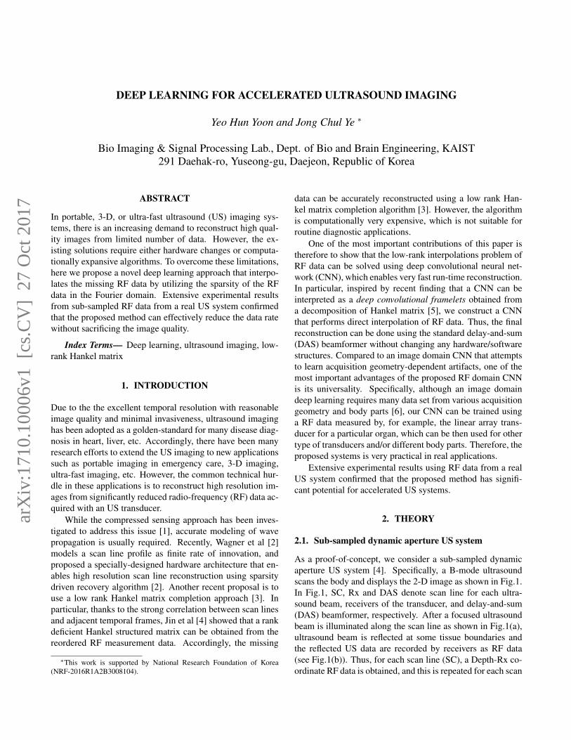
Fig. 4 illustrates B-mode ultrasound images acquired fromlinear array transducer. The proposed CNN-based interpola-
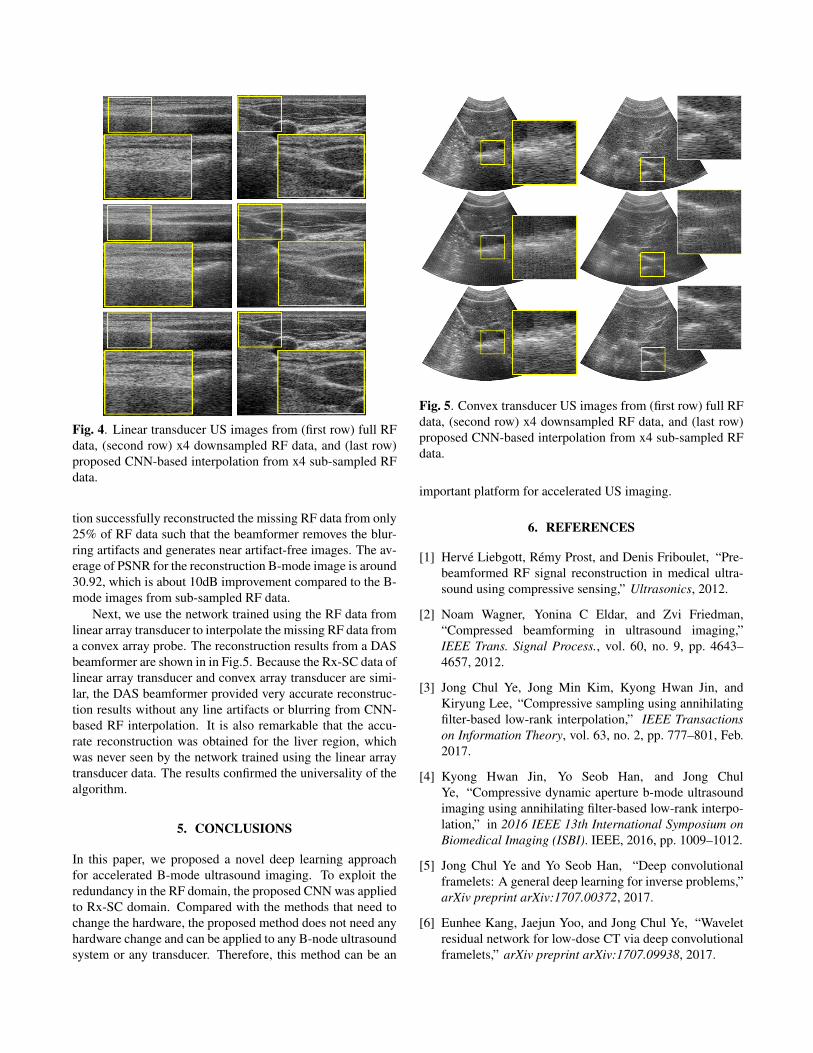
Fig. 4. Linear transducer US images from (first row) full RFdata, (second row) x4 downsampled RF data, and (last row)proposed CNN-based interpolation from x4 sub-sampled RFdata.
tion successfully reconstructed the missing RF data from only25% of RF data such that the beamformer removes the blur-ring artifacts and generates near artifact-free images. The av-erage of PSNR for the reconstruction B-mode image is around30.92, which is about 10dB improvement compared to the B-mode images from sub-sampled RF data.
Next, we use the network trained using the RF data fromlinear array transducer to interpolate the missing RF data froma convex array probe. The reconstruction results from a DASbeamformer are shown in in Fig.5. Because the Rx-SC data oflinear array transducer and convex array transducer are simi-lar, the DAS beamformer provided very accurate reconstruc-tion results without any line artifacts or blurring from CNN-based RF interpolation. It is also remarkable that the accu-rate reconstruction was obtained for the liver region, whichwas never seen by the network trained using the linear arraytransducer data. The results confirmed the universality of thealgorithm.
5. CONCLUSIONS
In this paper, we proposed a novel deep learning approachfor accelerated B-mode ultrasound imaging. To exploit theredundancy in the RF domain, the proposed CNN was appliedto Rx-SC domain. Compared with the methods that need tochange the hardware, the proposed method does not need anyhardware change and can be applied to any B-node ultrasoundsystem or any transducer. Therefore, this method can be an
Fig. 5. Convex transducer US images from (first row) full RFdata, (second row) x4 downsampled RF data, and (last row)proposed CNN-based interpolation from x4 sub-sampled RFdata.
important platform for accelerated US imaging.
6. REFERENCES
[1] Herve Liebgott, Remy Prost, and Denis Friboulet, “Pre-beamformed RF signal reconstruction in medical ultra-sound using compressive sensing,” Ultrasonics, 2012.
[2] Noam Wagner, Yonina C Eldar, and Zvi Friedman,“Compressed beamforming in ultrasound imaging,”IEEE Trans. Signal Process., vol. 60, no. 9, pp. 4643–4657, 2012.
[3] Jong Chul Ye, Jong Min Kim, Kyong Hwan Jin, andKiryung Lee, “Compressive sampling using annihilatingfilter-based low-rank interpolation,” IEEE Transactionson Information Theory, vol. 63, no. 2, pp. 777–801, Feb.2017.
[4] Kyong Hwan Jin, Yo Seob Han, and Jong ChulYe, “Compressive dynamic aperture b-mode ultrasoundimaging using annihilating filter-based low-rank interpo-lation,” in 2016 IEEE 13th International Symposium onBiomedical Imaging (ISBI). IEEE, 2016, pp. 1009–1012.
[5] Jong Chul Ye and Yo Seob Han, “Deep convolutionalframelets: A general deep learning for inverse problems,”arXiv preprint arXiv:1707.00372, 2017.
[6] Eunhee Kang, Jaejun Yoo, and Jong Chul Ye, “Waveletresidual network for low-dose CT via deep convolutionalframelets,” arXiv preprint arXiv:1707.09938, 2017.