death receptor pathway activation and increase of ros...
TRANSCRIPT
1
Death receptor pathway activation and increase of ROS production by the
triple epigenetic inhibitor, UVI5008
Angela Nebbioso1,6 , Raquel Pereira2,6, Harshal Khanwalkar3,6,8, Filomena Matarese4, José García-
Rodríguez2, Marco Miceli1, Colin Logie4, Valerie Kedinger3, Felicetto Ferrara7, Hendrik G.
Stunnenberg4,9 , Angel R. de Lera2,9 , Hinrich Gronemeyer3,9 , Lucia Altucci1,5,9
1 Dipartimento di Patologia generale, Seconda Università di Napoli, Vico L. de Crecchio 7,
80138, Napoli, IT; 2Departamento de Química Orgánica, Facultade de Química, Universidade de
Vigo, 36310 Vigo, ES; 3Department of Cancer Biology, Institut de Génétique et de Biologie
Moléculaire et Cellulaire, 67404 Illkirch Cedex, Strasbourg, FR; 4NCMLS, Department of
Molecular Biology, Radboud University, 6525 GA Nijmegen, NL; 5 CNR-IGB, Via P.Castellino
80100 Napoli, IT; 7 Ospedale Cardarelli, Napoli, IT
Running title: Mechanisms of UVI5008 epi-drug action
Keywords: epigenetic treatments, cancer, apoptosis, SIRT, HDAC
6 equal first authors
8 present address: Lupin Limited (Research Park), 46A/47A, Nande village, Pune- 411 042, State: Maharashtra,India
9 equal last authors; ,corresponding authors:
Prof. Hendrik G. Stunnenberg: NCMLS, Department of Molecular Biology, Radboud University, Nijmegen, NL; Ph
+31243610524; Fax +31243610520; [email protected]
Prof. Angel R. de Lera: Departamento de Química Orgánica, Universidade de Vigo, 36310 VIGO, ES; Ph
+34986812316; Fax+34986811940; [email protected]
Dr. Hinrich Gronemeyer: Department of Cancer Biology, IGBMC, F-67404 Illkirch, FR; Ph +33388653473; Fax
+33388653437; [email protected]
Prof. Lucia Altucci: Dipartimento di Patologia generale, Seconda Università degli Studi di Napoli, Vico L. De Crecchio
7, 80138 Napoli, IT; Ph +390815667569; Fax +390812144840; [email protected]
on October 15, 2018. © 2011 American Association for Cancer Research. mct.aacrjournals.org Downloaded from
Author manuscripts have been peer reviewed and accepted for publication but have not yet been edited. Author Manuscript Published OnlineFirst on October 6, 2011; DOI: 10.1158/1535-7163.MCT-11-0525

2
ABSTRACT
Deregulation of the epigenome is recognized as cause of cancer and epigenetic factors are
receiving major attention as therapeutic targets; yet the molecular mode of action of
existing epi-drugs is largely elusive. Here, we report on the decryption of the mechanism
of action of UVI5008, a novel epigenetic modifier, that inhibits histone deacetylases,
sirtuins and DNA methyltransferases. UVI5008 highly efficiently induces cancer cell-
selective death in a variety of models and exerts its activities in several human tumor
xenografts and genetic mouse models of human breast cancer in vivo. Its anticancer
activity involves independent activation of death receptors and ROS production.
Importantly, UVI5008 action is not critically dependent on p53, Bcl-2 modifying factor
(BMF), and/or TNF-related apoptosis-inducing ligand (TRAIL) as cell death is efficiently
induced in cells mutated or deficient for these factors limiting the risk of drug resistance
development and maximizing its application spectrum. The simultaneous modulation of
multiple (epigenetic) targets promises to open new avenues with unanticipated potential
against cancer.
on October 15, 2018. © 2011 American Association for Cancer Research. mct.aacrjournals.org Downloaded from
Author manuscripts have been peer reviewed and accepted for publication but have not yet been edited. Author Manuscript Published OnlineFirst on October 6, 2011; DOI: 10.1158/1535-7163.MCT-11-0525
3
INTRODUCTION
Cancer is a multistep process involving acquisition of unlimited replicative potential,
self-sustained growth with loss of sensitivity towards apoptogenic and check-point
controls (1). These aberrations are caused by a series of genetic events that involve proto-
oncogene activation, tumor suppressor gene (TSG) inactivation and senescence escape.
Studies on leukemogenesis have revealed the intimate relationship between initial
somatic mutations, often a chromosomal translocation, and epigenetic deregulation (2, 3).
In the case of acute promyelocytic leukemia (APL) the formation of a PML-RARα
oncofusion protein leads to multiple deregulations, that include HDAC recruitment (4) to
retinoic acid (RA) target genes resulting in obstruction of differentiation programs (2, 5).
While aberrant HDAC recruitment is a hallmark of APL, recent data suggest that HDAC
mis-targeting is necessary, but not sufficient for leukemogenesis (6), supporting the novel
concept that multiple targeting by combination therapy may achieve maximal anti-cancer
effects. This concept is also supported by the multiple genetic and epigenetic aberrations
present in cancer that might need a multi-target therapy approach. It has become clear
that cancer originates from, and is supported by epigenetic deregulation (7-9). Deposition
of epigenetic ‘marks’ on chromatin is accomplished by enzymes residing in multi-subunit
complexes (10). The control of the enzymatic machinery including histone acetylation
(HAT)/deacetylation (HDAC) and methylation/demethylation (11) or DNA methylation
(DNMT) (12) is central to transcription regulation. Chromatin modifying enzymes, in
particular HDACs and DNMTs, have emerged as new anticancer targets. The fact that
epigenetic modifications can be reversed makes epi-drugs promising for anti-tumor
therapy (13). Proof-of-principle comes from studies with HDAC inhibitors (HDACi) that
on October 15, 2018. © 2011 American Association for Cancer Research. mct.aacrjournals.org Downloaded from
Author manuscripts have been peer reviewed and accepted for publication but have not yet been edited. Author Manuscript Published OnlineFirst on October 6, 2011; DOI: 10.1158/1535-7163.MCT-11-0525
4
are in clinical practice (14).
HDACs are considered leading targets for therapy to reverse epi-aberrations (15). The
eighteen mammalian HDACs are divided in two families, the Zn+2-dependent HDACs
(Class I: HDACs 1-3, 8; Class II: HDACs 4-7, 9, 10; Class IV: HDAC11) and the NAD+-
dependent sirtuins (SIRTs1-7; Class III) (16). SIRT1 has only recently been implicated in
malignancies (17). DNMTs are responsible for CpG methylation within the genome.
Three active enzymes have been identified in mammals: DNMT1 responsible for
maintenance of pre-existing methylation and DNMT3a/b for de novo methylation (18).
Notwithstanding the clinical use of HDAC and other inhibitors, little is known about the
molecular mode of action. We -and others- have demonstrated that HDACis induce
TRAIL-apoptosis (TNF-related apoptosis inducing ligand/TNSF10/Apo2L) in acute
myeloid leukemia (AML) cells, in vivo AML models and ex vivo blasts (19, 20). Here we
report on UVI5008 molecular characterization and anti-cancer activities. UVI5008
inhibits three epi-enzymes and independently affects the death receptor (DR) pathway
and ROS production. UVI5008 targets p53, the Bcl-2 modifying factor BMF and/or
TRAIL in cancer cells harboring the wild-type factors as well as tumors, which are
deficient for one of them.
MATERIALS AND METHODS
Cell lines
Cell lines and primary cell cultures are deeply described as supplementary methods.
Briefly, U937, K562, K812-F, HL60 (leukemia), DU-145 (prostate), HCT116 (colon),
MDA-MB231 and MCF7 (breast) cancer cells were obtained from ATCC. HCT116 p53-/-
and HCT116-DK0 colon cancer cells were a gift from B. Vogelstein, John Hopkins
on October 15, 2018. © 2011 American Association for Cancer Research. mct.aacrjournals.org Downloaded from
Author manuscripts have been peer reviewed and accepted for publication but have not yet been edited. Author Manuscript Published OnlineFirst on October 6, 2011; DOI: 10.1158/1535-7163.MCT-11-0525
5
Kimmel Cancer Center, Baltimore, US. BJ, normal foreskin fibroblast and Ras
transformed BJELR cells were a gift from W. C. Hahn (Dana-Farber Cancer Institute,
Boston, US). For AML primary samples, bone marrow containing 80% to 90% leukemic
blasts was purified over Ficoll and processed as previously described (20). Primary
CD34+ normal progenitors (obtained from donors) have been cultured in ex vivo medium
as recommended by the supplier. This study was approved by the Ethical Committee of
the Second University of Naples. KD clones used in this study have been published
previously (20). or have been created by standard procedures using the Mission
technology (Sigma).
Inhibitors
SAHA (Merck), AZA (Sigma), EX527 (Alexis) and MS-275 (Bayer Schering Pharma)
were dissolved in DMSO and used at 5x10⁻⁶ M or as indicated. All other compounds
described were dissolved in DMSO and used at 1 or 5 μM.
Cell cycle, differentiation and cell death assays are described as supplementary methods
and carried out as in (20, 21).
Western blot analyses
Western blots were performed following supplier’s suggestions. For the determination of
p21WAF1/CIP1 and p16INK4 100 µg of extracts were separated on 15% polyacrylamide gels
and blotted. Western blots are shown for p21 (Transduction Laboratories), p16 and total
ERKs (Santa Cruz). For α-tubulin acetylation 25 µg of extracts were separated on 10%
polyacrylamide gels and blotted. Western blots are shown for acetylated α-tubulin, total
tubulin (Sigma) and total ERKs (Santa Cruz). For the detection of histone H3 acetylation
we used Upstate antibodies, whereas for histone H3K9 trimethylation, and H3K9, H3K14
and H3K18 acetylations we used Abcam antibodies.
on October 15, 2018. © 2011 American Association for Cancer Research. mct.aacrjournals.org Downloaded from
Author manuscripts have been peer reviewed and accepted for publication but have not yet been edited. Author Manuscript Published OnlineFirst on October 6, 2011; DOI: 10.1158/1535-7163.MCT-11-0525

6
Histone extraction protocol and MS analysis, P53K382 acetylation, p21 expression and
histone extraction in xenograft tumors, Xenograft and MMTV-MYC mouse models,
Isolation and digestion of genomic DNA are described as supplementary methods.
In vitro methylation of genomic DNA
Digested DNA was methylated with 8U of Sss1 methylase (New England Biolabs) in the
presence of 3.2 mM SAM for 2 h at 37 °C. After phenol-chloroform extraction, DNA
was precipitated and resuspended with 20 µL water.
Bisulfite treatment and PCR for RARβ and p16 methylation
Bisulfite treatment and PCR for RARβ and p16 methylation are described in the
Supplementary Methods
Fluorimetric human cell based HDAC1 and 4 assays and in vitro HDAC assays
HDAC assays were performed as previously described (21, 22)
In vitro assays of Sirt1 and Sirt2, DNMTs assay, ChIP assay, Immunofluorescence
assays are described in the Supplementary Methods. ChIP assays have been performed as
in (23).
RNA Preparation for RNA-Seq, Illumina High-Throughput Sequencing and
MethylCap-seq are described in the Supplementary methods and in (2, 24, 25).
RESULTS
UVI5008 efficiently Induces Apoptosis
We tested the activity of UVI5008 (Fig. 1A) activity on a series of cancer cells .
on October 15, 2018. © 2011 American Association for Cancer Research. mct.aacrjournals.org Downloaded from
Author manuscripts have been peer reviewed and accepted for publication but have not yet been edited. Author Manuscript Published OnlineFirst on October 6, 2011; DOI: 10.1158/1535-7163.MCT-11-0525
7
UVI5008 induced cell death (Fig. 1B) and apoptosis by activation of both initiator,
executor caspases (Fig. 1C) and loss of mitochondrial membrane potential in cancer cells
derived from of leukemias (U937, Ku-812F and K562) or solid tumors, such as breast
(MCF7), osteosarcoma (U2OS), prostate (DU145), colon carcinoma (HCT116) and
melanoma (A375) (Supplementary Fig. S1A and data not shown). Several cell lines
derived from colon, breast and prostate carcinomas, exhibited anti-proliferative responses
with IC50s ranging from about 200 nM to 3.1 µM (Supplementary Methods and data not
shown).
UVI5008 Anti-Cancer Action in vivo and ex vivo
To test UVI5008 anti-tumor activity in vivo, xenograft assays were performed with
luciferase-expressing human cancer cells. At 40 mg/kg UVI5008 (MW 710.46 g/mol)
efficiently blocked HCT116 colon carcinoma growth as visualized by in vivo imaging
(Fig. 2A) and quantified by direct photon counting (Fig. 2B). No adverse side effects
were noticed, while the same dose of SAHA (Vorinostat) (MW 264.33 g/mol) was lethal
(data not shown). Similarly, UVI5008 strongly inhibited MCF7 breast cancer growth in
vivo in presence of estradiol; tamoxifen co-treatment attenuated the growth without
affecting the median overall tumor size (Fig. 2C left panel). Importantly, UVI5008
strongly reduced tumor growth in a genetic mouse model (MMTV-myc) of human breast
cancer, when treatment was initiated as soon as the tumors became palpable (Fig. 2C,
right panel). The weight profiles of the treated vs. control groups supported the absence
of toxicity. Similar tumor growth inhibition by 40 mg/kg UVI5008 was observed in the
MMTV-cerbB2 breast cancer model (data not shown). Thus, UVI5008 displays a strong
anti-cancer effect not only in xenografts, but also in two genetic mouse models that
on October 15, 2018. © 2011 American Association for Cancer Research. mct.aacrjournals.org Downloaded from
Author manuscripts have been peer reviewed and accepted for publication but have not yet been edited. Author Manuscript Published OnlineFirst on October 6, 2011; DOI: 10.1158/1535-7163.MCT-11-0525
8
mimic human breast cancer. Pharmacokinetics analyses revealed half-lives of up to ~7 h
(p.o. application) for the in vivo formed glutathione conjugate of UVI5008 (see
Supplementary Methods).
UVI5008 mediated Tumor-Selective Apoptosis
To assess the cancer-selective potential of UVI5008 we first used the isogenic BJ
stepwise cellular tumorigenesis model (26). While primary fibroblasts (BJ) were largely
insensitive to UVI5008, 80% of the transformed BJELR cells were killed (Fig. 2D, left
panel).
The efficient induction of tumor selective cell death by UVI5008 prompted us to test its
efficacy in ex vivo assays in primary AML blasts. Within 24h, UVI5008 induced caspase
3 activation and apoptosis in 14/14 independent ex vivo blasts (Fig. 2E and
Supplementary Fig. S1B). No significant toxicity was seen in normal CD34+
progenitors, whereas all AML blasts were sensitive (Fig. 2D, right panel).
UVI5008 is a triple HDAC, DNMT and SIRT inhibitor
To decipher the basis of UVI5008 anticancer activity we assessed inhibition of DNA
methylation. Using methylation-specific PCR (MSP), we corroborated that UVI5008, like
its parental compound psammaplin A (27), induced significant demethylation of the
RARβ and p16INK4a promoters (Supplementary Fig. S2A). In keeping with the p16
promoter reactivation, UVI5008 induced an increase of p16 protein levels in U937 cells
that was higher than the one seen upon combined treatment with 5-aza-cytidine (5-aza-C)
and SAHA (Supplementary Fig. S2B, top panel). In vitro assays demonstrated that
on October 15, 2018. © 2011 American Association for Cancer Research. mct.aacrjournals.org Downloaded from
Author manuscripts have been peer reviewed and accepted for publication but have not yet been edited. Author Manuscript Published OnlineFirst on October 6, 2011; DOI: 10.1158/1535-7163.MCT-11-0525

9
UVI5008 acted as a direct inhibitor of DNMT3a (Supplementary Fig. S2B) and only
marginally inhibited DNMT1 (Supplementary Fig. S2B, lower panel), when compared
to the DNMT1 inhibitors RG108 (28) or SGI-1027 (29). To the best of our knowledge
UVI5008 is the first DNMT3a-selective inhibitor.
Next, we assessed whether UVI5008, like psammaplin A (30, 31), acts as HDAC
inhibitor. UVI5008 blocked the activity of HDAC1-4 both in vitro and in cell-based
assays (Fig. 3A and Supplementary Fig. S3). Moreover, UVI5008 treatment of U937
cells induced the well-known HDACi target p21, inhibited deacetylation of the
HDAC6/SIRT2 substrate α-tubulin and affected histone acetylation (H3K9, H3K14 and
H3K18) (Fig. 3B). UVI5008 also induced global histone H3 hyperacetylation in ex vivo
blasts as exemplified for AML #116 (Fig. 3C and data not shown), thus acting both in
vitro and ex vivo. Immunoblotting of tumors derived from both HCT116-xenografted
mice and ‘MMTV-myc’ model treated with UVI5008 revealed global (pan-H3) and
specific (H3K9, H3K14) hyperacetylation, thereby confirming UVI5008 HDACi action
in tumori. Immunohistochemical analysis showed a stronger induction of H3K9
acetylation in HCT116 xenografts of mice treated with UVI5008 than in animals treated
with SAHA (Fig. 3D bottom panel).
To investigate whether UVI5008 induced other histone modifications, we defined the
spectrum of modifications in bulk histones from U937 cells treated with UVI5008 (4h)
using mass spectrometric analyses. In addition to the increase of histone H3 acetylation
expected for an HDACi, we identified a histone H3-derived peptide that is acetylated at
K56 (Supplementary Fig. S2C and Supplementary Table S1). In yeast, deacetylation
of H3K56ac is mediated by the sirtuin homologues Hst3 and Hst4 (32), and in humans
on October 15, 2018. © 2011 American Association for Cancer Research. mct.aacrjournals.org Downloaded from
Author manuscripts have been peer reviewed and accepted for publication but have not yet been edited. Author Manuscript Published OnlineFirst on October 6, 2011; DOI: 10.1158/1535-7163.MCT-11-0525
10
H3K56ac acts as SIRT1 substrate (33). Hence, we investigated whether UVI5008
possesses sirtuin inhibitory (SIRTi) activity. In vitro assays with recombinant human
sirtuin-1 and -2 revealed that UVI5008 is a powerful inhibitor of SIRT1 and SIRT2 (Fig.
4A). Analyses with anti-H3K56ac antibodies (34) revealed H3K56 acetylation in
UVI5008-treated U937 cells, U2OS osteosarcoma cells (Fig. 4A-B) and other cell lines
(Fig. 4B and data not shown). The observation that tumors from HCT116-xenografted
mice and from the ‘MMTV-myc’ breast cancer genetic mouse model revealed increased
H3K56ac when treated with UVI5008 but not SAHA, established the in vivo SIRT
inhibitory activity of UVI5008 (Fig. 4B-C).
Considering that acetylation activates p53 (35) and that SIRT1 deacetylates p53K382
(36), we hypothesized that UVI5008 may sustain p53 activity. Indeed, treatment of
MCF7 and HCT116 cells with etoposide and UVI5008 increased p53K382 acetylation
similar to the SIRT1i EX527 (37), thus extending our observations that UVI5008 inhibits
sirtuins (Fig. 4C). Immunohistochemical analysis in tumors from HCT116-xenografted
mice treated with UVI5008 confirmed efficient SIRT inhibition in vivo (37) (Fig. 4D).
Our in vivo studies confirm that UVI5008, which displays a favorable PK (see
Supplementary methods for PK and PD study), reaches tumors in vivo to exert HDACi
and SIRTi activity and to display anticancer action at doses that may be tolerable and
efficacious in the context of a clinical trial. These results demonstrate that UVI5008 is a
potent HDAC and sirtuin inhibitor in vitro and in vivo.
UVI5008 re-Activates Tumor Suppressor Gene Programs
A measure of importance for the activity of epigenetic inhibitors and their application as
anti-cancer drugs is the ability to induce growth arrest by altering the expression of tumor
on October 15, 2018. © 2011 American Association for Cancer Research. mct.aacrjournals.org Downloaded from
Author manuscripts have been peer reviewed and accepted for publication but have not yet been edited. Author Manuscript Published OnlineFirst on October 6, 2011; DOI: 10.1158/1535-7163.MCT-11-0525
11
suppressor genes (TSG) such as the cyclin-dependent kinase inhibitor p21Waf1/Cip1
CDKN1A (38). UVI5008 efficiently induced transcription of p21Waf1/Cip1 in HCT116 cells
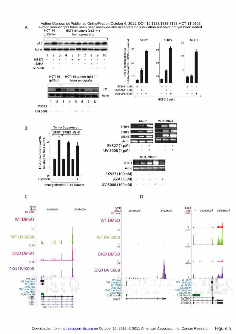
and in the corresponding xenografts in vivo (Fig. 5A). UVI5008 also induced p21Waf1/Cip1
expression in p53-/- HCT116 cells and xenografts, indicating that p21 targeting is p53-
independent (Fig. 5A). Expression of SFRP1, SFRP2 and MLH1 TSGs was strongly
induced in UVI5008-treated HCT116 cells (Fig. 5A, right panel), HCT116-xenografts,
MDA-MB231 and MCF7 cells, supporting the SIRTi activity of UVI5008 (Fig. 5B).
Note that UVI5008 mediated higher activation of SFRP1 expression when compared with
exposure to SIRTi or DNMTi alone (Fig. 5B).
We also tested the ability of UVI5008 to activate transcription from genes with DNA
methylated CpG-island in HCT116 cells and a double knockout of DNMT1 and
DNMT3b (“HCT116-DKO” cells) by RNA-seq; in parallel, the DNA methylation status
was determined by MethylCap (25). UVI5008 was able to partially reactivate loci such as
UCHL1 gene whose CpG-island is DNA methylated in line with its albeit weak DNMTi
action (Fig. 5C). Upon complete loss of DNA methylation at this CpG-island as in
HCT116-DKO, the gene is strongly reactivated. UVI5008 treatment of HCT116-DKO
did not further enhance transcription as compared to the parental HCT116 suggesting that
its DNMT but not HDAC and/or sirtuin inhibition caused the low but significant
activation of the UCHL1 locus in WT cells. TIMP3, a TSG known both to be repressed
by DNA methylation (39, 40) and to be SIRT1 target (41) was reactivated by UVI5008
only in HCT116-DKO cells and not in the untreated or wt cells (Fig. 5C). Possibly due to
its low DNMT-inhibitory activity UVI5008 could not overcome the repressive action of
DNA methylation. Induction of p21Waf1/Cip1 (CDKN1A) expression by UVI5008 in wt
on October 15, 2018. © 2011 American Association for Cancer Research. mct.aacrjournals.org Downloaded from
Author manuscripts have been peer reviewed and accepted for publication but have not yet been edited. Author Manuscript Published OnlineFirst on October 6, 2011; DOI: 10.1158/1535-7163.MCT-11-0525
12
and DKO cells (Fig. 5D) is most likely due to the non-DNMTi activities of UVI5008, as
the promoter-proximal CpG-island is not DNA methylated in wt or DKO HCT116 cells.
Mechanisms of Pro-Apoptotic UVI5008 action
The induction of apoptosis in cells, ex vivo and in vivo by UVI5008 and its bona fide
tumor-specific action prompted us to decipher the underlying molecular pathway(s). RT-
PCR and ELISA assays showed a strong increase of TNFSF10 mRNA and protein levels
upon UVI5008 exposure (Supplementary Fig. S4A) extending our previous findings
that the TRAIL is a target of HDACi (19, 20). In line with the transcription activation of
the TNFSF10 promoter by UVI5008, chromatin immunoprecipitation (ChIP) assays (23)
revealed a rapid increase in histone H3 acetylation at the TNFSF10 promoter as
exemplified by H3K9ac spreading to the TNFSF10 intron within 2h of treatment
(Supplementary Fig. S4B). Similar activation was seen at the promoter of the death
receptor-5 (DR5/TRAIL-R2; data not shown). These results show that UVI5008 activates
the tumor-selective TRAIL pathway, which accounts for, or contributes to its tumor-
selectivity.
To assess the contribution of the TRAIL pathway to tumor-selective activity of UVI5008,
we expressed a dominant-negative FADD (dnFADD) and the non-functional FADD
mutant (dnFADDmut); the dnFADD reduced UVI5008-induced death only to ~50%,
while completely blocking TNFSF10-induced apoptosis (Supplementary Fig. S4C).
Similarly, only a moderate reduction of apoptosis was detectable in UVI5008-treated
U937 cells expressing stably integrated shTNFSF10 (KD) (20), whereas a complete
on October 15, 2018. © 2011 American Association for Cancer Research. mct.aacrjournals.org Downloaded from
Author manuscripts have been peer reviewed and accepted for publication but have not yet been edited. Author Manuscript Published OnlineFirst on October 6, 2011; DOI: 10.1158/1535-7163.MCT-11-0525

13
reduction was obtained upon MS275, a bona fide ‘pure’ HDACi (Supplementary Fig.
S4C). Moreover, neither the simultaneous knockdown of TNFSF10 and p21 nor the
blocking of DR-mediated death pathways by neutralizing antibodies affected UVI5008
death (Supplementary Fig. S5A and B). These observations strongly suggest that while
TRAIL induction is a key factor in HDACi apoptosis, this is not the only mechanism by
which UVI5008 induces apoptotic pathways distinct from or additional to TRAIL.
Concomitant Death receptors and ROS activation cause UVI5008-mediated
apoptosis
To gain further insight into the UVI5008 death activities, we assessed the implication of
p53 and Bcl-2 modifying factor (BMF). The p53 gene is a SIRT target, whereas BMF
silencing was reported to rescue cells from HDACi-induced apoptosis (42) in some
cancers. Despite efficient knockdown of BMF and p53 no significant rescue from
UVI5008-induced apoptosis was detected (Fig. 6A) suggesting that neither p53 nor BMF
are singly essential. In agreement with these results, strong anti-tumor action of UVI5008
was observed in HCT116 p53-/- xenografts in vivo (Fig. 6A, right panel), re-emphasizing
its therapeutic potential for the therapy of p53 mutant or deficient cancers. On the other
hand, caspase 8 and 9 inhibition, or caspase 8 silencing resulted in a major inhibition
(Fig. 6B), implicating both the extrinsic and intrinsic pathways in UVI5008-induced
death. Interestingly, while necroptosis inhibition by Nec-1 did not affect UVI5008-
mediated death, N-acetyl-cysteine (NAC), a scavenger of reactive oxygen species (ROS),
significantly decreased it (Fig. 6C; compare lanes 3 and 7 with lanes 2 or 6). Most
on October 15, 2018. © 2011 American Association for Cancer Research. mct.aacrjournals.org Downloaded from
Author manuscripts have been peer reviewed and accepted for publication but have not yet been edited. Author Manuscript Published OnlineFirst on October 6, 2011; DOI: 10.1158/1535-7163.MCT-11-0525
14
importantly, NAC fully blocked UVI5008-induced apoptosis in cells stably expressing
shTNFSF10 (compare lanes 14 and 15). Strikingly, experiments carried out ex vivo in
primary AML blasts, corroborated the dual involvement of the TRAIL-induced death
signaling and ROS production as the main apoptotic mechanism of action of UVI5008
(Fig. 6D, lane 6). Thus, the anticancer activity of UVI5008 originates from the
cumulative activation of multiple pathways due to the concomitant activation of death
receptors and ROS production. More specifically, the triple action inhibitor UVI5008
displays i) strong anticancer action in vitro, ex vivo and in vivo, ii) reactivation of TSGs
in vitro and in vivo and iii) antitumor efficacy independent of p53 and BMF in vitro and
in vivo.
DISCUSSION
The fact that epigenetic modifications can be reversed by epi-drugs makes epi-
compounds highly promising for anti-tumor therapy. However, the results have been
lagging behind expectation. Investigation of the spectrum of epi-mutations in cancer has
only recently started and FDA (Food and Drug Administration)-approved epi-drugs are
currently limited by a number of factors. Due to their moderate toxicities, HDACis and
DNMTis entered therapy without a detailed molecular characterization. Beside this, no
real markers for their apoptotic action or patient’s response have been reported so far,
thus currently hampering clinical stratification. This lack of knowledge about pathways
and targets might have led to unfocused therapeutic applications of epi-drugs.
We have identified and characterized the mechanism resulting in the apoptogenic action
of UVI5008 in detail. We have previously reported that induction of TRAIL is part of the
on October 15, 2018. © 2011 American Association for Cancer Research. mct.aacrjournals.org Downloaded from
Author manuscripts have been peer reviewed and accepted for publication but have not yet been edited. Author Manuscript Published OnlineFirst on October 6, 2011; DOI: 10.1158/1535-7163.MCT-11-0525
15
action spectrum of HDACis (19, 20). Hence, the contribution of TRAIL tumor-selective
death by UVI5008 is likely to be linked to its HDACi action. Our observation that both
TNFSF10-silenced and TNFSF10-deficient cells (cell lines and blasts) undergo apoptosis
upon UVI5008 treatment strongly supports the contribution of SIRT and DNMT
inhibition to UVI5008 anti-cancer action. Our data suggests that UVI5008 may be
effective even in HDACi-resistant and hyper-mutated tumors. The same applies to tumors
in which p53 is silenced or mutated (Figures 5 and 6). Importantly, p53, BMF and
TRAIL are often altered in cancers, thus suggesting the beneficial application of
UVI5008 in multiple settings. Considering that SIRT1 mediates resistance to intrinsic
apoptosis in cancer (43), SIRT1i may enforce the higher UVI5008 apoptotic activity
possibly enhanced by its other inhibitory activities. We show that UVI5008 anticancer
activity is causally linked to DR pathway activation and ROS production (Fig. 6).
Although the involvement of oxidative stress in TRAIL-mediated apoptosis has been
reported (44), our data shows that only the concomitant impairment of both pathways
fully blocks UVI5008 action. Thus the targeting of these two molecular pathways in both
in vitro and ex vivo settings by UVI5008 is independent (Fig. 6). Interestingly, SIRT1
inhibits apoptosis induced by oxidative stress. SIRT1 deacetylates and activates FOXO1
(45), FOXO3a (46), and FOXO4 (47), promoting expression of the DNA repair factor
GADD45, and of the mitochondrial antioxidant enzyme manganese superoxide dismutase
(MnSOD). These proteins may contribute to the induction of ROS tolerance by SIRT1 in
tumors. Perturbation of the mechanisms that tightly couple ROS production, oxidative
stress signaling, and FOXO activity to the subsequent cellular response is a pivotal step
in cancer. Consequently, the ROS-FOXO pathway is a promising therapeutic target in
on October 15, 2018. © 2011 American Association for Cancer Research. mct.aacrjournals.org Downloaded from
Author manuscripts have been peer reviewed and accepted for publication but have not yet been edited. Author Manuscript Published OnlineFirst on October 6, 2011; DOI: 10.1158/1535-7163.MCT-11-0525
16
cancer, not only as it mediates chemotherapy response, but also because it underpins drug
resistance. As the intimate, reciprocal relation between FOXO and ROS is being
unraveled (48), new applications arise to apply SIRTis to circumvent resistance to
conventional drugs. The tight link between SIRTis and ROS production (48) may explain
UVI5008 ROS-dependent action and its higher anticancer action when compared to
single HDACi and SIRTi.
Analysing DNMT3a inhibition, UVI5008 effect on DNA methylation on the p16
promoter is comparable to that of 5-azacytidine, whereas the induction of p16 protein
expression by UVI5008 is higher (Supplementary Fig. S3), which likely results from the
simultaneous inhibition of DNMT3a and SIRT1/2. Indeed, the indication that upon
induction of DNA double strand breaks, SIRT1 recruitment to the E-cadherin promoter is
a pre-requisite for DNMT3 recruitment and subsequent heritable CpG methylation at the
promoter strongly favors the use of an anti-cancer drug that targets both SIRT and
DNMT. The reported de-regulation/over-expression of HDACs, DNMTs and SIRTs in
cancer (17, 33), also rationalizes UVI5008 use.
As suggested for HDACi and DNMTi combinations (49), also SIRT1i reactivates TSGs
without altering DNA hypermethylation (8). The fact that TSGs, such as TIMP3 (Fig. 5),
become re-expressed upon UVI5008 exposure is relevant. The mechanism of TIMP3
silencing in colon cancer and its role in tumorigenesis remain elusive. That DNA
demethylation fails to reactivate the gene indicates that multiple activities potentiated by
UVI5008 are needed for its re-expression. A further illustration that UVI5008 fulfills the
criteria of a 'multiple targeting' drug is the activation of SFRP1 (Fig. 5) which is far
higher with UVI5008 than with canonical combo-treatments, supporting the winning
on October 15, 2018. © 2011 American Association for Cancer Research. mct.aacrjournals.org Downloaded from
Author manuscripts have been peer reviewed and accepted for publication but have not yet been edited. Author Manuscript Published OnlineFirst on October 6, 2011; DOI: 10.1158/1535-7163.MCT-11-0525

17
strategy of applying a single drug exerting multiple-interventions against cancer.
The use of a single drug with multiple epi-activities, such as UVI5008, has great promise
for therapy, as the synergies between its activities may reduce both the effective dose and
development of resistance (50). The pharmaceutical development of a single compound is
less complex than that of combinatorial therapies, complicated by divergent
pharmacokinetics, pharmacodynamics and complex ADMET profiles. UVI5008 belongs
to this novel class of compounds, as it inhibits three distinct classes of epigenetic
enzymes, namely DNMT3a, HDACs, and sirtuins. The rationale at the basis of targeting
drugs could radically shift with the introduction of molecules interfering simultaneously
with multiple targets as this might be more effective than single target agents thus
representing a new generation of anti-cancer drugs. In this view the inhibition of at least
three epi-enzymes offers a promising alternative to combo-treatments of cancer using
‘poly-pharmacology’ approaches. Given the ability of cancer to bypass signaling routes
via complex networks, the simultaneous modulation of multiple epi-targets promises to
open new avenues against cancer.
DISCLOSURE OF POTENTIAL CONFLICT OF INTEREST
HGS, ARdL, HG and LA are inventors for the patent n° WO2008125988 entitled: ‘Novel
Derivatives of Psammaplin A, a method for their synthesis and their use for the
prevention or treatment of cancer’.
ACKNOWLEDGEMENTS
on October 15, 2018. © 2011 American Association for Cancer Research. mct.aacrjournals.org Downloaded from
Author manuscripts have been peer reviewed and accepted for publication but have not yet been edited. Author Manuscript Published OnlineFirst on October 6, 2011; DOI: 10.1158/1535-7163.MCT-11-0525
18
We are grateful to W. Hahn and R. Weinberg for stepwise tumorigenesis model. We
thank P. Jansen, C. Erb, and G. Lemaire for technical help; E. Carrillo de Santa Pau and
H. Marks, for bioinformatics analysis; V. Carafa and F. Rodríguez-Barrios for technical
help and discussions.
GRANT SUPPORT
This work was supported by: Associazione Italiana ricerca contro il cancro (AIRC to
LA), MIUR to LA, Association for International Cancer Research (AICR 00-108 to HG),
Ligue National Contre le Cancer (HG), ANR-07-EMPB-012-01 ‘EPI_DRUG’, MEC
(SAF2010-17935 FEDER to AdL), Programmi di Ricerca Scientifica di Rilevante
Interesse Nazionale (PRIN2009PX2T2E to LA), EU LSHC-CT-2005-518417
(EPITRON), HEALTH-F4-2007-200767 (APO-SYS), HEALTH-F2-2007-200620
(CANCERDIP), HEALTH-F4-2009-221952 (ATLAS).
REFERENCES
1. Hanahan, D. and Weinberg, R. A. Hallmarks of cancer: the next generation. Cell, 144: 646-674, 2011.
2. Martens, J. H., Brinkman, A. B., Simmer, F., Francoijs, K. J., Nebbioso, A., Ferrara, F. et al. PML-RARalpha/RXR alters the epigenetic landscape in acute promyelocytic leukemia. Cancer Cell, 17: 173-185, 2010.
3. Martens, J. H. and Stunnenberg, H. G. The molecular signature of oncofusion proteins in acute myeloid leukemia. FEBS Lett, 2010.
4. Grignani, F., De Matteis, S., Nervi, C., Tomassoni, L., Gelmetti, V., Cioce, M. et al. Fusion proteins of the retinoic acid receptor-alpha recruit histone deacetylase in promyelocytic leukaemia. Nature, 391: 815-818, 1998.
5. Altucci, L., Leibowitz, M. D., Ogilvie, K. M., de Lera, A. R., and Gronemeyer, H. RAR and RXR modulation in cancer and metabolic disease. Nat Rev Drug Discov, 6: 793-810, 2007.
6. Matsushita, H., Scaglioni, P. P., Bhaumik, M., Rego, E. M., Cai, L. F., Majid,
on October 15, 2018. © 2011 American Association for Cancer Research. mct.aacrjournals.org Downloaded from
Author manuscripts have been peer reviewed and accepted for publication but have not yet been edited. Author Manuscript Published OnlineFirst on October 6, 2011; DOI: 10.1158/1535-7163.MCT-11-0525
19
S. M. et al. In vivo analysis of the role of aberrant histone deacetylase recruitment and RAR alpha blockade in the pathogenesis of acute promyelocytic leukemia. J Exp Med, 203: 821-828, 2006.
7. Baylin, S. B. and Ohm, J. E. Epigenetic gene silencing in cancer - a mechanism for early oncogenic pathway addiction? Nat Rev Cancer, 6: 107-116, 2006.
8. Pruitt, K., Zinn, R. L., Ohm, J. E., McGarvey, K. M., Kang, S. H., Watkins, D. N. et al. Inhibition of SIRT1 reactivates silenced cancer genes without loss of promoter DNA hypermethylation. PLoS Genet, 2: e40, 2006.
9. Rodriguez-Paredes, M. and Esteller, M. Cancer epigenetics reaches mainstream oncology. Nat Med, 17: 330-339, 2011.
10. Bantscheff, M., Hopf, C., Savitski, M. M., Dittmann, A., Grandi, P., Michon, A. M. et al. Chemoproteomics profiling of HDAC inhibitors reveals selective targeting of HDAC complexes. Nat Biotechnol, 29: 255-265, 2011.
11. Chi, P., Allis, C. D., and Wang, G. G. Covalent histone modifications--miswritten, misinterpreted and mis-erased in human cancers. Nat Rev Cancer, 10: 457-469, 2010.
12. Szyf, M. Epigenetics, DNA methylation and chromatin modifying drugs. Ann. Rev. Pharmacol. Toxicol., 49: 243-263, 2009.
13. Mai, A. and Altucci, L. Epi-drugs to fight cancer: from chemistry to cancer treatment, the road ahead. Int J Biochem Cell Biol, 41: 199-213, 2009.
14. Lane, A. A. and Chabner, B. A. Histone deacetylase inhibitors in cancer therapy. J Clin Oncol, 27: 5459-5468, 2009.
15. Bolden, J. E., Peart, M. J., and Johnstone, R. W. Anticancer activities of histone deacetylase inhibitors. Nat Rev Drug Discov, 5: 769-784, 2006.
16. Finkel, T., Deng, C. X., and Mostoslavsky, R. Recent progress in the biology and physiology of sirtuins. Nature, 460: 587-591, 2009.
17. Huffman, D. M., Grizzle, W. E., Bamman, M. M., Kim, J. S., Eltoum, I. A., Elgavish, A. et al. SIRT1 is significantly elevated in mouse and human prostate cancer. Cancer Res, 67: 6612-6618, 2007.
18. Ooi, S. K. and Bestor, T. H. The colorful history of active DNA demethylation. Cell, 133: 1145-1148, 2008.
19. Insinga, A., Monestiroli, S., Ronzoni, S., Gelmetti, V., Marchesi, F., Viale, A. et al. Inhibitors of histone deacetylases induce tumor-selective apoptosis through activation of the death receptor pathway. Nat Med, 11: 71-76, 2005.
20. Nebbioso, A., Clarke, N., Voltz, E., Germain, E., Ambrosino, C., Bontempo, P. et al. Tumor-selective action of HDAC inhibitors involves TRAIL induction in acute myeloid leukemia cells. Nat Med, 11: 77-84, 2005.
21. Nebbioso, A., Manzo, F., Miceli, M., Conte, M., Manente, L., Baldi, A.et al. Selective class II HDAC inhibitors impair myogenesis by modulating the
on October 15, 2018. © 2011 American Association for Cancer Research. mct.aacrjournals.org Downloaded from
Author manuscripts have been peer reviewed and accepted for publication but have not yet been edited. Author Manuscript Published OnlineFirst on October 6, 2011; DOI: 10.1158/1535-7163.MCT-11-0525

20
stability and activity of HDAC-MEF2 complexes. EMBO Rep, 10: 776-782, 2009.
22. Lahm, A., Paolini, C., Pallaoro, M., Nardi, M. C., Jones, P., Neddermann, P.et al. Unraveling the hidden catalytic activity of vertebrate class IIa histone deacetylases. Proc Natl Acad Sci U S A, 104: 17335-17340, 2007.
23. Denissov, S., van Driel, M., Voit, R., Hekkelman, M., Hulsen, T., Hernandez, N. et al. Identification of novel functional TBP-binding sites and general factor repertoires. Embo J, 26: 944-954, 2007.
24. Akkers, R. C., van Heeringen, S. J., Jacobi, U. G., Janssen-Megens, E. M., Francoijs, K. J., Stunnenberg, H. G. et al. A hierarchy of H3K4me3 and H3K27me3 acquisition in spatial gene regulation in Xenopus embryos. Dev Cell, 17: 425-434, 2009.
25. Brinkman, A. B., Simmer, F., Ma, K., Kaan, A., Zhu, J., and Stunnenberg, H. G. Whole-genome DNA methylation profiling using MethylCap-seq. Methods, 2010.
26. Hanahan, D. and Weinberg, R. A. The hallmarks of cancer. Cell, 100: 57-70, 2000.
27. Piña, I. C., Gautschi, J. T., Wang, G. Y. S., Sanders, M. L., Schmitz, F. J., France, D. et al. Psammaplins from the sponge Pseudoceratina purpurea: Inhibition of both histone deacetylase and DNA methyltransferase. J. Org. Chem., 68: 3866-3873, 2003.
28. Brueckner, B., Boy, R. G., Siedlecki, P., Musch, T., Kliem, H. C., Zielenkiewicz, P. et al. Epigenetic reactivation of tumor suppressor genes by a novel small-molecule inhibitor of human DNA methyltransferases. Cancer Res, 65: 6305-6311, 2005.
29. Datta, J., Ghoshal, K., Denny, W. A., Gamage, S. A., Brooke, D. G., Phiasivongsa, P. et al. T. A new class of quinoline-based DNA hypomethylating agents reactivates tumor suppressor genes by blocking DNA methyltransferase 1 activity and inducing its degradation. Cancer Res, 69: 4277-4285, 2009.
30. Ahn, M. Y., Jung, J. H., Na, Y. J., and Kim, H. S. A natural histone deacetylase inhibitor, Psammaplin A, induces cell cycle arrest and apoptosis in human endometrial cancer cells. Gynecol Oncol, 108: 27-33, 2008.
31. Garcia, J., Franci, G., Pereira, R., Benedetti, R., Nebbioso, A., Rodriguez-Barrios, F. et al. R. Epigenetic profiling of the antitumor natural product psammaplin A and its analogues. Bioorg Med Chem, 2010.
32. Maas, N. L., Miller, K. M., DeFazio, L. G., and Toczyski, D. P. Cell cycle and checkpoint regulation of histone H3 K56 acetylation by Hst3 and Hst4. Mol Cell, 23: 109-119, 2006.
33. Das, C., Lucia, M. S., Hansen, K. C., and Tyler, J. K. CBP/p300-mediated acetylation of histone H3 on lysine 56. Nature, 459: 113-117, 2009.
on October 15, 2018. © 2011 American Association for Cancer Research. mct.aacrjournals.org Downloaded from
Author manuscripts have been peer reviewed and accepted for publication but have not yet been edited. Author Manuscript Published OnlineFirst on October 6, 2011; DOI: 10.1158/1535-7163.MCT-11-0525
21
34. Ozdemir, A., Spicuglia, S., Lasonder, E., Vermeulen, M., Campsteijn, C., Stunnenberg, H. G. et al. Characterization of lysine 56 of histone H3 as an acetylation site in Saccharomyces cerevisiae. J Biol Chem, 280: 25949-25952, 2005.
35. Tang, Y., Zhao, W., Chen, Y., Zhao, Y., and Gu, W. Acetylation is indispensable for p53 activation. Cell, 133: 612-626, 2008.
36. Avalos, J. L., Celic, I., Muhammad, S., Cosgrove, M. S., Boeke, J. D., and Wolberger, C. Structure of a Sir2 enzyme bound to an acetylated p53 peptide. Mol Cell, 10: 523-535, 2002.
37. Solomon, J. M., Pasupuleti, R., Xu, L., McDonagh, T., Curtis, R., DiStefano, P. S. et al. J. Inhibition of SIRT1 catalytic activity increases p53 acetylation but does not alter cell survival following DNA damage. Mol Cell Biol, 26: 28-38, 2006.
38. Gui, C. Y., Ngo, L., Xu, W. S., Richon, V. M., and Marks, P. A. Histone deacetylase (HDAC) inhibitor activation of p21WAF1 involves changes in promoter-associated proteins, including HDAC1. Proc Natl Acad Sci U S A, 101: 1241-1246, 2004.
39. Bai, Y. X., Yi, J. L., Li, J. F., and Sui, H. Clinicopathologic significance of BAG1 and TIMP3 expression in colon carcinoma. World J Gastroenterol, 13: 3883-3885, 2007.
40. Durr, M. L., Mydlarz, W. K., Shao, C., Zahurak, M. L., Chuang, A. Y., Hoque, M. O., Westra, W. H. et al. Quantitative methylation profiles for multiple tumor suppressor gene promoters in salivary gland tumors. PLoS One, 5: e10828, 2010.
41. Cardellini, M., Menghini, R., Martelli, E., Casagrande, V., Marino, A., Rizza, S. et al. TIMP3 is reduced in atherosclerotic plaques from subjects with type 2 diabetes and increased by SirT1. Diabetes, 58: 2396-2401, 2009.
42. Zhang, Y., Adachi, M., Kawamura, R., and Imai, K. Bmf is a possible mediator in histone deacetylase inhibitors FK228 and CBHA-induced apoptosis. Cell Death Differ, 13: 129-140, 2006.
43. Cohen, H. Y., Lavu, S., Bitterman, K. J., Hekking, B., Imahiyerobo, T. A., Miller, C. et al. A. Acetylation of the C terminus of Ku70 by CBP and PCAF controls Bax-mediated apoptosis. Mol Cell, 13: 627-638, 2004.
44. Lee, M. W., Park, S. C., Kim, J. H., Kim, I. K., Han, K. S., Kim, K. Y. et al. The involvement of oxidative stress in tumor necrosis factor (TNF)-related apoptosis-inducing ligand (TRAIL)-induced apoptosis in HeLa cells. Cancer Lett, 182: 75-82, 2002.
45. Motta, M. C., Divecha, N., Lemieux, M., Kamel, C., Chen, D., Gu, W. et al. Mammalian SIRT1 represses forkhead transcription factors. Cell, 116: 551-563, 2004.
46. Brunet, A., Sweeney, L. B., Sturgill, J. F., Chua, K. F., Greer, P. L., Lin, Y. et
on October 15, 2018. © 2011 American Association for Cancer Research. mct.aacrjournals.org Downloaded from
Author manuscripts have been peer reviewed and accepted for publication but have not yet been edited. Author Manuscript Published OnlineFirst on October 6, 2011; DOI: 10.1158/1535-7163.MCT-11-0525

22
al. Stress-dependent regulation of FOXO transcription factors by the SIRT1 deacetylase. Science, 303: 2011-2015, 2004.
47. van der Horst, A., Tertoolen, L. G., de Vries-Smits, L. M., Frye, R. A., Medema, R. H., and Burgering, B. M. FOXO4 is acetylated upon peroxide stress and deacetylated by the longevity protein hSir2(SIRT1). J Biol Chem, 279: 28873-28879, 2004.
48. Myatt, S. S., Brosens, J. J., and Lam, E. W. Sense and sensitivity: FOXO and ROS in cancer development and treatment. Antioxid Redox Signal, 14: 675-687, 2011.
49. Jones, P. A. and Baylin, S. B. The fundamental role of epigenetic events in cancer. Nat Rev Genet, 3: 415-428, 2002.
50. Overington, J. P., Al-Lazikani, B., and Hopkins, A. L. How many drug targets are there? Nat Rev Drug Discov, 5: 993-996, 2006.
FIGURE LEGENDS
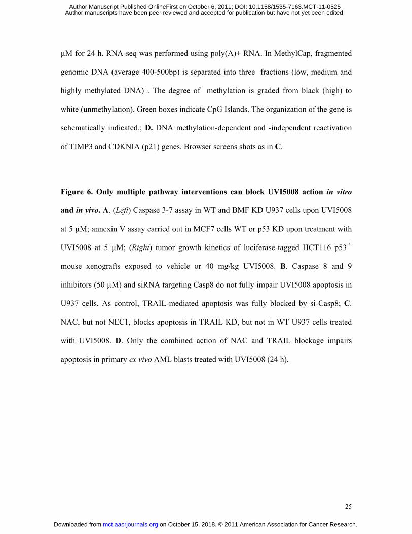
Figure 1. Structure and apoptogenic action of UVI5008 in U937 cells. A. Structure of
UVI5008; B. Apoptosis of UVI5008 at 40h revealed by pan-caspase activation assays;
C. Selective assays for activation of caspases 8, 9 and 3-7. In (B) and (C) UVI5008 was
used at µM UVI5008. Evaluation of the loss of mitochondrial potential after UVI5008
treatment (5 µM).
Figure 2. UVI5008 displays anticancer action in vivo and tumor-selective apoptosis.
A. False-color images revealing tumor growth in vivo of luciferase-tagged HCT116 cells,
xenografted into nude mice and treated with vehicle or 40mg/kg of UVI5008; the
increase of luminescence determined by direct photon counting reveals the growth
inhibitory effects; B. Kinetics of tumor mass development as in A; C. left panel. Tumor
mass development as in A of MCF7 xenografted mice treated with 40mg/kg of UVI5008;
on October 15, 2018. © 2011 American Association for Cancer Research. mct.aacrjournals.org Downloaded from
Author manuscripts have been peer reviewed and accepted for publication but have not yet been edited. Author Manuscript Published OnlineFirst on October 6, 2011; DOI: 10.1158/1535-7163.MCT-11-0525

23
right panel. Tumor mass development in MMTV-Myc transgenic mice treated with
40mg/kg of UVI5008; treatment was initiated when tumors became palpable; normalized
mouse weights are shown on the right side. D. Left panel. Primary BJ fibroblasts and
their tumorigenic derivatives were exposed to UVI5008 and survival was measured by
MTT assays; right panel. Survival rate of CD34+ cells and AML blasts upon UVI5008 (5
µM) treatment after 48 h. E. Example of the apoptotic UVI5008 action (20 h, 5 µM) in
the AML blast #116, both in bone marrow (BM) and periphery (P).
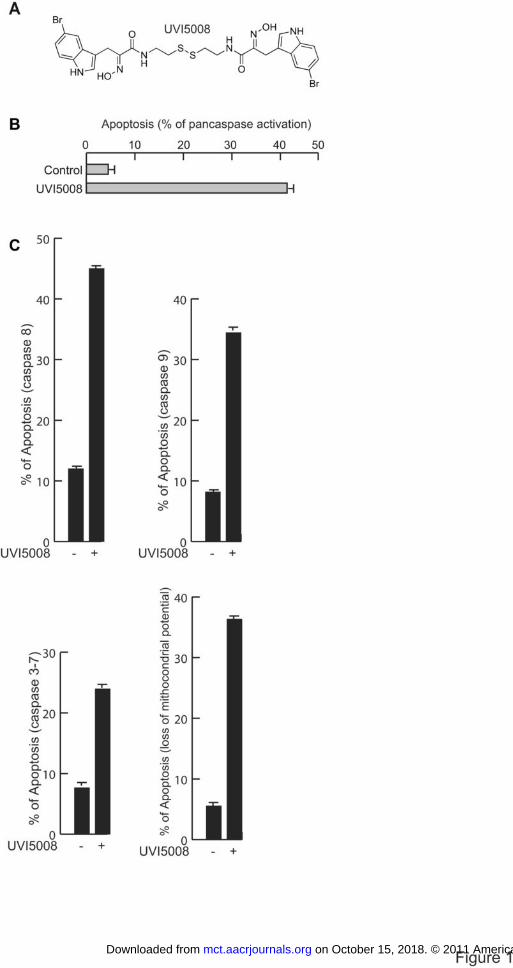
Figure 3. HDAC inhibitory activities of UVI5008 in vitro, ex vivo and in vivo. A. Cell-
based HDAC1 and HDAC4 assay carried out in MCF7 cells with UVI5008 at 5 µM; B.
Western blot analyses of p21, α-tubulin and histone H3 acetylation, K9H3me3x and
H3K9, H3K14, H3K18 acetylations in U937 cells after 24 h with UVI5008 at 5 µM; C.
Immunofluorescence of histone H3 acetylation levels in ex vivo AML 116 blasts; D.
Stimulation of histone H3 specific acetylations both in the tumors of MMTV-Myc
transgenic mice (data with HCT116 cells treated in vitro are shown for comparison) and
in xenografted HCT116 p53+/+ tumors treated with 40 mg/kg dose of UVI5008 and 20
mg/kg dose of SAHA analysed at day 22. Immunohistochemical staining for H3K9
acetylation in tumor sections from UVI5008 or SAHA-treated HCT116-xenografted
mice.
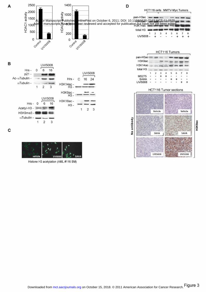
Figure 4. SIRT inhibitory activities of UVI5008 in vitro, ex vivo and in vivo. A.
Human recombinant SIRT1 and 2 assays in vitro with the indicated compounds; histone
H3K56 acetylation in U937 and U2OS cells treated for 24 h with UVI5008 (5 µM); B.
on October 15, 2018. © 2011 American Association for Cancer Research. mct.aacrjournals.org Downloaded from
Author manuscripts have been peer reviewed and accepted for publication but have not yet been edited. Author Manuscript Published OnlineFirst on October 6, 2011; DOI: 10.1158/1535-7163.MCT-11-0525
24
Western blot analyses of H3K56 acetylation in histone extracts of U937 cells treated with
UVI5008 (5 µM); C. Stimulation with UVI5008 (40 mg/kg dose) induced H3K56
specific acetylation in both tumors of MMTV-Myc transgenic mice (data with HCT116
cells treated in vitro are shown for comparison) and in xenografted HCT116 p53+/+
tumors analysed at day 22; H3K56ac immunohistochemical staining of tumor sections
from UVI5008-treated HCT116-xenografted mice. D. Top panel. Analysis of p53K382
acetylation in MCF7 cells after 6 h treatment with the indicated compounds; middle
panel. p53K382 acetylation levels in HCT116 cells treated in vitro; bottom panel.
p53K382 acetylation immunohistochemical staining of tumor sections from HCT116-
xenografted mice treated with 40mg/kg of UVI5008. Both UVI5008 and EX243, but not
MS275, stimulate p53K382 acetylation in HCT116 cells in vitro (middle panel) and in
tumori in treated xenografted mice (bottom panel).
Figure 5. UVI5008 displays Tumor Suppressor Gene reactivation in vitro and in vivo
A. Left panel. Western blot revealing p21 expression in HCT116 cells and in mice
bearing HCT116 p53+/+ and HCT116 p53-/- tumors; right panel. 48h treatment of
UVI5008 re-activates TSGs in HCT116 cells; B. Left panel. UVI5008 re-activates TSGs
in HCT116-based xenografts; 36B4 is an inert internal control RNA used for
normalization; (right panel, top) UVI5008 reactivates TSG in breast cancer cells treated
for 30h; (right panel, bottom) combined treatment with UVI5008 and AZA or EX527 and
AZA (36 h) synergizes in gene-re-expression in MDA-MB231 cells; C. Reactivation of
UCHL1 gene: browser screen shot of RNA-seq and DNA methylation (MethylCap-seq)
profiling of HCT116 (WT) and HCT116-DKO cells after treatment with UVI5008 at 5
on October 15, 2018. © 2011 American Association for Cancer Research. mct.aacrjournals.org Downloaded from
Author manuscripts have been peer reviewed and accepted for publication but have not yet been edited. Author Manuscript Published OnlineFirst on October 6, 2011; DOI: 10.1158/1535-7163.MCT-11-0525
25
µM for 24 h. RNA-seq was performed using poly(A)+ RNA. In MethylCap, fragmented
genomic DNA (average 400-500bp) is separated into three fractions (low, medium and
highly methylated DNA) . The degree of methylation is graded from black (high) to
white (unmethylation). Green boxes indicate CpG Islands. The organization of the gene is
schematically indicated.; D. DNA methylation-dependent and -independent reactivation
of TIMP3 and CDKNIA (p21) genes. Browser screens shots as in C.
Figure 6. Only multiple pathway interventions can block UVI5008 action in vitro
and in vivo. A. (Left) Caspase 3-7 assay in WT and BMF KD U937 cells upon UVI5008
at 5 µM; annexin V assay carried out in MCF7 cells WT or p53 KD upon treatment with
UVI5008 at 5 µM; (Right) tumor growth kinetics of luciferase-tagged HCT116 p53-/-
mouse xenografts exposed to vehicle or 40 mg/kg UVI5008. B. Caspase 8 and 9
inhibitors (50 µM) and siRNA targeting Casp8 do not fully impair UVI5008 apoptosis in
U937 cells. As control, TRAIL-mediated apoptosis was fully blocked by si-Casp8; C.
NAC, but not NEC1, blocks apoptosis in TRAIL KD, but not in WT U937 cells treated
with UVI5008. D. Only the combined action of NAC and TRAIL blockage impairs
apoptosis in primary ex vivo AML blasts treated with UVI5008 (24 h).
on October 15, 2018. © 2011 American Association for Cancer Research. mct.aacrjournals.org Downloaded from
Author manuscripts have been peer reviewed and accepted for publication but have not yet been edited. Author Manuscript Published OnlineFirst on October 6, 2011; DOI: 10.1158/1535-7163.MCT-11-0525
Published OnlineFirst October 6, 2011.Mol Cancer Ther Angela Nebbioso, Raquel Pereira, Harshal Khanwalkar, et al. production by the triple epigenetic inhibitor, UVI5008Death receptor pathway activation and increase of ROS
Updated version
10.1158/1535-7163.MCT-11-0525doi:
Access the most recent version of this article at:
Material
Supplementary
http://mct.aacrjournals.org/content/suppl/2011/10/03/1535-7163.MCT-11-0525.DC1
Access the most recent supplemental material at:
Manuscript
Authoredited. Author manuscripts have been peer reviewed and accepted for publication but have not yet been
E-mail alerts related to this article or journal.Sign up to receive free email-alerts
Subscriptions
Reprints and
To order reprints of this article or to subscribe to the journal, contact the AACR Publications
Permissions
Rightslink site. Click on "Request Permissions" which will take you to the Copyright Clearance Center's (CCC)
.http://mct.aacrjournals.org/content/early/2011/10/06/1535-7163.MCT-11-0525To request permission to re-use all or part of this article, use this link
on October 15, 2018. © 2011 American Association for Cancer Research. mct.aacrjournals.org Downloaded from
Author manuscripts have been peer reviewed and accepted for publication but have not yet been edited. Author Manuscript Published OnlineFirst on October 6, 2011; DOI: 10.1158/1535-7163.MCT-11-0525