d&c case presentation january 26, 2012. malpositioned central venous catheter right subclavian...
TRANSCRIPT
Transplant Surgery
D&C Case PresentationJanuary 26, 2012
Complication
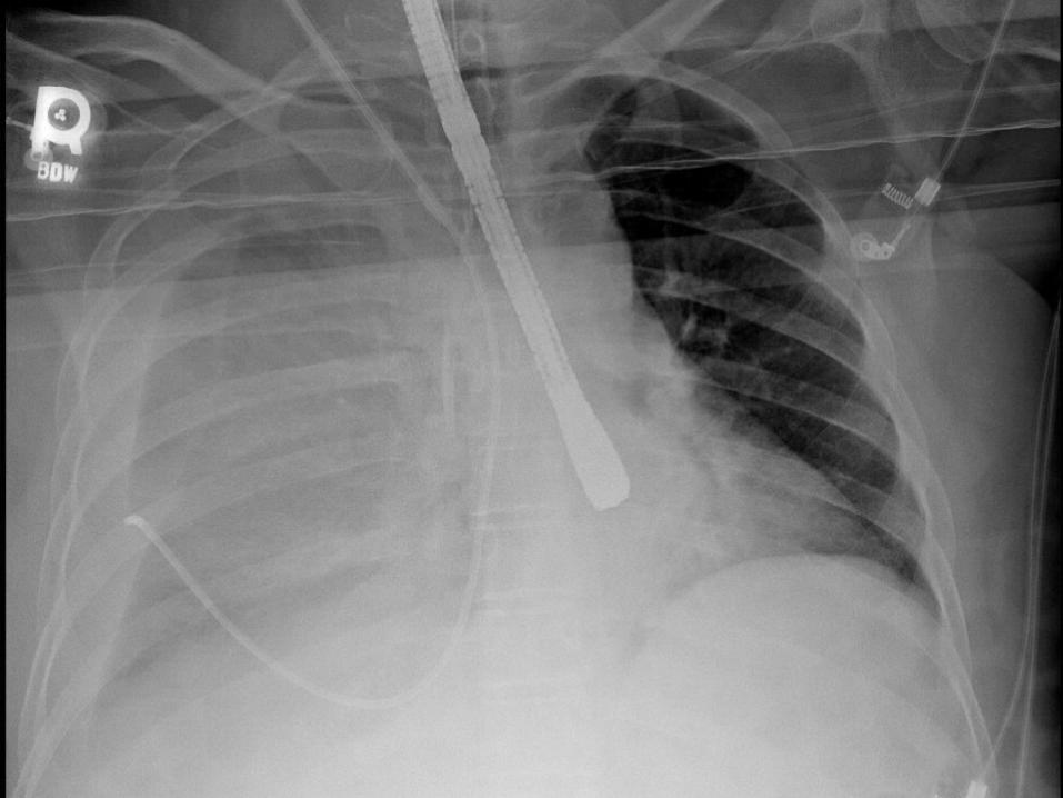
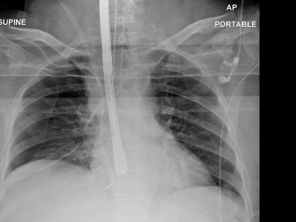
Malpositioned central venous catheter Right subclavian vein laceration Right subclavian artery laceration
Procedure: Central venous catheter insertion Deceased donor liver transplant
SH
63yo woman Cirrhosis due to Hepatitis C Moderate/Severe decompensation:
MELD 24 – Cr 1.76 ; Bilirubin 5.2; INR 1.4; Na 138
Weekly paracentesis for refractory ascities
Encephalopathy Presented for Liver transplant on Jan
14 Donor – 34yo man – DCD donor
SH
Taken to the operating room for deceased donor liver transplant
Central venous catheter MAC (9.0 Fr)
Swan-Ganz catheter Chest X-Ray
Central Line Complications
Arterial puncture Pneumothorax Arrythmia Thoracic Duct Injury Guidewire Loss Cardiac Perforation
Evens SRT. Surgical Pitfalls: Prevention and Management. Philadelphia: Saunders. 2009.
Ultrasound standard for IJ access1. Denys BG, Uretsky BF, Reddy PS. Ultrasound-assisted cannulation
of the internal jugular vein. A prospective comparison to the external landmark-guided technique. Circulation. 1993;87:1557-1562.
2. Gualtieri E, Deppe SA, Sipperly ME, Thompson DR. Subclavian venous catheterization: greater success rate for less experienced operators using ultrasound guidance. Crit Care Med. 1995;23:692-697.
3. Mallory DL, McGee WT, Shawker TH, Brenner M, Bailey KR, Evans RG, et al. Ultrasound guidance improves the success rate of internal jugular vein cannulation. A prospective, randomized trial. Chest. 1990;98:157-160.
4. Troianos CA, Jobes DR, Ellison N. Ultrasound-guided cannulation of the internal jugular vein. A prospective, randomized study. Anesth Analg. 1991;72:823-826.
5. Hilty WM, Hudson PA, Levitt MA, Hall JB. Real-time ultrasound-guided femoral vein catheterization during cardiopulmonary resuscitation. Ann Emerg Med. 1997;29:331-336.
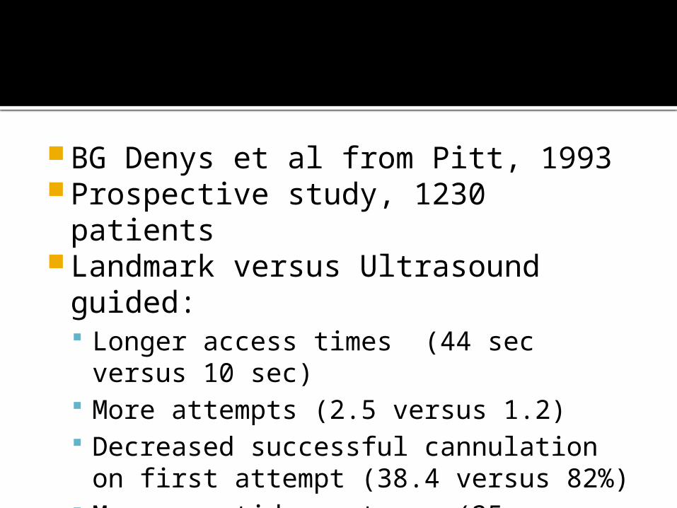
BG Denys et al from Pitt, 1993 Prospective study, 1230 patients Landmark versus Ultrasound guided:
Longer access times (44 sec versus 10 sec)
More attempts (2.5 versus 1.2) Decreased successful cannulation on
first attempt (38.4 versus 82%) More carotid punctures (25 versus 8)
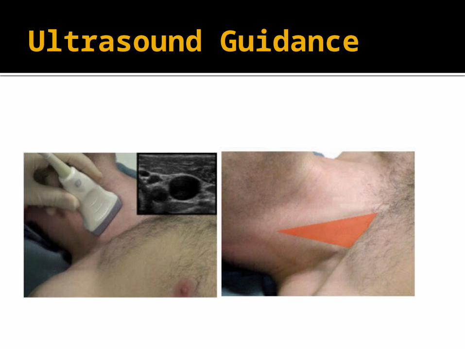
Ultrasound Guidance
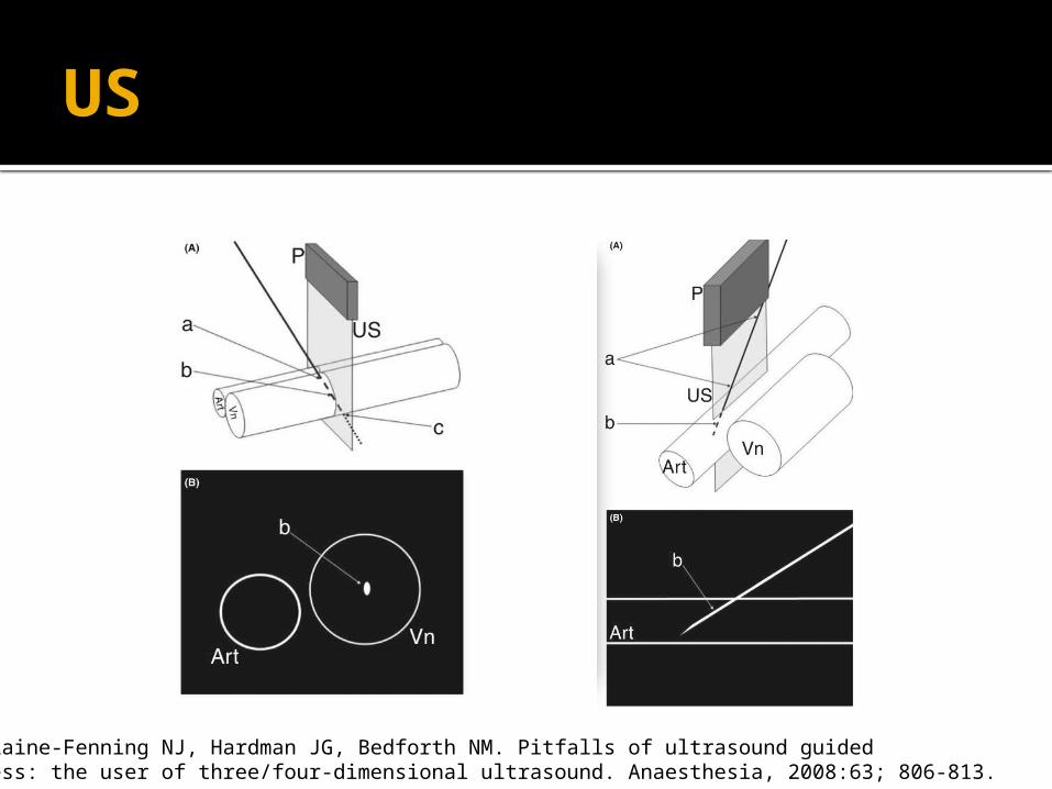
US
French JLH, Raine-Fenning NJ, Hardman JG, Bedforth NM. Pitfalls of ultrasound guidedVascular access: the user of three/four-dimensional ultrasound. Anaesthesia, 2008:63; 806-813.
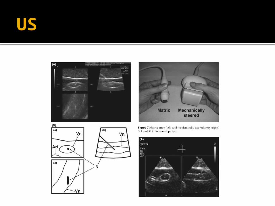
US
SH
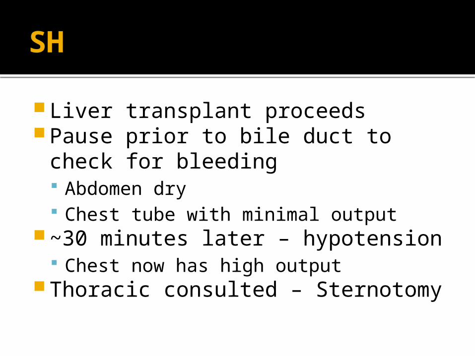
Liver transplant proceeds Pause prior to bile duct to check for
bleeding Abdomen dry Chest tube with minimal output
~30 minutes later – hypotension Chest now has high output
Thoracic consulted – Sternotomy
Ligation of Subclavian
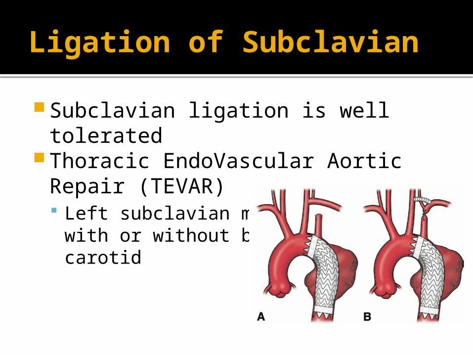
Subclavian ligation is well tolerated Thoracic EndoVascular Aortic Repair
(TEVAR) Left subclavian may be occluded with or
without bypass from left carotid
Open intervention
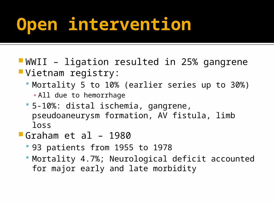
WWII – ligation resulted in 25% gangrene Vietnam registry:
Mortality 5 to 10% (earlier series up to 30%)▪ All due to hemorrhage
5-10%: distal ischemia, gangrene, pseudoaneurysm formation, AV fistula, limb loss
Graham et al – 1980 93 patients from 1955 to 1978 Mortality 4.7%; Neurological deficit accounted
for major early and late morbidity
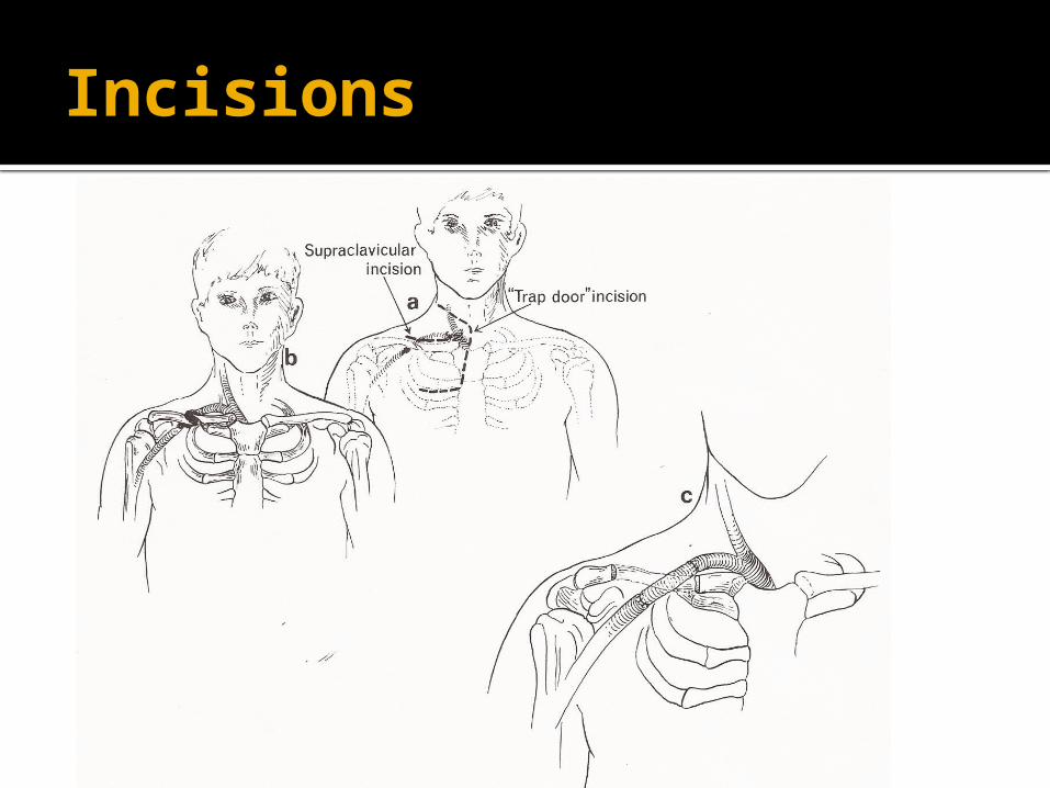
Incisions
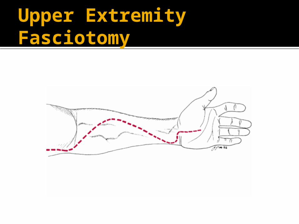
Upper Extremity Fasciotomy
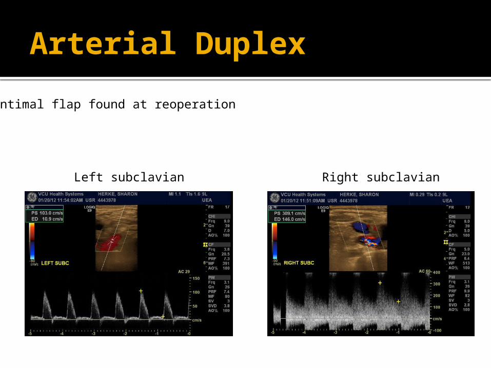
Arterial Duplex
Intimal flap found at reoperation
Left subclavian Right subclavian