david lacomis, md acquired diseases of muscle: histologic features
TRANSCRIPT

David Lacomis, MDDavid Lacomis, MD
AcquiredAcquiredDiseases of Diseases of
Muscle:Muscle:
Histologic FeaturesHistologic Features
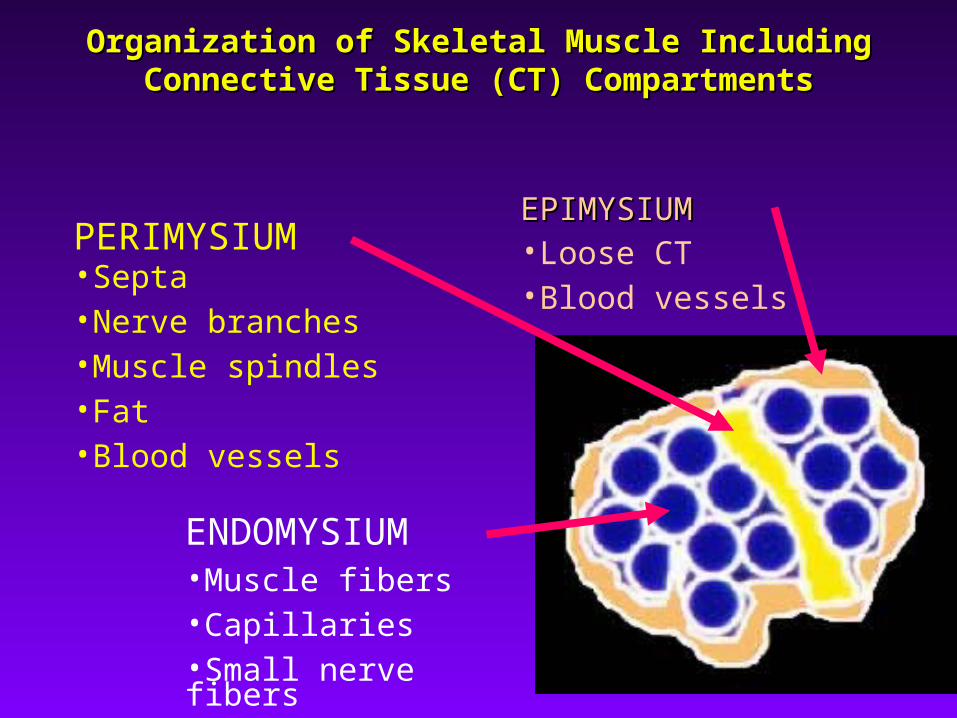
Organization of Skeletal Muscle Including Organization of Skeletal Muscle Including Connective Tissue (CT) CompartmentsConnective Tissue (CT) Compartments
EPIMYSIUMEPIMYSIUM•Loose CT•Blood vessels
PERIMYSIUM•Septa•Nerve branches•Muscle spindles•Fat•Blood vessels
ENDOMYSIUM•Muscle fibers•Capillaries•Small nerve fibers
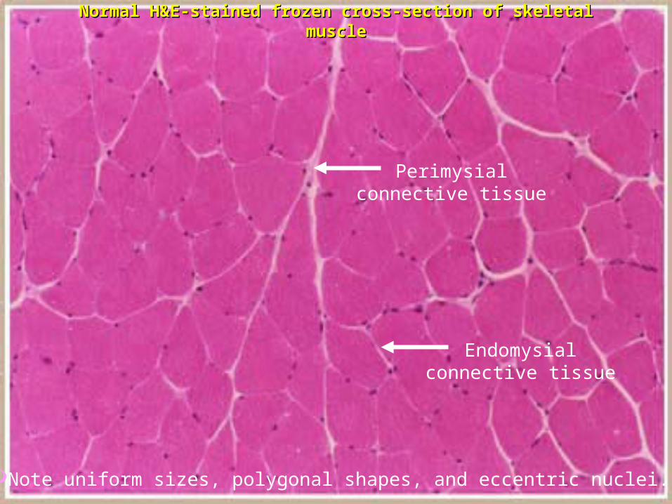
Perimysialconnective tissue
Endomysialconnective tissue
Normal H&E-stained frozen cross-section of skeletal Normal H&E-stained frozen cross-section of skeletal musclemuscle
Note uniform sizes, polygonal shapes, and eccentric nuclei.

Normal H&E-stained longitudinal paraffin sectionNormal H&E-stained longitudinal paraffin section
Note the banding pattern.Note the banding pattern. Nuclei are eccentrically placed.Nuclei are eccentrically placed.

Spindle
Nerve Twig
Normal Structures: Muscle SpindleNormal Structures: Muscle Spindleand Associated Nerve Fibersand Associated Nerve Fibers
Gomori trichromeGomori trichrome

Can be identified by the esterase reaction due Can be identified by the esterase reaction due to the presence of acetylcholinesterase.to the presence of acetylcholinesterase.
Neuromuscular JunctionsNeuromuscular Junctions

Neuromuscular JunctionNeuromuscular JunctionElectron MicroscopyElectron Microscopy
postsynaptic
presynaptic
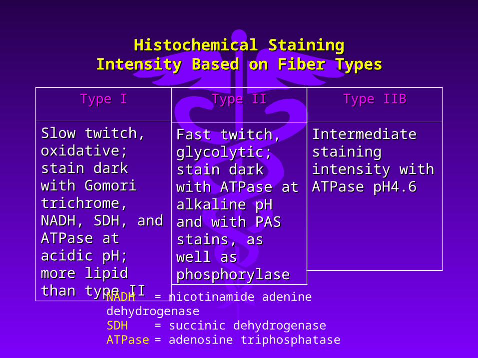
Histochemical Staining Intensity Histochemical Staining Intensity Based on Fiber TypesBased on Fiber Types
Type IType I
Slow twitch, Slow twitch, oxidative; stain oxidative; stain dark with Gomori dark with Gomori trichrome, NADH, trichrome, NADH, SDH, and ATPase SDH, and ATPase at acidic pH; more at acidic pH; more lipid than type IIlipid than type II
NADH = nicotinamide adenine dehydrogenaseSDH = succinic dehydrogenaseATPase = adenosine triphosphatase
Type IIBType IIB
Intermediate Intermediate staining intensity staining intensity with ATPase pH4.6with ATPase pH4.6
Type IIType II
Fast twitch, Fast twitch, glycolytic; stain glycolytic; stain dark with ATPase dark with ATPase at alkaline pH and at alkaline pH and with PAS stains, as with PAS stains, as well as well as phosphorylasephosphorylase
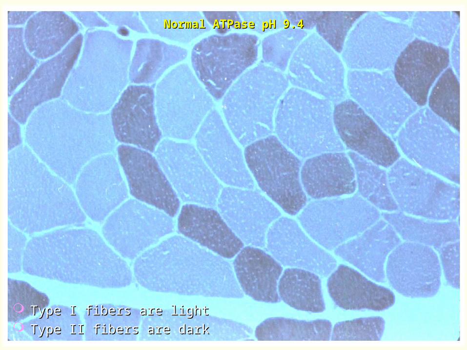
Type I fibers are lightType I fibers are light Type II fibers are darkType II fibers are dark
Normal ATPase pH 9.4Normal ATPase pH 9.4

Ultrastructure of a SarcomereUltrastructure of a Sarcomere
Extends from Z-band to Z-band.Extends from Z-band to Z-band.
Note arrangement of thick and thin filaments.Note arrangement of thick and thin filaments.
Z ZM
H band
ActinMyosin I bandI band
A band
A band includes overlap of actin & myosin.

Dark A-Dark A-bandsbands
Light I-Light I-bandsbands
Z-band is Z-band is present in present in the middle the middle of the light of the light bandband
Thin Thin filaments filaments are are attached at attached at the Z-bandthe Z-band
Normal electron microscopyNormal electron microscopy

Classification of MyopathiesClassification of Myopathies
ACQUIREDACQUIRED INHERITEDINHERITED
Inflammatory MyopathiesInflammatory Myopathies DystrophiesDystrophiesPolymositis (PM)Polymositis (PM) DystrophinopathiesDystrophinopathiesDermatomyositis (DM)Dermatomyositis (DM) Limb-GirdleLimb-GirdleInclusion body myositis (IBM)Inclusion body myositis (IBM) MyotonicMyotonicGranulomatous myositisGranulomatous myositis Facioscapulohumeral (FSHD)Facioscapulohumeral (FSHD)Infectious myositisInfectious myositis Oculopharyngeal (OPD)Oculopharyngeal (OPD)
ToxicToxic DistalDistal
EndocrineEndocrine CongenitalCongenital
MetabolicMetabolicMitochondrialMitochondrialGlycogen & lipid storageGlycogen & lipid storage

PolymyositisPolymyositisLongitudinal paraffin-embedded sectionLongitudinal paraffin-embedded section
Mononuclear inflammatory cell infiltrates and Mononuclear inflammatory cell infiltrates and many basophilic regenerating fibersmany basophilic regenerating fibers

PolymyositisPolymyositisLongitudinal paraffin-embedded section (higher power)Longitudinal paraffin-embedded section (higher power)
Regenerating fiber (non-specific)Regenerating fiber (non-specific) Fiber is basophilic due to presence of increased Fiber is basophilic due to presence of increased
RNA and DNA.RNA and DNA. Activated plump nuclei and prominent nucleoliActivated plump nuclei and prominent nucleoli

As regeneration advances, a myotube “bridge” is formed.As regeneration advances, a myotube “bridge” is formed.
PolymyositisPolymyositisLongitudinal paraffin-embedded section (higher power)Longitudinal paraffin-embedded section (higher power)
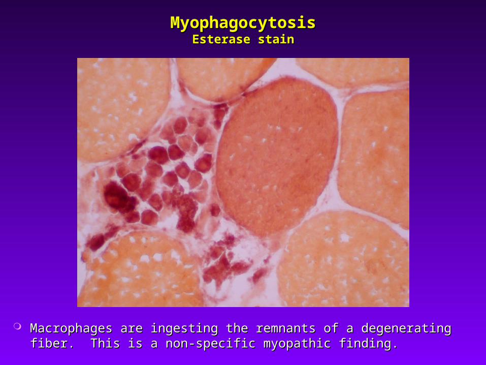
MyophagocytosisMyophagocytosisEsterase stainEsterase stain
Macrophages are ingesting the remnants of a degenerating fiber. Macrophages are ingesting the remnants of a degenerating fiber. This is a non-specific myopathic finding.This is a non-specific myopathic finding.

Invasion of a Non-necrotic Fiber by Invasion of a Non-necrotic Fiber by Inflammatory CellsInflammatory Cells
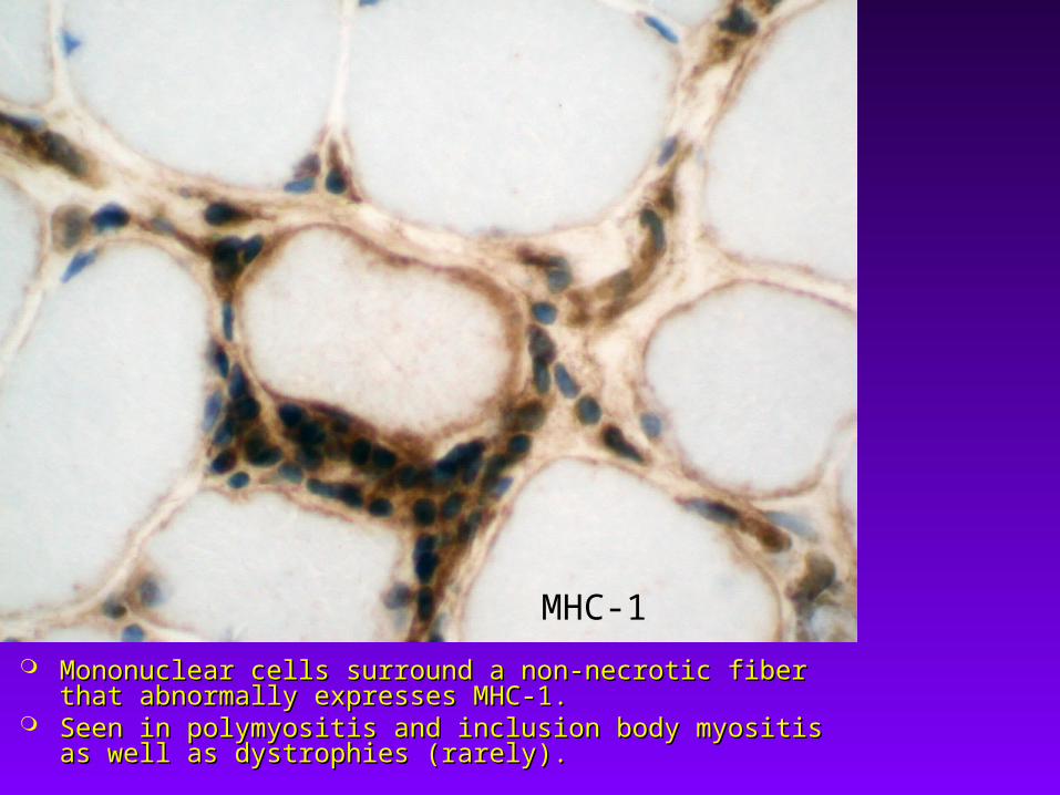
Mononuclear cells surround a non-necrotic fiber that Mononuclear cells surround a non-necrotic fiber that abnormally expresses MHC-1.abnormally expresses MHC-1.
Seen in polymyositis and inclusion body myositis as well as Seen in polymyositis and inclusion body myositis as well as dystrophies (rarely).dystrophies (rarely).
MHC-1

CD8
Inflammatory infiltrate in polymyositis is endomysial Inflammatory infiltrate in polymyositis is endomysial predominantly of the cytotoxic T-cell type.predominantly of the cytotoxic T-cell type.

DermatomyositisDermatomyositis
Perifascicular atrophyPerifascicular atrophy DegenerationDegeneration Inflammatory cells in the perimysium Inflammatory cells in the perimysium
surrounding a blood vesselsurrounding a blood vessel Inflammatory cells tend to be B-cells.Inflammatory cells tend to be B-cells.
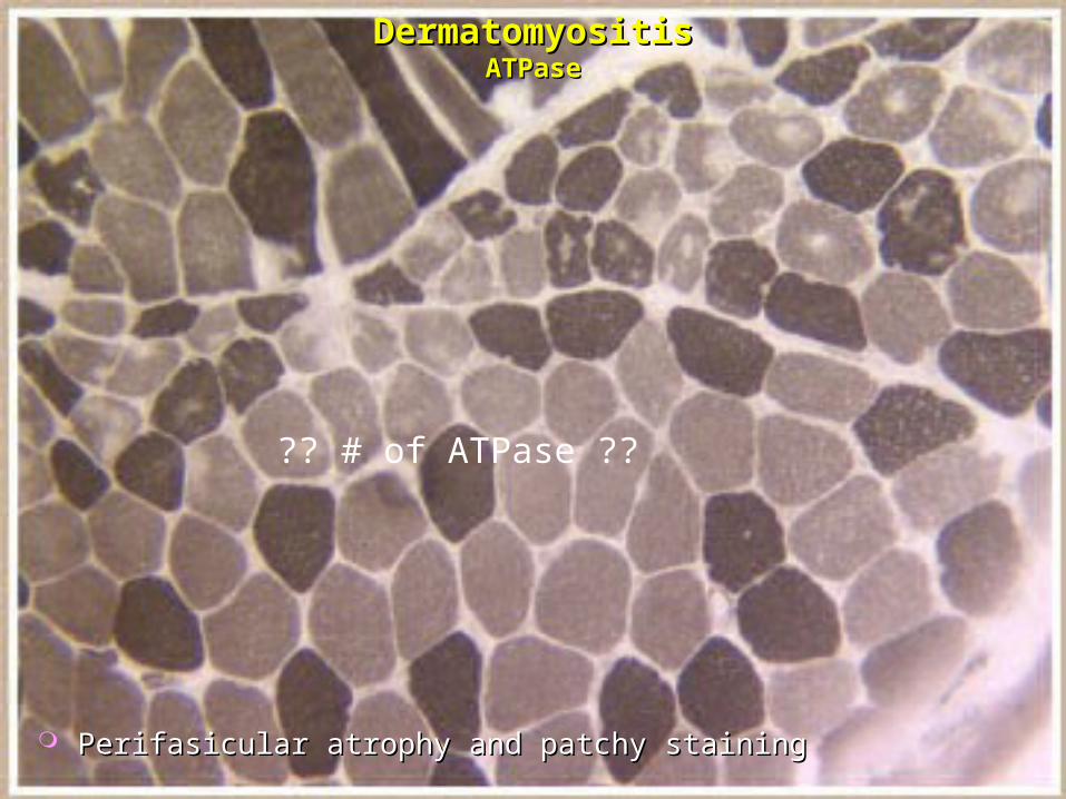
DermatomyositisDermatomyositisATPaseATPase
Perifasicular atrophy and patchy stainingPerifasicular atrophy and patchy staining
?? # of ATPase ??

The perifascicular fibers may have an abnormal purplish The perifascicular fibers may have an abnormal purplish appearance with Gomori trichrome.appearance with Gomori trichrome.

Perifascicular AtrophyPerifascicular AtrophyNADH-reacted sectionNADH-reacted section

Dermatomyositis

Dermatomyositis
B-cell
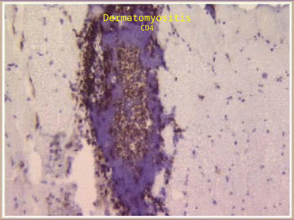
DermatomyositisCD4

DermatomyositisCD8

Dermatomyositis Inflammatory Infiltrate in SkinDermatomyositis Inflammatory Infiltrate in Skin
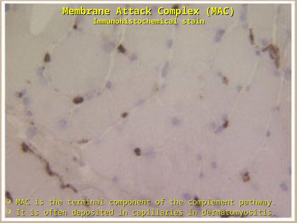
MAC is the terminal component of the complement pathway.MAC is the terminal component of the complement pathway. It is often deposited in capillaries in dermatomyositis.It is often deposited in capillaries in dermatomyositis.
Membrane Attack Complex (MAC)Membrane Attack Complex (MAC)Immunohistochemical stainImmunohistochemical stain

Membrane Attack Complex (MAC)Membrane Attack Complex (MAC)Immunohistochemical stainImmunohistochemical stain
Increased staining in capillaries in patients with Increased staining in capillaries in patients with dermatomyositisdermatomyositis
Degenerating fibers may also stain.Degenerating fibers may also stain.

DermatomyositisDermatomyositisElectron microscopyElectron microscopy
Tubuloreticular inclusion in a capillary endothelial cellTubuloreticular inclusion in a capillary endothelial cell
Invaded fiber
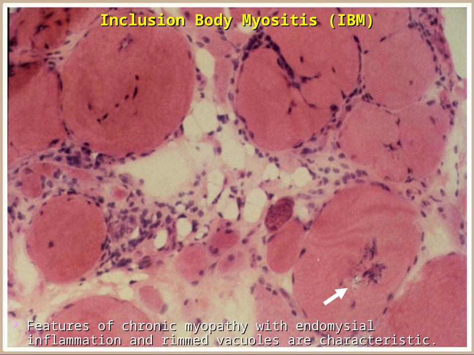
Features of chronic myopathy with endomysial inflammation and Features of chronic myopathy with endomysial inflammation and rimmed vacuoles are characteristic.rimmed vacuoles are characteristic.
Inclusion Body Myositis (IBM)Inclusion Body Myositis (IBM)
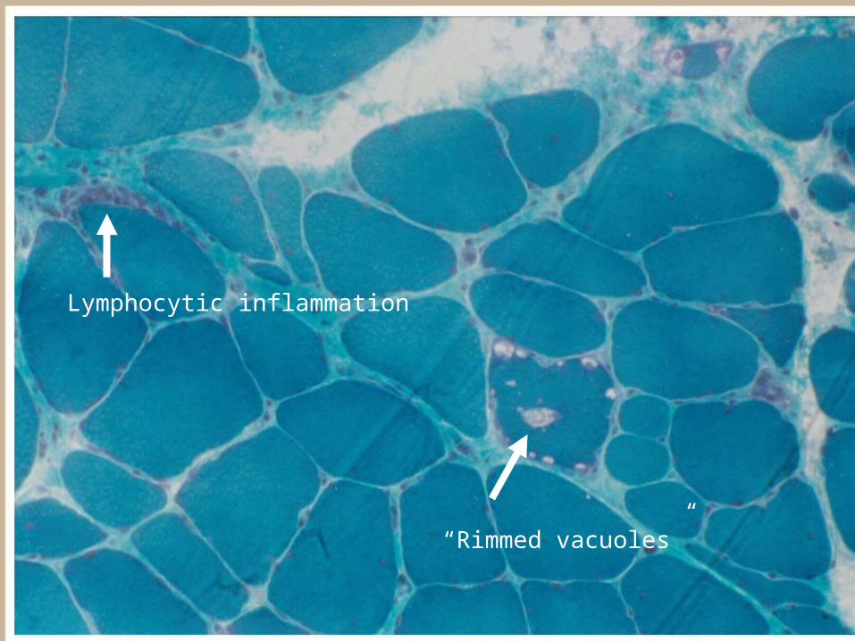
Lymphocytic inflammation
“Rimmed vacuoles”

Rimmed vacuoles may be “slit-like”

IBM: Vacuoles contain amyloid.IBM: Vacuoles contain amyloid.
Congo Red

IBM: VacuolesIBM: Vacuoles
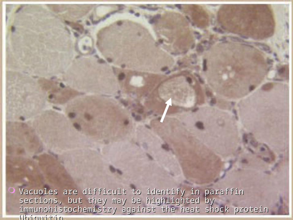
Vacuoles are difficult to identify in paraffin sections, but they Vacuoles are difficult to identify in paraffin sections, but they may be highlighted by immunohistochemistry against the heat may be highlighted by immunohistochemistry against the heat shock protein Ubiquitin.shock protein Ubiquitin.

IBM Eosinophilic Inclusion (Cytoid Body)IBM Eosinophilic Inclusion (Cytoid Body)Electron microscopyElectron microscopy

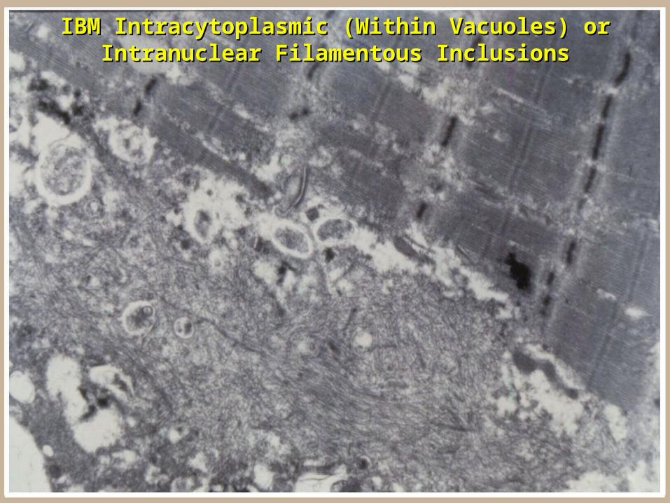
IBM Intracytoplasmic (Within Vacuoles) or IBM Intracytoplasmic (Within Vacuoles) or Intranuclear Filamentous InclusionsIntranuclear Filamentous Inclusions

Pyomyositis Gram Positive CocciPyomyositis Gram Positive Cocci

Granulomatous MyositisGranulomatous Myositisin a Patient with Sarciodosisin a Patient with Sarciodosis
Granulomas tend not to cause significant Granulomas tend not to cause significant damage to adjacent myofibers.damage to adjacent myofibers.
Giant cell
See picture Granuloma 1

Parasites: Trichinella spiralisParasites: Trichinella spiralis

Characteristic of mostCharacteristic of most
Endocrine Disturbance Type II Fiber Endocrine Disturbance Type II Fiber AtrophyAtrophy
ATPase pH9.4ATPase pH9.4

Inherited PolyneuropathyInherited PolyneuropathyChronic Neurogenic AtrophyChronic Neurogenic Atrophy
Groups of angulated atrophic fibersGroups of angulated atrophic fibers Marked variation in myofiber sizeMarked variation in myofiber size

Acute DenervationAcute DenervationNADH reactionNADH reaction
Manifested by small, darkly staining angulated fibersManifested by small, darkly staining angulated fibers
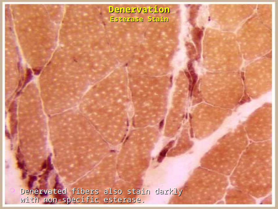
Denervated fibers also stain darkly with Denervated fibers also stain darkly with non-specific esterase.non-specific esterase.
DenervationDenervationEsterase StainEsterase Stain
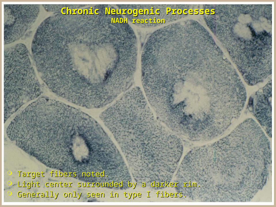
Target fibers noted.Target fibers noted. Light center surrounded by a darker rim.Light center surrounded by a darker rim. Generally only seen in type I fibers.Generally only seen in type I fibers.
Chronic Neurogenic ProcessesChronic Neurogenic ProcessesNADH reactionNADH reaction

Fiber type groupingFiber type grouping
Chronic Neurogenic AtrophyChronic Neurogenic AtrophyATPase reactionATPase reaction

Werdnig-Hoffman DiseaseWerdnig-Hoffman Disease(Spinal Muscular Atrophy Type I)(Spinal Muscular Atrophy Type I)

Werdnig-Hoffman DiseaseWerdnig-Hoffman Disease(Spinal Muscular Atrophy Type I)(Spinal Muscular Atrophy Type I)

Denervated fibers are atrophic but round.Denervated fibers are atrophic but round. Interspersed hypertrophic round fibers are usually noted.Interspersed hypertrophic round fibers are usually noted.
Werdnig-Hoffman DiseaseWerdnig-Hoffman Disease(Spinal Muscular Atrophy Type I)(Spinal Muscular Atrophy Type I)