d-galactose induced changes in enzymatic antioxidant ... · d-galactose induced changes in...
TRANSCRIPT
D-galactose Induced Changes in Enzymatic Antioxidant Status
in Rats of Different Ages
N. HADZI-PETRUSHEV1, V. STOJKOVSKI
2, D. MITROV
2, M. MLADENOV
1
1Faculty of Natural Sciences and Mathematics, Institute of Biology, “Ss. Cyril and Methodius”
University, Skopje, Macedonia
2Faculty of Veterinary Medicine, “Ss. Cyril and Methodius” University, Skopje, Macedonia
Corresponding author:
Nikola Hadzi-Petrushev, PhD
Faculty of Natural Sciences and Mathematics, Institute of Biology, “Ss. Cyril and Methodius”
University, P.O. Box 162, 1000 Skopje, Macedonia
E-mail: [email protected]
Phone: + 389 2 3249 606
Velimir Stojkovski, PhD
2Faculty of Veterinary Medicine, “Ss. Cyril and Methodius” University, 1000 Skopje,
Macedonia
Dine Mitrov, PhD
2Faculty of Veterinary Medicine, “Ss. Cyril and Methodius” University, 1000 Skopje,
Macedonia
Mitko Mladenov, PhD, Ass. Prof.
Faculty of Natural Sciences and Mathematics, Institute of Biology, “Ss. Cyril and Methodius”
University, P.O. Box 162, 1000 Skopje, Macedonia
Short title
D-gal Induced Changes in Antioxidant Status
Abstract
Aims
Considering the preexisting influence of the process of natural aging on antioxidant enzymes
activity and the level of lipid peroxidation, the age of the rats at which D-galactose (D-gal)
treatment is started could strongly impact the development of D-gal induced senescence. To
evaluate this, we subjected 1, 3 and 15 months old rats to D-gal treatment in parallel with having
appropriate placebos (0.9% saline).
Results
Our results showed elevated glutathione peroxidase (GPx) activity and no significant changes in
superoxide dismutase (SOD), catalase (CAT) and glutathione reductase (GR) activity or
malondialdehyde (MDA) levels in relation to natural aging. In mature and aged senescent livers
we observed positive correlation between increased ratio R=SOD/(GPx+CAT) and increased
MDA concentration. MDA levels seemed to correlate positively with the age of the animals at
which D-gal treatment had started. In the case of 3 and 15 months old rats there was D-gal
induced decrease in SOD and GR activity, but this effect of the treatment was not observed in 1
month old rats.
Conclusions
Our results imply that the changes in the antioxidant enzyme activities are not only under the
influence of the D-gal overload, but also depend on the developmental stage of the rats.
According to our results, with regard to enzymatic antioxidant capacity and the level of lipid
peroxidation, the best age for induction of senescence is somewhere after the third month.
Key words: D-galactose . Senescence
. Aging
. Antioxidant enzymes
. Lipid peroxidation
. Wistar
rats
Introduction
Aging is a multifactorial process associated with physiological decline. There is a
substantial amount of data supporting the positive relation between the process of aging and the
progressive decline in antioxidant function combined with increased mitochondrial ROS
(reactive oxygen species) generation and increased accumulation of oxidant products
(Kasapoglu and Ozben 2001; Navarro et al. 2004; Tian et al.1998). Some of these products are
generated by the process of lipid peroxidation that involves the oxidative deterioration of
polyunsaturated fatty acids from the cell membranes (Mladenov et al. 2006). The ROS-derived
unstable lipid peroxides disintegrate to different products including carbonyl compounds like
malondialdehyde (MDA). The measurement of MDA is a reliable method for assessment of the
level of lipid peroxidation and it is one of the most important biomarkers of aging (Pandey and
Rizvi 2010).
On the other hand, the most important line of defense is represented by the main
antioxidant enzymes. The three isoforms of superoxide dismutase (SOD) are responsible for the
elimination of superoxide anions, the most abundant form of ROS, produced mainly during
electron transfer in the mitochondrial respiratory chain (Halliwel 1991). As a result of this action,
hydrogen peroxide is generated and it can be further neutralized by the activity of catalase
(CAT). Hydrogen peroxide can be reduced by glutathione peroxidase (GPx), an enzyme that
functions as part of the glutathione cycle, along with glutathione reductase (GR). However,
conflicting results do exist and the association between antioxidant status, lipid peroxidation and
aging, seems to display variations depending on the species, strain, sex and examined organs of
the experimental animals (Rikans and Hornbrook 1997).
Animal models provide valuable research tools toward a better understanding of the
mechanisms underlying aging. A process very similar to natural aging can be induced by chronic
administration of D-galactose (D-gal) (Ho et al. 2003). An oversupply of D-gal leads to its
conversion to D-galacto-hexodialdose and hydrogen peroxide by galactose oxidase and to
galactitol by action of aldose reductase (Ho et al. 2003). These products can accumulate in cells,
which in turn causes osmotic and oxidative stress that may account for the acceleration of aging
(Kumar et al. 2011). Hence rodents treated with D-gal can be used as an animal model for
oxidative stress. That is based on one of the proposed mechanisms of D-gal induced senescence,
known to involve increased production of oxidants, changes in antioxidant enzymes activity and
accumulation of oxidative damage (He et al. 2009; Wei et al. 2005). The described processes
could be responsible for altered antioxidant capacity and increased MDA production, which has
been registered in studies involving D-gal induced aging (Liu et al. 2013; Banji et al. 2013;
Anand et al. 2012). Most of the studies involving D-gal accelerated aging are performed using
mice and less frequently rats as experimental animals, and they are mainly focused on the
neurodegenerative changes.
In view of the relative lack of data regarding D-gal induced aging in rats and considering
the preexisting influence of the process of natural aging on antioxidant enzymes activity, we
hypothesized that the age of the rats at which D-gal treatment is started, could strongly impact
the development of D-gal induced senescence. This might lead to animal models with varying
antioxidant status, irrespective of the equal D-gal dose and treatment duration. Therefore, one of
the aims of our study was to provide data about the antioxidant enzymes activity in rats of
different age, subjected to both natural and D-gal induced aging. We also determined the
concentration of MDA, to confirm the impact of altered enzymatic antioxidant capacity on the
level of lipid peroxidation in the liver and kidney of the experimental animals, since these tissues
are both metabolically active and show age-related increase in the level of lipid peroxidation
products (Ward 2005).
Furthermore, taking into account our previous studies (Hadzi-Petrushev et al. 2011;
Stojkovski et al. 2013), where we published that sensitivity to free radicals apparently depends
on the equilibrium between the formation of hydrogen peroxide from superoxide (in the
dismutation reaction catalyzed by SOD) and its degradation by GPx and CAT, rather than on the
activities of individual antioxidant enzymes, we lay down the basis for the second aim of this
study, which is to investigate the relationship between the ratio of activities of antioxidant
enzymes R=SOD/(GPx+CAT) and the levels of MDA in the liver and kidney of D-gal treated
rats. Additionally, considering the sex-dependent reactions on various stressors, the presented
methods could also be important in further studies regarding the development of D-gal induced
senescence in rats of different gender.
Material and Methods
Animals and experimental design
All experimental procedures were conducted in accordance with the Guiding Principles
for Care and Use of Laboratory Animals approved by the Macedonian Center for Bioethics. All
protocols were approved by the Animal Ethics Committee of the University “Ss. Cyril and
Methodius”, Skopje, R. Macedonia, in accordance with the International Guiding Principles for
Biomedical Research Involving Animals. Anesthetics were applied according to the standards
given by the guide of the EC Directive 86/609/EEC. Animals were anesthetized with an
intraperitoneal injection of thiopental sodium (Rhone-Poulenc Rorer Limited, Nenagh, Co
Tipperary, Ireland), 50 mg kg-1
b. wt. Male Wistar rats (n=48) were used for all protocols and
were maintained on a 12:12 light:dark cycle and fed with standard rat chow and water ad libitum.
All animals were divided into six groups (each containing 8 rats) depending on their age
and treatment with D-Gal. The process of induction of aging usually involves administration of
50-500 mg/kg D-gal for 6-8 weeks (Kumar and Rizvi 2013). Three groups consisted of placebo
(0.9% saline, 1.5 ml daily, subcutaneous) rats, at the age of 1 month (“young”), 3 months
(“mature”) and 15 months (“aged”). The other three groups of animals, starting at the same age,
for 42 consecutive days were treated with D-galactose (Sigma, St Louis, MO, USA), 300 mg kg-1
b.wt., subcutaneous, daily.
Tissue Preparation
The livers and kidneys were excised during laparotomy. Immediately upon collection all
tissue samples were washed with physiological salt solution and then were snap-frozen in liquid
nitrogen and stored at -80 °C until further analysis.
Assay for SOD activity
SOD activity was determined according to the method described by Marklund and
Marklund (1974), based on the ability of SOD to inhibit the autooxidation of pyrogallol. The
reaction mixture consisted of 50 mM Tris-HCl, pH 8.2, 1 mM diethylenetriamine pentaacetic
acid and sample. The reaction was initiated by the addition of pyrogallol (final concentration of
0.2 mM) and the absorbance measured kinetically at 420 nm, 25 °C, for 3 min. One unit of
activity is defined as the amount of sample needed to inhibit pyrogallol oxidation by 50 %. The
final results were expressed as U/mg protein.
Assay for CAT activity
CAT activity was determined by measuring the degradation of hydrogen peroxide
(H2O2), using the method described by Claiborne (1985). The reaction mixture (1 ml) consisted
of 50 mM potassium phosphate buffer, pH 7.0, 19 mM H2O2 and sample. The reaction was
initiated by the addition of H2O2 and the change in absorbance was followed at 240 nm, 25 °C,
for a time period of 30 s, taking measurements at 5 s intervals. In those conditions, the molar
extinction coefficient for H2O2 is 43.6 M-1
cm-1
. The activity of CAT was expressed as U/mg
protein, with one unit of activity being equal to the conversion of 1 µmol H2O2 per minute.
Assay for GPx activity
The activity of GPx was determined according to the method described by Lawrence and
Burk (1976). The activity was assayed by following the oxidation of NADPH at 340 nm for 3
min, 25 °C, in the presence of GR and GSH. The reaction mixtures containing 50 mM potassium
phosphate, pH 7.0, 1 mM sodium azide, 2 mM GSH, 0.2 mM NADPH, 1 U/ml GR, 1.5 mM
cumene hydroperoxide and sample, were incubated at 25 °C for 5 min. The reaction was initiated
by the addition of cumene hydroperoxide. One unit of activity was defined as the oxidation of
1μmol of NADPH per minute. Final results were expressed as U/mg protein.
Assay for GR activity
The rate of oxidation of NADPH by GSSG at 30 °C was used as a measure of GR
activity (Racker 1955). The reaction system contained 1 mM GSSG, 0.1 mM NADPH, 0.5 mM
EDTA, 100 mM potassium phosphate buffer, pH 7.5, and sample. The oxidation of 1 μmol of
NADPH per minute was defined as a unit of GR activity. The specific activity was expressed as
mU/mg protein.
The protein content of the liver and kidney samples was determined using the method
described by Lowry et al. (1951).
Malondialdehyde assay
The lipid peroxides were estimated in the liver and kidney homogenates using the
thiobarbituric acid (TBA) reactive substances tests, described by Ohkawa et al. (1979). After the
addition of 8.1 % sodium dodecyl sulfate, each sample or standard (1,1,3,3-tetraethoxypropane
solution) was vortexed and left at room temperature for 10 minutes. At the end of the incubation
period, 20 % acetic acid and 0,6 % thiobarbituric acid were added and the test tubes were placed
in a water bath at 90-95 °C for 1 hour. After that, they were cooled on ice and the colored
supernatant was obtained by adding a mixture of butanol:pyridine (15:1), vortexing and
centrifuging. The absorbance was measured at 535 nm. The results were expressed as nmol
MDA per g tissue.
Statistical Analysis
Results are presented as means ± standard error (SE). The effects of D-gal treatment and
aging were assessed using One-way analysis of variance (ANOVA). The post hoc test of Tukey
was performed in selected instances to evaluate further differences between group pairs.
Correlations were expressed using the Pearson’s product-moment correlation coefficient. All
analyses were performed with GraphPad Prism 4.0 (San Diego, CA, USA). p<0.05 was
considered statistically significant.
Results
Influence of aging and D-gal treatment on SOD activity in liver and kidney
The process of natural aging did not cause significant changes in SOD activity in both
liver and kidney, according to the results obtained when comparing the activity of SOD in young
placebo (YP) to mature and aged placebo rats (MP and AP, respectively). The treatment with D-
gal led to significant decrease in SOD activity, in both liver and kidney of mature and aged rats,
but there were no significant changes in the examined tissues of young animals. This was
concluded by comparing the SOD activity in the liver of MP to MT (mature treated) rats
(p<0.001), as well as SOD activity in the liver of AP to AT (aged treated) animals (p<0.001).
The same effect of the treatment was observed when making the equivalent comparisons in
kidney (MP to MT, p<0.05 and AP to AT, p<0.01). Additionally, in the young kidney, the lack
of effect of D-gal treatment, was further supplemented by the significantly higher SOD activity
in YT (young treated) compared to MT and AT rats (p<0.01 in both cases) (Fig. 1).
Influence of aging and D-gal treatment on CAT activity in liver and kidney
There were no significant differences in CAT activity in the liver between the groups of
YP, MP and AP, respectively. The same situation was observed in the kidney, confirming the
nonappearance of significant effects of the process of natural aging on CAT activity. Similarly,
the treatment with D-gal failed to cause significant changes to CAT activity in the liver and
kidney, regardless of the age of the rats at the beginning of the treatment. However, CAT activity
in the liver of MT was significantly higher compared to the activity in the liver of YT animals
(p<0.05) (Fig. 2).
Influence of aging and D-gal treatment on GPx activity in liver and kidney
The effect of natural aging on GPx activity was evident only in the kidney, when
comparing the activity in YP to AP rats (p<0.001) and MP to AP rats (p<0.01). Increased GPx
activity was also observed due to D-gal treatment in the liver of MT and AT rats (p<0.05 in both
cases) and in the kidney of MT (p<0.05). YT had GPx activity in the liver and kidney that was
not significantly different than the activity in the appropriate placebo groups. Moreover, GPx
activity in the liver of YT rats stayed significantly lower compared to the activity in MT and AT
animals (p<0.001 and p<0.01, respectively). In the kidney, GPx activity in YT rats was also
significantly lower than the activity in MT and AT rats (p<0.001 in both cases) (Fig. 3).
Influence of aging and D-gal treatment on GR activity in liver and kidney
The data presented on Fig. 4, demonstrated that GR activity in the liver and kidney did
not change significantly with the age. The treatment with D-gal, resulted in a significant decrease
in GR activity in the livers of MT rats (p<0.05) and in the kidney of MT and AT animals (p<0.05
in both cases). GR activity in the liver and kidney of YT rats did not change significantly
compared to the placebo groups and at the same time was not significantly different than GR
activity in the MT and AT rats.
Influence of aging and D-gal treatment on liver and kidney MDA levels
The results from our study showed that the effects of the process of natural aging were
not strong enough to cause significant changes in MDA levels in both liver and kidney. Quite
differently, the treatment with D-gal led to a significant increase in MDA concentration in the
liver and kidney of MT (p<0.001 for liver and p<0.05 for kidney) and AT rats (p<0.001 for
both). This effect of D-gal treatment was not observed in the examined tissues of young rats. The
MDA levels seemed to correlate positively with the age of the animals at which treatment with
D-gal had started. Hence, YT and MT rats had significantly lower MDA levels in the liver,
compared to AT (p<0.001 in both cases). In the kidney, MDA concentrations in MT and AT rats
did not differ significantly, but both were higher than the concentration in YT rats (p<0.001 for
both) (Fig. 5).
The relationship between antioxidant enzymes in liver and kidney
Changes in the equilibrium between the formation of hydrogen peroxide from superoxide
dismutation and its decomposition by other enzymes (GPx and CAT) are expressed by the ratio
R=SOD/(GPx+CAT) (Table 1). This ratio was significantly different when the values for MT
and AT livers (p=0.002) and kidneys (p=0.011) were compared to the values of their placebo
counterparts. In AT liver, this tendency was additionally corroborated by a positive correlation
between SOD and CAT (r=0.841, p=0.036). Significant positive correlations between the levels
of lipid peroxidation products (MDA) and the value of the ratio R were observed in MP livers
(r=0.983, p=0.0004) and kidneys (r=0.869, p=0.024) and AP livers only (r=0.865, p=0.026).
Moreover, the positive correlations between the liver ratio R and MDA in mature (r=0.841,
p=0.036) and aged placebo rats (r=0.946, p=0.004), were persistent even after the treatment with
D-gal (r=0.872, p=0.025; r=0.912, p=0.01, respectively). On the other side, the negative
correlation between MDA and SOD (r=−0.958, p=0.002) in YT liver and MDA vs CAT
(r=−0.916, p=0.01) in MT liver, once again potentiate the disintegrative potential of the D-gal on
antioxidant equilibrium. In the kidneys, there were no significant correlations among studied
variables after treatment with D-gal.
Discussion
The examined parameters were aimed to give us insight into whether the age of the rats at
the start of D-gal treatment should be taken into consideration, regarding the antioxidant status of
the subsequently established animal models of aging. In contrast to the general consensus, our
results showed no significant changes in the examined antioxidant status (SOD, CAT and GR
activity) in function of natural aging. Additionally, GPx activity was higher in mature and aged
rats compared to the group of young animals. In our experiment, the absence of a decline in
antioxidant enzyme activity could be explained in view of the age of the animals in all three
groups of rats. Our previous results (Hadzi-Petrushev et al. 2011, 2012) have shown that GPx
activity in liver and kidney decreases only after 18-20 months of age and CAT activity remains
unchanged. In some way, this is an expansion of the results of Tian et al. (1998) who have
shown that GPx activity in kidney decreases only after 24 months of age and does not change in
the liver. GPx activity may be preserved due to the fact that aging is not necessarily associated
with changes in the level of GSH (Kasapoglu and Ozben 2001) and the increased antioxidant
enzyme activity represents an adaptive response to increased levels of oxidative products. In this
manner, a decrease in one antioxidant may be compensated by an increase in another,
maintaining relatively sufficient antioxidant protection in the cells (Rikans et al. 1992). For
example, researchers have reported that 50 % of SOD and up to 70 % of CAT activity could be
inhibited in isolated hepatocytes and still this antioxidant capacity is sufficient to withstand
experimentally induced oxidative stress (Rikans et al. 1992; Orr et al. 1992). Also, Amstad et al.,
(1991), reported an overproduction of SOD in epidermal cells sensitive to superoxide and
hydrogen peroxide, whereas cells with overproduction of CAT were protected against the effects
of oxidants. Such protective effect of CAT was confirmed in our study, as we found significant
increase in its activity only in senescent mature liver. Further, in mature and aged senescent
livers we observed positive correlation between increased ratio R=SOD/(GPx+CAT) and
increased MDA. These results may support the theory of contribution of the antioxidant enzymes
to the oxidative stress in the senescent rats, due to decreased SOD and GR activity. Moreover,
the significant increase of MDA during senescence (MT and AT), found in this study,
corroborates our hypothesis that the balance of the oxygen metabolism is compromised in both
liver and kidney. In this sense, the negative correlations between MDA and SOD in YT livers
and MDA and CAT in MT livers represent an additional support to our claim.
On the other side, in the case of aged rats, our results did not show significant differences
in MDA levels in comparison to their younger counterparts. It has already been shown that lipid
peroxidation of brain and liver, expressed as MDA, shows no significant differences among rats
of different ages (Tian et al. 1998) and also no relationship was found between MDA levels and
age in humans (Block et al. 2003). Nevertheless, these data do not exclude increased MDA
production in relation to aging, as MDA may be subjected to rapid degradation i.e. high turnover,
so it is not possible to detect significant accumulation in chronic conditions (Holmes et al. 1992).
Our data from the D-gal treated groups of rats, showed that starting the treatment at different age
(3 or 15 months), could lead to the establishment of animal model of aging that completely
fulfills the accepted profile of senescence, regarding the antioxidant enzyme activity and the
level of MDA as marker of lipid peroxidation.
The increase in GPx activity after D-gal treatment in our experimental groups of mature
and aged rats is in agreement with the consideration that the oxidative status of the cells
primarily regulates the gene expression and the activity of antioxidant enzymes (Pahl and
Baererle 1994). The fact that the increase in MDA levels in mature and aged D-gal treated rats,
was followed by an unchanged GPx activity, leads to a reasoning that either the response of GPx
to increased pro-oxidant conditions is limited or the lack of change in GPx activity is a reason for
increased MDA levels. Also, GPx per se is not an efficient decomposer of hydrogen peroxide
(Lopez-Torres et al. 1993) and the absence of appropriate change in CAT activity in conditions
of increased ROS may impair the capacity of the antioxidant enzyme system, leading to
increased MDA levels.
The absence of significant D-gal-related changes in our group of young rats could be
observed in the light of the mandatory attention that must be given to the developmental changes
and the influence of aging on antioxidant enzymes (Wei and Lee 2002). The rate of free radical
production in mitochondria increases with age in animal tissues (Perez-Campo et al. 1998; Wei
and Lee 2002, Stojkovski et al.2013) and the aforementioned results may be elucidated by the
presence of low ROS production and better balance between the free radical scavenging enzymes
in young tissues, which is very important for the cellular resistance to oxidative stress. In this
direction, Kasapoglu and Ozben (2001) reported the presence of chemically modified enzymes
with decreased catalytic efficiency in aged tissues. Further, Zhang with coworkers (2010)
published that the activities of mitochondrial respiratory chain components (complex I, II, III and
IV) in rats are reduced in D-gal-induced mimetic aging, when the D-gal treatment is started at
the age of 5 months. Different studies (mainly performed on mice), corroborate our results,
highlighting the importance of the starting age of the animals in relation to D-gal induced
senescence. In this direction, Xu et al. (2009) reported that 3 and 6 months old mice are more
suitable for establishing a mimetic ageing model compared to 1.5 months old mice. Regarding
the use of rats, equivalent study has not been performed but experimental data showing increased
MDA level, and decreased SOD and CAT activity is characteristic for animals of various ages at
the start of the treatment (Liu et al. 2013; Lei et al. 2008).
The pathophysiological changes during natural and D-gal induced aging are related to the
production of ROS and oxidative stress and it is obvious that rats of different age react
differently to D-gal treatment. The pro-oxidant/antioxidant balance is successfully represented
and can be assessed by the antioxidant enzyme activities and the level of lipid peroxidation. As it
is shown by our study, in the D-gal model of accelerated aging, the changes in the antioxidant
enzyme activities are not only under the influence of the D-gal overload, but also depend on the
developmental stage of the animals. Senescence manifestation in old rats (15 months age) was
quite unclear and the changes in the examined parameters were less consistent compared to the
case of mature rats. Conversely, some of the D-gal induced changes in young rats (1 month age)
were not considerably dissimilar to the changes observed in mature rats. However, it is
reasonable to favor the choice of mature rats for the process of accelerated aging considering
their complete organ and body differentiation in contrast to the possibility of additional changes
relating to the natural maturation in young rats (Kwekel et al. 2010; Stuart et al. 2001). In
general, this implies on the existence of optimal starting age of treatment for the establishment of
D-gal-induced aging model in rats. In view of the enzymatic antioxidant status and the level of
lipid peroxidation, the obtained results indicate that, in our experimental settings, the mature rats
(3 months age) appeared to be the most appropriate for the development of an animal model of
aging.
References
AMSTAD P, PESKIN A, SHAH G, MIRULT ME, MORET R, ZBINDEN I, CERUTTI P: The
balance between Cu,Zn-superoxide dismutase and catalase affects the sensitivity of mouse
epidermal cells to oxidative stress. Biochemistry 30: 9305-9313, 1991.
ANAND KV, MOHAMED JAABIR MS, THOMAS PA, GERALDINE P: Protective role of
Chrysin against oxidative stress in D-galactose-induced aging in an experimental rat model.
Geriatr Gerontol Int 12(4): 741-750, 2012.
BANJI D, BANJI OJ, DASAROJU S, ANNAMALAI AR: Piperine and curcumin exhibit
synergism in attenuating D-galactose induced senescence in rats. Eur J Pharmacol 703(1-3): 91-
99, 2013.
BLOCK G, DIETRICH M, NORKUS EP, PACKER L: Oxidative stress in human populations.
In: Critical Reviews of Oxidative Stress and Aging: Advances in Basic Science, Diagnostics and
Intervention, CUTLER RG, RODRIGUEZ H (Eds.), World Scientific Publishing, Singapore, pp
870-880, 2003.
CLAIBORNE A: Catalase activity. In: CRC Handbook of Methods for Oxygen Radical
Research, GREENWALD RA (Ed.), CRC Press, Boca Raton, FL, 1985, pp 283-284.
HADZI-PETRUSHEV N, HRISTOV K, JANKULOVSKI N, MLADENOV M: L-2-oxothia-
zolidine-4-carboxylate influence on age- and heat exposure-dependent redox changes in rat's
blood plasma. J Physiol Sci 6: 437–442, 2011.
HADZI-PETRUSHEV N, JANKULOVSKI N, MILEV M, FILIPOVSKA P, GAGOV H,
GJORGIEVSKA E, MITROV D, SOPI R, HRISTOV K, MLADENOV M: L-2-oxothiazolidine-
4-carboxylate influence on age- and heat exposure-dependent peroxidation in rat liver and
kidney. J Therm Biol 37(5): 361-365, 2012.
HALLIWELL B: Reactive oxygen species in living systems: source, biochemistry and role in
human disease. Am J Med 91: 3C14S–3C22S, 1991.
HE M, ZHAO L, WEI MJ, YAO WF, ZHAO HS, CHEN FJ: Neuroprotective effects of (-)-
epigallocatechin-3-gallate on aging mice induced by D-galactose. Biol Pharm Bull 32(1): 55-60,
2009.
HO SC, LIU JH, WU RY: Establishment of the mimetic aging effect in mice caused by D-
galactose. Biogerontology 4: 15-18, 2003.
HOLMES GE, BERNSTEIN C, BERNSTEIN H: Oxidative and other DNA damages as the
basis of aging: A review. Mutat Res 275: 305-315, 1992.
KASAPOGLU M, OZBEN T: Alterations of antioxidant enzymes and oxidative stress markers
in aging. Exp Geront 36: 209-220, 2001.
KUMAR A, PRAKASH A, DOGRA S: Centella asiatica attenuates D-galactose-induced
cognitive impairment, oxidative and mitochondrial dysfunction in mice. Int J Alzheimers Dis
2011 2011: 347569, 2011.
KUMAR D, RIZVI SI: Plasma paraoxonase 1 arylesterase activity in d-galactose-induced aged
rat model: correlation with LDL oxidation and redox status. Aging Clin Exp Res, DOI
10.1007/s40520-013-0170-2.
KWEKEL JC, DESAI VG, MOLAND CL, BRANHAM WS, FUSCOE JC: Age and sex
dependent changes in liver gene expression during the life cycle of the rat. BMC Genomics 11:
675.
LAWRENCE RA, BURK RF: Glutathione peroxidase activity in selenium deficient rat liver.
Biochem Biophys Res Commun 71: 952-958, 1976.
LEI M, HUA X, XIAO M, DING J, HAN Q, HU G: Impairments of astrocytes are involved in
the D-galactose-induced brain aging. Biochem Biophys Res Commun 369: 1082-1087, 2008.
LIU YY, NAGPURE BV, WONG PT, BIAN JS: Hydrogen sulfide protects SH-SY5Y neuronal
cells against D-galactose induced cell injury by suppression of advanced glycation end product
formation and oxidative stress. Neurochem Int 62(5): 603-609, 2013.
LOPEZ-TORRES M, PEREZ-CAMPO R, ROJAS C, CADENAS S, BARJA G: Simultaneous
induction of SOD, glutathione reductase, GSH, and ascorbate in the liver and kidney correlates
with survival during aging. Free Radic Biol Med 15: 133-142, 1993.
LOWRY OH, ROSEBROUGH JN, FARR LA, RANDALL JR: Protein measurement with the
Folin phenol reagent. J Biol Chem 193: 265-275, 1951.
MARKLUND S, MARKLUND G: Involvement of superoxide anion radical in the autoxidation
of pyrogallol and a convenient assay for superoxide dismutase. Eur J Biochem 47: 469-474,
1974.
MLADENOV M, GJORGOSKI I, STAFILOV T, DURIDANOVA D: Effect of vitamin C on
lipid hydroperoxides and carbonyl group content of rat plasma depending on age and acute heat
exposure. J Therm Biol 31: 588-593, 2006.
NAVARRO A, GOMEZ C, LÓPEZ-CEPERO JM, BOVERIS A: Beneficial effects of moderate
exercise on mice aging: survival, behavior, oxidative stress, and mitochondrial electron transfer.
Am J Physiol Regul Integr Comp Physiol 286(3): R505-511, 2004.
OHKAWA H, OHISHI N, YAGI K: Assay for lipid peroxides in animal tissue by thiobarbituric
acid reaction. Anal Biochem 95: 351-358, 1979.
ORR WC, ARNOLD LA, SOHAL RS: Relationship between catalase activity, life span and
some parameters associated with antioxidant defenses in Drosophila melanogaster. Mech Ageing
Dev 63(3): 287-296, 1992.
PAHL HL, BAERERLE PA: Oxygen and the control of gene expression. BioEssays 16: 497-
502, 1994.
PANDEY KB, RIZVI SI: Markers of oxidative stress in erythrocytes and plasma during aging in
humans. Oxid Med Cell Longev 3(1): 2-12, 2010.
PEREZ-CAMPO R, LOPEZ-TORRES M, CADENAS S, ROJAS C, BARJA G: The rate of free
radical production as a determinant of the rate of aging: evidence from the comparative
approach. J Comp Physiol 168: 149-158, 1998.
RACKER E: Glutathione reductase from bakers' yeast and beef liver. J Biol Chem 217(2): 855-
865, 1955.
RIKANS LE, HORNBROOK KR: Lipid peroxidation, antioxidant protection and aging. Biochim
Biophys Acta 1362: 116-127, 1997.
RIKANS LE, SNOWDEN CD, MOORE DR: Effect of aging on enzymatic antioxidant defenses
in rat liver mitochondria. Gerontology 38: 133-138, 1992.
STOJKOVSKI V, HADZI-PETRUSHEV N, ILIESKI V, SOPI R, GJORGOSKI I, MITROV D,
JANKULOVSKI N, MLADENOV M: Age and heat exposure-dependent changes in antioxidant
enzymes activities in rat liver and brain mitochondria: Role of α-tocopherol. Physiol Res 62:
503-510, 2013.
STUART RO, BUSH KT, NIGAM SK: Changes in global gene expression patterns during
development and maturation of the rat kidney. PNAS 98(10): 5649-5654, 2001.
TIAN L, CAI Q, WEI H: Alterations of antioxidant enzymes and oxidative damage to
macromolecules in different organs of rats during aging. Free Radic Biol Med 24(9): 1477-1484,
1998.
WARD WF, QI W, REMMEN HV, ZACKERT WE, ROBERTS LJ, RICHARDSON A: Effects
of age and caloric restriction on lipid peroxidation: measurement of oxidative stress by F2-
isoprostane levels. J Gerontol A Biol Sci Med Sci 60: 847-851, 2005.
WEI H, LI L, SONG Q, AI H, CHU J, LI W: Behavioural study of the D-galactose induced
aging model in C57BL/6J mice. Behav Brain Res 157: 245-51, 2005.
WEI YH, LEE HC: Oxidative stress, mitochondrial DNA mutation, and impairment of
antioxidant enzymes in aging. Exp Biol Med 227: 671-682, 2002.
XU Y, WU T, JIN Y, FU Z: Effects of age and jet lag on the D-galactose induced aging process.
Biogerontology 10: 153-161, 2009.
ZHANG X, LIU W, NIU X, AN L: Systemic administration of Catalpol prevents D-galactose
induced mitochondrial dysfunction in mice. Neurosci Lett 473: 224-228, 2010.
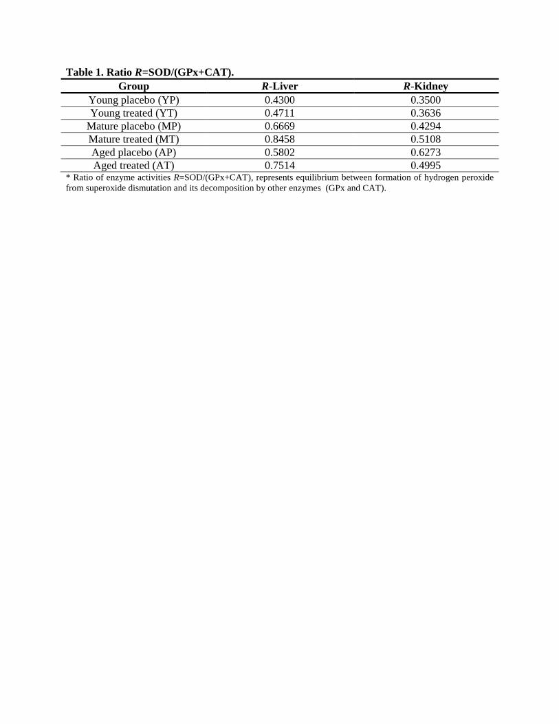
Table 1. Ratio R=SOD/(GPx+CAT).
Group R-Liver R-Kidney
Young placebo (YP) 0.4300 0.3500
Young treated (YT) 0.4711 0.3636
Mature placebo (MP) 0.6669 0.4294
Mature treated (MT) 0.8458 0.5108
Aged placebo (AP) 0.5802 0.6273
Aged treated (AT) 0.7514 0.4995 * Ratio of enzyme activities R=SOD/(GPx+CAT), represents equilibrium between formation of hydrogen peroxide
from superoxide dismutation and its decomposition by other enzymes (GPx and CAT).
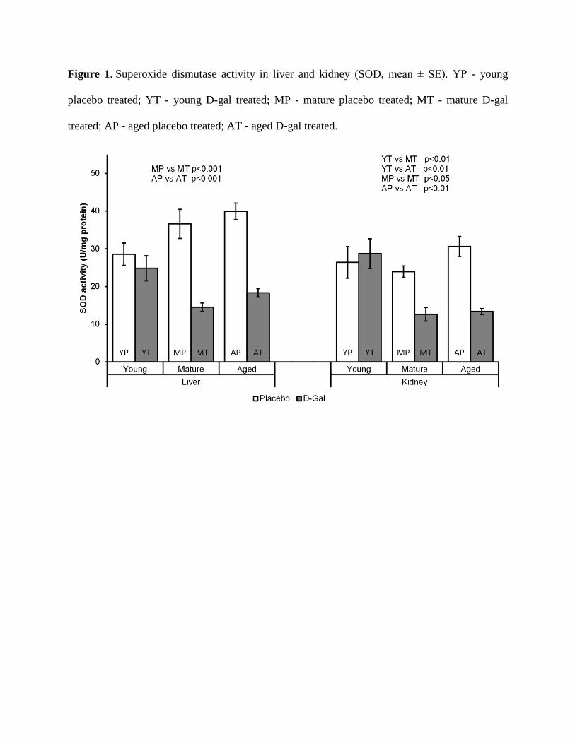
Figure 1. Superoxide dismutase activity in liver and kidney (SOD, mean ± SE). YP - young
placebo treated; YT - young D-gal treated; MP - mature placebo treated; MT - mature D-gal
treated; AP - aged placebo treated; AT - aged D-gal treated.
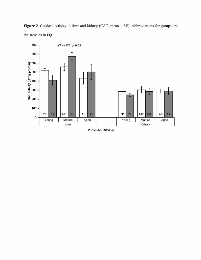
Figure 2. Catalase activity in liver and kidney (CAT, mean ± SE). Abbreviations for groups are
the same as in Fig. 1.

Figure 3. Glutathione peroxidase activity in liver and kidney (GPx, mean ± SE). Abbreviations
for groups are the same as in Fig. 1.
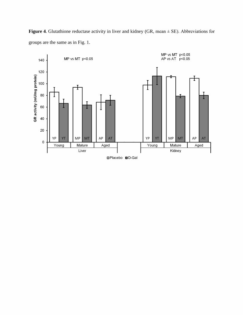
Figure 4. Glutathione reductase activity in liver and kidney (GR, mean ± SE). Abbreviations for
groups are the same as in Fig. 1.
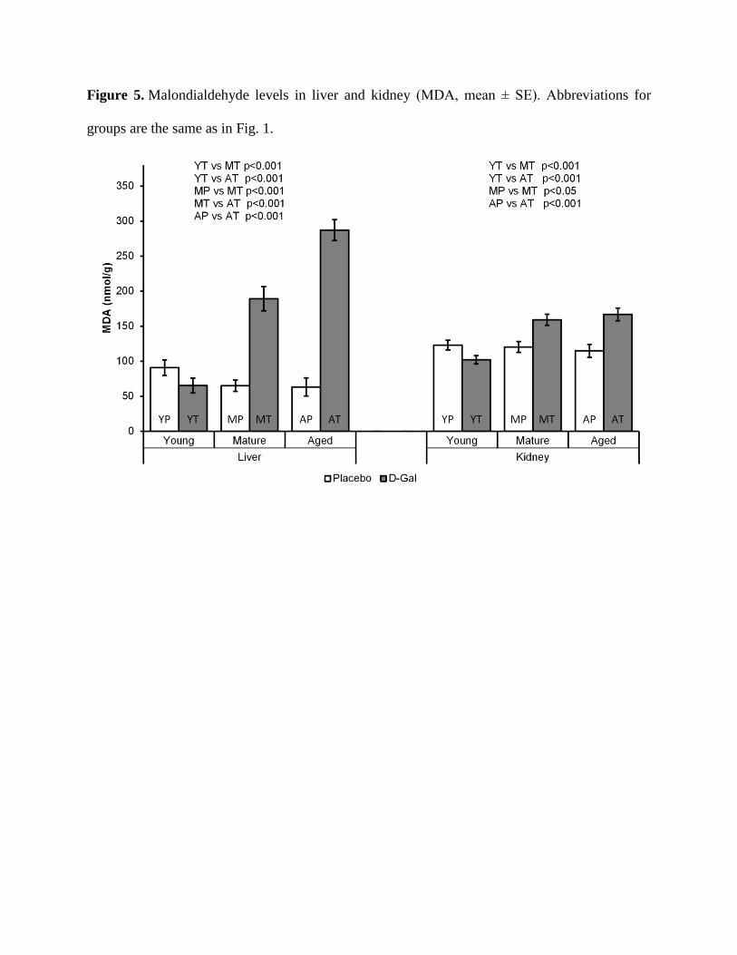
Figure 5. Malondialdehyde levels in liver and kidney (MDA, mean ± SE). Abbreviations for
groups are the same as in Fig. 1.