d- chapter 19 lecture powerpoint respiratory system anatomy and physiology
TRANSCRIPT
D- Chapter 19Lecture
PowerPointRespiratory
systemAnatomy and Physiology
2
Chapter 19
Respiratory System
3
19.1: Introduction• The respiratory system consists of passages that filter incoming air and transport it into the body, into the lungs, and to the many microscopic air sacs where gases are exchanged• Respiration is the process of exchanging gases between the atmosphere and body cells• It consists of the following events:
• Ventilation• External respiration• Transport of gases• Internal respiration• Cellular respiration
4
19.2: Why We Breathe• Respiration occurs on a macroscopic level at the organ system• Gas exchange, oxygen and carbon dioxide, occur at the cellular and molecular levels• Aerobic reactions of cellular respiration allow for:
• ATP production• Carbon dioxide generation forming carbonic acid
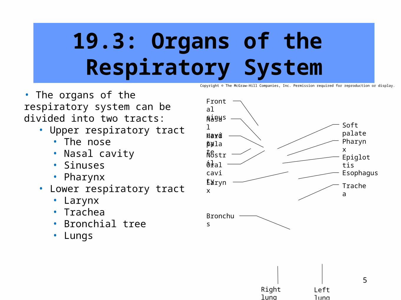
19.3: Organs of the Respiratory System
• The organs of the respiratory system can be divided into two tracts:
• Upper respiratory tract• The nose• Nasal cavity• Sinuses• Pharynx
• Lower respiratory tract• Larynx• Trachea• Bronchial tree• Lungs
Copyright © The McGraw-Hill Companies, Inc. Permission required for reproduction or display.
Larynx
Bronchus
Nostril
Right lung Left lung
Soft palate
Pharynx
Epiglottis
Esophagus
Frontalsinus
NasalcavityHardpalate
Oralcavity
Trachea
5
6
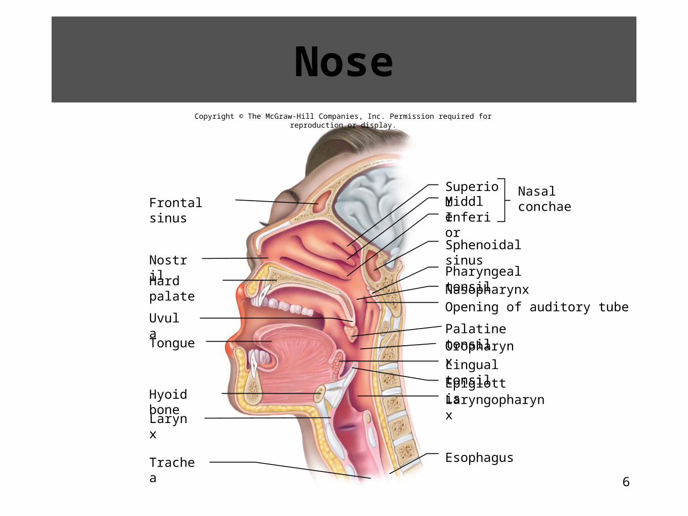
Nose
Frontal sinus
Nostril
Hard palate
Uvula
EpiglottisHyoid bone
Larynx
SuperiorMiddleInferior
Sphenoidal sinus
Pharyngeal tonsil
Nasopharynx
Palatine tonsilOropharynx
Lingual tonsil
Laryngopharynx
Esophagus
Tongue
Trachea
Nasalconchae
Opening of auditory tube
Copyright © The McGraw-Hill Companies, Inc. Permission required for reproduction or display.
7
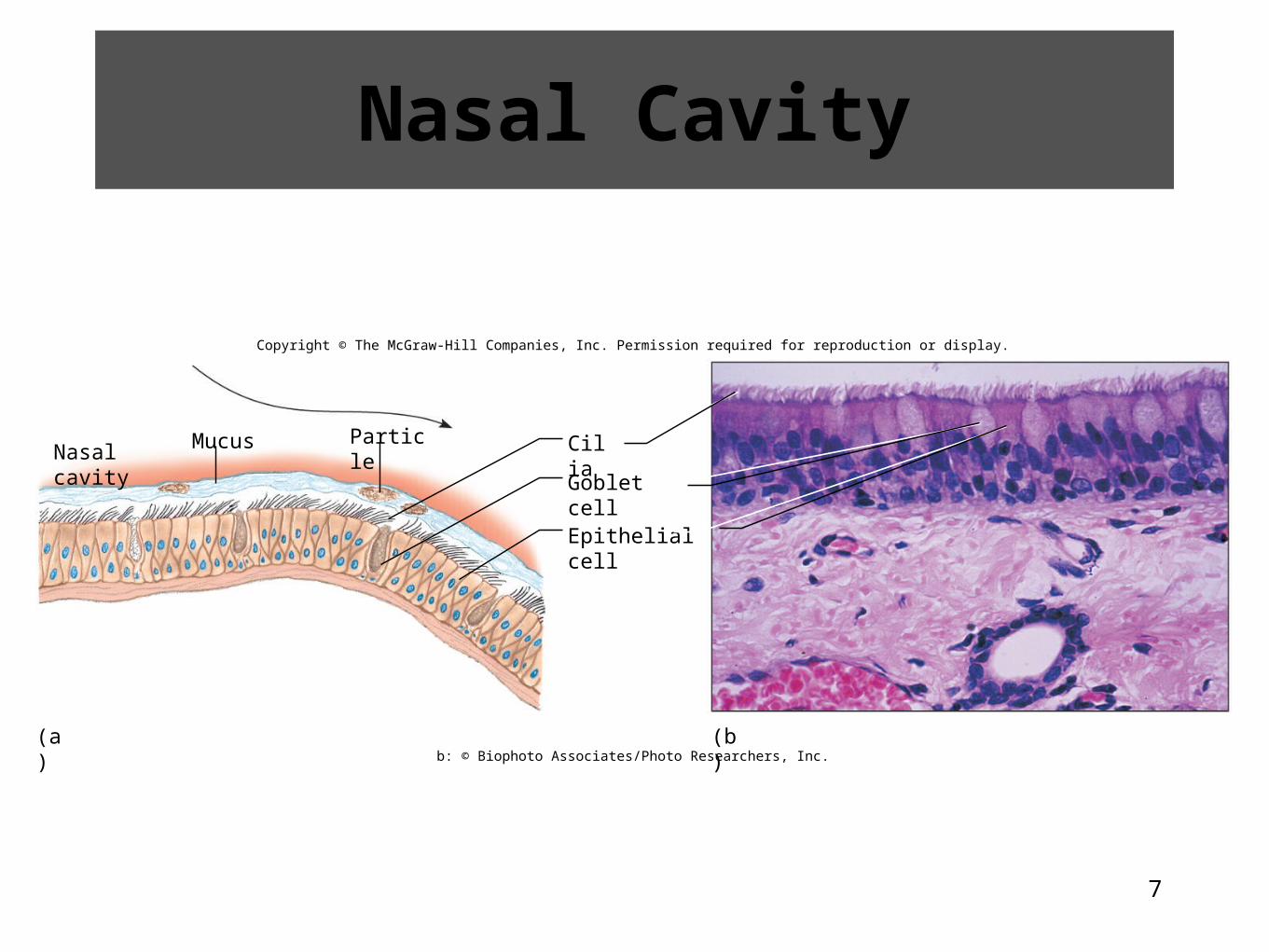
Nasal Cavity
Copyright © The McGraw-Hill Companies, Inc. Permission required for reproduction or display.
Mucus Particle
Goblet cell
CiliaNasal cavity
Epithelial cell
(b)(a)b: © Biophoto Associates/Photo Researchers, Inc.
8
Sinuses• The sinuses are air-filled spaces in the maxillary, frontal, ethmoid, and sphenoid bones of the skull
9
19.1 Clinical Application
The Effects of Cigarette Smoking on the Respiratory System
10
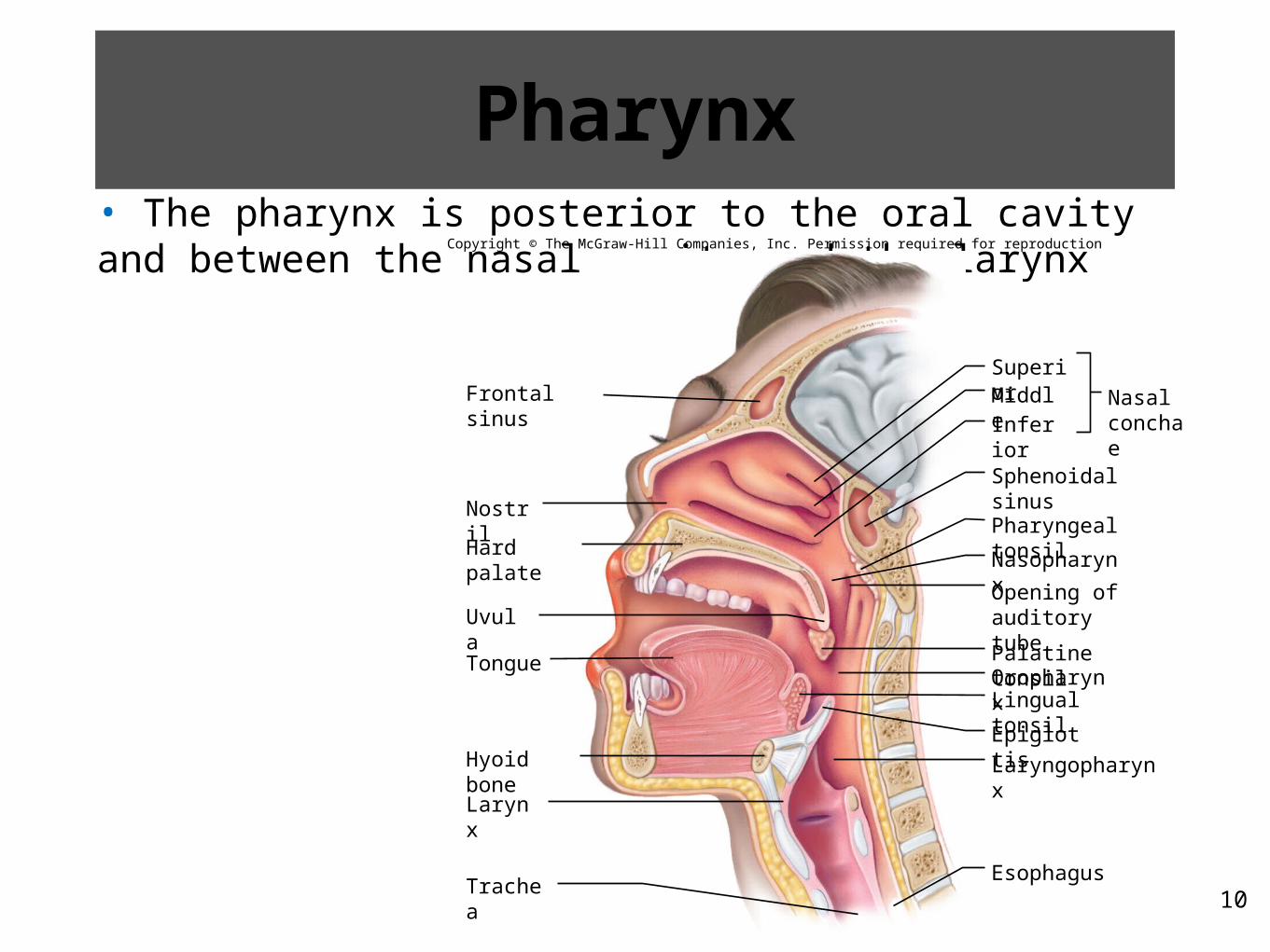
Pharynx• The pharynx is posterior to the oral cavity and between the nasal cavity and the larynx
Copyright © The McGraw-Hill Companies, Inc. Permission required for reproduction or display.
Frontal sinus
Nostril
Hard palate
Uvula
EpiglottisHyoid bone
Larynx
SuperiorMiddle
Inferior
Sphenoidal sinus
Pharyngeal tonsil
Nasopharynx
Palatine tonsilOropharynxLingual tonsil
Laryngopharynx
Esophagus
Tongue
Trachea
Nasalconchae
Opening ofauditory tube
11
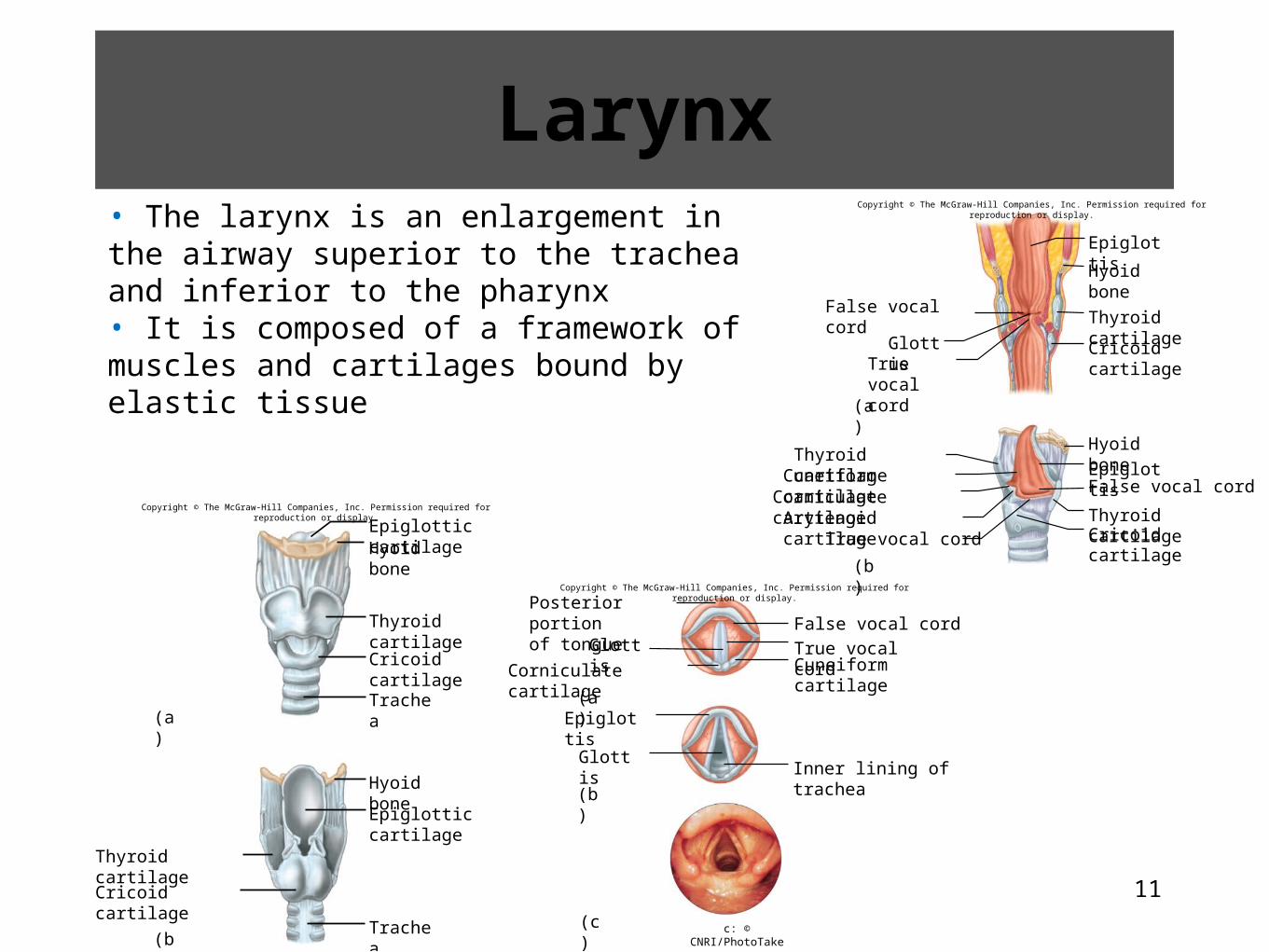
Larynx• The larynx is an enlargement in the airway superior to the trachea and inferior to the pharynx• It is composed of a framework of muscles and cartilages bound by elastic tissue
Epiglottic cartilageHyoid bone
Thyroid cartilage
Cricoid cartilage
Hyoid bone
Epiglottic cartilage
Thyroid cartilage
Cricoid cartilage
(b)
(a)
Copyright © The McGraw-Hill Companies, Inc. Permission required for reproduction or display.
Trachea
Trachea
False vocal cord
Glottis
Epiglottis
Hyoid bone
Thyroid cartilage
Cricoid cartilage
Hyoid bone
Epiglottis
Thyroid cartilageCricoid cartilage
(b)
(a)
False vocal cord
True vocalcord
Thyroid cartilageCuneiform cartilage
Corniculate cartilageArytenoid cartilage
True vocal cord
Copyright © The McGraw-Hill Companies, Inc. Permission required for reproduction or display.
Glottis
Corniculate cartilage
(a)
(b)
Epiglottis
Glottis
(c)
Posterior portionof tongue False vocal cord
True vocal cordCuneiform cartilage
Inner lining of trachea
c: © CNRI/PhotoTake
Copyright © The McGraw-Hill Companies, Inc. Permission required for reproduction or display.
Trachea
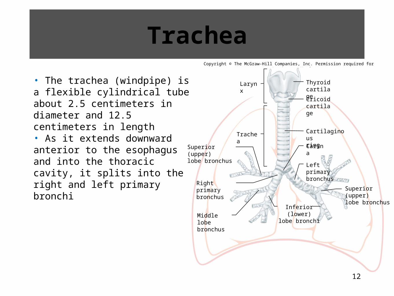
• The trachea (windpipe) is a flexible cylindrical tube about 2.5 centimeters in diameter and 12.5 centimeters in length• As it extends downward anterior to the esophagus and into the thoracic cavity, it splits into the right and left primary bronchi
Copyright © The McGraw-Hill Companies, Inc. Permission required for reproduction or display.
Larynx
Carina
Trachea
Superior (upper)lobe bronchus
Right primarybronchus
Middle lobebronchus
Inferior (lower)lobe bronchi
Thyroidcartilage
Cricoidcartilage
Cartilaginousring
Leftprimarybronchus
Superior (upper)lobe bronchus
12
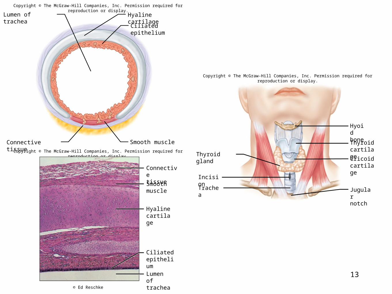
13
Hyaline cartilage
Ciliated epithelium
Smooth muscle
Lumen of trachea
Connective tissue
Copyright © The McGraw-Hill Companies, Inc. Permission required for reproduction or display.
Copyright © The McGraw-Hill Companies, Inc. Permission required for reproduction or display.
Connectivetissue
Hyalinecartilage
Ciliatedepithelium
Lumen oftrachea
Smoothmuscle
© Ed Reschke
Thyroid gland
Incision
Trachea
Hyoidbone
Thyroidcartilage
Cricoidcartilage
Jugularnotch
Copyright © The McGraw-Hill Companies, Inc. Permission required for reproduction or display.
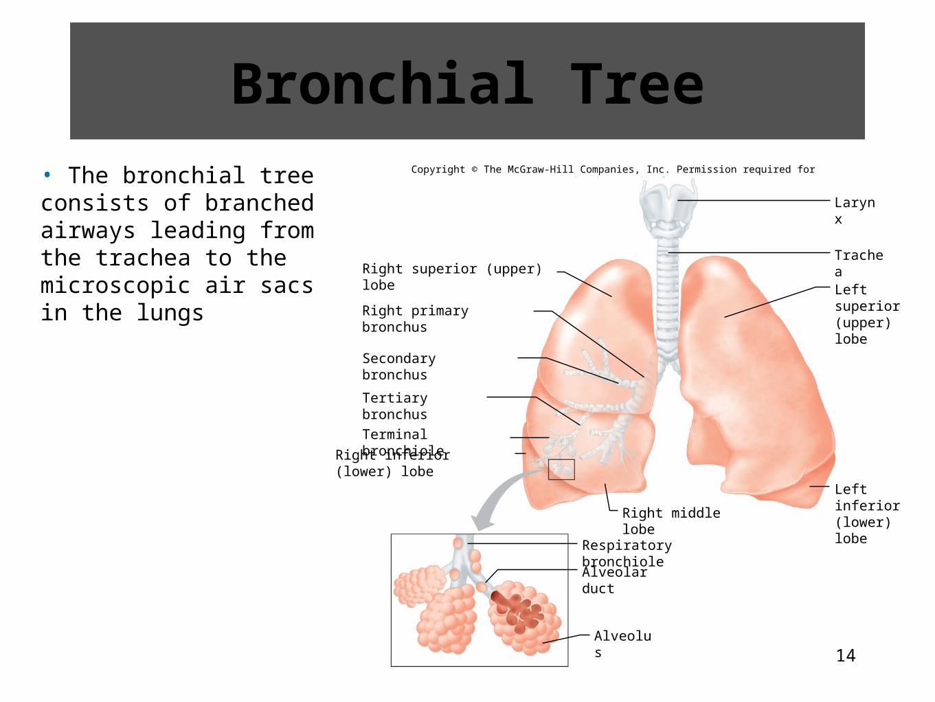
Bronchial Tree• The bronchial tree consists of branched airways leading from the trachea to the microscopic air sacs in the lungs
Copyright © The McGraw-Hill Companies, Inc. Permission required for reproduction or display.
Larynx
Right middle lobe
Right superior (upper) lobe
Right primary bronchus
Secondary bronchus
Right inferior (lower) lobe
Alveolar duct
Alveolus
Respiratory bronchiole
Tertiary bronchus
Terminal bronchiole
Trachea
Left superior(upper) lobe
Left inferior(lower) lobe
14

15
• The successive divisions of the branches from the trachea to the alveoli are:1.Right and left primary bronchi2.Secondary or lobar bronchi3.Tertiary or segmental bronchi4.Intralobular bronchioles (12-14 generations)5.Terminal bronchioles6.Respiratory bronchioles7.Alveolar ducts•Alveolar sacs1.Alveoli
Copyright © The McGraw-Hill Companies, Inc. Permission required for reproduction or display.
© Ralph Hutchings/Visuals Unlimited
Branches of the Bronchial Tree
16
Copyright © The McGraw-Hill Companies, Inc. Permission required for reproduction or display.
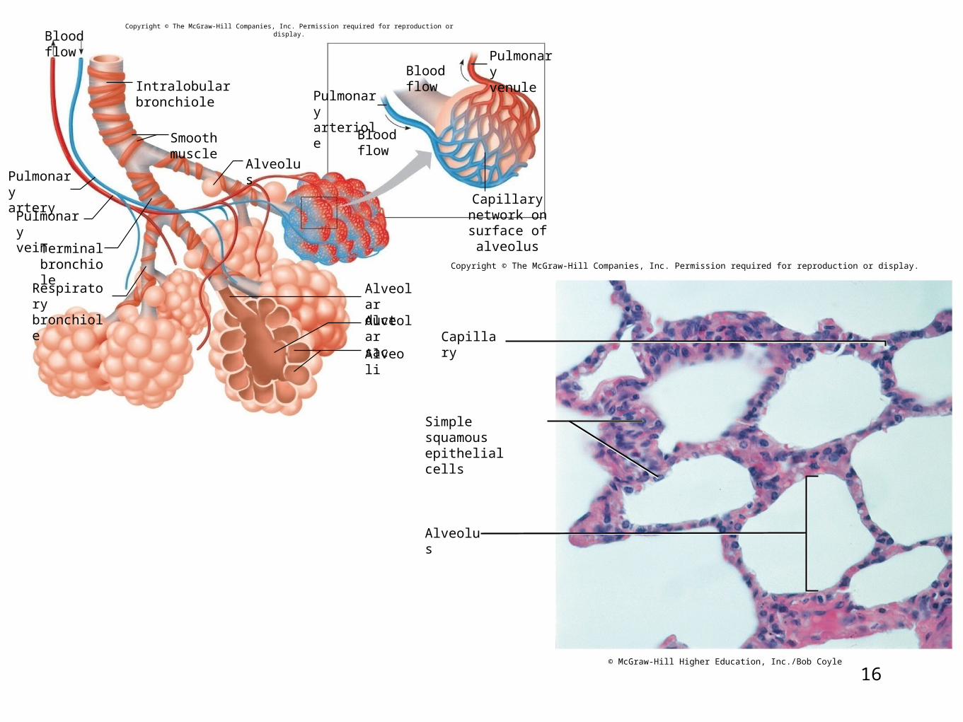
Intralobular bronchiole
Blood flow
Alveolus
Smooth muscle
Alveoli
Blood flow
Blood flow
Pulmonaryartery
Pulmonaryvein
Terminalbronchiole
Respiratorybronchiole
Pulmonaryarteriole
Pulmonaryvenule
Capillary network onsurface of alveolus
AlveolarductAlveolarsac
Copyright © The McGraw-Hill Companies, Inc. Permission required for reproduction or display.
Capillary
Alveolus
Simple squamousepithelial cells
© McGraw-Hill Higher Education, Inc./Bob Coyle
17
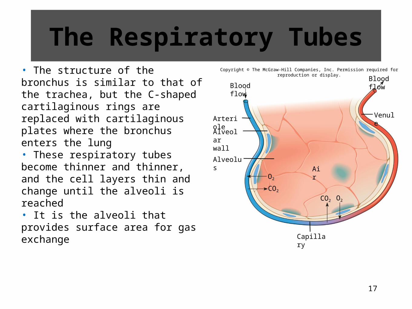
• The structure of the bronchus is similar to that of the trachea, but the C-shaped cartilaginous rings are replaced with cartilaginous plates where the bronchus enters the lung• These respiratory tubes become thinner and thinner, and the cell layers thin and change until the alveoli is reached• It is the alveoli that provides surface area for gas exchange
Copyright © The McGraw-Hill Companies, Inc. Permission required for reproduction or display.
Blood flowBlood flow
Arteriole
Alveolus
Capillary
Air
O2CO2
CO2
Alveolarwall
Venule
O2
The Respiratory Tubes
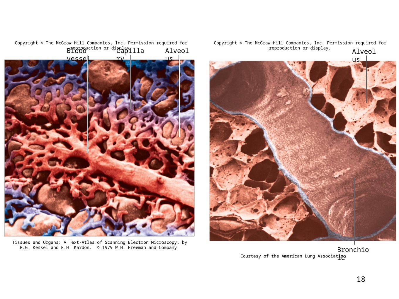
18
Copyright © The McGraw-Hill Companies, Inc. Permission required for reproduction or display.
Blood vessel Capillary Alveolus
Tissues and Organs: A Text-Atlas of Scanning Electron Microscopy, by R.G. Kessel and R.H. Kardon. © 1979 W.H. Freeman and Company
Copyright © The McGraw-Hill Companies, Inc. Permission required for reproduction or display.
Courtesy of the American Lung Association Bronchiole
Alveolus
19
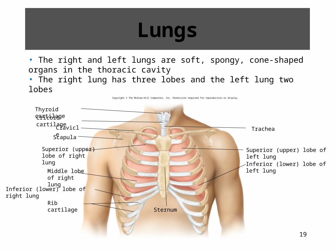
Lungs• The right and left lungs are soft, spongy, cone-shaped organs in the thoracic cavity• The right lung has three lobes and the left lung two lobes
Thyroid cartilage
Cricoid cartilage
Clavicle
Scapula
Rib cartilageSternum
Superior (upper)lobe of right lung
Middle lobeof right lung
Inferior (lower) lobe of right lung
Superior (upper) lobe of left lung
Inferior (lower) lobe of left lung
Trachea
Copyright © The McGraw-Hill Companies, Inc. Permission required for reproduction or display.
20
21
19.2 Clinical Application
Lung Irritants
22
19.4: Breathing Mechanism• Breathing or ventilation is the movement of air from outside of the body into the bronchial tree and the alveoli
• The actions responsible for these air movements are inspiration, or inhalation, and expiration, or exhalation
23
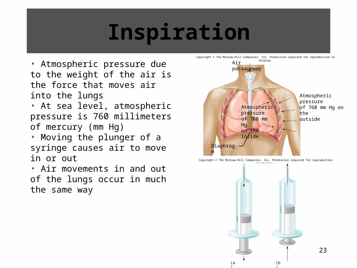
Inspiration• Atmospheric pressure due to the weight of the air is the force that moves air into the lungs• At sea level, atmospheric pressure is 760 millimeters of mercury (mm Hg)• Moving the plunger of a syringe causes air to move in or out• Air movements in and out of the lungs occur in much the same way
Copyright © The McGraw-Hill Companies, Inc. Permission required for reproduction or display.
Diaphragm
Air passageway
Atmospheric pressureof 760 mm Hg on theoutsideAtmospheric
pressureof 760 mm Hgon the inside
Copyright © The McGraw-Hill Companies, Inc. Permission required for reproduction or display.
(a) (b)
24
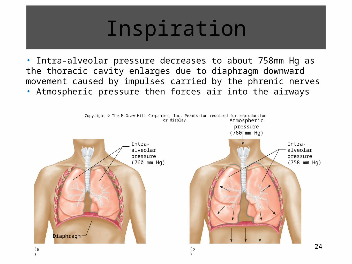
• Intra-alveolar pressure decreases to about 758mm Hg as the thoracic cavity enlarges due to diaphragm downward movement caused by impulses carried by the phrenic nerves• Atmospheric pressure then forces air into the airways
Copyright © The McGraw-Hill Companies, Inc. Permission required for reproduction or display.
Diaphragm
(a) (b)
Intra-alveolarpressure(760 mm Hg)
Atmospheric pressure(760 mm Hg)
Intra-alveolarpressure(758 mm Hg)
Inspiration
25
Copyright © The McGraw-Hill Companies, Inc. Permission required for reproduction or display.
(a) (b)
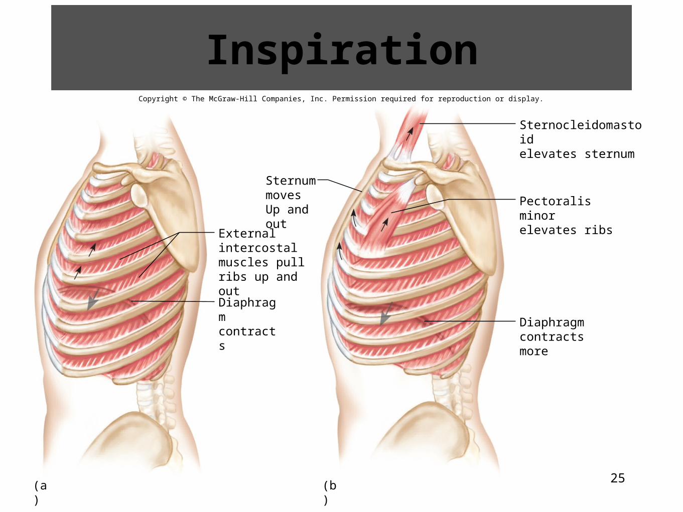
Externalintercostalmuscles pullribs up and out
Diaphragmcontracts
SternummovesUp and out
Sternocleidomastoidelevates sternum
Pectoralis minorelevates ribs
Diaphragmcontracts more
Inspiration

26
27
Expiration• The forces responsible for normal resting expiration come from elastic recoil of lung tissues and from surface tension• These factors increase the intra-alveolar pressure about 1 mm Hg above atmospheric pressure forcing air out of the lungs
28
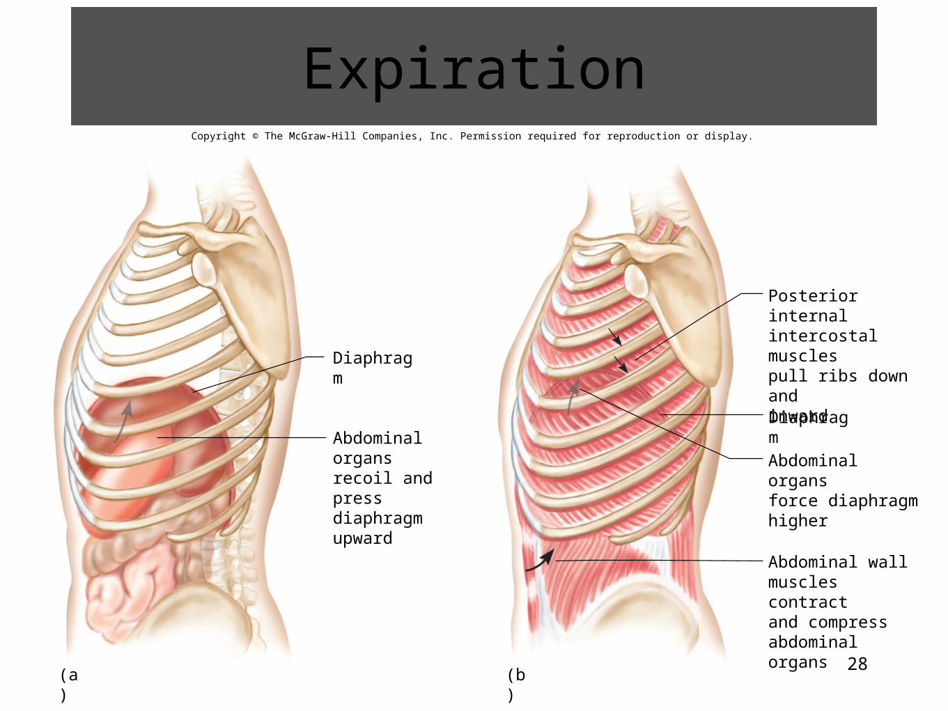
Copyright © The McGraw-Hill Companies, Inc. Permission required for reproduction or display.
Diaphragm
Diaphragm
(a) (b)
Abdominal organsrecoil and pressdiaphragm upward
Posterior internalintercostal musclespull ribs down andinward
Abdominal organsforce diaphragmhigher
Abdominal wallmuscles contractand compressabdominal organs
Expiration
29
30
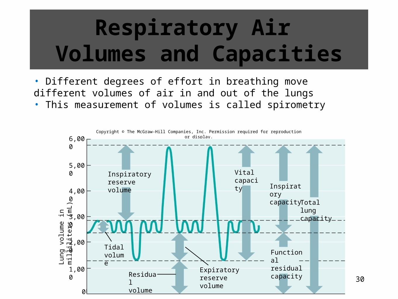
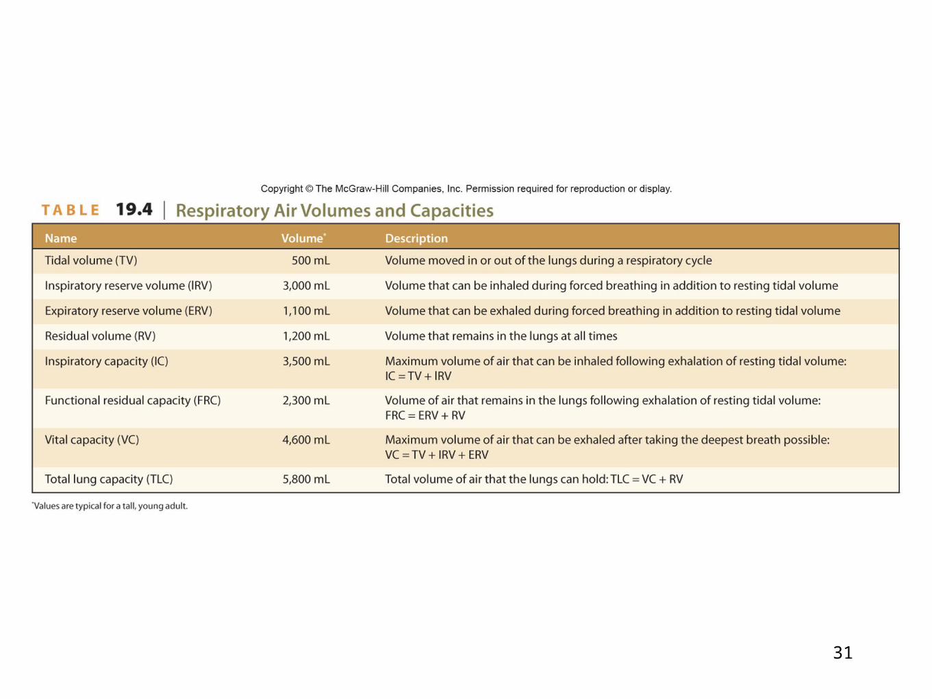
Respiratory Air Volumes and Capacities
• Different degrees of effort in breathing move different volumes of air in and out of the lungs• This measurement of volumes is called spirometry
Copyright © The McGraw-Hill Companies, Inc. Permission required for reproduction or display.
Lung
vo
lum
e in
mill
ilite
rs (
mL)
6,000
5,000
4,000
3,000
2,000
1,000
0
Inspiratoryreserve volume
Tidalvolume
Residualvolume
Expiratoryreserve volume
Vitalcapacity
Inspiratorycapacity
Total lungcapacity
Functionalresidualcapacity
31
32
Alveolar Ventilation
• The volume of new atmospheric air moved into the respiratory passages each minute is minute ventilation• It equals the tidal volume multiplied by the breathing rate• Much of the new air remains in the physiologic dead space• The tidal volume minus the physiologic dead space then multiplied by breathing rate is the alveolar ventilation rate• This is the volume of air that reaches the alveoli• This impacts the concentrations of oxygen and carbon dioxide in the alveoli
33
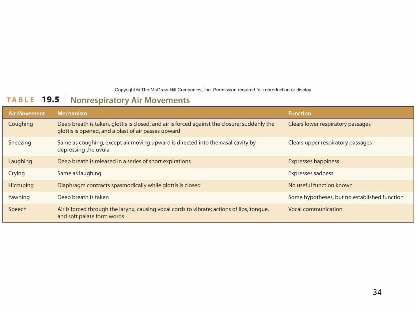
Nonrespiratory Air Movements
• Air movements other than breathing are called nonrespiratory movements• They clear air passages, as in coughing and sneezing, or express emotions, as in laughing and crying
34
35
19.3 Clinical Application
Respiratory Disorders That Decrease Ventilation: Bronchial Asthma and
Emphysema
36
19.5: Control of Breathing
• Normal breathing is a rhythmic, involuntary act that continues when a person is unconscious• Respiratory muscles can be controlled as well voluntarily
37
Respiratory Areas
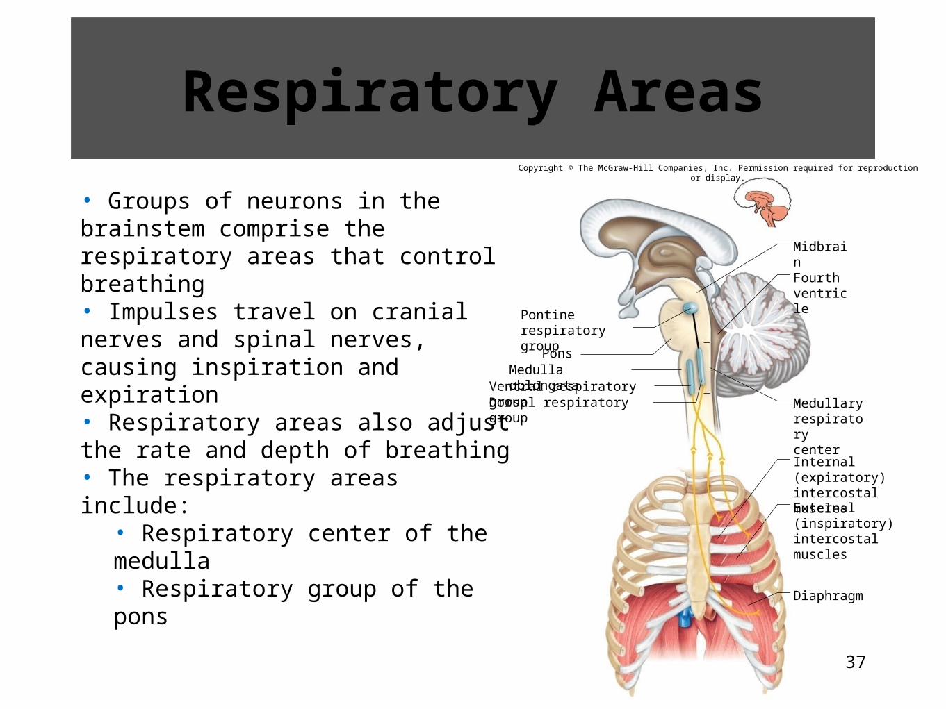
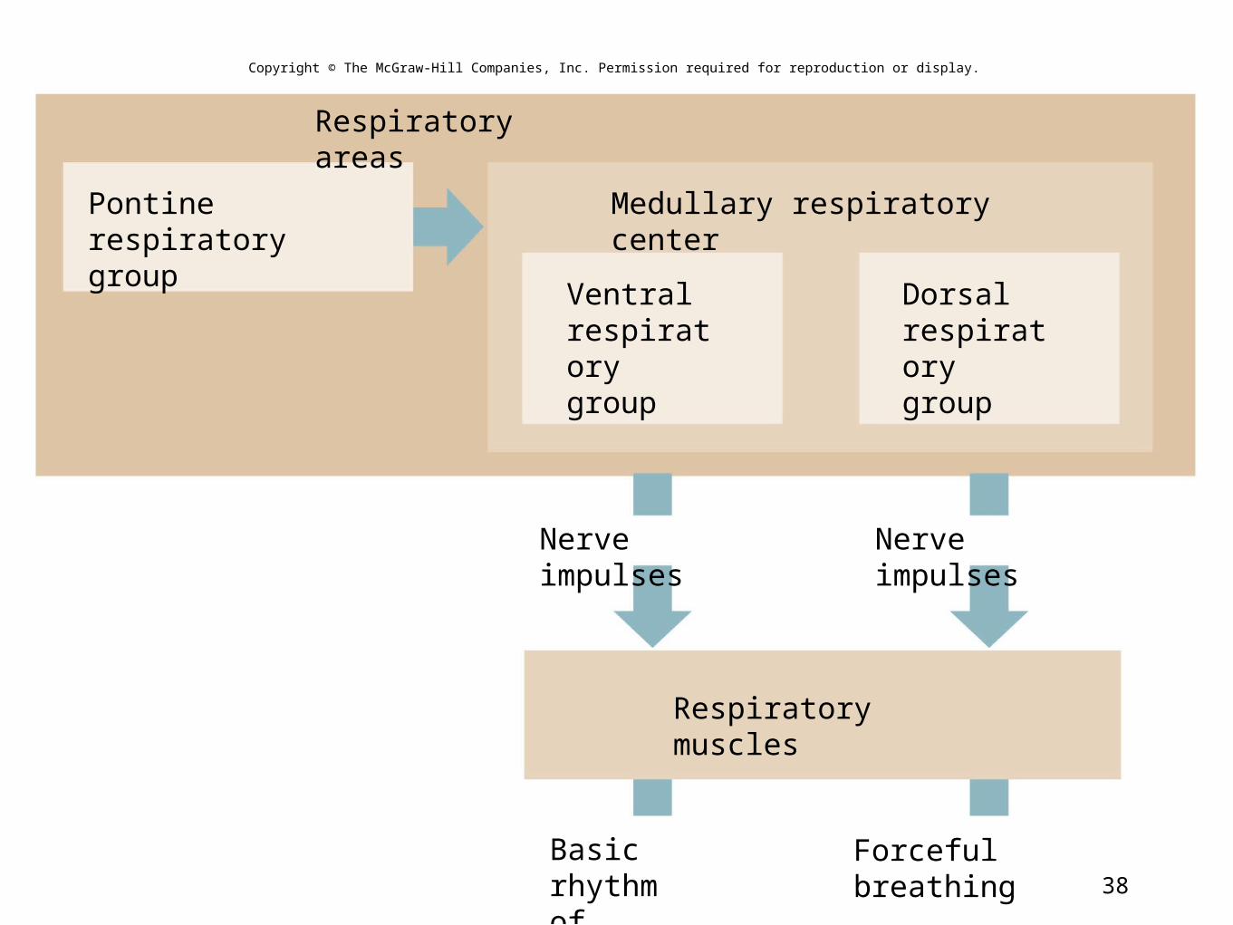
• Groups of neurons in the brainstem comprise the respiratory areas that control breathing• Impulses travel on cranial nerves and spinal nerves, causing inspiration and expiration• Respiratory areas also adjust the rate and depth of breathing• The respiratory areas include:
• Respiratory center of the medulla• Respiratory group of the pons
Copyright © The McGraw-Hill Companies, Inc. Permission required for reproduction or display.
Diaphragm
Medulla oblongataPons
Midbrain
Dorsal respiratory group
Pontine respiratorygroup
Ventral respiratory group
Fourthventricle
Medullaryrespiratorycenter
Internal (expiratory)intercostal muscles
External (inspiratory)intercostal muscles
38
Respiratory muscles
Forceful breathing
Nerve impulses Nerve impulses
Pontine respiratorygroup
Ventralrespiratorygroup
Dorsalrespiratorygroup
Medullary respiratory center
Respiratory areas
Basic rhythmof breathing
Copyright © The McGraw-Hill Companies, Inc. Permission required for reproduction or display.
39
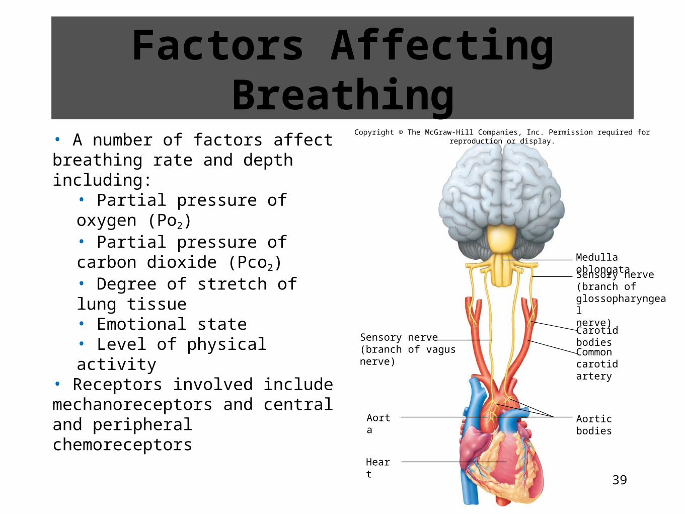
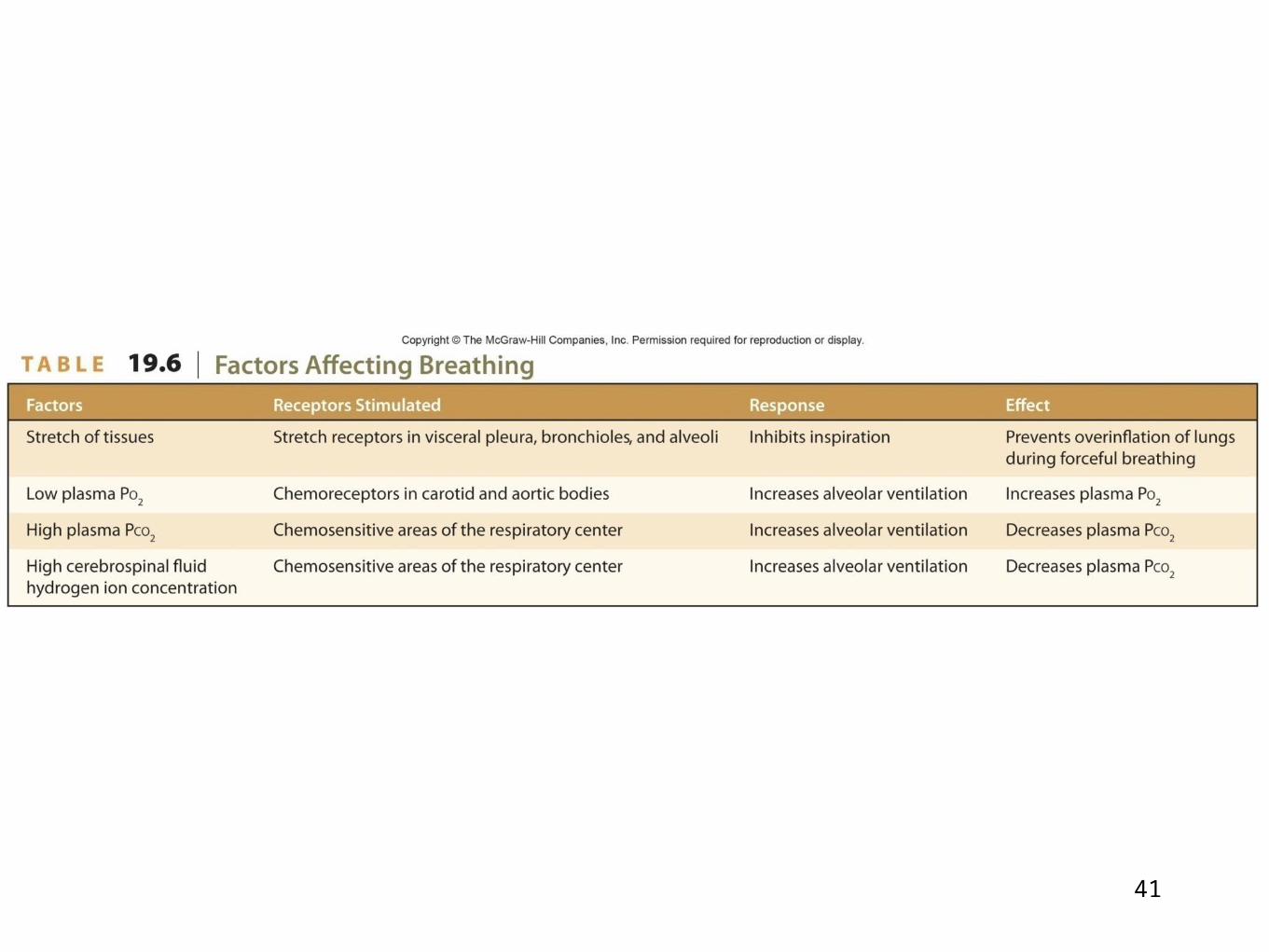
Factors Affecting Breathing
• A number of factors affect breathing rate and depth including:
• Partial pressure of oxygen (Po2)• Partial pressure of carbon dioxide (Pco2)• Degree of stretch of lung tissue• Emotional state• Level of physical activity
• Receptors involved include mechanoreceptors and central and peripheral chemoreceptors
Carotid bodies
Aorta
Heart
Aortic bodies
Medulla oblongata
Sensory nerve(branch ofglossopharyngealnerve)
Common carotidartery
Sensory nerve(branch of vagus nerve)
Copyright © The McGraw-Hill Companies, Inc. Permission required for reproduction or display.
40
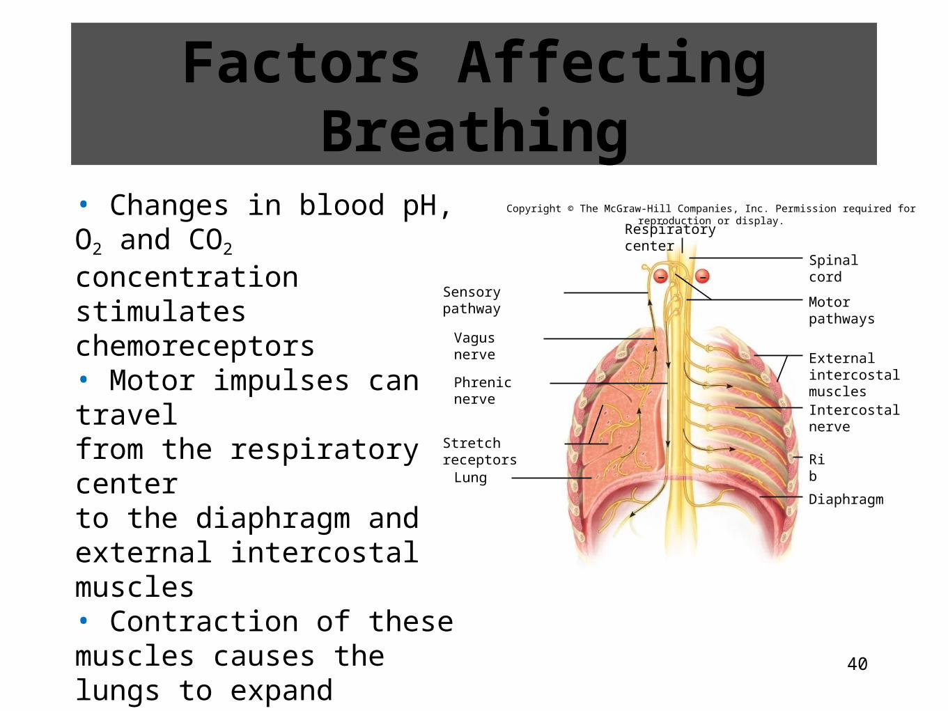
• Changes in blood pH, O2 and CO2 concentration stimulates chemoreceptors• Motor impulses can travelfrom the respiratory centerto the diaphragm and external intercostal muscles• Contraction of these muscles causes the lungs to expand stimulating mechanoreceptors in the lungs• Inhibitory impulses from the mechanoreceptors back to the respiratory center prevent overinflation of the lungs
Copyright © The McGraw-Hill Companies, Inc. Permission required for reproduction or display.
Respiratory center
Motor pathways
Spinal cord
Intercostal nerve
Rib
Diaphragm
Sensory pathway
Phrenic nerve
Stretch receptors
Lung
External intercostalmuscles
Vagus nerve
––
Factors Affecting Breathing
41
42
19.4 Clinical Application
Exercise and Breathing
43
19.6: Alveolar Gas Exchanges• The alveoli are the sites of the vital process of gas exchange between the air and the blood
44
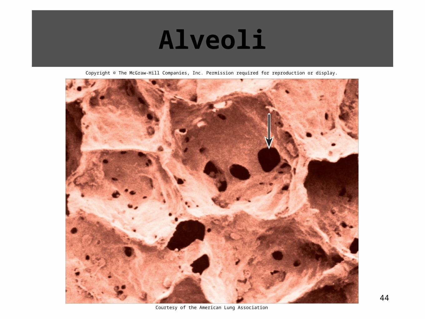
AlveoliCopyright © The McGraw-Hill Companies, Inc. Permission required for reproduction or display.
Courtesy of the American Lung Association
45
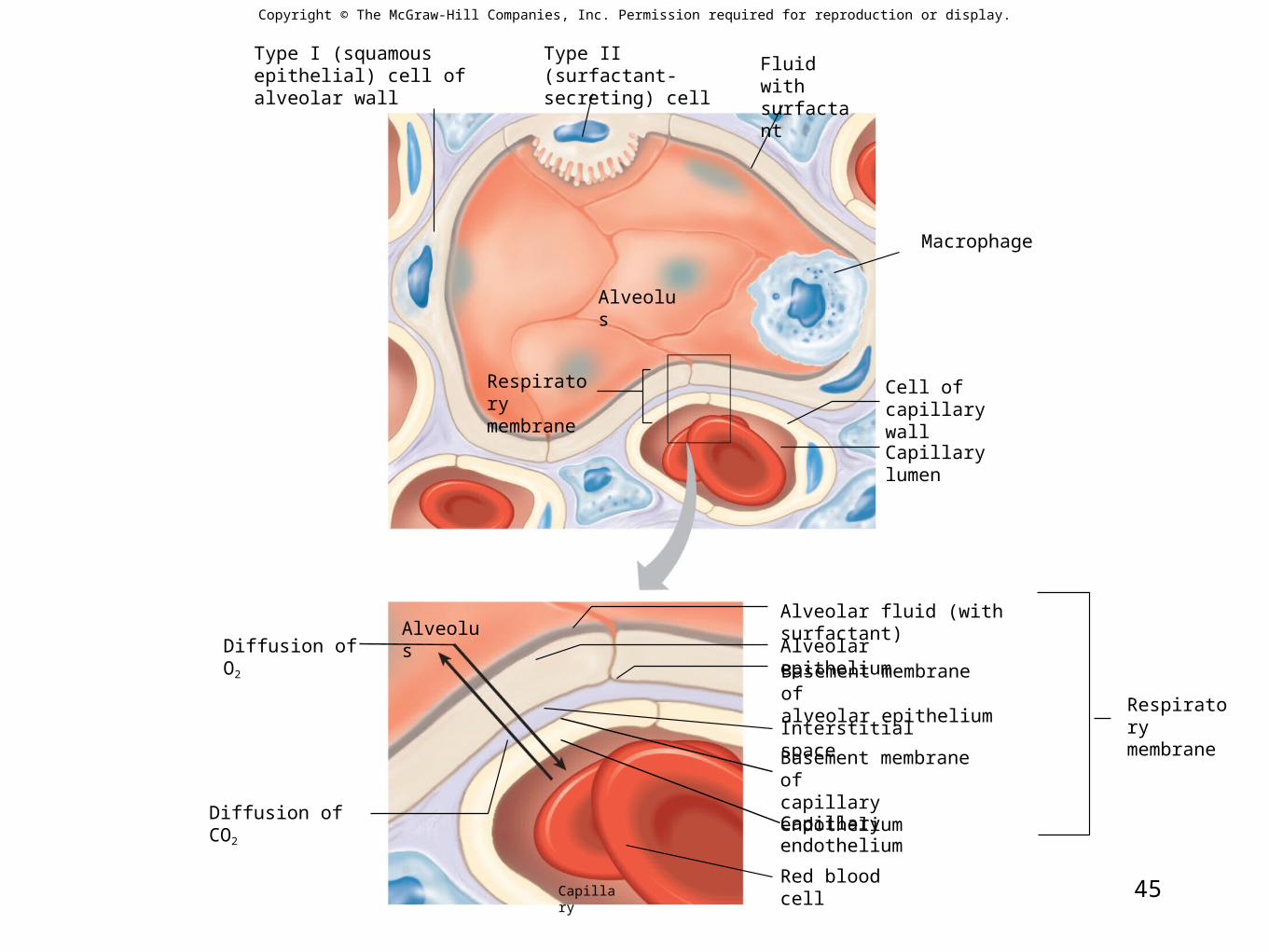
Capillary lumen
Alveolus
Macrophage
Capillary
Alveolus
Red blood cell
Diffusion of CO2
Diffusion of O2
Capillary endothelium
Interstitial space
Alveolar epithelium
Type I (squamous epithelial) cell of alveolar wall
Type II (surfactant-secreting) cell
Fluid withsurfactant
Respiratorymembrane
Cell ofcapillary wall
Alveolar fluid (with surfactant)
Basement membrane ofalveolar epithelium
Basement membrane ofcapillary endothelium
Respiratorymembrane
Copyright © The McGraw-Hill Companies, Inc. Permission required for reproduction or display.
46
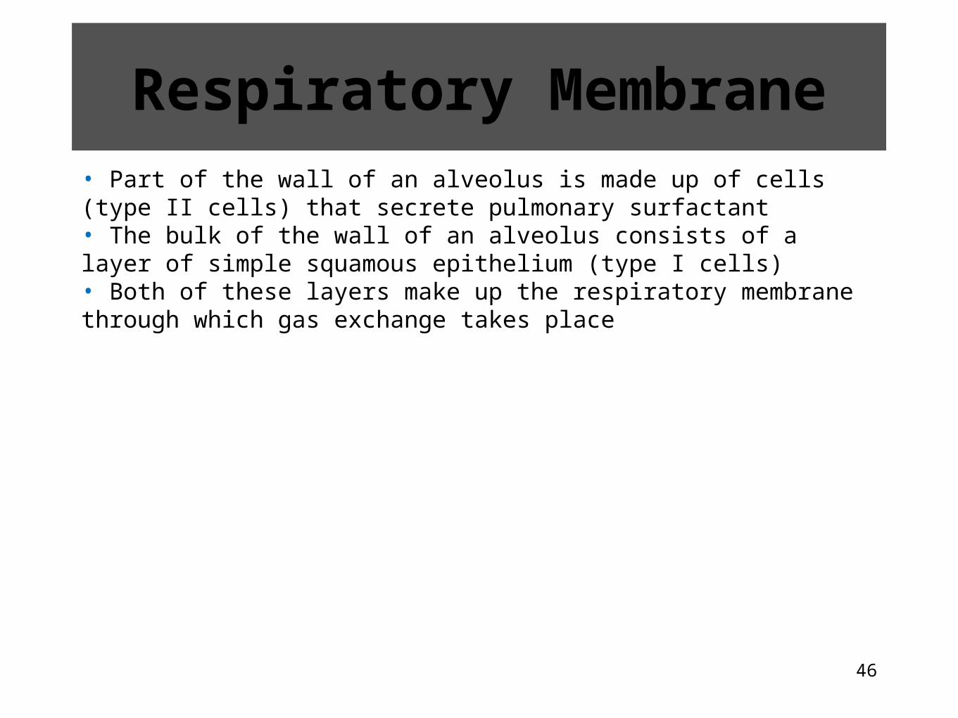
Respiratory Membrane• Part of the wall of an alveolus is made up of cells (type II cells) that secrete pulmonary surfactant• The bulk of the wall of an alveolus consists of a layer of simple squamous epithelium (type I cells)• Both of these layers make up the respiratory membrane through which gas exchange takes place
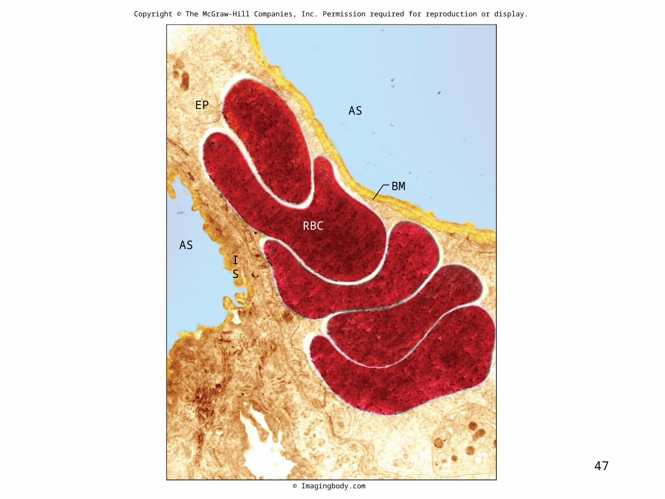
47
Copyright © The McGraw-Hill Companies, Inc. Permission required for reproduction or display.
AS
AS
BM
IS
RBC
EP
© Imagingbody.com
48
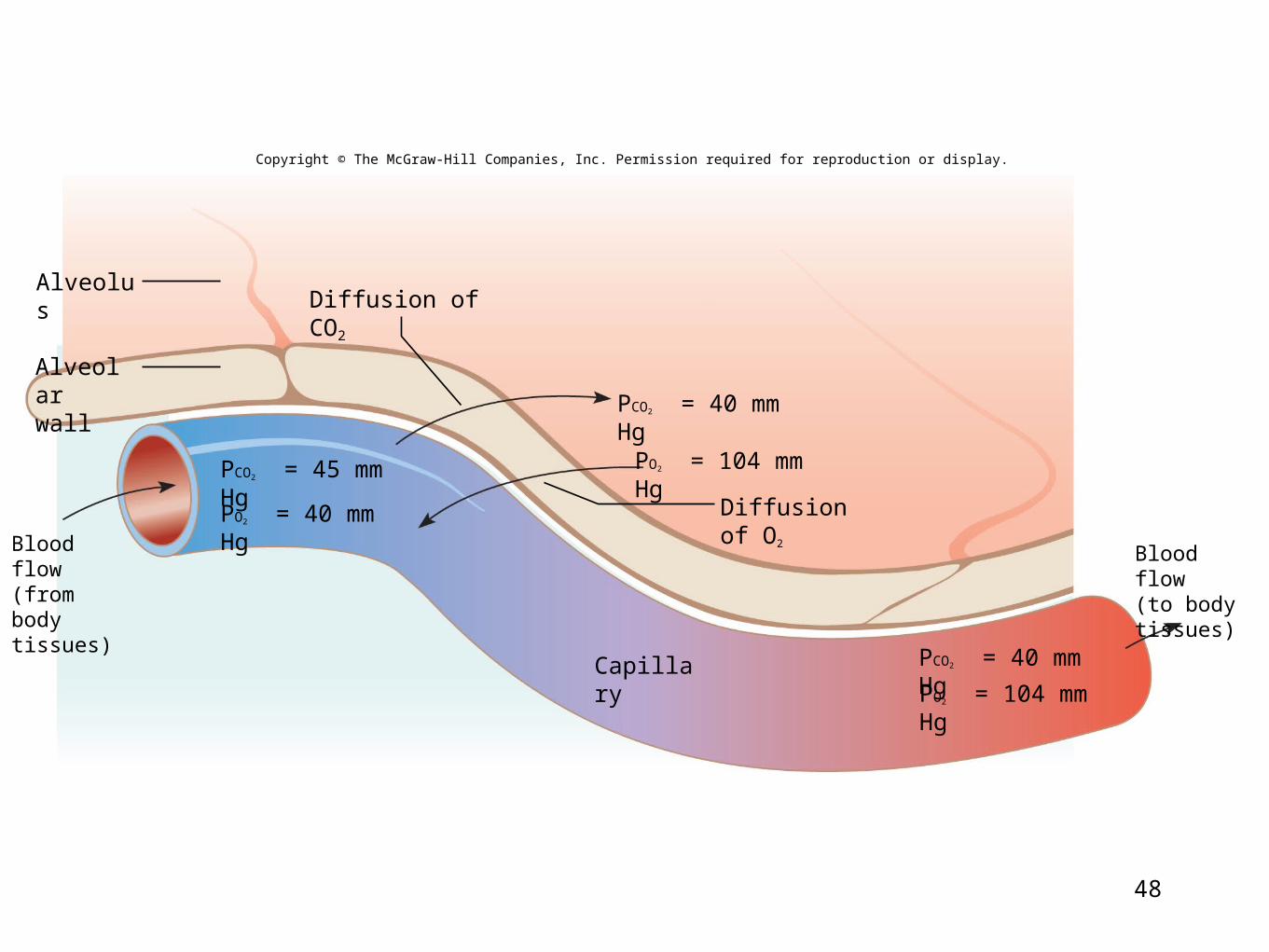
AlveolusDiffusion of CO2
Diffusion of O2
Capillary
Alveolarwall
Blood flow(from bodytissues)
Blood flow(to bodytissues)
PCO2 = 45 mm Hg
Copyright © The McGraw-Hill Companies, Inc. Permission required for reproduction or display.
PCO2 = 40 mm Hg
PO2 = 40 mm Hg
PO2 = 104 mm Hg
PO2 = 104 mm Hg
PCO2 = 40 mm Hg
49
Diffusion Through the Respiratory Membrane
• Molecules diffuse from regions where they are in higher concentration toward regions where they are in lower concentration• It is important to know the concentration gradient• In respiration, think in terms of gas partial pressures• Gases diffuse from areas of higher partial pressure to areas of lower partial pressure• The respiratory membrane is normally thin and gas exchange is rapid
• Increased diffusion is favored with more surface area, shorter distance, greater solubility of gases and a steeper partial pressure gradient• Decreased diffusion occurs from decreased surface area
50
19.5 Clinical Application
Effects of High Altitude
51
19.6 Clinical Application
Disorders That Impair Gas Exchange: Pneumonia, Tuberculosis, and Adult
Respiratory Distress Syndrome
52
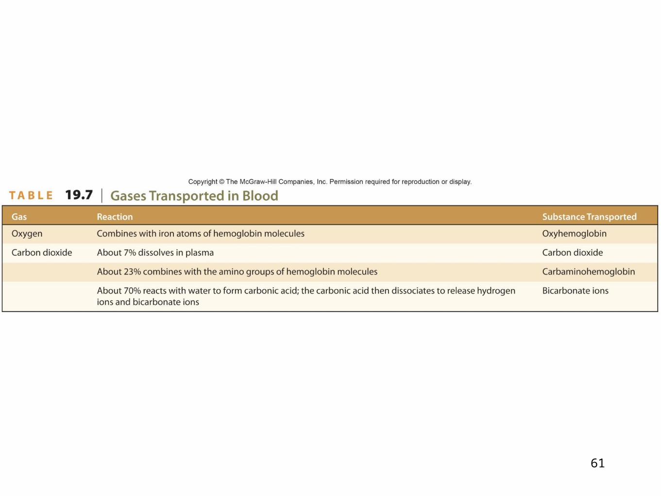
19.7: Gas Transport
• Blood transports O2 and CO2 between the lungs and the body cells• As the gases enter the blood, they dissolve in the plasma or chemically combine with other atoms or molecules
53
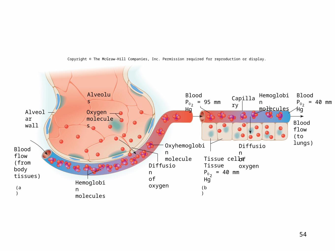
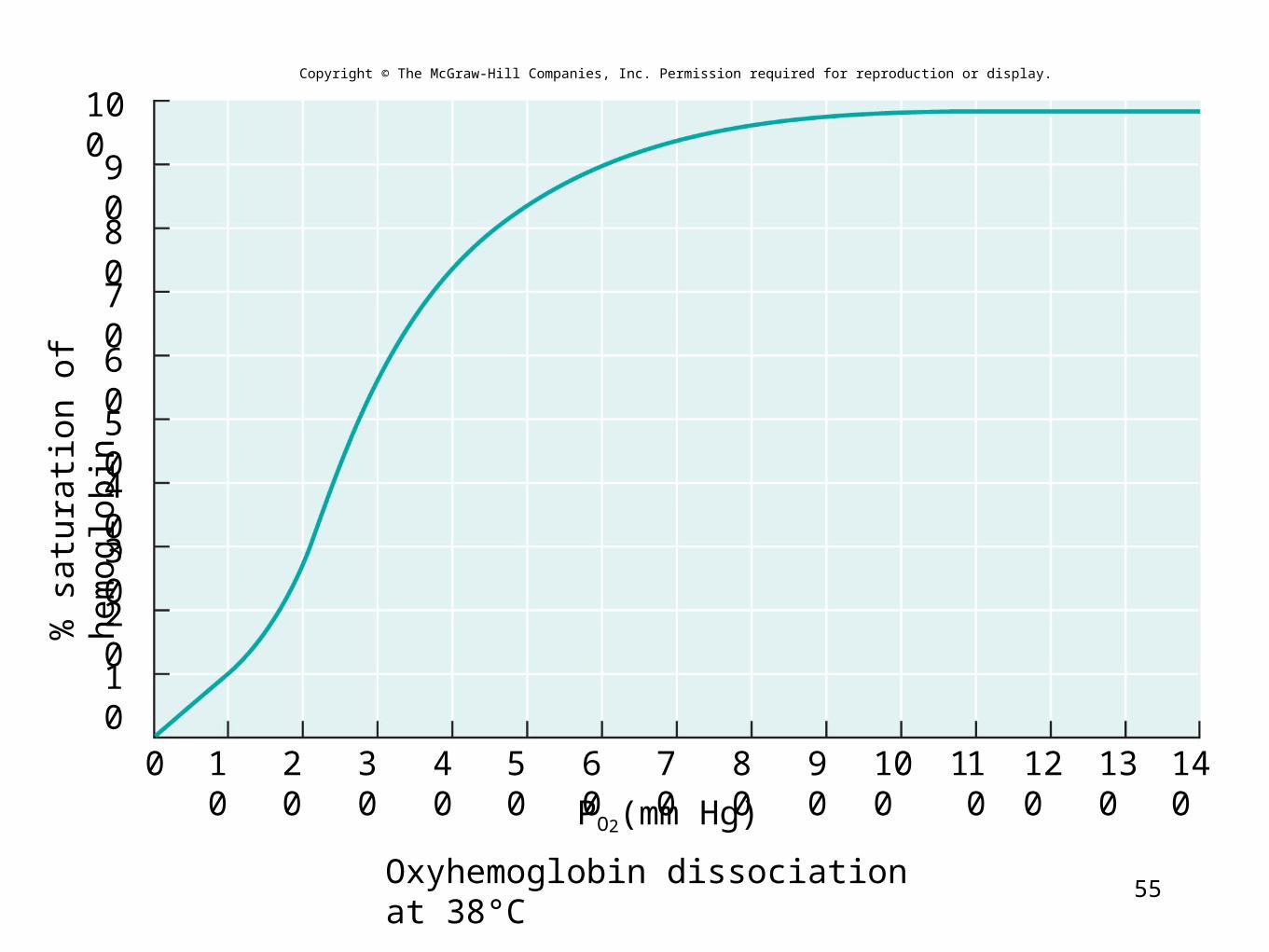
Oxygen Transport• Almost all oxygen carried in the blood is bound to the protein hemoglobin in the form of oxyhemoglobin• Chemical bonds between O2 and hemoglobin are relatively unstable• Oxyhemoglobin releases O2 into the body cells• About 75% of the O2 remains bound to hemoglobin in the venous blood ensuring safe CO2 levels and thereby pH
54
AlveolusCapillary
2
2
2
(a) (b)
Blood flow(from bodytissues)
Alveolarwall
Oxygenmolecules
Hemoglobinmolecules
Diffusionof oxygen
Oxyhemoglobinmolecule
Hemoglobinmolecules
Diffusionof oxygen
Blood flow(to lungs)
BloodPO = 40 mm Hg
BloodPO = 95 mm Hg
Tissue cellsTissuePO = 40 mm Hg
Copyright © The McGraw-Hill Companies, Inc. Permission required for reproduction or display.
55
Copyright © The McGraw-Hill Companies, Inc. Permission required for reproduction or display.
10
20
30
40
50
60
70
80
90
100
Oxyhemoglobin dissociation at 38°C
% s
atur
atio
n of
hem
oglo
bin
100 40 50 60 70 9080 100 110 120 130 14020 30
PO2(mm Hg)
56
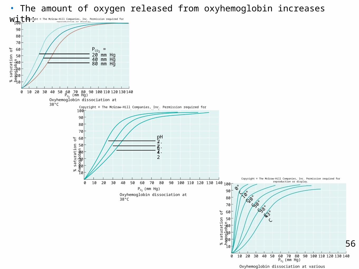
• The amount of oxygen released from oxyhemoglobin increases with: Copyright © The McGraw-Hill Companies, Inc. Permission required for reproduction or display.
10
20
30
40
50
60
70
80
90
100
% s
atur
atio
n of
hem
oglo
bin
100 40 50 60 70 9080 100 110 120 130 14020 30
Oxyhemoglobin dissociation at 38°C
20 mm Hg40 mm Hg80 mm Hg
PCO2 =
PO2 (mm Hg)
Copyright © The McGraw-Hill Companies, Inc. Permission required for reproduction or display.
10
20
30
40
50
60
70
80
90
100
Oxyhemoglobin dissociation at 38°C
% s
atur
atio
n of
hem
oglo
bin
100 40 50 60 70 9080 100 110 120 130 14020 30
7.67.47.2
pH =
PO2 (mm Hg)
10
20
30
40
50
60
70
80
90
100
% s
atur
atio
n of
hem
oglo
bin
100 40 50 60 70 9080 100 110 120 130 14020 30
43°C38
°C30°C20
°C10°C
0°C
PO2 (mm Hg)
Oxyhemoglobin dissociation at various temperatures
Copyright © The McGraw-Hill Companies, Inc. Permission required for reproduction or display.
57
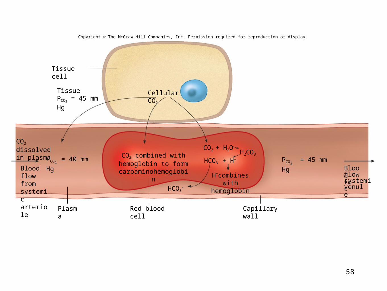
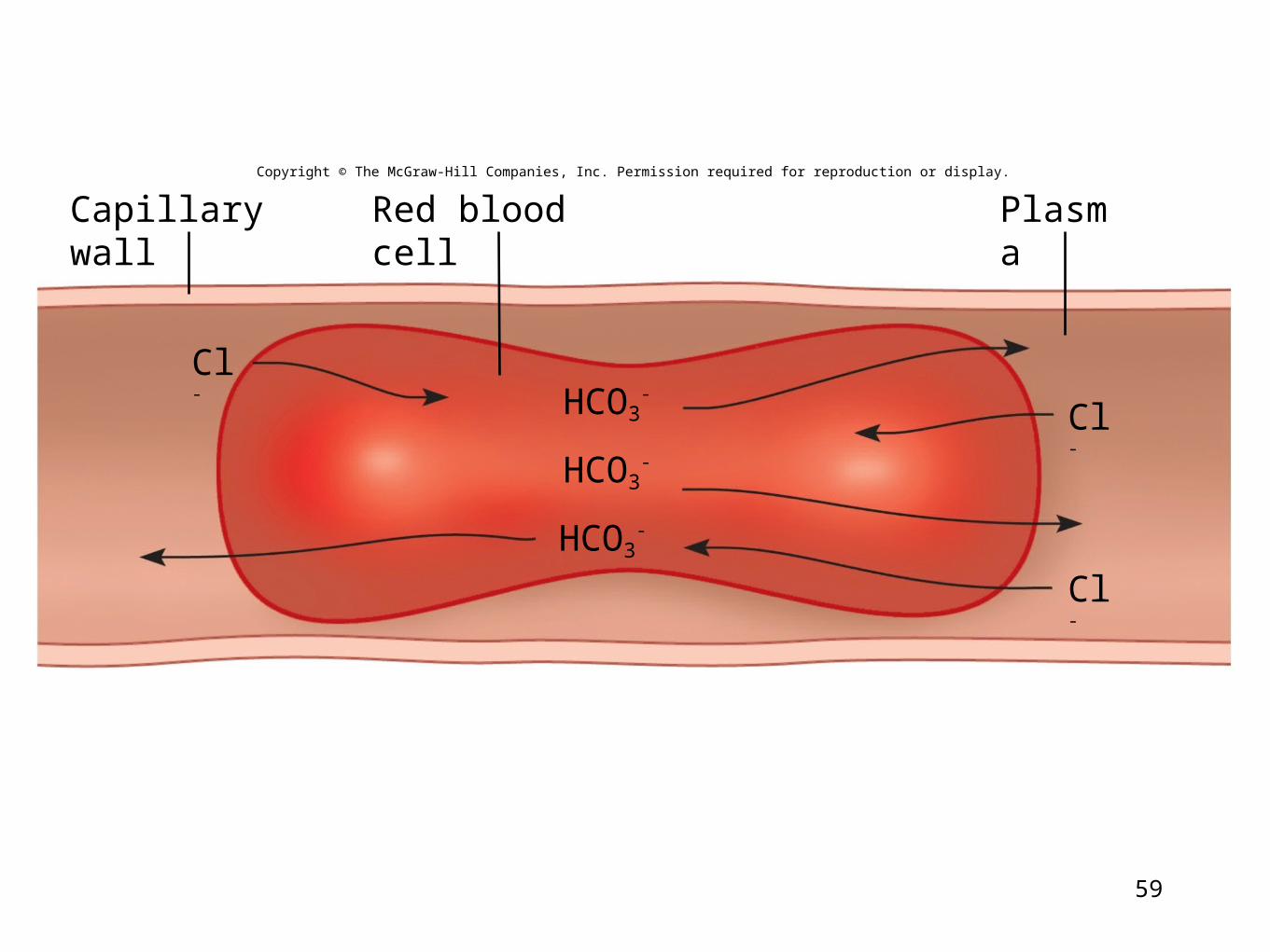
Carbon Dioxide Transport
• Blood flowing through capillaries gains CO2 because the tissues have a high Pco2 • The CO2 is transported to the lungs in one of three forms:
• As CO2 dissolved in plasma• As part of a compound with hemoglobin• As part of a bicarbonate ion
58
Tissue cell
Cellular CO2
Plasma Red blood cell Capillary wall
Bloodflow tosystemicvenule
TissuePCO2
= 45 mm Hg
Copyright © The McGraw-Hill Companies, Inc. Permission required for reproduction or display.
CO2 dissolvedin plasma
PCO2 = 40 mm Hg
Bloodflow fromsystemicarteriole
CO2 combined withhemoglobin to form
carbaminohemoglobin
H2CO3
H+combineswith hemoglobin
PCO2 = 45 mm HgHCO3
- + H+
CO2 + H2O
HCO3-
59
HCO3-
Red blood cell
HCO3-
Cl-
Cl-
Cl-
HCO3-
PlasmaCapillary wallCopyright © The McGraw-Hill Companies, Inc. Permission required for reproduction or display.
60
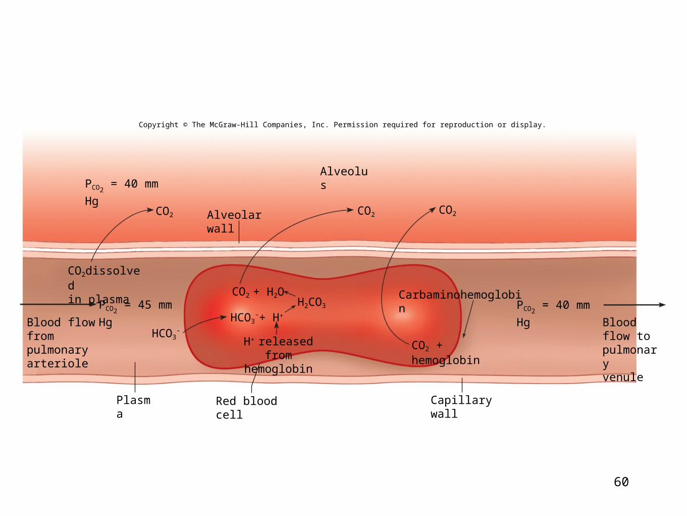
CO2CO2
Alveolus
Alveolar wall
Capillary wall
CO2
HCO3-
Carbaminohemoglobin
Plasma Red blood cell
PCO2 = 45 mm Hg
PCO2 = 40 mm Hg
CO2
CO2dissolvedin plasma
Blood flowfrom pulmonaryarteriole
HCO3-+ H+
H2CO3
H+ releasedfrom hemoglobin
Bloodflow topulmonaryvenule
PCO2 = 40 mm Hg
+ H2O
CO2 + hemoglobin
Copyright © The McGraw-Hill Companies, Inc. Permission required for reproduction or display.
61
62
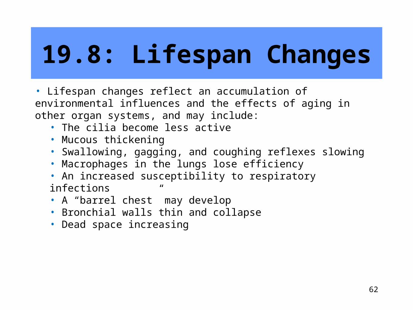
19.8: Lifespan Changes• Lifespan changes reflect an accumulation of environmental influences and the effects of aging in other organ systems, and may include:
• The cilia become less active• Mucous thickening• Swallowing, gagging, and coughing reflexes slowing• Macrophages in the lungs lose efficiency• An increased susceptibility to respiratory infections• A “barrel chest” may develop• Bronchial walls thin and collapse• Dead space increasing
63
Important Points in Chapter 19:Outcomes to be Assessed
19.1: Introduction
Identify the general functions of the respiratory system.
19.2: Why We Breathe
Explain why respiration is necessary for cellular survival.
19.3: Organs of the Respiratory System
Name and describe the locations of the organs of the respiratory system.
Describe the functions of each organ of the respiratory system.
19.4: Breathing Mechanism
Explain how inspiration and expiration are accomplished.
Name and define each of the respiratory air volumes and capacities.
64
Important Points in Chapter 19:Outcomes to be Assessed
Calculate the alveolar ventilation rate.
List several non-respiratory air movements and explain how each occurs.
19.5: Control of Breathing
Locate the respiratory areas and explain control of normal breathing.
Discuss how various factors affect breathing.
19.6: Alveolar Gas Exchanges
Define partial pressure and explain its importance in diffusion of gases.
Describe gas exchange in the pulmonary and systemic circuits.
65
Important Points in Chapter 19:Outcomes to be Assessed
Describe the structure and function of the respiratory membrane.
19.7: Gas Transport
Explain how the blood transports oxygen and carbon dioxide.
19.8: Lifespan Changes
Describe the effects of aging on the respiratory system.