cytokinin signaling activates wuschel … signaling activates wuschel expression during axillary...
TRANSCRIPT

Cytokinin Signaling Activates WUSCHEL Expression duringAxillary Meristem InitiationOPEN
JinWang,a,b CaihuanTian,a Cui Zhang,a Bihai Shi,a,b Xiuwei Cao,a,b Tian-Qi Zhang,c,d ZhongZhao,e Jia-WeiWang,c,f
and Yuling Jiaoa,b,1
a State Key Laboratory of Plant Genomics, Institute of Genetics and Developmental Biology, Chinese Academy of Sciences, andNational Center for Plant Gene Research, Beijing 100101, ChinabUniversity of Chinese Academy of Sciences, Beijing 100049, ChinacNational Key Laboratory of Plant Molecular Genetics, CAS Center for Excellence in Molecular Plant Sciences, Institute of PlantPhysiology and Ecology, Shanghai Institutes for Biological Sciences, Shanghai 200032, ChinadUniversity of Chinese Academy of Sciences, Shanghai 200032, Chinae School of Life Sciences, University of Science and Technology of China, Hefei, Anhui 230027, Chinaf ShanghaiTech University, Shanghai 200031, China
ORCID IDs: 0000-0002-4924-1716 (J.W.); 0000-0002-0794-5037 (C.T.); 0000-0002-6119-5572 (C.Z.); 0000-0001-8477-2048(B.S.); 0000-0001-5204-0061 (X.C.); 0000-0003-0270-1794 (T.-Q.Z.); 0000-0001-8044-0409 (Z.Z.); 0000-0003-3885-6296(J.-W.W.); 0000-0002-1189-1676 (Y.J.)
The homeodomain transcription factor WUSCHEL (WUS) defines the shoot stem cell niche, but the mechanisms underlyingthe establishment of WUS expression remain unclear. Here, we show that cytokinin signaling precedes WUS expression inleaf axils and activates WUS expression de novo in the leaf axil to promote axillary meristem initiation. Furthermore, type-BArabidopsis response regulator proteins, which are transcriptional activators in the cytokinin signaling pathway, directly bindto the WUS promoter and activate its expression. Finally, we show that cytokinin activation of WUS in the leaf axil correlateswith increased histone acetylation and methylation markers associated with transcriptional activation, supporting the factthat WUS expression requires a permissive epigenetic environment to restrict it to highly defined meristematic tissues. Takentogether, these findings explain how cytokinin regulates axillary meristem initiation and establish a mechanistic frameworkfor the postembryonic establishment of the shoot stem cell niche.
INTRODUCTION
Plant meristems are responsible for organogenesis, which cancontinue throughout the life of the plant. Shoot meristems harborstem cells located in the central zone (CZ), and these cells havea lower cell division rate. Some daughter cells of the CZ cellsreplenish themselves in the CZ, and other cells that are closer tothe peripheral zone form the organ primordia. The organizingcenter (OC) contains a small group of cells underneath theCZ andmaintains the stem character of stem cells in the CZ. A key factorregulating shoot stem cell specification is the WUSCHEL (WUS)homeobox transcription factor (Laux et al., 1996; Mayer et al.,1998).WUS is expressed in theOC,whichcomprises the stemcellniche of shoot meristems, including the shoot apical meristems(SAMs), axillary meristems (AMs), floral meristems (FMs), andadventitious shoot meristems.
Althoughwedonot knowhowWUSexpression is established ineach type of shoot meristem, extensive studies have shown bothhow WUS promotes shoot meristems and how WUS expressionis maintained in them. WUS function requires interaction with
transcriptional regulators of the HAIRY MERISTEM family (Zhouet al., 2015). WUS migrates from the OC to the CZ to activate theexpression of the negative regulator CLAVATA3 (CLV3), whichencodes a secretedpeptide (Fletcher et al., 1999; Yadav et al., 2011;Daum et al., 2014). The CLV3 peptide activates CLV1, a trans-membrane receptor kinase expressed in the OC, and this kinaseinhibitsWUS expression via a yet unknown signaling cascade (Clarket al., 1997; Ogawa et al., 2008). Thus, theWUS-CLV feedback loopformsa self-correctingmechanism thatmaintains a stemcell pool ofconstant size (Brand et al., 2000; Schoof et al., 2000).In addition, phytohormones also help maintain shoot stem cell
homeostasis. A positive-feedback loop between WUS functionand cytokinin provides positional cues for shoot meristem pat-terning (Leibfried et al., 2005; Gordon et al., 2009; Chickarmaneet al., 2012), although the underlying molecular mechanism hasnot been fully resolved. A number of additional WUS targets havebeen identified (Leibfried et al., 2005; Busch et al., 2010; Yadavet al., 2013), supporting the idea that WUS functions as a centralregulator of stem cells. In contrast to the indeterminate SAM andAM, the determinate FM only transiently maintains WUS ex-pression. The MADS transcription factor AGAMOUS (AG) is ac-tivated by WUS in the FM, and AG in turn terminates WUSexpression and thus determines floral bud growth (Lenhard et al.,2001; Lohmann et al., 2001; Liu et al., 2011). However, there isamajorgap inour understandingof themechanisms that establishthe initial expression of WUS.
1 Address correspondence to [email protected] author responsible for distribution of materials integral to the findingspresented in this article in accordance with the policy described in theInstructions for Authors (www.plantcell.org) is: Yuling Jiao ([email protected]).OPENArticles can be viewed without a subscription.www.plantcell.org/cgi/doi/10.1105/tpc.16.00579
The Plant Cell, Vol. 29: 1373–1387, June 2017, www.plantcell.org ã 2017 ASPB.

In addition to the SAM formed during embryogenesis, AMsform in the axils of leaves and develop into buds to enablebranching (McConnell and Barton, 1998; Wang et al., 2016). It-erative branching in perennial plants can lead to thousands ofterminal branches and thus determines aerial plant architecture(Wang and Li, 2008). The AM has the same developmental po-tential as the SAM to maintain itself and to initiate new organs.Genetic studies in Arabidopsis thaliana and other species haveshown that AM initiation is regulated by several transcriptionfactors, suchasLATERALSUPPRESSOR (LAS),REGULATOROFAXILLARY MERISTEMS (RAX), and REVOLUTA (REV) (Talbertet al., 1995; Greb et al., 2003; Müller et al., 2006). Genetic andmolecular studies revealed direct and indirect interactions amongthesegenes ina regulatory network (Ramanet al., 2008; Tianet al.,2014).WUS is expressed in the AM as in the SAM, but howWUSexpression is established during AM initiation remains enigmatic.The FM shares many similarities with the AM and has beensuggested to be a specialized AM (Long and Barton, 2000). HowWUS expression is established in the FM is also unknown.Our recent work showed that initiation of both the AM and theFM requires a cytokinin signaling pulse (Han et al., 2014; Wanget al., 2014b).
In this study, we report that cytokinin promotesWUS expressionto establish stem cell niches de novo during AM initiation. We thenshow that type-B Arabidopsis response regulator proteins (ARRs),which mediate the transcriptional response to cytokinin (Argyrosetal., 2008),bind to theWUSpromoter toactivate itsexpression.Wealso show that WUS activation requires permissive chromatinmodifications. In summary, we have provided a model for a directmolecular link explaining how shoot stem cell niches areestablished.
RESULTS
WUS Expression Is Activated de Novo during AM Initiation
To determine when WUS expression is established during AMinitiation, wemonitored the dynamics of leaf axilWUS expressionin plants grown under short-day conditions for 28 d. Using theProWUS:DsRed-N7 reporter line for WUS expression (Gordonet al., 2007), we found thatWUS expression is activated de novoprior toAMinitiation.WedidnotdetectDsRedexpression inyoungleaf axils. We observed that DsRed expression in cells of thesubepidermal layer started in the axil protrusions of the thirteentholdest leaf primordium (P13) (Figure 1A) and increased in P14 andolder leaf axils (Figure 1B), a time when AMs are morphologicallydetectable (Long and Barton, 2000; Greb et al., 2003). At thisstage, WUS is expressed in a small group of cells below thesecond cell layer of the AM (Figures 1A and 1B), resemblingWUSexpression in the SAM (Mayer et al., 1998). Thus, similar to the denovo activation of WUS expression during FM initiation (Mayeret al., 1998), de novo activation of WUS expression also occursduring AM initiation.
It has long been known that WUS is required for SAM and FMfunction (Laux et al., 1996), but it remains unknown ifWUS is requiredforAMintegrity.Therefore,weanalyzedbuddevelopment inthewus-1andwus-101mutants.Thewusmutantshaveabushyphenotypedue
to repetitive initiation of defective shootmeristems (Laux et al., 1996).We carefully analyzed these defective shootmeristems and foundthat they all originated from leaf axils and were defective AMs.Because of their defective SAM, wus plants have reduced apicaldominance and enhanced (terminated) axillary bud outgrowth. Onthe other hand, both wus-1 and wus-101 plants have a dramaticaxillary bud formation defect (Figures 1C to 1J). We followedanatomical changesat the leaf axils ofwild-type andwusplants. InP14 to P16 stage wild-type leaf axils, cells with a denser stainingcontent formmorphologically distinguishable bumps,whichmarkthe first morphological change associated with AM development(Figures 1D and 1H). In both wus-1 and wus-101 plants, densestaining cells and AM structure are lost in comparable stage leafaxils at a high frequency (Figures 1F and 1J). Consistently, there isa dramatic reduction of axillary buds in wus-1 (Figure 1C), with77% (87 out of 113) of leaf axils not supporting the formation ofaxillary buds (Figure 1F; Supplemental Figure 1B), 6% (7 out of113) of leaf axils forming one or two leaf-like structures(Supplemental Figure 1C), and 17% (19 out of 113) of leaf axilsforming terminated buds (with three or more leaves). The com-bination of reduced apical dominance and outgrowth of occa-sionally formed but terminated buds led to the observed bushyphenotype. Notably, the AM initiation defect of wus-101 can berescued by ProWUS:WUS-GFP (Figure 1K) (Daum et al., 2014).Nevertheless, the wus-1 and wus-101 plants also have severeSAMdefects, making it difficult to exclude an indirect effect of theSAM on AM initiation.Theexpressionof themeristematicgeneSHOOTMERISTEMLESS
(STM) marks leaf axil AM progenitor cells (Grbic and Bleecker, 2000;Long and Barton, 2000; Greb et al., 2003; Shi et al., 2016). Ex-amination of accumulation of the STM transcript in the wus-1mutant by RNA in situ hybridization revealed a leaf axil expressionpattern that is similar to the pattern in Ler wild-type plants(Supplemental Figure 2), suggesting that the maintenance ofmeristematic cells does not rely on WUS.Furthermore, WUS overexpression induced ectopic AM initia-
tion.We used an inducibleWUS overexpression line, pga6-1 (Zuoet al., 2002), in which b-estradiol induces constitutive WUSoverexpression, and a hormone-free leaf culture system (Wanget al., 2014b; Shi et al., 2016), in which we can quantify and live-image AM initiation. In isolated pga6-1 leaves in culture, we foundthat b-estradiol induction can lead to the formation of additionalAMs in the leaf axil (Supplemental Figures 3A and 3B). EctopicWUS expression also leads to ectopic AM initiation in cotyledonaxils, a phenotype indicating that AM initiation was enhanced(Wang et al., 2014a). Whereas wild-type Arabidopsis cotyledonslack axillary buds, we found that axillary buds could form in over50%(n>10)of cotyledonaxils afterWUS induction (SupplementalFigure 3C). To confirm that the ectopic AM initiation phenotypewas due to WUS overexpression, but not potential second-sitemutations, we generated an independent dexamethasone-inducible WUS overexpression line, ProUBQ10:WUS-GR. InProUBQ10:WUS-GR leaf axils, we similarly observed ectopic AMformation after WUS induction (Supplemental Figure 3D). Addi-tionally, a constitutive WUS overexpression line sef, which wasisolated as an activation tagging mutant (Xu et al., 2005), showedmultiple buds or branches per leaf axil (Supplemental Figures 3E
1374 The Plant Cell

Figure 1. Cytokinin Signaling Precedes and Overlaps with WUS Expression prior to AM Initiation.
(A) and (B) Serial transverse sections through a wild-type vegetative shoot apex showing expression of ProTCS:GFP-ER (green) and ProWUS:DsRed-N7(red) in leaf axils. Theorangearrow indicatesGFP in the leaf axil, andwhite arrows indicateoverlapping leaf axilGFPandDsRedsignals. Sectionsareorderedfrom apical (A) to basal (B) parts from the same plant.

and 3F). Taken together, these findings indicated that WUS ex-pression promoted AM initiation (Figure 1C).
Leaf Axil Cytokinin Signaling Precedes de NovoWUS Activation
Our recent work shows that a cytokinin signaling pulse occursprior to AM initiation (Han et al., 2014;Wang et al., 2014b).Wealsoused a cytokinin analog and a cytokinin antagonist to testwhetherAM initiation required cytokinin signaling. Treatment with thecytokinin analog 6-benzylaminopurine (BAP) caused productionof multiple axillary buds (Supplemental Figures 4A to 4C). Treat-ment of detached leaf axils with the phenylquinazoline compoundS-4893, a noncompetitive cytokinin antagonist that targets cyto-kinin receptors (Arata et al., 2010), severely compromised axillarybud formation (Supplemental Figure 4D).
Todetermine thedynamicsof local cytokininsignalingandWUSexpression in a developmental context, we examined the timingand location of expression ofProTCS:GFP-ER (Müller andSheen,2008), acytokininsignaling reporter, incombinationwithProWUS:DsRed-N7. Image analysis indicated that the leaf axil cytokininsignaling pulse emerged earlier than, and overlapped with, WUSexpressionduringAM initiation (Figures1Aand1B).Note that TCSsignals in the leaf axil were substantially stronger than that in theSAM (Supplemental Figure 5) (Wang et al., 2014b), implying thatthe AM and SAM require different levels of cytokinin signaling.Taken together, these results indicated that de novo WUS acti-vation during AM initiation is associated with a prior cytokininsignaling pulse in the same cells.
Wealsoanalyzedwhether perturbed leaf axil cytokinin signalingwas associated with defective WUS activation. AM initiation iscompromised in the las, rax, and revmutants (Talbert et al., 1995;Greb et al., 2003; Müller et al., 2006). Expression of the meri-stematic cell marker STM is maintained in las and rev mutants(Greb et al., 2003; Shi et al., 2016), suggesting that at least partialAM progenitor cell specification occurs in these mutants. Wefound that the leaf axils of these mutants lack the cytokinin sig-naling pulse (Figures 1L to 1O; Supplemental Figure 6A). In ad-dition, we could not detect WUS expression in the leaf axils ofthese mutants (Figures 1P to 1S; Supplemental Figure 6B). Thus,lack of leaf axil cytokinin signaling associated with leaf axil
WUS activation. Because cytokinin treatment can rescue AMinitiation defects in raxmutants (Wang et al., 2014b), we speculatethat cytokinin signaling activates WUS expression to enable AMinitiation.
Cytokinin Activates WUS Expression
To test whether cytokinin signaling can activateWUS expression,we treated shoot tissues with the cytokinin analog BAP ata concentration of 0.89 mM, which is within physiological levels(Corbesier et al., 2003). To enrich leaf axil tissues, we removedleaves and used the remaining shoot tissue for gene expressionanalysisbyRT-PCRwitha limitednumberofcycles (seeMethods).We found that a 4-h BAP treatment rapidly activated WUS ex-pression, even in the presence of the protein synthesis inhibitorcycloheximide (CHX) (Figure 2A), suggesting that activation ofWUS does not require de novo protein synthesis. Similarly, BAPactivation ofWUS expression was also found in the inflorescence(Supplemental Figure 7) (Gordon et al., 2009; Chickarmane et al.,2012), even when we used a physiological concentration of BAP(see Methods for details).By live-imaging the expression of the ProWUS:DsRed-N7 re-
porter and a functional ProWUS:WUS-GFP reporter (Daum et al.,2014), we found that BAP activated ectopic WUS expressioncenters de novo and substantially enlarged the endogenousWUSexpressing domain. We employed a hormone-free leaf culturesystem to live-image AM initiation (Wang et al., 2014b; Shi et al.,2016). In isolatedP15+ stage leaves, a 24-hBAP treatment inducedmultiple denovocenters ofWUSexpression in the leaf axil, so thatthe leaf axils formed multiple meristems, in contrast to the singlemeristem formed in untreated leaves (Figures 2B and 2C;Supplemental Figures 4A to 4C). In the leaf axil center whereWUSnormally is expressed, we observed a substantial enlargement ofthe WUS expression domain (Figures 2D to 2F; SupplementalFigures 8A to 8C, 9A to 9C, and 10). The expression ofWUS wasmaintained in the center of the leaf axil until a visible axillary budformed (Figures 2D to 2F). These results showed that local cy-tokinin signaling inducedWUS expression in leaf axils to promoteAM initiation. Consistent with this, mutants defective in cytokininsynthesis, perception, or signaling show defects in AM initiation(Wang et al., 2014b; Müller et al., 2015).
Figure 1. (continued).
(C)Schematic diagram of axillary buds of Ler andwus-1 plants. The thick black horizontal line represents the border between the youngest rosette leaf andthe oldest cauline leaf. For Ler, each column represents a single plant, and each square within a column represents an individual leaf axil. Forwus-1, eachcolumn representsasinglemainbranch, andbranches fromasingle plant aregrouped together. Thebottom row represents theoldest rosette leaf axils,withprogressively younger leaves above. Green, presence of an axillary bud; yellow, absence of an axillary bud; orange, one or two leaves in place of an axillarybud; red, a terminated axillary bud (with three or more leaves) in any particular leaf axil.(D) to (K) Longitudinal sections of vegetative shoot apices. Images show a protruding AM in the leaf axil of Ler (D), Col-0 (H), andProWUS:WUS-GFPwus-101 (K), but the lack of an AM in the leaf axil ofwus-1 (F) andwus-101 (J). Note the normal SAM in Ler (D), Col-0 (G), andProWUS:WUS-GFPwus-101 (K) incontrast to flat shoot apices in wus-1 (E) and wus-101 (I). Black arrows indicate leaf axils.(L) to (O)Expression ofProTCS:GFP-ER (green) in leaf axils. Images show transverse sections through shoot apices of Col-0 (L), las-101 (M), rax1-3 rax2-1rax3-1 (N), and rev-6 mutants (O). White arrows indicate GFP in leaf axils.(P) to (S) In situ hybridization ofWUS in the shoot apex. Images show transverse sections from Col-0 (P), las-101 (Q), rax1-3 rax2-1 rax3-1 (R), and rev-6mutants (S). Black arrows indicate WUS signal in leaf axils.Dottedboxes indicate the locationsof the regionsmagnified in the insets.Dotted lines indicate theoutlinesof leaf axils. Pn indicates leafprimordiumnumber,and (m/n) indicates m in n of biological repeats showing the displayed features. Bars = 50 mm.
1376 The Plant Cell

In contrast to mature leaves (P15+), ectopic cytokinin treatmentof immature leaf axils or other tissues did not lead to precociousWUS activation. In isolated P8 to P10 stage immature leaves, theexpression of ProWUS:DsRed-N7was detectable in the center ofthe leaf axil at;52h in culturewithout the addition ofBAP (Figures2D to 2F; Supplemental Figures 8A to 8C and 9A to 9C). TheexpressionofProCLV3:GFP-ER, aWUS target,wasdetected18hlater in thecells on topof theWUS-expressingcells (SupplementalFigures 9A to 9C). BAP treatment did not induce precociousWUS(and CLV3) expression (Supplemental Figure 9G). Nevertheless,BAP treatment induced additional de novo centers of WUS andCLV3 expression accompanying the emergence of the centralexpressiondomain (Figure2I).Also, the regionofProWUS:DsRed-N7 and ProCLV3:GFP-ER expression in the central domain wassubstantially enlarged after BAP treatment (Figures 2G to 2I;Supplemental Figures 8D to 8F, 9D to 9F, and 10). In particular,
BAP induced ectopicWUS expression in the epidermal cell layer(compared with Supplemental Figures 9B and 9E). The ectopicepidermal expression of WUS in the center of the leaf axil di-minished whenCLV3 expression appeared (Supplemental Figure9F). This may be explained by CLV3 inhibition ofWUS expression(Gaillochet and Lohmann, 2015). The BAP treatment did not in-duce WUS expression in differentiated cells, such as leaf bladecells. Thus, our imaging results also suggested that the preciseactivation ofWUS by cytokinins depended on the developmentalstage and cell location (i.e., cell type).
WUS Expression Requires a PermissiveEpigenetic Environment
Recent studies shows that the histone modification marker his-tone H3 lysine 27 trimethylation (H3K27me3), which is associated
Figure 2. Induction of WUS Expression by Cytokinin Treatment.
(A) A 4-h 0.89 mM BAP treatment inducedWUS expression in leaf-removed shoot apex tissues in both the absence and the presence of CHX. Error barsindicate the SD of three biological replicates, run in triplicate. **P < 0.01 (Student’s t test).(B) to (I)Time-lapse images ofWUS expression in leaf axils. BAP treatment caused rapid inductionof ectopicProWUS:DsRed-N7 inmature leaf axils (C)butdelayed induction in immature leaf axils ([G] to [I]).(B) and (C) A 24-h 0.89 mMBAP treatment (C), but not mock treatment (B), induced ectopic expression of ProWUS:DsRed-N7 (green) in the leaf axil of anisolated P15 stage leaf (arrow).(D) to (I) BAP treatment alters the WUS expression level, but not its timing. Time-lapse images showing expression of ProWUS:DsRed-N7 (green) andProCLV3:GFP-ER (blue) in isolated P9 leaf axil centers aftermock treatment ([D] to [F]) or 0.89mMBAP treatment ([G] to [I]). BAP treatment caused the leafaxil centerWUS expression domain to enlarge and activated ectopicWUS expression centers (arrows), but did not activate precociousWUS expression.The regions bordered by the redboxes in the insets in (B) and (D) roughly correspond to the imaged region. Asterisks in (D) to (F) and (G) to (I) label the samecells in corresponding time points. The cell membrane was labeled using FM4-64 (red). Bars = 50 mm.
Cytokinin Activates WUS during AM Initiation 1377

with transcriptional repression, is highly enriched at the WUSlocus in mature leaves, which have no WUS expression (Li et al.,2011; Liu et al., 2011). In addition, we found that histone H3 lysine4 trimethylation (H3K4me3), a histone modification marker as-sociated with transcriptional activation, was enriched at theWUSlocus in inflorescences containing WUS-expressing cells but notin mature leaves lacking WUS-expressing cells (SupplementalFigure 11).
Stage-specific sensitivity of leaf axil cells to BAP treatmentsuggested that epigenetic modifications may change duringleaf maturation. To test this hypothesis, we isolated the basal2- to 3-mm leaf axil tissues from immature and mature leavesand analyzed histone modifications. To accommodate thelimited sample amount, we used the ultralow input micrococcalnuclease-based native chromatin immunoprecipitation (ULI-NChIP) protocol (Brind’Amour et al., 2015). We found that theWUS genomic region showed higher levels of H3K27me3 andlower levels H3 acetylation in early stage (P8 to P10) leaf axilsthan in late stage (P15 to P17) axils (Figures 3A to 3D). Histoneacetylation increases the accessibility of DNA inside chromatinand promotes gene expression (Charron et al., 2009). Thus, theobserved temporal histone modification changes correlatewith activation of WUS expression in mature leaves, andstringent epigenetic modifications may prevent cytokinins fromactivating WUS expression in immature leaf axils and differenti-ated cells.
We next ectopically enhanced histone acetylation by apply-ing the histone deacetylation inhibitor trichostatin A (TSA). TSAtreatment, but not BAP treatment, increased histone H3 and H4acetylation of theWUS genomic region (Supplemental Figure 12),which allows transcription. Whereas TSA treatment alone mildlyincreased the WUS expression level, which may be due to en-dogenous cytokinins, a 4-h cotreatment with BAP and TSAcaused a 3.5-fold increase inWUS expression (Figure 3E). At thecellular level, we found that TSA enabled rapid de novo activationof WUS expression by BAP in differentiated leaf petiole cells(Figures 3F to 3I), explaining the dramatic increase ofWUS levelsby cotreatment with BAP and TSA.
We also used mutants that are defective in epigenetic re-pression of gene expression. The polycomb repressive complex(PRC) establishes the H3K27me3 mark and a repressive chro-matin configuration (Schuettengruber et al., 2007). CURLY LEAF(CLF) is a PRC2 core component catalyzing H3K27me3, andRING1a and RING1b are PRC1 core components that bind toH3K27me3, inhibiting transcription (Goodrich et al., 1997;Argyroset al., 2008; Xu and Shen, 2008). We detected precocious WUSexpression in young leaf axils of the clf-29mutant (SupplementalFigures13Ato13B). In the ring1a ring1bmutant,wedetectedWUSexpression in widely observed ectopic meristems (SupplementalFigure 13C). Furthermore, we found that the induction of WUSexpression by BAP was significantly enhanced in these mu-tants (Figure 3J). Consistent with this, BAP inducedmore budsin isolated leaf axils of clf-29 plants than in wild-type plants(Figure 3K). These results indicated that the PRC-mediatedH3K27me3 repressed WUS in earlier leaf axil and differenti-ated tissues. Taken together, these results supported the ideathat the induction of WUS required a permissive chromatinconfiguration.
Figure 3. TheEpigenetic EnvironmentAffectsCytokinin Induction ofWUSExpression.
(A) A diagram of the WUS genomic region with an arrow representing thetranscription start site. Black, coding sequences; dark gray, untranslatedregions; light gray, intron/intergenic regions; ATG and TAG, start and stopcodons. Black bold lines with Roman numerals indicate fragments am-plified by ChIP-qPCR. Gray bold lines with a to e indicate fragmentsshowing in Figure 5A.(B) to (D)The comparison of histonemodifications in early and late stage ofleaf axils. Images showULI-NChIPwithH3K27me3 (B), and histoneH3 (C)andH4 (D)acetylationofWUSgenomic regionsusingearly stage (P8 toP10)and late stage (P15 to P17) leaf axil tissues. Error bars represent SD, whichwas calculated from three technical replicates. Two biological replicatesgave similar results.(E) Expression of WUS after a 4-h 0.89 mM BAP and/or 1 mM TSAtreatment in shoot apex tissues. Leaves were removed before RNAisolation. Error bars indicate the SD of three biological replicates, run intriplicate.(F) to (I)Expression ofProWUS:DsRed-N7 (green) in P9 leaf petiole cells.Images show WUS expression 15 h after mock (F), 0.89 mM BAP (G),1mMTSA (H), or BAP andTSA (I) treatment. The regions bordered by theredbox in the insets in (F) roughly correspond to the imaged region.Notethat differentiated petiole cells were imaged. Bars = 50 mm.(J) A 0.89 mMBAP treatment inducedWUS expression in Col-0 wild-type,clf-29, and ring1a ring1b seedlings. Error bars indicate the SD of threebiological replicates, run in triplicate.(K) The number of buds in isolated leaf axils of Col-0 or clf-29mutants aftera 2-week mock or BAP treatment. Error bars indicate the SD (n$ 10). *P <0.05 and **P < 0.01 (Student’s t test).
1378 The Plant Cell

ARR1 Activates WUS Expression
ARR1 is a typical type-B ARR mediating the transcriptional re-sponse to cytokinin (Argyros et al., 2008). We previously showedthat the arr1-4 loss-of-functionmutant is defective in AM initiation(Wang et al., 2014b). Using a ProARR1:GFP-N7 reporter line, weobserved ARR1 expression in the leaf axil, prior to and duringAM initiation (Figure 4A). Furthermore, cell type-specific tran-scriptome data (Tian et al., 2014) show that many cytokinin sig-naling components, including other type-B ARRs, are expressedin the leaf axil (Supplemental Figure 14).
To test whether ARR1 was required for WUS activation in theleaf axil, we analyzed WUS expression in the arr1-4 mutant. Wecould not detect WUS expression in the leaf axil in arr1-4 plants(Figures 4B to 4G). We next tested whether ARR1 can activateWUS expression duringAM initiation using an inducible cytokinin-independent ARR1DDDK-MYC line, in which the N-terminal re-gion encompassing the DDK domain was deleted (Guan et al.,2014). Phosphorylation of the Asp residue in the receiver domainactivates the ability of the protein to promote the transcription oftarget genes. Because the DDK domain functions as a negativeregulatory motif, its removal causes constitutive activation oftranscription in the absence of cytokinin (Sakai et al., 2001). Wefound that activationofARR1DDDK-MYC resulted in rapid inductionof WUS expression both in shoot apex tissues and in the in-florescence within 8 h (Figure 4H; Supplemental Figure 15). Con-sistent with enhancedWUS expression, ectopic ARR1DDDK-MYCpromoted AM initiation and bud outgrowth, resulting in a bushyphenotype.However,wedidnotobserveectopicmeristem in leavesor on the stem, supporting the idea thatWUS expression activationrequires a permissive epigenetic environment.
We tested whether AM initiation defects in arr1-4 can be res-cued by restoring WUS expression using the inducible WUSoverexpression line pga6-1 (Zuo et al., 2002). In untreated arr1-4pga6-1 plants, very few of the first;10 rosette leaves, whichwereformed during the first 2 weeks of vegetative development, de-velopaxillarybuds (Figure4I).Startingat15daftergermination,wetreated shoot apexes of arr1-4 pga6-1 plants with b-estradiol toinduce WUS expression. We found that induction of WUS ex-pression rescued the axillary bud formation defects (Figure 4I;Supplemental Figure 16). In addition, a substantial portion of therosette leaves formed prior to treatment supported the formationof axillary buds after WUS induction (Figure 4I), suggesting thatmature leaf axil cells of arr1-4 plants are competent to respond toWUS activity. On the other hand, we did not observe precociousaxillary bud formation after b-estradiol induction of WUS ex-pression, suggesting thatWUS expression is not sufficient for AMinitiation. Taken together, these results indicated that ARR1regulated AM initiation through activatingWUS expression in theleaf axil. Because ARR10, 11, and 12 function redundantly withARR1 inpromotingAM initiation (Wangetal., 2014b), these relatedtype-B ARRs may also promote WUS expression.
Type-B ARRs Bind to the WUS Promoter to ActivateIts Expression
Because BAP induction of WUS did not require de novo proteinsynthesis (Figure2A),wespeculated thatARR1and related type-B
ARRs could bind directly to the WUS promoter region. Based onsequence conservation and the existence of the putative ARR1bindingsiteGAT(T/C) (Figures5Aand5B;Supplemental Figure17)(Sakai et al., 2000),we selected five regionsof theWUS locus (a–e)for analysis.Chromatin immunoprecipitation (ChIP) assays using either
shoot tissues with leaves removed or inflorescence tissuesshowed that ARR1DDDK-MYC strongly associated with regionsb,c, anddof theWUSpromoter region (Figure5C).To testwhetherARR1 can directly bind to these regions, we performed an in-dependent electrophoretic mobility shift assay (EMSA) andconfirmed that theDNAbindingdomain of ARR1bound to regionsb, c, and d (Figure 5D; Supplemental Figure 18). Region c showeda lower affinity for the ARR1 protein than did regions b and d.We also examined type-B ARR activation of WUS expression
using a transient transfection assay, chosen because transientlyexpressed reporter constructs would lack epigenetic mod-ifications that might interfere with ARR binding. Consistent withour ChIP and EMSA results, a transient transfection assay inprotoplastsdemonstrated thatARR1activated theWUSpromoter(Figure 5E; Supplemental Figures 19A and 19C). Regions b, c,and d are redundantly required for ARR1 activation (Figure 5F;Supplemental Figures 19B and 19D). Several other related type-BARRs (Argyros et al., 2008) are expressed in the leaf axil(Supplemental Figure 14), and their mutations can enhance AMinitiation defects in arr1 mutants (Wang et al., 2014b). We thentested whether ARR2, 10, 11, and 12 could also directly activateWUS expression in protoplasts. Our results indicated that all ofthese type-B ARRs activated WUS expression, although to dif-ferent extents (Figure 5G; Supplemental Figure 19E). This isconsistent with their redundant roles in promoting AM initiation.Taken together, these results indicated that ARR1 and other re-lated type-BARRs can bind to the promoter ofWUS to activate itsexpression.
DISCUSSION
WUS Expression Is Activated de Novo by CytokininSignaling during AM Initiation
Tremendous interest has focused on understanding how stemcells are specified in both animals and plants. Stem cell nichesprovide a microenvironment for stem cell fate determination andthe establishment of stem cell niches is central to stem cell bi-ology. In Arabidopsis, the expression ofWUS defines shoot stemcell niches (Mayer et al., 1998), as shown by overexpressionanalyses (Zuo et al., 2002; Xu et al., 2005). Thus, understandinghow WUS expression is regulated both spatially and temporallyprovides key information on stem cell niche specification. Severaltypes of shoot meristems can form in plants. The SAM is es-tablished during embryonic development and AMs initiate postembryonically from the axils of leaves. Cells in AMs and the SAMhave similar potential for indeterminate growth. After the floraltransition, determinate FMs form and initiate a limited number offloral organs. Adventitious shoot meristems may also form, es-pecially in tissue culture conditions. To understand howeach typeof shootmeristem is established, it is important to understand the
Cytokinin Activates WUS during AM Initiation 1379

mechanism of activation of WUS expression during shoot meri-stem initiation.
In this study, we focused on the de novo activation of WUSexpression during AM initiation. We previously showed that cy-tokinin promotes AM initiation (Han et al., 2014; Wang et al.,2014b). During AM initiation, the meristematic gene STM is highlyexpressed in the leaf axil (Grbic and Bleecker, 2000; Long andBarton, 2000; Greb et al., 2003; Shi et al., 2016). The leaf axilcytokinin signaling pulse may result from increased STM ex-pression because STM can promote cytokinin biosynthesis
(Jasinski et al., 2005;Yanai et al., 2005). In this study,weshow thatleaf axil cytokinin signaling directly increases the expression ofWUS, which defines stem cell niches and completes AM initiation(Figure 6). The expression of cytokinin signaling pathway genes inthe leaf axil (Figure 4A; Supplemental Figure 14), together with theSTM-promoted cytokinin biosynthesis in the leaf axil, leads to theobserved leaf axil-specific cytokinin signaling pulse and, moreimportantly, to de novo activation of WUS expression in the leafaxil (Figure 1). This activation is mediated by direct binding ofARR1 and related type-B ARRs, to the WUS promoter region
Figure 4. Activation of WUS Expression by ARR1 Is Required for AM Initiation.
(A) Expression of ProARR1:GFP-N7 in the leaf axil. Images show longitudinal sections through a vegetative shoot apex demonstrating expression ofProARR1:GFP-N7 (green) in the leaf axil prior to (upper panel) and during (lower panel) AM initiation. Arrows indicate GFP in leaf axils. The dotted lineindicates the outline of the bulged meristem. PI, propidium iodide.(B) to (G) In situ hybridization ofWUS. Images show serial transverse sections of vegetative shoot apexes in Col-0 ([B] to [D]) and the arr1-4mutant ([E] to[G]). Arrows indicateWUSsignal in leaf axils, anddotted lines indicate theoutlinesof leaf axils. Sectionsareordered fromapical ([B]and [E]) tobasal ([D]and[G]). The approximate distance from the summit of the SAM to section is given in the upper right-hand corner of each image. Note that (D) and (G) are fromregions more distant from the center of the same plants shown in (B) and (C), and (E) and (F). Bars in (A) to (G) = 50 mm.(H) Expression of WUS after 8-h mock treatment or induction of ARR1DDDK in leaf-removed shoot tissues. Error bars indicate the SD of three biologicalreplicates, run in triplicate. **P < 0.01 (Student’s t test).(I) Schematic diagram of axillary buds of arr1-4 pga6-1mutants with or without b-estradiol induction to activateWUS overexpression. See the legend toFigure1Cforadescriptionofsymbols.Green indicates thepresenceofanaxillarybud,andyellow indicates theabsenceofanaxillarybud.Plantsweregrownunder short-day conditions for 15dwithout treatment; leaf axil regionswere treatedwith 10mMb-estradiol every other day for another 15 dand then shiftedto long-day conditions without treatment until axillary buds were counted. The vertical line indicates leaves initiated during b-estradiol treatment.
1380 The Plant Cell
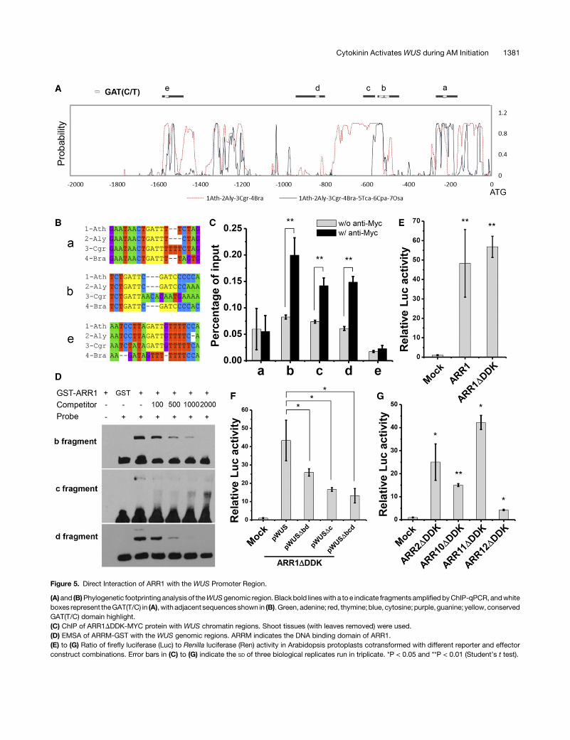
Figure 5. Direct Interaction of ARR1 with the WUS Promoter Region.
(A)and (B)Phylogenetic footprinting analysis of theWUSgenomic region.Blackbold lineswith a toe indicate fragments amplifiedbyChIP-qPCR, andwhiteboxes represent theGAT(T/C) in (A), with adjacent sequences shown in (B).Green, adenine; red, thymine; blue, cytosine; purple, guanine; yellow, conservedGAT(T/C) domain highlight.(C) ChIP of ARR1DDDK-MYC protein with WUS chromatin regions. Shoot tissues (with leaves removed) were used.(D) EMSA of ARRM-GST with the WUS genomic regions. ARRM indicates the DNA binding domain of ARR1.(E) to (G) Ratio of firefly luciferase (Luc) to Renilla luciferase (Ren) activity in Arabidopsis protoplasts cotransformed with different reporter and effectorconstruct combinations. Error bars in (C) to (G) indicate the SD of three biological replicates run in triplicate. *P < 0.05 and **P < 0.01 (Student’s t test).
Cytokinin Activates WUS during AM Initiation 1381

(Figure 5). The same regulatoryprinciple likely functionsduringFMinitiation (Supplemental Figures 7 and 15). In fact, floral bud de-velopment is also compromised in mutants defective in cytokininsynthesis, perception, or signaling, with phenotypes includingdefective floral bud initiation, fewer flowers, and early terminationof inflorescences (Higuchi et al., 2004; Nishimura et al., 2004;Argyros et al., 2008; Tokunaga et al., 2012). The same regulatorycircuits may also contribute to the de novo activation of WUSexpression during embryonic SAM and adventitious shoot mer-istem formation. Recent studies have proposed that SAM main-tenance requires cytokinin signaling (Gordon et al., 2009;Chickarmane et al., 2012). Therefore, the same regulatory prin-ciple may function during the maintenance of WUS expression.However, as shown by the ProTCS:GFP reporter, the SAM hasmuch weaker cytokinin signaling than the leaf axil (SupplementalFigure 5), resulting from a lower cytokinin concentration and/ora weaker cytokinin response. This suggests that type-B ARR-based transcriptional activation may be insufficient to maintainWUS expression. Whether other regulatory mechanisms exist formaintenance ofWUS expression in established shoot meristemsremains to be answered.
WUS Is Required for AM Initiation and Integrity
Inwusmutants, axillary buds are either absent or replaced by leaf-like structures that obviously lack indeterminate growth (Figures1C to 1K; Supplemental Figure 1). A substantial portion (77%) ofleaf axils are empty in wus-1, and most leaf axils lack discernibleshoot meristem structure in wus mutants (Figures 1D to 1K), in-dicating thatWUS is required for AM initiation. Although thewus-1and wus-101 alleles we used are strong alleles with severe SAMdefects, leaf patterning was mostly unaffected (Laux et al., 1996).Nevertheless, it is possible that the general effect ofWUS on SAMfunction has an indirect role onAM initiation in the leaf axil. Recentstudies also showed that formation of functional tiller buds in rice(Oryza sativa) requires the rice ortholog of WUS (Lu et al., 2015;Tanaka et al., 2015). In Arabidopsis wus mutants, leaf-likestructures or even-terminal branches can still form in a portion ofleaf axils (23% in wus-1; Figure 1C). Thus, additional regulatorsredundantly promote stem cell activities. Nevertheless, yet un-known stem cell activators are insufficient to maintain stem cellhomeostasis and indeterminacy (Figure 1C).
It is plausible that WUS is required for stem cell initiation topromote AM initiation. Alternatively, defective stem cell homeo-stasis alone could explain the lack of AM in wusmutants. Recentwork on embryonic shoot stemcell initiation indicated thatWUS isdispensable for stem cell initiation and that several members ofthe WUSCHEL-RELATED HOMEOBOX gene family redundantlyfunction in this process in embryos (Zhang et al., 2017). It remainsto be tested whetherWUS is required for stem cell initiation in theAM, in addition to its role in stem cell homeostasis.STM is also necessary for AM initiation (Shi et al., 2016), and
the expression of STM is maintained in the leaf axil in wus-1(Supplemental Figure 2). In contrast to the de novo activation ofWUS, STM expression is maintained in the leaf axil (Grbic andBleecker, 2000;LongandBarton, 2000;Grebetal., 2003;Shi et al.,2016). In fact, the expression patterns of WUS and STM are verydifferent between AM initiation and embryogenesis. During em-bryogenesis, the onset ofWUS expression at the 16-cell stage ismuch earlier than STM initiation at the late globular stage, whenthe embryo consists of;100 cells (Lenhard and Laux, 1999). ThecontinuousSTM expressionduringAM initiation is consistentwiththe STM functions in shielding meristematic cells from differen-tiation, allowing later stem cell initiation (Shi et al., 2016). Nev-ertheless, STM may also provide rudimentary stem cell initiationactivity in the absence of WUS (Brand et al., 2002).
Type-B ARRs Bind to the WUS Genomic Region
In addition to conducting genetic analysis, we provided multiplelines of evidence to support direct binding of ARR1 and relatedtype-B ARRs, to theWUS promoter. ChIP, EMSA, and protoplasttransactivation assays all demonstrated ARR binding to threediscrete regions (b, c, and d) of the WUS promoter (Figure 5).Whereas regionsbanddcontain thecanonical ARR1bindingcoremotif GAT(T/C) (Sakai et al., 2000), region c lacks thismotif. EMSAshowed weaker binding of ARR1 to region c compared with re-gions b and d (Figure 5D), implying an alternative DNA bindingmechanism. Promoter deletion analysis indicated that these threeregions are redundantly required for ARR activation of WUS ex-pression (Figure 5F). The wus-6 hypomorphic allele providesadditional support for the importance of type-B ARR binding sites.In wus-6, a 7-kb T-DNA insertion separates the type-B ARRbinding sites from the WUS open reading frame. The T-DNA in-sertion also caused a 95-bp deletion that partially overlaps withregion b. The expression ofWUS is substantially reduced inwus-6(Hamada et al., 2000), suggesting these evolutionarily conservedtype-B ARR binding regions and/or additional upstream regionsare important for WUS expression.Notably, aWUS promoter lacking all three regions still showed
activity, although the activity was much reduced (Figure 5F), in-dicating the existence of additional regulatory regions. One suchcandidate is a 57-bp region between regions c and d. A previousstudy showed that this region, when present in tandem and fusedto a minimal CaMV 35S promoter, could driveWUS expression inthe FM (Bäurle and Laux, 2005). Promoter deletion analysis alsoshowed that flanking regions of the 57-bp core sequence, whichcovers regionscandd,were required for optimal promoter activity(Bäurle and Laux, 2005). This 57-bp region likely is sufficient forthe maintenance of WUS expression, as this assay was done in
Figure 6. A Developmental Framework of AM Initiation.
In the leaf axil region, an auxin minimum (gray) at the early stage andasubsequent cytokinin signalingpulse (green) are required for AM initiation(Wang et al., 2014b). Later, cytokinin signaling de novo induces WUSexpression (red) to activate the stem cell niche and complete AM initiation.IAA, indole acetic acid; CK, cytokinin.
1382 The Plant Cell

wild-type plants with functional FMs and endogenous WUS ex-pression, but is insufficient for de novo activation of WUS ex-pression. It again highlights that de novo activation of WUSexpression prior to meristem initiation and maintenance ofWUS expression in established meristems could use differentmolecular mechanisms.
Epigenetic Regulation Restricts WUS Expression
In addition to type-B ARR regulation, our work suggests thatspatiotemporal epigenetic regulation refines WUS expression.Thus, hormones and epigenetic factors act in concert to governformation of the lateral shoot stem cell niche. Although leafaxils have highly defined cytokinin signaling (Figure 1), additionalcytokinin signaling centers exist in plants (Müller and Sheen,2008). Type-B ARRs also have broad expression (Figure 4A;Supplemental Figure 14) (Mason et al., 2004). However, mostcytokinin signaling centers do not activate WUS expression. In-stead, in addition to cytokinin signaling, existing meristematictissues are likely required to restrict spatiotemporal WUS ex-pression. Our data indicated that increasing the chromatin ac-cessibility by TSA treatment or in PRCmutants led to precociousand ectopic WUS expression and ectopic meristem formationfollowing cytokinin treatment (Figures 3E to 3K; SupplementalFigure 13). Following TSA treatment of wild-type plants or PRCmutants, the onset of WUS expression in the leaf axil occurredearlier, and ectopicWUS appeared in differentiated cells (Figures3F to 3I; Supplemental Figure 13) (Bratzel et al., 2010), resulting inectopic meristems (Figure 3K; Supplemental Figure 13C). Epi-genetic regulation is involved in the termination of WUS expres-sion in the FM (Liu et al., 2011), and related mechanisms maysuppress WUS expression in immature leave axil cells and indifferentiated tissues. During leaf maturation, leaf axil cells divide(Wang et al., 2014b; Shi et al., 2016), and epigeneticmodificationschange (Figures 3A to 3D). This observation suggests a celldivision-dependent induction, as was recently found in FM ter-mination (Sun et al., 2014). Cell division may dilute inhibitory cis-acting marks and/or trans-acting factors so that the chromatinenvironment would be permissive forWUS expression. BAP doesnot affect prohibiting factors but TSA removes such factors(Figures 3E to 3I; Supplemental Figure 12). TSA treatment leads todifferent histone acetylation profiles of theWUS promoter than doendogenous regulators, indicating different site specificity.Nevertheless, TSA was efficient in conditioning BAP activation ofWUS. The enrichment of H3K27me3, a marker of transcriptionalrepression, at theWUS locus inmature leaves and the enrichmentofH3K4me3, a transcriptional activationmarker, in inflorescencesmaybe causal in the regulation ofWUS expression (SupplementalFigure 11). Alternatively, these markers may simply reflect tran-scription status. The H3K4me3 binding protein REPRESSOR OFWUSCHEL1maycontribute to theepigenetic regulationofWUS inthe leaf axil (Han et al., 2008; Zhang et al., 2015).
The activity of the AM determines plant architecture and cropyield (McSteen and Leyser, 2005; Wang and Li, 2008; Yang andJiao, 2016). The finding that cytokinin activates WUS expressionprovides insight into how shoot stem cell niches are establishedandmay ultimately facilitate themanipulation of plant architectureto enhance crop yield.
METHODS
Plant Materials and Treatment Conditions
Arabidopsis thaliana ecotypes Col-0, Ler, and Ws were used as wild-typecontrols. Arabidopsis plants were grown under short-day conditions (8 hlight and 16 h dark at 22°C) for 30 d and then under long-day conditions(16h light and8hdarkat22°C) to inducefloweringbeforeaxillarybudswerecounted. The transgenic lines ProTCS:GFP, ARR1DDDK-MYC, ProWUS:WUS-GFP wus-101 (GK870H12), and ProUBQ10:WUS-GR are in the Col-0 background (Müller and Sheen, 2008; Daum et al., 2014; Guan et al.,2014), andProCLV3:GFP-ERProWUS:DsRed-N7 is in the Ler background(Reddy et al., 2004). Thewus-101, arr1-4,clf-29, and ring1a ring1bmutantsare in the Col-0 background (Goodrich et al., 1997; Argyros et al., 2008; Xuand Shen, 2008), the wus-1 mutant is in the Ler background (Laux et al.,1996), and thepga6-1and sefmutantsare in theWsbackground (Zuoetal.,2002; Xu et al., 2005). The las, rax, and rev mutants have been previouslydescribed (Talbert et al., 1995; Greb et al., 2003; Müller et al., 2006). Forin vitro leaf culture, P8 to P11 leaves were taken from plants grown onMurashige and Skoog medium in short-day conditions for 15 to 17 d.Detached leaves were cultured on Murashige and Skoog medium sup-plemented with 0.1 mg/L inositol acid and 0.5 mg/L folic acid under short-day conditions (Steeves et al., 1957; Wang et al., 2014b).
For chemical treatments, 0.89 mMBAP (Sigma-Aldrich), 30 mMS-4893(3-[(6-chloro-4-phenylquinazolin-2-yl) amino] propan-1-ol; Vitas-M Lab-oratory) (Arata et al., 2010), 10 mMCHX (Sigma-Aldrich), and/or 1 mM TSA(Sigma-Aldrich) were used to treat 3-week-old short-day grown plants.For detached leaf culture, the solutionwas added to the leaf axil region. Forseedlings and inflorescences, tissues were soaked in the solution. Forinducible WUS expression, pga6-1 and arr1-4 pga6-1 plants were grownunder short-day conditions for 15 d, treated with 10 mM b-estradiol everyother day for 15 d, and then shifted to long-day conditions withoutb-estradiol treatment until axillary buds were counted (Zuo et al., 2002;Wangetal., 2014b).Weused6 to16plants for phenotypic analysis (Figures1C, 3K, and 4I; Supplemental Figures 3 and 7).
In Situ Hybridization and Microscopy
In situ hybridization was performed as previously described (Wang et al.,2014b). The digoxigenin-labeledWUS probe contained nucleotides 382 to1075 bp downstream of the start codon. Shoots were fixed and sectionedfollowing previously described methods (Wang et al., 2014b). Images ofsections and plants were taken by a Nikon SMZ1000 stereoscopic mi-croscope or an Olympus BX60microscope equipped with a Nikon DS-Ri1camera. Scanning electron microscopy was performed using a HitachiS-3000N variable pressure scanning electron microscope after standardtissue preparation (Wang et al., 2014b). For confocal microscopy, samplepreparation was performed as previously described (Wang et al., 2014b).Images were taken with a Nikon A1 confocal microscope. Excitation anddetection window setups for GFP, DsRed, 49,6-diamidino-2-phenylindole,DsRed, GFP, FM4-64 (to label the cell membrane), and autofluorescencewere previously described (Qi et al., 2014; Wang et al., 2014b). Ten totwenty replicates (in three batches) were analyzed.
RT-PCR and RT-qPCR
Total RNAwas extracted from shoot tissues (with leaves removed),matureleaves, or inflorescences with the AxyPrep Multisource RNA MiniPrep kit(Corning). Arabidopsis shoot tissue is mainly composed of leaves, makingleaf axil tissues low in abundance. AsWUS expression is restricted to leafaxils,whereaxillary buds form,we removed leaves toenrich leaf axil tissueswith WUS expression, so that its expression could be reliably detected.First-strand cDNA synthesis was performed using the TransScriptOne-Step gDNA Removal and cDNA Synthesis SuperMix (TransGen).
Cytokinin Activates WUS during AM Initiation 1383

Quantitative RT-PCR was performed on a Bio-Rad CFX96 real-time de-tection system using the KAPA SYBR FAST qPCR kit (KAPA Biosystems)(Tian et al., 2014).ACTIN2was used as the reference gene to normalize therelative expression for quantitative RT-PCR analysis. RNAs from threebatches of independently prepared plant materials (biological replicateswith using different plants), each run in triplicate (technical replication),were analyzed. Mean and SD of biological replicates were used to presentthe data (Figures 2–5).
Phylogenetic Footprinting
The sequences of ;2000 bp upstream of the WUS start codon werealigned, and the degree of sequence divergence was quantified across forseven seed plant species (Arabidopsis thaliana, Arabidopsis lyrata, Capsellagrandiflora,Brassica rapa,Theobromacacao,Caricapapaya, andOryza sativa).All sequenceswereobtained fromPhytozome11.0 (https://phytozome.jgi.doe.gov/pz/portal.html). Sequence alignment (Supplemental File 1) was performedwith ClustalX version 2.1 (Larkin et al., 2007) with gap opening = 10, gap ex-tension = 0.2, delay divergent sequence = 30%, and turning off of negativematrix and Gonnet series for protein weight matrix. The phylogenetic tree forWUS (Supplemental File 1) was calculated with MEGA version 6.06 based onprotein sequences (Tamura et al., 2013) using the neighbor-joining method(SaitouandNei,1987).Confidence intervalsonphylogenieswere inferredbythebootstrap method (Felsenstein, 1985). The evolutionary distances werecomputed using the Poisson correction method (Zuckerkandl and Pauling,1965) and are in units of the number of amino acid substitutions per site. Allpositions containing gaps and missing data were eliminated. BigFoot,aBayesianalignmentandphylogenetic footprintingsoftware,was thenused toalign genomic sequences and score the degree of conservation with defaultsettings (Satija et al., 2009).
ChIP
ChIP experiments were performed according to published protocols (Hanet al., 2014). Inflorescence or shoot tissues (with leaves removed) ofARR1DDDK-MYC plants were induced with 10 mM b-estradiol for 8 h.Samples of more than 800 mg of tissue were harvested and fixed with 1%(v/v) 4°C formaldehyde at 4°C (Han et al., 2014). Immunoprecipitationswere performed with or without anti-MYC (ab9132; Abcam). The pre-cipitated DNA was isolated, purified, and used as a template for quanti-tative RT-qPCR. For detection of histone modifications, seedlings,inflorescence, and leaves from Col-0 plants, and anti-H3K4m3 (ab8580;Abcam), anti-H3K27me3 (07-449; Millipore), anti-acetyl-Histone H3 (06-599; Millipore), and anti-acetyl-Histone H4 (06-598; Millipore) were used.
ULI-NChIP was performed according to published protocols withmodifications (Brind’Amour et al., 2015). The basal 2 to 3 mm of leaf axiltissues from early stage (P8 to P10) or late stage (P15 to P17) leaveswerewasisolated from 4- to 5-week-old wild-type Col-0 plants. Tissues from 30 to40 leaf axils were used for each replicate. Tissues were fully ground with in30 mL Galbraith buffer (45 mM MgCl2, 30 mM sodium citrate, and 20 mMMES,pH7.0) ina1.5-mL tube.Thepestlewaswashedwithadditional20mLGalbraith buffer into the same tube. Nuclei were spun down at 1000g for10 min at 4°C. The supernatant was discarded, and the sediment wasresuspended with 50 mL nuclear isolation buffer (NUC-101; Sigma-Aldrich). The subsequent chromatin preparation was based on micrococcalnuclease fragmentation at 37°C for 7 min. Chromatin was precleared with10 mL of 1:1 protein A:protein G Dynabeads (Life Technologies) and thenimmunoprecipitated with 1 mg of antibody in antibody-bead complexes at4°C overnight. Protein-DNA-bead complexes were washed twice with400 mL low salt wash buffer (20 mM Tris-HCl, pH 8.0, 150 mM NaCl, 1%Triton X-100, and 0.1% SDS) and twice with high-salt wash buffer (20 mMTris-HCl, pH 8.0, 500mMNaCl, 1%Triton X-100, and 0.1%SDS). Protein-DNAcomplexeswere eluted in 30mLChIP elution buffer (100mMNaHCO3
and 1% SDS). DNA was purified using phenol/chloroform and ethanol
precipitation (Brind’Amour et al., 2015). The concentration of purified DNAwas measured using the Qubit dsDNA HS Assay kit (1674653; ThermalFisher) before the DNA was used as the template for PCR analysis.
EMSA
The DNA binding domain of ARR1 (ARRM; amino acids 236–299) fusedwith the GST tag was produced in a prokaryotic expression system aspreviously described (Tian et al., 2014). Biotin-labeled probes were am-plified using 59 biotin-labeled primers synthesized by SangonBiotech, andcorresponding competitor probes were amplified using primers of thesame sequences without labeling. Binding reactions and competitionexperiments were performed as described (Tian et al., 2014).
Transient Expression in Protoplasts
Full-length coding sequences ofARR1, 460 bp downstream of the start codonof ARR1 (ARR1DDDK), 433 bp downstream of the start codon of ARR2(ARR2DDDK), 400bpdownstreamof thestartcodonofARR10 (ARR10DDDK),382 bp downstream of the start codon of ARR11 (ARR11DDDK), and 400 bpdownstream of the start codon of ARR12 (ARR12DDDK) were amplified fromArabidopsiscDNAbyPCRand insertedbetween theKpnI andBstBIsitesof theProUC19-p35S-FLAG-RBS vector (Feng et al., 2012). To generate ProWUS:Luc, 1708 bp upstream of the start codon ofWUSwas amplified and insertedbetween theEcoRI andSacI sitesof theProFRK1:Lucvector (Fenget al., 2012).WUS promoter deletions (ProWUSD:Luc) were obtained by inverse PCR usingthe ProWUS:Luc as the template. The conserved GAT(C/T) motifs were re-moved inProWUSDbd (Supplemental Figure 19B), and2617 to2599bpwereremoved in ProWUSDc.
Arabidopsis protoplasts were isolated from leaves of plants grownunder short-day conditions for 5 to 6weeks. TheProUC19-Pro35S-ARRs-FLAG-RBS vector was cotransformedwith ProWUS:Luc (firefly luciferase)andPro35S:Ren (Renilla luciferase) into protoplasts and incubated at roomtemperatureovernight underweak light. The relativeLucactivity (as relativeLuc/Ren ratio) was detected with the dual-luciferase report assay system(Promega) andusing aPromegaGLOMAX96microplate luminometer. Theefficiency of transient expression was quantified by western immuno-blotting using anti-FLAG (A8592; Sigma-Aldrich).
Accession Numbers
Sequence data from this article can be found in the GenBank/EMBL da-tabase and/or the Arabidopsis Genome Initiative database under thefollowing accession numbers: ACTIN2 (At3g18780), ARR1 (At3g16857),ARR2 (At4g16110), ARR10 (At4g31920), ARR11 (AT1G67710), ARR12(At2g25180), CLF (At2g23380), CLV3 (At2g27250), LAS (AT1G55580),WUS/PGA6 (At2g17950), RAX1 (AT5G23000), RAX2 (AT2G36890), RAX3(AT3G49690), REV (AT5G60690), RING1A (AT5G44280), RING1B(AT1G03770), STM (At1g62360), and UBQ10 (AT4G05320).
Supplemental Data
Supplemental Figure 1. Defective Axillary Bud Formation in the wus-1Mutant.
Supplemental Figure 2. In Situ Hybridization of STM in the wus-1Mutant.
Supplemental Figure 3. Bud Formation after WUS Overexpression.
Supplemental Figure 4. Regulation of Axillary Bud Initiation byCytokinin.
Supplemental Figure 5. The Cytokinin Signaling Pulse Is MuchStronger in the Leaf Axil Than in the SAM.
Supplemental Figure 6. Lack of the Leaf Axil Cytokinin SignalingPulse and WUS Expression in the rax1-3 Mutant.
1384 The Plant Cell

Supplemental Figure 7. BAP Induction of WUS Expression in theInflorescence.
Supplemental Figure 8. Expression ofWUS-GFP in Response to BAPTreatment in Immature Leaf Axils.
Supplemental Figure 9. Expression ofWUS and CLV3 in Response toBAP Treatment in the Center Region of Immature Leaves.
Supplemental Figure 10. BAP Treatment Enlarges the ExpressionDomain of WUS and CLV3.
Supplemental Figure 11. H3K4me3 Is Associated with the WUSChromatin Region.
Supplemental Figure 12. Histone Acetylation of the WUS GenomicRegion Increases Following TSA Treatment.
Supplemental Figure 13. In Situ Hybridization of WUS in clf-29 andring1a ring1b Mutants.
Supplemental Figure 14. Expression of Cytokinin Signaling andBiosynthesis Genes in the Leaf Axil.
Supplemental Figure 15. ARR1 Induction of WUS in the Inflorescence.
Supplemental Figure 16. Buds Formed by WUS Induction AreIdentical to Normal Buds.
Supplemental Figure 17. Coding Sequence-Based Phylogenetic Treeof WUS from Seven Seed Plant Species.
Supplemental Figure 18. ARR1 No Longer Binds the Mutated Regionb in an EMSA.
Supplemental Figure 19. Type-B ARRs Induce WUS Expression.
Supplemental Table 1. List of Primers.
Supplemental File 1. Text File of the Alignment Used for thePhylogenetic Analysis Shown in Supplemental Figure 17.
ACKNOWLEDGMENTS
We thank Ying Wang for critical reading of the manuscript, Kang Chong,KlausTheres, LinXu,YunyuanXu, andJianruZuo for seeds, Jian-MinZhoufor the help with the transient expression assay, and Xian Sheng Zhang forexchangingunpublished results. Thisworkwas fundedbyNational NaturalScience Foundation of China (NSFC) Grant 31430010, by National BasicResearch Program of China (973 Program) Grant 2014CB943500, by theNational Program for Support of Top-Notch Young Professionals, and bythe State Key Laboratory of Plant Genomics. C.T. is amember of theYouthInnovation Promotion Association of the Chinese Academy of Sciences(2017139).
AUTHOR CONTRIBUTIONS
Y.J. conceived the project. J.W., C.T., C.Z., B.S., and X.C. performedexperiments and analyzed data. T.-Q.Z., Z.Z., and J.-W.W. contributedreagents. J.W. and Y.J. wrote the manuscript.
Received July 20, 2016; revised April 25, 2017; accepted June 1, 2017;published June 2, 2017.
REFERENCES
Arata, Y., Nagasawa-Iida, A., Uneme, H., Nakajima, H., Kakimoto,T., and Sato, R. (2010). The phenylquinazoline compound S-4893 isa non-competitive cytokinin antagonist that targets Arabidopsis
cytokinin receptor CRE1 and promotes root growth in Arabidopsisand rice. Plant Cell Physiol. 51: 2047–2059.
Argyros, R.D., Mathews, D.E., Chiang, Y.H., Palmer, C.M.,Thibault, D.M., Etheridge, N., Argyros, D.A., Mason, M.G.,Kieber, J.J., and Schaller, G.E. (2008). Type B response regu-lators of Arabidopsis play key roles in cytokinin signaling and plantdevelopment. Plant Cell 20: 2102–2116.
Bäurle, I., and Laux, T. (2005). Regulation of WUSCHEL transcriptionin the stem cell niche of the Arabidopsis shoot meristem. Plant Cell17: 2271–2280.
Brand, U., Grünewald, M., Hobe, M., and Simon, R. (2002). Regu-lation of CLV3 expression by two homeobox genes in Arabidopsis.Plant Physiol. 129: 565–575.
Brand, U., Fletcher, J.C., Hobe, M., Meyerowitz, E.M., and Simon,R. (2000). Dependence of stem cell fate in Arabidopsis on a feed-back loop regulated by CLV3 activity. Science 289: 617–619.
Bratzel, F., López-Torrejón, G., Koch, M., Del Pozo, J.C., andCalonje, M. (2010). Keeping cell identity in Arabidopsis requiresPRC1 RING-finger homologs that catalyze H2A monoubiquitination.Curr. Biol. 20: 1853–1859.
Brind’Amour, J., Liu, S., Hudson, M., Chen, C., Karimi, M.M., andLorincz, M.C. (2015). An ultra-low-input native ChIP-seq protocolfor genome-wide profiling of rare cell populations. Nat. Commun. 6:6033.
Busch, W., et al. (2010). Transcriptional control of a plant stem cellniche. Dev. Cell 18: 849–861.
Charron, J.B., He, H., Elling, A.A., and Deng, X.W. (2009). Dynamiclandscapes of four histone modifications during deetiolation inArabidopsis. Plant Cell 21: 3732–3748.
Chickarmane, V.S., Gordon, S.P., Tarr, P.T., Heisler, M.G., andMeyerowitz, E.M. (2012). Cytokinin signaling as a positional cue forpatterning the apical-basal axis of the growing Arabidopsis shootmeristem. Proc. Natl. Acad. Sci. USA 109: 4002–4007.
Clark, S.E., Williams, R.W., and Meyerowitz, E.M. (1997). TheCLAVATA1 gene encodes a putative receptor kinase that controlsshoot and floral meristem size in Arabidopsis. Cell 89: 575–585.
Corbesier, L., Prinsen, E., Jacqmard, A., Lejeune, P., VanOnckelen, H., Périlleux, C., and Bernier, G. (2003). Cytokininlevels in leaves, leaf exudate and shoot apical meristem of Arabi-dopsis thaliana during floral transition. J. Exp. Bot. 54: 2511–2517.
Daum, G., Medzihradszky, A., Suzaki, T., and Lohmann, J.U. (2014).A mechanistic framework for noncell autonomous stem cell induction inArabidopsis. Proc. Natl. Acad. Sci. USA 111: 14619–14624.
Felsenstein, J. (1985). Confidence limits on phylogenies: An ap-proach using the Bootstrap. Evolution 39: 783–791.
Feng, F., Yang, F., Rong, W., Wu, X., Zhang, J., Chen, S., He, C.,and Zhou, J.M. (2012). A Xanthomonas uridine 59-monophosphatetransferase inhibits plant immune kinases. Nature 485: 114–118.
Fletcher, J.C., Brand, U., Running, M.P., Simon, R., and Meyerowitz, E.M.(1999). Signaling of cell fate decisions by CLAVATA3 in Arabidopsis shootmeristems. Science 283: 1911–1914.
Gaillochet, C., and Lohmann, J.U. (2015). The never-ending story:from pluripotency to plant developmental plasticity. Development142: 2237–2249.
Goodrich, J., Puangsomlee, P., Martin, M., Long, D., Meyerowitz,E.M., and Coupland, G. (1997). A Polycomb-group gene regulateshomeotic gene expression in Arabidopsis. Nature 386: 44–51.
Gordon, S.P., Chickarmane, V.S., Ohno, C., and Meyerowitz, E.M.(2009). Multiple feedback loops through cytokinin signaling controlstem cell number within the Arabidopsis shoot meristem. Proc. Natl.Acad. Sci. USA 106: 16529–16534.
Gordon, S.P., Heisler, M.G., Reddy, G.V., Ohno, C., Das, P., andMeyerowitz, E.M. (2007). Pattern formation during de novo
Cytokinin Activates WUS during AM Initiation 1385

assembly of the Arabidopsis shoot meristem. Development 134:3539–3548.
Grbic, V., and Bleecker, A.B. (2000). Axillary meristem developmentin Arabidopsis thaliana. Plant J. 21: 215–223.
Greb, T., Clarenz, O., Schafer, E., Müller, D., Herrero, R., Schmitz,G., and Theres, K. (2003). Molecular analysis of the LATERALSUPPRESSOR gene in Arabidopsis reveals a conserved control mecha-nism for axillary meristem formation. Genes Dev. 17: 1175–1187.
Guan, C., Wang, X., Feng, J., Hong, S., Liang, Y., Ren, B., and Zuo,J. (2014). Cytokinin antagonizes abscisic acid-mediated inhibitionof cotyledon greening by promoting the degradation of abscisic acid in-sensitive5 protein in Arabidopsis. Plant Physiol. 164: 1515–1526.
Hamada, S., Onouchi, H., Tanaka, H., Kudo, M., Liu, Y.G., Shibata,D., MacHida, C., and Machida, Y. (2000). Mutations in the WU-SCHEL gene of Arabidopsis thaliana result in the development ofshoots without juvenile leaves. Plant J. 24: 91–101.
Han, P., Li, Q., and Zhu, Y.X. (2008). Mutation of Arabidopsis BARD1causes meristem defects by failing to confine WUSCHEL expres-sion to the organizing center. Plant Cell 20: 1482–1493.
Han, Y., Zhang, C., Yang, H., and Jiao, Y. (2014). Cytokinin pathwaymediates APETALA1 function in the establishment of determinatefloral meristems in Arabidopsis. Proc. Natl. Acad. Sci. USA 111:6840–6845.
Higuchi, M., et al. (2004). In planta functions of the Arabidopsis cy-tokinin receptor family. Proc. Natl. Acad. Sci. USA 101: 8821–8826.
Jasinski, S., Piazza, P., Craft, J., Hay, A., Woolley, L., Rieu, I.,Phillips, A., Hedden, P., and Tsiantis, M. (2005). KNOX action inArabidopsis is mediated by coordinate regulation of cytokinin andgibberellin activities. Curr. Biol. 15: 1560–1565.
Larkin, M.A., et al. (2007). Clustal W and Clustal X version 2.0. Bio-informatics 23: 2947–2948.
Laux, T., Mayer, K.F., Berger, J., and Jürgens, G. (1996). The WU-SCHEL gene is required for shoot and floral meristem integrity inArabidopsis. Development 122: 87–96.
Leibfried, A., To, J.P., Busch, W., Stehling, S., Kehle, A., Demar, M.,Kieber, J.J., and Lohmann, J.U. (2005). WUSCHEL controls mer-istem function by direct regulation of cytokinin-inducible responseregulators. Nature 438: 1172–1175.
Lenhard, M., and Laux, T. (1999). Shoot meristem formation andmaintenance. Curr. Opin. Plant Biol. 2: 44–50.
Lenhard, M., Bohnert, A., Jürgens, G., and Laux, T. (2001). Termi-nation of stem cell maintenance in Arabidopsis floral meristems byinteractions between WUSCHEL and AGAMOUS. Cell 105: 805–814.
Li, W., Liu, H., Cheng, Z.J., Su, Y.H., Han, H.N., Zhang, Y., andZhang, X.S. (2011). DNA methylation and histone modificationsregulate de novo shoot regeneration in Arabidopsis by modulatingWUSCHEL expression and auxin signaling. PLoS Genet. 7:e1002243.
Liu, X., Kim, Y.J., Müller, R., Yumul, R.E., Liu, C., Pan, Y., Cao, X.,Goodrich, J., and Chen, X. (2011). AGAMOUS terminates floralstem cell maintenance in Arabidopsis by directly repressing WU-SCHEL through recruitment of Polycomb Group proteins. Plant Cell23: 3654–3670.
Lohmann, J.U., Hong, R.L., Hobe, M., Busch, M.A., Parcy, F.,Simon, R., and Weigel, D. (2001). A molecular link between stemcell regulation and floral patterning in Arabidopsis. Cell 105: 793–803.
Long, J., and Barton, M.K. (2000). Initiation of axillary and floralmeristems in Arabidopsis. Dev. Biol. 218: 341–353.
Lu, Z., Shao, G., Xiong, J., Jiao, Y., Wang, J., Liu, G., Meng, X.,Liang, Y., Xiong, G., Wang, Y., and Li, J. (2015). MONOCULM 3, anortholog of WUSCHEL in rice, is required for tiller bud formation.J. Genet. Genomics 42: 71–78.
Müller, B., and Sheen, J. (2008). Cytokinin and auxin interaction inroot stem-cell specification during early embryogenesis. Nature453: 1094–1097.
Müller, D., Schmitz, G., and Theres, K. (2006). Blind homologousR2R3 Myb genes control the pattern of lateral meristem initiation inArabidopsis. Plant Cell 18: 586–597.
Müller, D., Waldie, T., Miyawaki, K., To, J.P., Melnyk, C.W., Kieber,J.J., Kakimoto, T., and Leyser, O. (2015). Cytokinin is required forescape but not release from auxin mediated apical dominance.Plant J. 82: 874–886.
Mason, M.G., Li, J., Mathews, D.E., Kieber, J.J., and Schaller, G.E.(2004). Type-B response regulators display overlapping expressionpatterns in Arabidopsis. Plant Physiol. 135: 927–937.
Mayer, K.F., Schoof, H., Haecker, A., Lenhard, M., Jürgens, G., andLaux, T. (1998). Role of WUSCHEL in regulating stem cell fate in theArabidopsis shoot meristem. Cell 95: 805–815.
McConnell, J.R., and Barton, M.K. (1998). Leaf polarity and meristemformation in Arabidopsis. Development 125: 2935–2942.
McSteen, P., and Leyser, O. (2005). Shoot branching. Annu. Rev.Plant Biol. 56: 353–374.
Nishimura, C., Ohashi, Y., Sato, S., Kato, T., Tabata, S., andUeguchi, C. (2004). Histidine kinase homologs that act as cytoki-nin receptors possess overlapping functions in the regulation ofshoot and root growth in Arabidopsis. Plant Cell 16: 1365–1377.
Ogawa, M., Shinohara, H., Sakagami, Y., and Matsubayashi, Y.(2008). Arabidopsis CLV3 peptide directly binds CLV1 ectodomain.Science 319: 294.
Qi, J., Wang, Y., Yu, T., Cunha, A., Wu, B., Vernoux, T., Meyerowitz,E., and Jiao, Y. (2014). Auxin depletion from leaf primordia con-tributes to organ patterning. Proc. Natl. Acad. Sci. USA 111: 18769–18774.
Raman, S., Greb, T., Peaucelle, A., Blein, T., Laufs, P., and Theres,K. (2008). Interplay of miR164, CUP-SHAPED COTYLEDON genesand LATERAL SUPPRESSOR controls axillary meristem formationin Arabidopsis thaliana. Plant J. 55: 65–76.
Reddy, G.V., Heisler, M.G., Ehrhardt, D.W., and Meyerowitz, E.M.(2004). Real-time lineage analysis reveals oriented cell divisionsassociated with morphogenesis at the shoot apex of Arabidopsisthaliana. Development 131: 4225–4237.
Saitou, N., and Nei, M. (1987). The neighbor-joining method: a newmethod for reconstructing phylogenetic trees. Mol. Biol. Evol. 4: 406–425.
Sakai, H., Aoyama, T., and Oka, A. (2000). Arabidopsis ARR1 andARR2 response regulators operate as transcriptional activators.Plant J. 24: 703–711.
Sakai, H., Honma, T., Aoyama, T., Sato, S., Kato, T., Tabata, S., andOka, A. (2001). ARR1, a transcription factor for genes immediatelyresponsive to cytokinins. Science 294: 1519–1521.
Satija, R., Novák, A., Miklós, I., Lyngsø, R., and Hein, J. (2009).BigFoot: Bayesian alignment and phylogenetic footprinting withMCMC. BMC Evol. Biol. 9: 217.
Schoof, H., Lenhard, M., Haecker, A., Mayer, K.F., Jürgens, G., andLaux, T. (2000). The stem cell population of Arabidopsis shootmeristems in maintained by a regulatory loop between the CLAV-ATA and WUSCHEL genes. Cell 100: 635–644.
Schuettengruber, B., Chourrout, D., Vervoort, M., Leblanc, B., andCavalli, G. (2007). Genome regulation by polycomb and trithoraxproteins. Cell 128: 735–745.
Shi, B., et al. (2016). Two-step regulation of a meristematic cellpopulation acting in shoot branching in Arabidopsis. PLoS Genet.12: e1006168.
Steeves, T.A., Gabriel, H.P., and Steeves, M.W. (1957). Growth insterile culture of excised leaves of flowering plants. Science 126:350–351.
1386 The Plant Cell

Sun, B., Looi, L.S., Guo, S., He, Z., Gan, E.S., Huang, J., Xu, Y.,Wee, W.Y., and Ito, T. (2014). Timing mechanism dependent on celldivision is invoked by Polycomb eviction in plant stem cells. Sci-ence 343: 1248559.
Talbert, P.B., Adler, H.T., Parks, D.W., and Comai, L. (1995). TheREVOLUTA gene is necessary for apical meristem development andfor limiting cell divisions in the leaves and stems of Arabidopsisthaliana. Development 121: 2723–2735.
Tamura, K., Stecher, G., Peterson, D., Filipski, A., and Kumar, S.(2013). MEGA6: Molecular Evolutionary Genetics Analysis version6.0. Mol. Biol. Evol. 30: 2725–2729.
Tanaka, W., Ohmori, Y., Ushijima, T., Matsusaka, H., Matsushita,T., Kumamaru, T., Kawano, S., and Hirano, H.Y. (2015). Axillarymeristem formation in rice requires the WUSCHEL ortholog TILL-ERS ABSENT1. Plant Cell 27: 1173–1184.
Tian, C., et al. (2014). An organ boundary-enriched gene regulatorynetwork uncovers regulatory hierarchies underlying axillary meri-stem initiation. Mol. Syst. Biol. 10: 755.
Tokunaga, H., Kojima, M., Kuroha, T., Ishida, T., Sugimoto, K.,Kiba, T., and Sakakibara, H. (2012). Arabidopsis lonely guy (LOG)multiple mutants reveal a central role of the LOG-dependent path-way in cytokinin activation. Plant J. 69: 355–365.
Wang, Q., Hasson, A., Rossmann, S., and Theres, K. (2016). Divideet impera: boundaries shape the plant body and initiate new mer-istems. New Phytol. 209: 485–498.
Wang, Q., Kohlen, W., Rossmann, S., Vernoux, T., and Theres, K.(2014a). Auxin depletion from the leaf axil conditions competencefor axillary meristem formation in Arabidopsis and tomato. Plant Cell26: 2068–2079.
Wang, Y., and Li, J. (2008). Molecular basis of plant architecture.Annu. Rev. Plant Biol. 59: 253–279.
Wang, Y., Wang, J., Shi, B., Yu, T., Qi, J., Meyerowitz, E.M., andJiao, Y. (2014b). The stem cell niche in leaf axils is established byauxin and cytokinin in Arabidopsis. Plant Cell 26: 2055–2067.
Xu, L., and Shen, W.H. (2008). Polycomb silencing of KNOX genesconfines shoot stem cell niches in Arabidopsis. Curr. Biol. 18: 1966–1971.
Xu, Y.Y., Wang, X.M., Li, J., Li, J.H., Wu, J.S., Walker, J.C., Xu, Z.H.,and Chong, K. (2005). Activation of the WUS gene induces ectopicinitiation of floral meristems on mature stem surface in Arabidopsisthaliana. Plant Mol. Biol. 57: 773–784.
Yadav, R.K., Perales, M., Gruel, J., Girke, T., Jönsson, H., andReddy, G.V. (2011). WUSCHEL protein movement mediates stem cellhomeostasis in the Arabidopsis shoot apex. Genes Dev. 25: 2025–2030.
Yadav, R.K., Perales, M., Gruel, J., Ohno, C., Heisler, M., Girke, T.,Jönsson, H., and Reddy, G.V. (2013). Plant stem cell maintenanceinvolves direct transcriptional repression of differentiation program.Mol. Syst. Biol. 9: 654.
Yanai, O., Shani, E., Dolezal, K., Tarkowski, P., Sablowski, R.,Sandberg, G., Samach, A., and Ori, N. (2005). Arabidopsis KNOXIproteins activate cytokinin biosynthesis. Curr. Biol. 15: 1566–1571.
Yang, M., and Jiao, Y. (2016). Regulation of axillary meristem initiation bytranscription factors and plant hormones. Front. Plant Sci. 7: 183.
Zhang, Y., Jiao, Y., Liu, Z., and Zhu, Y.X. (2015). ROW1 maintainsquiescent centre identity by confining WOX5 expression to specificcells. Nat. Commun. 6: 6003.
Zhang, Z., Tucker, E., Hermann, M., and Laux, T. (2017). A molec-ular framework for the embryonic initiation of shoot meristem stemcells. Dev. Cell 40: 264–277.e4.
Zhou, Y., Liu, X., Engstrom, E.M., Nimchuk, Z.L., Pruneda-Paz,J.L., Tarr, P.T., Yan, A., Kay, S.A., and Meyerowitz, E.M. (2015).Control of plant stem cell function by conserved interacting tran-scriptional regulators. Nature 517: 377–380.
Zuckerkandl, E., and Pauling, L. (1965). Evolutionary divergence andconvergence in proteins. In Evolving Genes and Proteins, V. Vogeland H.J. Bryson, eds (Academic Press), pp. 97–166.
Zuo, J., Niu, Q.W., Frugis, G., and Chua, N.H. (2002). The WUSCHELgene promotes vegetative-to-embryonic transition in Arabidopsis.Plant J. 30: 349–359.
Cytokinin Activates WUS during AM Initiation 1387

DOI 10.1105/tpc.16.00579; originally published online June 2, 2017; 2017;29;1373-1387Plant Cell
and Yuling JiaoJin Wang, Caihuan Tian, Cui Zhang, Bihai Shi, Xiuwei Cao, Tian-Qi Zhang, Zhong Zhao, Jia-Wei Wang
Expression during Axillary Meristem InitiationWUSCHELCytokinin Signaling Activates
This information is current as of May 25, 2018
Supplemental Data /content/suppl/2017/08/24/tpc.16.00579.DC2.html /content/suppl/2017/06/05/tpc.16.00579.DC1.html
References /content/29/6/1373.full.html#ref-list-1
This article cites 78 articles, 35 of which can be accessed free at:
Permissions https://www.copyright.com/ccc/openurl.do?sid=pd_hw1532298X&issn=1532298X&WT.mc_id=pd_hw1532298X
eTOCs http://www.plantcell.org/cgi/alerts/ctmain
Sign up for eTOCs at:
CiteTrack Alerts http://www.plantcell.org/cgi/alerts/ctmain
Sign up for CiteTrack Alerts at:
Subscription Information http://www.aspb.org/publications/subscriptions.cfm
is available at:Plant Physiology and The Plant CellSubscription Information for
ADVANCING THE SCIENCE OF PLANT BIOLOGY © American Society of Plant Biologists