current topics - pediatric dentistry · and radiologic textbooks. 7,8,9) since dental radiology...
TRANSCRIPT
PEDIATRIC DENTISTRY/Copyright © 1980 byThe American Academy of Pedodontics/Vol. 2, No. 2 CURRENT TOPICS
[] []
Risk-benefit considerationsin pedodontic radiologyRichard W. Valachovic, D.M.D.Alan G. Lurie, D.D.S., Ph.D.
AbstractThe principal risk to the pediatric patient from diag-
nostic radiologic procedures is cancer induction, whilediagnostic yield resulting in improved patient care is theprincipal benefit. It is imperative that high yield criteriabe established for the radiologic examination of the pedo-dontic patient. Epidemiologic studies on human popula-tions exposed to ionizing radiation are presented whichindicate that at extremely low doses a linear or quadraticrelationship exists betweer~ increasing radiation dose andincreasing cancer induction. Animal and in vitro laboratorystudies are discussed which support this concept, andwhich suggest that dose fractionation and interactions oflow-level radiation with other environmental agents mayenhance carcinogenesis. The most efficient means of dosereduction is through the appropriate use of radiographsonly when there is a predicted diagnostic yield which isexpected to impact on the patient’s treatment. Determina-tion of the appropriate radiologic examination is madefollowing the completion of a thorough history and clinicalexamination. Screening with radiographs is shown to bean inappropriate, low-yield procedure with an unfavorablerisk-benefit ratio. Specific clinical indications for radio-logic examinations are presented and discussed. Whilethere are specific indications for panoramic radiographs, itis a specialized radiologic technique and its widespreaduse in pedodontics as a screening and diagnostic tool isquestioned. A variety of technical methods to reduce pa-tient exposure are discussed including the use of beam-guiding film-holding field-size-limiting devices.
IntroductionThere has recently been considerable discussion in
the public media regarding possible risks of cancerinduction in humans from exposure to low-level ioniz-
Accepted: February 5, 1980.
ing radiation. The incident at Three Mile Island inPennsylvania during the spring of 1979 has focusedattention on nuclear power plants as a sourceof such radiation. Unfortunately, this has deflectedpublic awareness from the increasing use of dentaland medical diagnostic X-rays as a significant sourceof ionizing radiation (see Tables 1 and 2). It is esti-mated that 90 percent of the total man-made radia-tion dose to which the population of the United Statesis exposed is from medical and dental uses of radia-tion. 1
Nearly every practicing dentist has some instrumentfor taking radiographs in his or her office. Dentistsare generally taught to rely heavily on X-ray films toconfirm or supplement their clinical examination.However, every X-ray exposure carries with it a riskto the patient, and such risk considerations are evenmore critical in the radiologic examination of theyoung patient. It has been established that childrenare significantly more susceptible to radiation-inducedcarcinogenesis than adults,z,3
It is clear that the dentist who treats children mustbe especially careful in his or her utilization of diag-nostic radiology. This paper will attempt to brieflyreview the biological basis for considering low-levelX-radiation as a carcinogen and suggest ways of maxi-mizing clinical diagnostic radiologic utilization whileminimizing patient exposure.
Radiobiological ConsiderationsReview of Terms
An understanding of certain radiologic terms is es-sential to the discussion. Table 3 lists a number ofthese terms and the definitions which we will use. (Amore comprehensive discussion of these and relatedterms may be found in government publications4,5,e
128
and radiologic textbooks.7,8,9) Since dental radiologyemploys low kilovoltage X rays, the numerical valuesof roentgens (R), fads and reins are. very similar andessentially interchangeable. For the sake of unifor-mity, we have elected to use the term roentgen in itsabbreviated form "R" throughout our discussion. Table4 presents some approximate skin entry doses for com-monly encountered diagnostic and therapeutic radio-logical procedures. More detailed dosimetric measure-ments may be found in recent publications of Bengts-son1° and Danforth and Gibbs)1
The Dose-Response Relationship
Following the atomic bombings of Hiroshima andNagasaki at the end of World War II, the major pop-ulation risk from low-level radiation exposure was per-ceived as non-lethal genetic damage which could becarried and perhaps amplified through subsequentgenerations. Subsequent studies of atomic bomb sur-vivors and numerous genetic studies on animalsshowed less dramatic genetic effects than had beenpredicted and resulted in a reappraisal of relative pa-tient risks from low-level exposures.~’,6,12 In recentyears, somatic damage and primarily cancer inductionto the exposed individual has become the primaryconcern of agencies establishing risk estimates andsafety guidelines.4,5,13 There has been a recent revivalof interest in genetic effects and new data and analy-tic techniques appear to be leading towards new andmore accurate genetic risk estimates to the populationfrom low-level radiation exposures. Based on the 1977UNSCEAR report, 6 Danforth and Gibbszz have calcu-lated the risk of inducing a non-lethal, transmittablemutation with a harmful clinical effect to be 30 cases/billion full-mouth radiographic examinations of 16-22films each. This type of analysis is relatively new andtheoretical. The facts that exposures to organs at can-cer risk-bone marrow, thyroid gland, salivary gland-are considerably greater than to the gonads in dentalradiographic procedures, and that quantitative risk es-timates for carcinogenesis are orders of magnitudegreater than for genetic damage support carcinogene-sis as the principal radiation risk to be considered fordental patients.
The degree of risk of cancer inductibn in humansfollowing exposures to diagnostic levels of X-radiationis a highly controversial area. In the initial years fol-lowing the identification of carcinogenesis as a majorpopulation risk following radiation exposure, argu-ments centered on the presence or absence of a "thres-hold dose"; in other words, a particular dose belowwhich there would be no risk of cancer induction.However, epidemeiologic studies of human popula-tions exposed to ionizing radiation following atomicbomb blasts, occupational exposures in nuclear reactor
Table 1. Sources of significant radiation exposures tothe population
Whole Body ExposureSource (torero/year)
Natural radiationMan-made radiation
Medical and dental diagnosticWeapons testing falloutOccupational exposures andnuclear power generation
102
734
<1
Source: National Academy of Sciences Advisory Committeeon the Bological Effects of Ionizing Radiation. Considerationsof health benefit-cost analysis for activities involving ionizingradiation exposure and alternatives. Washington, D.C. 1977.53
Table 2. Distribution of medical and dental X-rayexaminations in the year 1970
Body Area NumberType of Examination (Million)
Chest (Thorax)Upper abdomenLower abdomenUpper extremitiesLower extremitiesHead, neck and otherGastrointestinal seriesBarium enemaAll other fluoroscopic examinationsDENTAL RADIOGRAPHY
651517101210
6.63.52.5
68
From: USPHS. Population exposure to X-rays. U.S. 1970. Foodand Drug Administration, DHEW Publication (FDA) 73-8047,DHEW, Washington, D.C., 1973.93
plants, and diagnostic or therapeutic medical radiol-ogy have shown that, at least for the maiority of tu-mors, there is probably no threshold dose.2,~4,15J6 Pres-ent day arguments tend to be concerned more withthe shape of the cancer induction dose-response curveat low radiation doses: whether it is linear, quadratic,or "convex upwards" for a particular tumor or groupof tumors.
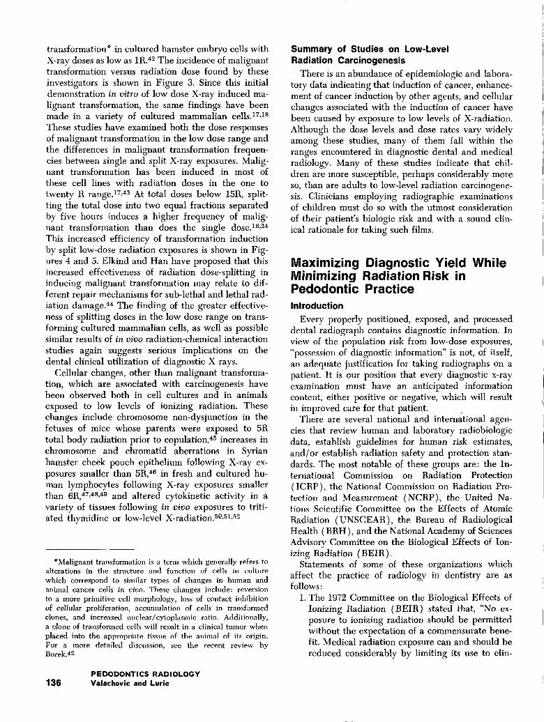
Figure 1 shows four possible dose-response curvesfor cancer incidence vs. increasing radiation dose. Atmoderate (50-500R) to high dose levels (above500R), there is a substantial amount of data on ex-posed human populations, and these data support alinear relationship between increasing radiation doseand increasing incidences of a variety of can-cers.-°,6,13,1~ At extremely high doses, the incidence of
PEDIATRIC DENTISTRYVolume 2, No. 2 129
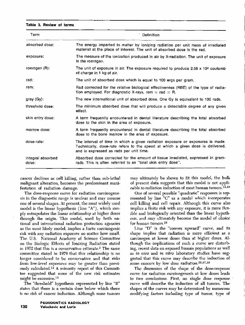
Table 3. Review of terms
Term ’Definition
absorbed dose:
exposure:
roentgen (R):
rad:
rein:
g ray (Gy):
threshold dose:
skin entry dose:
marrow dose:
dose-rate:
integral absorbeddose:
The energy imparted to matter by ionizing radiation per unit mass of irradiatedmaterial at the place of interest. The unit of absorbed dose is the rad.
The measure of the ionization produced in air by X-radiation. The unit of exposureis the roentgen.
The unit of exposure in air. The exposure required to produce 2.58 x 10⁴ coulombof charge in 1 kg of air.
The unit of absorbed dose which is equal to 100 ergs per gram.
Rad corrected for the relative biological effectiveness (RBE) of the type of radia-tion employed. For diagnostic X-rays, rem = rad = R.
The new international unit of absorbed dose. One Gy is equivalent to 100 rads.
The minimum absorbed dose that will produce a detectable degree of any giveneffect.
A term frequently encountered in dental literature describing the total absorbeddose to the skin in the area of exposure.
A term frequently encountered in dental literature describing the total absorbeddose to the bone marrow in the area of exposure.
The interval of time in which a given radiation exposure or exposures is made.Technically, dose-rate refers to the speed at which a given dose is delivered,and is expressed as rads per unit time.
Absorbed dose corrected for the amount of tissue irradiated, expressed in gram-rads. This is often referred to as "total skin entry dose".
cancer declines as cell killing, rather than sub-lethalmalignant alteration, becomes the predominant mani-festation of radiation damage,
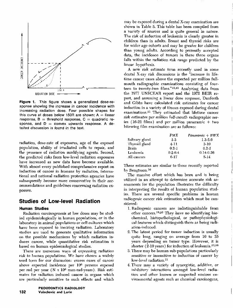
The dose-response curve for radiation carcinogene-sis in the diagnostic range is unclear and may assumeone of several shapes. At present, the most widely usedmodel is the linear hypothesis (line "A"), which sim-.ply extrapolates the linear relationship at higher dosesthrough the origin. This model, used by both na-tional and international radiation protection agenciesas the most likely model, implies a finite carcinogenicrisk with any radiation exposure no matter how small.The U.S. National Academy of Science Committeeon the Biologic Effects of Ionizing Radiation statedin 1972 that this is a conservative estimate.~ The samecommittee stated in 1979 that this relationship is nolonger considered to be conservative and that risksfrom low-level exposures may be greater than previ-ously calculated, lz A minority report of this Commit-tee suggested that some of the new risk estimatesmight be excessive.1~
The "threshold" hypothesis represented by line "B"states that there is a certain dose below which thereis no risk of cancer induction. Although some tumors
may ultimately be shown to fit this model, the bulkof present data suggests that this model is not appli-cable to radiation induction of most human tumors.13,1"~
One of several possible "quadratic" responses is rep-resented by line "C" as a model which incorporatescell killing and cell repair. Although this curve alsoimplies a finite risk with any exposure, it is more flex-ible and biologically oriented than the linear hypoth-esis, and may ultimately become the model of choicefor human tumors.15
Line "D" is the "convex upward" curve, and itsshape implies that radiation is more efficient as acarcinogen at lower doses than at higher doses. Al-though the implications of such a curve are disturb-ing, recent data on exposed human populations as wellas in vivo and in vitro laboratory studies have sug-gested that this curve may describe the induction ofsome cancers by low dose radiation.15Jr,~s
The discussion of the shape of the dose-responsecurve for radiation carcinogenesis at low doses leadsto two conclusions. First, no single dose response ̄curve will describe the induction of all tumors. Theshapes of the curves may be determined by numerousmodifying factors including type of tumor, type of
PEDODONTICS RADIOLOGY130 Valachovic and Lurie
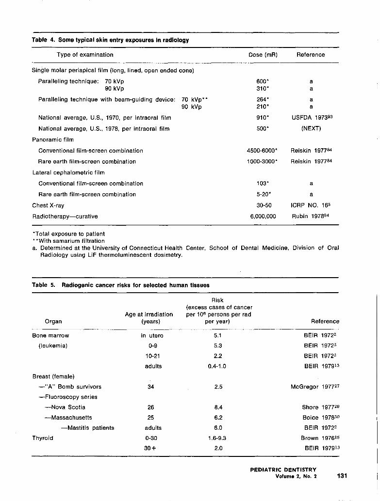
Table 4. Some typical skin entry exposures in radiology
Type of examination Dose (mR) Reference
single molar periapical film (long, lined, open ended cone)
Paralleling technique: 70 kVp90 kVp
Paralleling technique with beam-guiding device:
National average, U.S., 1970, per intraoral film
National average, U.S., 1978, per intraoral film
Panoramic film
Conventional film-screen combination
Rare earth film-screen combination
Lateral cephalometric film
Conventional film-screen combination
Rare earth film-screen combination
Chest X-ray
Radiotherapy--curative
70 kVp**90 kVp
600" a310" a
264" a210" a
910" USFDA 197393
5OO* (NEXT)
4500-6000* Reiskin 197784
1000-3000" Reiskin 1977~
103" a
5-20* a
30-50 ICRP NO. 16~
6,000,000 Rubin 197894
*Total exposure to patient**With samarium filtrationa. Determined at the University of Connecticut Health Center, School of Dental Medicine, Division of Oral
Radiology using LiF thermoluminescent dosimetry.
Table 5. Radiogenic cancer risks for selected human tissues
Risk(excess cases of cancer
Age at irradiation per 106 persons per radOrgan (years) per year) Reference
Bone marrow in utero 5.1 BEIR 1972~
(leukemia) 0-9 5.3 BEIR 1972~
10-21 2.2 BEIR 1972"~
adults 0.4-1.0 BEIR 197913
Breast (female)
--"A" Bomb survivors 34 2.5 McGregor 197727
--Fluoroscopy series
--Nova Scotia 26 8.4 Shore 197729
mMassachusetts 25 6.2 Boice 19783o
--Mastitis patients adults 6.0 BEIR 1972-~
Thyroid 0-30 1.6-9.3 Brown 19761~
30+ 2.0 BEIR 197913
PEDIATRIC DENTISTRYVolume 2, No. 2 131
i~0 ~
RADIATION DOSE ~V
Figure 1. This figure shows a generalized dose-re-sponse showing the increase in cancer incidence withincreasing radiation dose. Four possible shapes forthis curve at doses below 150R are shown: A ---- linearresponse, B = threshold response, C = quadratic re-sponse, and D ---- convex upwards response. A de-tailed discussion is found in the text.
radiation, dose-rate of exposures, age of the exposedpopulation, ability of irradiated cells to repair, andthe presence of radiation modifying agents. Second,the predicted risks from low-level radiation exposureshave increased as new data have become available.With almost every published comprehensive report oninduction of cancer in humans by radiation, interna-tional and national radiation protection agencies havesubsequently become more conservative in their rec-ommendations and guidelines concerning radiation ex-posure.
Studies of Low-level RadiationHuman Studies
Radiation carcinogenesis at low doses may be stud-ied epidemiologically in human populations, or in thelaboratory in animal populations or cell cultures whichhave been exposed to ionizing radiation. Laboratorystudies are used to generate qualitative informationon the possible mechanisms by which radiation in-duces cancer, while quantitative risk estimation isbased on human epidemiological studies.
There are numerous ways of expressing radiationrisk to human populations. We have chosen a widelyused term for our discussion: excess cases of cancerabove expected incidence per l0 s persons exposedper rad per year (N x l0s man-rad-years). Risk esti-mates for radiation induced cancer in organs whichare particularly sensitive to such effects and which
may be exposed during a dental X-ray examination areshown in Table 5. This table has been compiled froma variety of sources and is quite general in nature.The risk of induction of leukemia is clearly greater inchildren than in adults. Breast and thyroid risks arefor wider age cohorts and may be greater for childrenthan young adults. According to presently accepteddata, the incidence of tumors in these three organsfalls within the radiation risk range predicted by thelinear hypothesis.
A new risk estimate term recently used in somedental X-ray risk discussions is the "ix]crease in life-time cancer cases above the expected per million full-mouth radiographic examinations consisting of four-teen to twenty-two films."10,11 Analyzing data fromthe 1977 UNSCEAR report and the 1972 BEIR re-port, and assuming a linear dose response, Danforthand Gibbs have calculated risk estimates for cancerinduction in a variety of tissues exposed during dentalexaminations.11 They estimated that lifetime cancerrisk estimates per million full-mouth radiographic ser-ies (16-22 films) and per million panoramic + twobitewing film examination are as follows:
FMX Panoramic + BWXSalivary gland 1-3 1.3-2.6Thyroid gland 4-11 3-10Brain 0.2-1 0.2-1Leukemia 0.2-0.4 0.14-0.26All cancers 6-17 5-14
These estimates are similar to those recently reportedby Bengtsson.~°
The massive effort which has been and is beingutilized in an attempt to determine accurate risk as-sessments for the population illustrates the difficultyin interpreting the results of human population stud-ies. There are several specific problems in humanradiogenic cancer risk estimation which must be con-sidered:
1. Radiogenic cancers are indistinguishable fromother cancers.19,2° They have no identifying bio-chemical, histopathological, or pathophysiologi-cal features which distinguish them as being radi-ation-induced.
2. The latent period for tumor induction is usuallyquite long, ranging on average from 10 to 35years depending on tumor type. However, it isshorter (2-10 years ) for induction of leukemia.13,16
3. There may be human sub-populations particularlysensitive or insensitive to induction of cancer bylow-level radiation.2~
4. There may a variety of synergistic, additive, orinhibitory interactions amongst low-level radia-tion and other known or suspected noxious en-vironmental agents such as chemical carcinogens,
PEDODONTICS RADIOLOGY132 Valachovic and Lurie
oncogenic viruses, tumor promoters and muta-gens.22,23
5. The effects of splitting up the radiation exposureinto several smaller exposures over many years onthe carcinogenicity of the radiation are not welldefined.18,24,25
6. Most risk estimates are expressed in terms ofwhole body exposure. Most diagnostic radiolog-ical procedures are partial body exposures. Pres-ently, there are no data to support or deny theconcept that irradiating one-tenth of the body re-suits in one-tenth of the cancer risk of irradiatingthe entire body.
7. Most low-risk estimates are derived by extrapo-lating the results of populations exposed to dosesin the moderate-to-high dose range (50-500R), there are few control studies of populations ex-posed to low doses. A mechanistic biological ba-sis for such extrapolations remains to be estab-lished.
The effect of delivering a lifetime radiation dose asmultiple small exposures at varying time intervals, re-ferred to as "fractionating the dose," upon the carcin-ogenic potential of that radiation dose is of criticalinterest to the pedodontist. The practice of repeatedradiologic exposures at frequent intervals duringchildhood and young adulthood could be consideredas fractionation of a moderate (greater than 50R)radiation dose. For example, based on the nationalaverage of 500 mR skin entry dose per intraoral filmin the U.S. in 1978,28 a 20-film series taken every fiveyears from childhood through 60 years of age woulddeliver, through repeated low-dose exposures, a totalexposure to the patient of approximately 120R.
In a recent review, Brown presented data whichsuggested that the frationation of X-ray doses and thereduction of the dose per fraction increases the car-cinogenicity of x- and gamma-radiation in both hu-man female breast and human bone marrow tissues.15
Table 5 shows that in atomic bomb survivors who re-ceived an acute gamma ray and neutron exposure, therisk estimate for breast cancer induction was 2.5 (ex-cess cancer above the expected incidence).27 Mastitispatients receiving fractionated X-ray exposures at ahigh dose per fraction had a risk estimate of 8.3.28
Fluoroscopy patients receiving fractionated X-rays at alow dose per fraction had a risk estimate of 6.2-8.4.~9,39
A similar observation can be made for leukemia in-duction in humans. Atomic bomb survivors, victims ofan acute gamma ray exposure, had a leukemia riskestimate of 0.5 excess cancer beyond the expected in-cidence. Ankylosing spondylitic 31 and mastitis 28 pa-tients who received fractionated therapeutic X-ray
treatments, ranging from 275R to 2750R, have re-spective leukemia risk estimates of 0.5 and 2.4.
These leukemia and breast cancer data suggest in-creasing efficiency of radiation to induce cancer withfractionation. It is interesting to consider the analogybetween the effects of fractionation on leukemia andbreast cancer induction in these studies, and the over-all cancer risk to patients repeatedly exposed to den-tal X-ray exams, particularly during childhood andyoung adulthood.
A controversial suggestion concerning induction ofleukemia in children was made by Bross and Natara-jan in their 1973 Tristate Study.21 This was a retro-spective matched case/control study which examineda large group of children with leukemia and analyzedsimilarities and contrasts in their medical and radia-tion exposure histories with their cohorts. They foundat least two population sub-groups: one group withand the other group without leukemia associated withexposure to low level radiation, such as diagnostic X-rays in utero. It was concluded that not only are chil-dren more susceptible to radiation carcinogenesis thanadults, but that within the population of childrenthere may be sub-groups "orders of magnitude moresensitive" than the total population of children. How-ever, serious questions regarding the validity of thestatistical methodology and sensitive sub-populationconcept have recently appeared.13
There have been several papers analyzing carcino-genesis in workers at the Hanford Nuclear PowerPlant in Richland, Washington, some of which haveclaimed substantially greater leukemia risks in adultsfrom low level radiation than those shown in Table5.21,32 However, these data have been the subject ofconsiderable debate in the literature, ~,34,35 are basedon extremely complex statistical analyses, and thushave not been presented in this discussion.
Most data on radiation cancer induction in humansappears to fall within the predicted linear models,thus indicating a small but definite increased cancerrisk with any radiation exposure, no matter how small.Additionally, there is evidence that there is a greatercancer induction risk for children than for adults dueto the greater proliferative activity in growing tissues.This is clearly shown in the leukemia and thyroid dataof Table 4. Human population studies have tended tosupport conservative (linear as opposed to threshold)estimates of risk, and estimates of risks have increasedas new data have become available. (Such increasedrisk estimates are evidenced by the growing estimatesfor radiogenic thyroid carcinoma induction from theModan3~ study, which had a risk estimate of 6.1 in1973av but has recently been revised upwards to10.9.15) Although the reality of risk from low-level ex-posures appears clear from human studies, the lack of
PEDIATRIC DENTISTRYVolume 2, No. 2 133
80-
~ 60
~ 8o-
u~ 40-
20-
I I I I I I I I
10 20 30 40
WEEKS AFTER INITIAL TREATN~ENT
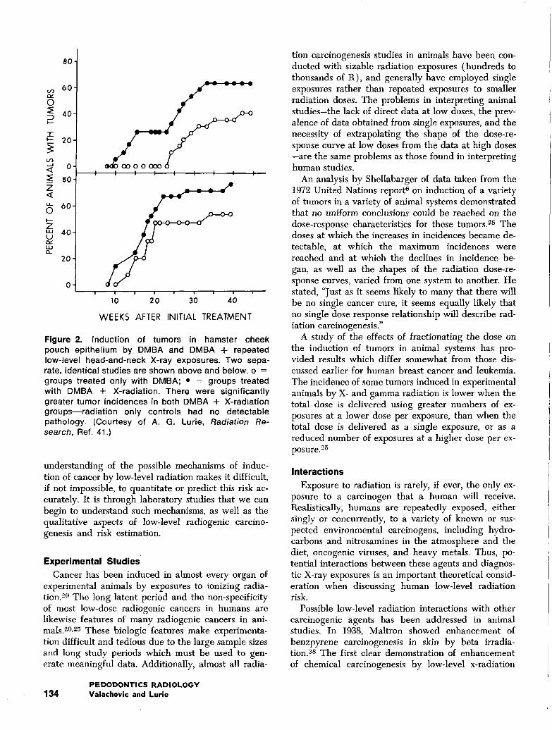
Figure 2. Induction of tumors in hamster cheekpouch epithelium by DMBA and DMBA + repeatedlow-level head-and-neck X-ray exposures. Two sepa-rate, identical studies are shown above and below, o =groups treated only with DMBA; ̄ -- groups treatedwith DMBA + X-radiation. There were significantlygreater tumor incidences in both DMBA -I- X-radiationgroups--radiation only controls had no detectablepathology. (Courtesy of A. G. Lurie, Radiation Re-search, Ref. 41.)
understanding of the possible mechanisms of induc-tion of cancer by low-level radiation makes it difficult,ff not impossible, to quantitate or predict this risk ac-curately. It is through laboratory studies that we canbegin to understand such mechanisms, as well as thequalitative aspects of low-level radiogenic carcino-genesis and risk estimation.
Experimental Studies
Cancer has been induced in almost every organ ofexperimental animals by exposures to ionizing radia-tion. -°° The long latent period and the non-specificityof most low-dose radiogenic cancers in humans arelikewise features of many radiogenic cancers in ani-mals.2°,~5 These biologic features make experimenta-tion difficult and tedious due to the large sample sizesand long study periods which must be used to gen-erate meaningful data. Additionally, almost all radia-
tion carcinogenesis studies in animals have been con-ducted with sizable radiation exposures (hundreds tothousands of R), and generally have employed singleexposures rather than repeated exposures to smallerradiation doses. The problems in interpreting animalstudies-the lack of direct data at low doses, the prev-alence of data obtained from single exposures, and thenecessity of extrapolating the shape of the dose-re-sponse curve at low doses from the data at high doses-are the same problems as those found in interpretinghuman studies.
An analysis by Shellabarger of data taken from the1972 United Nations report6 on induction of a varietyof tumors in a variety of animal systems demonstratedthat no uniform conclusions could be reached on thedose-response characteristics for these tumors.25 Thedoses at which the increases in incidences became de-tectable, at which the maximum incidences werereached and at which the declines in incidence be-gan, as well as the shapes of the radiation dose-re-sponse curves, varied from one system to another. Hestated, "Just as it seems likely to many that there willbe no single cancer cure, it seems equally likely thatno single dose response relationship will describe rad-iation carcinogenesis."
A study of the effects of fractionating the dose onthe induction of tumors in animal systems has pro-vided results which differ somewhat from those dis-cussed earlier for human breast cancer and leukemia.The incidence of some tumors induced in experimentalanimals by X- and gamma radiation is lower when thetotal dose is delivered using greater numbers of ex-posures at a lower dose per exposure, than when thetotal dose is delivered as a single exposure, or as areduced number of exposures at a higher dose per ex-posure.25
Interactions
Exposure to radiation is rarely, if ever, the only ex-posure to a carcinogen that a human will receive.Realistically, humans are repeatedly exposed, eithersingly or concurrently, to a variety of known or sus-pected environmental carcinogens, including hydro-carbons and nitrosamines in the atmosphere and thediet, oncogenic viruses, and heavy metals. Thus, po-tential interactions between these agents and diagnos-tic X-ray exposures is an important theoretical consid-eration when discussing human low-level radiationrisk.
Possible low-level radiation interactions with othercarcinogenic agents has been addressed in animalstudies. In 1938, Maltron showed enhancement ofbenzpyrene carcinogenesis in skin by beta irradia-tion. 38 The first clear demonstration of enhancementof chemical carcinogenesis by low-level x-radiation
PEDODONTICS RADIOLOGY134 Valachovi¢ and Lurie
1.0
.001 ~ I I , I I ~ I I ,I 10 100 000
x-ray absorbed dose (tad1
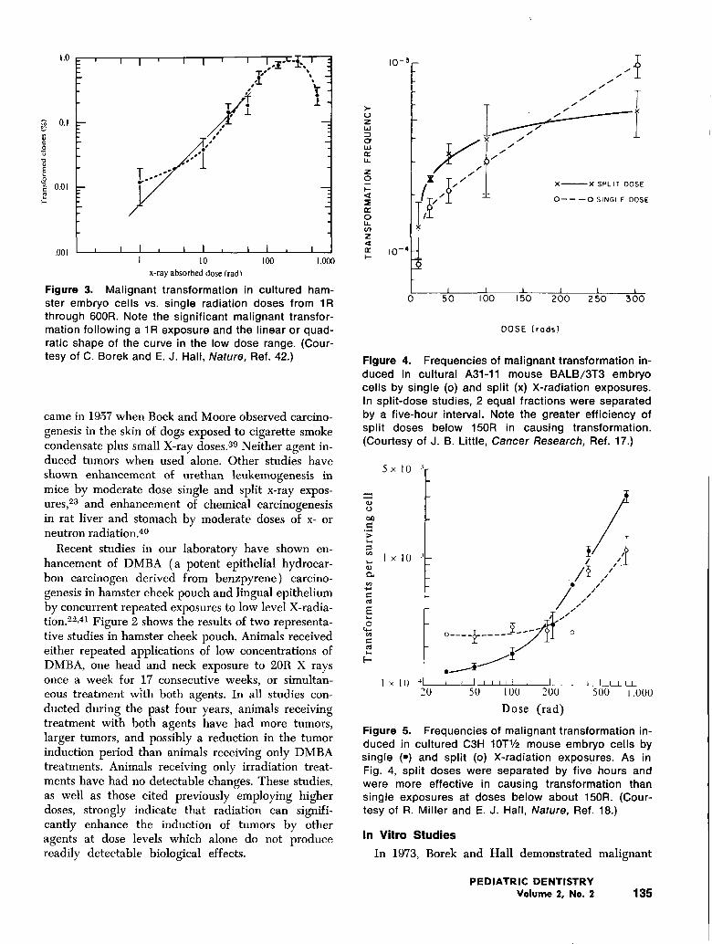
Figure 3. Malignant transformation in cultured ham-ster embryo cells vs. single radiation doses from 1Rthrough 800R. Note the significant malignant transfor-mation following a 1R exposure and the linear or quad-ratic shape of the curve in the low dose range. (Cour-tesy of C. Borek and E. J. Hall, Nature, Fief. 42.)
came in 1957 when Bock and Moore observed carcino-genesis in the skin of dogs exposed to cigarette smokecondensate plus small X-ray doses.39 Neither agent in-duced tumors when used alone. Other studies haveshown enhancement of urethan leukemogenesis inmice by moderate dose single and split x-ray expos-ures, 23 and enhancement of chemical carcinogenesisin rat liver and stomach by moderate doses of x- orneutron radiation.4o
Recent studies in our laboratory have shown en-hancement of DMBA (a potent epithelial hydrocar-bon carcinogen derived from benzpyrene) carcino-genesis in hamster cheek pouch and lingual epitheliumby concurrent repeated exposures to low level X-radia-tion. ~-2,41 Figure 2 shows the results of two representa-tive studies in hamster cheek pouch. Animals receivedeither repeated applications of low concentrations ofDMBA, one head and neck exposure to 20R X raysonce a week for 17 consecutive ~veeks, or simultan-eous treatment ~v]th both agents. In all studies con-
ducted during the past four years, animals receivingtreatment with both agents have had more tumors,larger tumors, and possibly a reduction in the tumorinduction period than animals receiving only DMBAtreatments. Animals receiving only irradiation treat-ments have had no detectable changes. These studies,
as well as those cited previously employing higherdoses, strongly indicate that radiation can signifi-cantly enhance the induction of tumors by otheragents at dose levels which alone do not producereadily detectable biological effects.
"~ T//- I X~X SPLIT OOSE±
10-4
0 501 I I I I
I00 150 200 250 300
DOSE (rods)
Figure 4. Frequencies of malignant transformation in-duced in cultural A31-11 mouse BALB/3T3 embryocells by single (o) and split (x) X-radiation exposures.In split-dose studies, 2 equal fractions were separatedby a five-hour interval, Note the greater efficiency ofsplit doses below 150R in causing transformation.(Courtesy of J. B. Little, Cancer Research, Ref. 17.)
5x 10
Ix I0
Ix 10 ~ I ~ I ~ ~1 , t ~ ~ 1 , , ~20 50 100 200 500 ~ .000
Dose (rad)
Fisure 5. Frequencies of malignant transformation in-duced in cultured C3H IOTV~ mouse emb~o cells bysingle (~) and split (o) X-radiation exposures. As Fig. 4, split doses were separated by five hours andwere more effective in causing transformation thansingle exposures at doses below about 150R. (Cour-tesy of R. Miller and E. J. Hall, Nature, Ref. 18.)
In Vitro Studies
In 1973, Borek and Hall demonstrated malignant
PEDIATRIC DENTISTRYVolume 2, No. 2 135
transformation* in cultured hamster embryo cells withX-ray doses as low as 1R.42 The incidence of malignanttransformation versus radiation dose found by theseinvestigators is shown in Figure 3. Since this initialdemonstration in vitro of low dose X-ray induced ma-lignant transformation, the same findings have beenmade in a variety of cultured mammalian cells.17,18
These studies have examined both the dose responsesof malignant transformation in the low dose range andthe differences in malignant transformation frequen-cies between single and split X-ray exposures. Malig-nant transformation has been induced in most ofthese cell lines with radiation doses in the one totwenty R range.17,43 At total doses below 15R, split-ting the total dose into two equal fractions separatedby five hours induces a higher frequency of malig-nant transformation than does the single dose.~8,24
This increased efficiency of transformation inductionby split low-dose radiation exposures is shown in Fig-ures 4 and 5. Elkind and Han have proposed that thisincreased effectiveness of radiation dose-splitting ininducing malignant transformation may relate to dif-ferent repair mechanisms for sub-lethal and lethal rad-iation damage.4~ The finding of the greater effective-ness of splitting doses in the low dose range on trans-forming cultured mammalian cells, as well as possiblesimilar results of in vivo radiation-chemical interactionstudies again suggests serious implications on thedental clinical utilization of diagnostic X rays.
Cellular changes, other than malignant transforma-tion, which are associated with carcinogenesis havebeen observed both in cell cultures and in animalsexposed to low levels of ionizing radiation. Thesechanges include chromosome non-dysiunction in thefetuses of mice whose parents were exposed to 5Rtotal body radiation prior to copulation,45 increases inchromosome and chromatid aberrations in Syrianhamster cheek pouch epithelium following X-ray ex-posures smaller than 5R,4~ in fresh and cultured hu-man lymphocytes following X-ray exposures smallerthan 6R,47,48,49 and altered cytokinetic activity in avariety of tissues following in vivo exposures to triti-ated thymidine or low-level X-radiation.5°,~,5°-
*Malignant transformation is a term which generally refers toalterations in the structure and function of cells in culturewhich correspond to similar types of changes in human andanimal cancer cells in vivo. These changes include: reversionto a more primitive cell morphology, loss of contact inhibitionof cellular proliferation, accumulation of ceils in transformedclones, and increased nuclear/cytoplasmic ratio. Additionally,a clone of transformed cells will result in a clinical tumor whenplaced into the appropriate tissue of the animal of its origin.For a more detailed discussion, see the recent review byBorek.42
Summary of Studies on Low-LevelRadiation Carcinogenesis
There is an abundance of epidemiologic and labora-tory data indicating that induction of cancer, enhance-ment of cancer induction by other agents, and cellularchanges associated with the induction of cancer havebeen caused by exposure to low levels of X-radiation.Although the dose levels and dose rates vary widelyamong these studies, many of them fall within theranges encountered in diagnostic dental and medicalradiology. Many of these studies indicate that chil-dren are more susceptible, perhaps considerably moreso, than are adults to low-level radiation carcinogene-sis. Clinicians employing radiographic examinationsof children must do so with the utmost considerationof their patient’s biologic risk and with a sound clin-ical rationale for taking such films.
Maximizing Diagnostic Yield WhileMinimizing Radiation Risk inPedodontic PracticeIntroduction
Every properly positioned, exposed, and processeddental radiograph contains diagnostic information. Inview of the population risk from low-dose exposures,"possession of diagnostic information" is not, of itself,an adequate justification for taking radiographs on apatient. It is our position that every diagnostic x-rayexamination must have an anticipated informationcontent, either positive or negative, which will resultin improved care for that patient.
There are several national and international agen-cies that review human and laboratory radiobiologicdata, establish guidelines for human risk estimates,and/or establish radiation safety and protection stan-dards. The most notable of these groups are: the In-ternational Commission on Radiation Protection(ICRP), the National Commission on Radiation Pro-tection and Measurement (NCRP), the United Na-tions Scientific Committee on the Effects of AtomicRadiation (UNSCEAR), the Bureau of RadiologicalHealth ( BRH ), and the National Academy of SciencesAdvisory Committee on the Biological Effects of Ion-izing Radiation (BEIR).
Statements of some of these organizations whichaffect the practice of radiology in dentistry are asfollows:
1. The 1972 Committee on the Biological Effects ofIonizing Radiation (BEIR) stated that, "No ex-posure to ionizing radiation should be permittedwithout the expectation of a commensurate bene-fit. Medical radiation exposure can and should bereduced considerably by limiting its use to clin-
PEDODONTICS RADIOLOGY136 Valachovic and Lurie
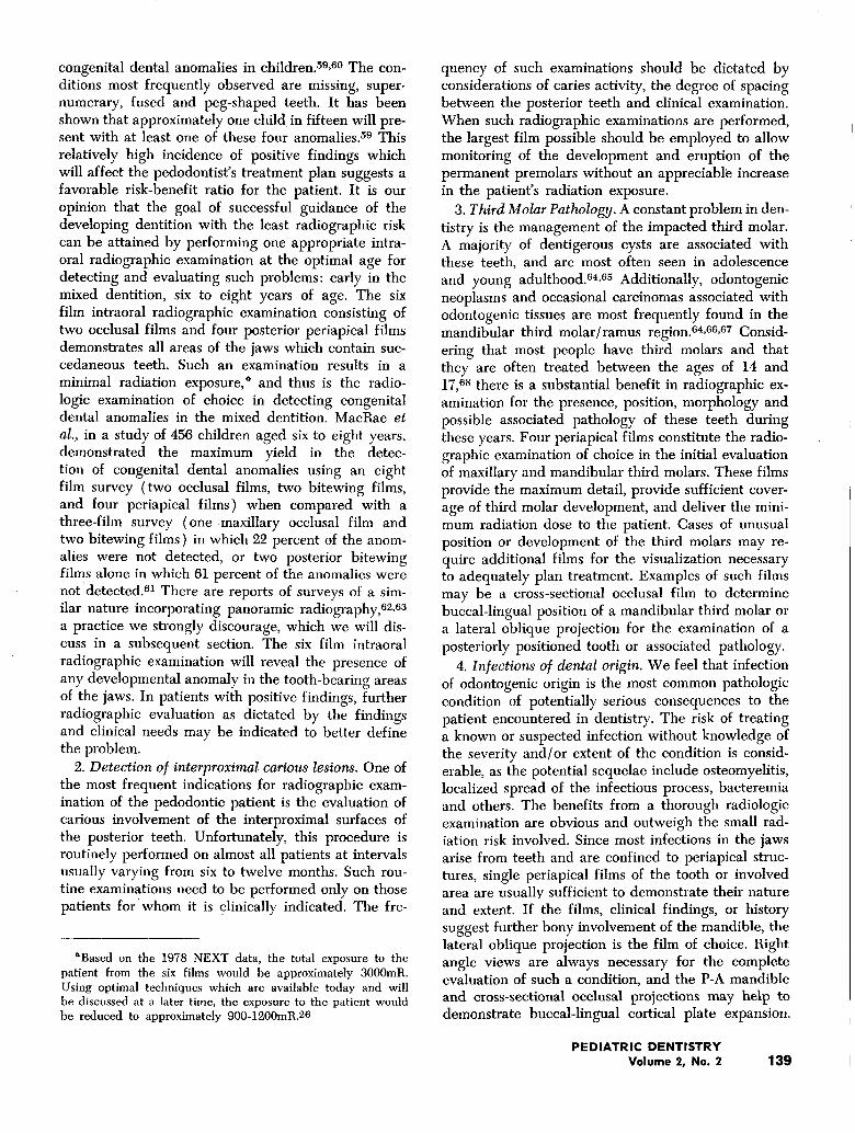
Table 6. Clinical situations for which radiographsmay be indicated
History of painEvidence of swelling
Positive neurologic findings in face and jawsTrauma to teeth, jaws and/or lips
Mobility of teethUnexplained bleeding
Deep periodontal pocketingFistula formation
Unexplained sensitivity of teethEvaluation of sinus condition
Unusual eruption of teethUnusual spacing or migration of teeth
Lack of response to conventional dental treatmentUnusual tooth morphology, calcification or color
Evaluation of growth abnormalitiesAltered occlusal relationships
Aid in diagnosis of systemic diseaseAssessment of dental involvement in established
systemic diseaseFamilial history of dental anomalies
Post-operative evaluationOthers
ically indicated procedures utilizing efficient ex-posure techniques and optimal operation of radia-tion equipment.’’2
2. The National Committee on Radiation Protectionand Measurements (NCRP) has recommendedthat, "Exposure which fulfills no useful purposeshould be reduced to an absolute minimum . . . amajor effort is justified to assure that the radia-tion field does not extend beyond the area of theX-ray film or fluoroscopic screen or significantlybeyond the region to be examined."4
3. The Food and Drug Administration (FDA) hasproposed regulations which will mandate qualityassurance programs in any diagnostic radiologyfacility (including the private dental office) reduce the radiation exposure burden to theAmerican population. The commissioner of theFDA stated that such regulations are necessarybecause "... data from several sources indicatethat many diagnostic radiology facilities are pro-ducing poor quality images and giving unneces-sary patient radiation exposure.’’54 As of the dateof the submission of this paper, no final actionhad been taken in this area.
In addition, the American Dental Association Councilon Dental Materials, Instruments, and Equipment hasrecommended that, "The decision to use diagnosticradiography rests on professional judgment of its he-
cessity for the benefit of the total health of the pa-tient. This decision having been made, it then be-comes the duty of the dental professional to producea maximum yield of information per unit of X-ray ex-posure."55
The interest of state and federal governments in theexamination and possible future regulation of all as-pects of the diagnostic radiological sciences has stead-ily increased during recent years. Areas being consid-ered by such agencies as the President’s InteragencyTask Force on Ionizing Radiation, CongressionalHearings on ionizing radiation, the Commissioner ofthe FDA, and a number of state legislative commit-tees and regulatory agencies include machine per-formance, film characteristics, operator training andlicensing, professional and auxiliary education, qualityassurance programs, and facility licensing and inspec-tion programs. Eight states have instituted specificradiologic licensing examinations for dentists separatefrom their State Dental Board Examinations. Severalother states are considering similar action.
How does the practicing dentist meet such radia-tion safety and practice criteria while maintainingthe level of diagnostic radiology needed for excel-lence in clinical practice? We feel that these needs aremet in two ways: (1) the establishment of high diag-nostic yield criteria to be used in the determination ofthe need for films; and (2) the execution of such ex-aminations incorporating techniques which maximizethe diagnostic yield while minimizing radiation ex-posure for every film.
Suggested Criteria for Radiologic Examinationof Children and Adolescents
We shall define high yield criteria as those clinicalor historical findings for which radiographic examina-tions are likely to provide confirming or clarifying in-formation. These radiographic examinations shouldhave a high probability of affecting the diagnosis andtreatment of a problem which, ff left untreated, posesa potential health hazard greater than that associatedwith the radiographic exposure.
Historically, dentists have used radiographic exam-inations for a variety of documentary and screeningpurposes. Documentary purposes include films takenfor insurance company post-treatment verification,teaching files, slide collections, patient education,completeness of records, "making sure everything isOK," and the evaluation of marginal adaptation ofrestorations. Such purposes rarely provide a benefitto the patient, and we disapporve of such practicesbecause they expose patients to a radiation risk with-out a commensurate or greater benefit.
A screening examination is one in which specific di-agnostic procedures are performed in a population
PEDIATRIC DENTISTRYVolume 2, No. 2 137
specifically at risk with a view towards discoveringoccult disease of a life-threatening nature whichwould be otherwise undetected. Such a populationmay be that of an entire country, an entire city, orthe private practice patients of an individual dentist.For a screening procedure to be effective, positivefindings must be followed by appropriate treatment.If the screening procedure entails risk to the popula-tion, then the benefits accrued from the discovery andtreatment of occult disease must outweigh this risk.
The only radiographic screening procedures pres-ently employed, to our knowledge, are mammographyin post-menopausal women, where there is an estab-lished and significant risk-benefit ratio in favor of thepatient,56,57 and radiographic screening for diseases ofthe iaws in dentistry where a risk-benefit ratio hasnever been established and is likely to be quite un-favorable for the patient population being screened.Although there are diseases which are unique to theiaws, the incidences of serious diseases of the iawshave not been demonstrated to be greater than thoseinvolving the general skeleton. A total body skeletalradiographic survey for the detection of occult diseasein the absence of clinical findings is not practiced any-where in the world. In addition, often-cited examplesof such diseases of the iaws are cysts, abnormallyformed teeth, odontogenic neoplasia or cancer. Theonly diseases in such a list with expected serious con-sequences are cancers and aggressive odontogenicneoplasms of the iaws, both of which are exceedinglyrare in the pediatric age group. When such lesions dooccur, they almost always have presenting clinicalsigns and symptoms which in the absence of any rad-iographic examination would be suggestive of an ab-normality requiring further evaluation.
The rational use of radiology in pedodontics re-quires definition of clinical and historical criteriawhich are likely to require a radiologic examination toallow a practitioner to proceed with the best possibletreatment: high yield criteria. Table 6 lists a varietyof positive clinical and historical findings which arelikely to require radiographic examination. Many ofthe clinical situations listed in this table are obvious.For example, the dentist to whom a pedodontie pa-tient presents with a history of .pain, evidence ofswelling, or after traumatic iniury, must have thebenefit of radiologic examination available in orderto carry out the best treatment. However, some of thesituations listed in this table require discussion.
Fistula formation in the pediatric age group is us-ually indicative of a localized furcational or periapi-cal infection of dental origin. Radiographic examina-tion is necessary, but one should be selective in thenumber and types of films used. Each film shouldhave an indication based on the clinical situation;
there is no reason to simply order a "full-mouth ser-ies." The same holds true for the evaluation of un-usual eruption of teeth. For example, a seven-year oldpresents with erupting maxillary permanent lateral in-cisors but only one erupting permanent central incisor.Consideration of the absence or displacement of theunerupted incisor must be made, and an individualperiapical or occlusal film is the indicated radiograph-ic examination. Another situation which the pedodon-tist is faced with is the evaluation of the medicallyexceptional child. A dental radiographic examinationmay be helpful in the diagnosis of systemic disease;however, the taking of numerous radiographs in a pa-tient with an established systemic disease but no clin-ical evidence of dental involvement to "see what’s go-ing on" is not an indicated procedure because it ex-poses the patient to a radiation dose without having asignificant potential benefit to that patient.
Whenever diagnostic radiology is to be utilized, thepracticing dentist should make an intelligent decisionof which radiographs will provide the informationneeded at the lowest radiation exposure possible.There may be times when the decision should be thatno radiographs are indicated. Although we do notascribe to broad rules to dictate which radiographsto take for which problems, there are certain commonsituations in pedodontics which require discussion.
Common Clinical Indications for RadiologyFive clinical situations which the pedodontist is
commonly confronted with in which radiographs areusually indicated for a thorough evaluation are: (1)detection of congenital dental anomalies in the mixeddentition in patients undergoing comprehensive den-tal care; (2) detection of interproximal carious les-ions; (3) third molar evaluation; (4) infection; (5) trauma. Each of these indications for radiographicexamination requires a consideration of the risk-bene-fit ratio. These situations and examination rationalesare shown in Table 7.
1. Detection of congenital dental anomalies. Thecomprehensive dental care of the pedodontic patientincludes the management of the dentition in a waythat permits the most harmonious oeclusal relation-ships to develop. The dentist must be able to antici-pate any condition which may complicate the treat-ment plan. The mixed dentition space analysis is oftenused to detect potential problems which may be de-veloping as the occlusal pattern is established. Undi-agnosed congenital anomalies of tooth number, size,shape and location have a potential impact on the suc-cessful management of the developing dentition. Suchpathology generally is detectable only with radio-graphs.58
Various authors have investigated the incidence of
PEDODONTICS RADIOLOGY138 Va|achovic and Lurie
congenital dental anomalies in children. 59,6° The con-ditions most frequently observed are missing, super-numerary, fused and peg-shaped teeth. It has beenshown that approximately one child in fifteen will pre-sent with at least one of these four anomalies.59 Thisrelatively high incidence of positive findings whichwill affect the pedodontist’s treatment plan suggests afavorable risk-benefit ratio for the patient. It is ouropinion that the goal of successful guidance of thedeveloping dentition with the least radiographic riskcan be attained by performing one appropriate intra-oral radiographic examination at the optimal age fordetecting and evaluating such problems: early in themixed dentition, six to eight years of age. The sixfilm intraoral radiographic examination consisting oftwo occlusal films and four posterior periapicalfilmsdemonstrates all areas of the iaws which contain suc-cedaneous teeth. Such an examination results in aminimal radiation exposure,* and thus is the radio-logic examination of choice in detecting congenitaldental anomalies in the mixed dentition. MacRae etal., in a study of 456 children aged six to eight years,demonstrated the maximum yield in the detec-tion of congenital dental anomalies using an eightfilm survey (two occlusal films, two bitewing films,and four periapical films) when compared with three-film survey (one maxillary occlusal film andtwo bitewing films) in which 22 percent of the anom-alies were not detected, or two posterior bitewingfilms alone in which 61 percent of the anomalies werenotdetected.61 There are reports of surveys of a sim-ilar nature incorporating panoramic radiography,6~,63
a practice we strongly discourage, which we will dis-cuss in a subsequent section. The six film intraoralradiographic examination will reveal the presence ofany developmental anomaly in the tooth-bearing areasof the iaws. In patients with positive findings, furtherradiographic evaluation as dictated by the findingsand clinical needs may be indicated to better definethe problem.
2. Detection of interproximal carious lesions. One ofthe most frequent indications for radiographic exam-ination of the pedodontic patient is the evaluation ofcarious involvement of the interproximal surfaces ofthe posterior teeth. Unfortunately, this procedure isroutinely performed on almost all patients at intervalsusually varying from six to twelve months. Such rou-tine examinations need to be performed only on thosepatients for’whom it is ~linically indicated. The fre-
*Based on the 1978 NEXT data, the total exposure to thepatient from the six films would be approximately 3000mR.Using optimal techniques which are available today and willbe discussed at a later time, the exposure to the patient wouldbe reduced to approximately 900-1200mR.26
quency of such examinations should be dictated byconsiderations of caries activity, the degree of spacingbetween the posterior teeth and clinical examination.When such radiographic examinations are performed,the largest film possible should be employed to allowmonitoring of the development and eruption of thepermanent premolars without an appreciabl~ increasein the patient’s radiation exposure.
3. Third Molar Pathology. A constant problem in den-tistry is the management of the impacted third molar.A maiority of dentigerous cysts are associated withthese teeth, and are most often seen in adolescenceand young adulthood.64,6~ Additionally, odontogenicneoplasms and occasional carcinomas associated withodontogenic tissues are most frequently found in themandibular third molar/ramus region. 64,66,6r Consid-ering that most people have third molars and thatthey are often treated between the ages of 14 and17,68 there is a substantial benefit in radiographic ex-amination for the presence, position, morphology andpossible associated pathology of these teeth duringthese years. Four periapical films constitute the radio-graphic examination of choice in the initial evaluationof maxillary and mandibular third molars. These filmsprovide the maximum detail, provide sufficient cover-age of third molar development, and deliver the mini-mum radiation dose to the patient. Cases of unusualposition or development of the third molars may re-quire additional films for the visualization necessaryto adequately plan treatment. Examples of such filmsmay be a cross-sectional occlusal film to determinebuccal-lingual position of a mandibular third molar ora lateral oblique proiection for the examination of aposteriorly positioned tooth or associated pathology.
4. Infections of dental origin. We feel that infectionof odontogenic origin is the most common pathologiccondition of potentially serious consequences to thepatient encountered in dentistry. The risk of treatinga known or suspected infection without knowledge ofthe severity and/or extent of the condition is consid-erable, as the potential sequelae include osteomyelitis,localized spread of the infectious process, bacteremiaand others. The benefits from a thorough radiologicexamination are obvious and outweigh the small rad-iation risk involved. Since most infections in the iawsarise from teeth and are confined to periapical struc-tures, single periapical films of the tooth or involvedarea are usually sufficient to demonstrate their nature
and extent. If the films, clinical findings, or historysuggest further bony involvement of the mandible, thelateral oblique proiection is the film of choice. Rightangle views are always necessary for the completeevaluation of such a condition, and the P-A mandibleand cross-sectional occlusal projections may help todemonstrate buccal-lingual cortical plate expansion.
PEDIATRIC DENTISTRYVolume 2, No. 2 139
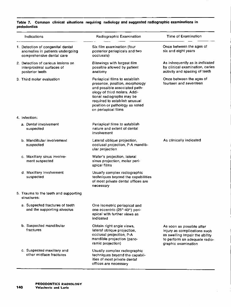
Table 7. Common clinical situations requiring radiology and suggested radiographic examinations inpedodontics
Indications Radiographic Examination Time of Examination
1. Detection of congenital dentalanomalies in patients undergoingcomprehensive dental care
2. Detection of carious lesions oninterproximal surfaces ofposterior teeth
3. Third molar evaluation
4. Infection:
a. Dental involvementsuspected
b. Mandibular involvementsuspected
c. Maxillary sinus involve-ment suspected
d. Maxillary involvementsuspected
5. Trauma to the teeth and supportingstructures:
a. Suspected fractures of teethand the supporting alveolus
b. Suspected mandibularfractures
c. Suspected maxillary andother midface fractures
Six film examination (fourposterior periapicals and twoocclusals)
Bitewings with largest filmpossible allowed by patientanatomy
Periapical films to establishpresence, position, morphologyand possible associated path-ology of third molars. Addi-tional radiographs may berequired to establish unusualposition or pathology as notedon periapical films
Periapical films to establishnature and extent of dentalinvolvement
Lateral oblique projection,occlusal projection, P-A mandib-ular projection
Water’s projection, lateralsinus projection, molar peri-apical films
Usually complex radiographictechniques beyond the capabilitiesof most private dental offices arenecessary
One isometric periapical andone eccentric (25o-40°) peri-apical with further views asindicated
Obtain right angle views,lateral oblique projection,occlusal projection, P-Amandible projection (pano-ramic projection)
Usually complex radiographictechniques beyond the capabil-ities of most private dentaloffices are necessary
Once between the ages ofsix and eight years
As infrequently as is indicatedby clinical examination, cariesactivity and spacing of teeth
Once between the ages offourteen and seventeen
As clinically indicated
As soon as possible afterinjury as complications suchas swelling impair the abilityto perform an adequate radio-graphic examination
PEDODONTICS RADIOLOGY140 Valachovic and Lurie
Extensive osseous involvement of the maxilla is rareand requires films usually beyond the capacity of thedental office. Infections of the maxillary sinus arebest demonstrated by the molar periapical film, andthe lateral sinus and Water’s proiections.
5. Trauma. The pedodontic patient who presentswith localized trauma to the teeth and iaws requiresa thorough historical and clinical evaluation. The po-tential complications of undiagnosed or improperlydiagnosed traumatic injuries are numerous, and sincemost are osseous or dental in nature, such complica-tions may be obviated by an appropriate and thoroughradiographic examination of the likely areas of in-iury.69,7°,71 As with infection, therefore, the obvioussubstantial benefit which results from the completedemonstration of the injuries via radiographic exam-ination leading to appropriate treatment outweighsthe radiation risk.
Andreasen has discussed the need for multiple rad-iographic views in the examination of suspected frac-tured teeth. 7~ From an optical point of view, a two-film examination consisting of one standard and oneeccentric periapical film, with the tube head rotatedand the film in the standard position, of the trauma-tized tooth usually will demonstrate the presence orabsence of a fracture of the root. If a fracture is docu-mented, or if the clinical evidence is not consistentwith the radiographic findings, further radiographsmay be necessary. For example, if there is clinicalevidence that the root apex of a maxillary incisortooth has been displaced into or through the buccalplate, the lateral view using an extraorally placed peri-apical or occlusal film has been suggested.7’2 However,as this technique does not necessarily demonstratecollimation requirements and as it is an extraoral film,it may violate both federal and state regulations. Al-ternatives are intraoral placement of dental films inthe buccal vestibule or use of an extraoral cassette. Ifthe clinical evidence suggests a root fracture which isnot demonstrated on initial radiographic examination,further eccentric periapical films may be indicated.
Fractures of the mandible and midface usually pre-sent with compelling historical and/or clinical find-ings. These patients will generally be treated by anoral surgeon or hospital-based dental service. The di-agnostic work-up usually will involve a radiographicexamination, thus the most prudent course with sucha patient in a private office is referral without radio-graphs, since any films would most likely be dupli-cated at the time of treatment at the surgeon’s officeor in the hospital.
For those practitioners who are involved in the careof facial and mandibular fractures, the following gen-eral principles apply to the radiographic examinationof such traumatized patients. In the case of suspected
mandibular fractures, right angle views are essentialto adequately demonstrate the existence of a fractureand the nature of any displacement. The lateral obliqueproiection demonstrates the mandibular body andramus, and the open mouth Towne proiection demon-strates the condylar head and neck, while the P-Amandible, right angle mandibular occlusal, and/orsubmentovertex projections demonstrate the body, ra-mus and symphysis in right angle proiections. Therehave been recent preliminary studies conducted atthe Vanderbilt University Medical Center which sug-gest that the panoramic proiection may be a particu-larly high yield procedure when used to confirm thepresence of a clinically suspected fracture.* Radio-graphic examination of fractures of the midface andcranial bones may be complex and dictated by theclinical situation.
Follow-up of Infection and Trauma. Radiographscan be effective means of following osseous responseto therapy after trauma and/or infection. Keeping inmind the requirement that there must be a 30 percentto 60 percent alteration in the mineral content of thetissue to result in a detectable radiographic change,7a
an adequate time interval should be allowed beforetaking follow-up films. For example, iust as there maybe no radiographic evidence of an acute osteomyelitisin an obviously clinically ill patient until three weeksafter onset of the disease, a healing mandible withprevious widespread chronic osteomyelitis may stillpresent radiographically as seriously diseased for sev-eral weeks after positive response to therapy. In orderto reduce the number of follow-up films, every effortmust be made to duplicate as closely as possible thepatient positioning and exposure factors used in theoriginal examination. This, of course, requires thor-ough quality control and record keeping at all times.
Extraoral Curved Surface Panoramic Radiography
The current popularity and misunderstanding of ex-traoral panoramic radiology in pedodontic practice re-quires discussion. Following the introduction of extra-oral panoramic machines into the United States in theearly 1960s, its supposed virtues of low dose, ease ofoperation, patient education capabilities, patient com-fort, and high diagnostic content were extolled, andthere has been subsequent widespread use of this pro-cedure for a variety of clinical situations.74,75,76,77 It isour contention that although there are specific clin-ical indications for an extraoral panoramic film, thevast maiority of panoramic radiographs taken in theUnited States today are inappropriate, unnecessary,and potentially inaccurate and confusing. Extraoral
*s. J. Gibbs, Department of Radiology, Vanderbi|t Univer-sity, Nashville, Tennessee 37232, personal communication.
PEDIATRIC DENTISTRYVolume 2, No. 2 141
panoramic radiology substantially adds to the popu-lation radiation burden with little if any concomitantbenefit to the patients.
The extraoral panoramic film is a tomogram-an op-tical slice which intentionally blurs structures in frontand in back of the part of the obiect being examined.Although tomographic units used in medical radiologycan be operated in such a way as to finely controlthe areas being demonstrated or blurred, the pan-oramic unit produces a film with fixed cut thicknessesand limited flexibility in patient positioning. Varia-tions in patient positions and anatomic configurationsresult in a lack of precise control over the anatomicareas in focus on the film. Obiects in and about theiaws, not in the plane of focus of the cut, may be par-tially or completely absent in the resultant film. Thisapplies to obiects as large as impacted teeth. 78 Addi-tionally, there are inherent distortion artifacts fre-quently observed in a variety of areas.79,8°,81
There are several articles dealing with relative dosi-metry of panoramic and conventional intraoral radi-ography, and these studies have explained patient ex-posures and/or absorbed doses in a variety of waysincluding: local skin entry dose, total skin entry dose,integral absorbed dose, marrow absorbed dose, thy-roid dose, and most recently marrow equivalent dose.In most of these studies, skin and marrow doses fromthe panoramic films have been lower than a conven-tional full-mouth intraoral radiographic examinationdue to the dose reducing quality of film/screen com-binations. 82,83 Recent studies have indicated that theuse of the new extremely high speed rare earth imag-ing systems can result in a substantial dose reductionbeyond current levels.84
It must be remembered that there are centers of ro-tation in the patient’s head which receive greater ex-posures in extraoral panoramic radiography than inintraoral radiography, most notably thyroid, salivarygland and pharyngeal-lymphoid regions. 8~ RecentMonte Carlo dosimetric studies suggest that-the in-creased lifetime cancer risk per million persons for asingle panoramic film is quite similar to that from acollimated, 16-22 film full-mouth series. 1°,~ The ex-posure to the thyroid gland is of particular concern intaking panoramic films in the pediatric age group. Asdiscussed earlier, the thyroid gland in children is apal-ticularly sensitive organ to low-level radiation car-cinogenesis. Myers et al. s’2 have recently discussed thenecessity of shielding the thyroid gland, especially inchildren, during panoramic and cephalometric radio-graphic procedures. In their discussion, they pointedout the more superior anatomic position of this glandin the child, thus placing it closer to the primarybeam field than in the adult.
Perhaps the single greatest excess contribution to
the patient radiation exposure from panoramic radi-ography occurs when suspected positive findings onpanoramic films generally require additional plainfilms* to be taken. These films are needed to clearlydemonstrate and define suspicious positive findingswhich are poorly shown on the panoramic film due toits inherent distortion. The plain films usually couldhave been ordered at the outset if a thorough andthoughtful clinical and historical evaluation were ob-tained, and would have demonstrated the pathologywithout the need for the panoramic film. Addition-ally, panoramic artifacts in the midline and molarregions often suggest the presence of pathology andrequire plain films which reveal nothing more thannormal anatomy. The patient has been unnecessarilyexposed twice. Artifacts which may be suggestive ofcystic or neoplastic disease are lucencies in the man-dibular midline, maxillary antral and tuberosity re-gions and the mandibular molar-ramus region. A moreextensive discussion of such artifacts may be foundin a recent publication by Reiskin.sl
The panoramic radiograph is often used as a popu-lation screening device. Even if there were a favor-able risk-benefit ratio from radiologic screening forserious occult dental disease, the panoramic filmwould be a poor choice since it neither demonstratesthe entire dental and osseous content of the iaws norpossesses adequate sharpness and definition. It is rare-ly possible to arrive at a secure radiographic diagnosisbased on this film alone. Screening for dental diseaseusing a panoramic film is comparable to screening forlung cancer with a random cut tomogram in oneplane of the lungs.
There are several reasons cited in the literature fortaking panoramic radiographs on children. These in-clude the relative rate of success of the examination,patient comfort, ease and speed of the examination,and the usefulness of the film as a patient educationaid. 6~,75,77,86 Clearly, these reasons for taking pan-oramic films are not related to the clinical needs of thepatient. They do not contribute appreciably to any-thing but the convenience of the dentist and the radia-tion exposure of the patient. Although one could arguethat the higher yield of diagnostic quality films isvalid, this shows a lack of adequate technique in per-forming conventional high yield radiologic examina-tions, since techniques are available to do so.
While the majority of pediatric panoramic exam-inations appear to be inappropriate and unnecessary,there are certainly indications for the taking of suchfilms. They are particularly useful for patients in
*"Plain films" are films in which all structures between theradiation source and the film appear in focus on the fihn. Allintraoral films are plain films.
PEDODONTICS RADIOLOGY142 Valachovic and Lurie
whom conventional radiology cannot be performed.Such patients would include: those in intermaxillaryfixation; some post-trauma patients; patients who arenot able to tolerate films or instruments in theirmouths; and perhaps, those with suspected mandibu-lar fractures. As with all other forms of tomography,the panoramic projection is a specialized examinationwhich has a place in clinical dentistry when well de-fined clinical criteria and iudicious use result in a fa-vorable risk-benefit ratio.
Techniques for Minimizing Patient Exposure
Thus far, we have discussed the rationale for de-cision making in pedodontic radiology based on con-sideration of the individual needs of each patient. Thisis the manner in which the greatest reduction in thepediatric population radiation exposure in dentistrycan be accomplished.
The means of reducing the radiation dose to theabsolute minimum is through the use of state-of-the-art technique and equipment. Bengtsson in a recentreview of maxillofacial aspects of radiation protectiondiscussed the strong influence of technical factors onthe radiation dose to the patient. 1° He stated that ex-aminers not particularly interested in the radiationdose easily give more than twice the dose given byexaminers strongly interested in patient radiation pro-tection.
There are numerous publications in the dental lit-erature which discuss radiological techniques andequipment for use in pedodontics which will result insignificant patient dose reduction.8r,88,89,9°,91 We shalllimit our discussion to a brief review of these tech-niques and consideration of beam-guiding, field-size-limiting, film-holding devices.
1. Variable voltage equipment allows the use of thehighest possible kVp for the desired clinical result,thus reducing the exposure of the skin and superficialstructures. A recently developed alternative to thevariable kilovoltage machine is the fixed 70 kVp ma-chine with samarium filtration and small focal spotsize which permits higher contrast radiology than 80to 90 kVp units while delivering a significantly re-duced radiation dose to the patient.81
2. Fast speed film (speed group "D") combinedwith high milliamperage machines allow the minimalexposure time with a reduction in exposure and mo-tion distortion.
3. Wrap-around leaded apron~ and thyroid shieldssubstantially reduce exposures to critical body sitesand alleviate patient concern regarding gonadal ex-posure.
4. Optimal processing chemistry eliminates the needfor overexposing and underdeveloping films which
only adds to the patient’s radiation burden and re-suits in poor film quality.
5. A daily quality assurance program for optimalmachine and processing chemistry performance shouldbe instituted in every pedodontic office. Deteriorationof machine or chemistry performance can result in in-creased patient exposure in a variety of ways.
6. Use of double film packs allows an original radio-graphic record to be sent to another practitioner whilepermitting an identical set to be maintained in the of-fice and provides insurance against loss of one set.
7. The use of beam-guiding, field-size-limiting, film-holding instruments for intraoral radiology is prob-ably the most significant advantage in patient dose re-duction, film quality improvement, and attainment ofconsistently successful films in recent decades. Fed-eral regulations state that for medical diagnostic radi-ology, the beam size must not exceed the receptor size.Although intraoral dental radiology is not included inthese regulations, the technology to restrict the inci-dent beam to the size of the film is readily availablein the market. Such devices have been shown to dra-matically reduce radiation doses to marrow, thyroid,and other tissues outside of the area being examined.92
This is accomplished not only by restricting the beamsize to that of the receptor but by the incorporation ofa metal shield behind the X-ray films, on some ofthese devices, which absorbs that part of the primarybeam passing through the film packet. Additionally,such devices improve film quality by reducing inci-dent scatter and maximizing proiection geometric re-lationships between teeth, alveolar bone, and the pri-mary beam.9~ These devices reduce the number of re-takes by allowing precise positioning of the films andpermit sequential films of the same area to be takenusing the same proiection geometry throughout theprolonged treatment period. Every pedodontist shouldhave such instruments in his or her office.
SummaryThe risk of cancer induction in children by exposure
to low-level X-radiation demands the thoughtful useof diagnostic radiology in pedodontics. The possiblerelationships between decreasing radiation dose andcancer induction are discussed, and human, animaland cell culture studies of radiation carcinogenesisand malignant transformation are presented to clarifyand explore these low-level radiation risk concepts.
The concept of high yield criteria to determine theneed for radiographs is presented and the applicationsof such criteria to pediatric patients with caries, trau-ma, infection, third molar and congenital abnormali-ties are discussed. Screening and panoramic radio-
PEDIATRIC DENTISTRYVolume 2, No. 2 143
graphic examinations are also considered. Techniquesfor reducing patient exposures to an absolute mini-mum are presented.
Acknowledgments
The authors gratefully acknowledge the thoughtfulcomments of Dr. S. J. Gibbs, H. D. Toubman, A. B.Reiskin, and J. A. Hargreaves concerning the manu-script, the permission of Drs. J. B. Little, C. Borek,E. J. Hall and R. Miller for allowing us to reproducetheir data, and Ms. S. A. Beauchene for her excellent
manuscript typing.
References1. U.S. Dept. of H.E.W.: "The Report of the Interagency
Task Force on the Health Effects of Ionizing Radiation,"Washington, D.C.: The U.S. Dept. of H.E.W., 1979.
2. National Academy of Sciences, Advisory Committee on theBiological Effects of Ionizing Radiation: "Report on theEffects on Populations of Exposure to Low Levels ofIonizing Radiation," Washington, D.C.: National ResearchCouncil, 1972.
3. Mole, R. H.: "’Late Effects of Radiation: Carcinogenesis,"Br Med Bull, 29:78-83, 1973.
4. National Council on Radiation Protection and Measure-ments: "Basic Radiation Protection Criteria, NCRP No.39," Washington, D.C.: N.C.R.P. Publishers, 1971.
5. International Commission on Radiological Protection: "Pro-tection of the Patient in X-ray Diagnosis, ICRP No. 16,"Oxford: Pergamon Press, 1970.
6. United Nations Scientific Committee on the Effects ofAtomic Radiation: "Sources and Effects of Ionizing Ra-diation," New York: United Nations, 1977.
7. Fnchs, A. W.: "Principles of Radiographic Exposure andProcessing," 2nd ed., Springfield: Charles C. Thomas,1972.
8. Pizzarello, D. J. and Witcofski, R. L.: "Medical RadiationBiology," Philadelphia: Lea & Febiger, 1972.
9. Potchen, J. E., Koehler, P. R., and Davis, D. O.: "Princi-ples of Diagnostic Radiology," New York: McGraw-Hall,1971.
10. Bengtsson, G.: "Maxillo-facial Aspects of Radiation Pro-tection Focused on Recent Research Regarding CriticalOrgans," Dentomax Radiol, 7:5-14, 1978.
11. Danforth, R. A. and Gibbs, S. J.: "’Diagnostic Dental Ra-diation: What is the Risk?" I Cal Dent Assoc, In Press.
12. National Academy of Sciences: "The Biological Effects ofAtomic Radiation: Report of the Committee on the GeneticEffects of Atomic Radiation," Washington, D.C.: NationalResearch Council, 1956.
13. National Academy of Sciences, Advisory Conrmittee onthe Biological Effects of Ionizing Radiations: "Report onthe Effects on Populations of Exposure to Lo~v Levels ofIonizing Radiation," \Vashington, D.C.: National ResearchCouncil, 1979.
14. Casarett, G. W.: "Possible Effects of Relatively LowLevels of Radiation," Curr Prob Radiol, 11I:3-41, 1973.
15. Brown, J. M.: "Linearity vs. Non-linearity of Dose Re-sponse f6r Radiation Carcinogenesis," Health Phys, 31:231-245, 1976.
16. Ishimaru, M., Ishimaru, T., and Belsky, J. L.: "Incidenceof Leukemias in Atomic Bomb Survivors Belonging to aFixed Cohort in Hiroshima and Nagasaki, 1950-71,"J Radiat Res, 19:262-282, 1978.
17. Little, J. B.: "Quantitative Studies of Radiation Trans-formation with the A31-11 Mouse BALB/3T3 CelI Line,"Canc Res, 39:1474-1480, 1979.
18. Miller, R., and Hall, E. J.: "X-ray Dose Fractionation andOncogenic Transformations in Cultured Mouse EmbryoCells," Nature, 272:58-60, 1978.
19. Rubin, P. and Casarett, G. W.: "Clinical Radiation Path-ology, Volume II," Philadelphia: W. B. Saunders Com-pany, 1968, pp. 881-893.
20. Casarett, G. W.: "Experimental Radiation Carcinogenesis,"Prog Exp Tumor Res, 7:49-82, 1965.
21. Bross, I. D. J. and Natarajan, N.: "Leukemia from Lo~v-Level Radiation," N Eng J Med, 287:107-110, 1972.
22. Lurie, A. G. and Cutler, L. S.: "Effects of Low-LevelX-radiation on DMBA-Induced Lingual Tumorigenesis inSyrian Hamsters," J Natl Canc Inst, 63:147-152, 1979.
23. Goldfeter, A.: "Urethan and X-ray Effects on Mice of aTumor-Resistant Strain, X/GF," Canc Res, 32:2771-2777,1972.
24. Borek, C. and Hall, E. J.: "Effects of Split Doses ofX-Rays on Neoplastic Transformation of Siog]e Cells,"Nature, 252:499-501, 1974.
25. Shellabarger, C. J.: "Radiation Carcinogenesis: Labora-tory Studies," Cancer, 37:1090-1096, 1976.
26. Wochos, J. F., Detorie, N. A., and Cameron, J. R.: "Pa-tient Exposure From Diagnostic X-Rays: An Analysis of1972-1975 NEXT Data," Health Physics, 36:127-134,1979.
27. McGregor, D. H., Land, C. E., Choi, K., Tokuoka, S.,Liu, P. I., Wakabayashi, T., and Beebe, G. \V.: "BreastCancer Incidence Among Atomic Bomb Survivors, Hiro-shima and Nagasaki, 1950-1969," ] Natl Canc Inst, 59:799-811, 1977.
28. Shore, R. E., Hempehnann, L. H., Ko~valuk, E., Mansur,P. S., Pasternack, B. S., Albert, R. E., and Haughie, G. E.:"Breast Neoplasms in Women Treated with X-Rays forAcute Postpartum Mastitis," ] Natl Canc Inst, 59:813-822,1977.
29. Grundy, G. W. and Uzman, B. G.: "Breast Cancer As-sociated ~vith Repeated Fluoroscopy," ] Natl Canc Inst,51:1339-1340, 1973.
30. Boice, J. D., Rosenstein, M., and Trout, E. D.: "Estima-tion of Breast Doses and Breast Cancer Risk Associated~vith Repeated Fluoroscopic Chest Examinations of Womenwith Tuberculosis," Radiat Res, 73:373-390, 1978.
31. Court Bro~vn, W. M. and Doll, R.: "Mortality from Can-cer and Other Causes After Radiotherapy for AnkylosingSpondylitis," Brit Med 1, 2:1327-1332, 1965.
32. Mancuso, T. F., Stewart, A., and Kneale, G.: "RadiationExposures of Hanford Workers Dying from Cancer andOther Causes," Health Phys, 33:369-385, 1977.
33. Anderson, T. W.: "Radiation Exposure of Hanford Work-ers: A Critique of the Mancuso, Stewart, and Kneale Re-port," Health Phys, 35:743-750, 1978.
34. Hutchison, G. B., MacMahon, B., Jablon, S., and Land,C. E.: "Review of the Report by Mancuso, Stewart, andKneale of Radiation Exposure of Hanford Workers," HealthPhys, 37:207-220, 1979.
35. Marks, S. and Gilbert, E. S.: "Comments on HanfordMortality Data," Tenth Midyear Symposium of the HealthPhysics Society, Saratoga Springs, N.Y., 1976.
PEDODONTICS RADIOLOGY144 Valachovic and Lurie

36. Modan, B., Mart, H., Baidatz, D., Steinitz, R., and Levin,S. G.: "Radiation Induced Head and Neck Tumors,"Lancet, 1:277-’279, 1974.
37. Gesell, T. F.: "Radiation Induced Head and Neck Tu-mors," Lancet, 1:815, 1974.
38. Maltron, J. E.: "Production of Epithelial Tumor By aCombination of Beta-Radiation and Painting with Benzpy-rene," Am J Canc, 32:76-79, 1938.
39. Bock, F. G. and Moore, G. E.: "Carcinogenic Activity ofCigarette Smoke Condensate. I. The Effect of Traumaand Remote X-irradiation," I Natl Canc Inst, 22:401-411,1959.
40. Vogel, H. H. and Zaldivar, R.: "Co-Carcinogenesis: TheInteraction of Chemical and Physical Agents," Radiat Res,47:644-659, 1971.
41. Lurie, A. G.: "Enhancement of DMBA Tumorigenesis inHamster Cheek pouch Epithelimaa by Repeated Exposuersto Lo~v-Level X-radiation," Radiat Res, 72:499-511, 1977.
42. Borek, C. and Hall, E. J.: "Transformation of MammalianCells in vitro by Low Doses of X-rays," Nature, 243:450-453, 1973.
43. Terzaghi, M. and Little, J. B.: "X-radiation-InducedTransformation in a C3H Mouse Embryo-Derived CellLine," Canc Res, 36:1367-1374, 1976.
44. Elkind, M. M. and Han, A.: "Neoplastic Transformationand Dose Fractionation: Does Repair of Damage Play aRole?" Radiat Res, 79:233-240, 1979.
45. Yamamoto, M., Shimada, T., Endo, A., and Watanbe, G.:"Effects of Low-Dose X-irradiation on the ChromosomalNon-Dysjunction in Aged Mice," Nature, 244:206-208,1973.
46. White, S. C.: "’Effects of Low-Level X-radiation onChromosomes," ] Dent Res, 46(supp):1177-1181, 1967.
47. Bloom, A. and Tiio, J.: "’In vivo Effects of DiagnosticX-irradiation on Human Chromosomes," N Eng J Med,270:1341-1344, 1964.
48. Brewen, J. G.: "X-ray Induced Chromosome Aberrationsin the Corneal Epithelium of the Chinese Hamster," Sci-ence, 138:820-823, 1962.
49. Ciola, B.: "Effects of Low Kilovoltage X-rays on Cul-tured Human Peripheral Leukocytes," I Dent Res, 49(supp):969-978, 1970.
50. Olss6n, L.: "Effects of Tritium-Labeled Pyrimidine Nu-cleosides on Epithelial Cell Proliferation in the Mouse. I.Cytodynamic Pertubations in Normal Circadian RhythmsAfter a Single Iniection of 3H-Thymidine," Radiat Res,
68:258-274, 1976.51. Gibbs, S. J. and Casarett, G. W.: "’Influences of a Circa-
dian Rhythm and Mitotic Delay From Tritiated Thymidineon Cytokinetic Studies in Hamster Cheek Pouch Epithel-ium," Radiat Res, 40:588-600, 1969.
52. Lurie, A. G.: "Effects of Low-Level X-irradiation on theMitotic Index of Hamster Basal Epithelial Cells in vivo,’"
I Dent Res, 48:1049-1053, 1969.53. National Academy of Sciences, Advisory Committee on the
Biological Effects of Ionizing Radiations: "’Considerationsof Health Benefit-Cost Analysis for Activities Involvin~zIonizing Radiation Exposure and Alternatives," Wash-ington, D.C.: National Research Council, 1977.
54. "Diagnostic Radiology Facilities Quality Assurance Pro-grams," Federal Register, No. 83, 43:18207-18213, 28April 1978.
55. American Dental Association Council on Dental Materialsand Devices: "Recommendations in Radiographic Prac-tices, March 1978," J Am Dent Assoc, 96:485-486, 1978.
56. Fox, S. H., Moskowitz, M., Saenger, E. L. , et al.: "Bene-fit/Risk Analysis of Aggressive Mammographic Screening,"Radiology, 128:359-365, 1978.
57. Boice, J. D., Land, C. E., Shore, R. E., et al.: "Risk ofBreast Cancer Following Low-Dose Radiation Exposure,"Radiology, 131:589-597, 1979.
58. Budde, N. T. and Doykos, J. D.: "The Necessity for Full-Mouth Radiographs in Dentistry for Children," ] MassDent Assoc, 18:85-89, 1969.
59. Castaldi, C. R., Bodnarchuk, A., MacRae, P. D., andZecherl, W. A.: "Incidence of Congenital Anomalies inPermanent Teeth in a Group of Canadian Children Aged6-9," I Can Dent Assoc, 32:154-159, 1966.
60. Clayton, J. M.: "Congenital Dental Anomalies Occurringin 3557 Children," J Dent Child, 23:206-208, 1956.
61. MacRae, P. O., Bodnarchuk, A., Castaldi, C. R., andZacherl, W. A.: Detection of Congenital Dental Anoma-lies-How Many Films?" I Dent Child, 35:107-114, 1968.
62. Khanna, S. L. and Harrop, T. J.: "A Five Film OralRadiographic Survey for Children," ] Dent Child, 40:42-48, 1973.
63. Kogan, S. L., Stephens, R. G., and Reid, J. A.: "The Se-lection of the Most Efficient Radiographic Survey forYoung Adult Patients," J Can Dent Assoc, 44:317-319,1978.
64. Bernick, S.: "Dentigerous Cysts of the Jaws," Oral Surg,2:914-921, 1949.
65. Main, D. M. G.: "Epithelial Jaw Cysts-A Clinicopatholog-ical Reappraisal," Brit J Oral Surg, 8:114-125, 1970.
66. Gorlin, R. J.: "Odontogenic Tumors," in Thoma’s OralPathology, ed. Gorlin, R. J. and Goldman, H. M., St.Louis: C. V. Mosby Company, 1970.
67. Fredrickson, C. and Cherrick, H. M.: "Central Mucoepi-dermoid Carcinoma of the Jaws," ] Oral Med, 33:80-85,1978.
68. Laskin, D. M.: "Evaluation of the Third Molar Problem,"] Am Dent Assoc, 82:824-827, 1971.
69. Andreasen, J. O.: "Traumatic Injuries of the Teeth," St.Louis: The C. V. Mosby Company, 1972.
70. Andreasen, J. O. and Hiorting Hansen, E.: "Intra-alveolarRoot Fractures: Radiographic and Histologic Study of 50Cases," J Oral Surg, 25:414-426, 1967.
71. Boulis, Z. F., Dick, R., and Barnes, N..R.: "Head Injuriesin Children-Aetiology, Symptoms, Physical Findings andX-ray Wastage," Brit J Radiol, 51:851-854, 1978.
72. Andreasen, J. O.: "Examination and Diagnosis of Trau-matic Injuries," Paper Presented at the Thirteenth AnnualMeeting of The American Academy of Pedodontics, MiamiBeach, Florida, May 29 to June 1, 1977.
73. Lachman, E.: "Osteoporosis: The Potentialities and Limi-tations of Its Roentgenographic Diagnosis," Am ] Roent-genol Rad Ther Nucl Med, 74:712-716, 1955.
74. Horton, P. S., Sippy, F. H., and Higa, L. H.: "PanoramicRadiography-An Adjunct," Oral Surg, 43:473-477, 1977.
75. Bean, L. R. and Issac, H. K.: "X-Ray and the Child Pa-tient," Dent Clin N A, 17:13-24, 1973.
76. Pelton, W. J. and Bethart, H.: "The Value of PanoramicRadiographs," Alabama I Med Sci, 10:21-25, 1973.
77. Graber, T. M.: "Panoramic Radiography in OrthodonticDiagnosis," Am J Orthod, 53:799-803, 1967.
78. McVaney, T. P. and Kalkwarf, K. L.: "Misdiagnosis of anImpacted Supernumerary Tooth from a Panoramic Radio-graph," Oral Surg, 41:678-681, 1967.
79. American Dental Association, Council on Dental Materialsand Devices: "Advantages and Disadvantages of the Use
PEDIATRIC DENTISTRYVolume 2, No. 2 145
of Dental Tomographic Radiography," ] Am Dent Assoc,94:147, 1977.
80. White, S. C. and Weissman, D. D.: "Relative Discern-ment of Lesions By Intraoral and Panoramic Radiography,"] Am Dent Assoc, 95:1117-1121, 1977.
81. Reiskin, A. B.: "Advances in Oral Radiology, Volume 12,"Postgraduate Dental Handbook Series, Littleton, MA:P.S.G. Publishing Company, 1980.
82. Myers, D. R., Shoal, H. K., Wege, W. R., Carlton, W. H.,and Gilbert, M. A.: "’Radiation Exposure During Pano-ramic Radiography in Children," Oral Surg, 46:588-593,1978.
83. Block, A. J., Goeppe, R. A., and Mason, E. W.i "ThyroidRadiation Dose During Panoramic and Cephalometric Den-tal X-Ray Examinations," Angle Orthod, 47:17-24, 1977.
84. Reiskin, A. B., Hummel, E., Kirchhof, S., and Freedman,M. L.: "Rare Earth Imaging in Dental Radiology," ] PreoDent, 4:7-13, 1977.
85. Bushong, S. C., Glaze, S. A., Foster, J. K., and Copley,R. L.: "Panoramic Dental Radiography for Mass Screen-ing?" Health Phys, 25:489-494, 1973.
86. Phillips, J. E.: "Panoramic Radiography," Dent Clin NA,12:561-570, 1968.
87. Overend, J. K.: "Dose Reduction in Dental Radiography,"Brit Dent I, 141:87-89, 1976.
88. White, G. E. and Tsamtsouris, A.: "’The Use and Abuseof Radiographs in the Primary Dentition," Qunit Int, 8:59-62, 1977.
89. Gibbs, S. J., Crabtree, C. L., and Johnson, O. N.: "AnEducational Approach to Voluntary Improvement of Den-tal Radiological Practices," ] Am Dent Assoc, 95:562-569,1977.
90. Benusi~, K. P.: "The Buceal Bitewing Radiograph for theChild Patient," Northwest Dent, 56:186-187, 1977.
91. Alcox, R. W.: "Biological Effects and Radiation Protec-tion in the Dental Office," Dent Clin N A, 22:517-532,1978.
92. Medwedeff, F. M., Knox, W. H., and Latimer, P.: "ANew Device to Reduce Patient Radiation and ImproveDental Film Quality," Oral Surg, 29:376-386, 1970.
93. Bureau of Radiological Health and National Center forHealth Statistics: "Population Exposure to X-Rays, U.S.,1970. DHEW (FDA) 73-8047," Washington, D.C.: Su-perintendent of Documents, 1973.
94. Rubin, P. (ed.): "Clinical Oncology for Medical Studentsand Physicians, 5th ed.," New York: American CancerSociety, 1978.
RICHARD W. VALACHOVIC is an Assistant Por-
lessor, University of Connecticut Health Center,School of Dental Medicine, Division of Oral Radiolo-gy, Farmington, CT.
ALAN G. LURIE is an Associate Professor, Univer-sity of Connecticut Health Center, School of DentalMedicine, Division of Oral Radiology, Farmington,CT.
Requests for reprints may be sent to Richard W.Valachovic, University of Connecticut Health Center,School of Dental Medicine, Division of Oral Radiolo-gy, 263 Farmington Avenue, Farmington, CT 06032.
PEOODONTICS RADIOLOGY"Valachovi¢ and Lurie