current concepts in gynecologic pathology - … gynecologic... · pathway alterations involved in...
TRANSCRIPT

Current Concepts in
Gynecologic Pathology
Rouzan G Karabakhtsian, MD, PhD
Assistant Professor
University of Kentucky Medical Center [email protected]
October 15, 2011
OHIIO SOCIETY OF PATHOLOGISTS
Fall Meeting
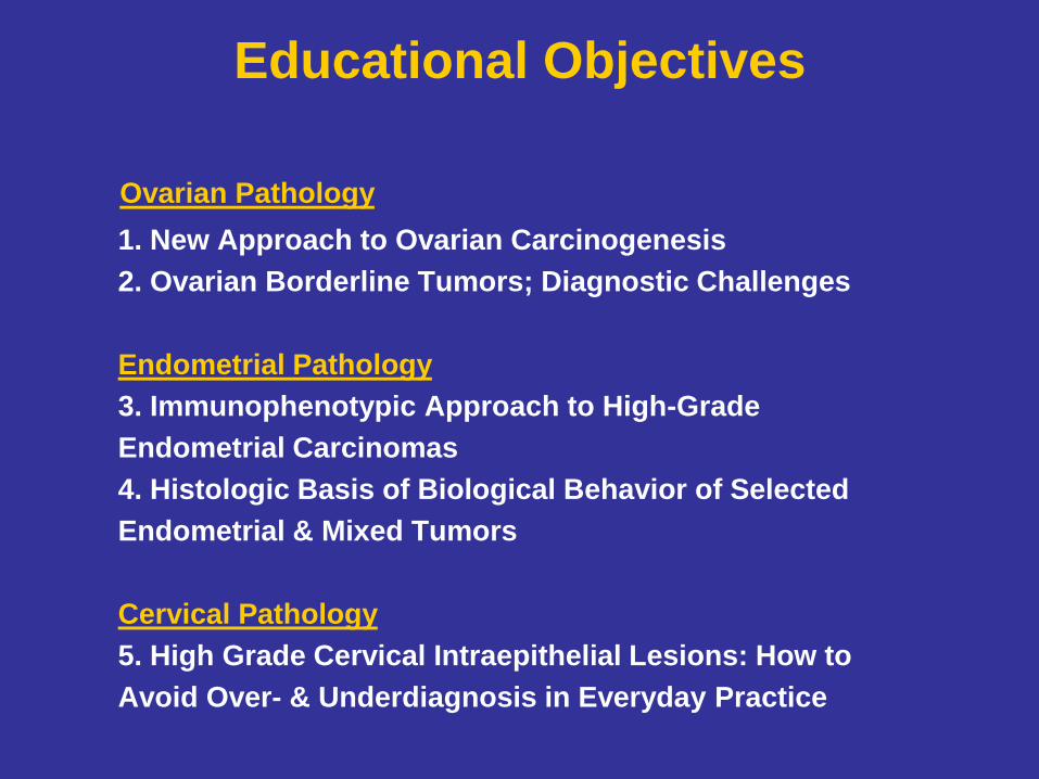
Educational Objectives
Ovarian Pathology
1. New Approach to Ovarian Carcinogenesis
2. Ovarian Borderline Tumors; Diagnostic Challenges
Endometrial Pathology
3. Immunophenotypic Approach to High-Grade
Endometrial Carcinomas
4. Histologic Basis of Biological Behavior of Selected
Endometrial & Mixed Tumors
Cervical Pathology
5. High Grade Cervical Intraepithelial Lesions: How to
Avoid Over- & Underdiagnosis in Everyday Practice
Abbreviations
• Epithelial ovarian cancer – EOC
• Carcinoma – CA
• Serous carcinoma – SC
• Mucinous carcinoma – MUC
• Endometrioid carcinoma – EMC
• Clear cell carcinoma – CCC
• Transitional cell carcinoma – TCC
• Malignant mixed müllerian tumor – MMMT
• Carcinosarcoma – CS
• Undifferentiated carcinoma – UDCA
• Dedifferentiated carcinoma – DDCA • Cystadenofibroma – CAF
• Low malignant potential – LMP
• Borderline tumor – BT
• Atypical proliferative tumor – APT
• Micropapillary serous ca – MPSC
• Intraepithelial carcinoma – IEC

1. New Approach to
Ovarian Carcinogenesis
Dualistic Model
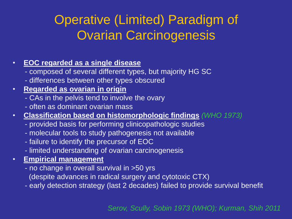
Operative (Limited) Paradigm of
Ovarian Carcinogenesis
• EOC regarded as a single disease
- composed of several different types, but majority HG SC
- differences between other types obscured
• Regarded as ovarian in origin
- CAs in the pelvis tend to involve the ovary
- often as dominant ovarian mass
• Classification based on histomorphologic findings (WHO 1973)
- provided basis for performing clinicopathologic studies
- molecular tools to study pathogenesis not available
- failure to identify the precursor of EOC
- limited understanding of ovarian carcinogenesis
• Empirical management
- no change in overall survival in >50 yrs
(despite advances in radical surgery and cytotoxic CTX)
- early detection strategy (last 2 decades) failed to provide survival benefit
Serov, Scully, Sobin 1973 (WHO); Kurman, Shih 2011
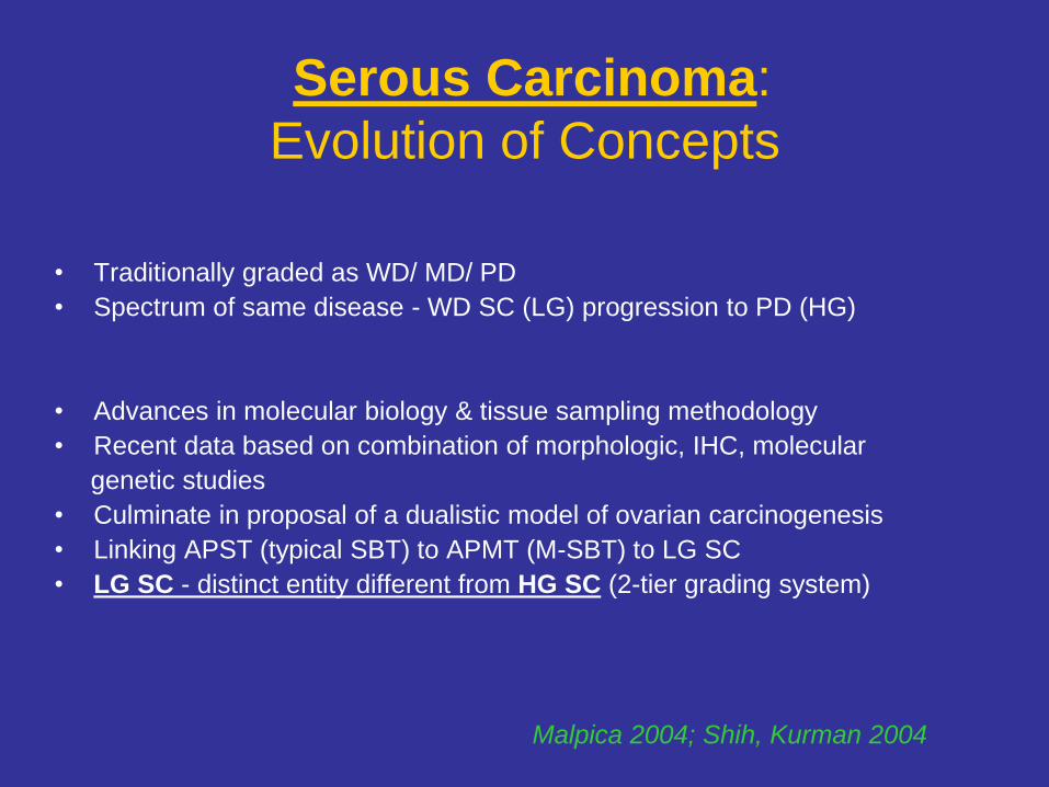
Serous Carcinoma:
Evolution of Concepts
• Traditionally graded as WD/ MD/ PD
• Spectrum of same disease - WD SC (LG) progression to PD (HG)
• Advances in molecular biology & tissue sampling methodology
• Recent data based on combination of morphologic, IHC, molecular
genetic studies
• Culminate in proposal of a dualistic model of ovarian carcinogenesis
• Linking APST (typical SBT) to APMT (M-SBT) to LG SC
• LG SC - distinct entity different from HG SC (2-tier grading system)
Malpica 2004; Shih, Kurman 2004
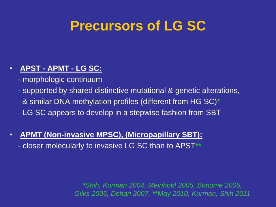
Precursors of LG SC
• APST - APMT - LG SC:
- morphologic continuum
- supported by shared distinctive mutational & genetic alterations,
& similar DNA methylation profiles (different from HG SC)*
- LG SC appears to develop in a stepwise fashion from SBT
• APMT (Non-invasive MPSC), (Micropapillary SBT):
- closer molecularly to invasive LG SC than to APST**
*Shih, Kurman 2004, Meinhold 2005, Bonome 2005,
Gilks 2005, Dehari 2007, **May 2010, Kurman, Shih 2011
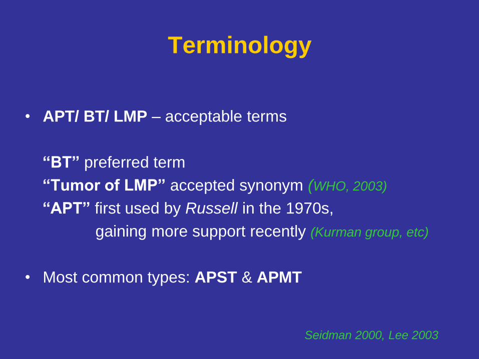
Terminology
• APT/ BT/ LMP – acceptable terms
“BT” preferred term
“Tumor of LMP” accepted synonym (WHO, 2003)
“APT” first used by Russell in the 1970s,
gaining more support recently (Kurman group, etc)
• Most common types: APST & APMT
Seidman 2000, Lee 2003
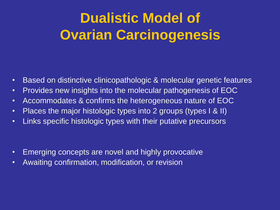
Dualistic Model of
Ovarian Carcinogenesis
• Based on distinctive clinicopathologic & molecular genetic features
• Provides new insights into the molecular pathogenesis of EOC
• Accommodates & confirms the heterogeneous nature of EOC
• Places the major histologic types into 2 groups (types I & II)
• Links specific histologic types with their putative precursors
• Emerging concepts are novel and highly provocative
• Awaiting confirmation, modification, or revision
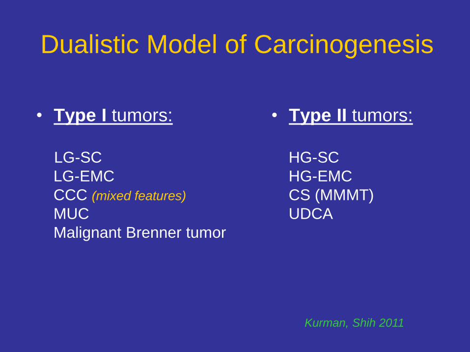
Dualistic Model of Carcinogenesis
• Type I tumors:
LG-SC
LG-EMC
CCC (mixed features)
MUC
Malignant Brenner tumor
• Type II tumors:
HG-SC
HG-EMC
CS (MMMT)
UDCA
Kurman, Shih 2011
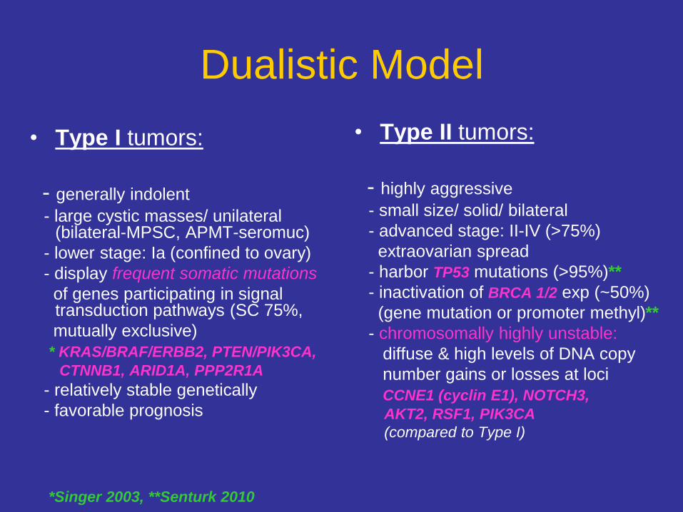
Dualistic Model
• Type I tumors:
- generally indolent
- large cystic masses/ unilateral (bilateral-MPSC, APMT-seromuc)
- lower stage: Ia (confined to ovary)
- display frequent somatic mutations
of genes participating in signal transduction pathways (SC 75%,
mutually exclusive)
* KRAS/BRAF/ERBB2, PTEN/PIK3CA,
CTNNB1, ARID1A, PPP2R1A
- relatively stable genetically
- favorable prognosis
• Type II tumors:
- highly aggressive
- small size/ solid/ bilateral
- advanced stage: II-IV (>75%)
extraovarian spread
- harbor TP53 mutations (>95%)**
- inactivation of BRCA 1/2 exp (~50%)
(gene mutation or promoter methyl)**
- chromosomally highly unstable:
diffuse & high levels of DNA copy
number gains or losses at loci
CCNE1 (cyclin E1), NOTCH3,
AKT2, RSF1, PIK3CA
(compared to Type I)
*Singer 2003, **Senturk 2010
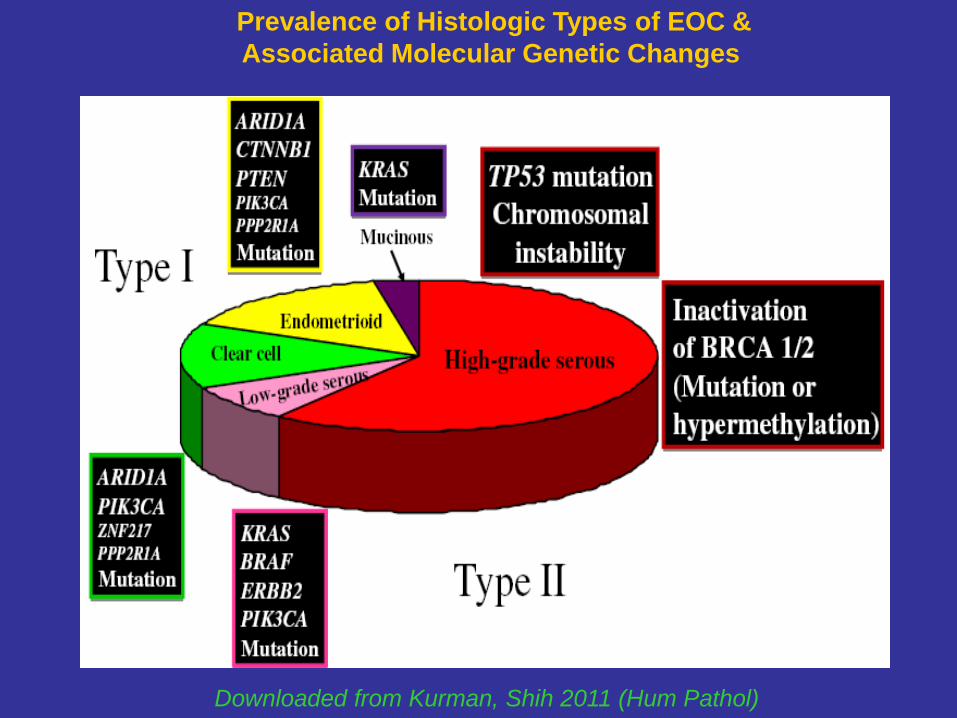
Prevalence of Histologic Types of EOC &
Associated Molecular Genetic Changes
Downloaded from Kurman, Shih 2011 (Hum Pathol)
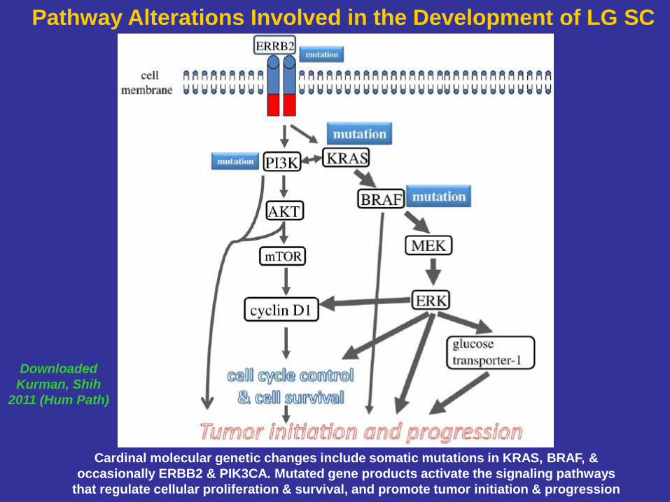
Pathway Alterations Involved in the Development of LG SC
Cardinal molecular genetic changes include somatic mutations in KRAS, BRAF, &
occasionally ERBB2 & PIK3CA. Mutated gene products activate the signaling pathways
that regulate cellular proliferation & survival, and promote tumor initiation & progression
Downloaded
Kurman, Shih
2011 (Hum Path)

Pathway Alterations Involved in the
Development of Other EOC
• LG EMC:
- mutations that deregulate PI3K/ PTEN signaling pathway
- mutations of CTNNB1 gene (encodes b-catenin)
• CCC:
- activating mutation of PIK3CA
- somatic inactivity mutation of ARID1A (tumor suppressor gene)
• MUC:
- mutation in KRAS gene
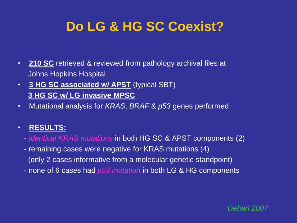
Do LG & HG SC Coexist?
• 210 SC retrieved & reviewed from pathology archival files at
Johns Hopkins Hospital
• 3 HG SC associated w/ APST (typical SBT)
3 HG SC w/ LG invasive MPSC
• Mutational analysis for KRAS, BRAF & p53 genes performed
• RESULTS:
- Identical KRAS mutations in both HG SC & APST components (2)
- remaining cases were negative for KRAS mutations (4)
(only 2 cases informative from a molecular genetic standpoint)
- none of 6 cases had p53 mutation in both LG & HG components
Dehari 2007
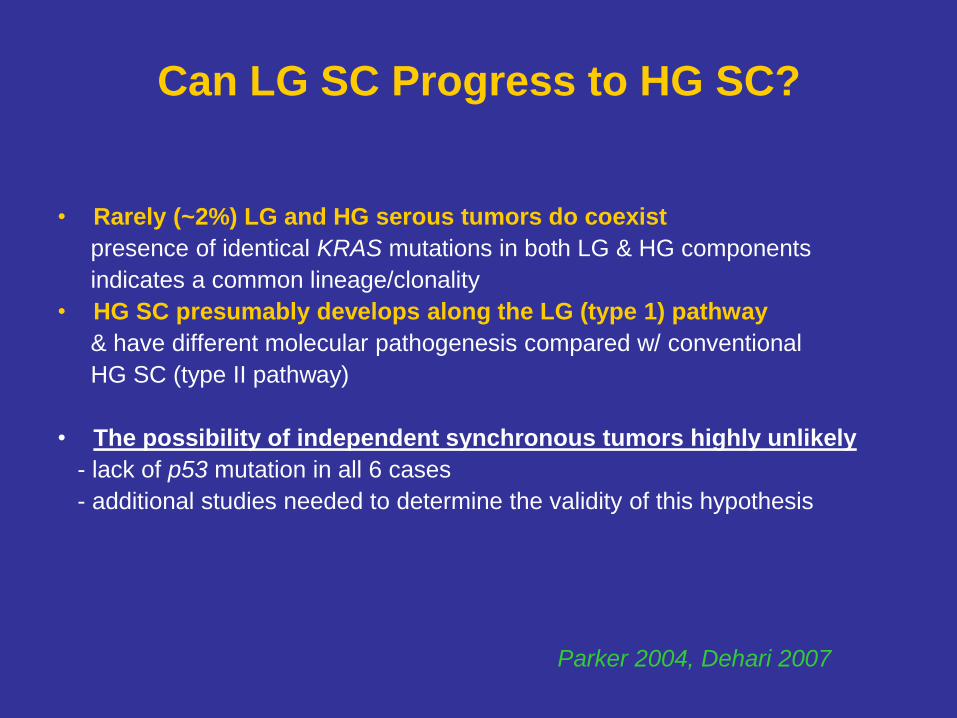
Can LG SC Progress to HG SC?
• Rarely (~2%) LG and HG serous tumors do coexist
presence of identical KRAS mutations in both LG & HG components
indicates a common lineage/clonality
• HG SC presumably develops along the LG (type 1) pathway
& have different molecular pathogenesis compared w/ conventional
HG SC (type II pathway)
• The possibility of independent synchronous tumors highly unlikely
- lack of p53 mutation in all 6 cases
- additional studies needed to determine the validity of this hypothesis
Parker 2004, Dehari 2007
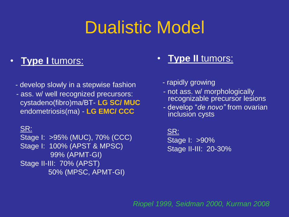
Dualistic Model
• Type I tumors:
- develop slowly in a stepwise fashion
- ass. w/ well recognized precursors:
cystadeno(fibro)ma/BT- LG SC/ MUC
endometriosis(ma) - LG EMC/ CCC
SR:
Stage I: >95% (MUC), 70% (CCC)
Stage I: 100% (APST & MPSC)
99% (APMT-GI)
Stage II-III: 70% (APST)
50% (MPSC, APMT-GI)
• Type II tumors:
- rapidly growing
- not ass. w/ morphologically recognizable precursor lesions
- develop “de novo” from ovarian inclusion cysts
SR:
Stage I: >90%
Stage II-III: 20-30%
Riopel 1999, Seidman 2000, Kurman 2008
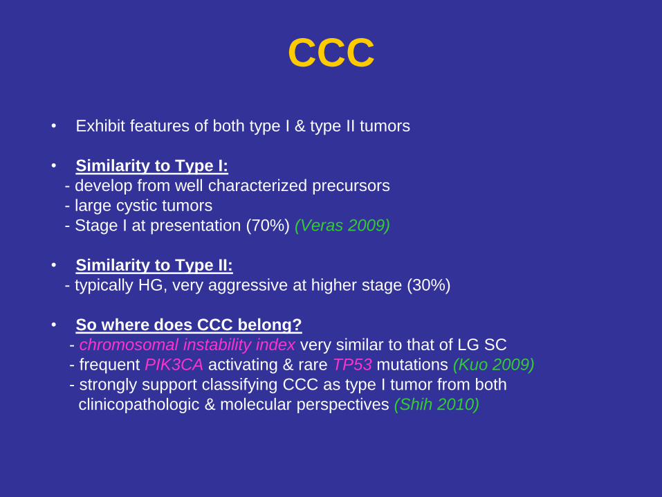
CCC
• Exhibit features of both type I & type II tumors
• Similarity to Type I:
- develop from well characterized precursors
- large cystic tumors
- Stage I at presentation (70%) (Veras 2009)
• Similarity to Type II:
- typically HG, very aggressive at higher stage (30%)
• So where does CCC belong?
- chromosomal instability index very similar to that of LG SC
- frequent PIK3CA activating & rare TP53 mutations (Kuo 2009)
- strongly support classifying CCC as type I tumor from both
clinicopathologic & molecular perspectives (Shih 2010)
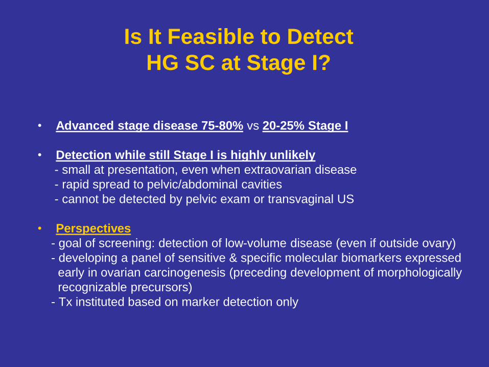
Is It Feasible to Detect
HG SC at Stage I?
• Advanced stage disease 75-80% vs 20-25% Stage I
• Detection while still Stage I is highly unlikely
- small at presentation, even when extraovarian disease
- rapid spread to pelvic/abdominal cavities
- cannot be detected by pelvic exam or transvaginal US
• Perspectives
- goal of screening: detection of low-volume disease (even if outside ovary)
- developing a panel of sensitive & specific molecular biomarkers expressed
early in ovarian carcinogenesis (preceding development of morphologically
recognizable precursors)
- Tx instituted based on marker detection only
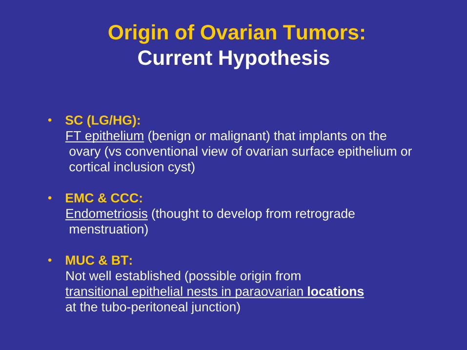
Origin of Ovarian Tumors:
Current Hypothesis
• SC (LG/HG):
FT epithelium (benign or malignant) that implants on the
ovary (vs conventional view of ovarian surface epithelium or
cortical inclusion cyst)
• EMC & CCC:
Endometriosis (thought to develop from retrograde
menstruation)
• MUC & BT:
Not well established (possible origin from
transitional epithelial nests in paraovarian locations
at the tubo-peritoneal junction)
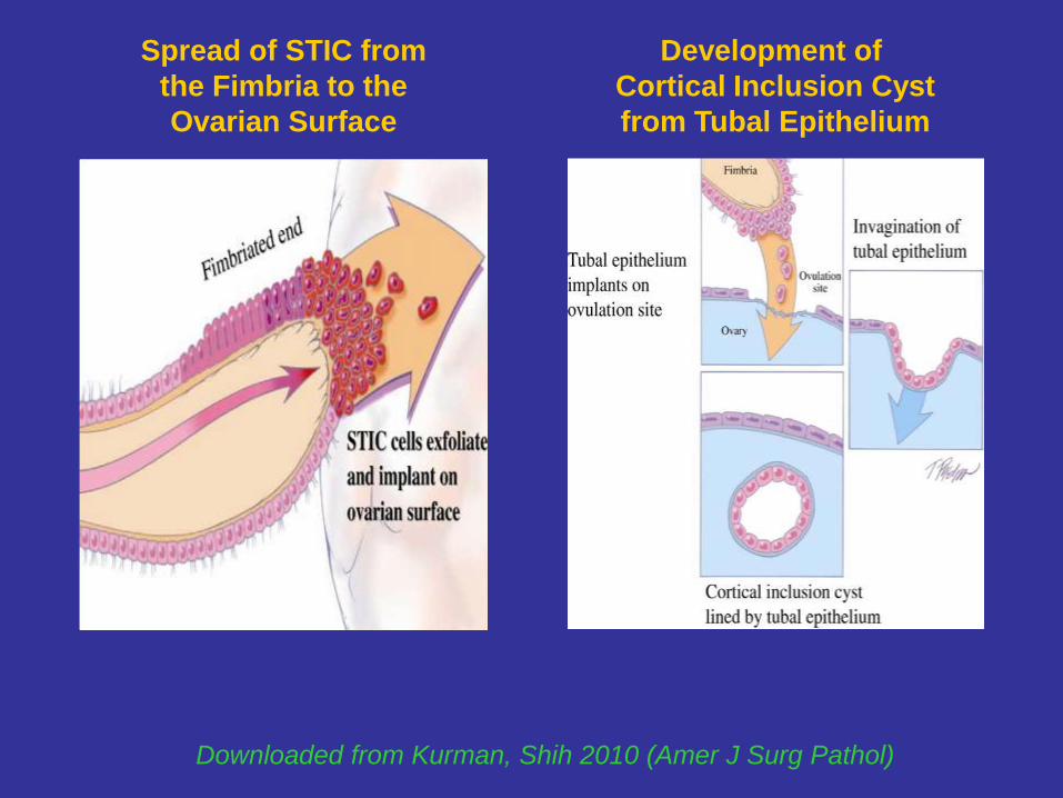
Development of
Cortical Inclusion Cyst
from Tubal Epithelium
Downloaded from Kurman, Shih 2010 (Amer J Surg Pathol)
Spread of STIC from
the Fimbria to the
Ovarian Surface
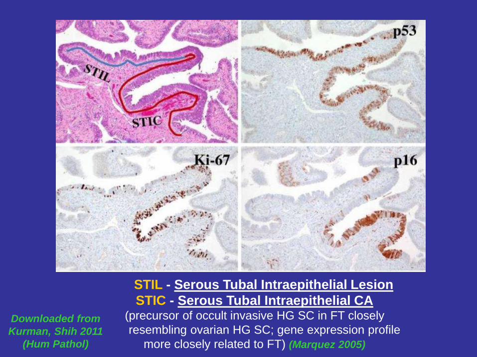
STIL - Serous Tubal Intraepithelial Lesion
STIC - Serous Tubal Intraepithelial CA (precursor of occult invasive HG SC in FT closely
resembling ovarian HG SC; gene expression profile
more closely related to FT) (Marquez 2005)
Downloaded from
Kurman, Shih 2011
(Hum Pathol)
Downloaded from Kurman, Shih 2011 (Hum Pathol)
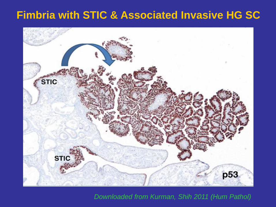
Fimbria with STIC & Associated Invasive HG SC
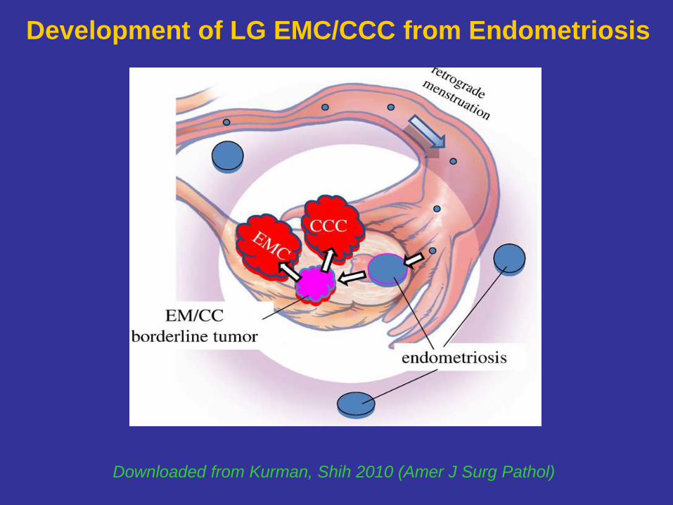
Development of LG EMC/CCC from Endometriosis
Downloaded from Kurman, Shih 2010 (Amer J Surg Pathol)
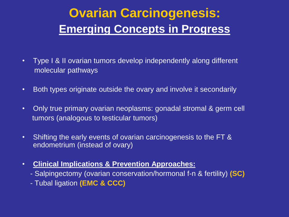
Ovarian Carcinogenesis:
Emerging Concepts in Progress
• Type I & II ovarian tumors develop independently along different
molecular pathways
• Both types originate outside the ovary and involve it secondarily
• Only true primary ovarian neoplasms: gonadal stromal & germ cell
tumors (analogous to testicular tumors)
• Shifting the early events of ovarian carcinogenesis to the FT & endometrium (instead of ovary)
• Clinical Implications & Prevention Approaches:
- Salpingectomy (ovarian conservation/hormonal f-n & fertility) (SC)
- Tubal ligation (EMC & CCC)

2. Ovarian Borderline Tumors:
Diagnostic Challenges
Ovarian Epithelial Tumor Categories
• Cystadeno(fibro)ma
• Atypical proliferative tumor (BT, LMP)
• Intraepithelial carcinoma (non-invasive)
• Microinvasion/ Microinvasive carcinoma
• Invasive Carcinoma
BORDERLINE OVARIAN TUMORS:
A WEB-BASED ATLAS
NCI Borderline Ovarian Tumor Workshop, Bethesda, MD, August 27-28, 2003
Introduction It is said that a picture is worth more than a thousand words.
Pathologists believe that a picture with a few descriptive words is
…priceless. The purpose of this web page is to offer practicing
surgical pathologists and pathology residents didactic samples of
ovarian borderline tumors (also called atypical proliferative tumors or
tumors of LMP) and the tumors that enter into their differential Dx.

BORDERLINE OVARIAN TUMORS:
A WEB-BASED ATLAS
NCI Borderline Ovarian Tumor Workshop, Bethesda, MD, August 27-28, 2003
“One of the most difficult areas in gynecological pathology is the
spectrum of diseases that fall between the categories of clear cut
benign epithelial lesion and clear cut invasive carcinomas.
Pathologists sometimes disagree on terminology, even on the
definition of apparently self-explanatory terms such as "destructive
invasion", "severe nuclear atypia" and "microinvasion". For this
reason, the National Institute of Health convened pathologists,
clinical & surgical oncologists, epidemiologists and basic scientists
interested in this field for the Borderline Ovarian Tumor Workshop”.

DISCLAIMER:
This selection of images is for educational purposes only. The views
are those of the contributors and do not reflect endorsement by the
NIH or the University of Illinois at Chicago. The images contain
statements related to rendered diagnostic opinions. In view of the wide
range of opinions regarding the tumors under discussion, none of
these statements is intended as, or should be interpreted as
representing the "standard of care."
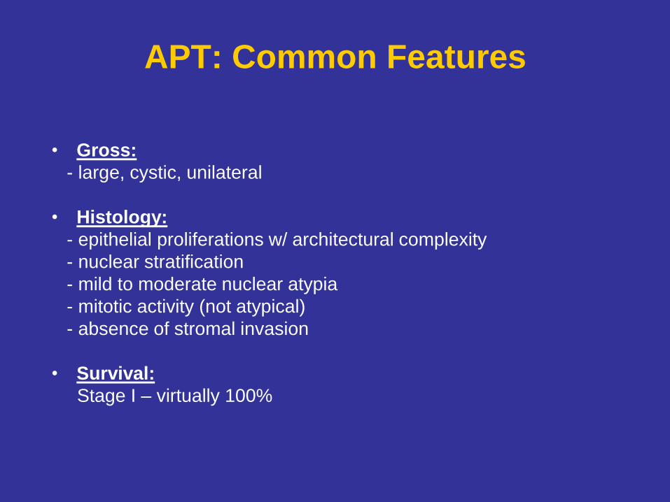
APT: Common Features
• Gross:
- large, cystic, unilateral
• Histology:
- epithelial proliferations w/ architectural complexity
- nuclear stratification
- mild to moderate nuclear atypia
- mitotic activity (not atypical)
- absence of stromal invasion
• Survival:
Stage I – virtually 100%
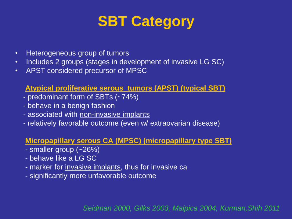
SBT Category
• Heterogeneous group of tumors
• Includes 2 groups (stages in development of invasive LG SC)
• APST considered precursor of MPSC
Atypical proliferative serous tumors (APST) (typical SBT)
- predominant form of SBTs (~74%)
- behave in a benign fashion
- associated with non-invasive implants
- relatively favorable outcome (even w/ extraovarian disease)
Micropapillary serous CA (MPSC) (micropapillary type SBT)
- smaller group (~26%)
- behave like a LG SC
- marker for invasive implants, thus for invasive ca
- significantly more unfavorable outcome
Seidman 2000, Gilks 2003, Malpica 2004, Kurman,Shih 2011
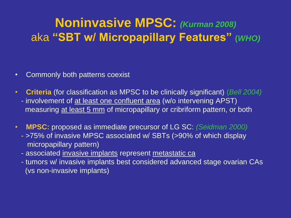
Noninvasive MPSC: (Kurman 2008)
aka “SBT w/ Micropapillary Features” (WHO)
• Commonly both patterns coexist
• Criteria (for classification as MPSC to be clinically significant) (Bell 2004)
- involvement of at least one confluent area (w/o intervening APST)
measuring at least 5 mm of micropapillary or cribriform pattern, or both
• MPSC: proposed as immediate precursor of LG SC: (Seidman 2000)
- >75% of invasive MPSC associated w/ SBTs (>90% of which display
micropapillary pattern)
- associated invasive implants represent metastatic ca
- tumors w/ invasive implants best considered advanced stage ovarian CAs
(vs non-invasive implants)
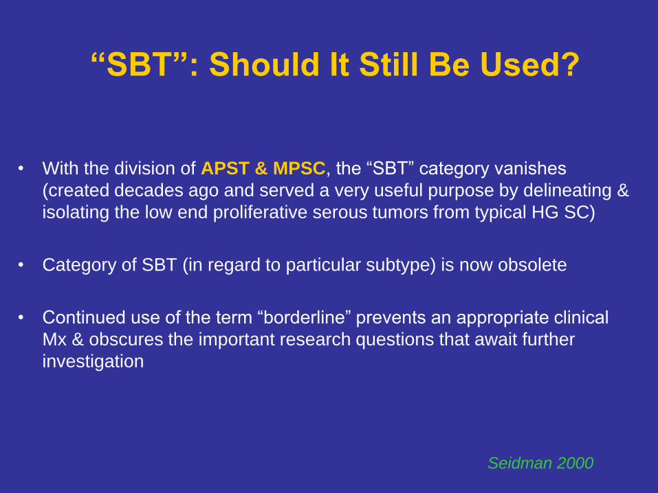
“SBT”: Should It Still Be Used?
• With the division of APST & MPSC, the “SBT” category vanishes
(created decades ago and served a very useful purpose by delineating &
isolating the low end proliferative serous tumors from typical HG SC)
• Category of SBT (in regard to particular subtype) is now obsolete
• Continued use of the term “borderline” prevents an appropriate clinical
Mx & obscures the important research questions that await further
investigation
Seidman 2000
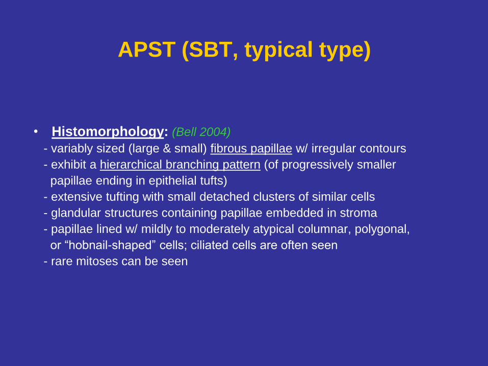
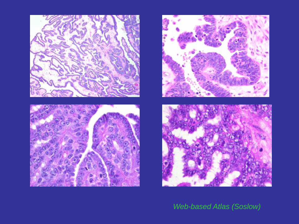
APST (SBT, typical type)
• Histomorphology: (Bell 2004)
- variably sized (large & small) fibrous papillae w/ irregular contours
- exhibit a hierarchical branching pattern (of progressively smaller
papillae ending in epithelial tufts)
- extensive tufting with small detached clusters of similar cells
- glandular structures containing papillae embedded in stroma
- papillae lined w/ mildly to moderately atypical columnar, polygonal,
or “hobnail-shaped” cells; ciliated cells are often seen
- rare mitoses can be seen
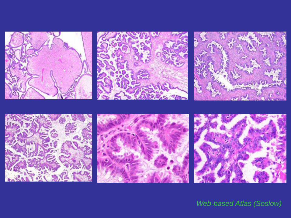
Web-based Atlas (Soslow)
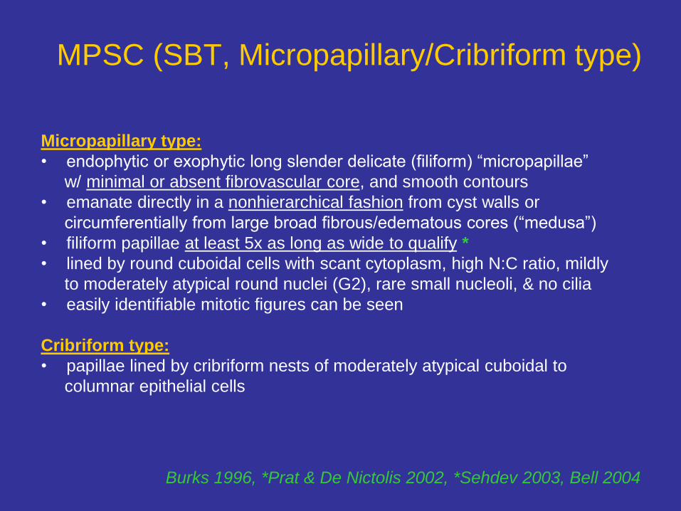
MPSC (SBT, Micropapillary/Cribriform type)
Micropapillary type:
• endophytic or exophytic long slender delicate (filiform) “micropapillae”
w/ minimal or absent fibrovascular core, and smooth contours
• emanate directly in a nonhierarchical fashion from cyst walls or
circumferentially from large broad fibrous/edematous cores (“medusa”)
• filiform papillae at least 5x as long as wide to qualify *
• lined by round cuboidal cells with scant cytoplasm, high N:C ratio, mildly
to moderately atypical round nuclei (G2), rare small nucleoli, & no cilia
• easily identifiable mitotic figures can be seen
Cribriform type:
• papillae lined by cribriform nests of moderately atypical cuboidal to
columnar epithelial cells
Burks 1996, *Prat & De Nictolis 2002, *Sehdev 2003, Bell 2004
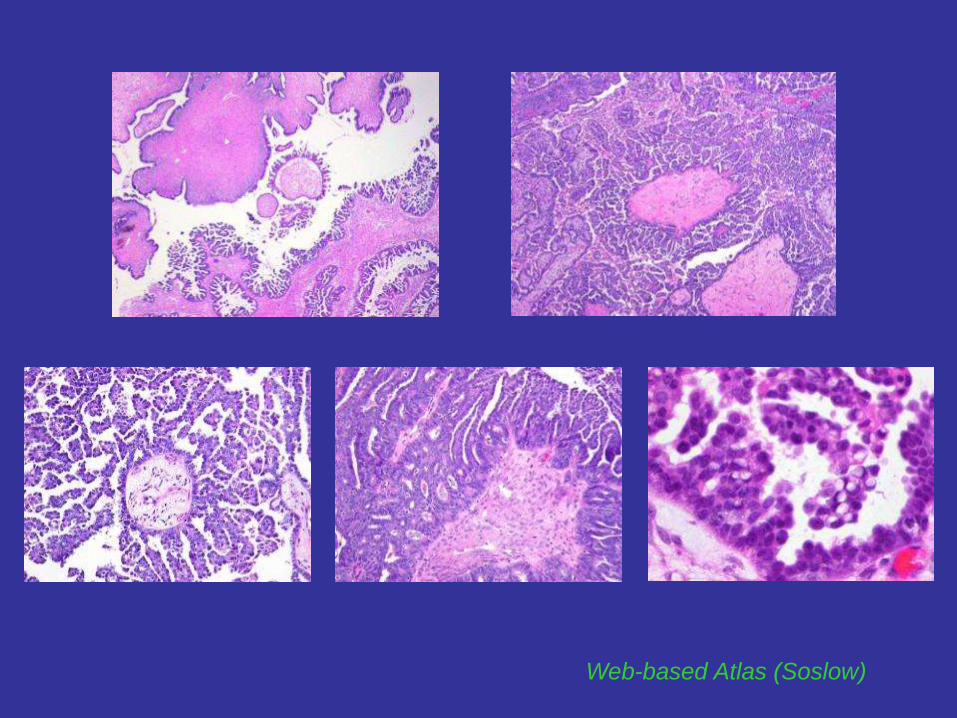
Web-based Atlas (Soslow)
Small Foci of Tumor w/ Borderline Morphology
within an Otherwise Benign CAF:
Possible approaches (Borderline Ovarian Tumor Workshop, 2003):
1) Ignoring it
2) Diagnosing the entire tumor as borderline
3) Diagnosing the tumor as benign & focally borderline, no further comment
4) Diagnosing as in (3), but with a comment suggesting that the lesion is
unlikely to behave aggressively
5) Diagnosing the tumor as benign and mentioning the presence of the
borderline focus/foci only in the comment
This uncertainty reflects the lack of consensus
- unclear whether transition to BT should occur at 5%, 10%, 15% or >
- even 100% BT (Stage I) extremely unlikely to behave aggressively
- certainly smaller proportions should be at least equally benign
- universal agreement with 1% or 2% involvement
SBT with Microinvasion:
Does It Matter?
• SBTs with small foci of intrastromal tumor cells ≤3-5 mm or <10mm2
• Often do not elicit a significant stromal reaction
• Can be found in 10-15% of SBTs
• Eosinophilic cell pattern (most common)
- isolated cells/small nests w/ abundant eosinophilic cytoplasm in stroma
- surrounded by clear spaces (retraction)
- not ass. w/ unfavorable Px (SR 100% after 6.7 yrs follow-up)
- does not alter clinical behavior (no clinical relevance)
• Micropapillary pattern (uncommon)
- displays micropapillae (resembles LG SC)
- may be designated also as “Microinvasive CA”
- represents higher risk lesion w/ clinical course analogous to LG SC
Tavassoli 1988, Bell & Scully 1990, Longacre 1993,
Seidman 2000, Bell 2004, Kurman 2008
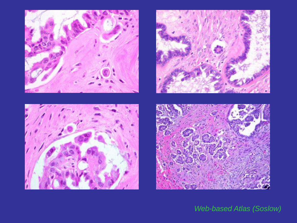
Web-based Atlas (Soslow)
SBT - Prognostic Indicators
• Stage of the disease
• Nature of peritoneal implants/ extraovarian disease (Seidman 2000)
- survival data analyzed on 467 advanced stage SBTs
- 104 had invasive implants (22% of all implants)
- responsible for 67% of all tumor-related deaths (7-fold higher)
- distinction of invasive vs noninvasive implants may be extremely difficult
- in particular, with associated marked desmoplastic response
(desmoplastic non-invasive implants)
- closely simulates host response of invasive CA
(most difficult lesions to assess)
Peritoneal Implants
• Architecturally complex peritoneal proliferations ass. w/ SBTs
• Seen in 20-30% of SBTs at presentation
• Noninvasive & invasive
• Noninvasive implants:
- superficial location on the surfaces of peritoneum/ submesothelial/
between omental lobules, along the septa
- lack tissue invasion (micropapillary architecture within the stroma,
or solid or papillary nests with a cleft)
1) epithelial type - without a stromal response
2) desmoplastic type - accompanied by a marked stromal reaction
McCaughey 1984, Scully 1999, Seidman 2000

Peritoneal Implants
• Noninvasive
- ass. w/ APST
- represent reactive mesothelial hyperplasia or “true” implants
from benign proliferative lesions
- behave in a benign fashion
- SR: 95.3%
(w/ improved criteria ~100%)
• Invasive
- ass. w/ MPSC
- represent metastatic LG SC
(initially occur on the surface,
but eventually invade; reason
why some pts w/ “noninvasive
implants” die of disease)
- behave as CAs
- SR: 66% (at 7.4 yrs f/u)
Seidman 2000, Russell 2002, Kurman & Shih 2011
Do Invasive Implants Ever Arise from
APST (Typical SBT) ?
• Very unlikely! Implants ass. w/ APST are non-invasive (Kurman 2008)
• Reported invasive implants ass. w/ APST probably contained occult
areas of unsampled MPSC (adequate sampling!)
• If no areas of MPSC found, question whether implants truly invasive
• Micropapillary architecture: strong predictor of invasive implants
• Adequate sampling (Borderline Ovarian Tumor Workshop, 2003)*
- at least 1 section/cm for tumors smaller than 10cm
- 2 sections/cm for larger tumors (excluding smooth-walled cystic areas)
- when micropapillary foci histologically, further sampling encouraged
- grossly negative omentum sampled extensively: at least 1 section/2cm
McCaughey 1984, *Silverberg 2003, Kurman 2008, 2011
Non-Invasive Implants:
Epithelial Type
• Branching papillary proliferations w/ small epithelial tufts & buds on
peritoneal surface or smoothly contoured submesothelial invaginations
- well-defined/ frequently rounded free floating structures surrounded
by reactive mesothelium
- papillae with fibrous or hyalinized cores, +/- psammomatous Ca++
- lined by mild to moderately atypical cuboidal to columnar cells
- no destruction of underlying stroma
• Permeation of stroma by individual eosinophilic cells (w/o invasion
of underlying tissue) should be included in non-invasive category
McCaughey 1984, Prat & De Nictolis 2002, Kurman 2008
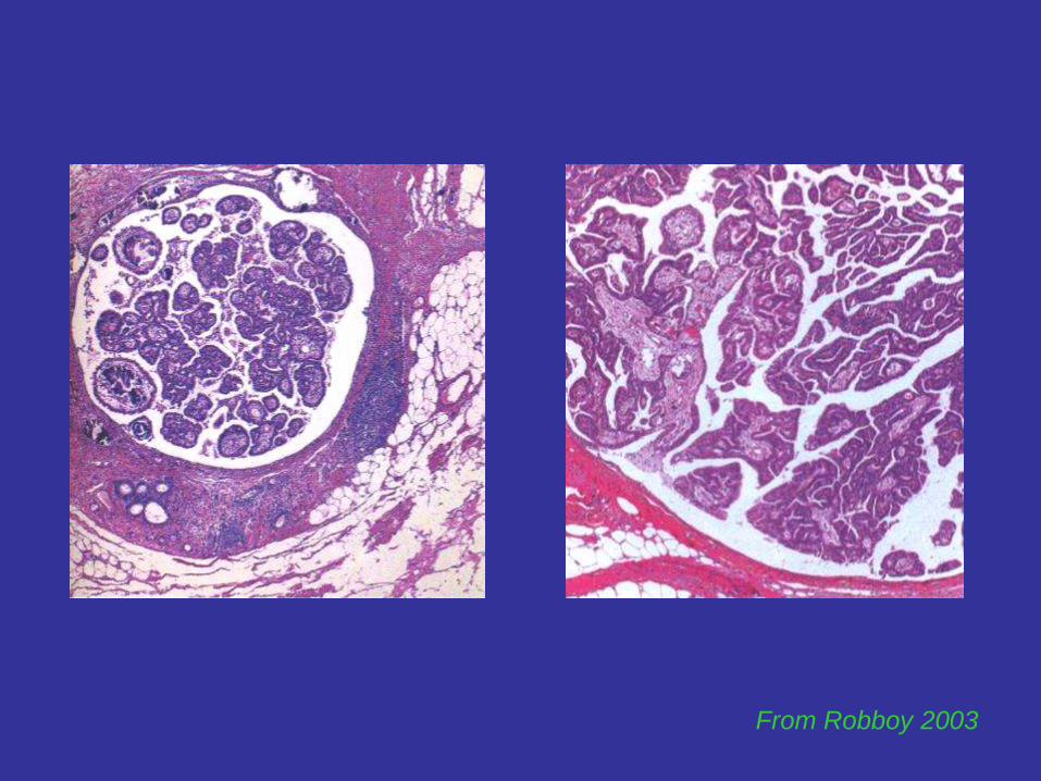
From Robboy 2003
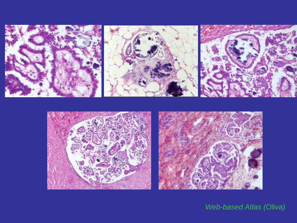
Web-based Atlas (Oliva)
Web-based Atlas (Seidman)
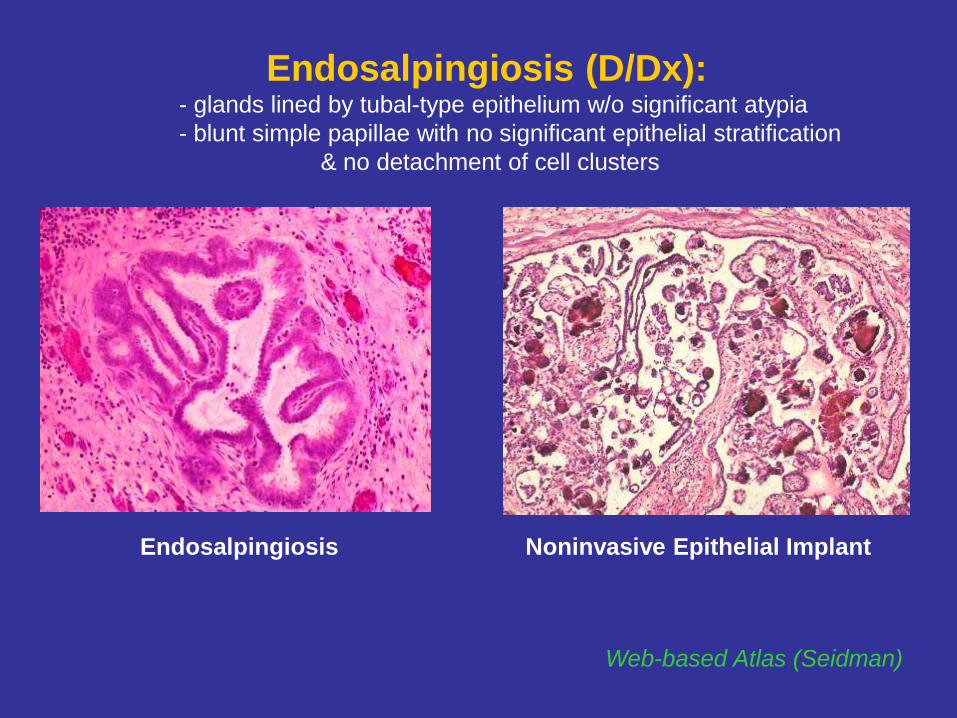
Endosalpingiosis Noninvasive Epithelial Implant
Endosalpingiosis (D/Dx): - glands lined by tubal-type epithelium w/o significant atypia
- blunt simple papillae with no significant epithelial stratification
& no detachment of cell clusters

Non-Invasive Implants:
Desmoplastic Type
• Gland-like & papillary structures, single & small nests of cells, in
edematous/ inflamed fibrous tissue (“stuck on” the peritoneal surfaces)
- circumscribed w/ sharp interface with normal tissue
- abundant but superficial stromal reaction
- low epithelium:stroma ratio (prevalence of stroma vs epithelium)
- totally disorganized arrangement of bland epithelial elements,
psammoma bodies, mesothelial cells and inclusions
- complex but bland epithelial elements merging w/ adjacent stromal cells, although sometimes clefts may be seen
- stroma with maturing/organizing granulation tissue appearance
(“tissue-culture” type fibroblasts or spindled mesothelial cells)
- typically ass. w/ inflammatory cells & recent hemorrhage
• Often both types of noninvasive implants present in same patient
McCaughey 1984, Bell & Kurman 2001, Russel 2002
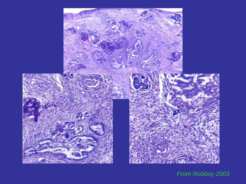
From Robboy 2003
Web-based Atlas (Oliva)
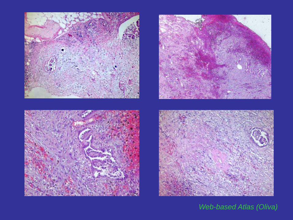
Invasive Implants
• Definition varies from study to study (expanded/less restrictive criteria)
• Irregular infiltrative carcinomatous deposits (papillae surr. by a space, angulated glands/interanastomosing, single cells) deeply penetrating stroma
- irregular infiltrative margin of underlying tissue/fat
- distinct from the surrounding stroma
- higher epithelium:stroma ratio
- more ordered radial growth/orientation
- relatively uniform papillary or confluent cribriform aggregates
- HG cytology
• Micropapillary architecture or small solid nests or papillae
surrounded by a clear space or cleft (resembling LG SC)
w/o true invasion of underlying tissue
McCaughey 1984, Bell 2001, Russel 2002, Kurman 2008
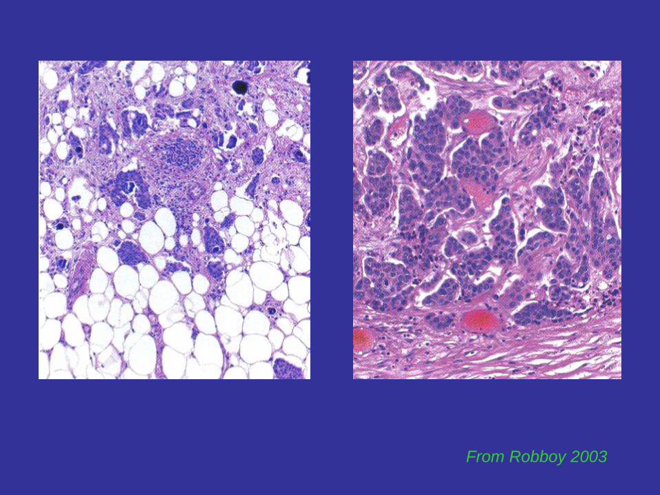
From Robboy 2003
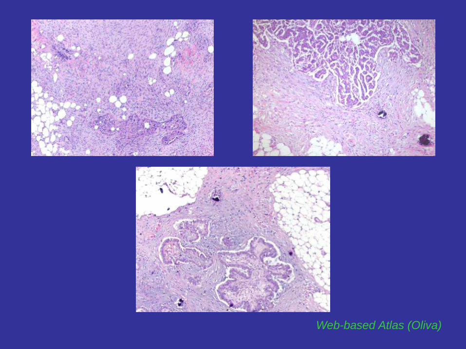
Web-based Atlas (Oliva)
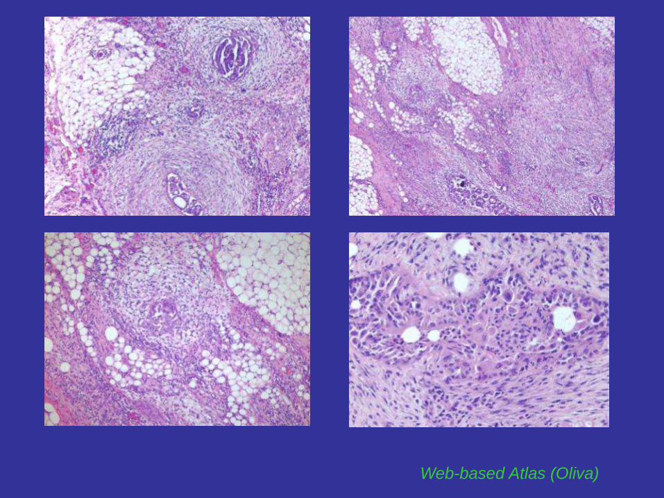
Web-based Atlas (Oliva)
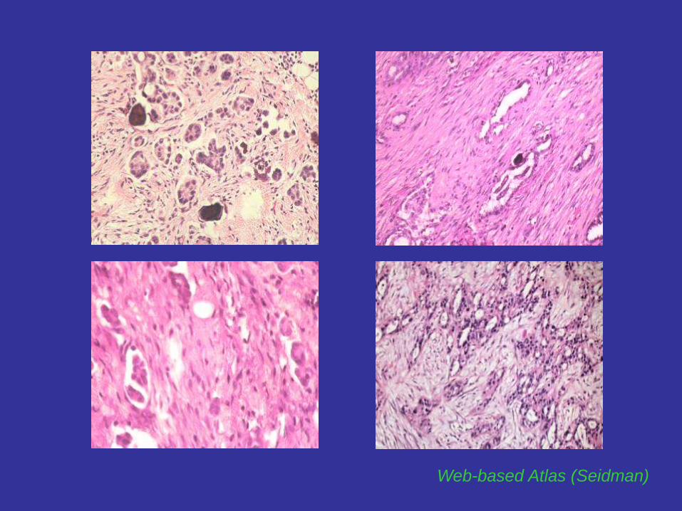
Web-based Atlas (Seidman)

Lymph Node Lesions Associated w/ SBTs
• Endosalpingiosis (benign tubal-type/müllerian glandular inclusions)
• Mesothelial hyperplasia involving lymph node sinusoids
• Individual cells/clusters w/ abundant eosinophilic cytoplasm
in predominantly subcapsular sinuses
- nature unclear, exfoliated mesothelial cells?
- similar cells present at the surface of SBTs in the ovary
• Small papillary clusters/glandular inclusions resembling primary
tumor, usually just beneath the capsule
- majority associated w/ endosalpingiosis in the same node
- no stromal invasion
Clement 1996, Seidman 2000, Robboy 2002
Lymph Node Involvement:
Clinically Significant?
• Regional lymph node involvement:
- not ass. with adverse prognosis in patients with peritoneal implants
(SR 98% after 6.5 yrs follow-up)
- do not represent functional metastatic CA (reported w/o primary tumor)*
- proposed independent origin within the lymph node (transition from
müllerian inclusions)
• Distant lymph node involvement:
- may rarely be seen at presentation (more commonly w/ tumor recurrence)**
**Goldman 1993, *Prade 1995, Seidman 2000, Robboy 2002
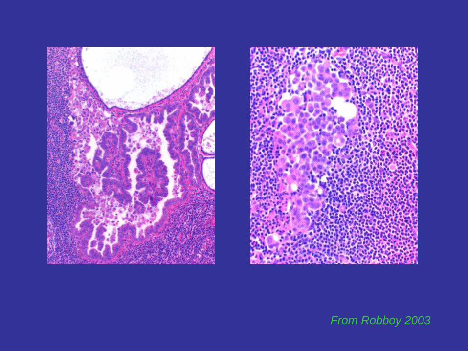
From Robboy 2003
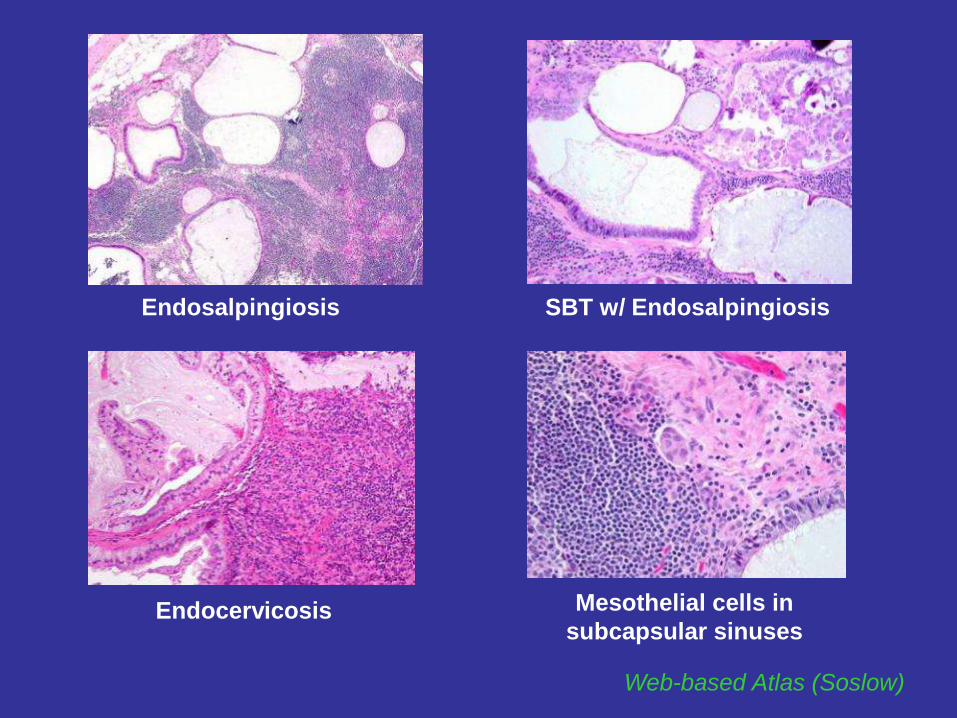
Web-based Atlas (Soslow)
Endosalpingiosis SBT w/ Endosalpingiosis
Endocervicosis Mesothelial cells in
subcapsular sinuses
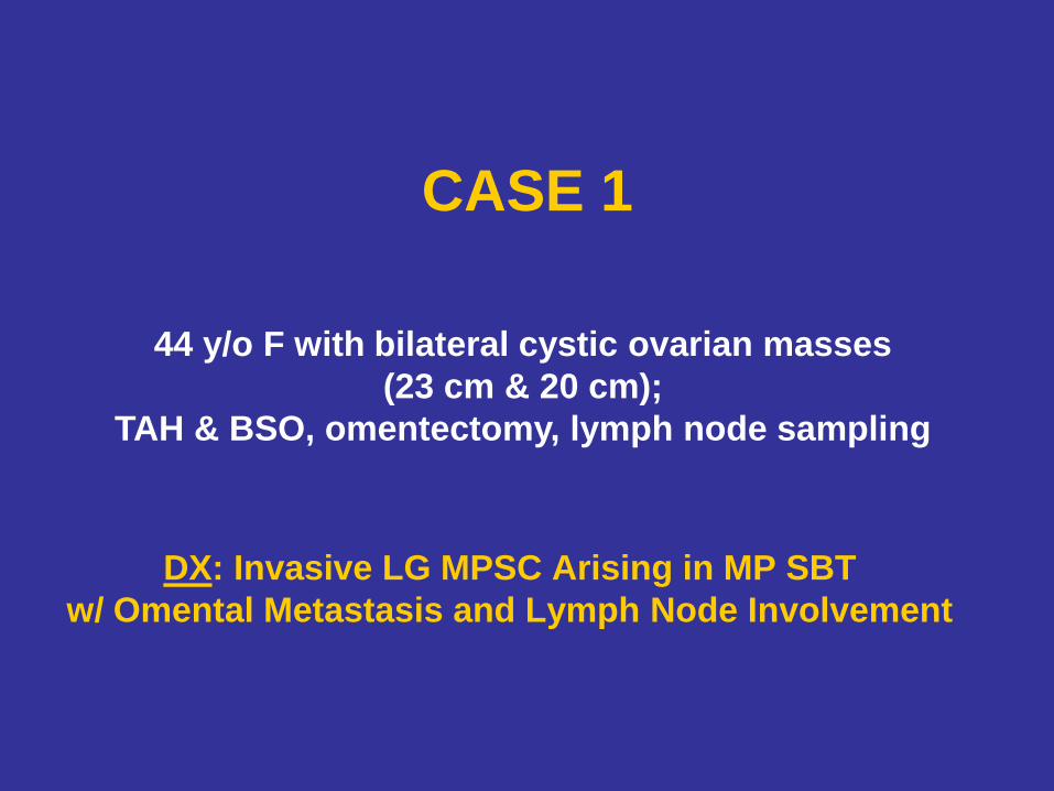
CASE 1
DX: Invasive LG MPSC Arising in MP SBT
w/ Omental Metastasis and Lymph Node Involvement
44 y/o F with bilateral cystic ovarian masses
(23 cm & 20 cm);
TAH & BSO, omentectomy, lymph node sampling
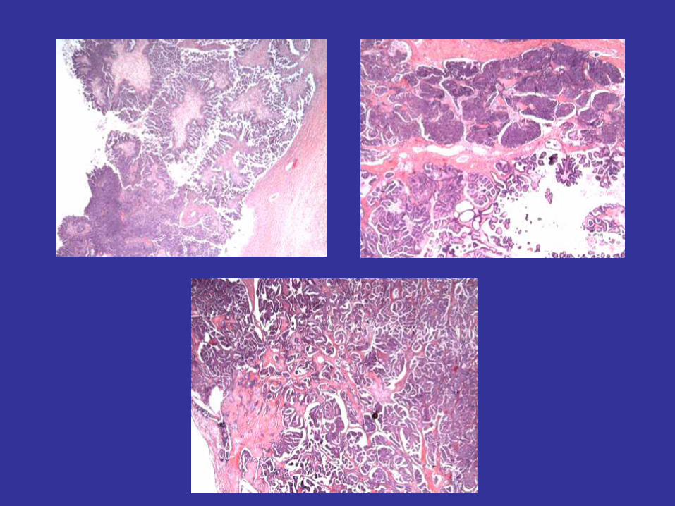
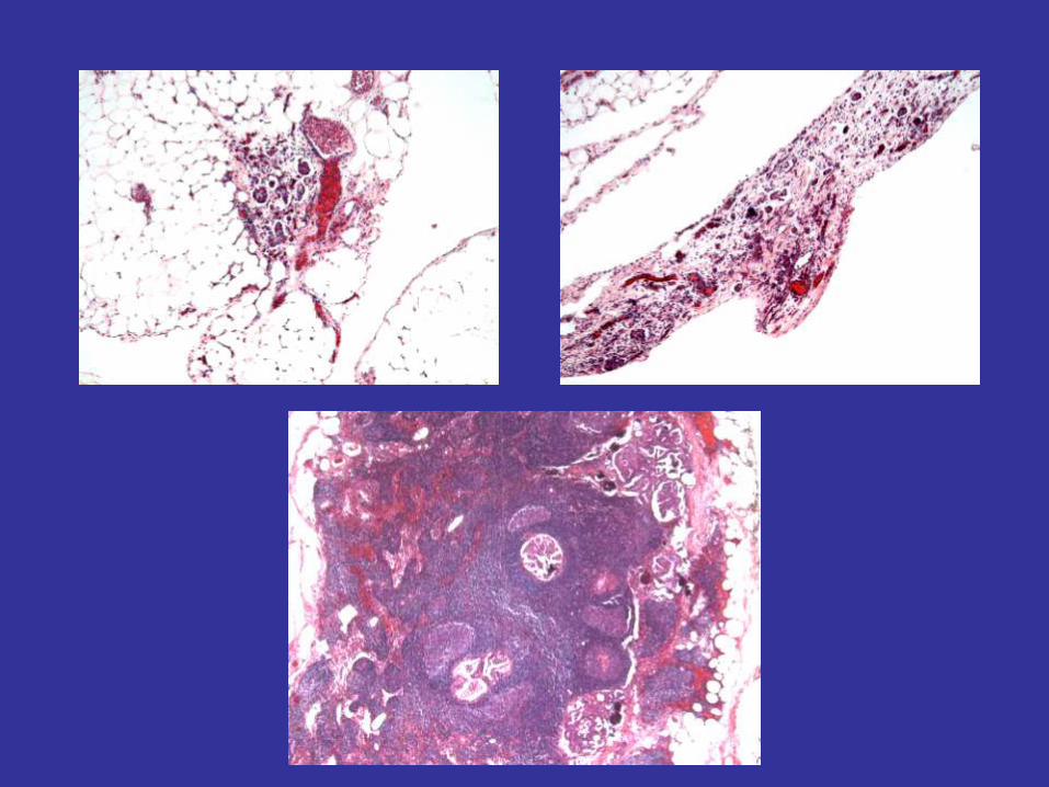
HG vs LG (Micropapillary) SC
• Rarely, HG SC can mimic SBT (grow in non-invasive pattern)
• HG SC:
- predominantly papillary (“MD”) or solid architecture (“PD”)
- severe cytologic atypia (>3-fold nuclear atypia, frequent mitoses
>12/10 HPF, prominent nucleoli)
• LG SC:
- delicate micropapillary architecture
- relatively small tumor cells with uniform nuclei & minimal atypia
- inconspicuous nucleoli
- less frequent mitoses
Malpica 2004, Dehari 2007
Web-based Atlas (Soslow)
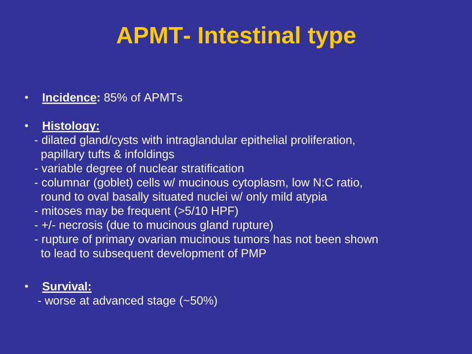
APMT- Intestinal type
• Incidence: 85% of APMTs
• Histology:
- dilated gland/cysts with intraglandular epithelial proliferation,
papillary tufts & infoldings
- variable degree of nuclear stratification
- columnar (goblet) cells w/ mucinous cytoplasm, low N:C ratio,
round to oval basally situated nuclei w/ only mild atypia
- mitoses may be frequent (>5/10 HPF)
- +/- necrosis (due to mucinous gland rupture)
- rupture of primary ovarian mucinous tumors has not been shown
to lead to subsequent development of PMP
• Survival:
- worse at advanced stage (~50%)
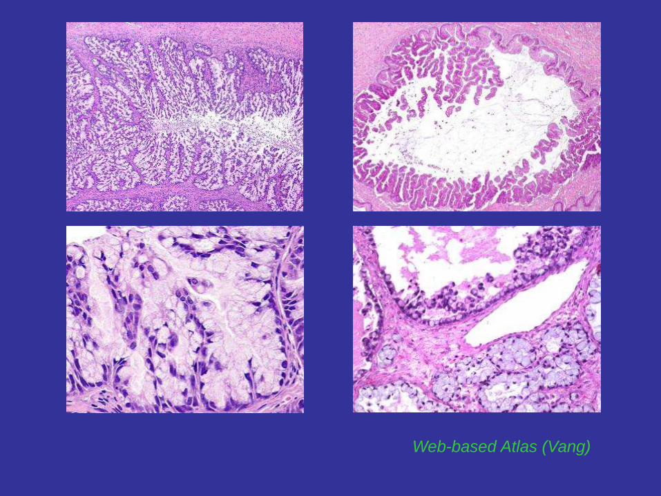
Web-based Atlas (Vang)
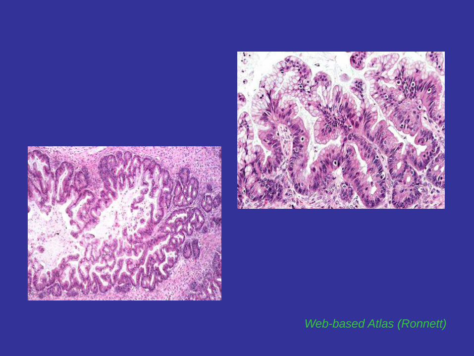
Web-based Atlas (Ronnett)
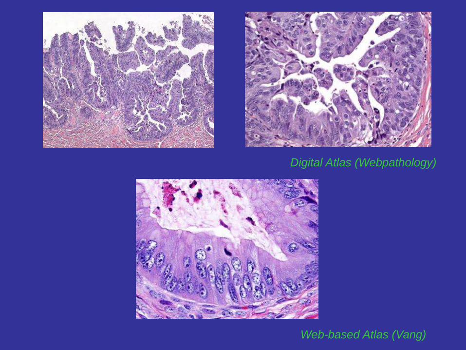
APMT - Endocervical type
• Incidence: 15% of APMT (younger age-group)
• Classical (Müllerian) mucinous:
- pure endocervical-type epithelium
- high association w/ endometriosis
• Seromucinous:
- mixed serous (cuboidal/dense eosinophilic/ciliated) & endocervical-type
(columnar/abundant eosinophilic cytoplasm, apical mucin)
- cellular papillae w/ atypical (reactive) cells (resembles SBT/MSBT)
- typical neutrophilic infiltrates within papillae, luminal mucin & stroma
• Mixed cell-type mucinous:
- heterogeneous cell population including serous, endocervical-type mucinous,
endometrioid, hobnail & “indifferent cells w/ abundant eosinophilic cytoplasm”
• Survival ~100%:
overwhelmingly benign behavior at all stages (>Stage I, IEC, microinv CA)
Shappell 2002
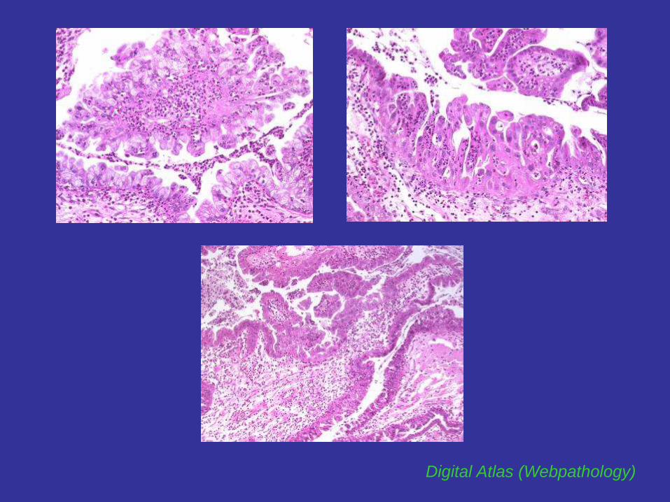
Digital Atlas (Webpathology)
Web-based Atlas (Soslow)
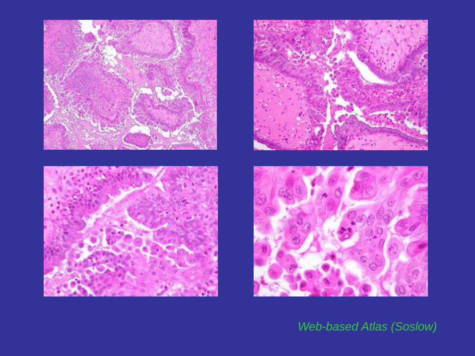
APMT with IEC
• Histology:
- severe cytologic atypia (sole Dx criterion), regardless of architecture
enlarged round to oval nuclei, high N:C ratio, some degree loss of
cytoplasmic mucin, irregularly distributed (vesicular) chromatin,
prominent nucleoli, mitoses
- complex cribriform intraglandular/intracystic growth w/ prominent
epithelial tufting, detached papillae & stratification (typical of APMT)
[does not qualify in the absence of severe atypia]
• Survival:
- essentially 100% for Stage I (95%, Riopel 1999)
(lower SR in older studies due to metastasis simulating primary
ovarian MUC or primary MUC w/ unsampled destructive invasion)
Digital Atlas (Webpathology)
Web-based Atlas (Vang)
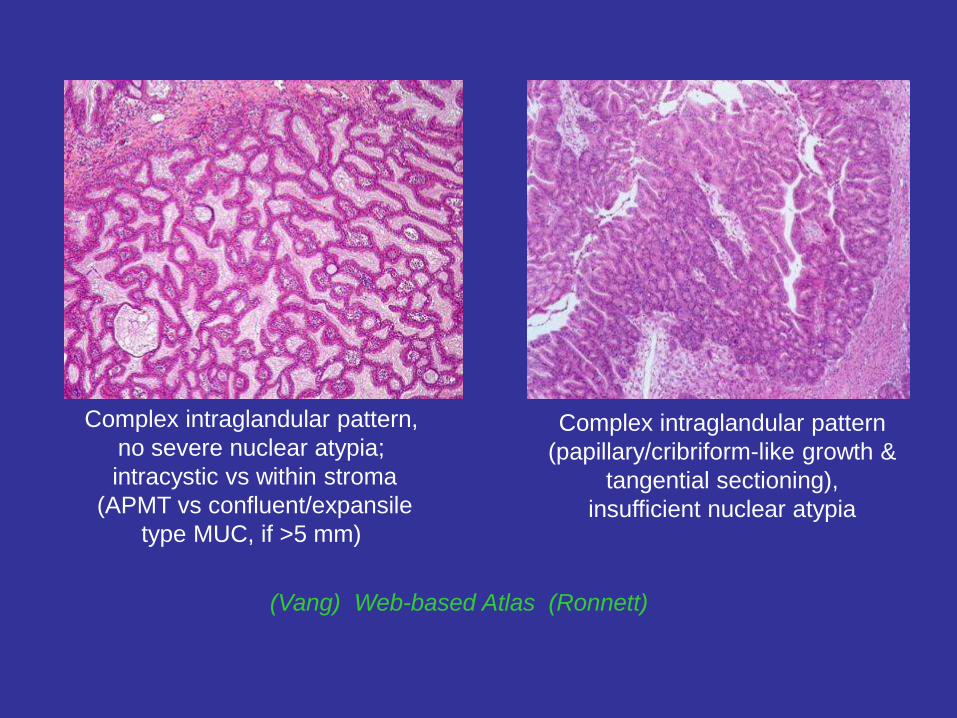
Complex intraglandular pattern,
no severe nuclear atypia;
intracystic vs within stroma
(APMT vs confluent/expansile
type MUC, if >5 mm)
(Vang) Web-based Atlas (Ronnett)
Complex intraglandular pattern
(papillary/cribriform-like growth &
tangential sectioning),
insufficient nuclear atypia
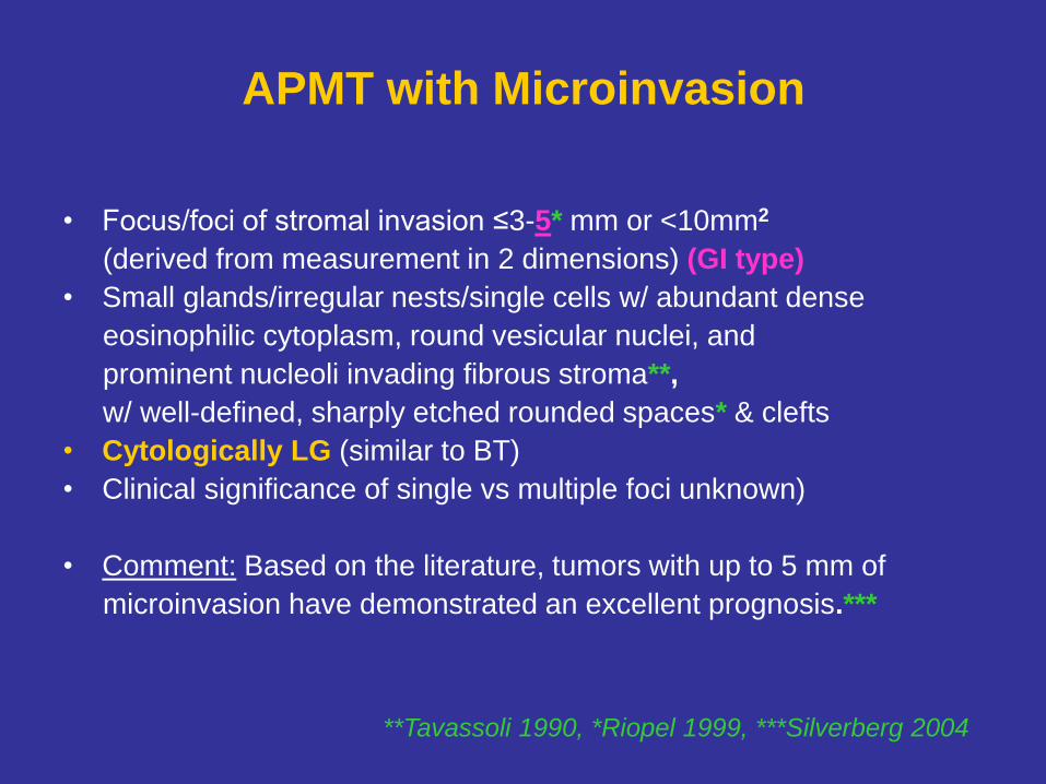
APMT with Microinvasion
• Focus/foci of stromal invasion ≤3-5* mm or <10mm2
(derived from measurement in 2 dimensions) (GI type)
• Small glands/irregular nests/single cells w/ abundant dense
eosinophilic cytoplasm, round vesicular nuclei, and
prominent nucleoli invading fibrous stroma**,
w/ well-defined, sharply etched rounded spaces* & clefts
• Cytologically LG (similar to BT)
• Clinical significance of single vs multiple foci unknown)
• Comment: Based on the literature, tumors with up to 5 mm of
microinvasion have demonstrated an excellent prognosis.***
**Tavassoli 1990, *Riopel 1999, ***Silverberg 2004

APMT with Microinvasive CA
• Small nests of cells or micropapillae, often surrounded by
clear spaces, haphazardly infiltrating fibrous or myxoid stroma, or
grow in confluent pattern of back-to-back glands or micropapillae,
w/ HG cytology (moderate-marked atypia)**
and/or
• Small invasive foci arising in tumor w/ IEC
• Appear to represent transitional stage in ovarian mucinous carcinogenesis*
• Clinical significance unclear (more studies needed)
**Bell & Scully 1990, *Riopel 1999
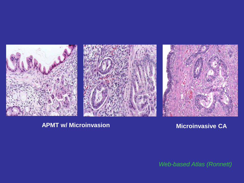
Web-based Atlas (Ronnett)
APMT w/ Microinvasion Microinvasive CA
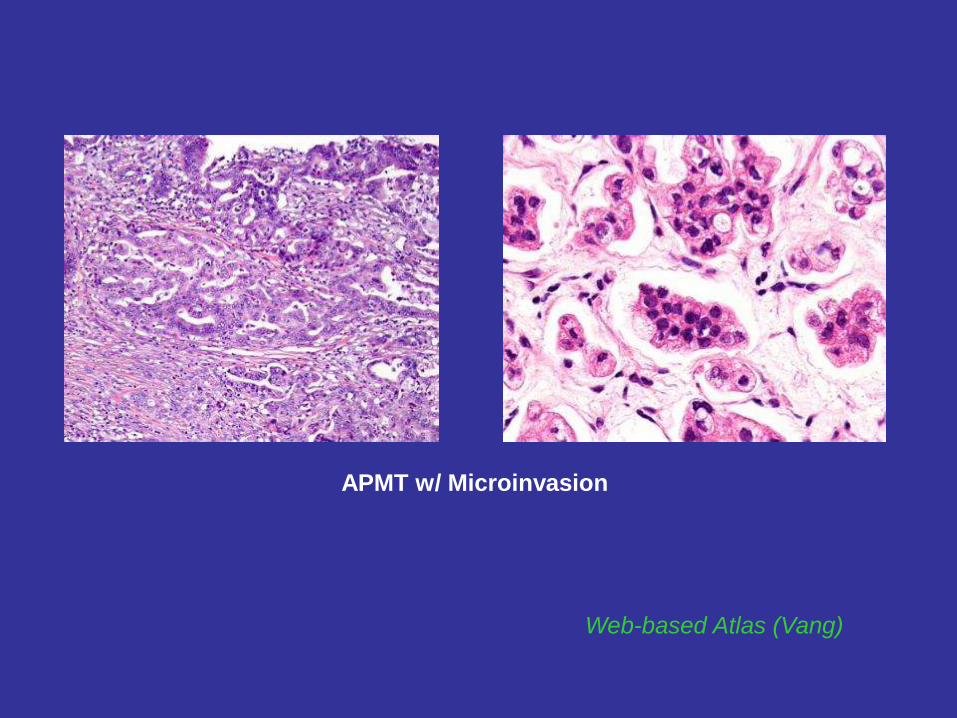
Web-based Atlas (Vang)
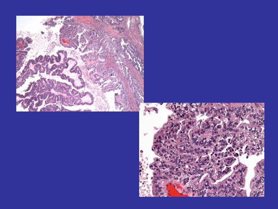
APMT w/ Microinvasion
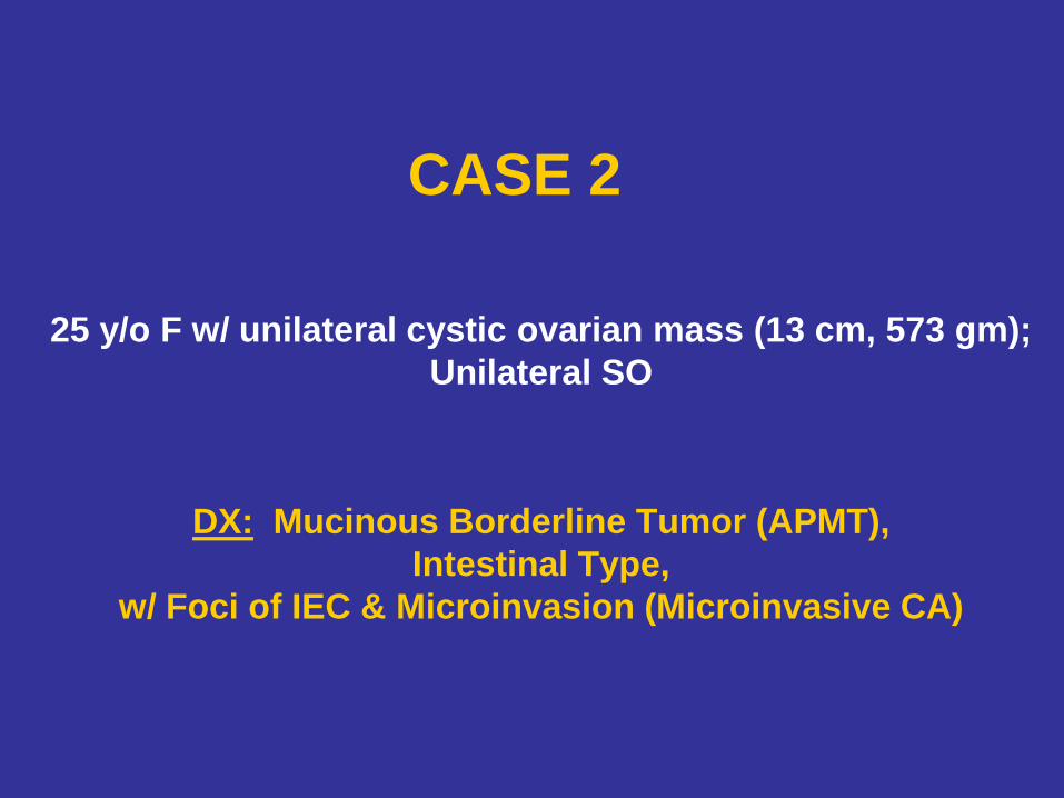
CASE 2
25 y/o F w/ unilateral cystic ovarian mass (13 cm, 573 gm);
Unilateral SO
DX: Mucinous Borderline Tumor (APMT),
Intestinal Type,
w/ Foci of IEC & Microinvasion (Microinvasive CA)
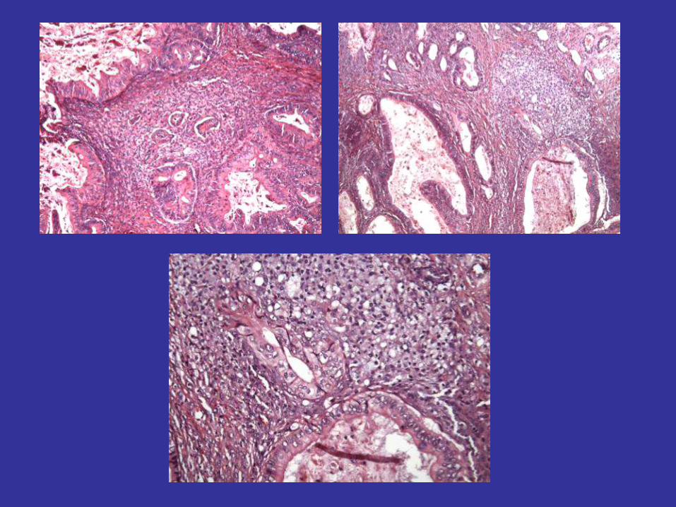
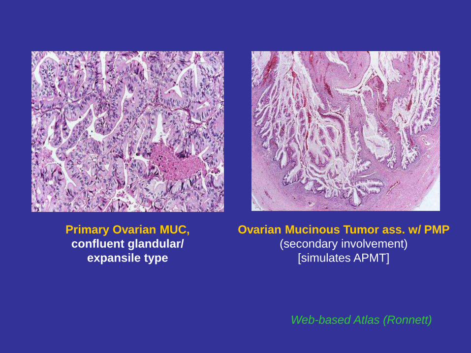
Types of Invasion in Primary MUC
• Confluent glandular/cribriform (expansile) type
- common pattern (often ass. w/ APMT, which replaces ovary)
- resembles AEH or WD EMC of endometrium
- complex/confluent/cribriform back-to-back glandular proliferation
w/o intervening stroma (interconnected labyrinthine, uninterrupted by
normal ovarian stroma) and/or papillary growth
- lacks destructive stromal invasion
• Infiltrative (destructive) type (frankly invasive MUC)
- less common pattern (D/Dx: Metastatic MUC)
- small mucinous glands/nests/individual cells w/ eosinophilic cytoplasm
surrounded by clear spaces, haphazardly/ irregularly infiltrating stroma
- foci of destructive stromal invasion >3-5 mm or >10 mm2
(>5 mm = the sole feature that correlated w/ poor Px)
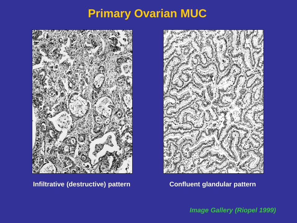
Infiltrative (destructive) pattern
Image Gallery (Riopel 1999)
Confluent glandular pattern
Primary Ovarian MUC
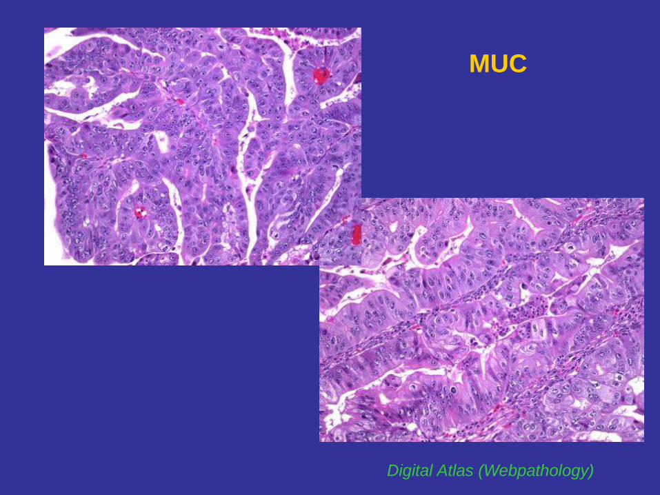
MUC
Digital Atlas (Webpathology)
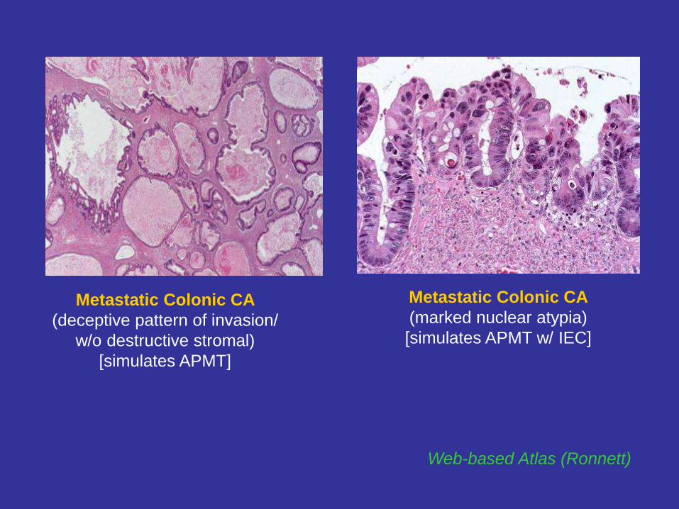
Metastatic MUC Involving Ovary
• Bilateral, usually small (<10-12 cm vs >15 APMT), GI type
• Unilateral tumor ass. w/ extraovarian disease, GI type
• Superficial cortical/surface ovarian involvement (vs stromal APMT)
• Characteristic nodular infiltrative pattern of invasion
multiple small nodules w/ aggregates of small to medium-sized,
well-formed and irregularly infiltrative glands clustered together &
separated from adjacent nodules by preserved and compressed
ovarian stroma/structures (vs confluent diffuse)
• LVI, particularly in the hilum
• Ass. w/ prominent pseudomyxoma peritonei (PMP) ~90%
(vs absent/minimal in APMT)
• Survival (5yr SR):
Poor - 11% vs 91% (Stage I) & 51% (all Stages) Ovarian MUC*
Riopel 1999
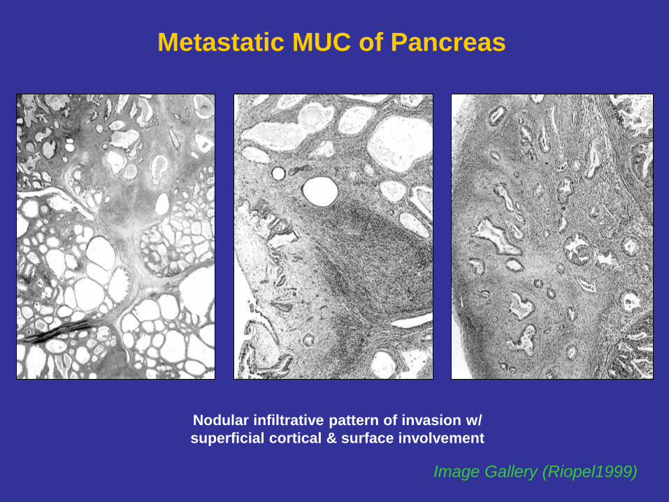
Metastatic MUC of Pancreas
Nodular infiltrative pattern of invasion w/
superficial cortical & surface involvement
Image Gallery (Riopel1999)
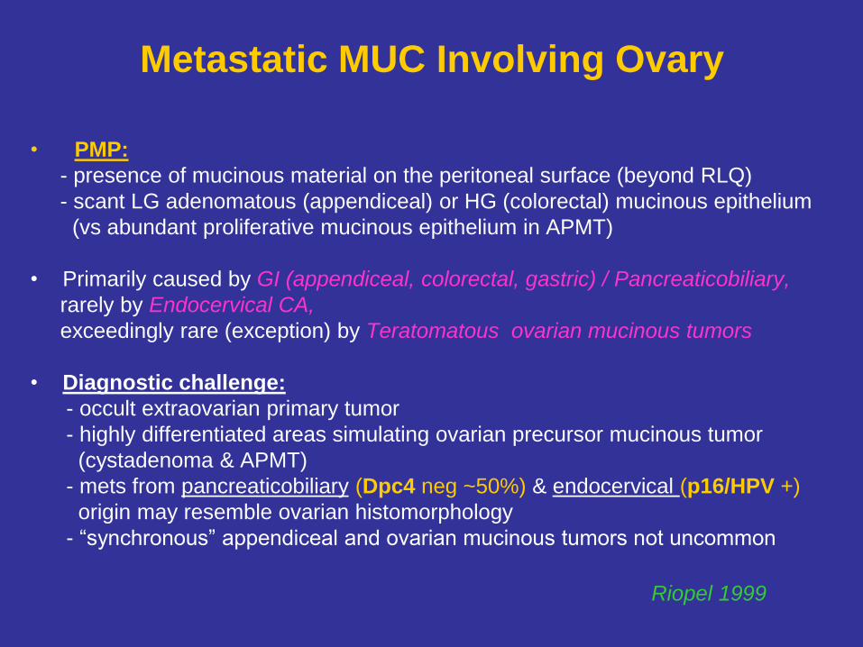
Metastatic MUC Involving Ovary
• PMP:
- presence of mucinous material on the peritoneal surface (beyond RLQ)
- scant LG adenomatous (appendiceal) or HG (colorectal) mucinous epithelium
(vs abundant proliferative mucinous epithelium in APMT)
• Primarily caused by GI (appendiceal, colorectal, gastric) / Pancreaticobiliary,
rarely by Endocervical CA,
exceedingly rare (exception) by Teratomatous ovarian mucinous tumors
• Diagnostic challenge:
- occult extraovarian primary tumor
- highly differentiated areas simulating ovarian precursor mucinous tumor
(cystadenoma & APMT)
- mets from pancreaticobiliary (Dpc4 neg ~50%) & endocervical (p16/HPV +)
origin may resemble ovarian histomorphology
- “synchronous” appendiceal and ovarian mucinous tumors not uncommon
Riopel 1999
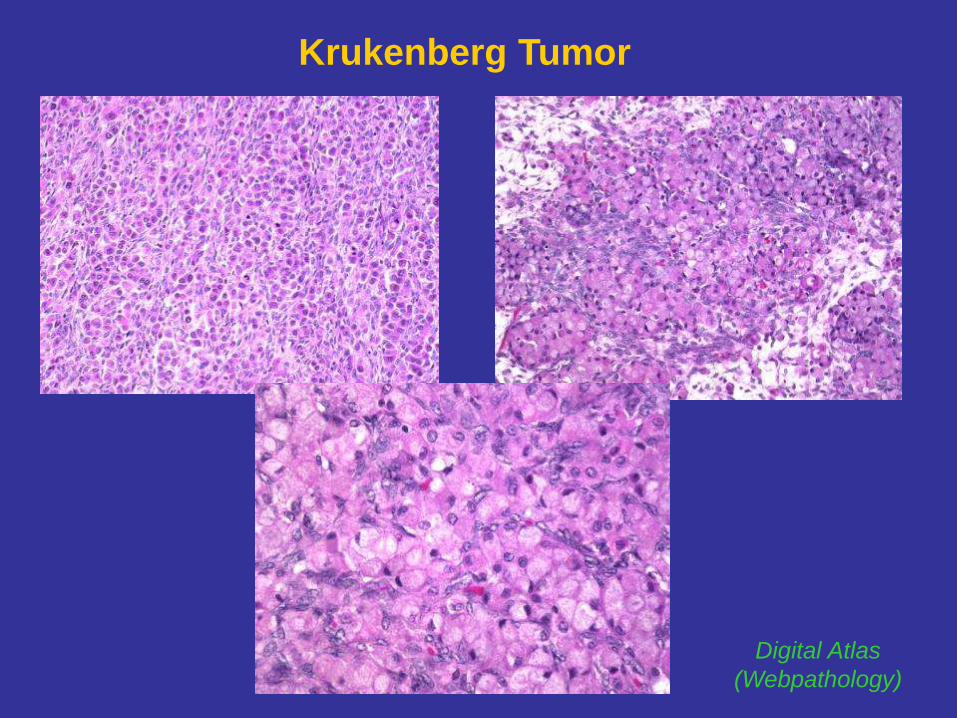
Krukenberg Tumor
Digital Atlas
(Webpathology)
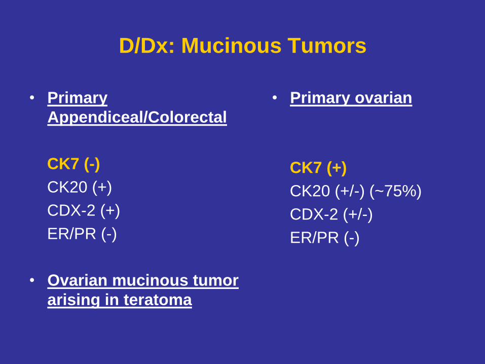
D/Dx: Mucinous Tumors
• Primary
Appendiceal/Colorectal
CK7 (-)
CK20 (+)
CDX-2 (+)
ER/PR (-)
• Ovarian mucinous tumor
arising in teratoma
• Primary ovarian
CK7 (+)
CK20 (+/-) (~75%)
CDX-2 (+/-)
ER/PR (-)
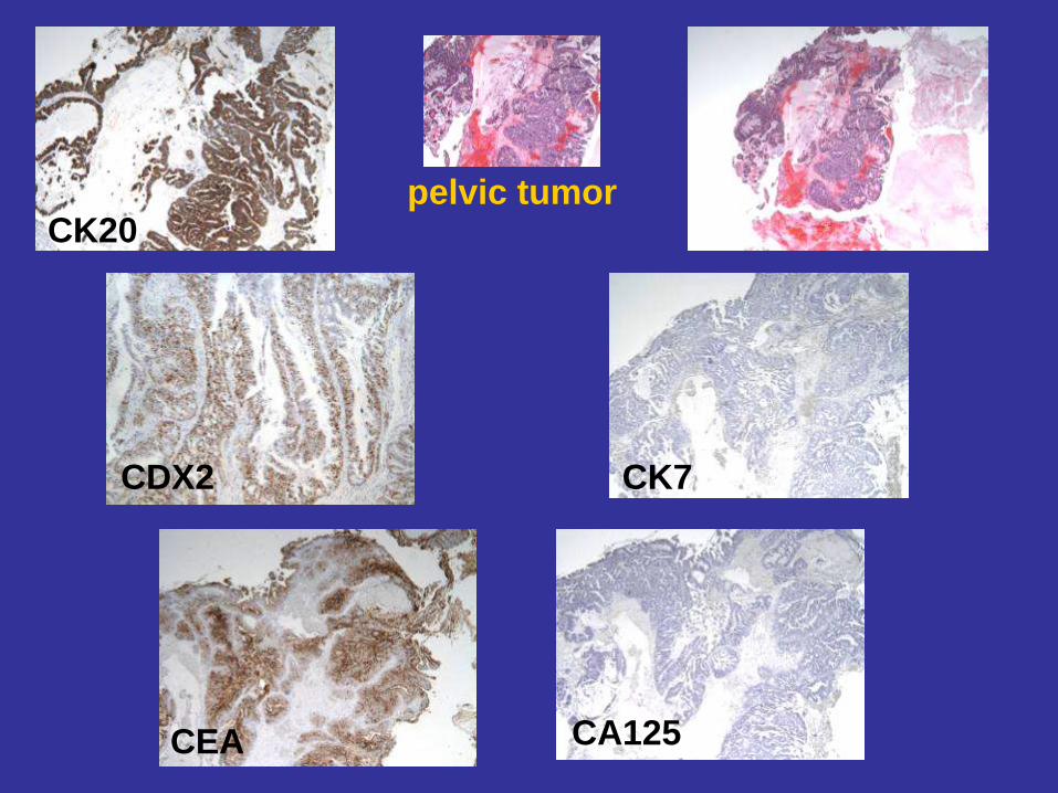
CK7
CA125
CK20
CDX2
CEA
pelvic tumor
Web-based Atlas (Ronnett)
Primary Ovarian MUC,
confluent glandular/
expansile type
Ovarian Mucinous Tumor ass. w/ PMP
(secondary involvement)
[simulates APMT]
Web-based Atlas (Ronnett)
Metastatic Colonic CA
(deceptive pattern of invasion/
w/o destructive stromal)
[simulates APMT]
Metastatic Colonic CA
(marked nuclear atypia)
[simulates APMT w/ IEC]
Endometrioid Tumors
• APET:
- marked glandular crowding with budding and branching glands
- mild to moderate cytologic atypia
• APET w/ IEC:
- marked cytologic atypia
(nuclear enlargement, vesicular chromatin, prominent nucleoli)
• APET w/ microinvasion (<5mm or ≤10mm2):
infiltrative pattern (more common)
- haphazardly arranged glands/nests ass. w/ an altered stroma
confluent pattern (less common)
- cribriform architecture within glands as opposed to within stroma
(not universally accepted as pattern of invasion)
• Small foci of WD CA arising within an endometriotic cyst (2 mm)*
• Implants: very rare and poorly documented
*Seidman 1996
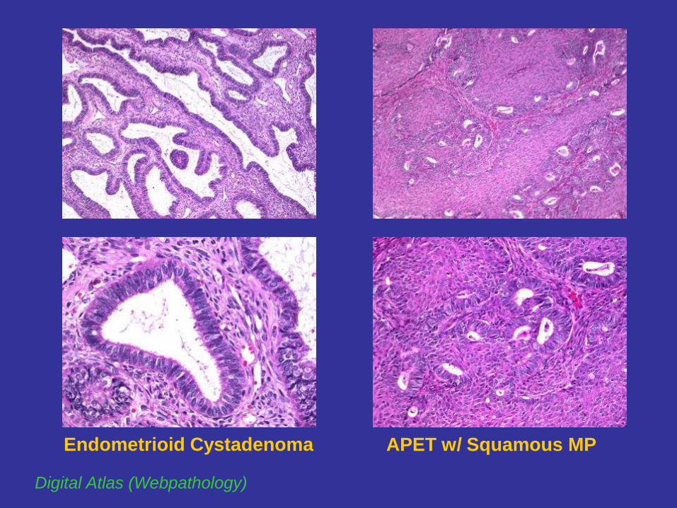
APET w/ Squamous MP
Digital Atlas (Webpathology)
Endometrioid Cystadenoma
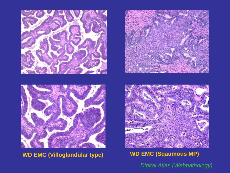
WD EMC (Villoglandular type) WD EMC (Sqaumous MP)
Digital Atlas (Webpathology)

D/Dx: Endometrioid Tumors
Colorectal
CK7 (-)
CK20 (+)
CDX-2 (+)
ER/PR (-)
Primary ovarian Endocervical
CK7 (+) CK7 (+)
CK20 (-) CK20 (-)
CDX-2 (-) CDX-2 (-)
ER/PR (+) ER/PR (-)
p16 (+)
HPV-ISH (+)
Clear Cell Tumors
• AFs and APTs very uncommon
• Almost all tumors in CC category are CAs
• AF to AF-like patterns of CCC represent morphologic continuum*
• No uniformly accepted diagnostic criteria:
AF vs APT
AF/APT w/ cytologic atypia vs APT with IEC vs
AF-like patterns of CCC w/ subtle cytologic atypia
• Thresholds vary between GYN pathologists
Yamamoto 2007, Vang 2008
Clear Cell AF vs APT
• Both resemble one another:
- round simple glands within a fibromatous stroma
- round, dense, and eosinophilic intraluminal secretions
- epithelium often flattened (obvious CC diff. may not be apparent)
- at least focally: hobnail cells
• Clear Cell AF:
- glands widely spaced apart by fibroblastic stroma
- lined by 1-2 layers of w/o stratification
- cuboidal or flattened cells w/ abundant clear cytoplasm
- no nuclear atypia
• Clear Cell AF – Borderline (APCCT):
- marked glandular crowding +/- small solid nests
- more irregular gland contours & stratification
- some degree of atypia
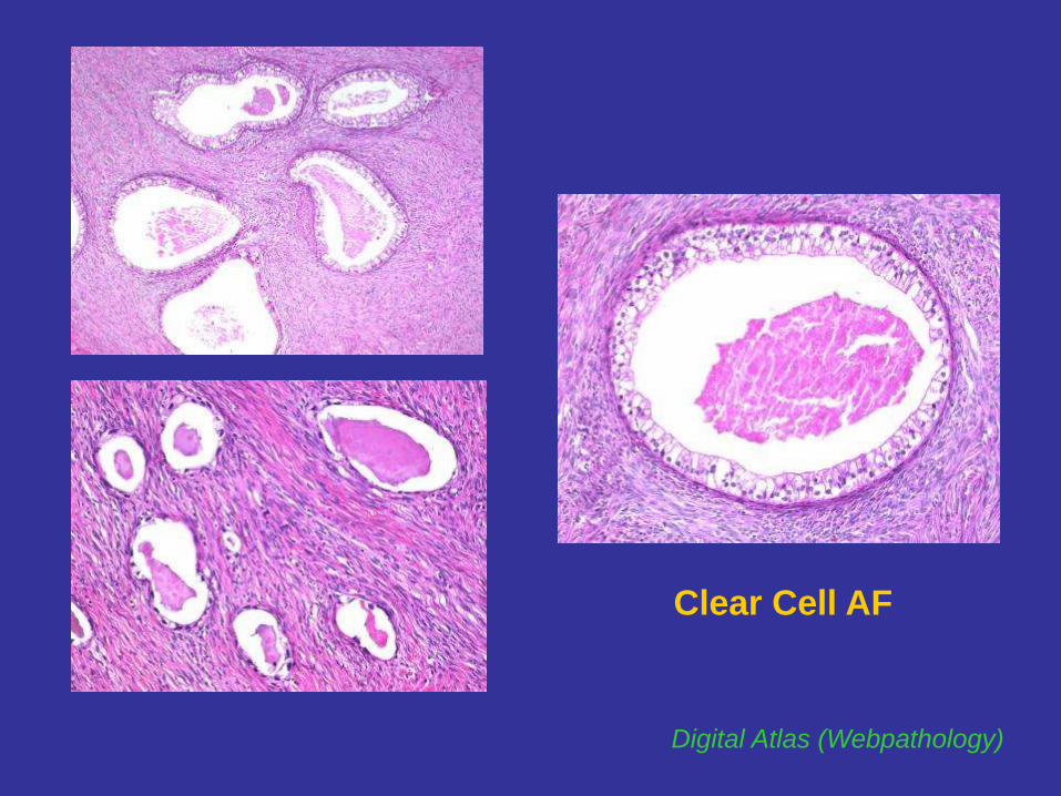
Clear Cell AF
Digital Atlas (Webpathology)
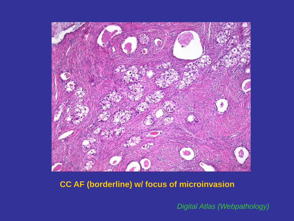
CC AF (borderline) w/ focus of microinvasion
Digital Atlas (Webpathology)
Marked Cytologic Atypia in CC Tumors
- exact level of atypia sufficient for IEC or CCC not well-defined
- degree of nuclear atypia in AF/APT can overlap w/ CCC
- some CCC can have deceptively bland nuclei
• APCCT w/ IEC (Tavassoli 2003; Kurman 2008)
- marked nuclear atypia w/o stromal invasion
- criteria not well-defined
• APCCT w/ microinvasion (Bell 1985)
- focus <3 mm w/ glands, small solid nests/single cells w/ malignant nuclei
haphazardly arranged within desmoplastic, myxoid, or edematous stroma
- 2/3 patients were alive at 4 & 6 years, 1 patient recurred at 3 years
• Adenofibroma-like pattern of CCC
- at least focal areas w/ combination of glandular crowding, marked nuclear
atypia & epithelial stratification beyond the level expected for APCCT
• Implants:
- extra-ovarian implants at the time of surgery have not been described
- frequent “implants of endometriosis” elsewhere in the pelvis (Veras 2009)
CCC
• General rule for the distinction of APT from CA (WHO Classification)
- presence of stromal invasion
• CCC – an exception to this rule
1) infiltrating or confluent patterns (obviously invasive)
2) intracystic proliferative pattern (appear to proliferate within a cyst)
- may not have apparent stromal invasion of the traditional type
- conventionally diagnosed as CA (generally aggressive behavior)
• CCC – typically not graded (unlike other histologic types)
- grade has not correlated with Px (in several studies)
- present at Stage I in 70% (Veras 2009)
- generally less responsive to CTX than SC
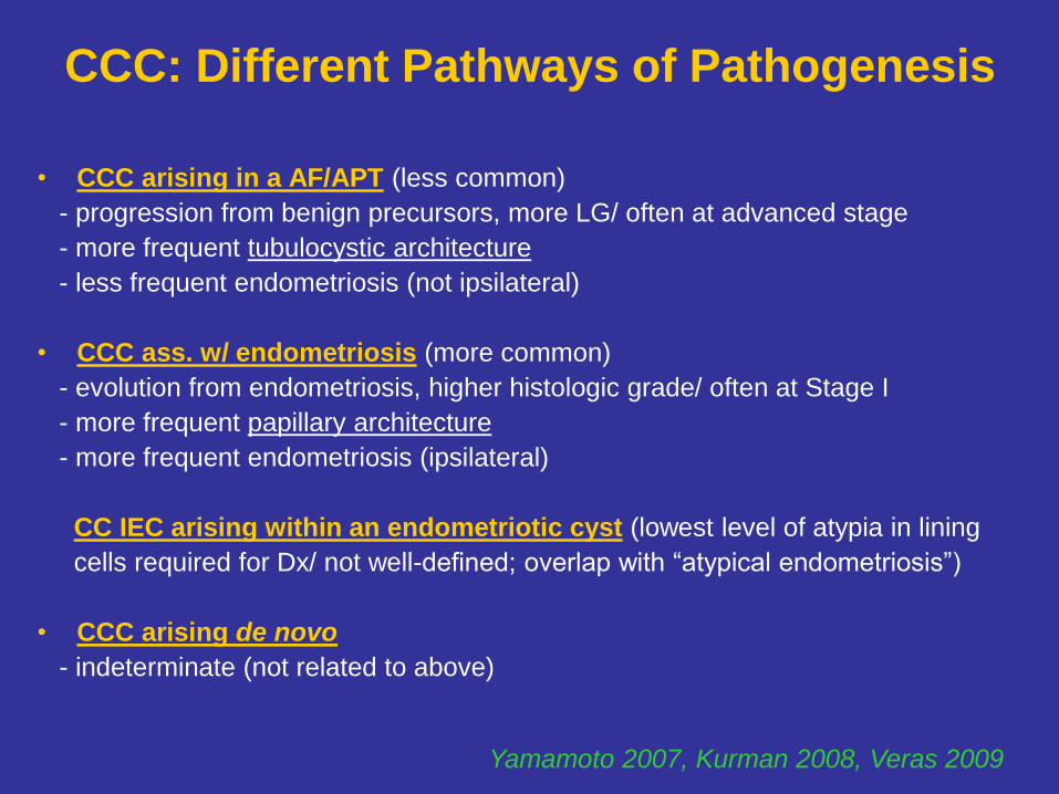
CCC: Different Pathways of Pathogenesis
• CCC arising in a AF/APT (less common)
- progression from benign precursors, more LG/ often at advanced stage
- more frequent tubulocystic architecture
- less frequent endometriosis (not ipsilateral)
• CCC ass. w/ endometriosis (more common)
- evolution from endometriosis, higher histologic grade/ often at Stage I
- more frequent papillary architecture
- more frequent endometriosis (ipsilateral)
CC IEC arising within an endometriotic cyst (lowest level of atypia in lining
cells required for Dx/ not well-defined; overlap with “atypical endometriosis”)
• CCC arising de novo
- indeterminate (not related to above)
Yamamoto 2007, Kurman 2008, Veras 2009
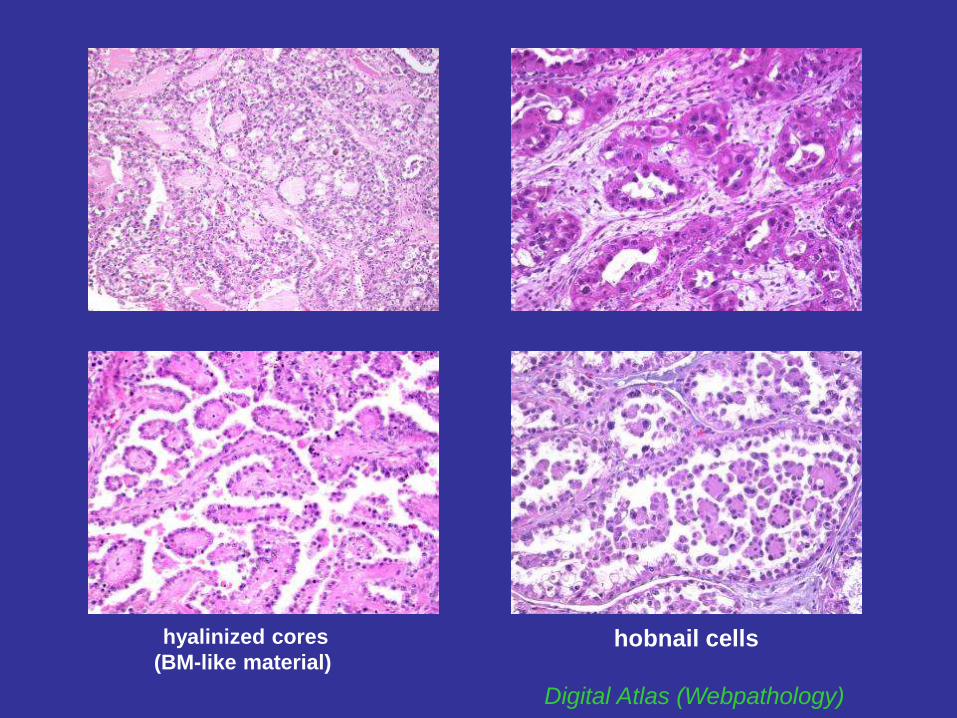
Digital Atlas (Webpathology)
hyalinized cores
(BM-like material) hobnail cells
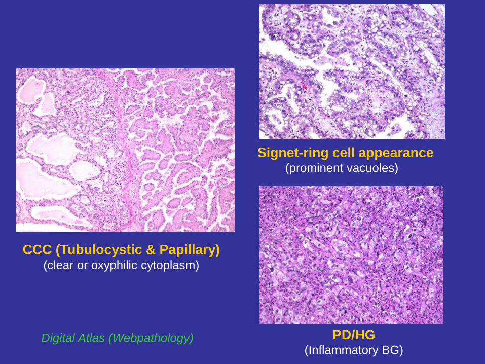
CCC (Tubulocystic & Papillary) (clear or oxyphilic cytoplasm)
Digital Atlas (Webpathology) PD/HG (Inflammatory BG)
Signet-ring cell appearance (prominent vacuoles)
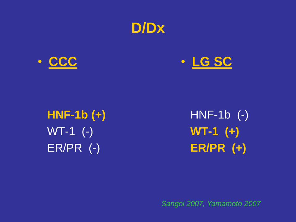
D/Dx
• CCC
HNF-1b (+)
WT-1 (-)
ER/PR (-)
• LG SC
HNF-1b (-)
WT-1 (+)
ER/PR (+)
Sangoi 2007, Yamamoto 2007
Transitional Cell Tumors
• Benign Brenner tumor (majority)
- small solid nests of transitional cells w/ sharply defined outlines
- in dense fibroblastic stroma
- bland cells w/ pale cytoplasm & oval nuclei w/ longitudinal grooves,
and small inconspicuous nucleoli
- ass. w/ mucinous lesions (juxtaposed/abrupt transition/hybrid) (~25%)
• APBT/ Borderline Brenner tumor (rare)
- crowded complex nests & large papillary fronds
- exuberant proliferation w/ tongues protruding into luminal spaces
- some degree of cytologic atypia, mitoses (~ LG Urothelial CA)
• IEC and microinvasion (not specifically defined)
• Malignant Brenner tumor (rare)
- HG nuclei (~ HG Uroth CA), invasion/desmoplastic stroma, Ca++
- BG component of precursor lesions (BBT/APBT)
• TCC (not uncommon), often bilateral
- HG invasive CA in the absence of precursor lesions
- mixed w/ HG CA: SC, EMC, MUC, CCC
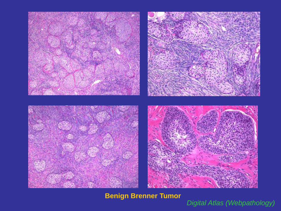
Digital Atlas (Webpathology) Benign Brenner Tumor
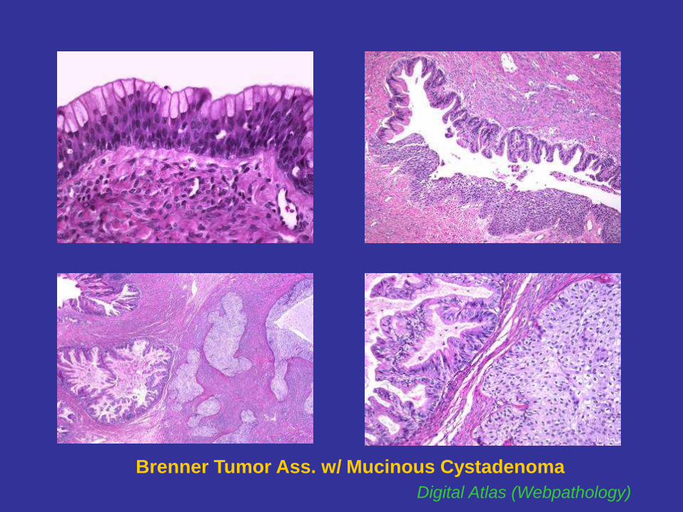
Brenner Tumor Ass. w/ Mucinous Cystadenoma Digital Atlas (Webpathology)
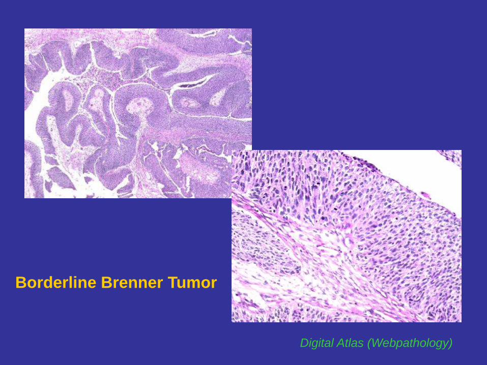
Borderline Brenner Tumor
Digital Atlas (Webpathology)
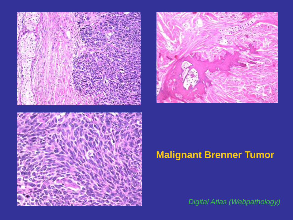
Malignant Brenner Tumor
Digital Atlas (Webpathology)
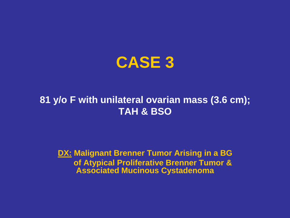
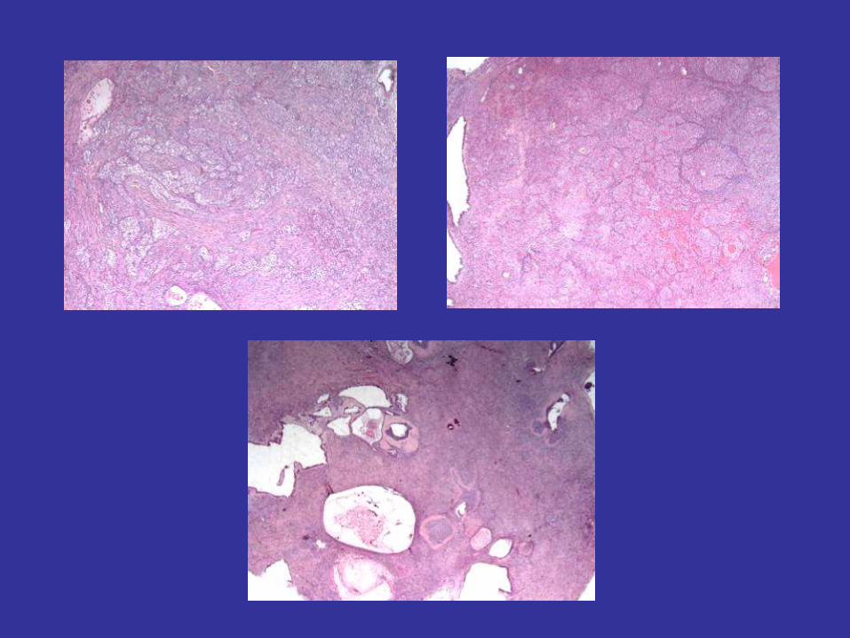
CASE 3
81 y/o F with unilateral ovarian mass (3.6 cm);
TAH & BSO
DX: Malignant Brenner Tumor Arising in a BG
of Atypical Proliferative Brenner Tumor & Associated Mucinous Cystadenoma
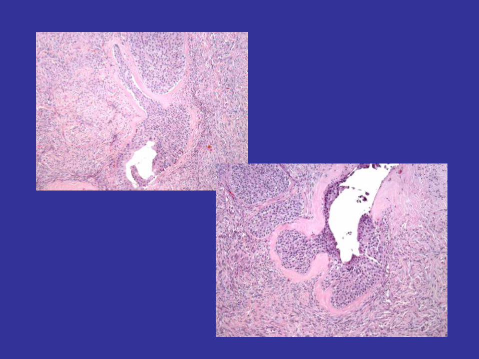
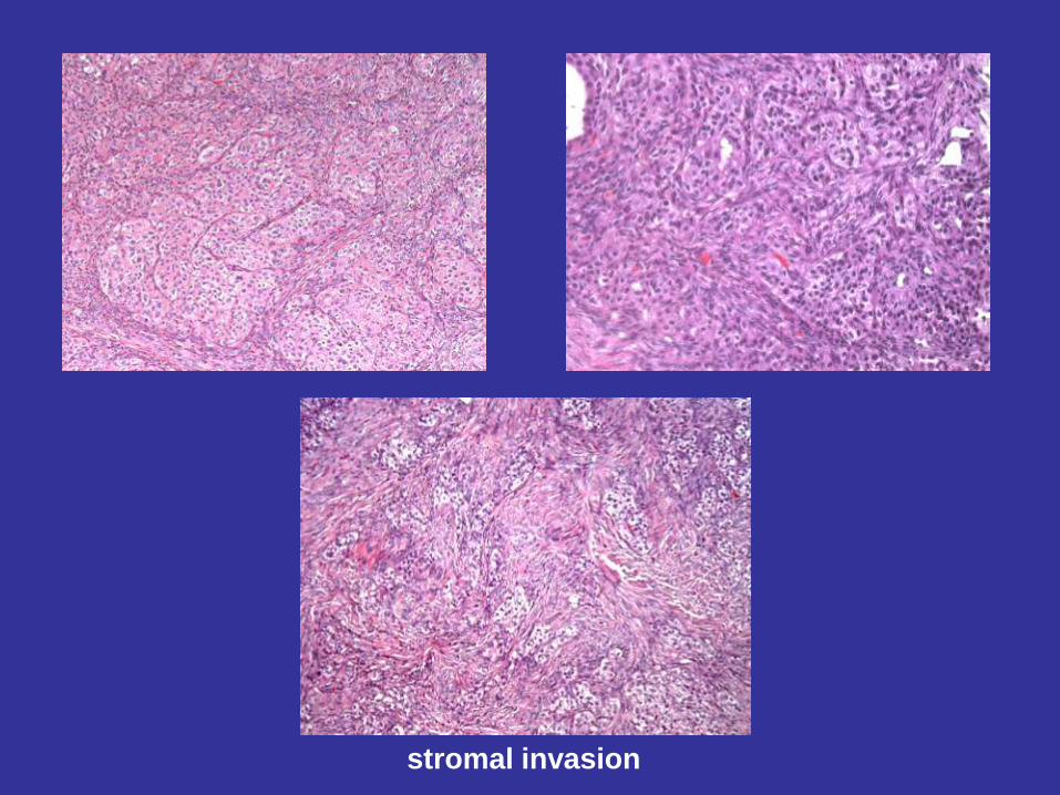
stromal invasion
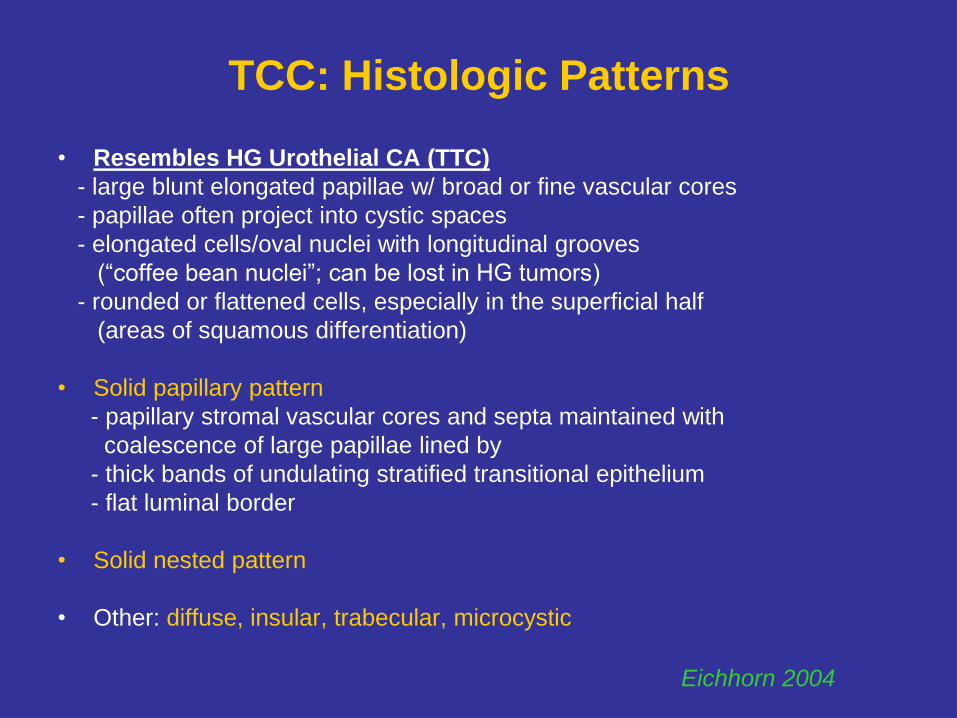
TCC: Histologic Patterns
• Resembles HG Urothelial CA (TTC)
- large blunt elongated papillae w/ broad or fine vascular cores
- papillae often project into cystic spaces
- elongated cells/oval nuclei with longitudinal grooves
(“coffee bean nuclei”; can be lost in HG tumors)
- rounded or flattened cells, especially in the superficial half
(areas of squamous differentiation)
• Solid papillary pattern
- papillary stromal vascular cores and septa maintained with
coalescence of large papillae lined by
- thick bands of undulating stratified transitional epithelium
- flat luminal border
• Solid nested pattern
• Other: diffuse, insular, trabecular, microcystic
Eichhorn 2004
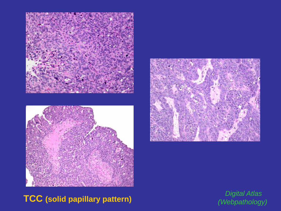
TCC (solid papillary pattern) Digital Atlas
(Webpathology)
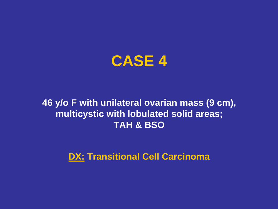
CASE 4
46 y/o F with unilateral ovarian mass (9 cm),
multicystic with lobulated solid areas;
TAH & BSO
DX: Transitional Cell Carcinoma
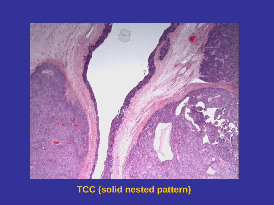
TCC (solid nested pattern)

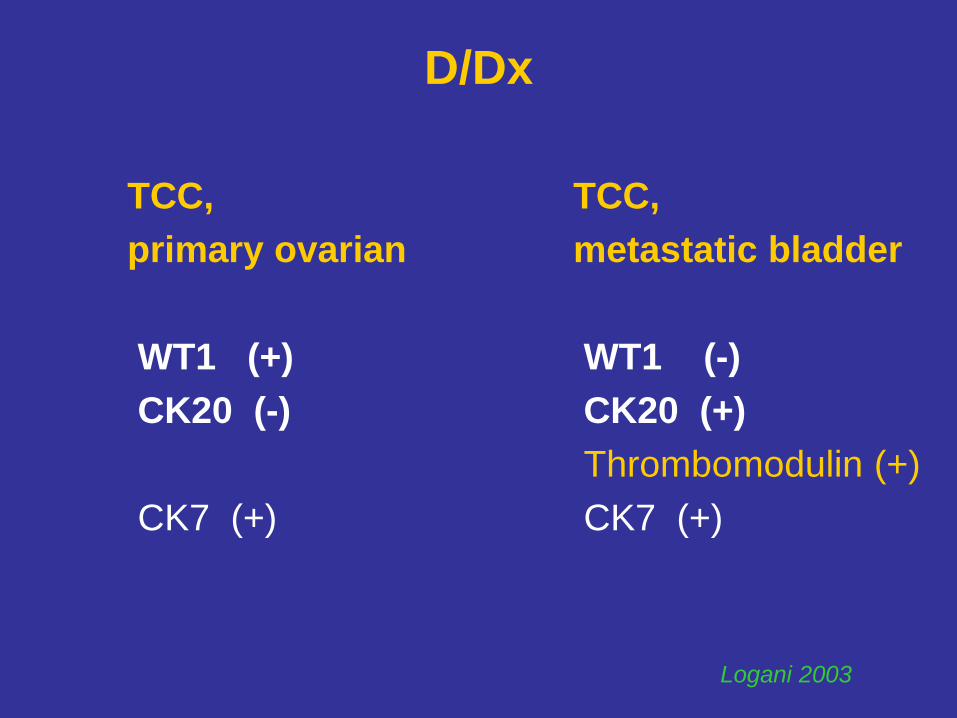
D/Dx
TCC,
primary ovarian
WT1 (+)
CK20 (-)
CK7 (+)
TCC,
metastatic bladder
WT1 (-)
CK20 (+)
Thrombomodulin (+)
CK7 (+)
Logani 2003

Kentucky Farm
House

3. Histologic Basis of Biological Behavior:
Selected Endometrial,
& Mixed Tumors
Current Concepts
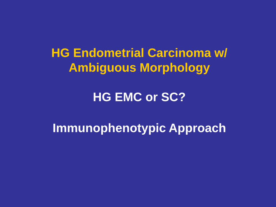
HG Endometrial Carcinoma w/
Ambiguous Morphology
HG EMC or SC?
Immunophenotypic Approach
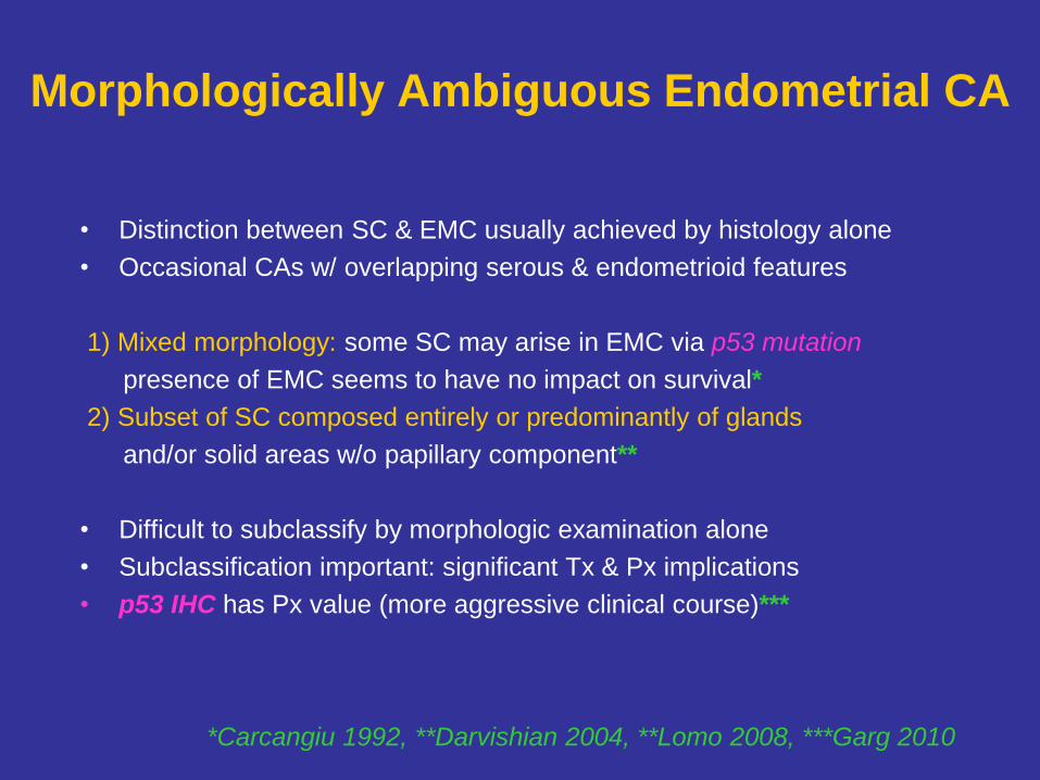
Morphologically Ambiguous Endometrial CA
• Distinction between SC & EMC usually achieved by histology alone
• Occasional CAs w/ overlapping serous & endometrioid features
1) Mixed morphology: some SC may arise in EMC via p53 mutation
presence of EMC seems to have no impact on survival*
2) Subset of SC composed entirely or predominantly of glands
and/or solid areas w/o papillary component**
• Difficult to subclassify by morphologic examination alone
• Subclassification important: significant Tx & Px implications
• p53 IHC has Px value (more aggressive clinical course)***
*Carcangiu 1992, **Darvishian 2004, **Lomo 2008, ***Garg 2010
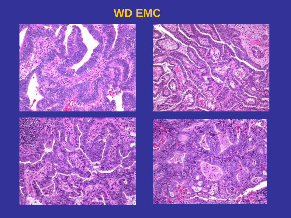
WD EMC
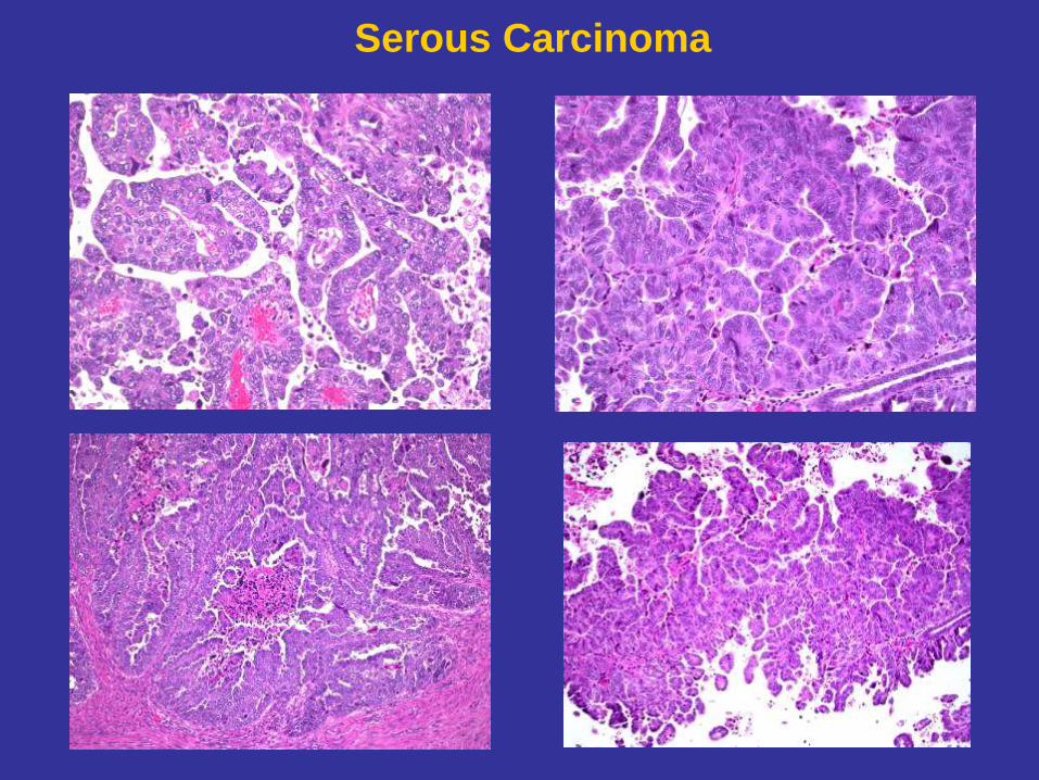
Serous Carcinoma

Thanks Thanks
Glandular Architecture & HG Nuclei
SC or HG EMC?
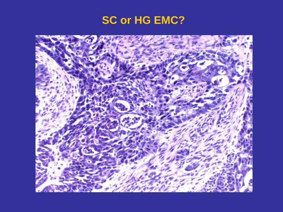
SC or HG EMC?
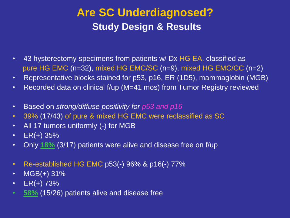
Are SC Underdiagnosed?
Study Design & Results
• 43 hysterectomy specimens from patients w/ Dx HG EA, classified as
pure HG EMC (n=32), mixed HG EMC/SC (n=9), mixed HG EMC/CC (n=2)
• Representative blocks stained for p53, p16, ER (1D5), mammaglobin (MGB)
• Recorded data on clinical f/up (M=41 mos) from Tumor Registry reviewed
• Based on strong/diffuse positivity for p53 and p16
• 39% (17/43) of pure & mixed HG EMC were reclassified as SC
• All 17 tumors uniformly (-) for MGB
• ER(+) 35%
• Only 18% (3/17) patients were alive and disease free on f/up
• Re-established HG EMC p53(-) 96% & p16(-) 77%
• MGB(+) 31%
• ER(+) 73%
• 58% (15/26) patients alive and disease free
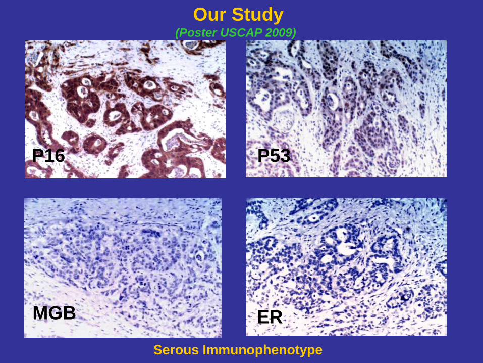
P16 P53
MGB ER
Our Study (Poster USCAP 2009)
Serous Immunophenotype
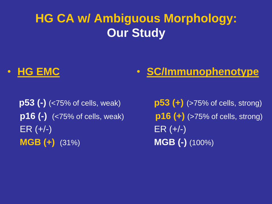
HG CA w/ Ambiguous Morphology:
Our Study
• HG EMC
p53 (-) (<75% of cells, weak)
p16 (-) (<75% of cells, weak)
ER (+/-)
MGB (+) (31%)
• SC/Immunophenotype
p53 (+) (>75% of cells, strong)
p16 (+) (>75% of cells, strong)
ER (+/-)
MGB (-) (100%)
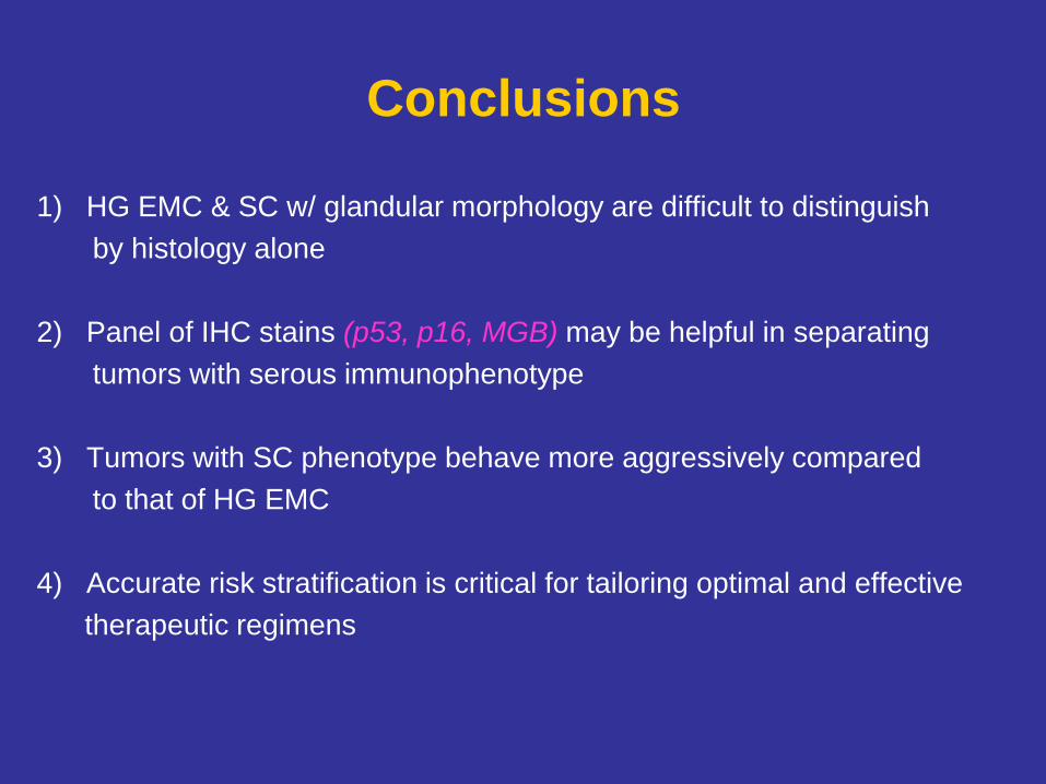
Conclusions
1) HG EMC & SC w/ glandular morphology are difficult to distinguish
by histology alone
2) Panel of IHC stains (p53, p16, MGB) may be helpful in separating
tumors with serous immunophenotype
3) Tumors with SC phenotype behave more aggressively compared
to that of HG EMC
4) Accurate risk stratification is critical for tailoring optimal and effective
therapeutic regimens
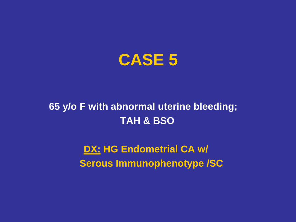
CASE 5
65 y/o F with abnormal uterine bleeding;
TAH & BSO
DX: HG Endometrial CA w/
Serous Immunophenotype /SC
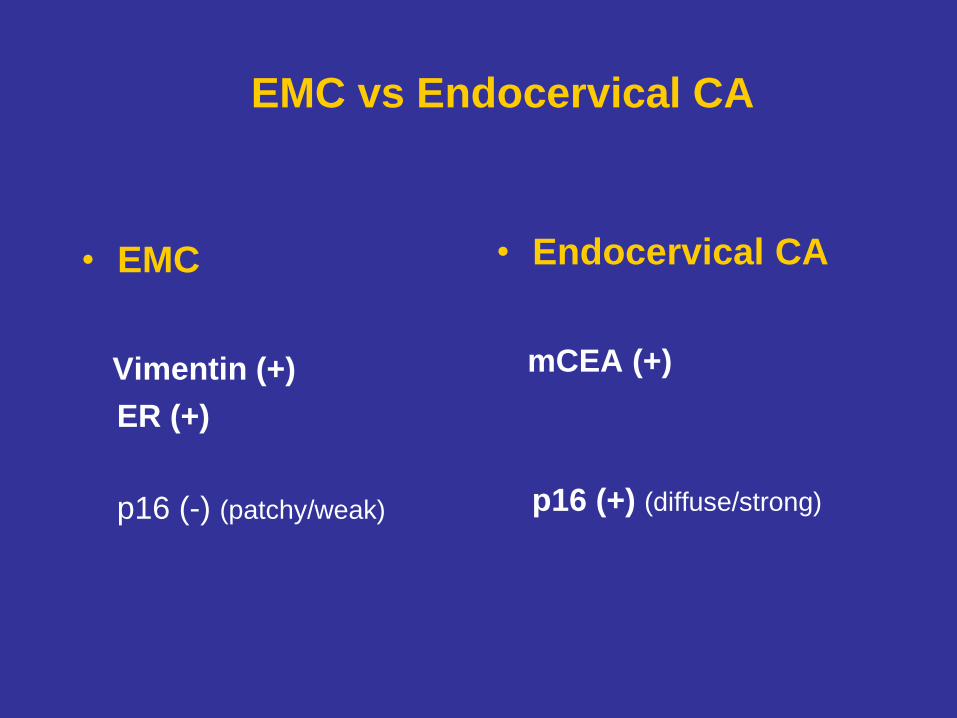
EMC vs Endocervical CA
• EMC
Vimentin (+)
ER (+)
p16 (-) (patchy/weak)
• Endocervical CA
mCEA (+)
p16 (+) (diffuse/strong)
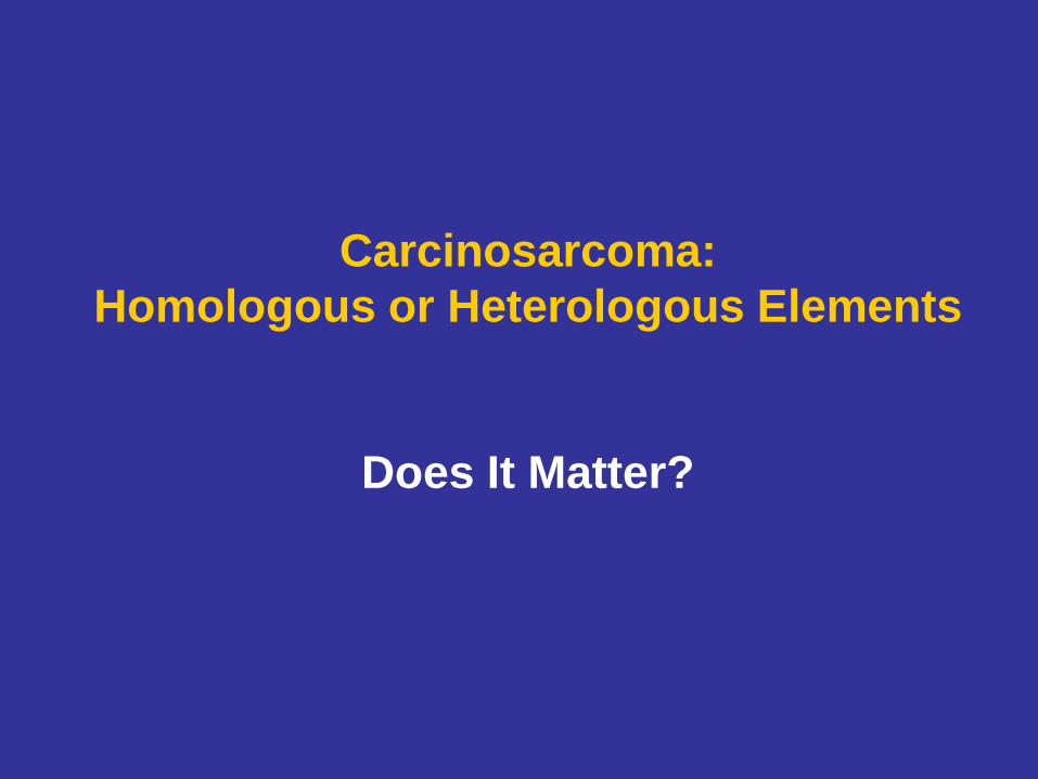
Carcinosarcoma:
Homologous or Heterologous Elements
Does It Matter?

CS (MMMT)
• WHO definition:
- neoplasm composed of admixed malignant epithelial &
mesenchymal components
• Biphasic tumor with both Endometrial CA & Sarcoma
- considered CA with “metaplastic” sarcomatoid features
(McCluggage 2002)
- occur predominantly in postmenopausal women/ bleeding
- biologically aggressive, often present at advanced stage
- metastasis usually as CA or CA-predominant (intraabdominal),
rarely as sarcoma (distant sites: lung, etc)
- 5-yr SR of <35% (Yamada 2000)
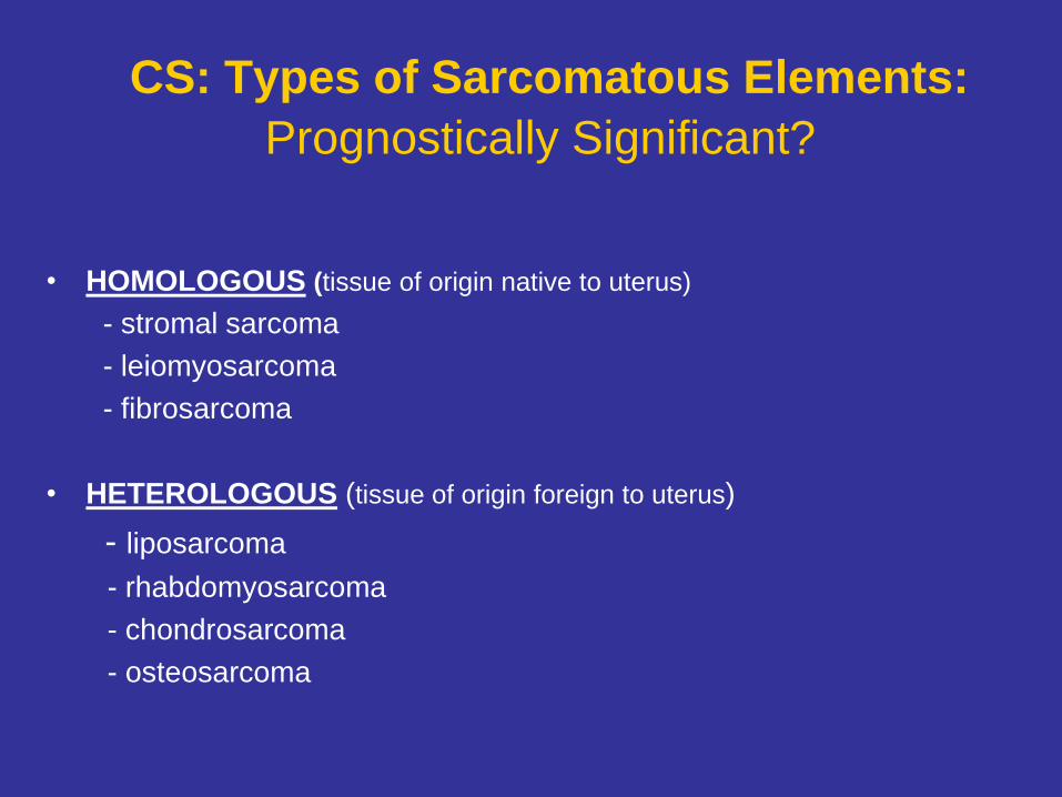
CS: Types of Sarcomatous Elements:
Prognostically Significant?
• HOMOLOGOUS (tissue of origin native to uterus)
- stromal sarcoma
- leiomyosarcoma
- fibrosarcoma
• HETEROLOGOUS (tissue of origin foreign to uterus)
- liposarcoma
- rhabdomyosarcoma
- chondrosarcoma
- osteosarcoma
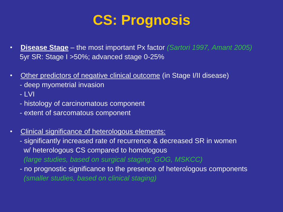
CS: Prognosis
• Disease Stage – the most important Px factor (Sartori 1997, Amant 2005)
5yr SR: Stage I >50%; advanced stage 0-25%
• Other predictors of negative clinical outcome (in Stage I/II disease)
- deep myometrial invasion
- LVI
- histology of carcinomatous component
- extent of sarcomatous component
• Clinical significance of heterologous elements:
- significantly increased rate of recurrence & decreased SR in women
w/ heterologous CS compared to homologous
(large studies, based on surgical staging: GOG, MSKCC)
- no prognostic significance to the presence of heterologous components
(smaller studies, based on clinical staging)
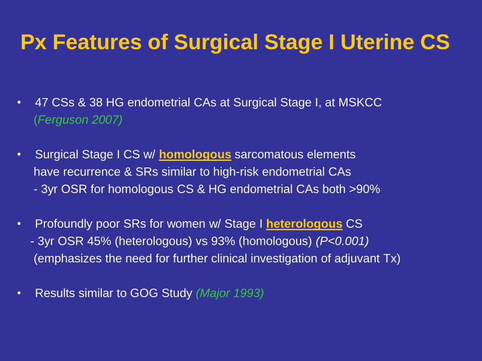
Px Features of Surgical Stage I Uterine CS
• 47 CSs & 38 HG endometrial CAs at Surgical Stage I, at MSKCC
(Ferguson 2007)
• Surgical Stage I CS w/ homologous sarcomatous elements
have recurrence & SRs similar to high-risk endometrial CAs
- 3yr OSR for homologous CS & HG endometrial CAs both >90%
• Profoundly poor SRs for women w/ Stage I heterologous CS
- 3yr OSR 45% (heterologous) vs 93% (homologous) (P<0.001)
(emphasizes the need for further clinical investigation of adjuvant Tx)
• Results similar to GOG Study (Major 1993)
Digital Atlas (Webpathology)

(Digital Atlas (Webpathology)
Digital Atlas (Webpathology)
CASE 6
61 y/o F w/ working Dx of Endometrial CA;
TAH & BSO
DX: Carcinosarcoma (MMMT) w/
Homologous & Heterologous Elements
EMC w/
Prominent Spindle Cell Component:
Carcinosarcoma (homologous) or
Metaplasia ?
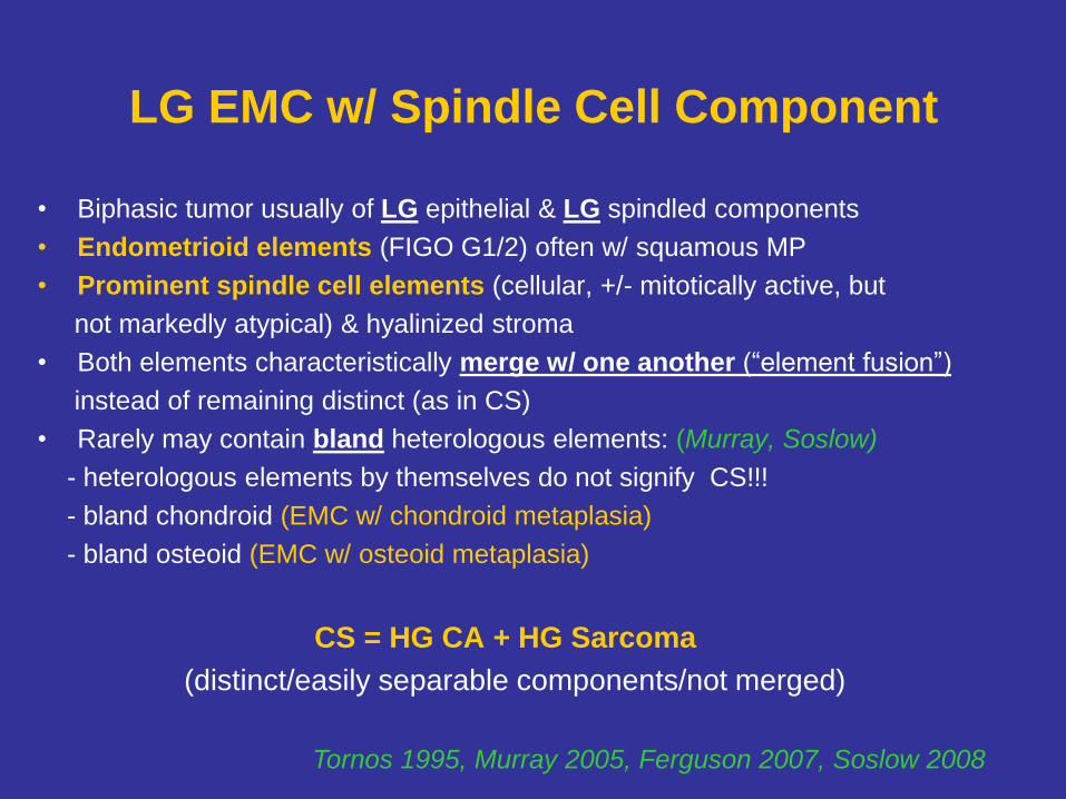
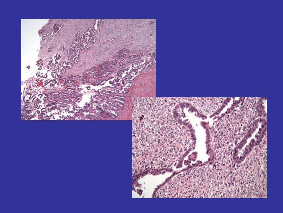
LG EMC w/ Spindle Cell Component
• Biphasic tumor usually of LG epithelial & LG spindled components
• Endometrioid elements (FIGO G1/2) often w/ squamous MP
• Prominent spindle cell elements (cellular, +/- mitotically active, but
not markedly atypical) & hyalinized stroma
• Both elements characteristically merge w/ one another (“element fusion”)
instead of remaining distinct (as in CS)
• Rarely may contain bland heterologous elements: (Murray, Soslow)
- heterologous elements by themselves do not signify CS!!!
- bland chondroid (EMC w/ chondroid metaplasia)
- bland osteoid (EMC w/ osteoid metaplasia)
CS = HG CA + HG Sarcoma
(distinct/easily separable components/not merged)
Tornos 1995, Murray 2005, Ferguson 2007, Soslow 2008
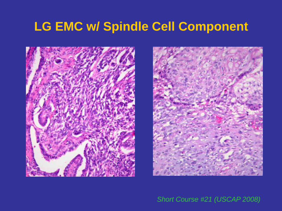
LG EMC w/ Spindle Cell Component
Short Course #21 (USCAP 2008)
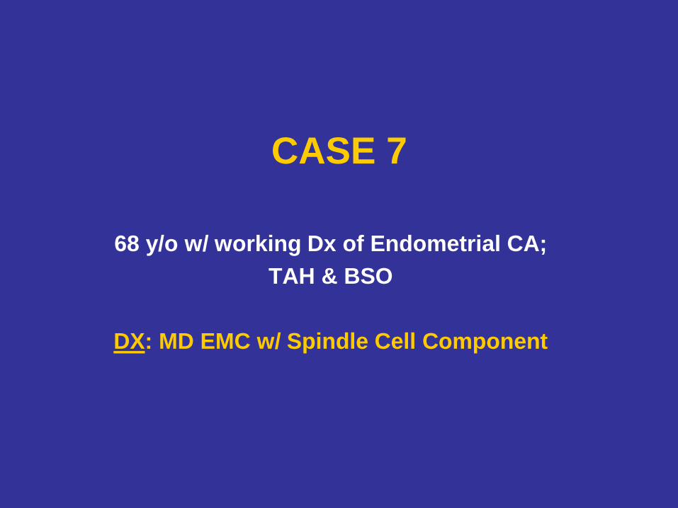
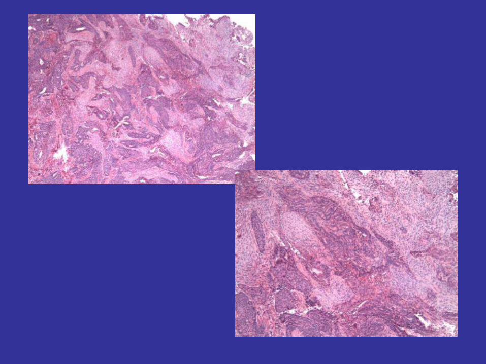
CASE 7
68 y/o w/ working Dx of Endometrial CA;
TAH & BSO
DX: MD EMC w/ Spindle Cell Component

EMC w/
Squamous Differentiation
Does It Affect Grading?

EMC w/ Squamous Differentiation
• Common finding in EMC ~50%
• Criteria for squamous differentiation:
- keratinization demonstrated w/ standard staining technique
- intercellular bridges and/or
- three or more of the following 4 criteria:
a) sheet-like growth without gland formation or palisading
b) sharp cell margins
c) eosinophilic and thick or glassy cytoplasm
d) decreased N/C ratio compared to foci elsewhere in the tumor
Tavassoli 2003
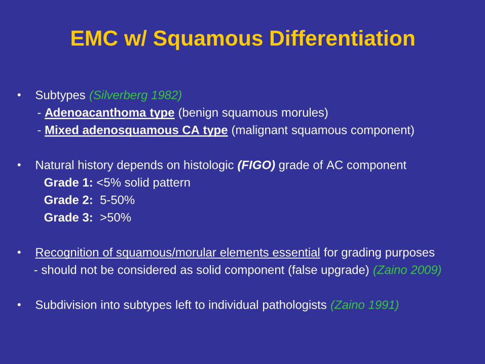
EMC w/ Squamous Differentiation
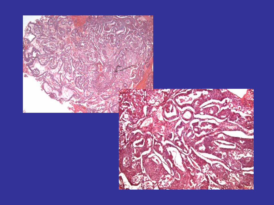
• Subtypes (Silverberg 1982)
- Adenoacanthoma type (benign squamous morules)
- Mixed adenosquamous CA type (malignant squamous component)
• Natural history depends on histologic (FIGO) grade of AC component
Grade 1: <5% solid pattern
Grade 2: 5-50%
Grade 3: >50%
• Recognition of squamous/morular elements essential for grading purposes
- should not be considered as solid component (false upgrade) (Zaino 2009)
• Subdivision into subtypes left to individual pathologists (Zaino 1991)
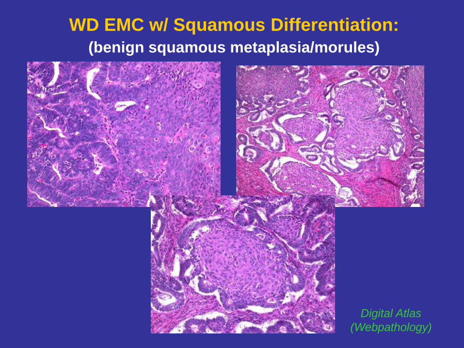
WD EMC w/ Squamous Differentiation:
(benign squamous metaplasia/morules)
Digital Atlas
(Webpathology)
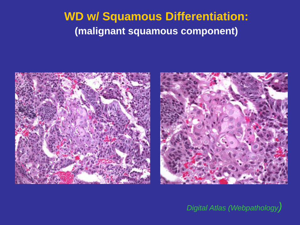
WD w/ Squamous Differentiation:
(malignant squamous component)
Digital Atlas (Webpathology)
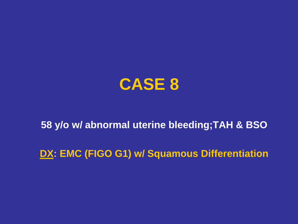
CASE 8
58 y/o w/ abnormal uterine bleeding;TAH & BSO
DX: EMC (FIGO G1) w/ Squamous Differentiation
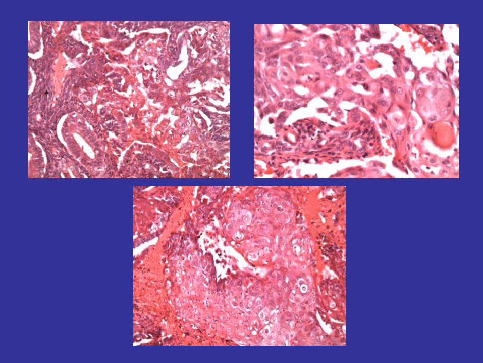
LG EMC Associated w/
Undifferentiated Carcinoma
A New Type of
De-Differentiated Carcinoma?
Undifferentiated CA
• HG/aggressive CA of endometrium (9% of all endometrial CAs)
• Under-recognized and poorly studied (frequently Dx as G3 EMC)
• Recognition extremely important when associated w/ G1/G2 EMC
• Important to Dx solid areas as UD & avoid evaluation them as
solid component of EMC (glandular elements within solid component)
• Lack of recognition of UDCA as part of mixed CA w/ ass. EMC
can lead to misclassification of the tumor as EMC G2/G3:
UDCA + G1 EMC: FIGO G2 EMC
UDCA + G2 EMC: FIGO G3 EMC
• Significant difference in biologic behavior/ clinical outcome:
- G2 EMC: excellent Px
- G3 EMC: IM Px
- UDCA: poor Px (irrespective of the extent of LG component)
UDCA ass. w/ differentiated areas = DDCA
Silva 2006, Silva 2007, Tafe 2010
Dedifferentiated CA
• UDCA (Silverberg 2003, WHO classification):
- neoplasm lacking any evidence of differentiation
• DDCA (Silva 2007)
- UDCA ass. w/ differentiated areas (recently described entity)
- UDCA related to FIGO G1/G2 EMC either in primary tumor or recurrence
- reflects de-differentiation (Silva 2006)
Importance of recognition of UD component from clinical behavior standpoint
UDCA/ DDCA: (Altrabulsi 2005)
- 54% presented at advanced stage/ may pursue fulminant clinical course
- did not respond to conventional CTX regimen used for EMC
- 75% died of the disease
FIGO G3 EMC:
- presented w/ advanced stage in only 30%
- 30% died of the disease
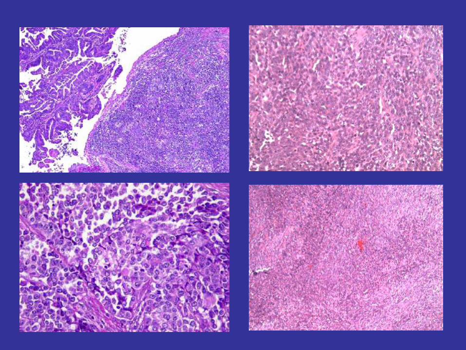
DDCA: Histomorphology • Gross features: nonspecific (white & fleshy w/ foci of necrosis)
• Microscopic features:
- LG (WD/MD) EMC juxtaposed with distinct UDCA
- usually look biphasic with abrupt transition/ necrotic foci
- solid sheets of dyshesive monotonous round to oval cells (+/- pleom)
- enlarged nuclei, occasional prominent nucleoli, mitoses 10/10hpf
- D/Dx: EMC G2/3 - intermixed glandular & solid/ similar cell type
• IHC profile:
- inconsistent expression of epithelial markers (HG EMC: strong/diffuse)
- heterogeneous staining for keratins: (-)/ weakly (+)/ focal (+++)
(in 80-90% cases only 5-10% tumor cells panCK+)
- variable expression of EMA, Vimentin, ER/PR
- only focal (<10%) expression NE markers (40% cases)
• Association w/ defects in DNA mismatch repair system,
including Lynch syndrome (Garg 2011)
Altrabulsi 2005, Silva 2006, Silva 2007, Ferguson 2007, Tafe 2010, Giordano 2011
CASE 9
85 y/o w/ working Dx of Endometrial CA;
TAH & BSO
DX: De-differentiated CA
(LG EMC Associated w/ UDCA)
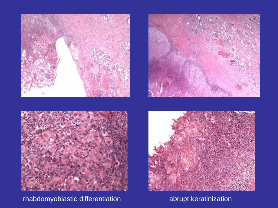
rhabdomyoblastic differentiation abrupt keratinization
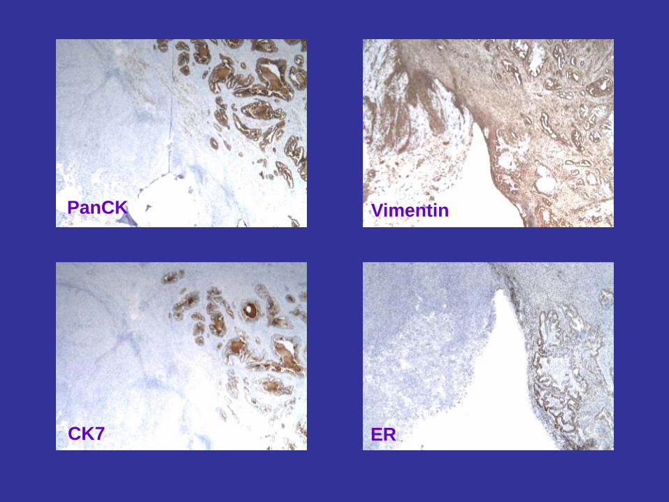
PanCK
CK7
Vimentin
ER

KENTUCKY HORSE FARM
4. HG Cervical Intraepithelial Lesions
(CIN 2+)
How to Avoid Over- & Underdiagnosis
in Everyday Practice
ARE CIN2 LESIONS OVERDIAGNOSED?
Selected Immunopanel Can Prevent
Unnecessary Cervical Excisions
EUROGIN 2010 CONGRESS -
CERVICAL CANCER PREVENTION:
20 Years of Progress and
A Path to the Future
Monte Carlo (Monaco) Feb 17-20, 2010
R. Karabakhtsian1, M.Cibull1, C. DeSimone2 Departments of 1Pathology and 2Gynecologic Oncology
University of KY Medical Center, Lexington, KY, USA

CIN2 Lesions of Uterine Cervix:
Background
• May present diagnostic challenge
• Particularly in small cervical biopsies & focal disease
• Can be mimicked by atypical cellular changes
• Recognized interobserver variability
• Will prompt aggressive clinical management
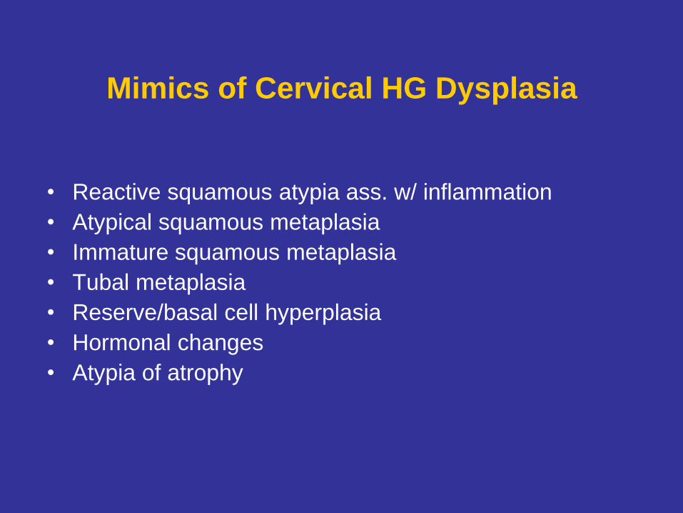
Mimics of Cervical HG Dysplasia
• Reactive squamous atypia ass. w/ inflammation
• Atypical squamous metaplasia
• Immature squamous metaplasia
• Tubal metaplasia
• Reserve/basal cell hyperplasia
• Hormonal changes
• Atypia of atrophy
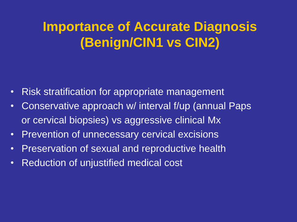
Importance of Accurate Diagnosis
(Benign/CIN1 vs CIN2)
• Risk stratification for appropriate management
• Conservative approach w/ interval f/up (annual Paps
or cervical biopsies) vs aggressive clinical Mx
• Prevention of unnecessary cervical excisions
• Preservation of sexual and reproductive health
• Reduction of unjustified medical cost
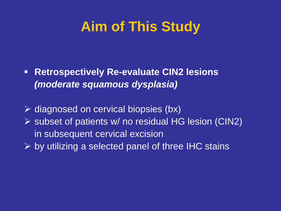
Aim of This Study
Retrospectively Re-evaluate CIN2 lesions
(moderate squamous dysplasia)
diagnosed on cervical biopsies (bx)
subset of patients w/ no residual HG lesion (CIN2)
in subsequent cervical excision
by utilizing a selected panel of three IHC stains
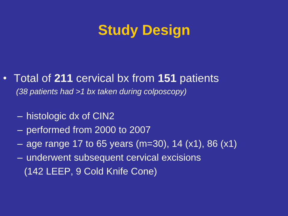
Study Design
• Total of 211 cervical bx from 151 patients (38 patients had >1 bx taken during colposcopy)
– histologic dx of CIN2
– performed from 2000 to 2007
– age range 17 to 65 years (m=30), 14 (x1), 86 (x1)
– underwent subsequent cervical excisions
(142 LEEP, 9 Cold Knife Cone)
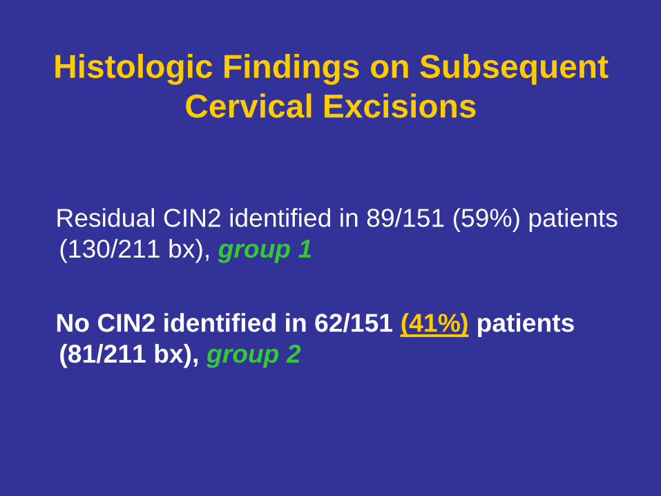
Histologic Findings on Subsequent
Cervical Excisions
Residual CIN2 identified in 89/151 (59%) patients
(130/211 bx), group 1
No CIN2 identified in 62/151 (41%) patients
(81/211 bx), group 2
Study Design (cont’d)
• Tissue blocks of 72/81 cervical biopsies from 56/62 patients (group 2) were stained for three IHC markers
• Remaining 9 biopsies from 6 patients did not have sufficient tissue for IHC work-up
• All H&E and immunostained slides were independently reviewed by two pathologists
Commercial Antibodies Used for
IHC Work-Up
• P16INK4a (CINtecR Histology Kit, MTM Labs)
• MIB-1 (DAKO, Carpinteria, CA, USA)
• ProExTMC (BD Diagnostics)
About Markers
• P16INK4a (P16) - surrogate marker for an activated oncogene
expression of HR-HPV in dysplastic cells
(biomarker of cervical dysplasia)
(major etiologic agent for cervical ca)
• MIB-1 (Ki-67) - mAB to nuclear AG Ki-67 present in
proliferating cells (proliferation marker)
(complimentary surrogate biomarker for
HPV-related squamous dysplasia)
• ProExTMC - ABs against proteins associated w/ aberrant
S-phase cell-cycle induction (MCM2 & TOP2A)
(proliferation marker)
(complimentary surrogate biomarker)
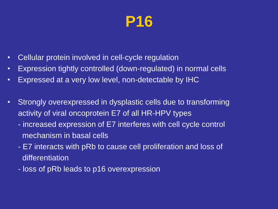
P16
• Cellular protein involved in cell-cycle regulation
• Expression tightly controlled (down-regulated) in normal cells
• Expressed at a very low level, non-detectable by IHC
• Strongly overexpressed in dysplastic cells due to transforming
activity of viral oncoprotein E7 of all HR-HPV types
- increased expression of E7 interferes with cell cycle control
mechanism in basal cells
- E7 interacts with pRb to cause cell proliferation and loss of
differentiation
- loss of pRb leads to p16 overexpression
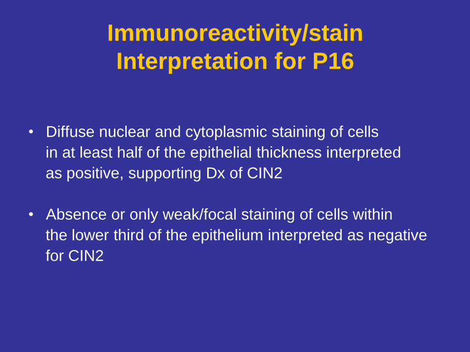
Immunoreactivity/stain
Interpretation for P16
• Diffuse nuclear and cytoplasmic staining of cells
in at least half of the epithelial thickness interpreted
as positive, supporting Dx of CIN2
• Absence or only weak/focal staining of cells within
the lower third of the epithelium interpreted as negative
for CIN2
Immunoreactivity/stain
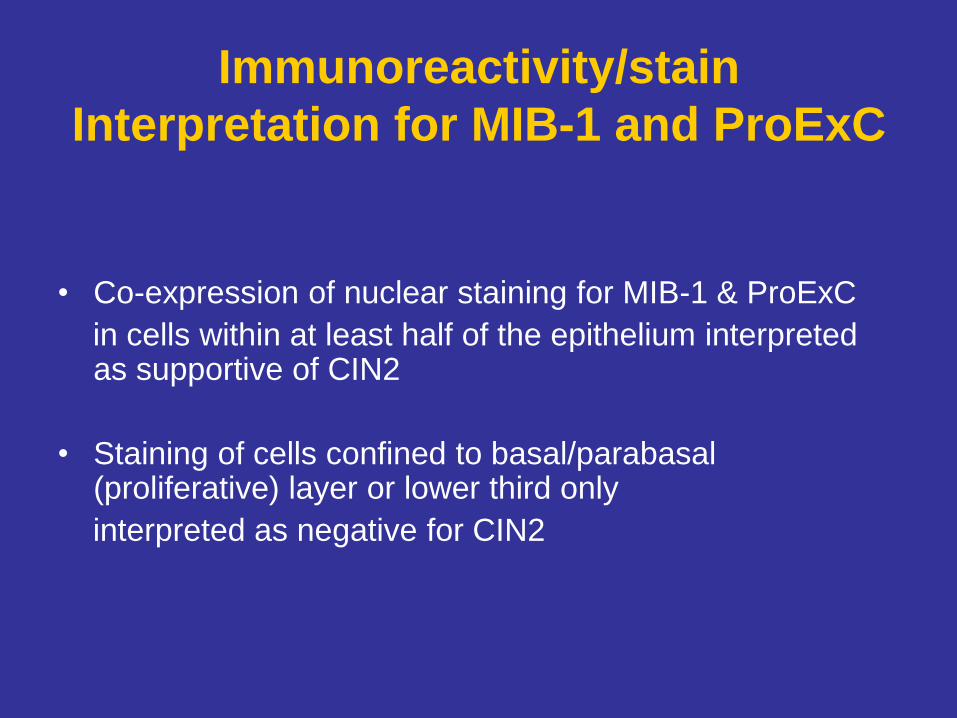
Interpretation for MIB-1 and ProExC
• Co-expression of nuclear staining for MIB-1 & ProExC
in cells within at least half of the epithelium interpreted as supportive of CIN2
• Staining of cells confined to basal/parabasal (proliferative) layer or lower third only
interpreted as negative for CIN2
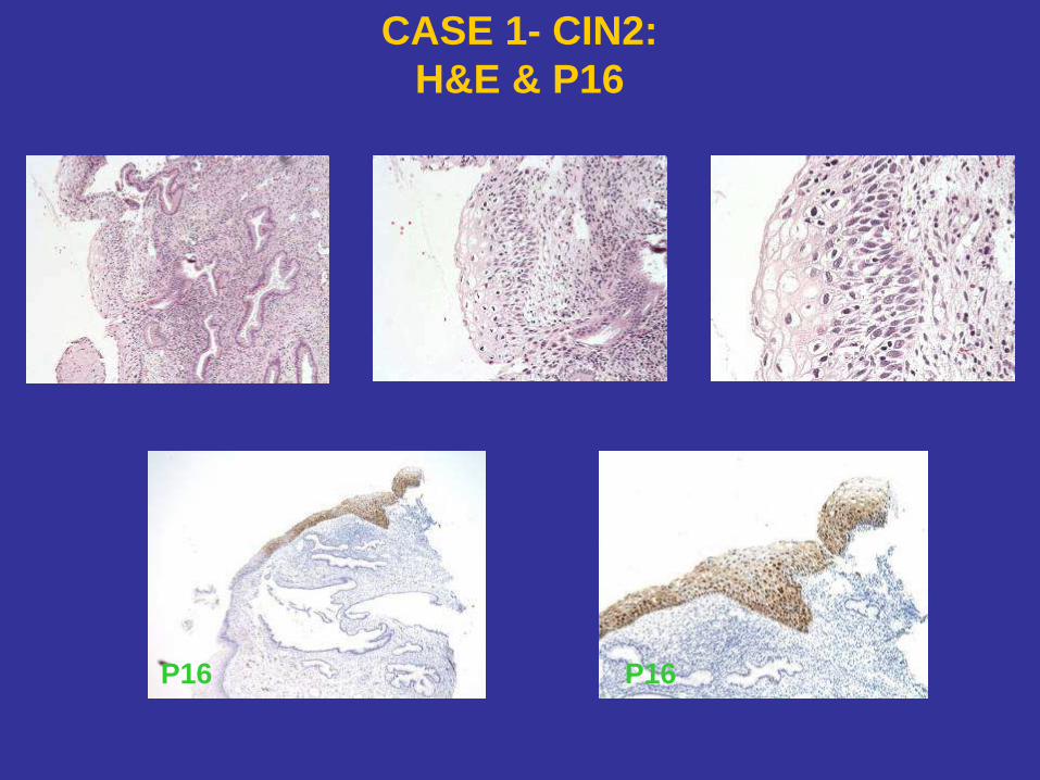
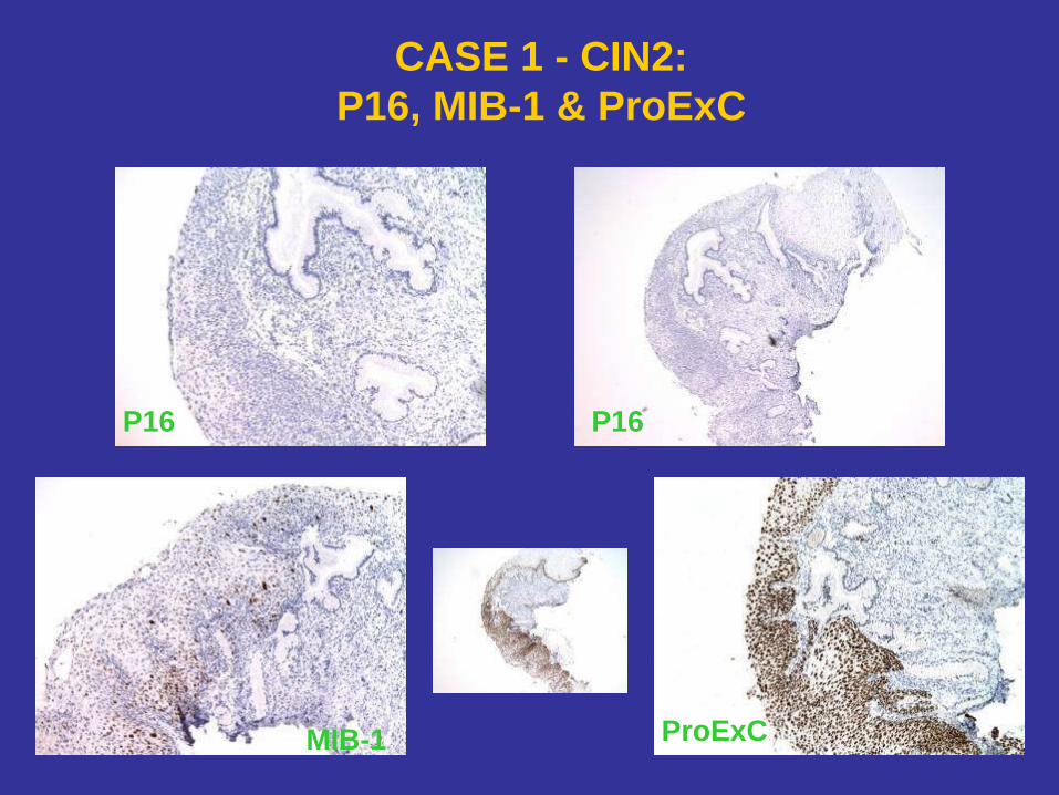
CASE 1- CIN2:
H&E & P16
P16 P16
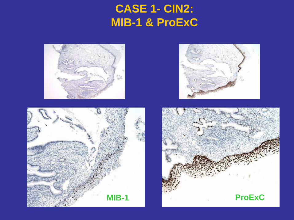
CASE 1- CIN2:
MIB-1 & ProExC
MIB-1 ProExC
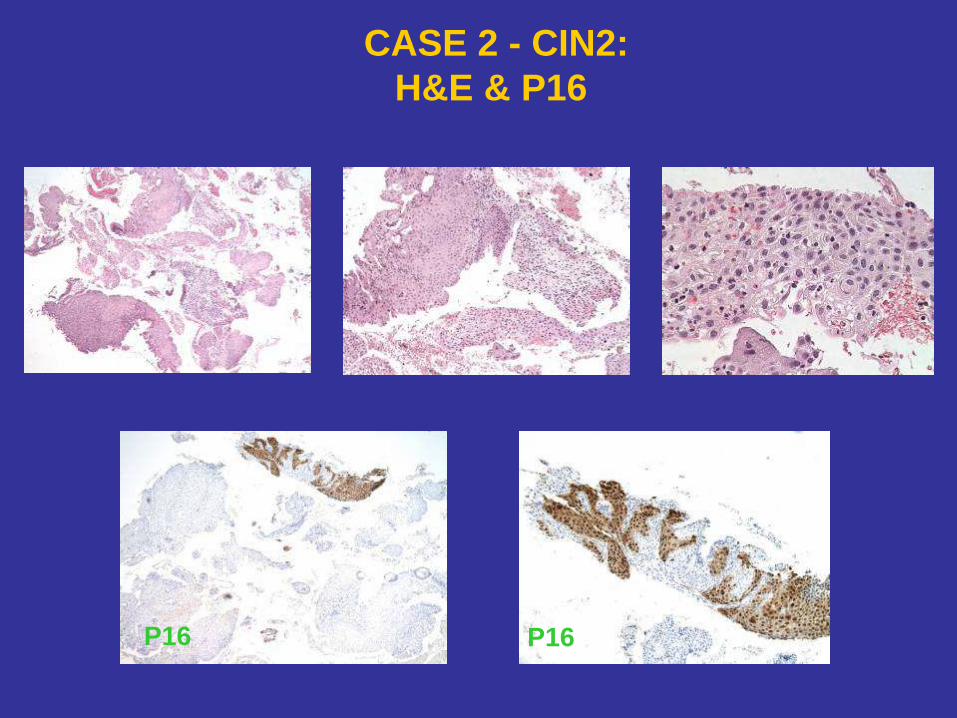
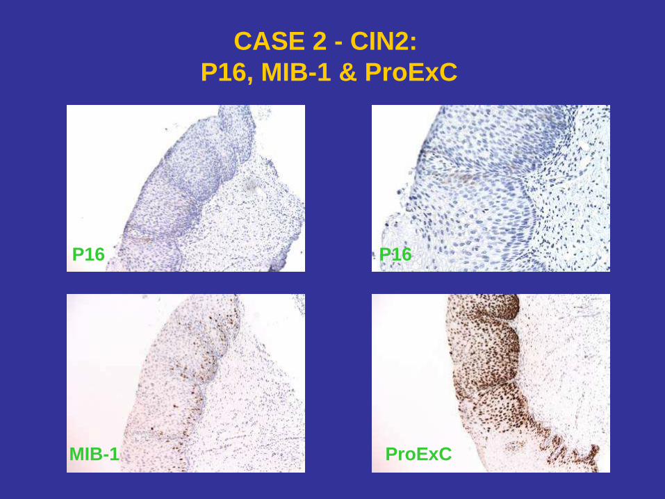
CASE 2 - CIN2:
H&E & P16
P16 P16
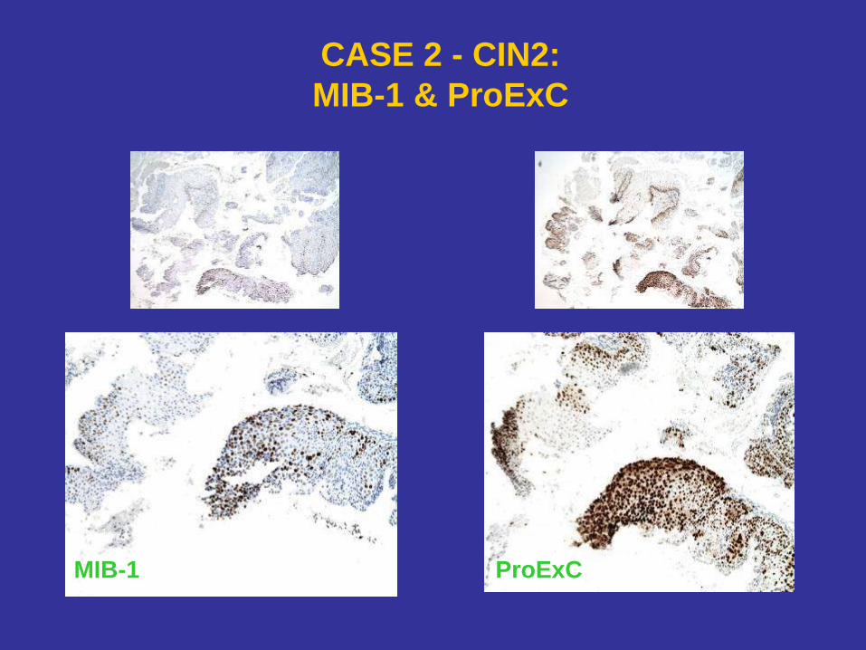
CASE 2 - CIN2:
MIB-1 & ProExC
MIB-1 ProExC
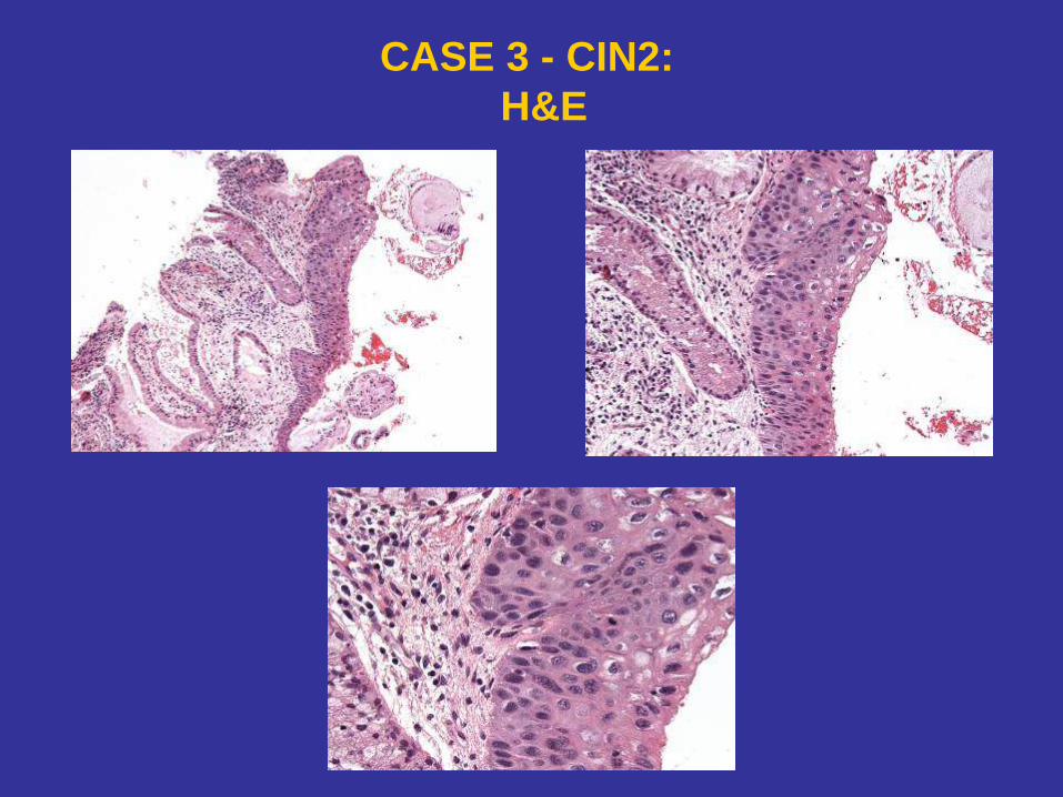
CASE 3 - CIN2:
H&E
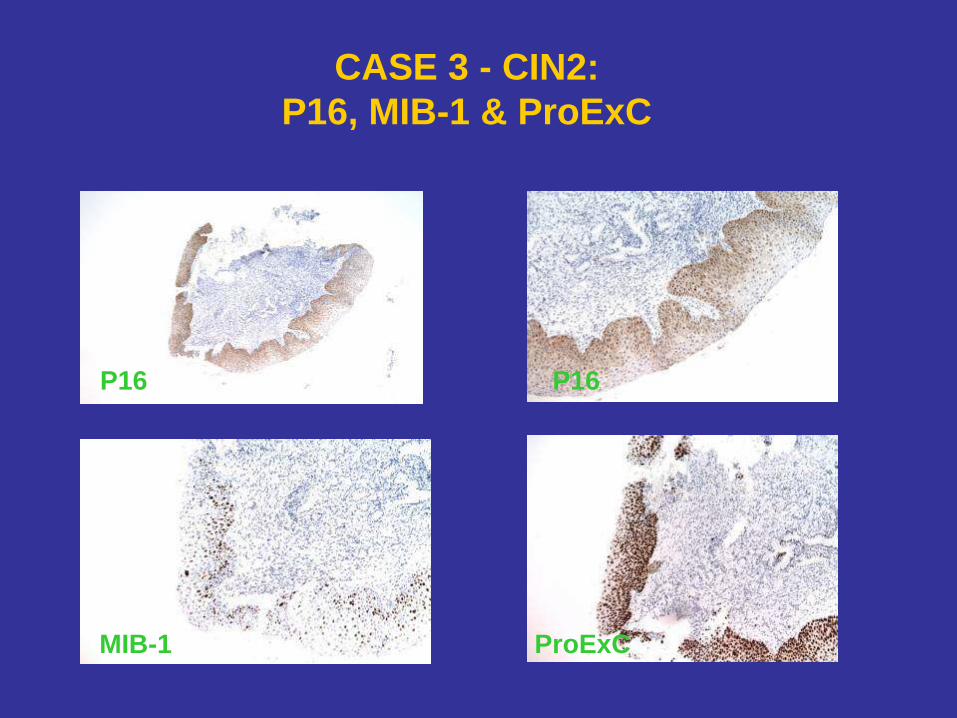
CASE 3 - CIN2:
P16, MIB-1 & ProExC
ProExC
MIB-1
P16 P16
CASE 4 - CIN2:
P16, MIB-1 & ProExC
P16 MIB-1 ProExC
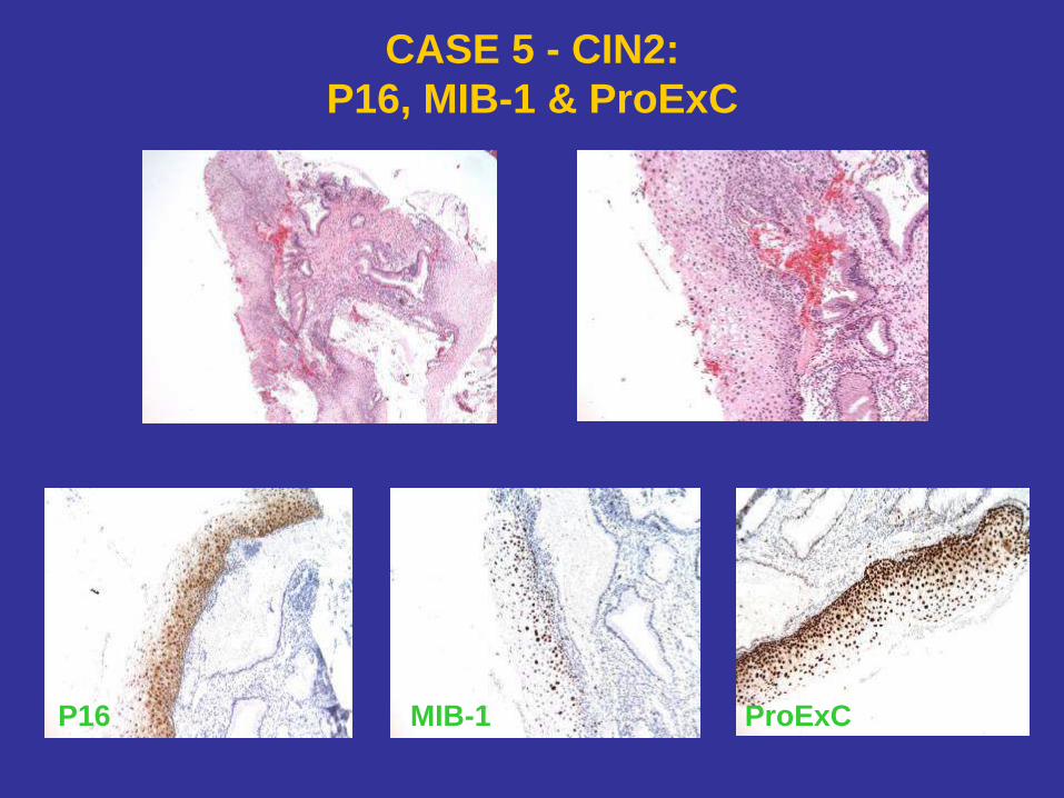
CASE 5 - CIN2:
P16, MIB-1 & ProExC
P16 MIB-1 ProExC
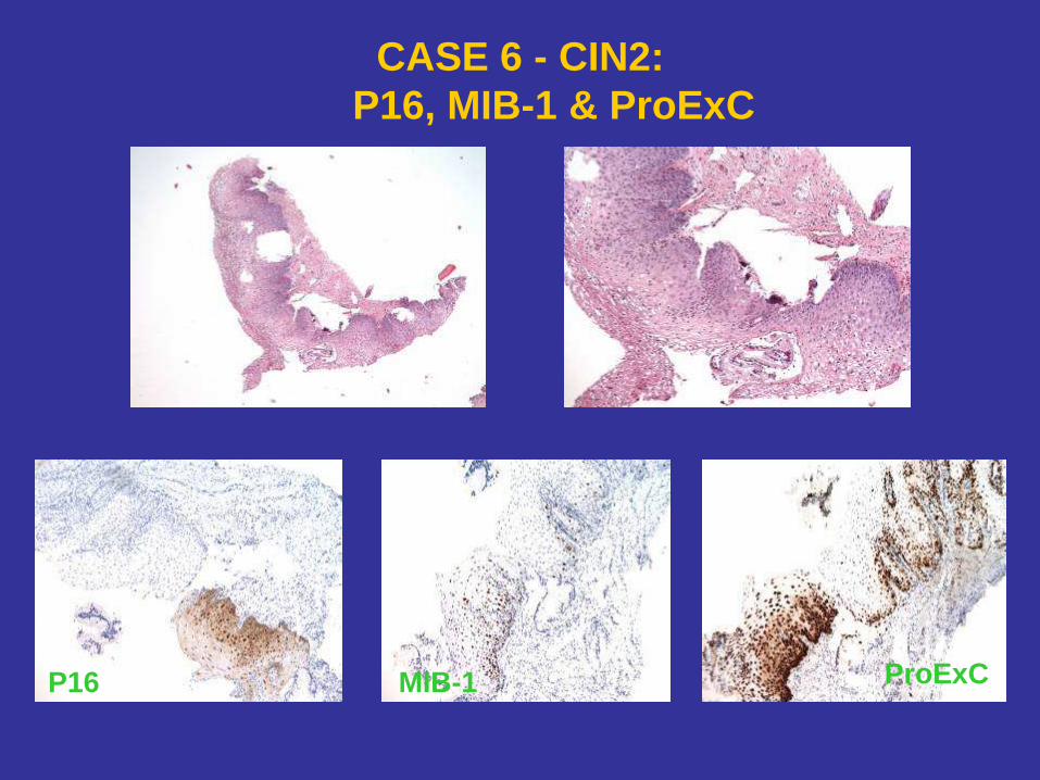
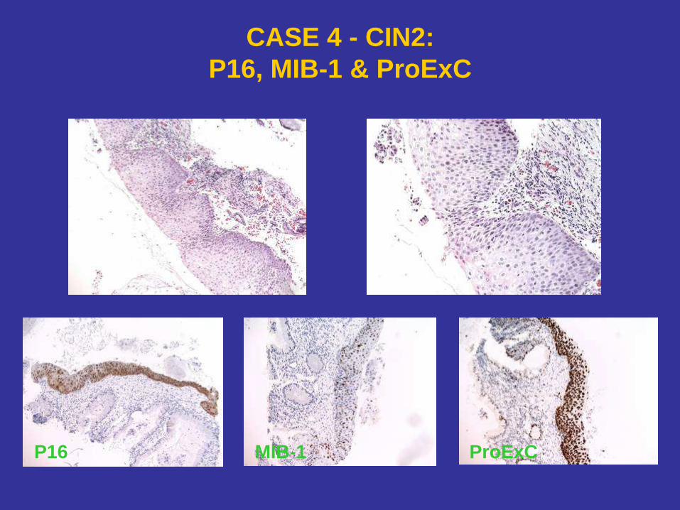
CASE 6 - CIN2:
P16, MIB-1 & ProExC
P16 MIB-1 ProExC
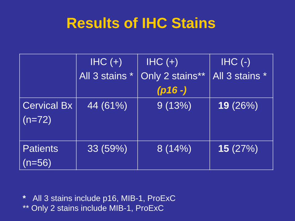
Results of IHC Stains
IHC (+)
All 3 stains *
IHC (+)
Only 2 stains**
(p16 -)
IHC (-)
All 3 stains *
Cervical Bx
(n=72)
44 (61%)
9 (13%)
19 (26%)
Patients
(n=56)
33 (59%) 8 (14%) 15 (27%)
* All 3 stains include p16, MIB-1, ProExC
** Only 2 stains include MIB-1, ProExC
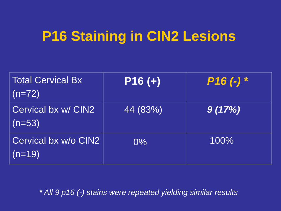
P16 Staining in CIN2 Lesions
Total Cervical Bx
(n=72)
P16 (+) P16 (-) *
Cervical bx w/ CIN2
(n=53)
44 (83%) 9 (17%)
Cervical bx w/o CIN2
(n=19)
0% 100%
* All 9 p16 (-) stains were repeated yielding similar results
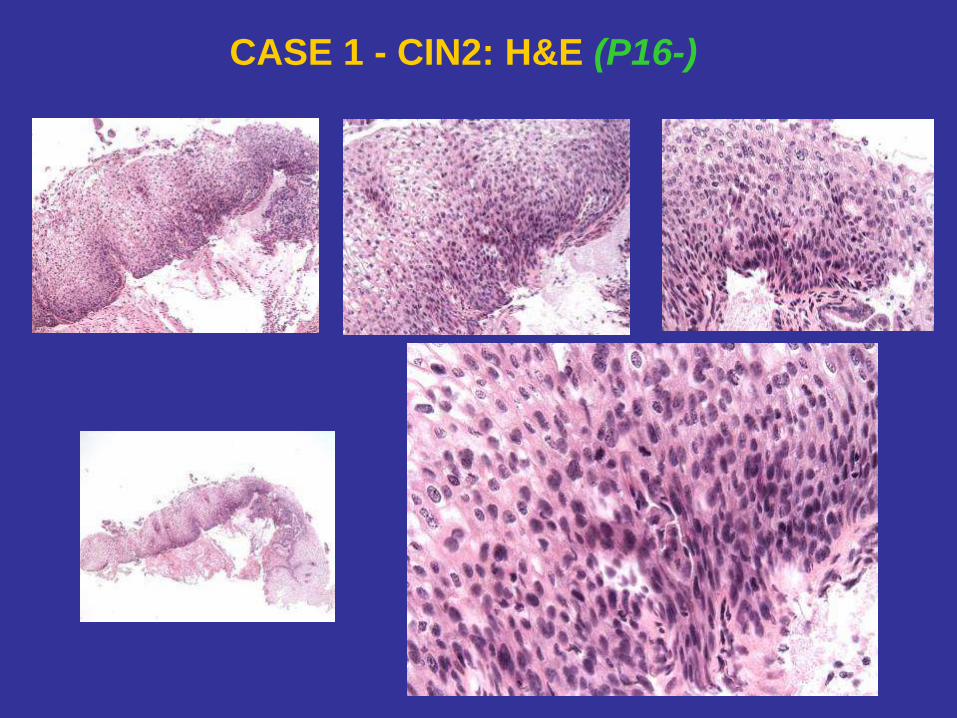
CASE 1 - CIN2: H&E (P16-)

CASE 1 - CIN2:
P16, MIB-1 & ProExC
ProExC MIB-1
P16 P16
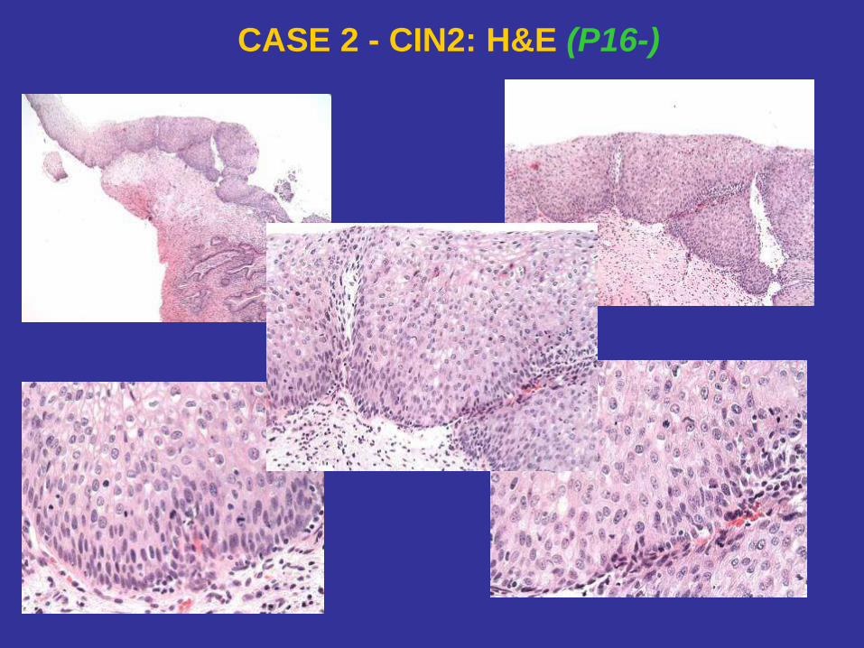
CASE 2 - CIN2: H&E (P16-)

P16 Staining in CIN2 Lesions
Total Cervical Bx
(n=72)
P16 (+) P16 (-) *
Cervical bx w/ CIN2
(n=53)
44 (83%) 9 (17%)
Cervical bx w/o CIN2
(n=19)
0% 100%
* All 9 p16 (-) stains were repeated yielding similar results

CASE 1 - CIN2: H&E (P16-)
CASE 1 - CIN2:
P16, MIB-1 & ProExC
ProExC MIB-1
P16 P16

CASE 2 - CIN2: H&E (P16-)
CASE 2 - CIN2:
P16, MIB-1 & ProExC
P16 P16
MIB-1 ProExC
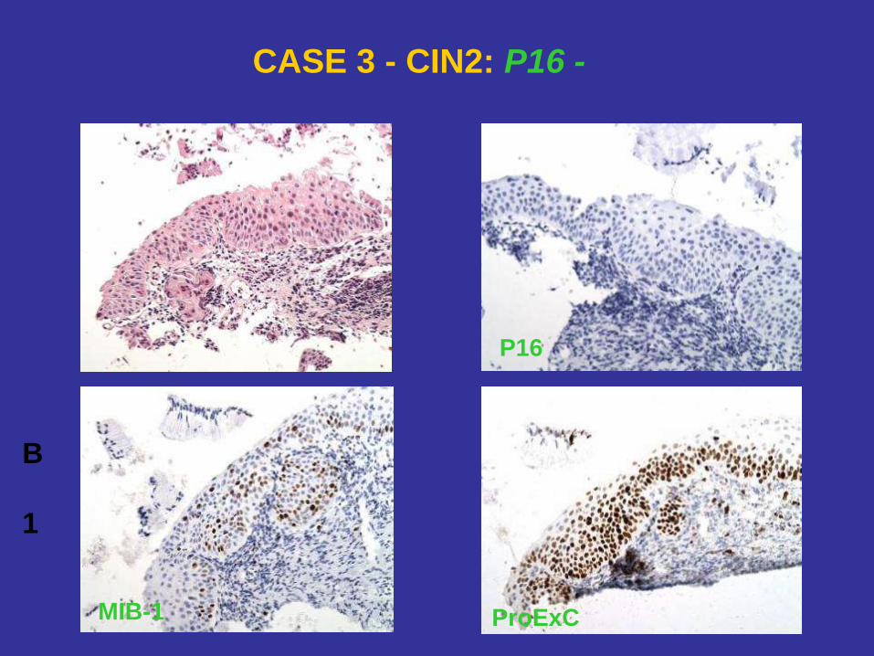
CASE 3 - CIN2: P16 -
B
1
ProExC
P16
MIB-1
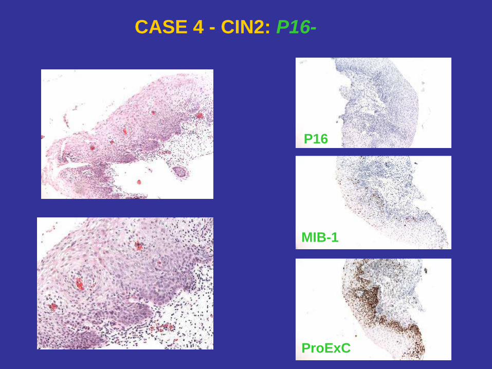
CASE 4 - CIN2: P16-
P16
MIB-1
ProExC
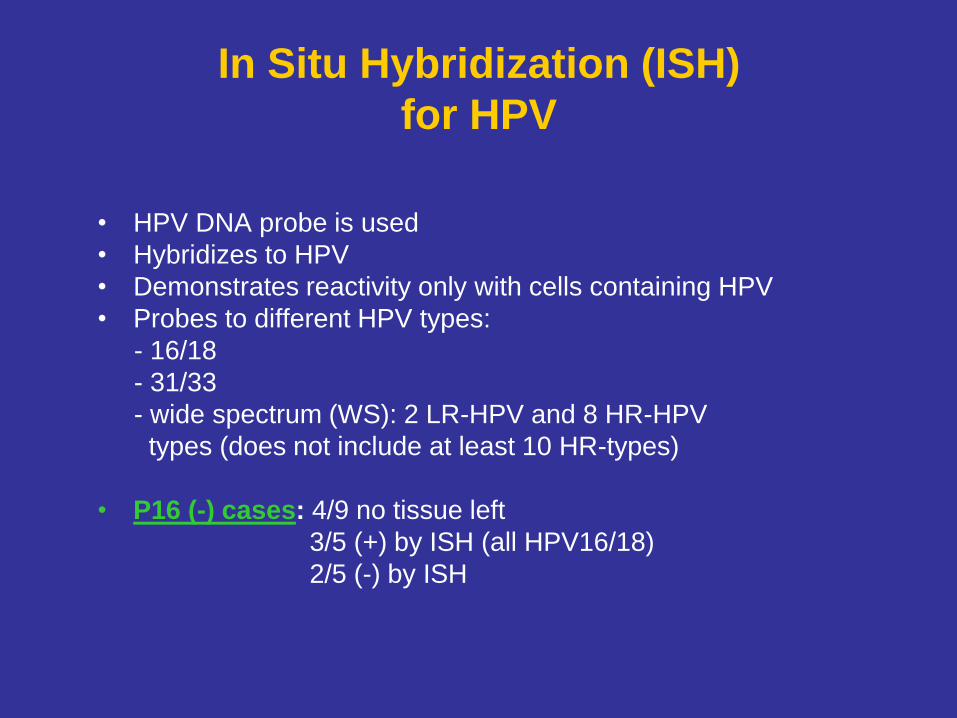
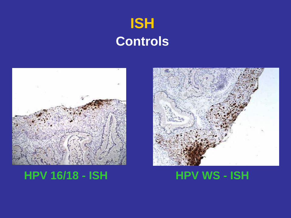
In Situ Hybridization (ISH)
for HPV
• HPV DNA probe is used
• Hybridizes to HPV
• Demonstrates reactivity only with cells containing HPV
• Probes to different HPV types:
- 16/18
- 31/33
- wide spectrum (WS): 2 LR-HPV and 8 HR-HPV
types (does not include at least 10 HR-types)
• P16 (-) cases: 4/9 no tissue left
3/5 (+) by ISH (all HPV16/18)
2/5 (-) by ISH
ISH
Controls
HPV 16/18 - ISH HPV WS - ISH
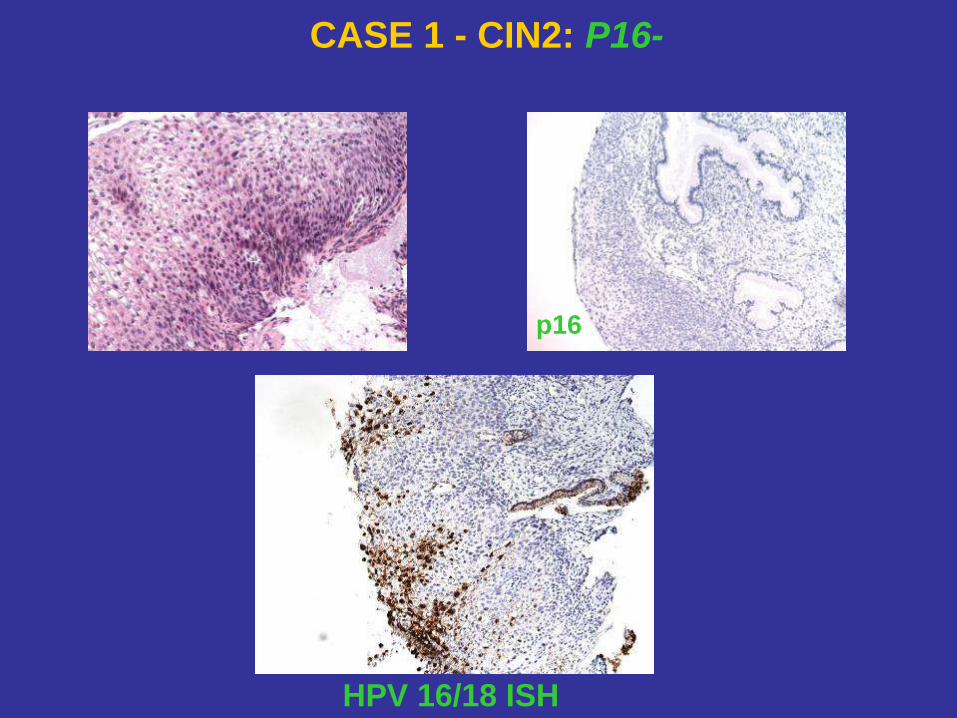
CASE 1 - CIN2: P16-
HPV 16/18 ISH
p16
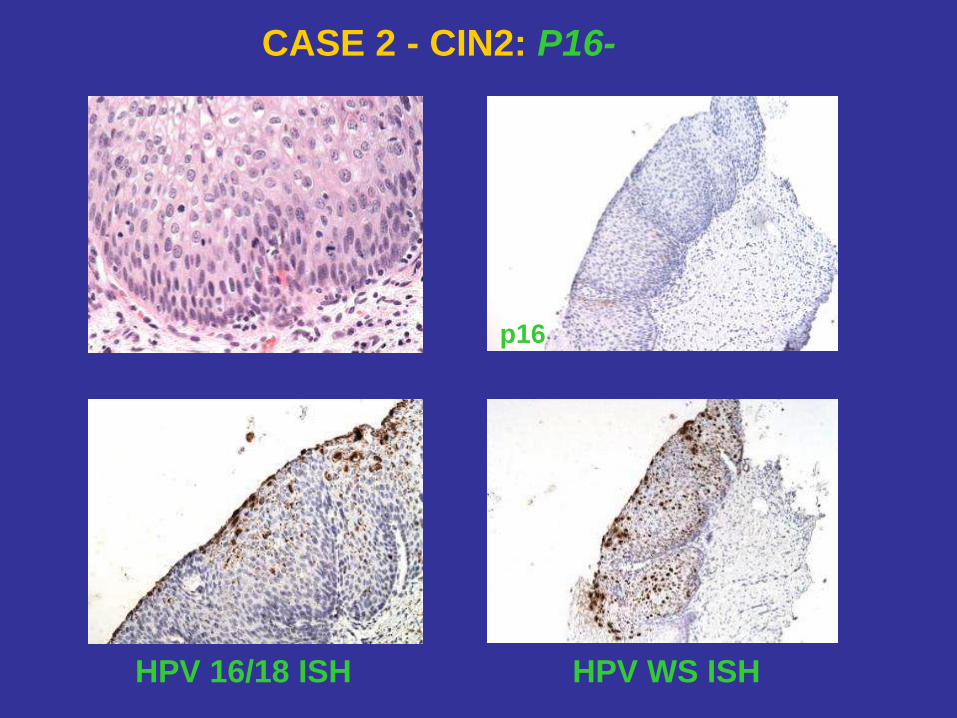
CASE 2 - CIN2: P16-
HPV 16/18 ISH HPV WS ISH
p16
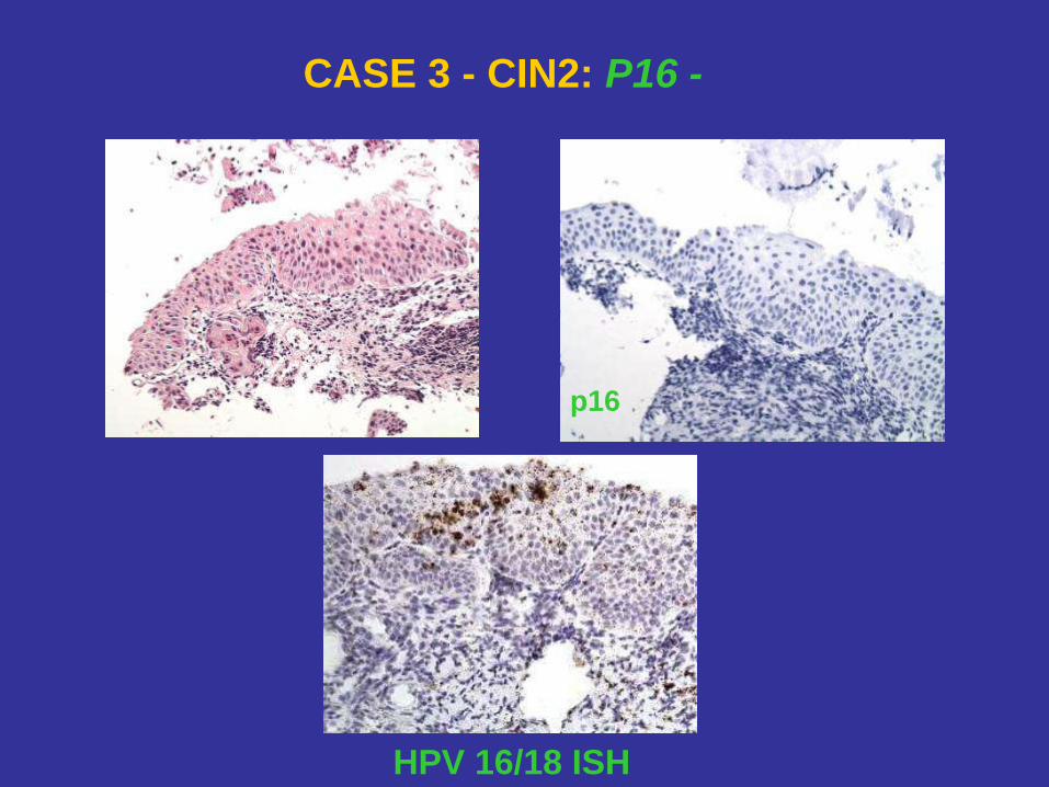
CASE 3 - CIN2: P16 -
HPV 16/18 ISH
p16
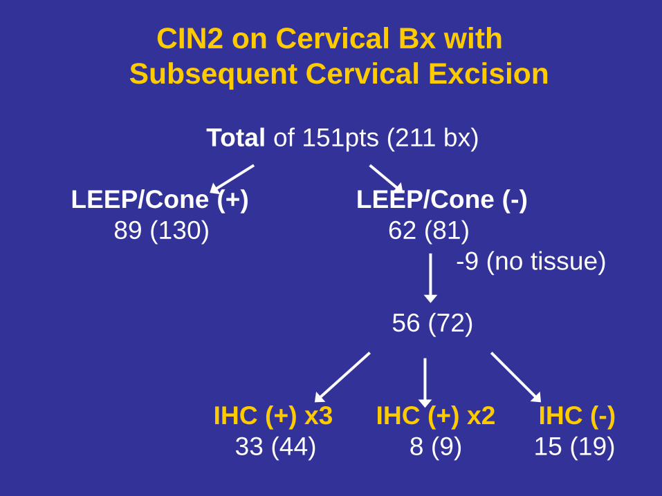
CIN2 on Cervical Bx with
Subsequent Cervical Excision
Total of 151pts (211 bx)
LEEP/Cone (+) LEEP/Cone (-)
89 (130) 62 (81)
-9 (no tissue)
56 (72)
IHC (+) x3 IHC (+) x2 IHC (-)
33 (44) 8 (9) 15 (19)
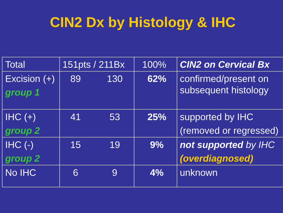
CIN2 Dx by Histology & IHC
Total 151pts / 211Bx 100% CIN2 on Cervical Bx
Excision (+)
group 1
89 130 62% confirmed/present on
subsequent histology
IHC (+)
group 2
41 53 25% supported by IHC
(removed or regressed)
IHC (-)
group 2
15 19 9% not supported by IHC
(overdiagnosed)
No IHC 6 9 4% unknown
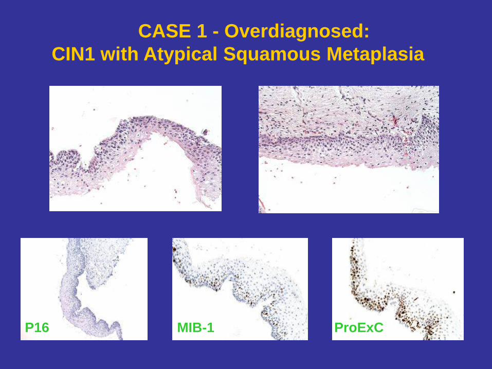
CASE 1 - Overdiagnosed:
CIN1 with Atypical Squamous Metaplasia
P16 MIB-1 ProExC
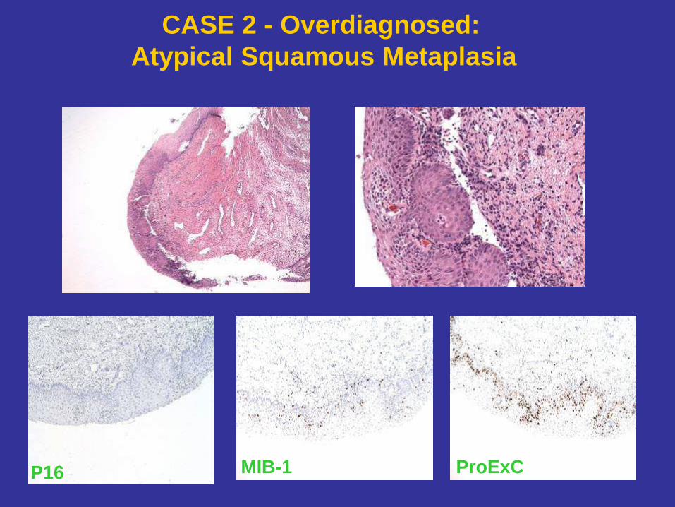
CASE 2 - Overdiagnosed:
Atypical Squamous Metaplasia
P16 MIB-1 ProExC
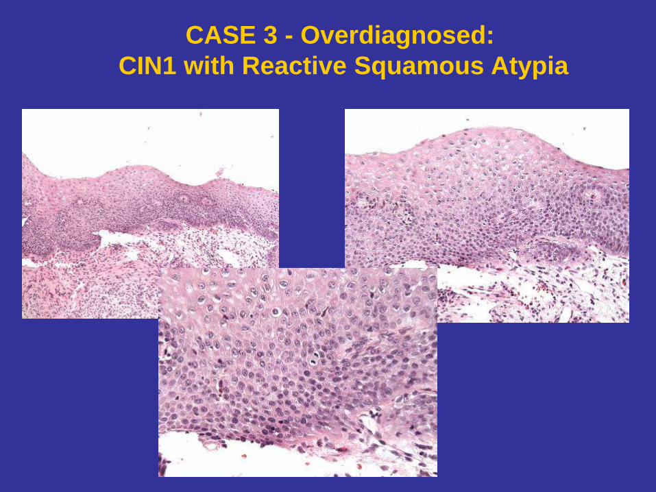
CASE 3 - Overdiagnosed:
CIN1 with Reactive Squamous Atypia
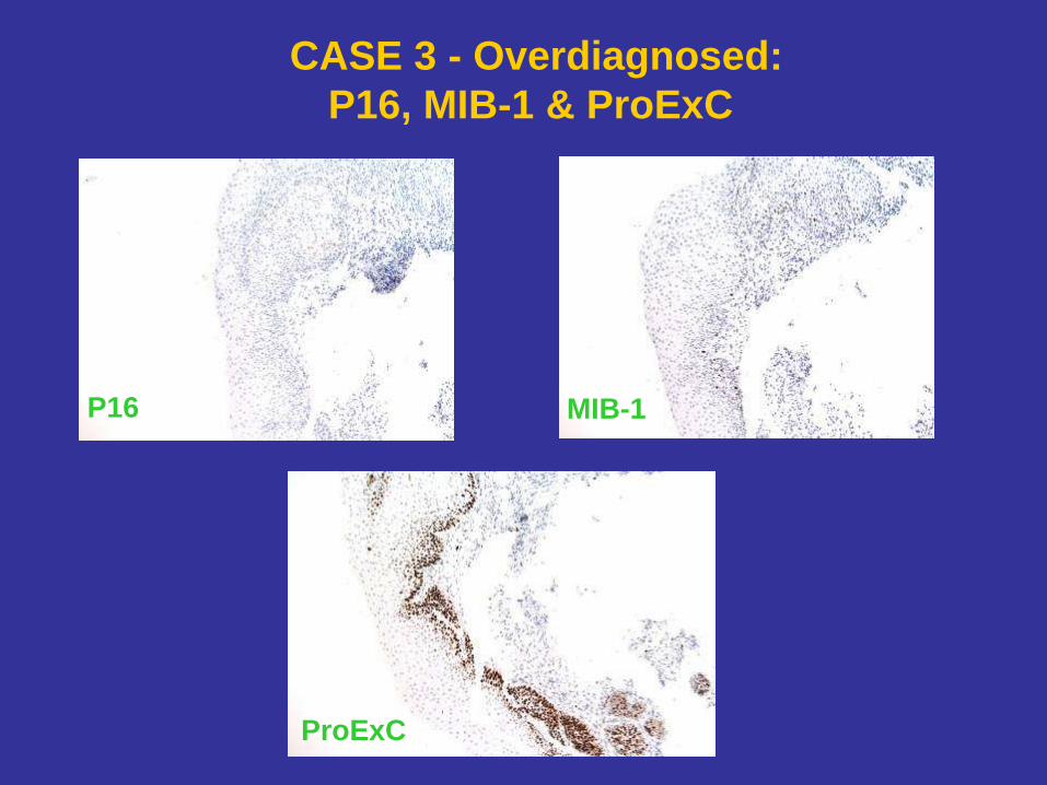
CASE 3 - Overdiagnosed:
P16, MIB-1 & ProExC
P16 MIB-1
ProExC

CASE 4 - Overdiagnosed:
Atypical Immature Metaplasia
P16 MIB-1 ProExC
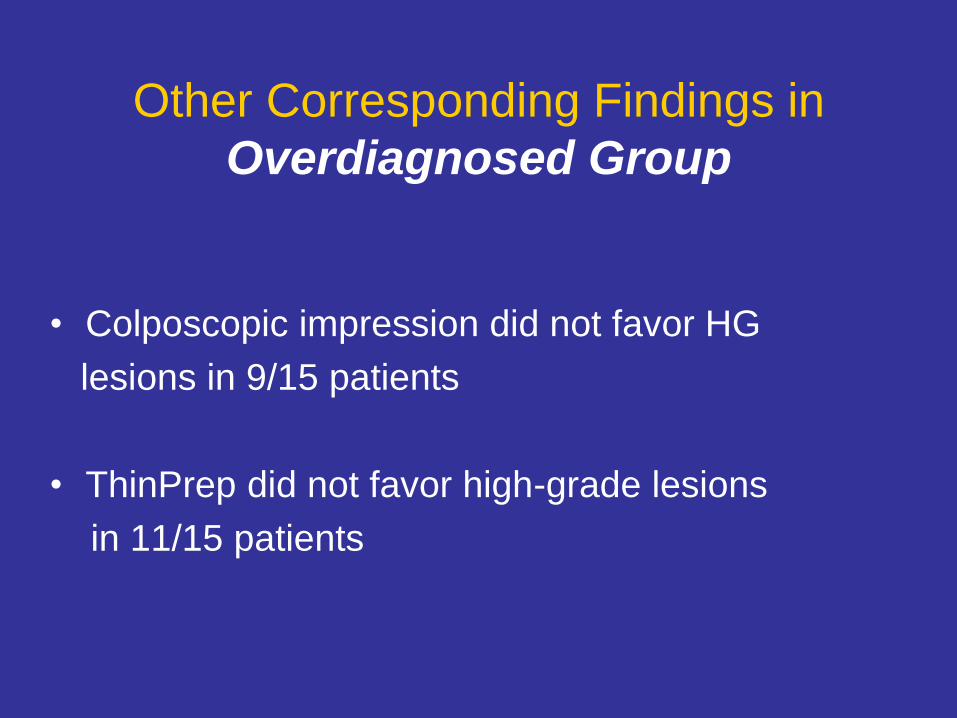
Other Corresponding Findings in
Overdiagnosed Group
• Colposcopic impression did not favor HG
lesions in 9/15 patients
• ThinPrep did not favor high-grade lesions
in 11/15 patients
Conclusions
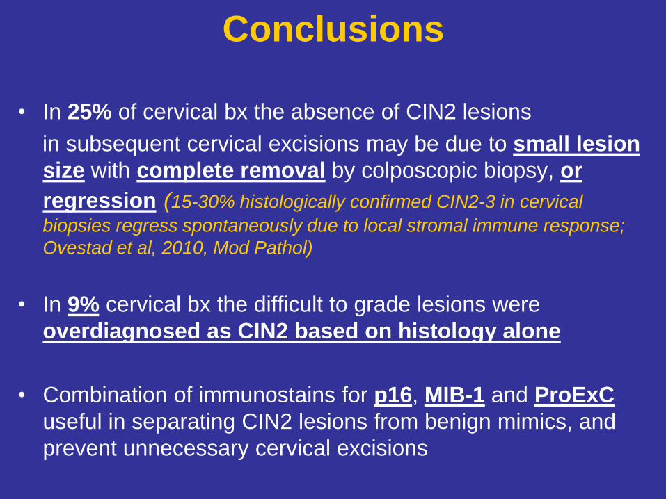
• In 25% of cervical bx the absence of CIN2 lesions
in subsequent cervical excisions may be due to small lesion
size with complete removal by colposcopic biopsy, or
regression (15-30% histologically confirmed CIN2-3 in cervical
biopsies regress spontaneously due to local stromal immune response;
Ovestad et al, 2010, Mod Pathol)
• In 9% cervical bx the difficult to grade lesions were
overdiagnosed as CIN2 based on histology alone
• Combination of immunostains for p16, MIB-1 and ProExC
useful in separating CIN2 lesions from benign mimics, and
prevent unnecessary cervical excisions
Conclusions
• Immunostaining patterns for proliferation markers appear to vary somewhat in distribution and intensity; scattered & dispersed pattern for MIB-1 vs
diffuse & intense staining for ProExC
• In rare p16 (-) cases co-expression of MIB-1 and ProExC can be helpful in supporting Dx of CIN2
• Caution should be exercised when interpreting MIB-1 and ProExC in a setting of associated inflammation, low clinical and histologic suspicion for HG dysplasia, and in the absence of p16 reactivity since findings may reflect reactive changes to inflammation
Conclusions
• P16 over-expression was present in 83%, and absent in 17% of CIN2 lesions in this study (compatible with Dr. Stoler’s UVA group study at 87%, Am J Surg Pathol, August 2010)
• 3/5 P16 (-) cases positive with in-situ hybridization (ISH) for HPV types 16/18
• While not extremely sensitive for CIN2 lesions (83%), p16 appears to be highly specific “key” biomarker for HR-HPV
ass. cervical disease, critical when dealing w/ Dx challenges, contributes greatly to accuracy of CIN2 Dx
• Whether absence of p16 over-expression in small subset of CIN2 lesions reflects less potential for disease progression remains to be further investigated
References
• Altrabulsi B, Malpica A, Deavers MT, Bodurka DC, Broaddus R, Silva EG. Undifferentiated carcinoma of the endometrium. Am J Surg Pathol 2005;29:1316-21.
• Amant F, Cadron I, Fuso L, et al. Endometrial carcinosarcomas have a different prognosis and pattern of spread compared to high-risk epithelial endometrial cancer. Gynecol Oncol 2005;98:274-80.
• Bell DA, Scully RE. Benign and borderline clear cell adenofibromas of the ovary. Cancer 1985;56:2922-31.
• Bell DA, Longacre TA, Prat J, et al. Serous Borderline (Low Malignant Potential, Atypical Proliferative) Ovarian Tumors: Workshop Perspectives. Hum Pathol 2004;35(8):28-42.
• Bell DA, Scully RE. Ovarian serous borderline tumors with stromal microinvasion: a report of 21 cases. Hum Pathol 1990;21:397-403.
• Bell KA, Smith Sehdev AE, Kurman RJ: Refined diagnostic criteria for implants associated with ovarian atypical proliferative serous tumors (borderline) and micropapillary serous carcinomas. Am J Surg Pathol 2001;25:419-32.
• Bell DA, Weinstock MA, Scully RE. Peritoneal implants of ovarian serous borderline tumors. Histologic features and prognosis. Cancer 1988;62:2212-22.
• Borderline Ovarian Tumors: A Web-Based Atlas. NCI Borderline Ovarian Tumors Workshop. Bethesda, MD, August 27-28, 2003.
• Bonome T, Lee JY, Park DC, et al. Expression profiling of serous low malignant potential, low-grade, and high-grade tumors of the ovary. Cancer Res 2005; 65: 10602-12.
• Burks RT, Sherman ME, Kurman RJ. Micropapillary serous carcinoma of the ovary. A distinctive low-grade carcinoma related to serous borderline tumors.
Am J Surg Pathol 1996;20:1319-30.
References
• Carcangiu ML, Chambers JT. Uterine papillary serous carcinoma: a study of 108
cases with emphasis on the prognostic significance of associated endometrioid
carcinoma, absence of invasion, and concomitant ovarian carcinoma. Gynecol Oncol
1992;47:298-305.
• Clement PB, Young RH, Oliva E, et al: Hyperplastic mesothelial cells within
abdominal lymph nodes: Mimic of metastatic ovarian carcinoma and serous
borderline tumor – A report of two cases associated with ovarian neoplasms. Mod.
Pathol 1996; 9:879-88.
• Darvishian F, Hummer AJ, Thaler HT, et al. Serous endometrial cancers that mimic
endometrioid adenocarcinomas: a clinicopathologic and immunohistochemical study
of a group of problematic cases. Am J Surg Pathol 2004;28:1568-78.
• Dehari R, Kurman RJ, Logani S, Shih IM. The development of high-grade serous
carcinoma from atypical proliferative (borderline) serous tumors and low-grade
micropapillary serous carcinoma: a morphologic and molecular genetic analysis. Am
J Surg Pathol 2007;31:1007-12.
• Eichhorn JH, Young RH. Transitional cell carcinoma of the ovary: A morphologic
study of 100 cases with emphasis on differential diagnosis. Am J Surg Pathol
2004;28:453-63.
• Female Genital Tract – Digital Atlas Index. WebPathology. Last Updated 8/1011.
• Ferguson SE, Tornos K, Hummer A, Barakat RR, Soslow RA. Prognostic Features of
Surgical Stage I Uterine Carcinosarcoma. Am J Surg Pathol 2007;31(11):1653-61.

References
• Galgano MT, Castle PE, Atkins KA, Brix WK, Nassau SR, Stoller MH. Using Biomarkers as Objective Standards in the Diagnosis of Cervical Biopsies. Am J Surg Pathol 2010;34(8):1077-87.
• Garg K, Leitao Jr MM, Wynveen CA, Sica GL, Shia J, Shi W, Soslow RA. P53 overexpression in morphologically ambiguous endometrial carcinomas correlates with adverse clinical outcomes. Mod Pathol 2010;23:80-92.
• Garg K, Soslow RA. Endometrial Undifferentiated Carcinomas. Pathology Case Reviews 2011;16(3):115-8.
• Gilks CB, Vanderhyden BC, Zhu S, van de Rijn M, Longacre TA. Distinction between serous tumors of low malignant potential and serous carcinoma based on global mRNA expression profiling. Gynecol Oncol 2005;96:684-94.
• Gilks CB, Alkushi A, You JJ et al. Advanced-stage serous borderline tumors of the ovary: A clinicopathological study of 49 cases. Int J Gynecol Pathol 2003; 22:29-36.
• Giordano G, D’Adda T, Bottarelli L, Lombardi M, Brigati F, Berretta R, Merisio C. Two cases of low-grade endometrioid carcinoma associated with undifferentiated carcinoma of the uterus (dedifferentiated carcinoma): A molecular study. Pathol. Oncol. Res. Epub. 30 March 2011.
• Hsu CY, Kurman RJ, Vang R, et al. Nuclear size distinguishes low- from high-grade ovarian serous carcinoma and predicts outcome. Hum Pathol 2005;36: 1049-54.
• Kuo KT, Mao TL, Jones S, Veras E, Ayhan A, Wang TL, Glas R, Slamon D, Velculescu VE, Kurman RJ et al. Frequent activating mutations of PIK3CA in ovarian clear cell carcinoma. Am J Pathol 2009;174:1597-601.
• Kurman RJ, Shih IM. The origin and pathogenesis of epithelial ovarian cancer: a proposed unifying theory. Am J Surg Pathol 2010;34:433-43.
• Kurman RJ, Ronnett BM and Vang R. Surface epithelial neoplasms of the ovary:
References New concepts of pathogenesis, diagnostic criteria, and persistent controversies, with
an emphasis on “borderline” tumors. USCAP 2008 Syllabus (Short Course #48).
• Kurman RJ, Shih IM. Molecular pathogenesis and extraovarian origin of epithelial ovarian cancer – Shifting the paradigm. Human Pathology 2011;42:918-31.
• Lee KR, Tavassoli FA, Prat J, et al. Tumors of the ovary and peritoneum: Surface epithelial-stromal tumours, in Tavassoli FA, Devilee P (eds): World Health Organization Classification of Tumors: Pathology and Genetics of Tumours of the Female Genital Organs. Lyon, France, IARC Press, 2003,113-45.
• Lomo L, Nucci MR, Lee KR, et al. Histologic and immunohistochemical decision making in endometrial adenocarcinoma. Mod Pathol 2008;21:937-42.
• Logani S, Oliva E, Amin MB, et al. Immunoprofile of ovarian tumors with putative transitional cell (urothelial) differentiation using novel urothelial markers: Histogenetic and diagnostic implications. Am J Surg Pathol 2003;27:1434-41.
• Longacre TA, Oliva E, Soslow RA. Mesenchymal Neoplasms of the Female Genital Tract. A Clinical and Morphologic Overview Emphasizing Differential Diagnosis and Prognostic Indices. USCAP 97th Annual Meeting (Short Course #21).
• Major FJ, Blessing JA, Silverberg SG, et al. Prognostic factors in early stage uterine sarcoma. A Gynecologic Oncology Group study. Cancer 1993;71(4 suppl):1702-9.
• Malpica A, Deavers MT, Lu K, et al. Grading ovarian serous carcinoma using a two-tier system. Amer J Surg Pathol 2004;28(4):496–504.
• Marquez RT, Baggerly KA, Patterson AP, et al. Patterns of gene expression in different histotypes of epithelial ovarian cancer correlate with those in normal fallopian tube, and colon. Clin Cancer Res 2005;11:6116-26.
• May T, Virtanen C, Sharma M, et al. Low malignant potential tumors with micropapillary features are molecularly similar to low grade serous carcinoma of the ovary. Gynecol Oncol 2010;117:9-17.
References
• McCauhey WTE, Kirk ME, Lester W, et al. Peritoneal epithelial lesions associated with proliferative serous tumors of the ovary. Histopathology 1984;8:195-208.
• McCluggage WG. Uterine carcinosarcomas (malignant mixed mullerian tumors) are metaplastic carcinomas. Int J Gynecol Cancer 2002;12:687-90.
• Meinhold-Heerlein I, Bauerschlag D, Hilpert F, et al. Molecular and prognostic distinction between serous ovarian carcinomas of varying grade and malignant potential. Oncogene 2005;24:1053-65.
• Murray SK, Clement PB, Young RH. Endometrioid carcinomas of the uterine corpus with sex cord-like formations, hyalinization, and other unusual morphologic features: a report of 31 cases of a neoplasm that may be confused with carcinosarcoma and other uterine neoplasms. Am J Surg Pathol 2005;29:157-66.
• Ovestad IT, Gudlaugsson E, Skaland I, Malpica A, Kruse AJ, Janssen EAM, Baak JPA. Local immune response in the microenvironment of CIN2-3 with and without spontaneous regression. Mod Pathol 2010;23:1231-40.
• Pecorelli S, Benedet JL, Creasman WT, et al. FIGO staging of gynecologic cancer. 1994-1997 FIGO Committee on Gynecologic Oncology. International Federation of Gynecology and Obstetrics. Int J Gynaecol Obstet 1999;64:5-10.
• Prade M, Spatz A, Bentley R, et al. Borderline and malignant serous tumor arising in the lymph nodes: Evidence of origin in benign glandular inclusions. Int J Gynecol Pathol 1995;14:87-91.
• Prat J, De Nictolis M: Serous borderline tumors of the ovary: A long-term follow-up study of 137 cases, including 18 with a micropapillary pattern and 20 with microinvasion. Am J Surg Pathol 2002;26:1111-28.
• Riopel MA, Ronnett BM, Kurman RJ. Evaluation of diagnostic criteria and behavior of ovarian intestinal-type mucinous tumors: atypical proliferative (borderline) tumors and
References
intraepithelial, microinvasive, invasive and metastatic carcinomas. The Amer J of Surg Pathol 1999;23(6):617-35. (Image Gallery)
• Russell P, Robboy SJ, Anderson MC. Epithelial/stromal ovarian tumors. In: Pathology of the Female Reproductive Tract. (Robboy SJ et al) 2002:544-55.
• Sangoi AR, Soslow RA, Teng NN, et al. Ovarian clear cell carcinoma with prominent papillary architecture: A potential mimic of serous tumor of low malignant potential [Abstract]. Mod Pathol 2007;20(Suppl 2):213A (#976).
• Sartori E, Bazzurini L, Gadducci A et al. Carcinosarcoma of the uterus: a clinicopathological multicenter CTF study. Gynecol Oncol 1997;67:70-5.
• Scully RE: World Health Organization International Histologic Classification of Tumors: Histologic Typing of Ovarian Tumors. Berlin, Germ, Springer-Verlag, 1999.
• Seidman JD and Kurman RJ. Ovarian Serous Borderline Tumors: A Critical Review of the Literature with Emphasis on Prognostic Indicators. Hum Pathol 2000;31:9-27.
• Seidman JD. Prognostic importance of hyperplasia and atypia in endometriosis. Int j Gynecol Pathol 1996;15:1-9.
• Senturk E, Cohen S, Dottino PR, Martignetti JA. A critical re-appraisal of BRCA1 methylation studies in ovarian cancer. Gynecol Oncol 2010;119:376-83.
• Serov SF, Scully RE, Sobin LH. International Histological Classification of Tumours (No. 9). Histological typing of ovarian tumours. Geneva: WHO; 1973.
• Shappell H, Riopel MA, Smith-Sehdev AE, et al. Diagnostic criteria and behavior of ovarian seromucinous (endocervical-type mucinous and mixed cell-type) tumors: Atypical proliferative (borderline) tumors and intraepithelial, microinvasive, and invasive carcinomas. Am J Surg Pathol 2002;26:1529-41.
• Shih IM. Gynecologic Pathology Specialty Conference handout. USCAP 2010.
• Shih IM, Kurman RJ. Ovarian tumorigenesis – a proposed model based on
References morphological and molecular genetic analysis. Am J Pathol 2004;164:1511-8.
• Shih IM, Chen L, Wang CC, et al. Distinct DNA methylation profiles in ovarian serous
neoplasms and their implications in ovarian carcinogenesis. Am J Obstet Gynecol 2010;584:e1-e22.
• Silva EG, Deavers MT, Bodurka DC, et al. Association of low-grade endometrioid carcinoma of the uterus and ovary with undifferentiated carcinoma: a new type of dedifferentiated carcinoma? Int J Gynecol Pathol 2006;25:52-8. Erratum in: Int J Gynecol Pathol 2006;25:304.
• Silva EG, Deavers MT, Malpica A. Undifferentiated carcinoma of the endometrium: a review. Pathology 2007;39(1):134-8.
• Silverberg SG, Bell DA, Kurman RJ, et al. Borderline Ovarian Tumors: Key Points and Workshop Summary. Hum Pathol 2004;35(8):910-7.
• Silverberg SG. Significance of squamous elements in carcinoma of the endometrium:
a review. Prog Surg Pathol 1982;4:115-36.
• Silverberg SG, Kurman RJ, Nogales F, et al. Epithelial tumors and related lesions. In: Tavassoli FA, Devilee P, eds. Tumors of the Breast and Female Genital Organs: World Health Organization Classification of Tumours. Lyon: IARC Press, 2003;227.
• Singer G, Oldt R III, Cohen Y, et al. Mutations in B-raf and K-ras characterize the development of low-grade ovarian serous carcinoma. J Nat Cancer Inst 2003; 95:474-6.
• Smith Sehdev AE, Sehdev PS, Kurman RJ. Noninvasive and invasive micropapillary (low-grade) serous carcinoma of the ovary:A clinicopathologic analysis of 135 cases. Am J Surg Pathol 2003;27:725-36.
• Tafe LJ, Garg K, Chew I, Tornos C, Soslow RA. Endometrial and ovarian carcinomas with undifferentiated components: clinically aggressive and frequently underrecognized neoplasms. Modern Pathol 2010;23:781-9.
References
• Tavasolli FA: Serous tumor of low malignant potential with early stromal invasion
(serous LMP with microinvasion). Mod Pathol 1990;1:407-14.
• Tavassoli FA, Deville P, (eds). World Health Organization Classification of Tumors. Pathology and Genetics of Tumors of the Breast and Female Genital Organs. Lyon, France: IARC Press, 2003.
• Tornos C, Silva EG, Ordonez NG, et al. Endometrioid carcinoma of the ovary with a prominent spindle-cell component, a source of diagnostic confusion. A report of 14 cases. Am J Surg Pathol 1995;19:1343-53.
• Vang R. Surface epithelial-stromal tumors of the ovary: Endometrioid, Clear cell, and Transitional cell types. Supplemental Handout (Short Course #48).
• Veras E, Mao TL, Ayhan A, et al. Cystic and adenofibromatous clear cell carcinomas of the ovary: distinctive tumors that differ in their pathogenesis and behavior: a
clinicopathologic analysis of 122 cases. Am J Surg Pathol;33:844-53.
• Yamada SD, Burger RA, Brewster WR, et al. Pathologic variables and adjuvant therapy as predictors of recurrence and survival for patients with surgically evaluated carcinosarcoma of the uterus. Cancer 2000; 88:2782-2786.
• Yamamoto S, Tsuda H, Yoshikawa T, et al. Clear cell adenocarcinomas associated with clear cell adenofibromatous components: A subgroup of ovarian clear cell adenocarcinoma with distinct clinicopathologic characteristics. Am J Surg Pathol 2007;31:999-1006.
• Zaino RJ, Kurman R, Herbold D, Gliedman J, Bundy BN, Voet R, et al. The significance of squamous differentiation in endometrial carcinoma: data from a Gynecologic Oncology Group study. Cancer 1991; 68: 2293-302.
• Zaino RJ. FIGO Staging of Endometrial Adenocarcinoma: A Critical Review and Proposal. Int J Gynecol Pathol 2009;28(1):1-9.

THANK YOU