curcumin prolongs graft survival in mouse corneal allografts…corneal transplantation •corneal...
TRANSCRIPT
Curcumin prolongs graft survival in mouse corneal allografts
Dr. Radhakrishna G Pillai
Department of Life Sciences
University of Calicut
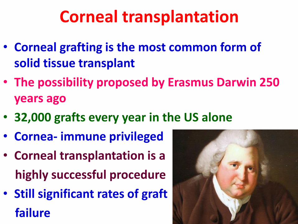
Corneal transplantation
• Corneal grafting is the most common form of solid tissue transplant
• The possibility proposed by Erasmus Darwin 250 years ago
• 32,000 grafts every year in the US alone
• Cornea- immune privileged
• Corneal transplantation is a
highly successful procedure
• Still significant rates of graft
failure
Rejection of corneal transplants
• long-term survival -approximately 75% at 5 years and 60% at 10 years
• high risk groups, the incidence of graft failure is higher
• The most common form of graft failure- immune reaction against the allogeneic grafts
• Important to develop strategies to prevent or reduce the impact of graft rejection
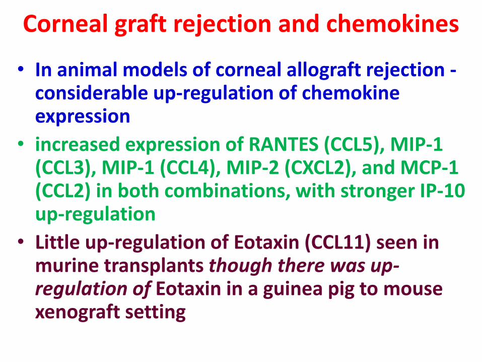
Corneal graft rejection and chemokines
• In animal models of corneal allograft rejection -considerable up-regulation of chemokine expression
• increased expression of RANTES (CCL5), MIP-1 (CCL3), MIP-1 (CCL4), MIP-2 (CXCL2), and MCP-1 (CCL2) in both combinations, with stronger IP-10 up-regulation
• Little up-regulation of Eotaxin (CCL11) seen in murine transplants though there was up-regulation of Eotaxin in a guinea pig to mouse xenograft setting
Corneal graft rejection and chemokines
• In murine grafts performed into a vascularized corneal bed (high risk graft) -more rapid and higher up-regulation of – MIP-1, MIP-2, eotaxin, MCP-1, and RANTES early after
transplantation (before onset of rejection) when compared with low risk grafts
– This is associated with an increase in infiltrating macrophages and neutrophils
• Treatment of animals with agents to prevent rejection resulted in a decrease in chemokines that were measured
• vMIPII experience
Curcumin- anti-inflammatory agent
• Extensively used in Ayurvedic preparations for centuries
– Antioxidant
– Anti-inflammatory
– Antiseptic activity
– Analgesic
• Inhibits nuclear factor NFĸB – regulates angiogenesis
• Constraints VEGF and angiopoietin
– Prevents angiogenesis
Curcumin- an inflammatory agent
• Previous reports revealed the effect of curcumin in – Scavenging NO
– blocking the release of cytokines like; • IL1β, IL4, IL6, IL10, IL17, IL22
• TNFα
• MCP1
• Almost 100 companies produces curcumin products
• Over 100 clinical trials focusing on chronic diseases including diabetes and cancer
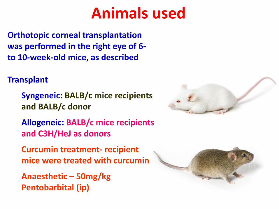
Animals used Orthotopic corneal transplantation was performed in the right eye of 6- to 10-week-old mice, as described Transplant
Syngeneic: BALB/c mice recipients and BALB/c donor
Allogeneic: BALB/c mice recipients and C3H/HeJ as donors
Curcumin treatment- recipient mice were treated with curcumin
Anaesthetic – 50mg/kg Pentobarbital (ip)
Curcumin administration
• 1g Curcumin (Sigma) was dissolved in 10ml 0.1 N NaOH and was diluted to a concentration of 50 mg/ml using PBS which brought down the pH to 7.2
• The integrity of the curcumin was checked by TLC analysis
• The experimental group received 50mg/kg curcumin each day by IP injection
• Curcumin administration started on the day before transplantation
• The control group was sham given with NaOH+PBS in the same manner
• The mice received regular, non-fortified food pellets
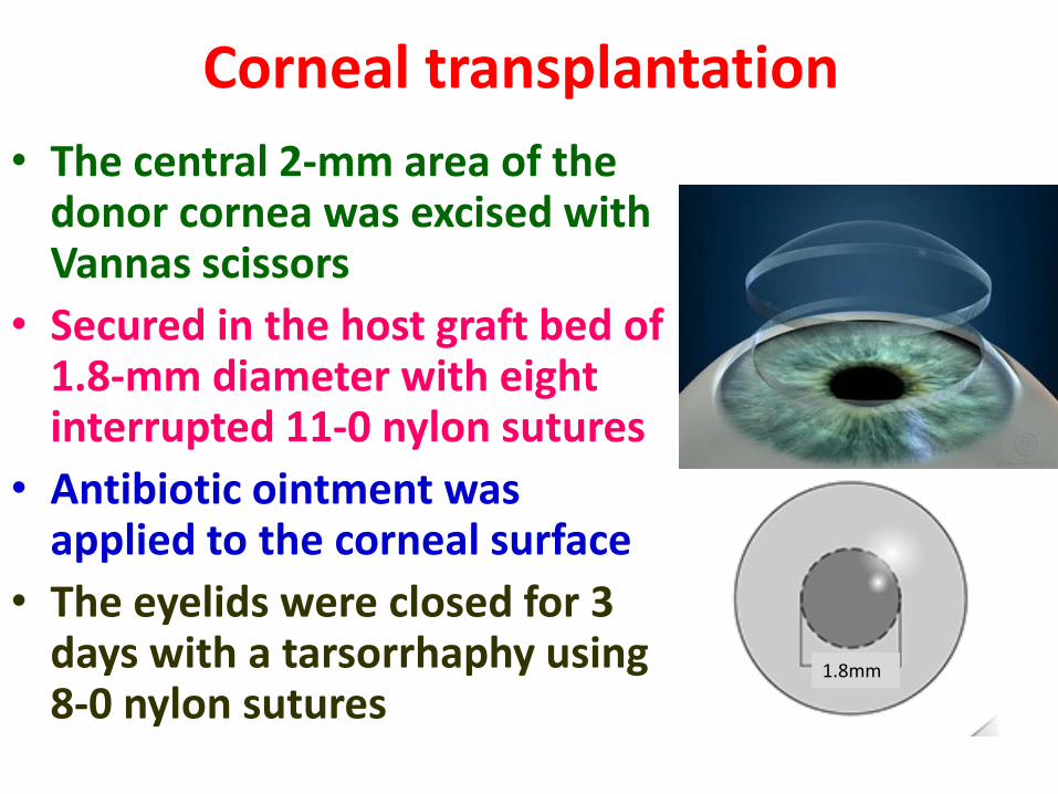
Corneal transplantation
• The central 2-mm area of the donor cornea was excised with Vannas scissors
• Secured in the host graft bed of 1.8-mm diameter with eight interrupted 11-0 nylon sutures
• Antibiotic ointment was applied to the corneal surface
• The eyelids were closed for 3 days with a tarsorrhaphy using 8-0 nylon sutures
1.8mm 1.8mm
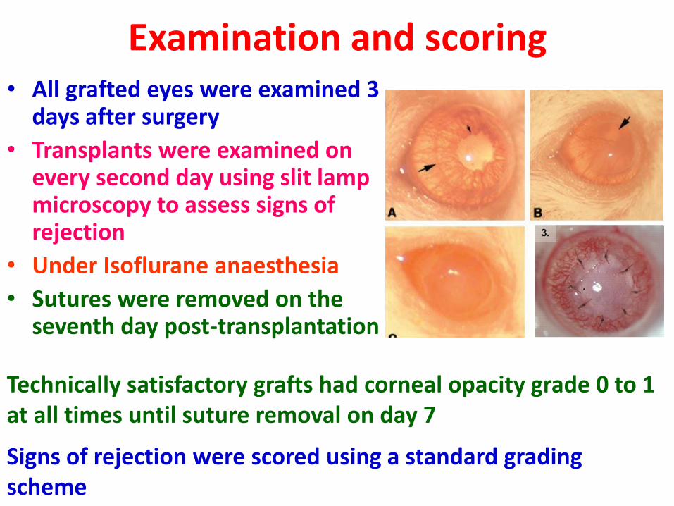
Examination and scoring • All grafted eyes were examined 3
days after surgery
• Transplants were examined on every second day using slit lamp microscopy to assess signs of rejection
• Under Isoflurane anaesthesia
• Sutures were removed on the seventh day post-transplantation
Technically satisfactory grafts had corneal opacity grade 0 to 1 at all times until suture removal on day 7
Signs of rejection were scored using a standard grading scheme
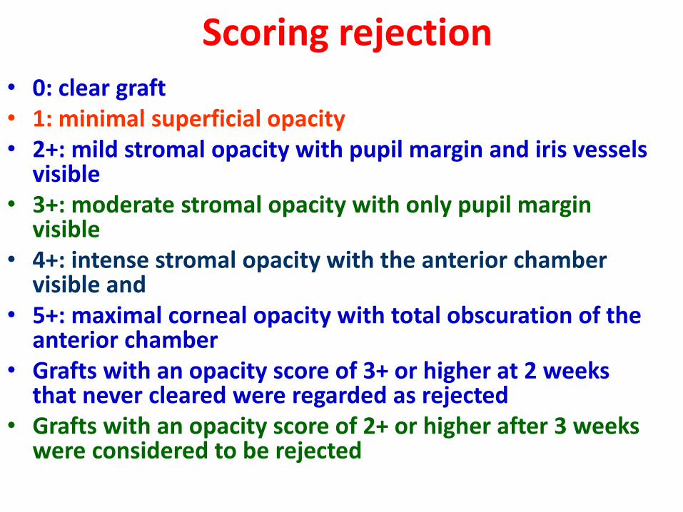
Scoring rejection • 0: clear graft • 1: minimal superficial opacity • 2+: mild stromal opacity with pupil margin and iris vessels
visible • 3+: moderate stromal opacity with only pupil margin
visible • 4+: intense stromal opacity with the anterior chamber
visible and • 5+: maximal corneal opacity with total obscuration of the
anterior chamber • Grafts with an opacity score of 3+ or higher at 2 weeks
that never cleared were regarded as rejected • Grafts with an opacity score of 2+ or higher after 3 weeks
were considered to be rejected
Detection of iNOS Protein by Western Blotting
• Protein extracted by homogenization with protein extraction buffer (50 mM Tris-HCl (pH 7.4), 150 mM NaCl, 10 mM EDTA, 1% Triton X-100, 1% SDS, and 1% protease inhibitors) and
• Separated by 8% SDS-PAGE and transferred to PVDF membranes at 90 V for 2 hours on ice (Mini-PROTEAN tetra cell; Bio-rad Laboratories, Richmond, CA, USA)
• The membranes were blocked with 5% blocking buffer
• Dissolved nonfat dried milk in TBST as blocking buffer (1X TBST- 1X TBST : 50 mM Tris.HCl, pH 7.4, 150 mM NaCl, 0.1% Tween 20)
• Incubated with diluted anti-iNOS (1 : 1,000, milipore, Temecula,
CA, USA)
• anti-β-tubulin (1 : 500,000, Abcam, Cambridge Science Park, UK)
• in 5% blocking buffer for overnight with shaking
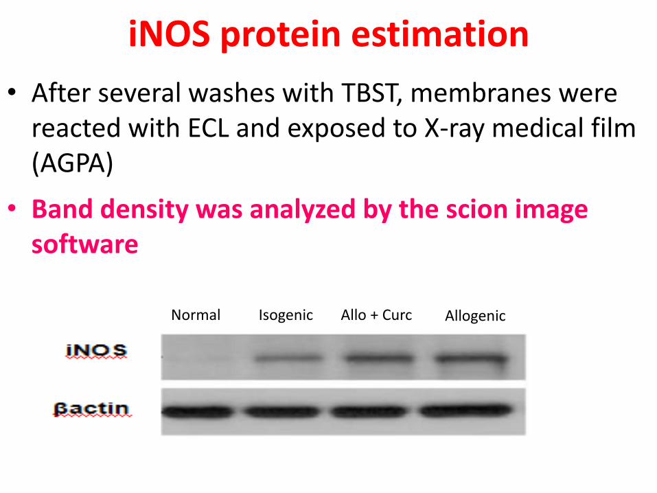
iNOS protein estimation
• After several washes with TBST, membranes were reacted with ECL and exposed to X-ray medical film (AGPA)
• Band density was analyzed by the scion image software
Normal Isogenic Allo + Curc Allogenic
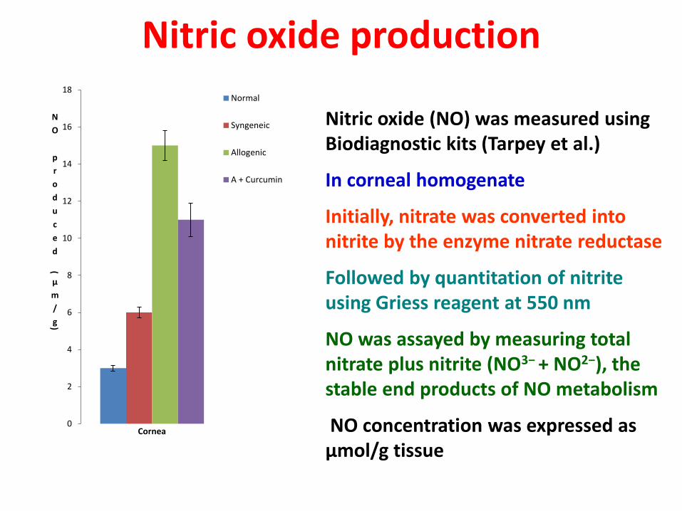
Nitric oxide production
Nitric oxide (NO) was measured using Biodiagnostic kits (Tarpey et al.)
In corneal homogenate
Initially, nitrate was converted into nitrite by the enzyme nitrate reductase
Followed by quantitation of nitrite using Griess reagent at 550 nm
NO was assayed by measuring total nitrate plus nitrite (NO3− + NO2−), the stable end products of NO metabolism
NO concentration was expressed as μmol/g tissue
0
2
4
6
8
10
12
14
16
18
N
O
p
r
o
d
u
c
e
d
(
μ
m
/
g)
Cornea
Normal
Syngeneic
Allogenic
A + Curcumin
NO/iNOS
• iNOS gene is under the transcriptional control of variety of inflammatory cytokines
• Increase in iNOS activity reported in other transplants – eg myocardial allografts – led to rejection
• Activates guanylyl cyclase • NO reacts with SO radicals forming peroxynitrite • Role of iNOS in transplant rejection not yet defined
– Rats- cytotoxic – Human –low/medium expression found beneficial
• Further studies are needed
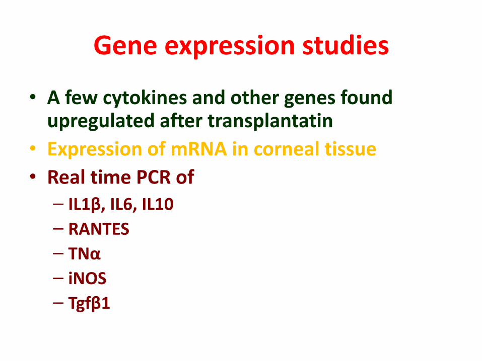
Gene expression studies
• A few cytokines and other genes found upregulated after transplantatin
• Expression of mRNA in corneal tissue
• Real time PCR of – IL1β, IL6, IL10
– RANTES
– TNα
– iNOS
– Tgfβ1
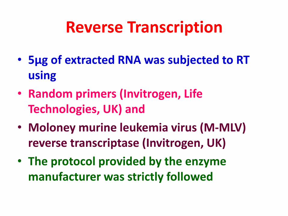
Reverse Transcription
• 5μg of extracted RNA was subjected to RT using
• Random primers (Invitrogen, Life Technologies, UK) and
• Moloney murine leukemia virus (M-MLV) reverse transcriptase (Invitrogen, UK)
• The protocol provided by the enzyme manufacturer was strictly followed
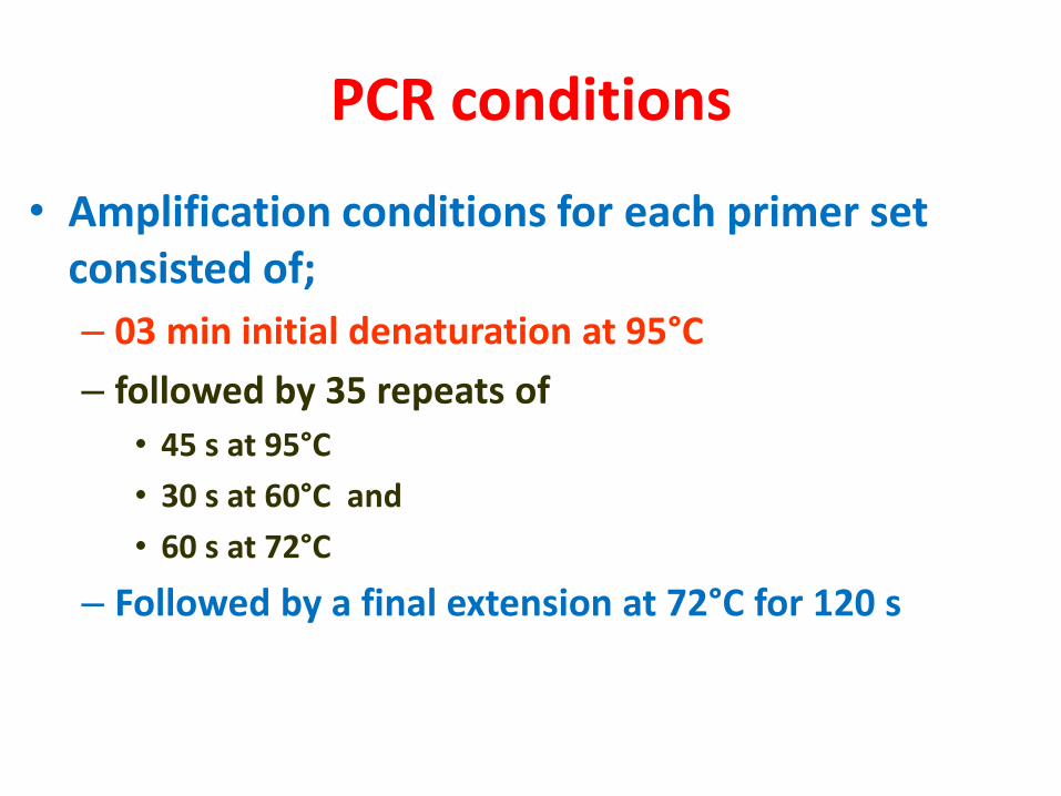
PCR conditions
• Amplification conditions for each primer set consisted of;
– 03 min initial denaturation at 95°C
– followed by 35 repeats of
• 45 s at 95°C
• 30 s at 60°C and
• 60 s at 72°C
– Followed by a final extension at 72°C for 120 s
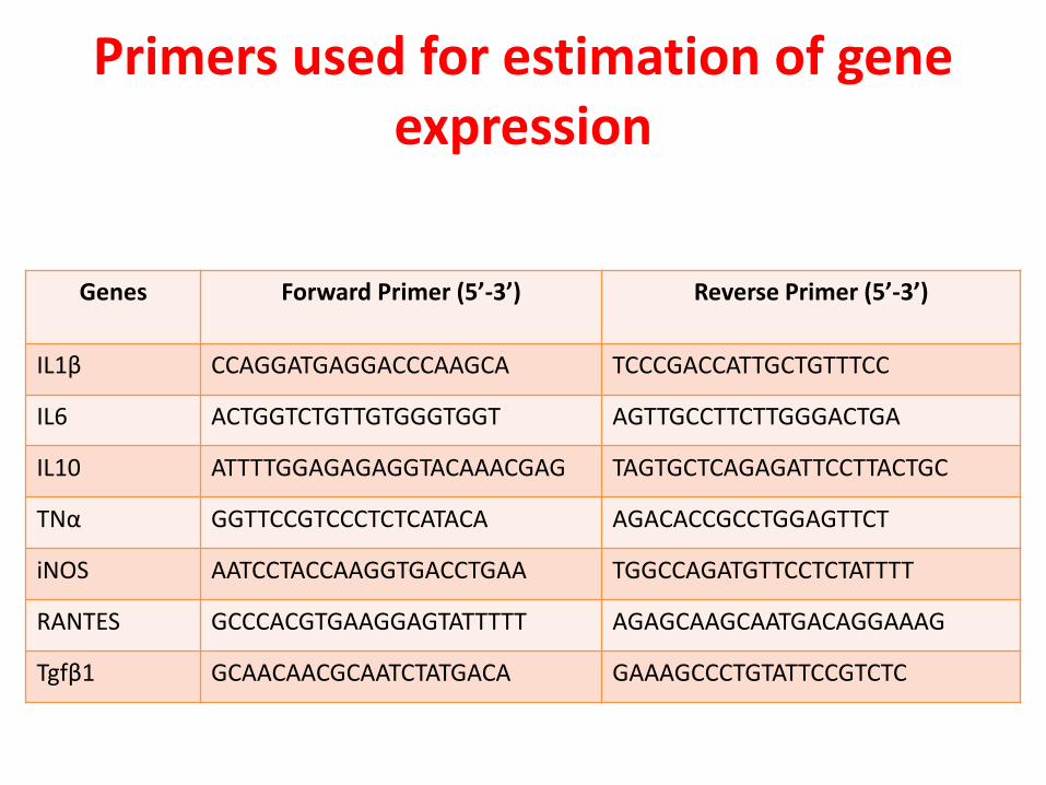
Primers used for estimation of gene expression
Genes Forward Primer (5’-3’)
Reverse Primer (5’-3’)
IL1β CCAGGATGAGGACCCAAGCA TCCCGACCATTGCTGTTTCC
IL6 ACTGGTCTGTTGTGGGTGGT AGTTGCCTTCTTGGGACTGA
IL10 ATTTTGGAGAGAGGTACAAACGAG TAGTGCTCAGAGATTCCTTACTGC
TNα GGTTCCGTCCCTCTCATACA AGACACCGCCTGGAGTTCT
iNOS AATCCTACCAAGGTGACCTGAA TGGCCAGATGTTCCTCTATTTT
RANTES GCCCACGTGAAGGAGTATTTTT AGAGCAAGCAATGACAGGAAAG
Tgfβ1 GCAACAACGCAATCTATGACA GAAAGCCCTGTATTCCGTCTC
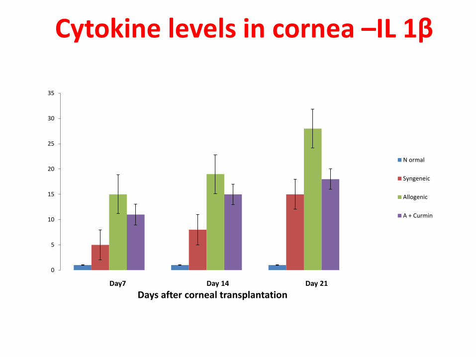
Cytokine levels in cornea –IL 1β
0
5
10
15
20
25
30
35
Day7 Day 14 Day 21
Days after corneal transplantation
N ormal
Syngeneic
Allogenic
A + Curmin
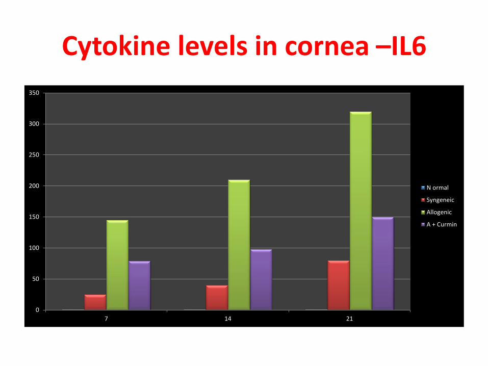
Cytokine levels in cornea –IL6
0
50
100
150
200
250
300
350
7 14 21
N ormal
Syngeneic
Allogenic
A + Curmin
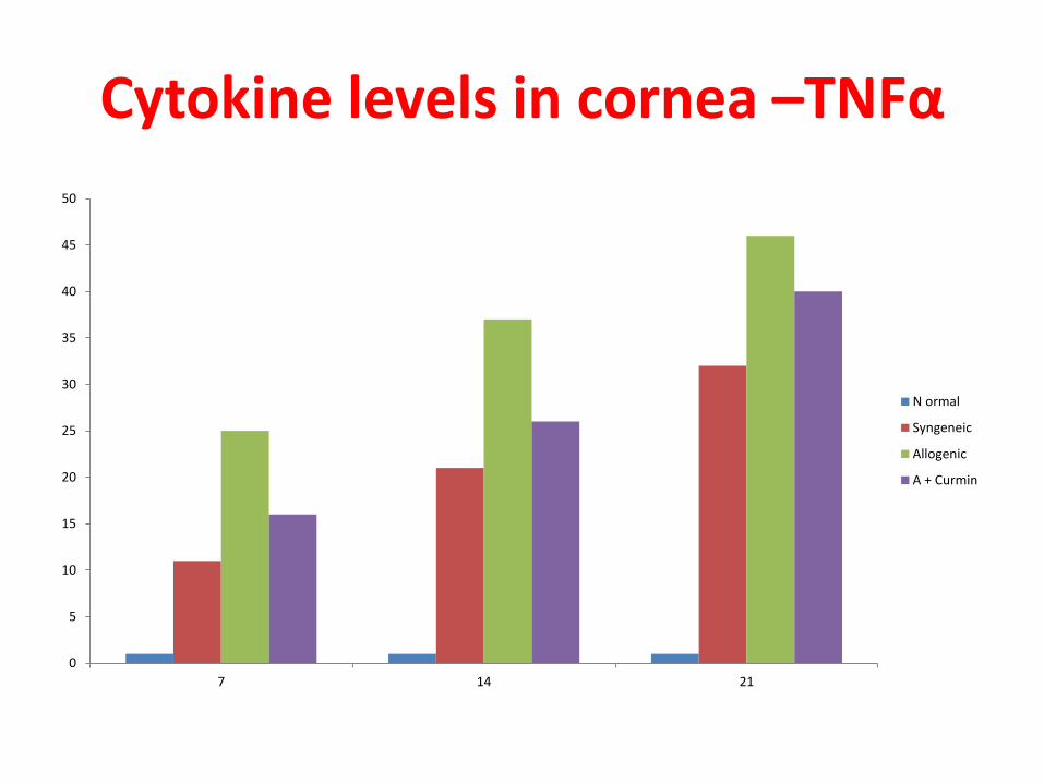
Cytokine levels in cornea –TNFα
0
5
10
15
20
25
30
35
40
45
50
7 14 21
N ormal
Syngeneic
Allogenic
A + Curmin
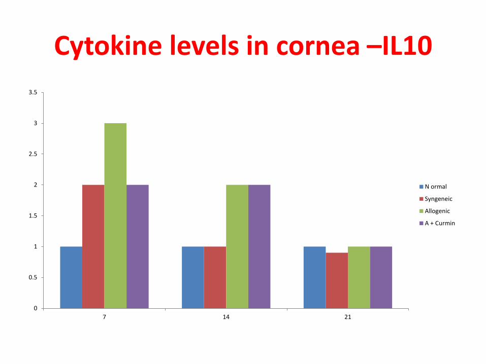
Cytokine levels in cornea –IL10
0
0.5
1
1.5
2
2.5
3
3.5
7 14 21
N ormal
Syngeneic
Allogenic
A + Curmin
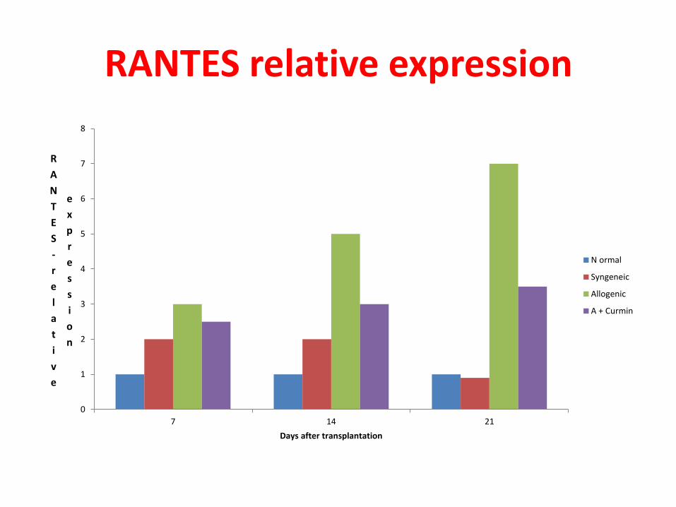
RANTES relative expression
0
1
2
3
4
5
6
7
8
7 14 21
R
A
N
T
E
S
-
r
e
l
a
t
i
v
e
e
x
p
r
e
s
s
i
o
n
Days after transplantation
N ormal
Syngeneic
Allogenic
A + Curmin
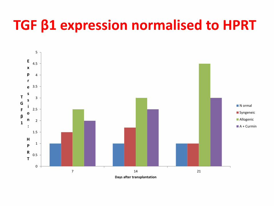
TGF β1 expression normalised to HPRT
0
0.5
1
1.5
2
2.5
3
3.5
4
4.5
5
7 14 21
T
G
F
β
1
E
x
p
r
e
s
s
i
o
n
:
H
P
R
T
Days after transplantation
N ormal
Syngeneic
Allogenic
A + Curmin
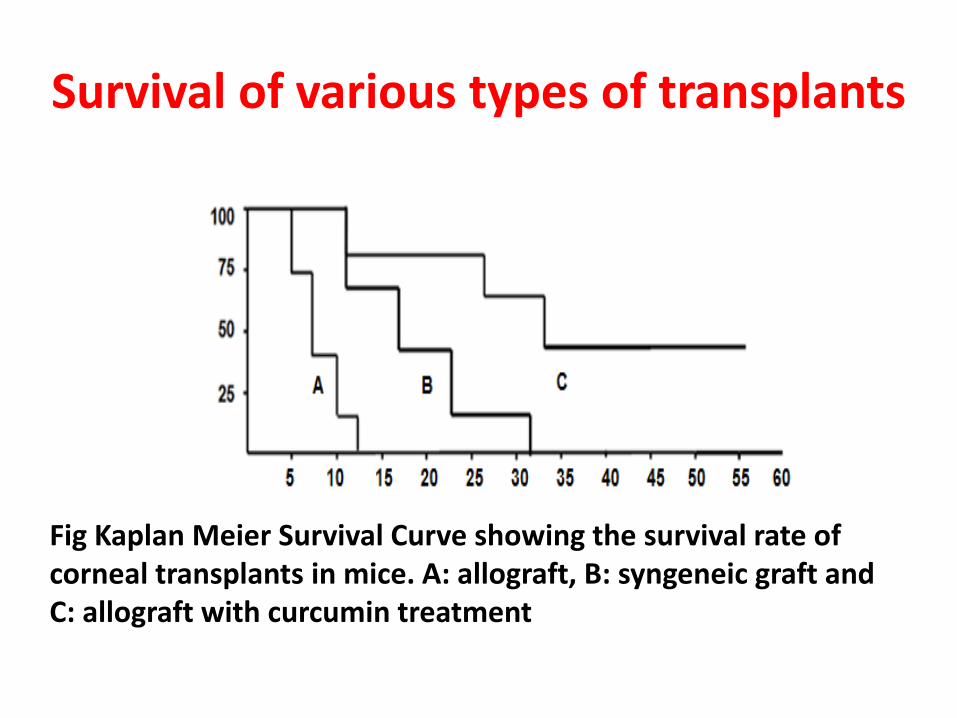
Survival of various types of transplants
Fig Kaplan Meier Survival Curve showing the survival rate of corneal transplants in mice. A: allograft, B: syngeneic graft and C: allograft with curcumin treatment
Discussion
• Expression of iNOS and chemokines were nominal in normal cornea
• Tranplantation increased expression of all
• Allogenic transplants showed highest increase
• Curcumin administration reduced expression
• Survival curve shows the influence of change in expression
• Curcumin offer a potential alternative to anti-inflammatory drugs in transplantation
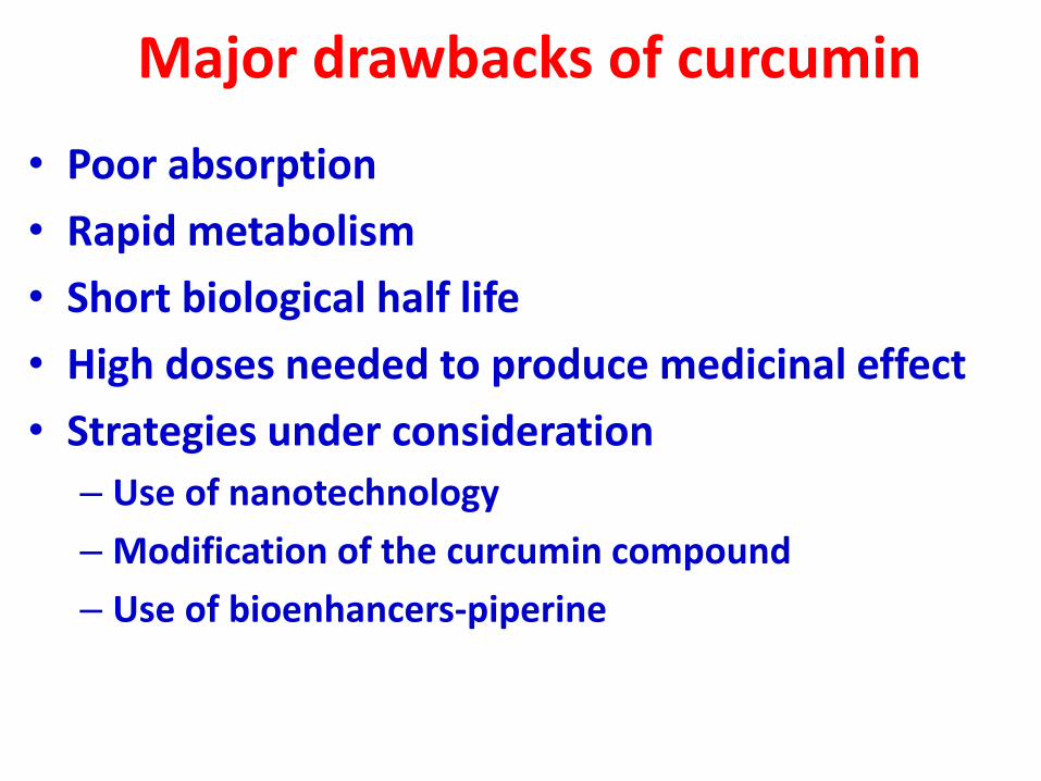
Major drawbacks of curcumin
• Poor absorption
• Rapid metabolism
• Short biological half life
• High doses needed to produce medicinal effect
• Strategies under consideration
– Use of nanotechnology
– Modification of the curcumin compound
– Use of bioenhancers-piperine