cumhuriyet Üniversitesi - dergipark
TRANSCRIPT
Cumhuriyet Üniversitesi
Diş Hekimliği Fakültesi
Dergisi
Cumhuriyet Dental Journal
http://dergipark.gov.tr/cumudj
http://dergi.cumhuriyet.edu.tr/cumudj
ISSN 1302-5805 e-ISSN 2146-2852 Volume/20 – Number/3 2017
CUMHURİYET ÜNİVERSİTESİ
Diş Hekimliği Fakültesi
Dergisi
Cumhuriyet Dental
Journal
An official publication of the
Faculty of Dentistry,
Cumhuriyet University, Issues
are published 3 times a year.
Our Faculty Journal first went
into press in 1998.
http://dergipark.gov.tr/cumudj
http://dergi.cumhuriyet.edu.tr/cumudj
ISSN 1302-5805
e-ISSN 2146-2852
Volume/20-Number/3-2017
Diş Hekimliği Fakültesi
Dergisi Adına Sahibi (Owner)
Prof.Dr.İhsan HUBBEZOĞLU
DEKAN V. (Dean)
Baş Editör
(Editor-in-Chief)
Prof.Dr.İhsan HUBBEZOĞLU
Editörler
(Associate Editors)
Doç.Dr.Vildan BOSTANCI
Doç.Dr.Derya Ö.DOĞAN
Yrd.Doç.Dr.Oğuzhan GÖRLER
Yrd.Doç.Dr.Recai ZAN
Yrd.Doç.Dr.Burak BULDUR
Yayın Kurulu
(Editorial Board)
Prof.Dr.Hakan DEVELİOĞLU
Doç.Dr.Derya Ö.DOĞAN
Yrd.Doç.Dr.Oğuzhan GÖRLER
Yrd.Doç.Dr.Recai ZAN
Yrd.Doç.Dr.Burak BULDUR
Yayın Kurulu Sekreteri
(Secretary)
Serap BEKİŞ
Telf: 03462191010/2775
E-mail: [email protected]
Cumhuriyet Dental Journal
2017; Volume:20 Issue:3
BİLİMSEL DANIŞMA KURULU (SCIENTIFIC ADVISORY BOARD)
Adil NALÇACI (Ankara Ü.)
Ahmet ALTAN (G.O.P.Ü.)
Ahmet Berhan YILMAZ (Atatürk Ü.)
Alpdoğan KANTARCI (Boston U.)
Ali ERDEMİR (Kırıkkale Ü.)
Ali Hakan DEVELİOĞLU (Cumhuriyet Ü.)
Alparslan DİLSİZ (Atatürk Ü.)
Alper KAPDAN (Cumhuriyet Ü.)
Arife KAPDAN (Cumhuriyet Ü.)
Arlin KİREMİTCİ (Hacettepe Ü.)
Arzu MÜJDECİ (Ankara Ü.)
Arzu TEZVERGİL MUTLUAY (University of Turku)
Aslıhan ÜŞÜMEZ (Serbest Diş Hekimi)
Ayşegül GÖZE SAYGIN (Cumhuriyet Ü.)
Banu ERMİŞ (S.Demirel Ü.)
Burak BULDUR (Cumhuriyet Ü.)
Cafer TÜRKMEN (Marmara Ü.)
Defne YALÇIN YELER (Cumhuriyet Ü.)
Derya ÖZDEMİR DOĞAN (Cumhuriyet Ü.)
Diğdem EREN (Cumhuriyet Ü.)
Emine Gülşah GÖKTOLGA AKIN (Cumhuriyet Ü.)
Emine PİRİM GÖRGÜN (Cumhuriyet Ü.)
Emrah SOYLU(G.O.P.Ü.)
Ercan Cenk DORUK (Cumhuriyet Ü.)
Esengül BEKAR (G.O.P.Ü.)
Faik TUĞUT (Cumhuriyet Ü.)
Fatih ÖZNURHAN (Cumhuriyet Ü.)
Fatma ÇAĞLAYAN (Atatürk Ü.)
Feridun HÜRMÜZLÜ (Cumhuriyet Ü.)
Filiz AYKENT (Serbest Diş Hekimi)
Funda BAYINDIR (Atatürk Ü.)
Füsun ÖZER (İzmir Bozyaka E.ve Arş.Hast.)
Giray BOLAYIR (Cumhuriyet Ü.)
Gülfem ERGÜN (Gazi Ü.)
Gülsüm DURUK (İnönü Ü.)
Hakan GÖKTÜRK (G.O.P.Ü.)
Hakan İŞCAN (Acıbadem Sağlık Gr.)
Hakan TERZİOĞLU (Ankara Ü.)
Hale CİMİLLİ (Marmara Ü.)
Halenur ALTAN (G.O.P.Ü.)
Hamid JAFARZADEH (Mashhad U.)
Hare GÜRSOY (Yeditepe Ü.)
Hasan YELER (Cumhuriyet Ü.)
Hatice BALCI YÜCE (G.O.P.Ü.)
Hatice ÖZDEMİR (Atatürk Ü.)
Hayati Murat AKGÜL (Atatürk Ü.)
Haydar ALBAYRAK (Erciyes Ü.)
Işıl SARIKAYA (G.O.P.Ü.)
Jale GÖRÜCÜ (Hacettepe Ü.)
Kerem KILIÇ (Erciyes Ü.)
Kezban Meltem ÇOLAK TOPCU (Atatürk Ü.)
Kürşat ER (Akdeniz Ü.)
Mehmet Emre COŞKUN (Cumhuriyet Ü.)
Mehmet KAYAHAN (Okan Ü.)
Muhammed SÜMBÜLLÜ (Atatürk Ü.)
Murat ÜNAL (Cumhuriyet Ü.)
Mustafa GÜNDOĞDU (Atatürk Ü.)
Mustafa MUTLUAY (University of Turku)
Mutlu OZCAN (University Of Zurich)
Neslihan ŞİMŞEK (İnönü Ü.)
Nihat AKBULUT (G.O.P.Ü.)
Nurhan ÖZTAŞ (Gazi Ü.)
Özden ÖZEL BEKTAŞ (Cumhuriyet.Ü)
Regina PALMA-DİBB (São Paulo U.)
Sadullah ÜÇTAŞLI (Ankara Ü.)
Sema BELLİ (Selçuk Ü.)
Sevcan KURTULMUŞ YILMAZ (Yakın Doğu Ü.)
Sibel AKBULUT(G.O.P.Ü.)
Sivakumar NUVVULA (N.D.C.H.)
Şenay CANAY (Hacettepe Ü.)
Şeyda HERGÜNER-SİSO
Tamer TAŞDEMİR (K.A.T.Ü.)
Tuğrul ASLAN (Erciyes Ü.)
T. Peyami HOCAOĞLU(Cumhuriyet Ü.)
Tülin POLAT (İnönü Ü.)
Ulvi GÜRSOY (University of Turku)
Victor FEİTOSA
Yağmur ŞENER (Konya Ü.)
Yakup ÜSTÜN (Erciyes Ü.)
Yasemin KULAK ÖZKAN (Marmara Ü.)
Yeliz HAYRAN (G.O.P.Ü.)
Yurdanur UÇAR (Çukurova Ü.)
Zeynep ÖZKURT KAYAHAN (Yeditepe Ü.)
Cumhuriyet Dental Journal
2017; Volume:20 Issue:3
Cumhuriyet Dental Journal
GUIDELINES FOR AUTHORS
Authors are requested to submit their original
manuscript and figures via the online submission and
editorial system for Cumhuriyet Dental Journal.
Using this online system, authors may submit
manuscripts and track their progress through the
system to publication. Reviewers can download
manuscripts and submit their opinions to the editor.
Editors can manage the whole
submission/review/revise/publish process.
Format
General
Manuscript length depends on manuscript type. In
general, research and clinical science articles should
not exceed 20 to 12 double-spaced, typed pages
(excluding references, legends, and tables). Clinical
Reports and Technique articles should not exceed 4
to 5 pages. Paper dimensions should be 8.5 × 11
inches with 2.5 cm margins on all sides.
use normal, plain font (12-point Times New Roman)
number all pages consecutively.
indent or space paragraphs.
Articles should be arranged in the following
order. Title, Abstract, Introduction, Materials and
Methods, Results, Discussion, Conclusions,
Acknowledgements, References,
Tables and Legends to Illustrations.
Title page
-Title
-Authors (first name, middle initial, surname) e.g.
Faik Tugut, DDS, PhD,a
-Authors' addresses (abbreviated) e.g. aAssistant Professor, Department of Prosthodontics,
Faculty of Dentistry, Cumhuriyet University, Sivas,
Turkey.
-If the research was presented before an organized
group, type the name of the organization and the
location and date of the meeting.
PLEASE UPLOAD TITLE PAGE APART FROM
MANUSCRIPT.
TITLE PAGE SHOULD UPLOAD AS A
SUPPLEMENTARY FILE.
-Corresponding Author details (essential): Name,
complete address, phone, fax, and E-mail
numbers
Abstract
Should not exceed 300 words and should be
presented under the following subheadings:
Objectives, Materials and Methods; Results;
Conclusions (For Reviews: Objectives; Data;
Sources; Study selection; Conclusions). These
subheadings should appear in the text of the
summary. Provide a short, nonstructured, 1-
paragraph abstract that briefly summarizes the
problem encountered and treatment administered for
clinical report.
Keywords
Up to 10 keywords should be supplied e.g. Er: YAG
laser, composite resin, adhesion.
Introduction
This must be presented in a structured format,
covering the following subjects, although not under
subheadings: succinct statements of the issue in
question; the essence of existing knowledge and
understanding pertinent to the issue; and the aims
and objectives of the research being reported.
Materials and methods
-describe the procedures and analytical techniques.
-identify names and sources of all commercial
products e.g.
-magnetic attachment (Hyper Slim 5513, Hitachi
Metals, Tokyo, Japan)
Results
-refer to appropriate tables and figures.
-report statistical findings.
Discussion
-discuss the results of the study.
-agreement with other studies should also be stated.
-identify the limitations of the present study, and
suggest areas for future research.
Conclusions
-concisely list conclusions that may be drawn from
the research.
-do not simply restate the results.
Acknowledgements
-If the work was supported by a grant or any other
kind of funding, supply the name of the supporting
organization and the grant number.
References
-References must be identified in the body of the
article with superscript Arabic numerals.
-The complete reference list, double spaced and in
numerical order, should follow the Conclusions
section but start on a separate page. Only references
cited in the text should appear in the reference list.
-Do not include unpublished data or personal
communications in the reference list.
Cumhuriyet Dental Journal
2017; Volume:20 Issue:3
Journal reference style:
Akin H, Coskun ME, Sari F, Tugut F. Mechanical
success and failure of the different types of dental
implants: two years follow up study. Cumhuriyet
Dent J 2009;2:121-124.
Book reference style:
Hilton TJ. Direct posterior composite restorations.
In: Schwartz RS, Summitt JB, Robbins JW (eds).
Fundamentals of Operative Dentistry. Chicago:
Quintessence,1996:207-228.
Tables and Figures
All tables and figures must be thoroughly discussed
in the text of the manuscript.
Tables
• one table to a page, each with a title.
• number tables in order of mention using Arabic
numerals. Do not list tables in parts (eg, Table Ia, Ib,
etc.). Each should have its own number.
• must be able to "stand alone" apart from text.
• when appropriate, standard deviations of
values should be indicated in parentheses; (do NOT
use ± notation).
• results of statistical analysis must be included,
use superscript letters to indicate significant
differences.
• for explanatory footnotes, use symbols (*,
#,**,##).
Figures
• do not import the figures into the text file.
• figures grouped together should have similar
dimensions and be labelled "A, B, C", etc.
• figures should be arranged to the width of 80
mm.
• color and black-and-white photographs should
be created and saved at a minimum of 300 dots per
inch (dpi).
• figures should be saved in jpeg format.
• The electronic image files must be named so
that the figure number and format can be easily
identified. For example, a Figure 1 in jpeg format
should be named fig 1. Multipart figures must be
clearly identifiable by the file names: fig 1A, fig 1B,
fig 1C, etc.
Graphs
• unique, concise axis labels; do not repeat the
Figure caption.
• uniform size for graphs of similar type.
• type size that will be easily read when the graph
is reduced to one column width.
• lines that are thick and solid (100% black).
Figure legends
• list together on a separate page.
• should be complete and understandable apart
from the text.
• include key for symbols or abbreviations used
in Figures.
Cumhuriyet Dental Journal
2017; Volume:20 Issue:3
İÇİNDEKİLER / CONTENTS
ARAŞTIRMA / RESEARCH
145-151 Assessment Of The Relationships Between Deleterious Oral Habits That May Cause
Orthodontic Anomalies And Psychological And Socio-Demographic Factors
Ortodontik Anomalilere Sebep Olabilen Kötü Alışkanlıkların Psikolojik ve
Sosyodemografik Faktörlerle İlişkisinin Değerlendirilmesi
Zeynep ÇOBAN BÜYÜKBAYRAKTAR, Cenk DORUK
152-160 The Effect of Diode Laser as an Adjunct to Periodontal Treatment on Clinical
Periodontal Parameters and Halitosis: A Randomized Controlled Clinical Trial
Periodontal Tedaviye Destek Olarak Kullanılan Diyot Lazerin Periodontal Klinik
Parametrelere Ve Halitozis Üzerine Etkileri: Randomize Kontrollü Klinik Çalışma
Mükerrem HATİPOĞLU, Zeliha AYTEKİN, Özlem DALTABAN, Rasih FELEK,
Mehmet Ziya FIRAT, Kemal ÜSTÜN
161-168 Effect of Two Activated Bleaching Techniques on Surface Roughness of Different
Esthetic Restorative Materials
İki Aktive Olan Beyazlatma Tekniğinin Farklı Estetik Restoratif Materyallerin Yüzey
Pürüzlülüğü Üzerine Etkisi
Tuncay ALPTEKIN, Özgün Yusuf ÖZYILMAZ, Filiz AYKENT, Haluk Barış KARA
169-174 Nonsurgical Closure of Oroantral Communications Using Occlusal Splints
Oroantral Açıklıkların Okluzal Splintler ile Cerrahisiz Kapatılması
Nükhet KÜTÜK, Ahmet Emin DEMİRBAŞ, Canay YILMAZ ASAN, Burcu BAŞ,
Alper ALKAN
175-179 Awareness About Periodontitis and Pre-Term Low Birth Weight Infants Among
Gynecologists In Chennai- A Questionnaire Study
Chennaideki Jinekologların Periodontitis ve Düşük Doğum Ağırlıklı Prematüre Bebekler
Hakkındaki Farkındalığı: Bir Anket Çalışması
Hafsa ISMAIL, Radhika ARJUNKUMAR
Cumhuriyet Dental Journal
2017; Volume:20 Issue:3
OLGU SUNUMU / CASE REPORT
180-184 A Case Report of Transoral Removal of Submandibular Gland Sialolith
Submandibular Tükürük Bezi Taşının Ağız İçinden Uzaklaştırılması: Bir Olgu Sunumu
Kumuda RAO, Subhas G BABU, Renita Lorina CASTELINO
185-190 Removal of Separated Instruments with Masserann Techniques: Two Case Reports
Masserann Tekniği ile Kırık Aletlerin Uzaklaştırılması: İki Olgu Sunumu
Hakan GÖKTÜRK, İsmail ÖZKOÇAK
191-197 Orthognathic Treatment Of A Patient With Class Iii Malocclusion And Severe
Facial Asymmetry: Report Of A Case
Sınıf III Malokluzyon ve Şiddetli Fasiyal Asimetriye Sahip Hastanın Ortognatik Cerrahi
ile Tedavisi: Olgu Sunumu
Hande GÖRÜCÜ COŞKUNER, İlken KOCADERELİ, Ersoy KONAŞ
198-203 Importance of Bifid Mandibular Canal in Implantology and in Oral Surgery:
Review of the Literature and Report of Three Cases
Bifid Mandibuler Kanalin İmplantoloji ve Oral Cerrahideki Önemi: Üç Vaka Raporu ile
Birlikte Literatürün Gözden Geçirilmesi
Nihat AKBULUT, Sibel AKBULUT, Bengi ÖZTAŞ, Şebnem KURŞUN, Emrah SOYLU,
Orhan GÜVEN
DERLEME / REVIEW
204-211 Parkinson’s Disease in Dentistry and Periodontology
Diş Hekimliğinde ve Periodontolojide Parkinson Hastalığı
Zeliha MUSLU, Hakan DEVELİOĞLU
Cumhuriyet Dental Journal: 2017; 20(3)
Doi: 10.7126/cumudj.341904 RESEARCH ARTICLES
145
Department of Orthodontics, Cumhuriyet Unversity School of Dentistry, Sivas, Turkey
ASSESSMENT OF THE RELATIONSHIPS BETWEEN DELETERIOUS ORAL
HABITS THAT MAY CAUSE ORTHODONTIC ANOMALIES AND
PSYCHOLOGICAL AND SOCIO-DEMOGRAPHIC FACTORS
Ortodontik Anomalilere Sebep Olabilen Kötü Alışkanlıkların Psikolojik ve
Sosyodemografik Faktörlerle İlişkisinin Değerlendirilmesi
Zeynep ÇOBAN BÜYÜKBAYRAKTAR, Cenk DORUK
Makale Kodu/Article Code : 341904
Makale Gönderilme Tarihi : 05.10.2017
Kabul Tarihi : 08.11.2017
ABSTRACT
Objectives: The aim of this study is to identify the relation
between the deleterious oral habits (DOH) which can
cause orthodontic anomalies such as finger sucking, nail
biting, lip chewing, bruxism and psychological and socio-
demographic factors.
Materials and Methods: 64 males, 71 females, between
the ages of 9 and 12, including their parents have been
included in our study. In our study, a survey form
consisting of Clinic Examination Data Form, Socio-
demographic Data Form, Children’s Depression Inventory
(CDI), Child State Trait Anxiety Inventory (CSTAI) has
been applied to patients.
Results: DOH have been observed in 62.5 % of the male
children and % 52.1 of the female children, but these
results are not statistically significant (p>0.05). No
statistically significant association was found between
DOH and CDI (p>0.05). No statistically significant
association was found between the CSTAI status section
score and trait section score and DOH (p>0.05).
Statistically significant associations were found between
family type and DOH (p<0.05).
Conclusions: While there was no association between
anxiety and depression and DOH, there was significant
association family type from socio-demographic factors
and DOH.
Key words: Finger Sucking, Nail Biting, Depression,
Anxiety
ÖZ
Amaç: Bu çalışmanın amacı; ortodontik anomalilere
sebep olabilen, parmak emme, tırnak yeme, dudak emme,
diş sıkma ve gıcırdatma gibi kötü alışkanlıkların psikolojik
ve sosyodemografik faktörlerle ilişkisini saptamaktır.
Gereç ve Yöntem: Çalışmamıza, yaşları 9 ila 12 arasında
değişen 64 erkek, 71 kız olmak üzere toplamda 135 çocuk
ve ebeveynleri dahil edilmiştir. Çalışmamızda, Klinik
Muayene Veri Formu, Sosyodemografik Veri Formu,
Çocuklar İçin Depresyon Ölçeği (ÇDÖ), Çocuklar İçin
Durumluk-Sürekli Kaygı Envanteri (ÇDSKE) şeklinde 4
bölümden oluşan anket formu, hastalara uygulanmıştır.
Bulgular: Erkek çocukların %62,5’inde, kız çocukların
ise %52,1’inde kötü alışkanlık görülmüştür ancak elde
edilen bu sonuç istatistiksel olarak anlamlı değildir
(p>0,05). Kötü alışkanlıklar ile ÇDÖ puanı arasında
istatistiksel açıdan anlamlı bir ilişki bulunamamıştır
(p>0,05). ÇDSKE durumluk bölüm puanı ve sürekli bölüm
puanı ile kötü alışkanlıklar arasında istatistiksel olarak
anlamlı bir ilişki bulunamamıştır (p>0,05). Aile tipi ile
kötü alışkanlıklar arasında ise istatistiksel açıdan anlamlı
ilişki bulunmuştur (p<0,05).
Sonuç: Kaygı ve depresyon ile kötü alışkanlıklar arasında
anlamlı bir ilişki bulunamazken, sosyodemografik
faktörlerden yalnız aile tipi ile kötü alışkanlıklar arasında
anlamlı bir ilişki bulunmuştur.
Anahtar kelimeler: Parmak Emme, Tırnak Yeme,
Depresyon, Kaygı
Büyükbayraktar Z. et al.
146
INTRODUCTION
Repetitive activities that occur automatically
are called habits. These repetitive behaviors are
often seen in childhood and many begin and end
by themselves.1 Habits such as finger sucking
and foreign body stabbing, which are
sometimes part of the psychosocial
development that breaks the physiological
development between the ages of 3 and 6 and
leads to pathology in the dentition are described
as deleterious oral habits (DOH). DOH could be
divided into 2 main groups:
1. Acquired DOH: When a child grows up,
they can easily leave this hint of habit and
switch to another habit.
2. Compulsive DOH: These habits are
constantly seen in children and when emotional
pressure becomes unbearable for a child, they
feel secure themselves with this habit. He feels
uneasy when he tries to get rid of habits.2
DOH such as finger sucking, lip chewing,
mouth breathing, nail biting, tongue thrusting
can be seen in children. It is known that DOH
seen in children sometimes cause orthodontic
anomalies that are impossible to cure.
Orthodontic deformation caused by DOH varies
with the severity of habits, frequency of
recurrence, duration of residence and tissue
strength.3
The reasons for these habits are different.
Several theories (psychoanalytic theory,
learning theory, insufficient sucking theory)
have been proposed to explain the etiology of
sucking behavior in particular. According to the
psychoanalytic theory which is one of these
theories, sucking behavior is instinct for the first
period of life. Unhindered, it should be
saturated in this period. According to those who
argue that sucking behaviors that transcend the
reflex or instinct dimension originate from
various spiritual problems, the problem should
be sought especially in child-mother or child-
sibling relationships.4,5 According to some
experts; sucking behavior that develops to elder
or older ages is a symptom of abnormal
psychological development.5, 6
Nail biting, which is another of the DOH,
emerges as a reaction in response to some
psychological disorders and in some children it
is seen that sucking habits have changed to nail
biting.1 Depression-style psychological
problems have been identified in more than half
of the families with children having nail biting.
Children with this habit should be assessed
emotionally.
The etiology of bruxism is still being
debate and in the theories; occlusal,
psychological, genetic and stress factors are
emphasized. It has been abandoned to think that
the phenomenon of bruxism is related to
occlusal discomfort alone. Today, there is a
common belief that etiology is related to more
than one factor and it is thought that there is a
central nervous system phenomena associated
with more stress and pain behavior.7
In our study, DOH such as finger sucking,
nail biting, lip chewing and bruxism were
evaluated in relation to psychological and
socio-demographic factors and comprehensive
data on etiology of these DOH were collected.
MATERIAL AND METHOD
Our study was conducted on the children and
their parents who applied to the Orthodontics
Department of the Dentistry of Cumhuriyet
University for examination between the dates of
10.12.2016 and 10.03.2017. In total, 135
children and their parents, 64 of whom are male
and 71 of whom are female were included in to
our study. The ages of the children are between
9 and 12.
Oral and written consent with ethics
committee approval for study were taken from
each patient and parents (Ethics committee
decision no: 2016-09/05, Date: 27.09.2016).
Patients who have not had any previous
orthodontic treatment and who do not have any
mental or physical disabilities that would
Assessment of the Relationships Between Deleterious Oral Habits that May Cause Orthodontic Anomalies and Psychological
and Socio-Demographic Factors
147
prevent them from responding to the
questionnaire were included in the study.
Habit group (HG): From the child patients
who have at least one of the DOH such as finger
sucking, lip chewing, nail biting and bruxism.
Habit free group (HFG): It is made up of
children who have not seen any of the DOH
mentioned.
The questionnaire consists of 4 sections
(Clinical Examination Data Form, Socio-
demographic Data Form, Children’s Depression
Inventory, Child State Trait Anxiety Inventory)
in total has been applied to collect information
about the underlying causes of DOH that
described the negative effects on mouth, teeth
and jaw system.
1. Clinical Examination Data Form: This
section includes intra oral and extra oral
examinations made by the observer and
questions directed to the patients’ parents. The
age and gender of the patients were recorded.
Whether or not DOH such as finger sucking,
nail biting, bruxism and lip chewing are present
or not is discussed and evaluated with the
patients’ parents.
2. Socio-demographic Data Form: In this
section, age, education, family type, number of
children living at home, settlement place and
monthly income are questioned. The answers to
the questions in this section are taken from the
guardians of patients.
3. Children’s Depression Inventory: In
childhood depression, among the self-
assessment scales, the most frequently used one
and the most frequently researched
psychometric features of a scale is the
Children’s Depression Inventory and it is a self-
assessment scale applicable to children aged 6-
17 years. It has been based on the views of
Kovacs which are 1. There is childhood
depression, 2. Observable and measurable, 3.
Features similar to adults. The Beck Depression
Scale is based on the questionnaire, but also
includes questions about the school-specific
situation for childhood depression, friendship,
and so on.8,9 The scale is filled in by reading to
the child or by the child. There are three
different options for each item on the 27-item
scale. The child is asked to choose the most
appropriate sentence for the last two weeks. For
example; 1. I feel sad sometimes from time to
time. 2. I feel sad often. 3. I always feel sorry
for myself. Each item takes 0, 1 or 2 points
according to the severity of the indication. The
maximum score is 54. The higher the score, the
more depressed it is. The cut point is
recommended as 19.8,9 Validity and reliability
studies in our country were made by ÖY10 and
pathology cut point was determined as 19
points.
4. Children’s State Trait Anxiety Inventory:
This scale, developed by Spielberger11, has two
subscales with multiple choice of 20 questions
for state and trait anxiety. Each item is scored 1,
2 or 3 according to the severity of the indication.
Trait Anxiety Scale: It aims to measure
persistent individual differences as well as
anxiety. The scale consists of 20 items. It
usually evaluates how the child feels according
to the frequecy of occurence. Expressions such
as "My nerves at home" or "My hands are titled"
are answered with one of the "almost never",
"sometimes" and "often" options. The scores to
be taken from the scale are between 20 and 60,
the increase of the scores represents the increase
in trait anxiety.
State Anxiety Scale: The children are asked to
evaluate how they feel at that moment and to
choose the most appropriate option such as "I
feel so angry, I feel angry, I do not feel angry".
The total number of items is 20. The lowest
score you can get is 20, the highest score is 60.
The state anxiety scale is suggested to be given
prior to the trait anxiety scale in practice, since
it is a scale that is susceptible to emotions/
disturbances that may occur in test conditions.
The study of validity and reliability of the scale
in our country was carried out by Özusta.12
Büyükbayraktar Z. et al.
148
Patients in both groups were examined
first, the necessary information was recorded by
the investigator and than a questionnaire was
given to the patients and their parents to answer
socio-demographic questions and psychological
inventory. Socio-demographic questions were
asked to be answered by guardian of patient and
psychological inventory of the patients were
asked to be answered by the patient.
Statistical method
The data obtained from our study were
uploaded to the SPSS (Ver: 15.0) program. In
the evaluation of the data; the mean, standard
deviation and frequency distributions were
examined. Subsequently, the significance test
between the two means in comparison of the
groups, Man Whitney U and Chi Square test
have been used. The level of error was taken as
0.05.
RESULTS
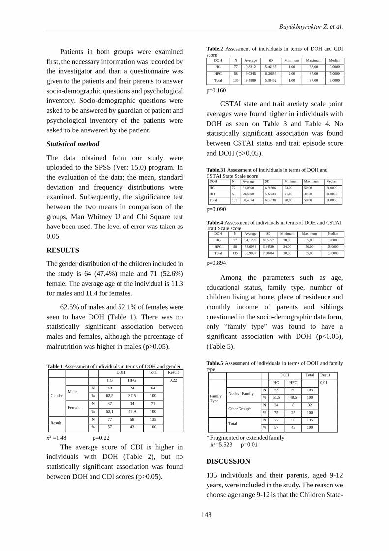
The gender distribution of the children included in
the study is 64 (47.4%) male and 71 (52.6%)
female. The average age of the individual is 11.3
for males and 11.4 for females.
62.5% of males and 52.1% of females were
seen to have DOH (Table 1). There was no
statistically significant association between
males and females, although the percentage of
malnutrition was higher in males (p>0.05).
Table.1 Assessment of individuals in terms of DOH and gender
x2 =1.48 p=0.22
The average score of CDI is higher in
individuals with DOH (Table 2), but no
statistically significant association was found
between DOH and CDI scores (p>0.05).
Table.2 Assessment of individuals in terms of DOH and CDI
score
p=0.160
CSTAI state and trait anxiety scale point
averages were found higher in individuals with
DOH as seen on Table 3 and Table 4. No
statistically significant association was found
between CSTAI status and trait episode score
and DOH (p>0.05).
Table.31 Assessment of individuals in terms of DOH and
CSTAI State Scale score
p=0.090
Table.4 Assessment of individuals in terms of DOH and CSTAI Trait Scale score
p=0.894
Among the parameters such as age,
educational status, family type, number of
children living at home, place of residence and
monthly income of parents and siblings
questioned in the socio-demographic data form,
only “family type” was found to have a
significant association with DOH (p<0.05),
(Table 5).
Table.5 Assessment of individuals in terms of DOH and family type
* Fragmented or extended family
x2=5.523 p=0.01
DISCUSSION
135 individuals and their parents, aged 9-12
years, were included in the study. The reason we
choose age range 9-12 is that the Children State-
DOH Total Result
Gender
HG HFG 0,22
Male N 40 24 64
% 62,5 37,5 100
Female N 37 34 71
% 52,1 47,9 100
Result N 77 58 135
% 57 43 100
DOH N Average SD Minimum Maximum Median
HG 77 9,8312 5,46135 1,00 33,00 9,0000
HFG 58 9,0345 6,20686 2,00 37,00 7,0000
Total 135 9,4889 5,78452 1,00 37,00 8,0000
DOH N Average SD Minimum Maximum Median
HG 77 31,0390 6,51606 23,00 50,00 28,0000
HFG 58 29,5690 5,42933 21,00 40,00 26,0000
Total 135 30,4074 6,09538 20,00 50,00 30,0000
DOH N Average SD Minimum Maximum Median
HG 77 34,1299 8,05957 28,00 55,00 30,0000
HFG 58 33,6034 6,44529 24,00 50,00 28,0000
Total 135 33,9037 7,38784 20,00 55,00 33,0000
DOH Total Result
Family
Type
HG HFG 0,01
Nuclear Family N 53 50 103
% 51,5 48,5 100
Other Group* N 24 8 32
% 75 25 100
Total N 77 58 135
% 57 43 100
Assessment of the Relationships Between Deleterious Oral Habits that May Cause Orthodontic Anomalies and Psychological
and Socio-Demographic Factors
149
Trait Anxiety Inventory we use in our study is
applicable to this age range.
In our study, the rate of DOH in males was
found higher than females. In the study done by
Leme et al., the rate of DOH was found higher
in girls than in boys8, but in some other studies
there was no significant relationship between
males and females in terms of DOH.9,10 Until
now, there is no consensus in the literature as to
whether there is a relationship between gender
and DOH. This can be explained by the
psychological character difference seen in the
individuals participating in the study.
Two theories have been proposed to
describe the etiology of DOH. One of them is
the psychoanalytic theory, which is defended by
Freud, and the other is the theory of learning.11
The link between psychoanalytic theory and
DOH remains unclear in the literature. The
work done in this subject is limited.12 We need
studies which are about psychosocial variables
such as depression and anxiety, which are
frequently seen in children and adolescents with
DOH. Thus, in our study, Children’s
Depression Inventory which is used frequently
and Children’s State Trait Anxiety Inventory
which is used for determining anxiety levels
have been used.
There was no statistically significant
relationship between anxiety and DOH in our
study; but CSTAI scores were found higher in
individuals with DOH. Similarly, in a study by
Dellazzana et al., it is found that anxiety scores
were higher in children with oral habits,
especially with mouth / tongue biting.13 Unlike
our study, Leme et al. and Tanaka et al. found a
significant relationship between DOH and
anxiety in their work.12
Although there was no statistically
significant relationship between depression and
oral habits in our study the average score of
depression in individuals with oral habits was
found to be higher. Similarly, in the study done
by Leme et al. there have been found more
depressive symptoms in children and
adolescents with oral habits when compared to
those not seen.8
Mothers and fathers are the individuals
having a basic role in child development. A
child modeling his or her parents can develop
positive or negative personality traits. The
increase in the socioeconomic level also causes
the individual to feel more comfortable and
stronger in the society, to trust himself and his
family, and to be accepted around his friends.
Otherwise, adjustment problems and behavioral
disorders may develop in children.14 Therefore,
the relationship between DOH and socio-
demographic factors was evaluated and there
was no statistically significant relationship
between DOH and mother's age, father's age,
mother's educational status, father's educational
status, number of children living at home, place
of residence and monthly income.
In a study similar to our study in 2009
when we look at their compliance problems
according totheir habitual disorders of the
students of the schools at different
socioeconomic levels, statistically significant
difference couldn’t be found when lower,
middle, upper socioeconomic levels are
compared according to variables of night
wetting, finger sucking, nail biting.15 Unlike our
study, in a study of the relationship between
socioeconomic factors and oral habits, it was
found that there are more oral habits in the
children of the parents having low
socioeconomic conditions. This is due to the
high likelihood that parents with low
socioeconomic conditions may not have
adequate information about oral health and
problems that may arise in the presence of oral
habits.9
There are many reasons why children
experience adjustment and behavior disorders.
One of these reasons is the divorce of parents or
the fragmentation of family. Family type has a
significant effect on the development of
children. There was a statistically significant
relationship between family type and DOH in
Büyükbayraktar Z. et al.
150
our study.16 In a similar research on primary
school students with different socioeconomic
conditions in Malatya province center, when
those with compliance problems and those with
no compliance problems are compared, it has
been determined that there is differences from
the aspects of tic, nail biting, finger sucking,
night wetting and school achievement. These
problems were more frequently observed in
those with adjustment problems, and school
performance was found to be lower in these
children.17 This data overlaps with the data
obtained with our work.
CONCLUSION
There was no statistically significant
relationship between anxiety and DOH;
however, the scores of CSTAI were found
higher in individuals with DOH. Despite the
fact that there is no statistically significant
relationship between depression and DOH, the
average score of depression in individuals with
oral habits is higher. There was a statistically
significant relationship between family type and
DOH.
It seems beneficial for us that there must be
done more comprehensive studies including
consultation with psychiatric departments in
larger patient groups about the relationship
between DOH and psychological and socio-
demographic factors.
REFERENCES
1. Shahraki N, Yassaei S, Moghadam MG.
Abnormal oral habits: a review. Journal of
Dentistry and Oral Hygiene 2012;2:12-15.
2. Finn SB. Clinical pedodontics. Saunders.
Philadelphia, 1998:370-80.
3. Enünlü N. Ortodontide kötü alışkanlıkların
önemi (tipik bir vak'a münasebetiyle)-The Role
of bad habits in orthodontics (report of a rare
case). Journal of Istanbul University Faculty of
Dentistry 1972;1:57-64.
4. Friman P. Thumb sucking in childhood,
feelings and their medical significance.
1987;29:11-14.
5. Haryett R, et al. Chronic thumb-sucking:
the psychologic effects and the relative
effectiveness of various methods of treatment.
American Journal of Orthodontics 1967;8:569-
585.
6. Hanna JC. Breast feeding versus bottle
feeding in relation to oral habits. Journal of
Dentistry for Children 1967;4:243-249.
7. Carlsson GE, Egermark I, Magnusson T.
Predictors of bruxism, other oral parafunctions,
and tooth wear over a 20-year follow-up period.
Journal of Orofacial Pain 2003;17(1).
8. Leme M, et al. Associations between
psychological factors and the presence of
deleterious oral habits in children and
adolescents. Journal of Clinical Pediatric
Dentistry 2014;4:313-317.
9. Facciolli Hebling SR, et al. Relationship
between malocclusion and behavioral,
demographic and socioeconomic variables: a
cross-sectional study of 5-year-olds. Journal of
Clinical Pediatric Dentistry 2008;1:75-79.
10. Stahl F, et al. Relationship between
occlusal findings and orofacial myofunctional
status in primary and mixed dentition. Journal
of Orofacial Orthopedics/Fortschritte der
Kieferorthopädie 2007;2:74-90.
11. Johnson E, Larson B. Thumb-sucking:
literature review. Journal of Dentistry for
Children 1993;6:385-391.
12. Tanaka OM, et al. Nailbiting, or
onychophagia: a special habit. American
Journal of Orthodontics and Dentofacial
Orthopedics 2008; 2:305-308.
13. Dellazzana AA, et al. Deleterıous oral
habits: relationship with the z-score boby mass
index and anxıety in chıldren. Revista
Conhecimento Online, 2017; 1:3-11.
Assessment of the Relationships Between Deleterious Oral Habits that May Cause Orthodontic Anomalies and Psychological
and Socio-Demographic Factors
151
14. Çetinkaya S, et al. Sivas il merkezinde
sosyoekonomik düzeyi farklı üç ilköğretim
okulu öğrencilerinin benlik saygısı düzeyi.
Klinik Psikiyatri 2006; 9:116-122.
15. Selimhocaoğlu A. Farklı sosyo-ekonomik
düzeylerdeki ilköğretim okullarında okuyan
öğrencilerin anne-babalarının değerlendirmesine
göre uyum sorunları (Kırşehir İl Örneği). Türk
Psikolojik Danışma ve Rehberlik Dergisi 2016;4
(32).
16. Aber JL, Jones S, Cohen J. The impact of
poverty on the mental health and development
of very young children. 2000.
17. Kaya M, et al. Malatya il merkezinde farklı
sosyoekonomik düzeydeki iki ilköğretim
okulunda demir eksikliği anemisi yaygınlığı.
2006.
İletişim Adresi
Zeynep ÇOBAN BÜYÜKBAYRAKTAR
Cumhuriyet Üniversitesi
Diş Hekimliği Fakültesi
Ortodonti ABD
Sivas, Türkiye
Tel: +905548005191
E-mail: [email protected]
Cumhuriyet Dental Journal: 2017; 20(3)
Doi: 10.7126/cumudj.369035 RESEARCH ARTICLES
152
1 Department of Periodontology, Faculty of Dentistry, Akdeniz University, Antalya, Turkey 2 Department of Animal Science, Biometry and Genetics, Faculty of Agriculture, Akdeniz University, Antalya, Turkey
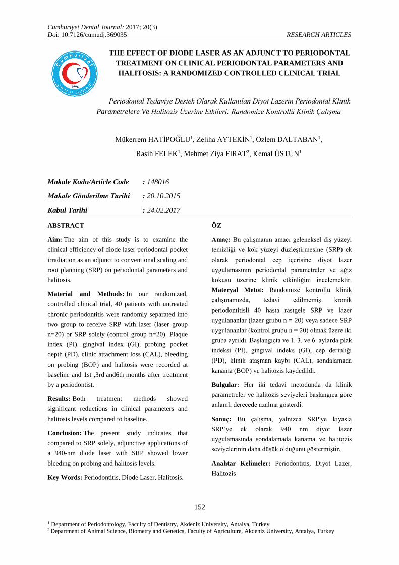
THE EFFECT OF DIODE LASER AS AN ADJUNCT TO PERIODONTAL
TREATMENT ON CLINICAL PERIODONTAL PARAMETERS AND
HALITOSIS: A RANDOMIZED CONTROLLED CLINICAL TRIAL
Periodontal Tedaviye Destek Olarak Kullanılan Diyot Lazerin Periodontal Klinik
Parametrelere Ve Halitozis Üzerine Etkileri: Randomize Kontrollü Klinik Çalışma
Mükerrem HATİPOĞLU1, Zeliha AYTEKİN1, Özlem DALTABAN1,
Rasih FELEK1, Mehmet Ziya FIRAT2, Kemal ÜSTÜN1
Makale Kodu/Article Code : 148016
Makale Gönderilme Tarihi : 20.10.2015
Kabul Tarihi : 24.02.2017
ABSTRACT
Aim: The aim of this study is to examine the
clinical efficiency of diode laser periodontal pocket
irradiation as an adjunct to conventional scaling and
root planning (SRP) on periodontal parameters and
halitosis.
Material and Methods: In our randomized,
controlled clinical trial, 40 patients with untreated
chronic periodontitis were randomly separated into
two group to receive SRP with laser (laser group
n=20) or SRP solely (control group n=20). Plaque
index (PI), gingival index (GI), probing pocket
depth (PD), clinic attachment loss (CAL), bleeding
on probing (BOP) and halitosis were recorded at
baseline and 1st ,3rd and6th months after treatment
by a periodontist.
Results: Both treatment methods showed
significant reductions in clinical parameters and
halitosis levels compared to baseline.
Conclusion: The present study indicates that
compared to SRP solely, adjunctive applications of
a 940-nm diode laser with SRP showed lower
bleeding on probing and halitosis levels.
Key Words: Periodontitis, Diode Laser, Halitosis.
ÖZ
Amaç: Bu çalışmanın amacı geleneksel diş yüzeyi
temizliği ve kök yüzeyi düzleştirmesine (SRP) ek
olarak periodontal cep içerisine diyot lazer
uygulamasının periodontal parametreler ve ağız
kokusu üzerine klinik etkinliğini incelemektir.
Materyal Metot: Randomize kontrollü klinik
çalışmamızda, tedavi edilmemiş kronik
periodontitisli 40 hasta rastgele SRP ve lazer
uygulananlar (lazer grubu n = 20) veya sadece SRP
uygulananlar (kontrol grubu n = 20) olmak üzere iki
gruba ayrıldı. Başlangıçta ve 1. 3. ve 6. aylarda plak
indeksi (PI), gingival indeks (GI), cep derinliği
(PD), klinik ataşman kaybı (CAL), sondalamada
kanama (BOP) ve halitozis kaydedildi.
Bulgular: Her iki tedavi metodunda da klinik
parametreler ve halitozis seviyeleri başlangıca göre
anlamlı derecede azalma gösterdi.
Sonuç: Bu çalışma, yalnızca SRP'ye kıyasla
SRP’ye ek olarak 940 nm diyot lazer
uygulamasında sondalamada kanama ve halitozis
seviyelerinin daha düşük olduğunu göstermiştir.
Anahtar Kelimeler: Periodontitis, Diyot Lazer,
Halitozis
Hatipoglu M. et al.
153
INTRODUCTION
Chronic periodontitis is an inflammatory
disease which develops against the microbial
plaque on tooth surface and ends in loss of
periodontal tissues.1 The prime target in
periodontal treatment is the elimination of all
factors which cause formation and cumulation
of the plaque.2
In nonsurgical periodontal treatment,
infection is aimed to be controlled by
mechanically removing supragingival and
subgingival calculus.3 The success of
periodontal treatment depends on totally
removing subgingival and supragingival
pathogens and enabling oral hygiene by the
patient after the treatment.4, 5
Tooth surface (SRP) is the most important
procedure in the treatment of periodontitis and
its clinical benefits were proved by many
studies.4, 6, 7 However, microbial component
may not be removed totally by mechanical
treatment in the presence of deep pockets.8
Therefore, many methods such as antibiotics,
antiseptics and lasers have been used in
addition to periodontal treatment. 9-12
Dental laser usage has been commonly
used recently. Different laser types are used for
dental purposes such as Er:YAG laser,
Er,Cr:YSGG laser, Nd:YAG laser, CO2 laser
and diode laser.13, 14
Diode laser is a semiconductor laser,
which uses combinations of elements such as
gallium (Ga), arsenide (Ar), aluminum (Al)
and indium (In) for the transformation of
electric energy into luminous energy.15,16
Diode laser, which can be used in soft tissue
implementations successfully, do not penetrate
into hard tissues.13 Many studies showed that
diode laser has an antibacterial activity when it
is used to support periodontal treatment.17-19
Moritz et al. suggest that diode laser as an
adjunct to SRP will decrease bacteria amount
and inflammation.17 They achieved positive
results in clinical parameters but irradiation
procedure caused morphological changes on
root surface.17, 19
The term 'halitosis' means bad breath
which can have local or systemic origins. 20, 21
10 per cent of halitosis cases develop because
of extra-oral reasons.22 Halitosis can be
observed in people of different ages and
negatively affect social interaction of the
person.23 Findings which belong to
epidemiological studies in different countries
cannot be compared because there are not
standart protocols for treatment and diagnosis
of halitosis.24
Most of extra oral factors which cause
halitosis are generated from respiratory tract or
otorhinolaryngologic diseases such as
tonsillitis, sinusitis and post-nasal drip. Bad
breath can occur rarely because of renal,
hepatic, endocrinological or gastrointestinal
reasons.22, 24-27 The cumulated materials can be
smelt by the breath within some certain
systemic diseases such as acetone smell within
diabetic patients or ammonia smell within urea
or cirrhotic patients.28 Periodontal diseases,
caries, bacteria plaque on the tongue,
insufficient salivation, stomatitis, in-mouth
neoplasm, extraction socket which is being
treated and smelly food consumption are
amongst in-mouth reasons of halitosis.22, 24, 29
Volatile sulphur compounds (VSC) are
formed as oral microorganisms which exist in
saliva, periodontal pockets, tongue and other
parts of the mouth proteolysate free amino
aside substrates, such as cysteine, sistine and
methionine which include sulphur.30 VSC is
mainly composed of hydrogen sulfide, methyl
mercaptan and dimethyl sulfide components
which are the main reasons of bad breath.31, 32
Two methods are implemented in the
evaluation of halitosis; the first one is
organoleptic method which is subjective. In
this method, breath of the patient is graded
through smelling by an educated and
experienced person. The method can give
variable values because it is a sense method
The Effect of Diode Laser as an Adjunct to Periodontal Treatment on Clinical Periodontal Parameters and Halitosis: A
Randomized Controlled Clinical Trial
154
besides it is not a good experience both for the
patient and the implementer.38, 39 The second
method is measuring VSC amount by gas
chromatography or halimeter which is an
objective method.40 When halitosis is
measured by a halimeter, nano VSC amount
can be defined.41-45 VSC amount within the
people with periodontal diseases are more than
healthy people because of the high rate of
bacteria plaque covering the tongue. Therefore,
there is a positive relation between the severity
of periodontal disease and VSC content.31
The main principle in the treatment of
halitosis is eliminating oral pathogens and
restraining bacterial bio film. Many methods
are adopted in order to treat halitosis such as
using mouthwash solutions with CHX,
hydrogen peroxide and essential oil, tongue
scraper or tongue brush. 70 per cent of oral
sulfides will be eliminated by tongue
cleaning.46 Chewing gum with mint and mouth
sprays is also used in order to cover the
smell.21, 24, 47-51
Our study aims at evaluating the
effectiveness of diode laser usage as an adjunct
to SPR both on periodontal parameters and
halitosis.
MATERIAL AND METHOD
Selection of the Participators and Design
of the Study
This study is a randomized controlled
clinical study. 40 adults (20 females, 20
males), who were selected from the patients
without periodontal treatment in the last 6
months, who consulted to Akdeniz University
Faculty of Dentistry Periodontology clinic
between September 2014 and March 2015 for
periodontal complaints or controls, were
included in the study. All patient provided
written permissions. The protocol of the study
was approved by Antalya Training Research
Hospital Non-pharmaceutical Clinic
Researches Ethical Commission (2014
decision no:46/10).
The people who have systemic diseases,
require regular medicine, are pregnant, smoke
and people with fixed partial denture were not
included in the study. The people have
minimum 14 teeth and at least 2 teeth that have
5mm pocket in each quadrant. The people were
only divided into 2 random groups as the ones
who have SRP treatment (control group KG
n=20) and who are implemented diode laser as
an adjunct to SRP (Laser group LG n=20)
Halitosis measurement
In our study halitosis measurements are
done by Halimeter (InterscanCorp., Chatsworth,
Ca, USA). People were asked not to consume
onion, garlic and spicy food and use mouthwash
before the implementation day. People were
also asked to breathe through the nose without
opening their mouths for a minute then
halimeter was placed into the mouth as not to
touch the patient's tongue and palate.
Clinical Procedure
Plaque index (PI), gingival index (GI),
clinical attachment level (CAL), probing
pocket depth (PD), bleeding on probing (BOP)
and halitosis measurements were carried out in
6 sections for each tooth after treatment prior
to treatment of the patients.
SRP was implemented via hand pieces
(Gracey Curettes, Hu-Friedy, Chicago, IL,
USA) and ultrasonic equipment (EMS SA CH
1260 Nyon, SWITZERLAND). 940 nm
indium-gallium-aluminum-phosphate diode
lasers (Epic, Biolase, Irvine, CA, USA) were
implemented in the same session under local
anesthesia. Total 15 J/cm2 power of laser in
1.5 W power with 20 ms frequency during 20
ms shots was implemented to periodontal
pocket. Laser irradiation was realized with
fiber optic ends which are of 300 µm diameter.
Fiber is implemented by parallelly locating on
root surface level inside the periodontal
pocket. Fiber laser is directed from apical to
coronal during light emission. It was
implemented in total 20 seconds as 10 second
Hatipoglu M. et al.
155
lingual to 10 seconds to each tooth in mezio-
distal direction in buccal angle.12
The patients were provided with detailed
oral hygiene training at the end of the session.
The training includes the usage of materials
such as dental floss and interdental brush
which are used for interdental cleaning and
how to clean dorsum and lateral of tongue with
brush and tongue cleaner.
Clinical index scores and halitosis levels
were measured after 1, 3 and 6 months after
the treatment. The patients were not informed
about their categories in the groups during the
treatment.
Whether they correlate normal
distribution or not is decided via Shapiro-
Wilks test for statistical assessment of datum.
Variance analysis, Tukey multiple comparison
tests and independent sample t-test were
carried out via an appropriate software for the
assessment of datum which are defined to
present normal distribution. (SPSS v20.0,
IBM, Chicago, IL, USA).
RESULTS
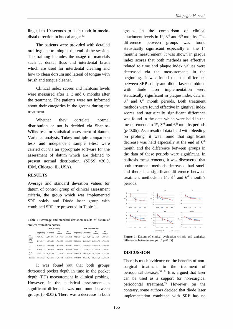
Average and standard deviation values for
datum of control group of clinical assessment
criteria, the group which was implemented
SRP solely and Diode laser group with
combined SRP are presented in Table 1.
Table 1: Average and standard deviation results of datum of
clinical evaluation criteria
It was found out that both groups
decreased pocket depth in time in the pocket
depth (PD) measurement in clinical probing.
However, in the statistical assessments a
significant difference was not found between
groups (p>0.05). There was a decrease in both
groups in the comparison of clinical
attachment levels in 1st, 3rd and 6th months. The
difference between groups was found
statistically significant especially in the 1st
month's measurement. It was shown in plaque
index scores that both methods are effective
related to time and plaque index values were
decreased via the measurements in the
beginning. It was found that the difference
between SRP solely and diode laser combined
with diode laser implementation were
statictically significant in plaque index data in
3rd and 6th month periods. Both treatment
methods were found effective in gingival index
scores and statistically significant difference
was found in the date which were held in the
measurements in 1st, 3rd and 6th months periods
(p<0.05). As a result of data held with bleeding
on probing, it was found that significant
decrease was held especially at the end of 6th
month and the difference between groups in
the data of these periods were significant. In
halitosis measurements, it was discovered that
both treatment methods decreased bad smell
and there is a significant difference between
treatment methods in 1st, 3rd and 6th month’s
periods.
Figure 1: Datum of clinical evaluation criteria and statistical
differences between groups. (*:p<0.05)
DISCUSSION
There is much evidence on the benefits of non-
surgical treatment in the treatment of
periodontal diseases.53 54 It is argued that laser
can be used as a support for non-surgical
periodontal treatment.55 However, on the
contrary, some authors decided that diode laser
implementation combined with SRP has no
SRP (Control) SRP + Diode Laser
Beginning 1st month 3rd
month
6th
month Beginning 1st month
3rd
month
6th
month
PD
(mm) 4,00±0,53 2,40±0,75 2,05±0,76 1,95±0,81 4,05±0,64 2,44±0,27 2,11±0,41 1,88±0,55
CAL
(mm) 2,74±0,59 1,87±0,41 1,76±0,55 1,81±0,60 3,03±0,65 2,32±0,59 2,09±0,74 1,75±0,58
PI 1,86±0,28 1,54±0,33 1,47±0,26 1,41±0,16 1,88±0,27 1,44±0,29 1,33±0,15 1,25±0,15
GI 1,96±0,28 1,65±0,27 1,54±0,28 1,41±0,31 1,98±0,25 1,42±0,27 1,17±0,13 1,14±0,11
BOP
(%) 74,8±7,59 49,4±8,56 42,1±9,73 31,5±7,23 75,9±6,79 49,6±8,93 40,1±9,80 21,7±8,16
Halitosis 83,4±17,2 70,1±8,56 61,8±10,2 58,2±10,8 83,9±14,9 64,5±10,1 55,4±11,4 52,4±8,69
The Effect of Diode Laser as an Adjunct to Periodontal Treatment on Clinical Periodontal Parameters and Halitosis: A
Randomized Controlled Clinical Trial
156
supremacy over SRP implementation solely in
terms of microbial parameters and gingival
inflammation.56, 57 In our study, it was
presented that both treatment prosedures caused
significant recovery in clinical parameters.
Dukic and colleagues expressed that
statistically significant recovery was held in
clinical parameters in the group which they
implemented SRP solely and the group
implemented with diode laser combined with
SRP in 6th and 18th weeks in a study, which
was carried out on people with chronic
periodontitis.2
Aykol and colleagues achieved progress
in periodontal pocket depth and bleeding on
probing per cent in the group they
implemented gallium-aluminum-arsenide after
periodontal treatment in 1st, 2nd and 7th day
through a similar study which they carried out
with chronic periodontitis patients.58
Kreisler and colleagues implemented
diode laser of 810 nm wave length to 2 random
quadrants of the patients after they treated 22
patients with chronic periodontitis via routine
SRP during their study. They presented that
more statistically significant decrease was held
in teeth mobility, pocket depth and clinical
attachment loss in the tooth which were
implemented laser when compared to control
group.59
In our study CAL, BOP and PI datum
were compared and decrease was found in both
groups in 1st, 3rd and 6th months. However, a
statistically significant recovery was achieved
in LG when compared to CG in the 1st month's
measurements. A statistically significant
decrease was held in BOP measurements in
LG when compared to CG in 6th month.
Whilst probing pocket depth decreased in
time in both groups in PD measurements a
statistically significant difference cannot be
found between groups.
Qadri and colleagues had better results in
periodontal pocket depth, plaque index and
gingival index in the areas where they
implemented laser when they carried out a
similar study design.60
Ustun and colleagues implemented diode
laser of 810 nm wave length to 2 random
quadrants of the patients after they treated 21
patients with chronic periodontitis via routine
SRP during their study. CAL, BOP and PI
datum were compared and decrease was found
in both groups in 1st, 3rd and 6th months. More
decrease was achieved in LG when compared
to CG in terms of PD levels in 1st, 3rd and 6th
months. Better results were found in LG when
compared to CG in terms of CAL and GI
levels in 3rd and 6th months. They could not
have found a statistically significant difference
between two groups in terms of PI.61
PI and GI datum were compared and a
decrease in both groups were found in our
study. However, in all of 1st, 3rd and 6th
months' measurements in GI levels of LG had
a statistically significantly decrease when
compared to CG. Whilst decrease was
observed in both groups within PI
measurements, a statistically significant
decrease was achieved in LG when compared
to CG in 3rd and 6th moths.
Many authors reported that periodontal
disease is a significant reason of halitosis.62-65
Periodontal pathogens generate endotoxine,
proteinase and VSC. Persson and colleagues
stated that Bacteroides melaninogenicus,
Treponema denticola, Porphyromonas
gingivalis and Prevotella intermedia types
produce VSC.66 VSC molecules, which are
produced by gram negative bacteria on tongue
and periodontal pocket, cause smell
formation.67
A number of studies showed the positive
relation between progression of periodontal
disease and VSC amount.35, 36 Morita and
Wang68 found a significant relation between
the severity of periodontal disease and VSC
amount in breath. They discovered that VSC
value was measured lower within people who
Hatipoglu M. et al.
157
are provided with periodontal treatment than
the ones who are not treated. They also
asserted that there is a correlation between
VSC level and bleeding on probing and
periodontal pocket depth. Coil and Tonzetich69
also found higher VSC levels in the people
with deep pockets which bleed during probing
than the people with shallow pockets with low
bleeding rate during probing.
A similar relation between periodontitis
and halitosis was also determined in our study.
VSC level in 1st, 3rd and 6th months were found
to be lower than the first measured level in
both groups which have periodontal treatment.
Dissimilarly, VSC level which is measured in
LG is found to be statistically significantly
lower than CG in the 1st and 3rd months.
In the light of this information, we can
state that laser treatment which is implemented
as an adjunct to SRP has a statistically
significant positive effect on clinical
parameters and halitosis. In addition to that
there is a need for new studies to discover in
which mechanisms does laser effect these
parameters.
REFERENCES
1. Armitage GC. Development of a
classification system for periodontal diseases and
conditions. Annals of periodontology / the
American Academy of Periodontology. 1999;
4:1-6.
2. Dukic W, Bago I, Aurer A, Roguljic M.
Clinical effectiveness of diode laser therapy as an
adjunct to non-surgical periodontal treatment: a
randomized clinical study. Journal of
periodontology. 2013; 84:1111-7.
3. Kaldahl WB, Kalkwarf KL, Patil KD, Molvar
MP, Dyer JK. Long-term evaluation of
periodontal therapy: I. Response to 4 therapeutic
modalities. Journal of periodontology. 1996;
67:93-102.
4. O'Leary TJ. The impact of research on scaling
and root planing. Journal of periodontology.
1986; 57:69-75.
5. Cugini MA, Haffajee AD, Smith C, Kent RL,
Jr., Socransky SS. The effect of scaling and root
planing on the clinical and microbiological
parameters of periodontal diseases: 12-month
results. Journal of clinical periodontology. 2000;
27:30-6.
6. Lindhe J, Nyman S. Scaling and granulation
tissue removal in periodontal therapy. Journal of
clinical periodontology. 1985; 12:374-88.
7. Kaldahl WB, Johnson GK, Patil KD,
Kalkwarf KL. Levels of cigarette consumption
and response to periodontal therapy. Journal of
periodontology. 1996; 67:675-81.
8. Sherman PR, Hutchens LH, Jr., Jewson LG.
The effectiveness of subgingival scaling and root
planing. II. Clinical responses related to residual
calculus. Journal of periodontology. 1990; 61:9-
15.
9. Mombelli A. Antimicrobial advances in
treating periodontal diseases. Frontiers of oral
biology. 2012; 15:133-48.
10. Sanz M, Teughels W, Group AoEWoP.
Innovations in non-surgical periodontal therapy:
Consensus Report of the Sixth European
Workshop on Periodontology. Journal of clinical
periodontology. 2008; 35:3-7.
11. Socransky SS, Haffajee AD. Dental biofilms:
difficult therapeutic targets. Periodontology
2000. 2002; 28:12-55.
12. Saglam M, Kantarci A, Dundar N, Hakki SS.
Clinical and biochemical effects of diode laser as
an adjunct to nonsurgical treatment of chronic
periodontitis: a randomized, controlled clinical
trial. Lasers in medical science. 2014; 29:37-46.
13. Aoki A, Sasaki KM, Watanabe H, Ishikawa I.
Lasers in nonsurgical periodontal therapy.
Periodontology 2000. 2004; 36:59-97.
14. Ishikawa I, Aoki A, Takasaki AA, Mizutani
K, Sasaki KM, Izumi Y. Application of lasers in
periodontics: true innovation or myth?
Periodontology 2000. 2009; 50:90-126.
15. Yiğit ŞB GM. Periodontolojide lazer. Selçuk
Üniversitesi Dişhekimliği Fakültesi Dergisi.
2007; 16:67-73.
The Effect of Diode Laser as an Adjunct to Periodontal Treatment on Clinical Periodontal Parameters and Halitosis: A
Randomized Controlled Clinical Trial
158
16. Research S, Therapy Committee of the
American Academy of P. Lasers in periodontics.
Journal of periodontology. 2002; 73:1231-9.
17. Moritz A, Gutknecht N, Doertbudak O,
Goharkhay K, Schoop U, Schauer P, et al.
Bacterial reduction in periodontal pockets
through irradiation with a diode laser: a pilot
study. Journal of clinical laser medicine &
surgery. 1997; 15:33-7.
18. Harris DM, Yessik M. Therapeutic ratio
quantifies laser antisepsis: ablation of
Porphyromonas gingivalis with dental lasers.
Lasers in surgery and medicine. 2004; 35:206-
13.
19. Moritz A, Schoop U, Goharkhay K, Schauer
P, Doertbudak O, Wernisch J, et al. Treatment of
periodontal pockets with a diode laser. Lasers in
surgery and medicine. 1998; 22:302-11.
20. Shimura M, Watanabe S, Iwakura M,
Oshikiri Y, Kusumoto M, Ikawa K, et al.
Correlation between measurements using a new
halitosis monitor and organoleptic assessment.
Journal of periodontology. 1997; 68:1182-5.
21. Quirynen M, Avontroodt P, Soers C, Zhao H,
Pauwels M, van Steenberghe D. Impact of
tongue cleansers on microbial load and taste.
Journal of clinical periodontology. 2004; 31:506-
10.
22. Tangerman A, Winkel EG. Intra- and extra-
oral halitosis: finding of a new form of extra-oral
blood-borne halitosis caused by dimethyl
sulphide. Journal of clinical periodontology.
2007; 34:748-55.
23. Morita M, Wang HL. Association between
oral malodor and adult periodontitis: a review.
Journal of clinical periodontology. 2001; 28:813-
9.
24. Bollen CM, Beikler T. Halitosis: the
multidisciplinary approach. International journal
of oral science. 2012; 4:55-63.
25. Amir E, Shimonov R, Rosenberg M. Halitosis
in children. The Journal of pediatrics. 1999;
134:338-43.
26. Marocchio LS, Junior DS, de Sousa SC,
Fabre RF, Raitz R. Multifocal diffuse oral
melanoacanthoma: a case report. Journal of oral
science. 2009; 51:463-6.
27. Scully C, Greenman J. Halitology (breath
odour: aetiopathogenesis and management). Oral
diseases. 2012; 18:333-45.
28. TAŞDOĞAN BE GY. Halitozis ve
Helikobakter pilori. . Güncel Gastroenteroloji
Dergisi. 2015; 15.
29. Torresyap G, Haffajee AD, Uzel NG,
Socransky SS. Relationship between periodontal
pocket sulfide levels and subgingival species.
Journal of clinical periodontology. 2003;
30:1003-10.
30. Sanz M, Roldan S, Herrera D. Fundamentals
of breath malodour. The journal of contemporary
dental practice. 2001; 2:1-17.
31. Yaegaki K, Sanada K. Volatile sulfur
compounds in mouth air from clinically healthy
subjects and patients with periodontal disease.
Journal of periodontal research. 1992; 27:233-8.
32. Yaegaki K, Coil JM. Examination,
classification, and treatment of halitosis; clinical
perspectives. Journal. 2000; 66:257-61.
33. Johnson PW, Yaegaki K, Tonzetich J. Effect
of volatile thiol compounds on protein
metabolism by human gingival fibroblasts.
Journal of periodontal research. 1992; 27:553-61.
34. Johnson P, Yaegaki K, Tonzetich J. Effect of
methyl mercaptan on synthesis and degradation
of collagen. Journal of periodontal research.
1996; 31:323-9.
35. Calenic B, Yaegaki K, Murata T, Imai T,
Aoyama I, Sato T, et al. Oral malodorous
compound triggers mitochondrial-dependent
apoptosis and causes genomic DNA damage in
human gingival epithelial cells. Journal of
periodontal research. 2010; 45:31-7.
36. Yaegaki K, Qian W, Murata T, Imai T, Sato
T, Tanaka T, et al. Oral malodorous compound
causes apoptosis and genomic DNA damage in
human gingival fibroblasts. Journal of
periodontal research. 2008; 43:391-9.
37. Ng W, Tonzetich J. Effect of hydrogen
sulfide and methyl mercaptan on the
permeability of oral mucosa. Journal of dental
research. 1984; 63:994-7.
Hatipoglu M. et al.
159
38. Rosenberg M, Kulkarni GV, Bosy A,
McCulloch CA. Reproducibility and sensitivity
of oral malodor measurements with a portable
sulphide monitor. Journal of dental research.
1991; 70:1436-40.
39. Kim DJ, Lee JY, Kho HS, Chung JW, Park
HK, Kim YK. A new organoleptic testing
method for evaluating halitosis. Journal of
periodontology. 2009; 80:93-7.
40. Murata T, Rahardjo A, Fujiyama Y, Yamaga
T, Hanada M, Yaegaki K, et al. Development of
a compact and simple gas chromatography for
oral malodor measurement. Journal of
periodontology. 2006; 77:1142-7.
41. Rosenberg M, McCulloch CA. Measurement
of oral malodor: current methods and future
prospects. Journal of periodontology. 1992;
63:776-82.
42. Kizhner V, Xu D, Krespi YP. A new tool
measuring oral malodor quality of life. European
archives of oto-rhino-laryngology : official
journal of the European Federation of Oto-
Rhino-Laryngological Societies. 2011;
268:1227-32.
43. Rosenberg M. Bad breath, diagnosis and
treatment. University of Toronto dental journal.
1990; 3:7-11.
44. Donaldson AC, Riggio MP, Rolph HJ, Bagg
J, Hodge PJ. Clinical examination of subjects
with halitosis. Oral diseases. 2007; 13:63-70.
45. Furne J, Majerus G, Lenton P, Springfield J,
Levitt DG, Levitt MD. Comparison of volatile
sulfur compound concentrations measured with a
sulfide detector vs. gas chromatography. Journal
of dental research. 2002; 81:140-3.
46. Rosenberg M. Bad breath and periodontal
disease: how related are they? Journal of clinical
periodontology. 2006; 33:29-30.
47. Raangs GC, Winkel EG, van Winkelhoff AJ.
In vitro antimicrobial effects of two antihalitosis
mouth rinses on oral pathogens and human
tongue microbiota. International journal of dental
hygiene. 2013; 11:203-7.
48. Tolentino Ede S, Chinellato LE, Tarzia O.
Saliva and tongue coating pH before and after
use of mouthwashes and relationship with
parameters of halitosis. Journal of applied oral
science : revista FOB. 2011; 19:90-4.
49. Wainwright M. Photodynamic antimicrobial
chemotherapy (PACT). The Journal of
antimicrobial chemotherapy. 1998; 42:13-28.
50. Saad S, Greenman J, Shaw H. Comparative
effects of various commercially available
mouthrinse formulations on oral malodor. Oral
diseases. 2011; 17:180-6.
51. Quirynen M, Mongardini C, van Steenberghe
D. The effect of a 1-stage full-mouth disinfection
on oral malodor and microbial colonization of
the tongue in periodontitis. A pilot study. Journal
of periodontology. 1998; 69:374-82.
52. Karlsson MR, Diogo Lofgren CI, Jansson
HM. The effect of laser therapy as an adjunct to
non-surgical periodontal treatment in subjects
with chronic periodontitis: a systematic review.
Journal of periodontology. 2008; 79:2021-8.
53. Cobb CM. Lasers in periodontics: a review of
the literature. Journal of periodontology. 2006;
77:545-64.
54. Slot DE, Kranendonk AA, Paraskevas S, Van
der Weijden F. The effect of a pulsed Nd:YAG
laser in non-surgical periodontal therapy. Journal
of periodontology. 2009; 80:1041-56.
55. Miyazaki A, Yamaguchi T, Nishikata J,
Okuda K, Suda S, Orima K, et al. Effects of
Nd:YAG and CO2 laser treatment and ultrasonic
scaling on periodontal pockets of chronic
periodontitis patients. Journal of periodontology.
2003; 74:175-80.
56. De Micheli G, de Andrade AK, Alves VT,
Seto M, Pannuti CM, Cai S. Efficacy of high
intensity diode laser as an adjunct to non-surgical
periodontal treatment: a randomized controlled
trial. Lasers in medical science. 2011; 26:43-8.
57. Ambrosini P, Miller N, Briancon S, Gallina S,
Penaud J. Clinical and microbiological
evaluation of the effectiveness of the Nd:Yap
laser for the initial treatment of adult
periodontitis. A randomized controlled study.
Journal of clinical periodontology. 2005; 32:670-
6.
58. Aykol G, Baser U, Maden I, Kazak Z, Onan
U, Tanrikulu-Kucuk S, et al. The effect of low-
The Effect of Diode Laser as an Adjunct to Periodontal Treatment on Clinical Periodontal Parameters and Halitosis: A
Randomized Controlled Clinical Trial
160
level laser therapy as an adjunct to non-surgical
periodontal treatment. Journal of periodontology.
2011; 82:481-8.
59. Kreisler M, Al Haj H, d'Hoedt B. Clinical
efficacy of semiconductor laser application as an
adjunct to conventional scaling and root planing.
Lasers in surgery and medicine. 2005; 37:350-5.
60. Qadri T, Miranda L, Tuner J, Gustafsson A.
The short-term effects of low-level lasers as
adjunct therapy in the treatment of periodontal
inflammation. Journal of clinical periodontology.
2005; 32:714-9.
61. Ustun K, Erciyas K, Sezer U, Senyurt SZ,
Gundogar H, Ustun O, et al. Clinical and
biochemical effects of 810 nm diode laser as an
adjunct to periodontal therapy: a randomized
split-mouth clinical trial. Photomedicine and
laser surgery. 2014; 32:61-6.
62. Loesche WJ, Kazor C. Microbiology and
treatment of halitosis. Periodontology 2000.
2002; 28:256-79.
63. Miyazaki H, Sakao S, Katoh Y, Takehara T.
Correlation between volatile sulphur compounds
and certain oral health measurements in the
general population. Journal of periodontology.
1995; 66:679-84.
64. Soder B, Johansson B, Soder PO. The relation
between foetor ex ore, oral hygiene and
periodontal disease. Swedish dental journal.
2000; 24:73-82.
65. Proietti MC, Reisser J, Marins LF,
Marcovaldi MA, Soares LS, Monteiro DS, et al.
Hawksbill x loggerhead sea turtle hybrids at
Bahia, Brazil: where do their offspring go? PeerJ.
2014; 2:e255.
66. Persson S, Edlund MB, Claesson R, Carlsson
J. The formation of hydrogen sulfide and methyl
mercaptan by oral bacteria. Oral microbiology
and immunology. 1990; 5:195-201.
67. Pham TA, Ueno M, Zaitsu T, Takehara S,
Shinada K, Lam PH, et al. Clinical trial of oral
malodor treatment in patients with periodontal
diseases. Journal of periodontal research. 2011;
46:722-9.
68. Morita M, Wang HL. Relationship of sulcular
sulfide level to severity of periodontal disease
and BANA test. Journal of periodontology. 2001;
72:74-8.
69. Coli JM, Tonzetich J. Characterization of
volatile sulphur compounds production at
individual gingival crevicular sites in humans.
The Journal of clinical dentistry. 1992; 3:97-103.
Correspondence Author
Dr. Mükerrem Hatipoğlu.
Akdeniz University
Faculty of Dentistry
Department of Periodontology.
07058 Antalya.
Phone: 05418838309
E-mail:[email protected]
Cumhuriyet Dental Journal: 2017; 20(3)
Doi: 10.7126/cumudj.369086 RESEARCH ARTICLES
161
1 Department of Conservative Dentistry, The Ministry of Health of Turkey, Balgat Dentaland Oral Health Center, Ankara, Turkey 2 Department of Prosthodontics, Faculty of Dentistry, Istanbul Medipol University, Istanbul, Turkey 3 Department of Prosthodontics, Faculty of Dentistry, Yildirim Beyazit University, Ankara, Turkey
EFFECT OF TWO ACTIVATED BLEACHING TECHNIQUES
ON SURFACE ROUGHNESS OF DIFFERENT ESTHETIC
RESTORATIVE MATERIALS
İki Aktive Olan Beyazlatma Tekniğinin Farklı Estetik Restoratif
Materyallerin Yüzey Pürüzlülüğü Üzerine Etkisi
Tuncay ALPTEKIN1, Özgün Yusuf ÖZYILMAZ2,
Filiz AYKENT3, Haluk Barış KARA2
Makale Kodu/Article Code : 192786
Makale Gönderilme Tarihi : 16.06.2016
Kabul Tarihi : 28.03.2017
ABSTRACT
Objectives: The aim of this in vitro study was to evaluate surface
roughness of six different restorative materials during office
bleaching procedures with blue light emitted diode (LED) and
diode laser photo activation.
Materials and Methods: Filtek TM supreme, Tetric Evo Ceram,
Tescera ATL, Clearfill Majesty Esthetic, Durafill VS and IPS
Empress 2 materials were evaluated in this study. Twenty
specimens, 10 mm in diameter and 2 mm thick, were fabricated
from each material using a teflon mold. All specimens were
randomly assigned to two groups (n=10). Group 1 received two
topical applications of 35% hydrogen peroxide for 20 s. And was
photoactivated using LED. Group 2 received topical application
of 46% hydrogen peroxide using diode laser. Surface roughness
values were measured prior to and following bleaching
procedures by using a profilometer. Data were analyzed
statistically, by one-way-analysis of variance (ANOVA), post-hoc
Tamhane's T2 and independent t tests.
Results: Surface roughness values for all restorative materials
tested increased after both bleaching procedures (p<0.05).
Tescera ATL bleached with diode laser photo activation showed
higher surface roughness value than LED activation (p<0.05).
However, there were no significant differences in two bleaching
methods for other restorative materials (p>0.05).
Conclusions: Although clinical effects depend on in-vivo
conditions, the effects of office bleaching agents should be known
and applied cautiously when a colored restoration is bleached or
a restoration is neighboured with the tooth bleached.
Keywords: Esthetic restorative materials, Dental porcelain, Teeth
bleaching, Surface roughness, Semiconductor lasers
ÖZ
Amaçlar: Bu in vitro çalışmanın amacı, mavi ışık yayan diyot
(LED) ve diyot lazer ile foto aktive olan ofis tipi beyazlatma
prosedürleri esnasında altı farklı restoratif materyalin yüzey
pürüzlülüğünü değerlendirmektir.
Materyal ve Metotlar: Bu çalışmada Filtek TM supreme, Tetric
Evo Ceram, Tescera ATL, Clearfill Majesty Esthetic, Durafill VS
ve IPS Empress 2 materyalleri kullanıldı. Yirmi örnek; her biri
10 mm çapında 2 mm kalınlığında olacak şekilde teflon kalıp
kullanılarak oluşturuldu. Bütün örnekler rastgele iki gruba ayrıldı
(n=10). Grup 1’ de topikal uygulanan %35’lik hidrojen peroksit
LED kullanılarak 20 saniye süreyle foto aktive edildi. Grup 2’
de %46’lık hidrojen peroksit diode lazer kullanılarak topikal
olarak uygulandı. Yüzey pürüzlülüğü değerleri başlangıç ve
beyazlatma işlemleri sonrasında olmak üzere profilometre
yardımıyla ölçüldü. Veriler tek-yönlü varyans analizi (ANOVA),
Tamhane's T2 ve bağımsız t testleri yapılarak istatiksel olarak
analiz edildi.
Sonuçlar: Yüzey pürüzlülüğü değerleri test edilen tüm restoratif
materyallerde her iki beyazlatma prosedüründe de artmıştır
(p<0,05). Diode lazer foto aktivasyonu ile beyazlatılan Tescera
ATL, LED ile aktive edilenden daha yüksek yüzey pürüzlülüğü
değeri gösterdi (p<0,05). Fakat diğer restoratif materyaller için iki
beyazlatma prosedüründe de önemli farklılıklar ortaya çıkmadı
(p>0,05).
Çıkarımlar: Klinik etkiler in-vivo şartlara bağlı olmasına rağmen
ofis tipi beyazlatma ajanlarının etkileri bilinmeli ve renklenmiş bir
restorasyon beyazlatıldığında veya beyazlatılmış diş ile komşu
restorasyon varlığında dikkatle kullanılmalıdır.
Anahtar kelimeler: Estetik restoratif materyaller, Dental
porselen, Diş beyazlatma, Yüzey pürüzlülüğü, Yarı iletken
lazerler
Alptekin T. et al.
162
INTRODUCTION
Dental bleaching is the best conservative
treatment to whitening the natural teeth in order
to improve aesthetic reasons.1,2 There are two
techniques for bleaching vital teeth: in-office
under supervision of a dentist, or at-home, with
the use of lower concentration of whitening
agents in special trays. In-office bleaching
systems produced more rapid results,
professional controls the contact of whitening
agents with soft tissues and avoidance of the
material ingestion. On the other hand, at-home
bleaching is being a lower cost procedure in
comparison with in-office treatment, being easy
to use by patients. Nevertheless, common
problems for this technique are sensitivity of the
tooth and soft tissue, incorrect use of by patients
and overexposure to the over-the-counter
products without professional control.3-7
Hydrogen peroxide, sodium perborate and
carbamide peroxide are the agents used for
dental bleaching procedures.6-8 To enhance or
accelerate the whitening process, heat
activation of bleaching agent by light, heat or
laser is described in the literature.9 Light-
activated bleaching is a method of tooth
whitening that can be achieved by utilizing
highly concentrated bleaching gel (35 to 50%
hydrogen peroxide). Halogen light, derived
from relatively low-cost technology, produces
high heat generation and loss of pulpal vitality.
LEDs (light emitting diodes) are also able to
produce light in a specific wavelength and have
low amounts of wasted energy and minimum
heat generation.10, 11 Laser technology has
rapidly developed during last two decades. Its
applications have been successfully
implemented in the medical professions.12 Laser
tooth bleaching officially started 1996, with the
approval of the argon laser (480 nm) and the
CO2 laser (10.6 µm).13,14 Today three types
lasers; argon (488/514 nm), CO2 (10.6 µm), and
diode (790-980 nm) lasers are approved for
tooth whitening according to US Food and Drug
Administration.10
In literature several reports exist regarding
the influence of bleaching agents on surface
morphology of composite15-20 and ceramic
materials15, 21, 22 and these studies present
controversial findings. Beside this there is no
information published regarding the potential
effects of activated bleaching techniques on the
surface roughness of the composites, ceramic
and ormocer. Therefore, the purpose of this
study was to evaluate the effect of two in-office
bleaching techniques (blue light-emitted diode
(LED) activated 35% HP and diode laser
activated 46% HP) on the surface roughness of
composite materials, ormocer and ceramic. Two
null hypothesis were purposed: 1) that the
surface roughness of restorative materials
would not affected by bleaching techniques. 2)
that there was no significant difference between
two bleaching techniques.
MATERIALS AND METHODS
The effect of two commercial in-office
bleaching techniques on the surface roughness
of 6 restorative materials was evaluated. The
materials, product names, and manufacturers
are listed in Table 1.
Table 1. Materials used
Twenty discs were fabricated by using
Teflon molds (10-mm diameter, 2 mm
thickness) from each of the restorative
materials. Teflon molds were positioned on a
transparent plastic matrix strip (Universal
strips; Extra Dental, Istanbul, Turkey) lying on
a glass plate and then filled with the restorative
composite materials. After having inserted the
materials into the teflon mold, a transparent
plastic matrix strip was put over them and a
Materials Abbreviation Manufacturer Type Lot Numbers
Filtek™ Supreme XT FS 3M ESPE Seefeld,
Germany Nanofilled 20090220
Clearfill Majesty™
Esthetic CME
Kuraray Medical
Inc, Okayama, Japan Nanofilled 0029AB
Tetric® EvoCeram TEC
Ivoclar Vivadent
AG, Schaan,
Liechtenstein
Nanohybrid K29326
Durafill® VS Df
Heraeus Kulzer
GmbH, Hanau,
Germany
Microfilled 010214
Tescera™ ATL TATL Bisco Inc.
Schaumburg , U.S.A Ormocer 0700004069
IPS Empress 2® IPS2
Ivoclar Vivadent
AG, Schaan,
Liechtenstein
Ceramic M22932
Effect of Two Activated Bleaching Techniques on Surface Roughness of Different Esthetic Restorative Materials
163
glass slide was placed in order to flatten the
surface. A glass slide was then placed over and
pressure was applied to extrude excess
materials. FiltekTM Supreme (FS), Clearfil
MajestyTM Esthetic (CME), Tetric®EvoCeram
(TEC) and Durafill (Df) composite materials
were then light polymerized for 40 s in two
steps through the glass slide with a blue light-
emitted diode (Bluephase Ivoclar Vivadent
Schaan, Liechtenstein) with irradiance of 470
mW/cm2, constantly monitored by a radiometer
(Curing Radiometer Model 100, Demetron
Corp., Danbury, CT, USA). A total of 80
composite specimens were made for this study.
Tescera ATL (TATL) specimens were
polymerized using the same light unit for 180 s
in accordance with the manufacturer’s
directions. As for the polymerization unit
(BISCO, Inc, Schaumburg, IL, USA) provided
for TATL specimens, it comprised two
specialized cups (one for pressure/light and one
for water/pressure/light/heat). TATL specimens
were placed in one increment and polymerized
with light polymerization cup for 5 minutes.
The specimens were then removed from first
cup and ormocer specimens were postcured in
the heat cup submerged in water at a
temperature of 120°C andunder a pressure of 6
bar for 13 minutes. A total of 20 ceromer
specimens were made. Composite and ormocer
specimens were regularized with a sequence of
600-, 1,000-, 1,200-grit aluminum oxide
abrasive papers under running water using the
Metaserve2000 polishing machine (Buehler
UK Ltd. Coventry, West Midlands, England)
with hand pressure to obtain a well-plane-
shaped surface that allowed positioning of
specimens for the roughness measurements.
And then specimens were polished with a felt
disc by same machine and a single investigator.
A leucite based all-ceramic (IPS Empress2,
IPS2) specimens (10 mm diameter and 2 mm
thickness) were waxed(S-U-Ceramo-Carving-
Wax, Schuler-Dental, Ulm, Germany) with
using a metal mold. The wax specimens were
sprued, and then pressed after investment. All
procedures were performed with IPS2 materials
following the manufacturer’s recommendations.
Ceramic specimens were then polished with
220-, 400-, 600-, 1000-grit aluminum oxide
abrasive papers under water and glazed
according to the manufacturer’s instructions. A
total of 20 ceramic specimens were made.
Finished specimens were cleaned in distilled
water with an ultrasonic cleaner (Biosonic UC 50,
ColteneWhaledent, Cuyahoga Falls, OH, USA)
for 5 minutes. Then, they were dried and stored in
distilled water at room temperature for 24 hours
before the test procedure.
A total of 120 specimens, 20 of each of the
composite materials, ormocer and ceramic were
fabricated and randomly divided into two
groups (n=10) according to bleaching
procedure. The first group specimens were
bleached with Whiteness HP (WHP) (Dentscare
LTDA, Joinville, Brazil) which is contain 35%
hydrogen peroxide (HP), as the bleaching agent.
The red activator was mixed the colorless
bleaching gel at the moment of use according to
the manufacturer’s instructions. The mixture
was applied on the surface of specimens with
approximately 1 mm thick layer for 10 minutes
and specimens were photoactivated with LED
for 20 s. Following this, the bleaching agents
were washed off. This procedure was repeated
4 times with a 2-min interval between them.
The second experimental group specimens
were bleached with Laserwhite 20 (LW) (MT
Promedt GmbH St. Ingbert, Germany) which is
contain 46% HP. The caps were removed from
both the activator and base gel syringes. The
two syringes were connected by twisting one
syringe onto other until fully tightened. To mix,
pushed one syringe into other and reversed
action for 25 times and it was applied
approximately 1 mm thick layer on the
specimen’s surface for 5 minutes and then
photoactivated witha diode laser (EzlaseTM
Laser, wavelength 980 nm, average power 7
watt, energy setting 200 J, continuous mode) for
30 s. The bleaching agents remained on the
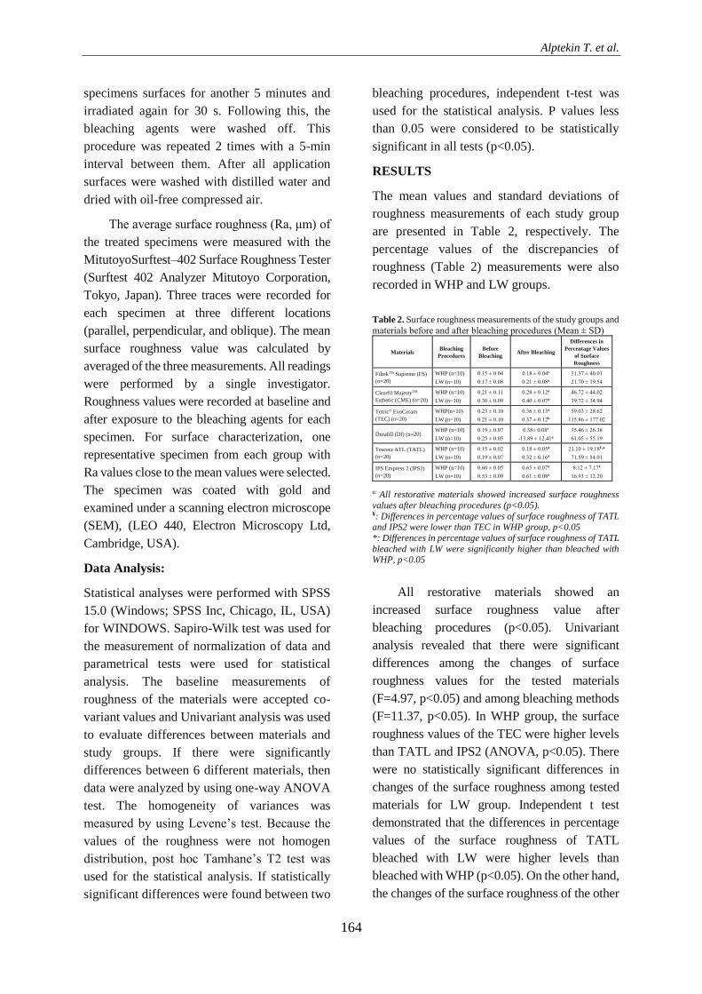
Alptekin T. et al.
164
specimens surfaces for another 5 minutes and
irradiated again for 30 s. Following this, the
bleaching agents were washed off. This
procedure was repeated 2 times with a 5-min
interval between them. After all application
surfaces were washed with distilled water and
dried with oil-free compressed air.
The average surface roughness (Ra, μm) of
the treated specimens were measured with the
MitutoyoSurftest–402 Surface Roughness Tester
(Surftest 402 Analyzer Mitutoyo Corporation,
Tokyo, Japan). Three traces were recorded for
each specimen at three different locations
(parallel, perpendicular, and oblique). The mean
surface roughness value was calculated by
averaged of the three measurements. All readings
were performed by a single investigator.
Roughness values were recorded at baseline and
after exposure to the bleaching agents for each
specimen. For surface characterization, one
representative specimen from each group with
Ra values close to the mean values were selected.
The specimen was coated with gold and
examined under a scanning electron microscope
(SEM), (LEO 440, Electron Microscopy Ltd,
Cambridge, USA).
Data Analysis:
Statistical analyses were performed with SPSS
15.0 (Windows; SPSS Inc, Chicago, IL, USA)
for WINDOWS. Sapiro-Wilk test was used for
the measurement of normalization of data and
parametrical tests were used for statistical
analysis. The baseline measurements of
roughness of the materials were accepted co-
variant values and Univariant analysis was used
to evaluate differences between materials and
study groups. If there were significantly
differences between 6 different materials, then
data were analyzed by using one-way ANOVA
test. The homogeneity of variances was
measured by using Levene’s test. Because the
values of the roughness were not homogen
distribution, post hoc Tamhane’s T2 test was
used for the statistical analysis. If statistically
significant differences were found between two
bleaching procedures, independent t-test was
used for the statistical analysis. P values less
than 0.05 were considered to be statistically
significant in all tests (p<0.05).
RESULTS
The mean values and standard deviations of
roughness measurements of each study group
are presented in Table 2, respectively. The
percentage values of the discrepancies of
roughness (Table 2) measurements were also
recorded in WHP and LW groups.
Table 2. Surface roughness measurements of the study groups and
materials before and after bleaching procedures (Mean ± SD)
a: All restorative materials showed increased surface roughness
values after bleaching procedures (p<0.05). ¥: Differences in percentage values of surface roughness of TATL and IPS2 were lower than TEC in WHP group, p<0.05
*: Differences in percentage values of surface roughness of TATL
bleached with LW were significantly higher than bleached with WHP, p<0.05
All restorative materials showed an
increased surface roughness value after
bleaching procedures (p<0.05). Univariant
analysis revealed that there were significant
differences among the changes of surface
roughness values for the tested materials
(F=4.97, p<0.05) and among bleaching methods
(F=11.37, p<0.05). In WHP group, the surface
roughness values of the TEC were higher levels
than TATL and IPS2 (ANOVA, p<0.05). There
were no statistically significant differences in
changes of the surface roughness among tested
materials for LW group. Independent t test
demonstrated that the differences in percentage
values of the surface roughness of TATL
bleached with LW were higher levels than
bleached with WHP (p<0.05). On the other hand,
the changes of the surface roughness of the other
Materials Bleaching
Procedures
Before
Bleaching After Bleaching
Differences in
Percentage Values
of Surface
Roughness
FiltekTM Supreme (FS)
(n=20)
WHP (n=10)
LW (n=10)
0.15 ± 0.04
0.17 ± 0.08
0.18 ± 0.04a
0.21 ± 0.08a
31.37 ± 40.01
21.70 ± 19.54
Clearfil MajestyTM
Esthetic (CME) (n=20)
WHP (n=10)
LW (n=10)
0.21 ± 0.11
0.30 ± 0.09
0.28 ± 0.12a
0.40 ± 0.07a
46.72 ± 44.02
39.72 ± 34.94
Tetric® EvoCeram
(TEC) (n=20)
WHP(n=10)
LW (n=10)
0.23 ± 0.10
0.21 ± 0.10
0.36 ± 0.13a
0.37 ± 0.12b
59.03 ± 28.62
115.86 ± 177.02
Durafill (Df) (n=20) WHP (n=10)
LW (n=10)
0.19 ± 0.07
0.25 ± 0.05
0.38± 0.08a
-13.89 ± 12.41a
35.46 ± 26.38
61.05 ± 55.19
Tescera ATL (TATL)
(n=20)
WHP (n=10)
LW (n=10)
0.15 ± 0.02
0.19 ± 0.07
0.18 ± 0.05a
0.32 ± 0.16a
21.10 ± 19.18¥,*
71.59 ± 54.01
IPS Empress 2 (IPS2)
(n=20)
WHP (n=10)
LW (n=10)
0.60 ± 0.05
0.53 ± 0.09
0.65 ± 0.07a
0.61 ± 0.08a
8.12 ± 7.17¥
16.93 ± 12.20
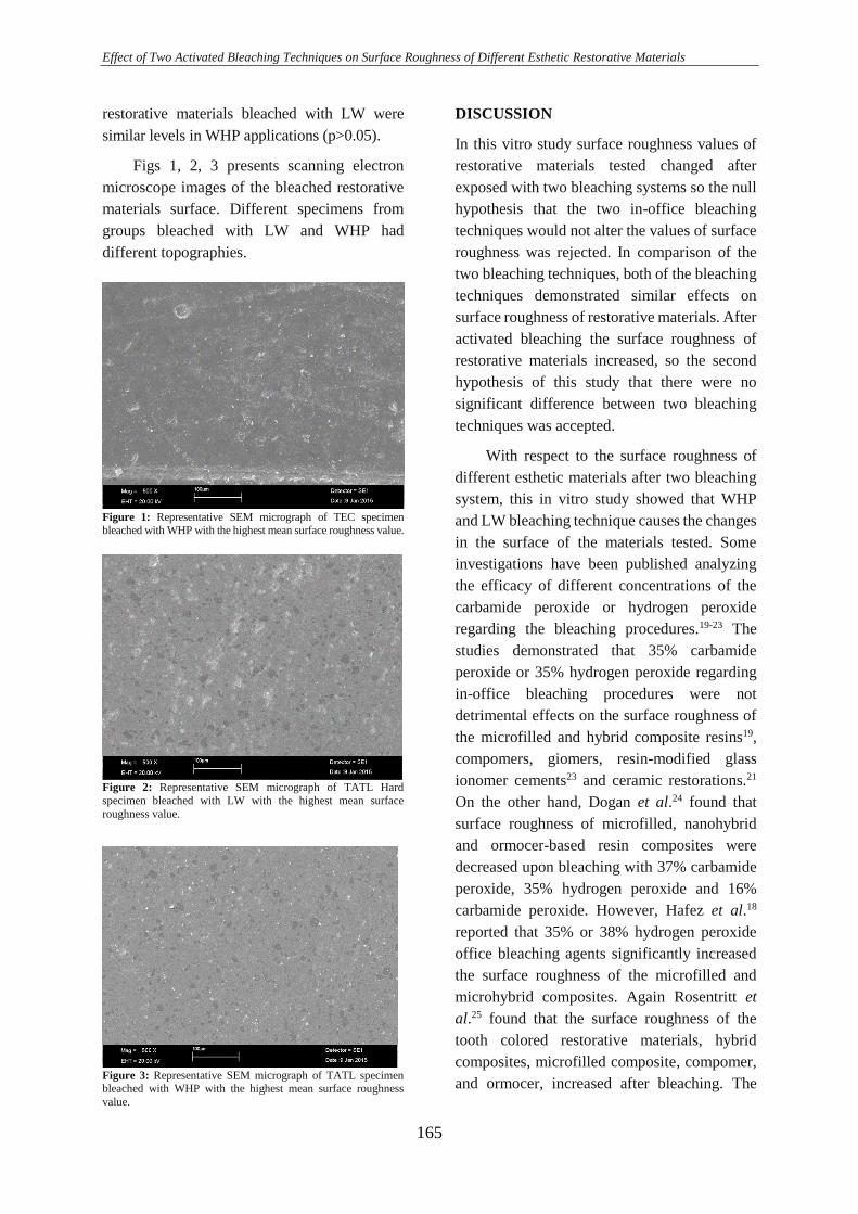
Effect of Two Activated Bleaching Techniques on Surface Roughness of Different Esthetic Restorative Materials
165
restorative materials bleached with LW were
similar levels in WHP applications (p>0.05).
Figs 1, 2, 3 presents scanning electron
microscope images of the bleached restorative
materials surface. Different specimens from
groups bleached with LW and WHP had
different topographies.
Figure 1: Representative SEM micrograph of TEC specimen bleached with WHP with the highest mean surface roughness value.
Figure 2: Representative SEM micrograph of TATL Hard
specimen bleached with LW with the highest mean surface
roughness value.
Figure 3: Representative SEM micrograph of TATL specimen bleached with WHP with the highest mean surface roughness
value.
DISCUSSION
In this vitro study surface roughness values of
restorative materials tested changed after
exposed with two bleaching systems so the null
hypothesis that the two in-office bleaching
techniques would not alter the values of surface
roughness was rejected. In comparison of the
two bleaching techniques, both of the bleaching
techniques demonstrated similar effects on
surface roughness of restorative materials. After
activated bleaching the surface roughness of
restorative materials increased, so the second
hypothesis of this study that there were no
significant difference between two bleaching
techniques was accepted.
With respect to the surface roughness of
different esthetic materials after two bleaching
system, this in vitro study showed that WHP
and LW bleaching technique causes the changes
in the surface of the materials tested. Some
investigations have been published analyzing
the efficacy of different concentrations of the
carbamide peroxide or hydrogen peroxide
regarding the bleaching procedures.19-23 The
studies demonstrated that 35% carbamide
peroxide or 35% hydrogen peroxide regarding
in-office bleaching procedures were not
detrimental effects on the surface roughness of
the microfilled and hybrid composite resins19,
compomers, giomers, resin-modified glass
ionomer cements23 and ceramic restorations.21
On the other hand, Dogan et al.24 found that
surface roughness of microfilled, nanohybrid
and ormocer-based resin composites were
decreased upon bleaching with 37% carbamide
peroxide, 35% hydrogen peroxide and 16%
carbamide peroxide. However, Hafez et al.18
reported that 35% or 38% hydrogen peroxide
office bleaching agents significantly increased
the surface roughness of the microfilled and
microhybrid composites. Again Rosentritt et
al.25 found that the surface roughness of the
tooth colored restorative materials, hybrid
composites, microfilled composite, compomer,
and ormocer, increased after bleaching. The
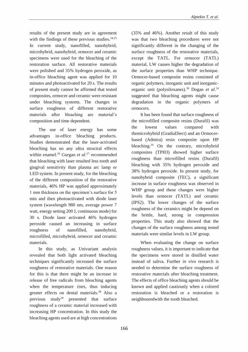
Alptekin T. et al.
166
results of the present study are in agreement
with the findings of these previous studies.24,25
In current study, nanofilled, nanohybrid,
microhybrid, nanohybrid, ormocer and ceramic
specimens were used for the bleaching of the
restoration surface. All restorative materials
were polished and 35% hydrogen peroxide, as
in-office bleaching agent was applied for 10
minutes and photoactivated for 20 s. The results
of present study cannot be affirmed that tested
composites, ormocer and ceramic were resistant
under bleaching systems. The changes in
surface roughness of different restorative
materials after bleaching are material’s
composition and time dependent.
The use of laser energy has some
advantages in-office bleaching products.
Studies demonstrated that the laser-activated
bleaching has no any ultra structral effects
within enamel.26 Gurgan et al.27 recommended
that bleaching with laser resulted less tooth and
gingival sensitivity than plasma arc lamp or
LED system. In present study, for the bleaching
of the different composition of the restorative
materials, 46% HP was applied approximately
1 mm thickness on the specimen’s surface for 5
min and then photoactivated with diode laser
system (wavelength 980 nm, average power 7
watt, energy setting 200 J, continuous mode) for
30 s. Diode laser activated 46% hydrogen
peroxide caused an increasing in surface
roughness of nanofilled, nanohybrid,
microfilled, microhybrid, ormocer and ceramic
materials.
In this study, as Univariant analysis
revealed that both light activated bleaching
techniques significantly increased the surface
roughness of restorative materials. One reason
for this is that there might be an increase in
release of free radicals from bleaching agents
when the temperature rises, thus inducing
greater effects on dental materials.28 Also a
previous study29 presented that surface
roughness of a ceramic material increased with
increasing HP concentration. In this study the
bleaching agents used are at high concentrations
(35% and 46%). Another result of this study
was that two bleaching procedures were not
significantly different in the changing of the
surface roughness of the restorative materials,
except the TATL. For ormocer (TATL)
material, LW causes higher the degradation of
the surface properties than WHP technique.
Ormocer-based composite resins consisted of
organic polymers, inorganic unit and inorganic-
organic unit (polysiloxanes).30 Dogan et al.24
suggested that bleaching agents might cause
degradation in the organic polymers of
ormocers.
It has been found that surface roughness of
the microfilled composite resins (Durafil) was
the lowest values compared with
themicrohybrid (GradiaDiect) and an Ormocer-
based (Admira) resin composite upon HP
bleaching.24 On the contrary, microhybrid
composites (TPH3) showed higher surface
roughness than microfilled resins (Durafil)
bleaching with 35% hydrogen peroxide and
38% hydrogen peroxide. In present study, for
nanohybrid composite (TEC), a significant
increase in surface roughness was observed in
WHP group and these changes were higher
levels than ormocer (TATL) and ceramic
(IPS2). The lower changes of the surface
roughness of the ceramics might be depend on
the brittle, hard, strong in compression
properties. This study also showed that the
changes of the surface roughness among tested
materials were similar levels in LW group.
When evaluating the change on surface
roughness values, it is important to indicate that
the specimens were stored in distilled water
instead of saliva. Further in vivo research is
needed to determine the surface roughness of
restorative materials after bleaching treatment.
The effects of office bleaching agents should be
known and applied cautiously when a colored
restoration is bleached or a restoration is
neighbouredwith the tooth bleached.
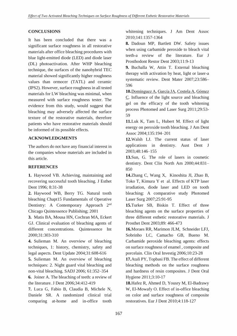
Effect of Two Activated Bleaching Techniques on Surface Roughness of Different Esthetic Restorative Materials
167
CONCLUSIONS
It has been concluded that there was a
significant surface roughness in all restorative
materials after office bleaching procedures with
blue light-emitted diode (LED) and diode laser
(DL) photoactivation. After WHP bleaching
technique, the surfaces of the nanohybrid TEC
material showed significantly higher roughness
values than ormocer (TATL) and ceramic
(IPS2). However, surface roughness in all tested
materials for LW bleaching was minimal, when
measured with surface roughness tester. The
evidence from this study, would suggest that
bleaching may adversely affected the surface
texture of the restorative materials, therefore
patients who have restorative materials should
be informed of its possible effects.
ACKNOWLEDGMENTS
The authors do not have any financial interest in
the companies whose materials are included in
this article.
REFERENCES
1. Haywood VB. Achieving, maintaining and
recovering successful tooth bleaching. J Esthet
Dent 1996; 8:31-38
2. Haywood WB, Berry TG. Natural tooth
bleaching Chapt15 Fundamentals of Operative
Dentistry: A Contemporary Approach 2nd
Chicago Quintessence Publishing; 2001
3. Matis BA, Mousa HN, Cochran MA, Eckert
GJ. Clinical evaluation of bleaching agents of
different concentrations. Quintessence Int
2000;31:303-310
4. Sulieman M. An overview of bleaching
techniques, 1: history, chemistry, safety and
legal aspects. Dent Update 2004;31:608-616
5. Sulieman M. An overview of bleaching
techniques: 2. Night guard vital bleaching and
non-vital bleaching. SADJ 2006; 61:352–354
6. Joiner A. The bleaching of teeth: a review of
the literature. J Dent 2006;34:412-419
7. Luca G, Fabio B, Claudia B, Michele N,
Daniele SR. A randomized clinical trial
comparing at-home and in-office tooth
whitening techniques. J Am Dent Assoc
2010;141:1357-1364
8. Dadoun MP, Bartlett DW. Safety issues
when using carbamide peroxide to bleach vital
teeth-a review of the literature. Eur J
Prosthodont Restor Dent 2003;11:9-13
9. Buchalla W, Attin T. External bleaching
therapy with activation by heat, light or laser-a
systematic review. Dent Mater 2007;23:586–
596
10. Domínguez A, García JA, Costela A, Gómez
C. Influence of the light source and bleaching
gel on the efficacy of the tooth whitening
process Photomed and Laser Surg 2011;29:53-
59
11. Luk K, Tam L, Hubert M. Effect of light
energy on peroxide tooth bleaching. J Am Dent
Assoc 2004;135:194–201
12. Walsh LJ. The current status of laser
applications in dentistry. Aust Dent J
2003;48:146–155
13. Sun, G. The role of lasers in cosmetic
dentistry. Dent Clin North Am 2000;44:831–
850
14. Zhang C, Wang X, Kinoshita JI, Zhao B,
Toko T, Kimura Y et al. Effects of KTP laser
irradiation, diode laser and LED on tooth
bleaching: A comparative study Photomed
Laser Surg 2007;25:91-95
15. Turker SB, Biskin T. Effect of three
bleaching agents on the surface properties of
three different esthetic restorative materials. J
Prosthet Dent 2003;89: 466-473
16. Moraes RR, Marimon JLM, Schneider LFJ,
Sobrinho LC, Camacho GB, Bueno M.
Carbamide peroxide bleaching agents: effects
on surface roughness of enamel , composite and
porcelain. Clin Oral Investig 2006;10:23-28
17. Atali PY, Topbasi FB. The effect of different
bleaching methods on the surface roughness
and hardness of resin composites. J Dent Oral
Hygiene 2011;3:10-17
18. Hafez R, Ahmed D, Yousry M, El-Badrawy
W, El-Mowafy O. Effect of in-office bleaching
on color and surface roughness of composite
restoratives. Eur J Dent 2010;4:118-127
Alptekin T. et al.
168
19. Sharafeddin F, Jamalipour G. Effects of 35%
carbamide peroxide gel on surface roughness
and hardness of composite resins. J Dent
(Tehran)2010;7: 6-12
20. Pruthi G, Jain V, Kandpal HC, Mathur VP,
Shah N. Effect of bleaching on color change and
surface topography of composite restorations.
Int J Dent 2010; 2010:695748. doi:
10.1155/2010/695748. Epub 2010 Dec 22
21. Zaki AA, Fahmy NZ. The effect of a
bleaching system on properties related to
different ceramic surface textures. J
Prosthodont 2009;18:223-229
22. de A Silva MF, Davies RM, Stewart B,
DeVizio W, Tonholo J, da Silva Júnior JG et al.
Effect of whitening gels on the surface
roughness of restorative materials in situ. Dent
Mater 2006;22:919-924
23. Wattanapayungkul P, Yap AU. Effects of in-
officebleaching products on surface finish
oftooth-colored restorations. Oper Dent
2003;28:15-19
24. Dogan A, Ozcelik S, Dogan OM,
Hubbezoglu I, Cakmak M, Bolayir G. Effects
of bleaching on roughness of dental composite
resins. J Adhes 2008;84: 897-917
25. Rosentritt M, Lang R, Plein T, Behr M,
Handel G. Discoloration of restorative materials
after bleaching application. Quintessence Inter
2005; 36:33-39
26. Goharkhay K, Schoop U, Wernisch J, Hartl
S, De Moor RT, Moritz A. Frequency doubled
neodymium:yttrium-aluminum-garnet and
diode laser-activated power bleaching--pH,
environmental scanning electron microscopy,
and colorimetric in vitro evaluations. Lasers
Med Sci 2009;24:339-346
27. Gurgan S, Cakir FY, Yazici E. Different
light-activated in-office bleaching systems: a
clinical evaluation. Lasers Med Sci
2010;25:817-822
28. Yu H, Li Q, Cheng H, Wang Y. The effects
of temperature and bleaching gels on the
properties of tooth-colored restorative
materials. J Prosthet Dent 2011;105:100-107
29. Abu-Eittah MR, Mandour MH. In vitro
study of the effect of three hydrogen peroxide
concentrations on the corrosion behavior and
surface topography of alumina-reinforced
dental ceramic. J Prosthodont 2011; 20:541-552
30. Hickel R, Dasch W, Janda R, Tyas M,
Anusavice K. New direct restorative materials.
Inter Dent J 1998;48:3-16
Correspondence Author
Özgün Yusuf ÖZYILMAZ
Istanbul Medipol University
Faculty of Dentistry,
Department of Prosthodontics,
Istanbul, Turkey
Phone: +902124607651
Fax: +902124607070
E-Mail: [email protected]
Cumhuriyet Dental Journal: 2017; 20(3)
Doi: 10.7126/cumudj.369104 RESEARCH ARTICLES
169
¹ Department of Oral and Maxillofacial Surgery, Faculty of Dentistry, Bezmi Alem University, İstanbul, Turkey. ² Department of Oral and Maxillofacial Surgery, Faculty of Dentistry, Erciyes University, Kayseri, Turkey. 3 Department of Oral and Maxillofacial Surgery, Faculty of Dentistry, Ondokuz Mayis University, Samsun, Turkey.
NONSURGICAL CLOSURE OF OROANTRAL COMMUNICATIONS
USING OCCLUSAL SPLINTS
Oroantral Açıklıkların Okluzal Splintler ile Cerrahisiz Kapatılması
Nükhet KÜTÜK1, Ahmet Emin DEMİRBAŞ2,
Canay YILMAZ ASAN2, Burcu BAŞ3, Alper ALKAN1
Makale Kodu/Article Code : 201578
Makale Gönderilme Tarihi : 19.09.2016
Kabul Tarihi : 04.03.2017
ABSTRACT
Purpose: Oroantral communications (OACs) may close
spontaneously, especially when the defect has a size
smaller than 2-3 mm, whereas larger openings require
surgical closure. The aim of this retrospective study was
to present our experince with non surgical closure of
small and large OACs with occlusal splints.
Materials and Methods: Clinical and radiographic data
of twenty patients who had used occlusal splints after the
occurance of an acute OAC were included in this study.
Two groups were created according to the size of the
defects: Group A: defects smaller (<) than 5 mm; and
Group B: 5 mm and wider (≥). In all patients, a well
fitted soft occlusal splint was placed for hermetic closure
of the opening. All patients were followed up weekly
and the splint use was stopped when complete
epithelization of the OAC was observed clinically. The
relationship between the size of OACs, treatment
outcome, and healing time was compared statistically.
Results: OAC was healed spontaneously in all patients,
except one. The healing time was found to be
significantly higher in goup B than in group A. No
significant difference was found between the groups with
respect to the success of the treatment.
Conclusion: The use of occlusal splints seems to
improve the spontaneus healing of the OACs.
Key words: Oroantral communication, occlusal splint,
nonsurgical closure
ÖZ
Amaç: Defekt boyutu 2-3 mm olan oroantral açıklıklar
spontan olarak iyileşebilirken büyük açıklıklarda cerrahi
müdahale gereklidir. Bu çalışmanın amacı, küçük ve
büyük oroantral açıklıkların, okluzal splintler ile
cerrahisiz tedavisi hakkında bilgi sunmaktır.
Gereç ve Yöntem: Bu çalışmaya akut oroantral açıklık
gelişen 12 hasta dahil edildi. Defektin boyutuna göre 2
grup oluşturuldu. Grup A: defektin 5 mm'den küçük
olduğu; Grup B: defektin 5 mm ve daha fazla olduğu
hastalardan oluşmaktaydı. Bütün hastalara açıklığın
kapatılması için yumuşak okluzal plak yapıldı. Hastalar
haftalık olarak takip edildi ve klinik olarak tam
epitelizasyon sağlandığında splint kullanımı bırakıldı.
Açıklıkların boyutu ile iyileşme süresi ve tedavi sonuçları
arasındaki ilişki istatistiksel olarak karşılaştırıldı.
Sonuçlar: Bir hasta dışında, tüm hastalarda oroantral
açıklıklar spontan olarak iyileşti. Grup B'nin iyileşme
süresinin Grup A'dan belirgin olarak fazla olduğu
görüldü. Tedavinin başarısı ile ilgili gruplar arasında
herhangi bir fark bulunamadı.
Sonuç: Oklüzal splintlerin kullanımı, oroantral
açıklıkların spontan olarak iyileşmesinde faydalı olabilir.
Anahtar Kelimeler: Oroantral açıklık, oklüzal splint,
cerrahisiz kapama
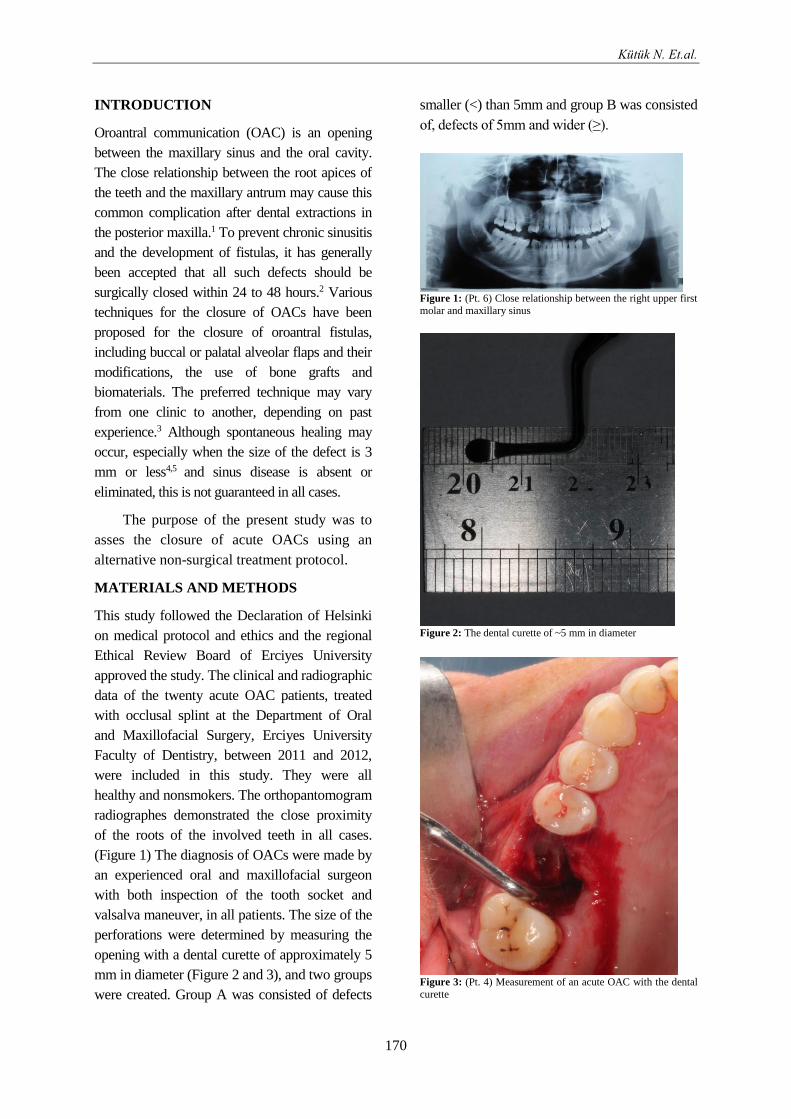
Kütük N. Et.al.
170
INTRODUCTION
Oroantral communication (OAC) is an opening
between the maxillary sinus and the oral cavity.
The close relationship between the root apices of
the teeth and the maxillary antrum may cause this
common complication after dental extractions in
the posterior maxilla.1 To prevent chronic sinusitis
and the development of fistulas, it has generally
been accepted that all such defects should be
surgically closed within 24 to 48 hours.2 Various
techniques for the closure of OACs have been
proposed for the closure of oroantral fistulas,
including buccal or palatal alveolar flaps and their
modifications, the use of bone grafts and
biomaterials. The preferred technique may vary
from one clinic to another, depending on past
experience.3 Although spontaneous healing may
occur, especially when the size of the defect is 3
mm or less4,5 and sinus disease is absent or
eliminated, this is not guaranteed in all cases.
The purpose of the present study was to
asses the closure of acute OACs using an
alternative non-surgical treatment protocol.
MATERIALS AND METHODS
This study followed the Declaration of Helsinki
on medical protocol and ethics and the regional
Ethical Review Board of Erciyes University
approved the study. The clinical and radiographic
data of the twenty acute OAC patients, treated
with occlusal splint at the Department of Oral
and Maxillofacial Surgery, Erciyes University
Faculty of Dentistry, between 2011 and 2012,
were included in this study. They were all
healthy and nonsmokers. The orthopantomogram
radiographes demonstrated the close proximity
of the roots of the involved teeth in all cases.
(Figure 1) The diagnosis of OACs were made by
an experienced oral and maxillofacial surgeon
with both inspection of the tooth socket and
valsalva maneuver, in all patients. The size of the
perforations were determined by measuring the
opening with a dental curette of approximately 5
mm in diameter (Figure 2 and 3), and two groups
were created. Group A was consisted of defects
smaller (<) than 5mm and group B was consisted
of, defects of 5mm and wider (≥).
Figure 1: (Pt. 6) Close relationship between the right upper first
molar and maxillary sinus
Figure 2: The dental curette of ~5 mm in diameter
Figure 3: (Pt. 4) Measurement of an acute OAC with the dental
curette
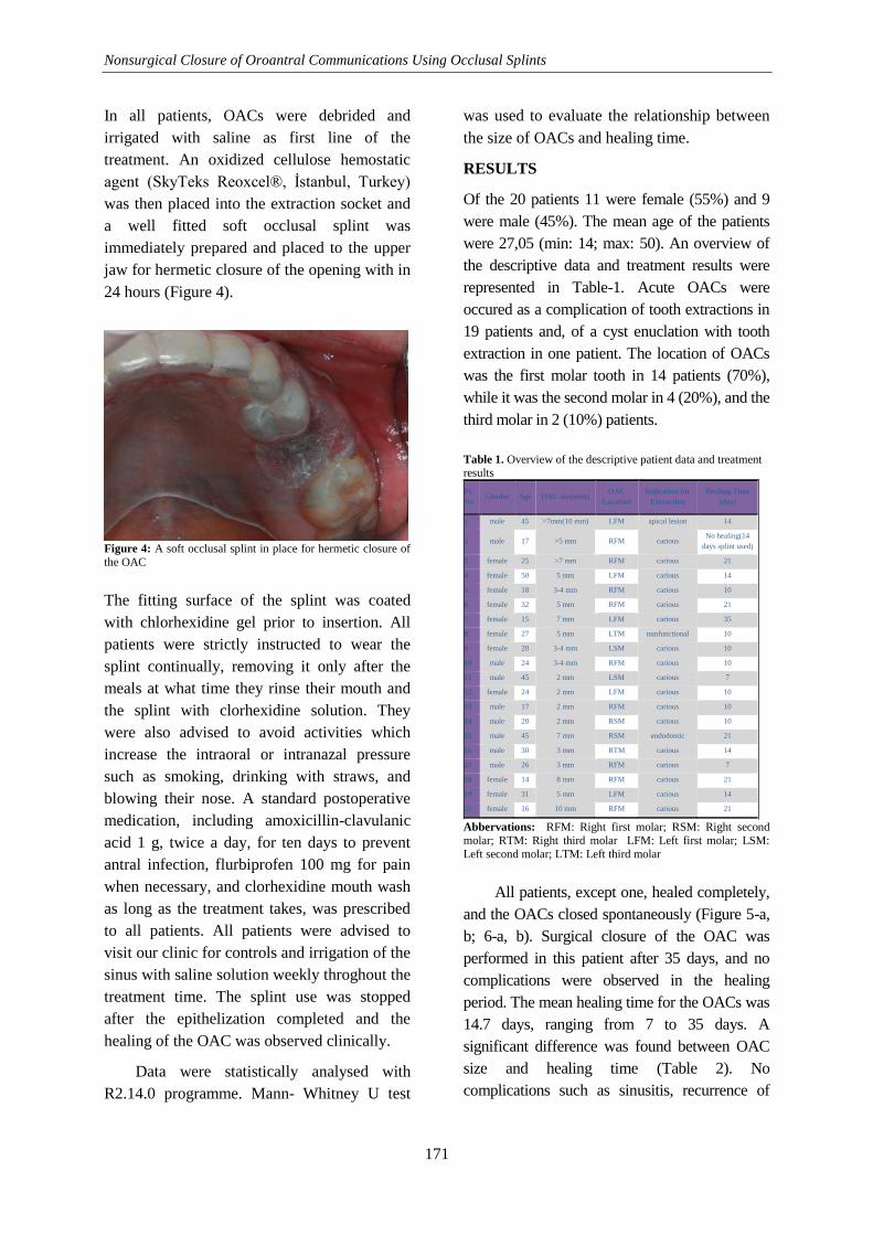
Nonsurgical Closure of Oroantral Communications Using Occlusal Splints
171
In all patients, OACs were debrided and
irrigated with saline as first line of the
treatment. An oxidized cellulose hemostatic
agent (SkyTeks Reoxcel®, İstanbul, Turkey)
was then placed into the extraction socket and
a well fitted soft occlusal splint was
immediately prepared and placed to the upper
jaw for hermetic closure of the opening with in
24 hours (Figure 4).
Figure 4: A soft occlusal splint in place for hermetic closure of
the OAC
The fitting surface of the splint was coated
with chlorhexidine gel prior to insertion. All
patients were strictly instructed to wear the
splint continually, removing it only after the
meals at what time they rinse their mouth and
the splint with clorhexidine solution. They
were also advised to avoid activities which
increase the intraoral or intranazal pressure
such as smoking, drinking with straws, and
blowing their nose. A standard postoperative
medication, including amoxicillin-clavulanic
acid 1 g, twice a day, for ten days to prevent
antral infection, flurbiprofen 100 mg for pain
when necessary, and clorhexidine mouth wash
as long as the treatment takes, was prescribed
to all patients. All patients were advised to
visit our clinic for controls and irrigation of the
sinus with saline solution weekly throghout the
treatment time. The splint use was stopped
after the epithelization completed and the
healing of the OAC was observed clinically.
Data were statistically analysed with
R2.14.0 programme. Mann- Whitney U test
was used to evaluate the relationship between
the size of OACs and healing time.
RESULTS
Of the 20 patients 11 were female (55%) and 9
were male (45%). The mean age of the patients
were 27,05 (min: 14; max: 50). An overview of
the descriptive data and treatment results were
represented in Table-1. Acute OACs were
occured as a complication of tooth extractions in
19 patients and, of a cyst enuclation with tooth
extraction in one patient. The location of OACs
was the first molar tooth in 14 patients (70%),
while it was the second molar in 4 (20%), and the
third molar in 2 (10%) patients.
Table 1. Overview of the descriptive patient data and treatment results
Pt.
No Gender Age OAC size(mm)
OAC
Location
Indication for
Extraction
Healing Time
(day)
1 male 45 >7mm(10 mm) LFM apical lesion 14
2 male 17 >5 mm RFM carious No healing(14
days splint used)
3 female 25 >7 mm RFM carious 21
4 female 50 5 mm LFM carious 14
5 female 18 3-4 mm RFM carious 10
6 female 32 5 mm RFM carious 21
7 female 15 7 mm LFM carious 35
8 female 27 5 mm LTM nunfunctional 10
9 female 20 3-4 mm LSM carious 10
10 male 24 3-4 mm RFM carious 10
11 male 45 2 mm LSM carious 7
12 female 24 2 mm LFM carious 10
13 male 17 2 mm RFM carious 10
14 male 20 2 mm RSM carious 10
15 male 45 7 mm RSM endodontic 21
16 male 30 3 mm RTM carious 14
17 male 26 3 mm RFM carious 7
18 female 14 8 mm RFM carious 21
19 female 31 5 mm LFM carious 14
20 female 16 10 mm RFM carious 21
Abbervations: RFM: Right first molar; RSM: Right second
molar; RTM: Right third molar LFM: Left first molar; LSM:
Left second molar; LTM: Left third molar
All patients, except one, healed completely,
and the OACs closed spontaneously (Figure 5-a,
b; 6-a, b). Surgical closure of the OAC was
performed in this patient after 35 days, and no
complications were observed in the healing
period. The mean healing time for the OACs was
14.7 days, ranging from 7 to 35 days. A
significant difference was found between OAC
size and healing time (Table 2). No
complications such as sinusitis, recurrence of

Kütük N. Et.al.
172
OAC, or infection occured during seven to nine
months of follow-up period.
Figure 5-a: (Pt. 6) Acute OAC Figure 5-b: (Pt. 6) Two months
of ~5 mm in diameter after the extraction
Figure 6-a: (Pt. 7) Acute OAC Figure 6-b: (Pt. 7) 2 months
of 7 mm in diameter after the extraction
Table 2. The relation between OAC size and healing time OAC size
<5 mm ≥ 5 mm P
Patients (n) 9 10
Healing time (median±Sd) 10.00±2.048 21.00±6.92 0.01
Mann Whitney U Test, p<0.05
DISCUSSION
OACs are pathological entities those are often
occured during or after dental treatments such as
surgical extractions, removing cysts, implant
surgery, trauma, tumors and pathological lesions
of the maxillary sinus.6 If the opening left
untreated, an oroantral fistula may develope and
patients may experience sinusitis within 48 hours
to 2 weeks.3, 6,7 To prevent sinus infection and the
development of a fistula, it has generally been
recommended that these defects should be closed
within 24 to 48 hours.2
Despite the simplicity and safety of most
of the procedures, each surgical technique has
its own drawbacks such as partial obliteration of
the vestibul depht requiring a secondary
vestibuloplasty when using buccal mucosal
flaps8; tissue bunching and liquid passage
beneath the flap4 when using palatal rotation
flaps, and an increased risk of infection related
to buccal fat pad flaps. Donor site morbidity is
one disadvantage, which further increases
postoperative pain and discomfort, depending
on the surgical technique applied. The need for
surgical expertise and equipment, postoperative
pain and swelling are also among the various
disadvantages.9 In addition, as a clinical
observation, surgical options may be avoided by
some patients, when they only expect a tooth
extraction procedure as their physician
describes the extent of a possible surgery.
Various nonsurgical techniques has been
proposed in the literature. The most preferred
technique is placing a gauze sponge dressing
that is supported with an eight suture or acrilic
splint for 48 h, to protect the blood clot in the
tooth socket.3 Resorbable hemostatic gauze
composed of reconstituted oxidized cullulose
was also used to maintain the blood clot in
place.10 Susan et al reported a 80% successful
rate with biodegradable polyurethane foam in
patients with OACs with diameters up to 7
mm.11 However, good oral hygen, patient
cooperation and close follow up is very
important in such cases.
The maximum size of OACs that tend to
heal spontaneously is controversial in the
literature. Most reports agreed on that small
defects of 1 to 2 mm heals spontaneously after
the formation of a blood clot.3, 6, 12, 13, 14, 15 Others
contend that defects smaller than 5 mm may
close naturally when there is no prior
infection.2,16 However, it is difficult to determine
the size of OACs clinically.17 In our study, we
measured the OACs with a dental curette of
approximately 5 mm in diameter, and, clasiffied
them into two groups according to the defect
sizes which were described either ‘less than 5
mm’ or ‘5 mm or greater’. Spontaneous healing
was observed at defects either less or larger than
5 mm. No significant difference was found
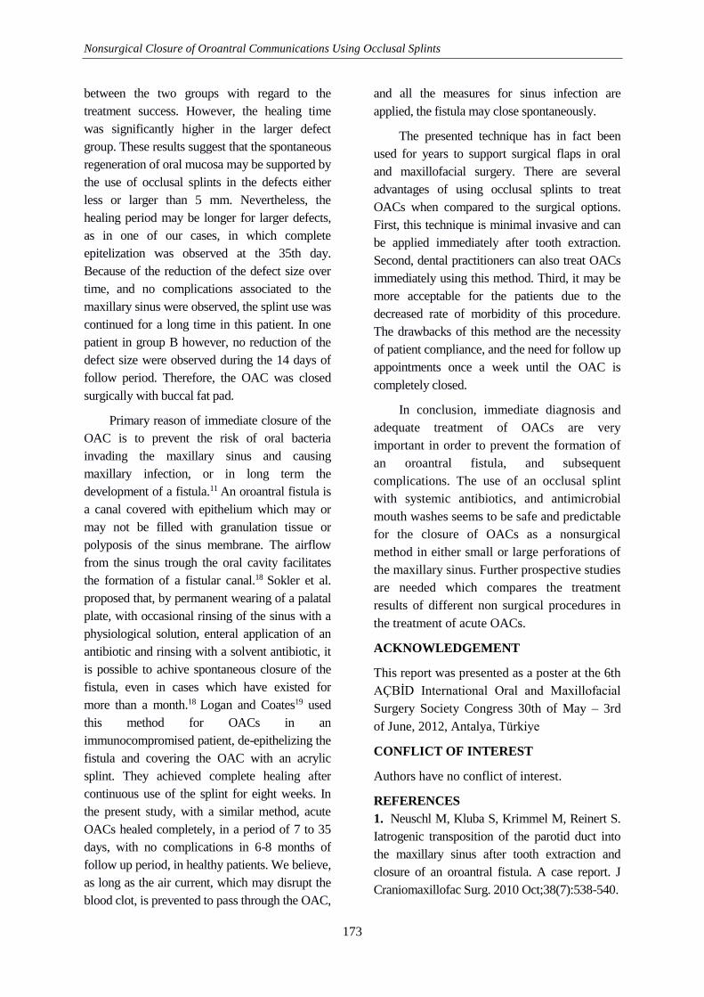
Nonsurgical Closure of Oroantral Communications Using Occlusal Splints
173
between the two groups with regard to the
treatment success. However, the healing time
was significantly higher in the larger defect
group. These results suggest that the spontaneous
regeneration of oral mucosa may be supported by
the use of occlusal splints in the defects either
less or larger than 5 mm. Nevertheless, the
healing period may be longer for larger defects,
as in one of our cases, in which complete
epitelization was observed at the 35th day.
Because of the reduction of the defect size over
time, and no complications associated to the
maxillary sinus were observed, the splint use was
continued for a long time in this patient. In one
patient in group B however, no reduction of the
defect size were observed during the 14 days of
follow period. Therefore, the OAC was closed
surgically with buccal fat pad.
Primary reason of immediate closure of the
OAC is to prevent the risk of oral bacteria
invading the maxillary sinus and causing
maxillary infection, or in long term the
development of a fistula.11 An oroantral fistula is
a canal covered with epithelium which may or
may not be filled with granulation tissue or
polyposis of the sinus membrane. The airflow
from the sinus trough the oral cavity facilitates
the formation of a fistular canal.18 Sokler et al.
proposed that, by permanent wearing of a palatal
plate, with occasional rinsing of the sinus with a
physiological solution, enteral application of an
antibiotic and rinsing with a solvent antibiotic, it
is possible to achive spontaneous closure of the
fistula, even in cases which have existed for
more than a month.18 Logan and Coates19 used
this method for OACs in an
immunocompromised patient, de-epithelizing the
fistula and covering the OAC with an acrylic
splint. They achieved complete healing after
continuous use of the splint for eight weeks. In
the present study, with a similar method, acute
OACs healed completely, in a period of 7 to 35
days, with no complications in 6-8 months of
follow up period, in healthy patients. We believe,
as long as the air current, which may disrupt the
blood clot, is prevented to pass through the OAC,
and all the measures for sinus infection are
applied, the fistula may close spontaneously.
The presented technique has in fact been
used for years to support surgical flaps in oral
and maxillofacial surgery. There are several
advantages of using occlusal splints to treat
OACs when compared to the surgical options.
First, this technique is minimal invasive and can
be applied immediately after tooth extraction.
Second, dental practitioners can also treat OACs
immediately using this method. Third, it may be
more acceptable for the patients due to the
decreased rate of morbidity of this procedure.
The drawbacks of this method are the necessity
of patient compliance, and the need for follow up
appointments once a week until the OAC is
completely closed.
In conclusion, immediate diagnosis and
adequate treatment of OACs are very
important in order to prevent the formation of
an oroantral fistula, and subsequent
complications. The use of an occlusal splint
with systemic antibiotics, and antimicrobial
mouth washes seems to be safe and predictable
for the closure of OACs as a nonsurgical
method in either small or large perforations of
the maxillary sinus. Further prospective studies
are needed which compares the treatment
results of different non surgical procedures in
the treatment of acute OACs.
ACKNOWLEDGEMENT
This report was presented as a poster at the 6th
AÇBİD International Oral and Maxillofacial
Surgery Society Congress 30th of May – 3rd
of June, 2012, Antalya, Türkiye
CONFLICT OF INTEREST
Authors have no conflict of interest.
REFERENCES
1. Neuschl M, Kluba S, Krimmel M, Reinert S.
Iatrogenic transposition of the parotid duct into
the maxillary sinus after tooth extraction and
closure of an oroantral fistula. A case report. J
Craniomaxillofac Surg. 2010 Oct;38(7):538-540.
Kütük N. Et.al.
174
2. von Wowern N: Correlation between the
development of an oroantral fistula and the size
of the corresponding bony defect. J Oral Surg.
1973; 31(2):98-102
3. Guven O: A clinical study on oroantral
fistulae. J Craniomaxillofac Surg. 1998;
26:267-271.
4. Anavi Y, Gal G, Silfen R, Calderon S.
Palatal rotation-advancement flap for delayed
repair of oroantral fistula: a retrospective
evaluation of 63 cases. Oral Surg Oral Med
Oral Pathol Oral Radiol Endod. 2003
Nov;96(5):527-534.
5. Abuabara A, Cortez AL, Passeri LA, de
Moraes M, Moreira RW. Evaluation of different
treatments for oroantral/oronasal
communications: experience of 112 cases. Int J
Oral Maxillofac Surg. 2006 Feb;35(2):155-158.
6. Lazow SK. Surgical management of the
oroantral fistula: flap procedures. Operative
Techniques in Otolaryngology-Head and Neck
Surgery. 1999;10(2):148-152.
7. Ashley RE. LIII A Method of Closing Antro-
Alveolar Fistulae. Annals of Otology, Rhinology
& Laryngology. 1939;48(3):632-642.
8. Meirelles RC, Neves-Pinto RM. Oroantral
fistula and genian mucosal flap: a review of 25
cases. Braz J Otorhinolaryngol. 2008 Jan-
Feb;74(1):85-90.
9. Obradovic O, Todorovic L, Pesic V.
Investigations of the buccal sulcus depth after
the use of certain methods of oro-antral
communication closure. Bull Group Int Rech
Sci Stomatol Odontol. 1981 Sep;24(3):209-214.
10. Gacic B, Todorovic L, Kokovic V, Danilovic
V, Stojcev-Stajcic L, Drazic R, Markovic A. The
closure of oroantral communications with
resorbable PLGA-coated beta-TCP root analogs,
hemostatic gauze, or buccal flaps: a prospective
study. Oral Surg Oral Med Oral Pathol Oral
Radiol Endod. 2009 Dec;108(6):844-850
11. Visscher SH, Van Minnen B, Bos RR.
Feasibility of conical biodegradable
polyurethane foam for closure of oroantral
communications. J Oral Maxillofac Surg. 2011
Feb;69(2):390-395.
12. Thoma K, Pajarola GF, Grätz KW,
Schmidlin PR. Bioabsorbable root analogue for
closure of oroantral communications after tooth
extraction: a prospective case-cohort study. Oral
Surg Oral Med Oral Pathol Oral Radiol Endod.
2006 May;101(5):558-564.
13. Herbert DC. Closure of a palatal fistula
using a mucoperiosteal island flap. Br J Plast
Surg. 1974 Oct;27(4):332-336.
14. Hanazawa Y, Itoh K, Mabashi T, Sato K.
Closure of oroantral communications using a
pedicled buccal fat pad graft. J Oral Maxillofac
Surg. 1995 Jul;53(7):771-775
15. Keresztesi K. Conservative therapy of
oroantral fistula. Osterr Z Stomatol. 1954
Jun;51(6):317-323.
16. van Minnen B, Stegenga B, van Leeuwen
MB, van Kooten TG, Bos RR. Nonsurgical
closure of oroantral communications with a
biodegradable polyurethane foam: A pilot study
in rabbits. J Oral Maxillofac Surg. 2007
Feb;65(2):218-222.
17. Visscher SH, van Minnen B, Bos RR.
Closure of oroantral communications: a review
of the literature. J Oral Maxillofac Surg. 2010
Jun;68(6):1384-1391.
18. Sokler K, Vuksan V, Lauc T. Treatment of
oroantral fistula. Acta Stomat Croat. 2002;
36:135–140.
19. Logan RM, Coates EA. Non-surgical
management of an oro-antral fistula in a patient
with HIV infection. Aust Dent J. 2003
Dec;48(4):255-258.
Correspondence Author
Canay YILMAZ ASAN
Erciyes Üniversitesi
Diş Hekimliği Fakültesi
Ağız Diş Çene Cerrahisi AD.
38039 Kayseri/ Turkey
Tel: +90 352 207 66 66
Fax: +90 352 438 06 57
E-mail: [email protected]
Cumhuriyet Dental Journal: 2017; 20(3)
Doi: 10.7126/cumudj.369109 RESEARCH ARTICLES
175
Saveetha Dental College, Department of Periodontics, India.
AWARENESS ABOUT PERIODONTITIS AND PRE-TERM LOW BIRTH
WEIGHT INFANTS AMONG GYNECOLOGISTS IN CHENNAI- A
QUESTIONNAIRE STUDY
Chennaideki Jinekologların Periodontitis ve Düşük Doğum Ağırlıklı Prematüre Bebekler
Hakkındaki Farkındalığı: Bir Anket Çalışması
Hafsa ISMAIL, Radhika ARJUNKUMAR
Makale Kodu/Article Code : 201578
Makale Gönderilme Tarihi : 25.06.2016
Kabul Tarihi : 28.03.2017
ABSTRACT
Background: Periodontal infection has been associated with
adverse pregnancy outcomes. Subsequently it is important that
gynecologists are educated about hormonal effects on women’s
oral health. The current study assessed gynecologist’s
awareness regarding the effect of female sex hormones on
periodontal health and preterm delivery.
Materials and methods: Sixty-six gynecologists from the city
of Chennai completed a questionnaire regarding awareness about
female hormonal and periodontal health and its effects on pre-
term low birth weight infants. Respondents were divided into
three groups: Group A, (doctors who practiced at a medical
colleges) Group B (doctors who practiced at private hospitals)
and Group C. (doctors who practiced at both medical colleges
and private hospitals) Survey responses were collected and data
between the groups were statistically compared.
Results: Our findings showed that most gynecologists were
aware and concerned about female patient’s oral health during
various hormonal phases. However, gynecologists practicing at
medical colleges (Group A) and gynecologists practicing at both
medical colleges and private hospitals (Group C) had
significantly greater health awareness than doctors practicing at
private hospitals. (Group B)
Conclusion: The results of the current study imply the fact that
there is adequate knowledge about the effects of hormonal
changes in the periodontal tissues. However, there is limited
incorporation of this knowledge, especially among gynecologists
who are exclusively into private practice.
Keywords: Periodontitis, preterm low birth weight, scaling, root
planing
ÖZ
Amaç: Periodontal enfeksiyon, gebeliğin olumsuz sonuçlarıyla
ilişkilendirilmiştir. Bundan dolayı jinekologların kadınların ağız
sağlığı üzerindeki hormonal etkileri konusunda eğitim alması
önemlidir. Mevcut araştırma kadın seks hormonlarının
periodontal sağlık ve erken doğum üzerindeki etkileri
konusundaki jinekoloğun farkındalığını değerlendirdi.
Gereç ve yöntem: Chennai şehrinden altmış altı jinekolog,
kadınlarda hormonal ve periodontal sağlık ile düşük doğum
ağırlıklı prematüre bebeklere etkileri konusundaki bir anketi
doldurdu. Katılımcılar, A Grubu (tıp fakültesinde çalışan
jinekologlar), B Grubu (özel hastanelerde çalışan jinekologlar)
ve C Grubu (hem tıp fakültelerinde hem de özel hastanelerde
çalışan jinekologlar) olmak üzere üç gruba ayrıldı. Anket
yanıtları toplandı ve gruplar arasındaki veriler istatistiksel olarak
karşılaştırıldı.
Bulgular: Çalışmanın sonuçları, çoğu jinekoloğun, çeşitli
hormonal fazlarda kadın hastalarının ağız sağlığı konusunda
bilgili olduklarını ve endişe duyduğunu ortaya koymuştur.
Bununla birlikte, tıp fakültelerinde çalışan jinekologlar (Grup A)
ve hem tıp fakültelerinde hem de özel hastanelerde çalışan
jinekologlar (Grup C), özel hastanelerde çalışan doktorlardan
(Grup B) istatiksel olarak anlamlı derecede daha fazla sağlık
bilincine sahip bulundu.
Sonuç: Bu çalışmanın sonuçları, hormonal değişikliklerin
periodontal dokular üzerindeki etkileri hakkında yeterli bilgi
bulunduğu sunmaktadır. Bununla birlikte, bu bilginin, özellikle
özel hastanelerde çalışan jinekologlar arasında sınırlı bir şekilde
göz önünde bulundurulması söz konusudur.
Anahtar Sözcükler: Periodontitis, prematüre düşük doğum
ağırlığı, diş yüzeyi temizliği, kök yüzeyi düzleştirme
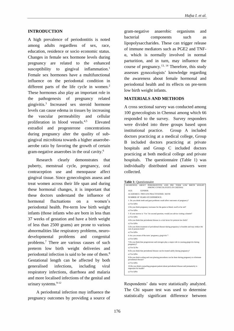
Hafsa I. et al.
176
INTRODUCTION
A high prevalence of periodontitis is noted
among adults regardless of sex, race,
education, residence or socio economic status.
Changes in female sex hormone levels during
pregnancy are related to the enhanced
susceptibility to gingival inflammation.1
Female sex hormones have a multifunctional
influence on the periodontal condition in
different parts of the life cycle in women.2
These hormones also play an important role in
the pathogenesis of pregnancy related
gingivitis.3 Increased sex steroid hormone
levels can cause edema in tissues by increasing
the vascular permeability and cellular
proliferation in blood vessels.4,5 Elevated
estradiol and progesterone concentrations
during pregnancy alter the quality of sub-
gingival microbiota towards a higher anaerobe-
aerobe ratio by favoring the growth of certain
gram-negative anaerobes in the oral cavity.6
Research clearly demonstrates that
puberty, menstrual cycle, pregnancy, oral
contraception use and menopause affect
gingival tissue. Since gynecologists assess and
treat women across their life span and during
these hormonal changes, it is important that
these doctors understand the influence of
hormonal fluctuations on a women’s
periodontal health. Pre-term low birth weight
infants (those infants who are born in less than
37 weeks of gestation and have a birth weight
of less than 2500 grams) are prone to various
abnormalities like respiratory problems, neuro-
developmental problems and congenital
problems.7 There are various causes of such
preterm low birth weight deliveries and
periodontal infection is said to be one of them.8
Gestational length can be affected by both
generalised infections, including viral
respiratory infections, diarrhoea and malaria
and more localised infections of the genital and
urinary systems.9-12
A periodontal infection may influence the
pregnancy outcomes by providing a source of
gram-negative anaerobic organisms and
bacterial components such as
lipopolysaccharides. These can trigger release
of immune mediators such as PGE2 and TNF-
α, which is normally involved in normal
parturition, and in turn, may influence the
course of pregnancy.13, 14 Therefore, this study
assesses gynecologists’ knowledge regarding
the awareness about female hormonal and
periodontal health and its effects on pre-term
low birth weight infants.
MATERIALS AND METHODS
A cross sectional survey was conducted among
100 gynecologists in Chennai among which 66
responded to the survey. Survey responders
were divided into three groups based upon
institutional practice. Group A included
doctors practicing at a medical college, Group
B included doctors practicing at private
hospitals and Group C included doctors
practicing at both medical college and private
hospitals. The questionnaire (Table 1) was
individually distributed and answers were
collected.
Table 1: Questionnaire
Respondents’ data were statistically analyzed.
The Chi square test was used to determine
statistically significant difference between
AWARENESS ABOUT PERIODONTITIS AND PRE TERM LOW BIRTH WEIGHT
AMONG GYNECOLOGISTS IN CHENNAI
AGE: GENDER:
ACADEMICS / PRIVATE PRACTITIONER / BOTH
NUMBER OF YEARS OF EXPERIENCE:
1. Do you think tooth and gum problems could affect outcomes of pregnancy?
a) Yes b)No
2.Do you think pregnancy increases for the gums to bleed, swell or be red?
a) Yes b)No
3. If your answer is ‘Yes’ for second question, would you advise visiting a dentist?
a) Yes b)No
4.Do you think that periodontal disease is a risk factor for preterm low birth?
a) Yes b)No
5.Do you think treatment of periodontal disease during pregnancy is feasible and may reduce the
risk of preterm birth?
a) Yes b)No
6.Are you aware of the term ‘pregnancy gingivitis’?
a) Yes b)No
7.Do you think that progesterone and estrogen play a major role in causing gingivitis during
pregnancy?
a) Yes b) No
8.Do you think that periodontal disease can be treated safely during pregnancy?
a) Yes b)No
9.Do you think scaling and root planing procedures can be done during pregnancy to eliminate
periodontal disease?
a) Yes b)No
10.Do you think counseling pregnant patient about periodontal disease and prematurity is
important for health?
a) Yes b)No
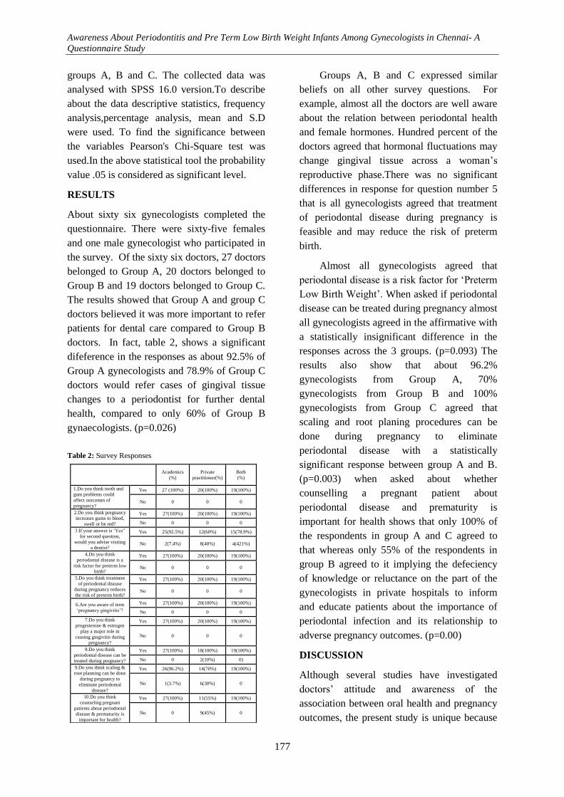
Awareness About Periodontitis and Pre Term Low Birth Weight Infants Among Gynecologists in Chennai- A
Questionnaire Study
177
groups A, B and C. The collected data was
analysed with SPSS 16.0 version.To describe
about the data descriptive statistics, frequency
analysis,percentage analysis, mean and S.D
were used. To find the significance between
the variables Pearson's Chi-Square test was
used.In the above statistical tool the probability
value .05 is considered as significant level.
RESULTS
About sixty six gynecologists completed the
questionnaire. There were sixty-five females
and one male gynecologist who participated in
the survey. Of the sixty six doctors, 27 doctors
belonged to Group A, 20 doctors belonged to
Group B and 19 doctors belonged to Group C.
The results showed that Group A and group C
doctors believed it was more important to refer
patients for dental care compared to Group B
doctors. In fact, table 2, shows a significant
difeference in the responses as about 92.5% of
Group A gynecologists and 78.9% of Group C
doctors would refer cases of gingival tissue
changes to a periodontist for further dental
health, compared to only 60% of Group B
gynaecologists. (p=0.026)
Table 2: Survey Responses
Groups A, B and C expressed similar
beliefs on all other survey questions. For
example, almost all the doctors are well aware
about the relation between periodontal health
and female hormones. Hundred percent of the
doctors agreed that hormonal fluctuations may
change gingival tissue across a woman’s
reproductive phase.There was no significant
differences in response for question number 5
that is all gynecologists agreed that treatment
of periodontal disease during pregnancy is
feasible and may reduce the risk of preterm
birth.
Almost all gynecologists agreed that
periodontal disease is a risk factor for ‘Preterm
Low Birth Weight’. When asked if periodontal
disease can be treated during pregnancy almost
all gynecologists agreed in the affirmative with
a statistically insignificant difference in the
responses across the 3 groups. (p=0.093) The
results also show that about 96.2%
gynecologists from Group A, 70%
gynecologists from Group B and 100%
gynecologists from Group C agreed that
scaling and root planing procedures can be
done during pregnancy to eliminate
periodontal disease with a statistically
significant response between group A and B.
(p=0.003) when asked about whether
counselling a pregnant patient about
periodontal disease and prematurity is
important for health shows that only 100% of
the respondents in group A and C agreed to
that whereas only 55% of the respondents in
group B agreed to it implying the defeciency
of knowledge or reluctance on the part of the
gynecologists in private hospitals to inform
and educate patients about the importance of
periodontal infection and its relationship to
adverse pregnancy outcomes. (p=0.00)
DISCUSSION
Although several studies have investigated
doctors’ attitude and awareness of the
association between oral health and pregnancy
outcomes, the present study is unique because
Academics
(%)
Private
practitioner(%)
Both
(%)
1.Do you think tooth and
gum problems could
affect outcomes of
pregnancy?
Yes 27 (100%) 20(100%) 19(100%)
No 0 0 0
2.Do you think pregnancy
increases gums to bleed,
swell or be red?
Yes 27(100%) 20(100%) 19(100%)
No 0 0 0
3.If your answer is ‘Yes”
for second question,
would you advise visiting
a dentist?
Yes 25(92.5%) 12(60%) 15(78.9%)
No 2(7.4%) 8(40%) 4(421%)
4.Do you think
periodontal disease is a
risk factor for preterm low
birth?
Yes 27(100%) 20(100%) 19(100%)
No 0 0 0
5.Do you think treatment
of periodontal disease
during pregnancy reduces
the risk of preterm birth?
Yes 27(100%) 20(100%) 19(100%)
No 0 0 0
6.Are you aware of term
‘pregnancy gingivitis’?
Yes 27(100%) 20(100%) 19(100%)
No 0 0 0
7.Do you think
progesterone & estrogen
play a major role in
causing gingivitis during
pregnancy?
Yes 27(100%) 20(100%) 19(100%)
No 0 0 0
8.Do you think
periodontal disease can be
treated during pregnancy?
Yes 27(100%) 18(100%) 19(100%)
No 0 2(10%) 0)
9.Do you think scaling &
root planning can be done
during pregnancy to
eliminate periodontal
disease?
Yes 26(96.2%) 14(70%) 19(100%)
No 1(3.7%) 6(30%) 0
10.Do you think
counseling pregnant
patients about periodontal
disease & prematurity is
important for health?
Yes 27(100%) 11(55%) 19(100%)
No 0 9(45%) 0
Hafsa I. et al.
178
it assessed gynecologists’ awareness of
pregnancy outcomes in addition to knowledge
about hormonal-induced gingival changes
throughout a woman’s reproductive phase. We
found that most gynecologists were well
educated and concerned about a female
patient’s oral health during different hormonal
phases. Our findings contribute to the literature
regarding gynecologists’ knowledge of oral
health, particularly periodontal health, for
female patients.15, 16
Our data showed that gynecologists from
Group A and C demonstrated greater
awareness of the need for dental follow-ups
compared to gynecologists from Group B. The
difference in awareness may be explained by
the fact that doctors in medical colleges
(Group A and Group C) are continually
updated with current knowledge for teaching
and research purposes. The present study
showed that while all of the gynecologists
knew about hormonal influences on the
periodontium, a few private practitioners were
less aware of the importance of a regular
dental check up and the to counsel pregnant
patients with regards to periodontal disease.
These findings are supported by Wilder et al
who concluded that obstetricians were well
aware of periodontal disease as a potential risk
factor for PLBW but showed limited
incorporation of this knowledge into clinical
practices.17 Almost all respondents agreed that
hormonal fluctuations might induce gingival
tissue changes across the female life cycle.
CONCLUSION
Preventive, routine and emergency dental care
procedures should be provided to pregnant
patients. Beyond treatment there is need for
pregnancy specific preventive care and oral
health education. It is recommended to
arrange training both dentists and obstetricians
in the oral health needs, screening and care of
pregnant patients, as a part of parental care.
REFERENCES
1. Silness J, Loe H, Periodontal disease in
pregnancy. II. Correlation between oral
hygiene and periodontal condition. Acta
Odontol Scand 1964; 22:121-135.
2. Gursoy M, Pajukanta R, Sorsa T, Kononen
E. Clinical changes in periodontium during
pregnancy and post-partum. J Clin Periodontol
2008;35:576-83.
3. Lopez NJ, Smith PC, Gutierrez J.
Periodontal therapy may reduce the risk of
preterm low birth weight in women with
periodontal disease: a randomized controlled
trial. J Periodontol 2002 73:911-924.
4. Lindhe J, Branemark PI. Changes in
vascular permeability after local application of
sex hormones. J Periodontal Res 1967; 2:259-
265.
5. Lindhe J, Branemark PI, Lundskog J.
Changes in vascular proliferation after local
application of sex hormones. J Periodontal Res
1967; 2:266-272.
6. Kornman KS, Loesche WJ, The
subgingival microbial flora during pregnancy.
J Periodontal Res 1980;15:111-122.
7. BK Yeo, LP Lim, Periodontal disease the
emergence of a risk for systemic conditions:
Pre term Low birth weight. Ann Acad Med
Singapore 2005; 34: 111-6.
8. Gibbs RS. The relationship between
infection and adverse pregnancy outcomes: an
overview. Ann Periodontol 2001;6:153-63
9. Bray RS, Anderson MJ. Falciparium
malaria and pregnancy. Trans R Soc Trop
Med Hyg 1979;73:427-31.
10. Andrews WW, Goldenberg RL, Hauth JC.
Preterm labor: emerging role of genital tract
infections. Infect Agents Dis 1995;4:196-211.
11. Gibbs RS, Romeo R, Hillier SL,
Eschenbach DA, Sweet RL. A review of
premature birth and subclinical infection. Am J
Obstet Gynecol 1992;166:1515-26.
12. Minkoff H, Grunebaum AN, Schwarz RH,
Feldman J, Cummings M, Crombleholme
W, et al. Risk factors for premature rupture of
membranes: a prospective study of the vagina
Awareness About Periodontitis and Pre Term Low Birth Weight Infants Among Gynecologists in Chennai- A
Questionnaire Study
179
flora in pregnancy. Am J Obstet Gynecol
1984;150:965-72.
13. Offenbacher S, Katz V, Fertik G, Collins J,
Boyd D, Maynor G, et al. Periodontal infection
as a possible risk factor for preterm low birth
weight. J Periodontol 1996; 67:1103-13.
14. Scannapieco FA. Position paper of The
American Academy of Periodontology:
periodontal disease as a potential risk factor for
systemic diseases. J Periodontol 1998;69:841-
50.
15. Apoorva, S.M., Suchetha, A. Effect of sex
hormones on periodontium. Indian J. Dent.
Sci. 2010; 2: 36-40.
16. Nutalapati R, Ramisetti A, Mutthineni RB,
Jampani ND, Kasagani SK. Awareness of
association between periodontitis and PLBW
among selected population of practising
gynecologists in Andhra Pradesh. Indian J
Dent Res 2011;22:735.
17. Wilder. R, Robinson.C, Jared.H.I.
Obstetricians’ knowledge and practice
behaviors concerning periodontal health and
preterm delivery and low birth weight. J Dent
hyg.2007; 81(4)81.
Correspondence Author
Hafsa Ismail
Saveetha Dental College
Department of Periodontics
India.
E-mail: [email protected]
Cumhuriyet Dental Journal: 2017; 20(3)
Doi: 10.7126/cumudj.369122 CLINICAL REPORTS
180
A. B. Shetty Memorial Institute of Dental Sciences, Department of Oral Medicine and Radiology, Nitte University, India.
A CASE REPORT OF TRANSORAL REMOVAL OF SUBMANDIBULAR
GLAND SIALOLITH
Submandibular Tükürük Bezi Taşının Ağız İçinden Uzaklaştırılması: Bir Olgu Sunumu
Kumuda RAO, Subhas G BABU, Renita Lorina CASTELINO
Makale Kodu/Article Code : 189565
Makale Gönderilme Tarihi : 17.05.2016
Kabul Tarihi : 20.01.2017
ABSTRACT
Salivary calculus or sialolithiasis is a disease that
affects the salivary glands characterized by the
formation of mineralized structures within the
glandular substance or excretory ducts of the
salivary gland. The formation of these salivary
stones is due to the crystallization of minerals in
saliva. It causes blockage of salivary ducts and
results in painful inflammation or sialadenitis of the
salivary gland. Among the salivary glands
submandibular gland has highest incidence of
sialolithiasis due its anatomic features. The patient
commonly experiences pain and/or edema when the
ducts are obstructed. The case report presented here
is of sialolithiasis of submandibular gland which
had caused pain and swelling in the floor of the
mouth.
Keywords: Salivary calculus, sialolithiasis,
salivary stones, submandibular gland, Wharton’s
duct
ÖZ
Tükrük bezi taşı veya siyalolitiyaz, tükrük bezini
etkileyen, glandüler madde veya tükrük bezinin
boşaltma kanallarındaki mineralize yapıların
oluşumu ile karakterize olan bir hastalıktır. Bu
tükrük taşlarının oluşumu tükrükteki minerallerin
kristalleşmesinden kaynaklanmaktadır. Tükürük
kanallarının tıkanmasına neden olur ve tükürük bezi
ağrılı inflamasyonu veya sialadenit ile sonuçlanır.
Tükrük bezleri arasında, submandibular bezin
anatomik özellikleri nedeniyle siyalolitiyaz
insidansının en yüksek olduğu görülmektedir.
Kanallar tıkandığı zaman hasta genellikle ağrı ve /
veya ödem görür. Bu olguda, ağız tabanında ağrı ve
şişme yaratan submandibular bezin sialolitiyazisi
sunulmaktadır.
Anahtar Kelimeler: Tükrük taşı, siyalolitiyazis,
tükrük taşları, submandibular bez, Wharton kanalı
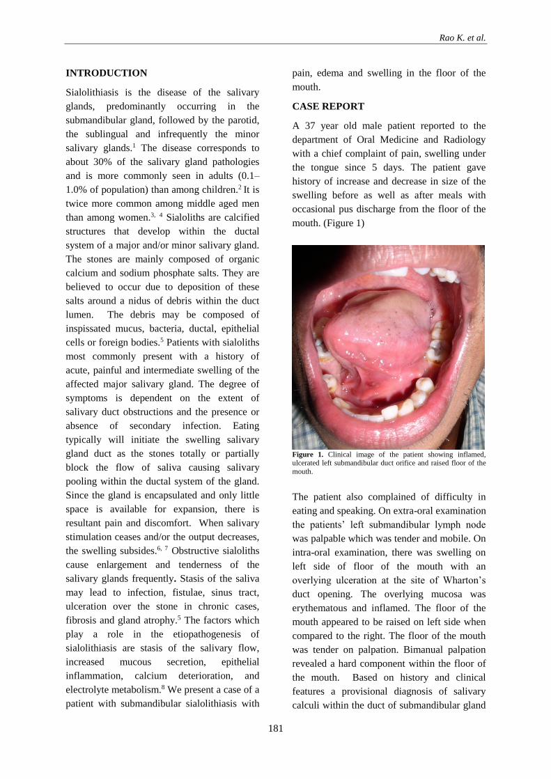
Rao K. et al.
181
INTRODUCTION
Sialolithiasis is the disease of the salivary
glands, predominantly occurring in the
submandibular gland, followed by the parotid,
the sublingual and infrequently the minor
salivary glands.1 The disease corresponds to
about 30% of the salivary gland pathologies
and is more commonly seen in adults (0.1–
1.0% of population) than among children.2 It is
twice more common among middle aged men
than among women.3, 4 Sialoliths are calcified
structures that develop within the ductal
system of a major and/or minor salivary gland.
The stones are mainly composed of organic
calcium and sodium phosphate salts. They are
believed to occur due to deposition of these
salts around a nidus of debris within the duct
lumen. The debris may be composed of
inspissated mucus, bacteria, ductal, epithelial
cells or foreign bodies.5 Patients with sialoliths
most commonly present with a history of
acute, painful and intermediate swelling of the
affected major salivary gland. The degree of
symptoms is dependent on the extent of
salivary duct obstructions and the presence or
absence of secondary infection. Eating
typically will initiate the swelling salivary
gland duct as the stones totally or partially
block the flow of saliva causing salivary
pooling within the ductal system of the gland.
Since the gland is encapsulated and only little
space is available for expansion, there is
resultant pain and discomfort. When salivary
stimulation ceases and/or the output decreases,
the swelling subsides.6, 7 Obstructive sialoliths
cause enlargement and tenderness of the
salivary glands frequently. Stasis of the saliva
may lead to infection, fistulae, sinus tract,
ulceration over the stone in chronic cases,
fibrosis and gland atrophy.5 The factors which
play a role in the etiopathogenesis of
sialolithiasis are stasis of the salivary flow,
increased mucous secretion, epithelial
inflammation, calcium deterioration, and
electrolyte metabolism.8 We present a case of a
patient with submandibular sialolithiasis with
pain, edema and swelling in the floor of the
mouth.
CASE REPORT
A 37 year old male patient reported to the
department of Oral Medicine and Radiology
with a chief complaint of pain, swelling under
the tongue since 5 days. The patient gave
history of increase and decrease in size of the
swelling before as well as after meals with
occasional pus discharge from the floor of the
mouth. (Figure 1)
Figure 1. Clinical image of the patient showing inflamed, ulcerated left submandibular duct orifice and raised floor of the
mouth.
The patient also complained of difficulty in
eating and speaking. On extra-oral examination
the patients’ left submandibular lymph node
was palpable which was tender and mobile. On
intra-oral examination, there was swelling on
left side of floor of the mouth with an
overlying ulceration at the site of Wharton’s
duct opening. The overlying mucosa was
erythematous and inflamed. The floor of the
mouth appeared to be raised on left side when
compared to the right. The floor of the mouth
was tender on palpation. Bimanual palpation
revealed a hard component within the floor of
the mouth. Based on history and clinical
features a provisional diagnosis of salivary
calculi within the duct of submandibular gland
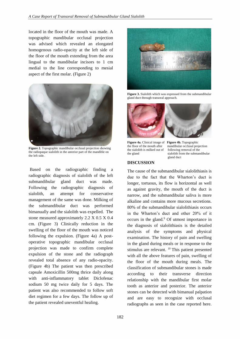
A Case Report of Transoral Removal of Submandibular Gland Sialolith
182
located in the floor of the mouth was made. A
topographic mandibular occlusal projection
was advised which revealed an elongated
homogenous radio-opacity at the left side of
the floor of the mouth extending from the area
lingual to the mandibular incisors to 1 cm
medial to the line corresponding to mesial
aspect of the first molar. (Figure 2)
Figure 2. Topographic mandibular occlusal projection showing the radiopaque sialolith in the anterior part of the mandible on
the left side.
Based on the radiographic finding a
radiographic diagnosis of sialolith of the left
submandibular gland duct was made.
Following the radiographic diagnosis of
sialolith, an attempt for conservative
management of the same was done. Milking of
the submandibular duct was performed
bimanually and the sialolith was expelled. The
stone measured approximately 2.2 X 0.5 X 0.4
cm. (Figure 3) Clinically reduction in the
swelling of the floor of the mouth was noticed
following the expulsion. (Figure 4a) A post-
operative topographic mandibular occlusal
projection was made to confirm complete
expulsion of the stone and the radiograph
revealed total absence of any radio-opacity.
(Figure 4b) The patient was then prescribed
capsule Amoxicillin 500mg thrice daily along
with anti-inflammatory tablet Diclofenac
sodium 50 mg twice daily for 5 days. The
patient was also recommended to follow soft
diet regimen for a few days. The follow up of
the patient revealed uneventful healing.
Figure 3. Sialolith which was expressed from the submandibular
gland duct through transoral approach.
Figure 4a. Clinical image of Figure 4b. Topographic
the floor of the mouth after mandibular occlusal projection the sialolith is milked out of following removal of the
the gland sialolith from the submandibular
gland duct
DISCUSSION
The cause of the submandibular sialolithiasis is
due to the fact that the Wharton’s duct is
longer, tortuous, its flow is horizontal as well
as against gravity, the mouth of the duct is
narrow, and the submandibular saliva is more
alkaline and contains more mucous secretions.
80% of the submandibular sialolithiasis occurs
in the Wharton’s duct and other 20% of it
occurs in the gland.9 Of utmost importance in
the diagnosis of sialolithiasis is the detailed
analysis of the symptoms and physical
examination. The history of pain and swelling
in the gland during meals or in response to the
stimulus are relevant. 10 This patient presented
with all the above features of pain, swelling of
the floor of the mouth during meals. The
classification of submandibular stones is made
according to their transverse direction
relationship with the mandibular first molar
tooth as anterior and posterior. The anterior
stones can be detected with bimanual palpation
and are easy to recognize with occlusal
radiographs as seen in the case reported here.
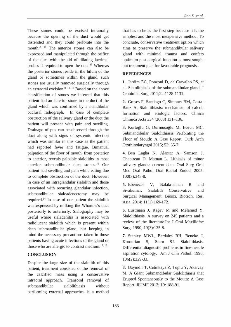
Rao K. et al.
183
These stones could be excised intraorally
because the opening of the duct would get
distended and they could perforate into the
mouth.8, 11 The anterior stones can also be
expressed and manipulated through the orifice
of the duct with the aid of dilating lacrimal
probes if required to open the duct.13 Whereas
the posterior stones reside in the hilum of the
gland or sometimes within the gland, such
stones are usually removed surgically through
an extraoral excision.8, 11, 13 Based on the above
classification of stones we inferred that this
patient had an anterior stone in the duct of the
gland which was confirmed by a mandibular
occlusal radiograph. In case of complete
obstruction of the salivary gland or the duct the
patient will present with pain and swelling.
Drainage of pus can be observed through the
duct along with signs of systemic infection
which was similar in this case as the patient
had reported fever and fatigue. Bimanual
palpation of the floor of mouth, from posterior
to anterior, reveals palpable sialoliths in most
anterior submandibular duct stones.10 Our
patient had swelling and pain while eating due
to complete obstruction of the duct. However,
in case of an intraglandular sialolith and those
associated with recurring glandular infection,
submandibular sialoadenectomy may be
required.14 In case of our patient the sialolith
was expressed by milking the Wharton’s duct
posteriorly to anteriorly. Sialography may be
useful where sialadenitis is associated with
radiolucent sialolith which is present within
deep submandibular gland, but keeping in
mind the necessary precautions taken in those
patients having acute infections of the gland or
those who are allergic to contrast medium.15, 16.
CONCLUSION
Despite the large size of the sialolith of this
patient, treatment consisted of the removal of
the calcified mass using a conservative
intraoral approach. Transoral removal of
submandibular sialolithiasis without
performing external approaches is a method
that has to be as the first step because it is the
simplest and the most inexpensive method. To
conclude, conservative treatment option which
aims to preserve the submandibular salivary
gland with minimal trauma and confers
optimum post-surgical function is most sought
out treatment plan for favourable prognosis.
REFERENCES
1. Jardim EC, Ponzoni D, de Carvalho PS, et
al. Sialolithiasis of the submandibular gland. J
Craniofac Surg 2011;22:1128-1131.
2. Grases F, Santiago C, Simonet BM, Costa-
Bauz A. Sialolithiasis: mechanism of calculi
formation and etiologic factors. Clinica
Chimica Acta 334 (2003) 131–136.
3. Kurtoğlu G, Durmuşoğlu M, Ecevit MC.
Submandibular Sialolithiasis Perforating the
Floor of Mouth: A Case Report. Turk Arch
Otorhinolaryngol 2015; 53: 35-7.
4. Ben Lagha N, Alantar A, Samson J,
Chapireau D, Maman L. Lithiasis of minor
salivary glands: current data. Oral Surg Oral
Med Oral Pathol Oral Radiol Endod. 2005;
100(3):345-8.
5. Ebenezer V, Balakrishnan R and
Sivakumar. Sialolith Conservative and
Surgical Management. Biosci. Biotech. Res.
Asia, 2014; 11(1):169-172.
6. Lustmaan J, Ragev M and Melamed Y.
Sialolithiasis. A survey on 245 patients and a
review of the literature.Int J Oral Maxillofac
Surg. 1990; 19(3):135-8.
7. Stanley MW1, Bardales RH, Beneke J,
Korourian S, Stern SJ. Sialolithiasis.
Differential diagnostic problems in fine-needle
aspiration cytology. Am J Clin Pathol. 1996;
106(2):229-33.
8. Bayındır T, Cetinkaya Z, Toplu Y, Akarcay
M. A Giant Submandibular Sialolithiasis that
Erupted Spontaneously to the Mouth: A Case
Report. JIUMF 2012; 19: 188-91.
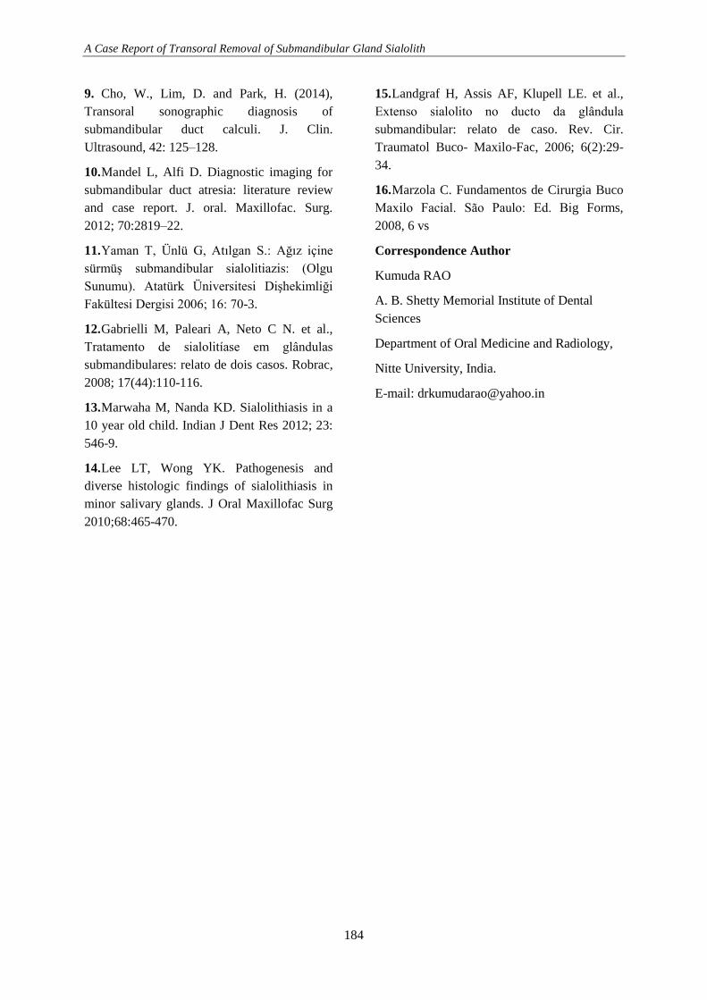
A Case Report of Transoral Removal of Submandibular Gland Sialolith
184
9. Cho, W., Lim, D. and Park, H. (2014),
Transoral sonographic diagnosis of
submandibular duct calculi. J. Clin.
Ultrasound, 42: 125–128.
10. Mandel L, Alfi D. Diagnostic imaging for
submandibular duct atresia: literature review
and case report. J. oral. Maxillofac. Surg.
2012; 70:2819–22.
11. Yaman T, Ünlü G, Atılgan S.: Ağız içine
sürmüş submandibular sialolitiazis: (Olgu
Sunumu). Atatürk Üniversitesi Dişhekimliği
Fakültesi Dergisi 2006; 16: 70-3.
12. Gabrielli M, Paleari A, Neto C N. et al.,
Tratamento de sialolitíase em glândulas
submandibulares: relato de dois casos. Robrac,
2008; 17(44):110-116.
13. Marwaha M, Nanda KD. Sialolithiasis in a
10 year old child. Indian J Dent Res 2012; 23:
546-9.
14. Lee LT, Wong YK. Pathogenesis and
diverse histologic findings of sialolithiasis in
minor salivary glands. J Oral Maxillofac Surg
2010;68:465-470.
15. Landgraf H, Assis AF, Klupell LE. et al.,
Extenso sialolito no ducto da glândula
submandibular: relato de caso. Rev. Cir.
Traumatol Buco- Maxilo-Fac, 2006; 6(2):29-
34.
16. Marzola C. Fundamentos de Cirurgia Buco
Maxilo Facial. São Paulo: Ed. Big Forms,
2008, 6 vs
Correspondence Author
Kumuda RAO
A. B. Shetty Memorial Institute of Dental
Sciences
Department of Oral Medicine and Radiology,
Nitte University, India.
E-mail: [email protected]
Cumhuriyet Dental Journal: 2017; 20(3)
Doi: 10.7126/cumudj.369127 CLINICAL REPORTS
185
Department of Endodontics, Faculty of Dentistry, Abant İzzet Baysal University, Bolu, Turkey.
REMOVAL OF SEPARATED INSTRUMENTS WITH MASSERANN
TECHNIQUES: TWO CASE REPORTS
Masserann Tekniği ile Kırık Aletlerin Uzaklaştırılması: İki Olgu Sunumu
Hakan GÖKTÜRK, İsmail ÖZKOÇAK
Makale Kodu/Article Code : 205221
Makale Gönderilme Tarihi : 23.10.2016
Kabul Tarihi : 13.01.2017
ABSTRACT
Instrument fracture is a procedural mishap that
prevents efficient cleaning and shaping of the
root canal. When bypassing the fracture fails,
the fragment must be removed. Many
techniques and devices have been described to
remove fragments from the root canal. The
Masserann Kit is a widely used instrument for
the removal of fragments. These clinical cases
describe use of the Masserann Kit to remove
separated instruments blocking the entire canal
in posterior teeth.
Keywords: Endodontics, Instrument removal,
Masserann Kit, Seperated file
ÖZ
Enstrüman kırığı, kök kanalının etkin şekilde
temizlenmesini ve şekillenmesini önleyen bir
prosedür hatasıdır. Kırık parçayı atlayarak
yanından geçilmesi (bypass) başarısız
olduğunda, parça çıkarılmalıdır. Kırık parçanın
kök kanalından uzaklaştırılması için birçok
teknik ve cihaz geliştirilmiştir. Masserann Kit,
kırık parçaların çıkarılması için yaygın olarak
kullanılan bir alettir. Bu klinik olgularda, arka
bölgedeki dişlerde tüm kanalı tıkayan kırık
parçaları uzaklaştırmak için Masserann Kit
kullanımını sunulmuştur.
Anahtar Kelimeler: Endodonti, Alet
uzaklaştırma, Masserann Kiti, Kırık eğe
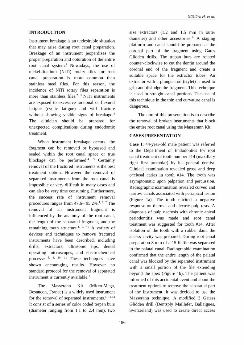
Göktürk H. et al.
186
INTRODUCTION
Instrument breakage is an undesirable situation
that may arise during root canal preparation.
Breakage of an instrument jeopardizes the
proper preparation and obturation of the entire
root canal system.1 Nowadays, the use of
nickel-titanium (NiTi) rotary files for root
canal preparation is more common than
stainless steel files. For this reason, the
incidence of NiTi rotary files separation is
more than stainless files.2, 3 NiTi instruments
are exposed to excessive torsional or flexural
fatigue (cyclic fatigue) and will fracture
without showing visible signs of breakage.4
The clinician should be prepared for
unexpected complications during endodontic
treatment.
When instrument breakage occurs, the
fragment can be removed or bypassed and
sealed within the root canal space or true
blockage can be performed.4, 5 Certainly
removal of the fractured instruments is the best
treatment option. However the removal of
separated instruments from the root canal is
impossible or very difficult in many cases and
can also be very time consuming. Furthermore,
the success rate of instrument removal
procedures ranges from 47.6– 95.2%.1, 6, 7 The
removal of an instrument fragment is
influenced by the anatomy of the root canal,
the length of the separated fragment, and the
remaining tooth structure.1, 5, 7-9 A variety of
devices and techniques to remove fractured
instruments have been described, including
drills, extractors, ultrasonic tips, dental
operating microscopes, and electrochemical
processes.5, 8, 10, 11 These techniques have
shown encouraging results. However no
standard protocol for the removal of separated
instrument is currently available.1
The Masserann Kit (Micro-Mega,
Besancon, France) is a widely used instrument
for the removal of separated instruments.1, 11-13
It consist of a series of color coded trepan burs
(diameter ranging from 1.1 to 2.4 mm), two
size extractors (1.2 and 1.5 mm in outer
diameter) and other accessories.14 A staging
platform and canal should be prepared at the
coronal part of the fragment using Gates
Glidden drills. The trepan burs are rotated
counter-clockwise to cut the dentin around the
coronal end of the fragment and create a
suitable space for the extractor tubes. An
extractor with a plunger rod (stylet) is used to
grip and dislodge the fragment. This technique
is used in straight canal portions. The use of
this technique in the thin and curvature canal is
dangerous.
The aim of this presentation is to describe
the removal of broken instruments that block
the entire root canal using the Masserann Kit.
CASES PRESENTATION
Case 1: 44-year-old male patient was referred
to the Department of Endodontics for root
canal treatment of tooth number #14 (maxillary
right first premolar) by his general dentist.
Clinical examination revealed gross and deep
occlusal caries in tooth #14. The tooth was
asymptomatic upon palpation and percussion.
Radiographic examination revealed curved and
narrow canals associated with periapical lesion
(Figure 1a). The tooth elicited a negative
response on thermal and electric pulp tests. A
diagnosis of pulp necrosis with chronic apical
periodontitis was made and root canal
treatment was suggested for tooth #14. After
isolation of the tooth with a rubber dam, the
access cavity was prepared. During root canal
preparation 8 mm of a 15 K-file was separated
in the palatal canal. Radiographic examination
confirmed that the entire length of the palatal
canal was blocked by the separated instrument
with a small portion of the file extending
beyond the apex (Figure 1b). The patient was
informed of this accidental event and about the
treatment options to remove the separated part
of the instrument. It was decided to use the
Masserann technique. A modified 3 Gatess
Glidden drill (Dentsply Maillefer, Ballaigues,
Switzerland) was used to create direct access

Removal of Separated Instruments with Masserann Techniques: Two Case Reports
187
to the fragment and a straight platform. Three
trepan burs (1, 2 and 3, respectively) were
subsequently used to remove the surrounding
dentin using a handpiece at 300 rpm in a
counter-clockwise rotation. The fragment was
not removed during trephining. A small
extractor tube with a diameter of 1.2 mm was
used to grip the broken instrument. An
extractor tube gripping the fragment was
gently rotated in a counter-clockwise direction
to retrieve the fragment. A radiograph was
taken to confirm instrument removal (Figure
1c). Preparation of all canals was completed
using a ProTaper Universal system with full
sequence up to F3 (PTU; Dentsply Tulsa
Dental, Tulsa, OK). The root canals were
copiously irrigated with 2.5% sodium
hypochlorite (Whitedentmed, Erhan Kimya,
İzmir, Turkey) and 17%
ethylenediaminetetraacetic acid (Imicryl Ltd.,
Konya, Turkey) during the preparation. The
root canals were filled with epoxy-amine resin-
based sealer (2Seal, VDW, Munich, Germany)
and gutta-percha points (Dentsply Maillefer,
Tulsa, OK) using the cold lateral condensation
technique. The tooth was provisionally sealed
with temporary filling material (Cavit G 3M
Espe, Seefeld, Germany). Two days later a
screw post (Nordin Screw Posts, H. Nordin
SA, Swiss) placed in the palatal root canal and
the access cavity was restored with composite
resin (3M Filtek Z-250, 3M ESPE, St. Paul,
MN, USA) (Figure 1d). The patient was then
referred to the prosthodontics clinic for a full
crown restoration. At the 6 month follow up
visit the tooth was asymptomatic without any
radiographic changes (Figure 1e). For this
reason, it was decided to follow the teeth
radiographically at intervals of six months.
However the tooth was not restored with a
crown and the patient was redirected to the
prosthodontics clinic.
Figure 1. (a) A pre-operative radiograph of Case 1. (b) Broken
instrument (15 K-file) in the entire palatal canal of maxillary
right first premolar. (c) A radiograph confirmed the removal of broken instrument. (d) A post-operative radiograph showing the
obturation and restoration. (e) Six month follow-up.
Case 2: A 44-year-old male patient was
referred to our clinic with complaints of pain
and swelling in the maxillary right region.
Clinically, tooth #16 and #17 had porcelain-
fused-to-metal crowns and were tender to
percussion. Radiographic examination revealed
that tooth #17 had a deep restoration under the
prosthesis and a wide radiolucent area in the
apical region (Figure 2a). After the removal of
prosthesis, an electric pulp test and cold
application were applied to tooth #17 and there
was no response these vitality tests. The tooth
was diagnosed with chronic apical
periodontitis. After isolation with a rubber
dam, the access cavity was prepared. During
root canal preparation a ProTaper Universal S1
(Dentsply, OK) file was fractured in the mesio-
buccal canal. Radiographic examination
revealed that the fragment was occupying the
entire length of the canal (Figure 2b).
Clinically, the broken fragment did not extend
to the pulp chamber. The fragment could not
be grasped coronally and it was decided to use
the Masserann technique. Three trepan burs (2,
3, and 4, respectively) were used to create
space between the fragment and the root canal
wall. The small extractor tube was used to grip
the fragment. The tube with the locked
fragment was gently rotated in a counter-
clockwise direction to retrieve the fragment. A
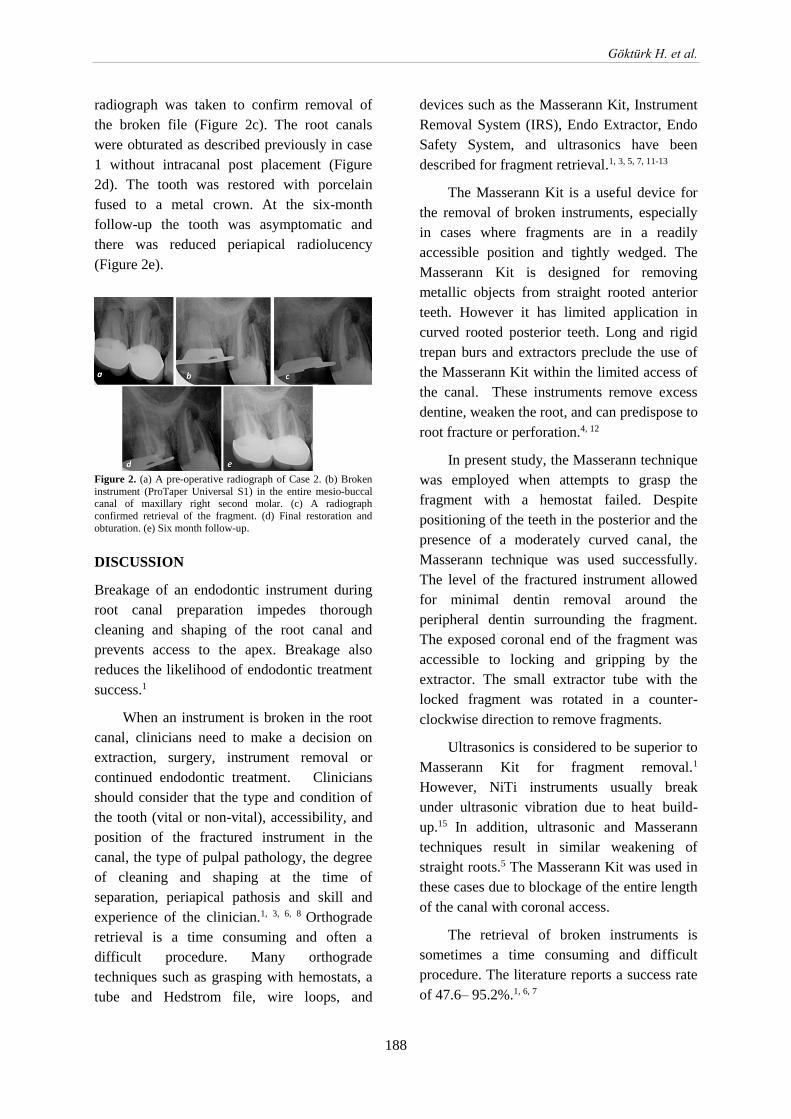
Göktürk H. et al.
188
radiograph was taken to confirm removal of
the broken file (Figure 2c). The root canals
were obturated as described previously in case
1 without intracanal post placement (Figure
2d). The tooth was restored with porcelain
fused to a metal crown. At the six-month
follow-up the tooth was asymptomatic and
there was reduced periapical radiolucency
(Figure 2e).
Figure 2. (a) A pre-operative radiograph of Case 2. (b) Broken
instrument (ProTaper Universal S1) in the entire mesio-buccal canal of maxillary right second molar. (c) A radiograph
confirmed retrieval of the fragment. (d) Final restoration and
obturation. (e) Six month follow-up.
DISCUSSION
Breakage of an endodontic instrument during
root canal preparation impedes thorough
cleaning and shaping of the root canal and
prevents access to the apex. Breakage also
reduces the likelihood of endodontic treatment
success.1
When an instrument is broken in the root
canal, clinicians need to make a decision on
extraction, surgery, instrument removal or
continued endodontic treatment. Clinicians
should consider that the type and condition of
the tooth (vital or non-vital), accessibility, and
position of the fractured instrument in the
canal, the type of pulpal pathology, the degree
of cleaning and shaping at the time of
separation, periapical pathosis and skill and
experience of the clinician.1, 3, 6, 8 Orthograde
retrieval is a time consuming and often a
difficult procedure. Many orthograde
techniques such as grasping with hemostats, a
tube and Hedstrom file, wire loops, and
devices such as the Masserann Kit, Instrument
Removal System (IRS), Endo Extractor, Endo
Safety System, and ultrasonics have been
described for fragment retrieval.1, 3, 5, 7, 11-13
The Masserann Kit is a useful device for
the removal of broken instruments, especially
in cases where fragments are in a readily
accessible position and tightly wedged. The
Masserann Kit is designed for removing
metallic objects from straight rooted anterior
teeth. However it has limited application in
curved rooted posterior teeth. Long and rigid
trepan burs and extractors preclude the use of
the Masserann Kit within the limited access of
the canal. These instruments remove excess
dentine, weaken the root, and can predispose to
root fracture or perforation.4, 12
In present study, the Masserann technique
was employed when attempts to grasp the
fragment with a hemostat failed. Despite
positioning of the teeth in the posterior and the
presence of a moderately curved canal, the
Masserann technique was used successfully.
The level of the fractured instrument allowed
for minimal dentin removal around the
peripheral dentin surrounding the fragment.
The exposed coronal end of the fragment was
accessible to locking and gripping by the
extractor. The small extractor tube with the
locked fragment was rotated in a counter-
clockwise direction to remove fragments.
Ultrasonics is considered to be superior to
Masserann Kit for fragment removal.1
However, NiTi instruments usually break
under ultrasonic vibration due to heat build-
up.15 In addition, ultrasonic and Masserann
techniques result in similar weakening of
straight roots.5 The Masserann Kit was used in
these cases due to blockage of the entire length
of the canal with coronal access.
The retrieval of broken instruments is
sometimes a time consuming and difficult
procedure. The literature reports a success rate
of 47.6– 95.2%.1, 6, 7
Removal of Separated Instruments with Masserann Techniques: Two Case Reports
189
Previous studies have shown that the
success rate of these procedures increases
when the fragment is visible using an operating
microscope.6, 8, 10 The probability of fragment
retrieval is reduced when the canal curvature
angle is increased.1,7 Excessive dentine
removal creates straight-line access within the
curved canal. This leads to weakening of the
tooth and perforation. Ideally, dentin removal
should be reduced as much as possible while
creating access to the fragment. In the present
cases, the coronal level of the fragment was
near the orifice of the canal and minimal dentin
removal was created to expose at least 1.5 mm
of the fragment in order to be able to grip the
fragment with the extractor.
A dilemma exists in fragment removal:
there is no clear evidence that retained
instrument fragments have any impact on
prognosis.9 Instrument fragment inhibit
cleaning and shaping of an infected root canal
system. Insufficient mechanical and chemical
treatment may have a negative impact on the
treatment outcome and reduce the chances of
successful treatment.8 However, fragment
removal procedures weaken the teeth.5 In the
present study, instrument fracture occurred in
the initial stage of the root canal preparation.
In both cases the canal could not be
sufficiently cleaned and shaped. The fragment
was very long and blocked the entire canal
system. It was impossible to bypass the broken
instrument. As a result it was decided to
remove the fragment using the Masserann Kit.
This report showed that the Masserann Kit
was successful in the retrieval of tightly
wedged fragments from maxillary premolar
and molar teeth. However it should be
employed with proper case selection. In the
hands of a skilled clinician, the Masserann Kit
is effective only in the straight portion of the
canal is cases with adequate surrounding
dentin and straight line accessibility.
In the present study, both cases were
diagnosed with chronic apical periodontitis. In
general, chronic apical periodontitis treated
with multiple-visit root canal treatment.
However, in the present both cases treated with
single-visit root canal treatment because of the
use of proper instrumentation and irrigation
procedures, aseptic operating procedures. Also
during the root canal filing there was no
exudate. Waltimo et al.16 report that, when
adequate bacterial reduction with NaOCl
irrigation was obtained, there was no
significance difference between single or
multi-visit treatment of chronic apical
periodontitis.
REFERENCES
1. Gencoglu N, Helvacioglu D. Comparison of
the different techniques to remove fractured
endodontic instruments from root canal
systems. Eur J Dent 2009;3:90-95.
2. Iqbal MK, Kohli MR, Kim JS. A
retrospective clinical study of incidence of root
canal instrument separation in an endodontics
graduate program: a PennEndo database study.
J Endod 2006;32:1048-1052.
3. Agrawal V, Kapoor S, Patel M. Ultrasonic
technique to retrieve a rotary nickel-titanium
file broken beyond the apex and a stainless
steel file from the root canal of a mandibular
molar: a case report. J Dent (Tehran)
2015;12:532-536.
4. Parashos P, Messer HH. Rotary NiTi
instrument fracture and its consequences. J
Endod 2006;32:1031-1043.
5. Gerek M, Baser ED, Kayahan MB, Sunay
H, Kaptan RF, Bayirli G. Comparison of the
force required to fracture roots vertically after
ultrasonic and Masserann removal of broken
instruments. Int Endod J 2012;45:429-434.
6. Ward JR, Parashos P, Messer HH.
Evaluation of an ultrasonic technique to
remove fractured rotary nickel-titanium
endodontic instruments from root canals: an
experimental study. J Endod 2003;29:756-
763.
7. Alomairy KH. Evaluating two techniques
on removal of fractured rotary nickel-titanium
Göktürk H. et al.
190
endodontic instruments from root canals: an in
vitro study. J Endod 2009;35:559-562.
8. Cuje J, Bargholz C, Hulsmann M. The
outcome of retained instrument removal in a
specialist practice. Int Endod J 2010;43:545-
554.
9. Panitvisai P, Parunnit P, Sathorn C, Messer
HH. Impact of a retained instrument on
treatment outcome: a systematic review and
meta-analysis. J Endod 2010;36:775-780.
10. Jadhav GR. Endodontic management of a
two rooted, three canaled mandibular canine
with a fractured instrument. J Conserv Dent
2014;17:192-195.
11. Brito-Junior M, Normanha JA, Camilo CC,
Faria-e-Silva AL, Saquy PC, Ferraz MA, et al.
Alternative techniques to remove fractured
instrument fragments from the apical third of
root canals: report of two cases. Braz Dent J
2015;26:79-85.
12. Yoldas O, Oztunc H, Tinaz C, Alparslan N.
Perforation risks associated with the use of
Masserann endodontic kit drills in mandibular
molars. Oral Surg Oral Med Oral Pathol Oral
Radiol Endod 2004;97:513-517.
13. Thirumalai AK, Sekar M, Mylswamy S.
Retrieval of a separated instrument using
Masserann technique. J Conserv Dent
2008;11:42-45.
14. Masserann J. "Entfernen metallischer
Fragmente aus Wurzelkanalen" (Removal of
metal fragments from the root canal). J Br
Endod Soc 1971;5:55-59.
15. Shen Y, Peng B, Cheung GS. Factors
associated with the removal of fractured NiTi
instruments from root canal systems. Oral Surg
Oral Med Oral Pathol Oral Radiol Endod
2004;98:605-610.
16. Waltimo T, Trope M, Haapasalo M,
Ørstavik D. Clinical efficacy of treatment
procedures in endodontic infection control and
one year follow-up of periapical healing. J
Endod 2005;31:863-6.
Correspondence Author
Hakan Göktürk
Abant Izzet Baysal University
Faculty of Dentistry
Department of Endodontics
Bolu 14100, Turkey
Phone: +90-374-254 10 00 ext-8453
Fax: +90-374-270 00 66
E-mail: [email protected]
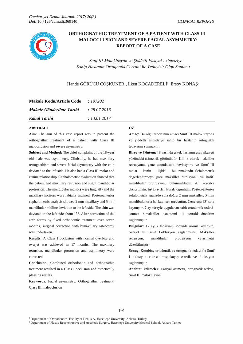
Cumhuriyet Dental Journal: 2017; 20(3)
Doi: 10.7126/cumudj.369140 CLINICAL REPORTS
191
1 Department of Orthodontics, Faculty of Dentistry, Hacettepe University, Ankara, Turkey 2 Department of Plastic Reconstructive and Aesthetic Surgery, Hacettepe University Medical School, Ankara Turkey
ORTHOGNATHIC TREATMENT OF A PATIENT WITH CLASS III
MALOCCLUSION AND SEVERE FACIAL ASYMMETRY:
REPORT OF A CASE
Sınıf III Malokluzyon ve Şiddetli Fasiyal Asimetriye
Sahip Hastanın Ortognatik Cerrahi ile Tedavisi: Olgu Sunumu
Hande GÖRÜCÜ COŞKUNER1, İlken KOCADERELİ1, Ersoy KONAŞ2
Makale Kodu/Article Code : 197202
Makale Gönderilme Tarihi : 28.07.2016
Kabul Tarihi : 13.01.2017
ABSTRACT
Aim: The aim of this case report was to present the
orthognathic treatment of a patient with Class III
malocclusion and severe asymmetry.
Subject and Method: The chief complaint of the 18-year
old male was asymmetry. Clinically, he had maxillary
retrognathism and severe facial asymmetry with the chin
deviated to the left side. He also had a Class III molar and
canine relationship. Cephalometric evaluation showed that
the patient had maxillary retrusion and slight mandibular
protrusion. The mandibular incisors were lingually and the
maxillary incisors were labially inclined. Posteroanterior
cephalometric analysis showed 2 mm maxillary and 5 mm
mandibular midline deviation to the left side. The chin was
deviated to the left side about 13°. After correction of the
arch forms by fixed orthodontic treatment over seven
months, surgical correction with bimaxillary osteotomy
was undertaken.
Results: A Class I occlusion with normal overbite and
overjet was achieved in 17 months. The maxillary
retrusion, mandibular protrusion and asymmetry were
corrected.
Conclusion: Combined orthodontic and orthognathic
treatment resulted in a Class I occlusion and esthetically
pleasing results.
Keywords: Facial asymmetry, Orthognathic treatment,
Class III malocclusion
ÖZ
Amaç: Bu olgu raporunun amacı Sınıf III malokluzyona
ve şiddetli asimetriye sahip bir hastanın ortognatik
tedavisini sunmaktır.
Birey ve Yöntem: 18 yaşında erkek hastanın esas şikayeti
yüzündeki asimetrik görüntüdür. Klinik olarak maksiller
retruzyonu, çene ucunda sola deviasyonu ve Sınıf III
molar kanin ilişkisi bulunmaktadır. Sefalometrik
değerlendirmeye göre maksiller retruzyonu ve hafif
mandibular protruzyonu bulunmaktadır. Alt keserler
dikleşmiştir, üst keserler labiale eğimlidir. Posteroanterior
sefalometrik analizde sola doğru 2 mm maksiller, 5 mm
mandibular orta hat kayması mevcuttur. Çene ucu 13° sola
kaymıştır. 7 ay süreyle uygulanan sabit ortodontik tedavi
sonrası bimaksiller osteotomi ile cerrahi düzeltim
sağlanmıştır.
Bulgular: 17 aylık tedavinin sonunda normal overbite,
overjet ve Sınıf I okluzyon sağlanmıştır. Maksiller
retruzyon, mandibular protruzyon ve asimetri
düzeltilmiştir.
Sonuç: Kombine ortodontik ve ortognatik tedavi ile Sınıf
I okluzyon elde edilmiş; kayıp estetik ve fonksiyon
sağlanmıştır.
Anahtar kelimeler: Fasiyal asimetri, ortognatik tedavi,
Sınıf III malokluzyon
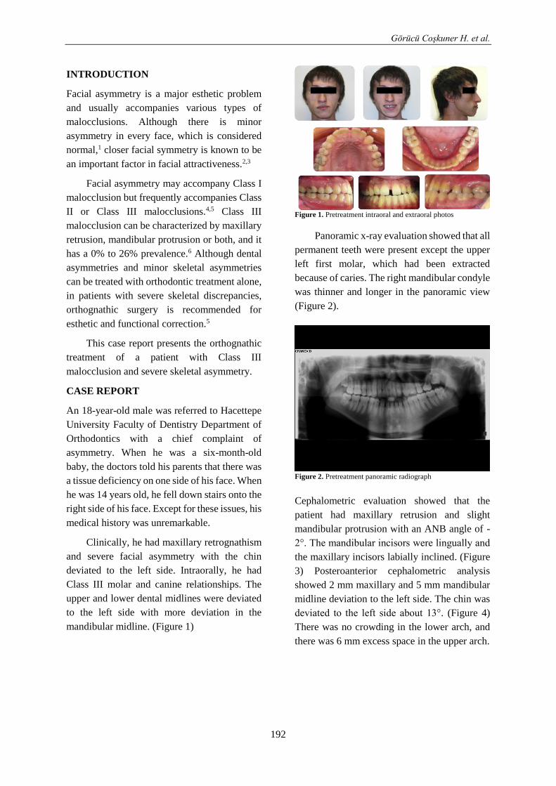
Görücü Coşkuner H. et al.
192
INTRODUCTION
Facial asymmetry is a major esthetic problem
and usually accompanies various types of
malocclusions. Although there is minor
asymmetry in every face, which is considered
normal,1 closer facial symmetry is known to be
an important factor in facial attractiveness.2,3
Facial asymmetry may accompany Class I
malocclusion but frequently accompanies Class
II or Class III malocclusions.4,5 Class III
malocclusion can be characterized by maxillary
retrusion, mandibular protrusion or both, and it
has a 0% to 26% prevalence.6 Although dental
asymmetries and minor skeletal asymmetries
can be treated with orthodontic treatment alone,
in patients with severe skeletal discrepancies,
orthognathic surgery is recommended for
esthetic and functional correction.5
This case report presents the orthognathic
treatment of a patient with Class III
malocclusion and severe skeletal asymmetry.
CASE REPORT
An 18-year-old male was referred to Hacettepe
University Faculty of Dentistry Department of
Orthodontics with a chief complaint of
asymmetry. When he was a six-month-old
baby, the doctors told his parents that there was
a tissue deficiency on one side of his face. When
he was 14 years old, he fell down stairs onto the
right side of his face. Except for these issues, his
medical history was unremarkable.
Clinically, he had maxillary retrognathism
and severe facial asymmetry with the chin
deviated to the left side. Intraorally, he had
Class III molar and canine relationships. The
upper and lower dental midlines were deviated
to the left side with more deviation in the
mandibular midline. (Figure 1)
Figure 1. Pretreatment intraoral and extraoral photos
Panoramic x-ray evaluation showed that all
permanent teeth were present except the upper
left first molar, which had been extracted
because of caries. The right mandibular condyle
was thinner and longer in the panoramic view
(Figure 2).
Figure 2. Pretreatment panoramic radiograph
Cephalometric evaluation showed that the
patient had maxillary retrusion and slight
mandibular protrusion with an ANB angle of -
2°. The mandibular incisors were lingually and
the maxillary incisors labially inclined. (Figure
3) Posteroanterior cephalometric analysis
showed 2 mm maxillary and 5 mm mandibular
midline deviation to the left side. The chin was
deviated to the left side about 13°. (Figure 4)
There was no crowding in the lower arch, and
there was 6 mm excess space in the upper arch.
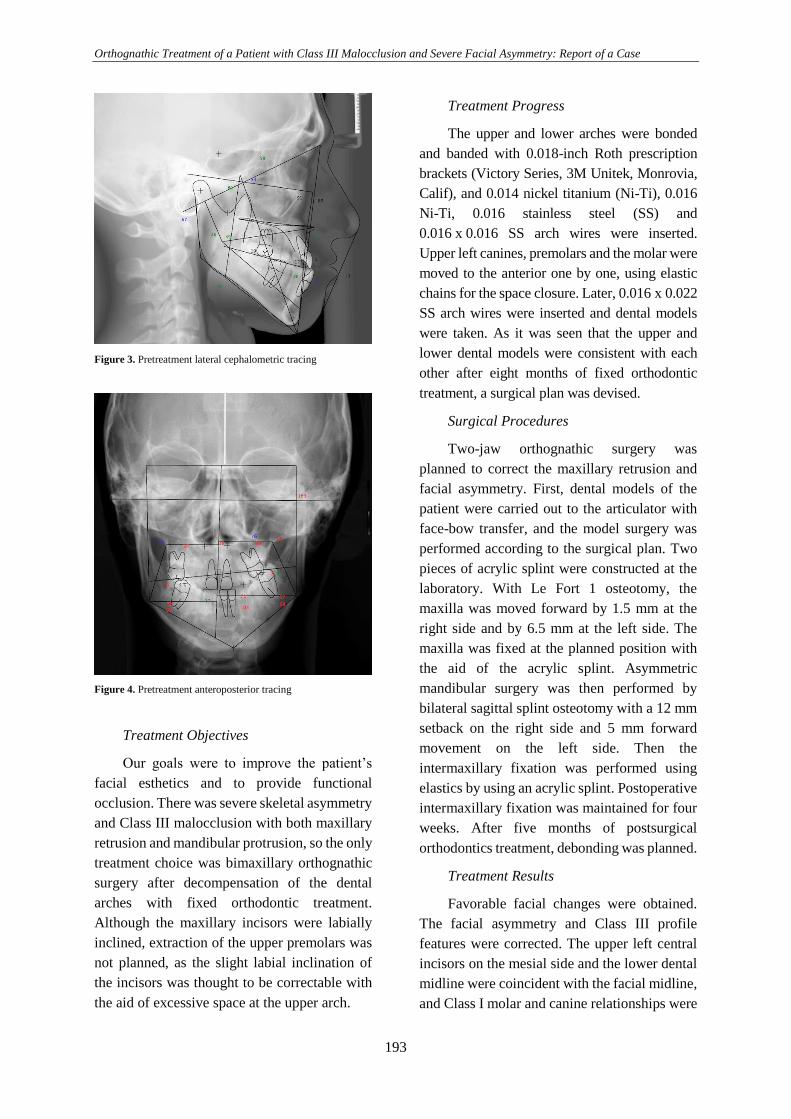
Orthognathic Treatment of a Patient with Class III Malocclusion and Severe Facial Asymmetry: Report of a Case
193
Figure 3. Pretreatment lateral cephalometric tracing
Figure 4. Pretreatment anteroposterior tracing
Treatment Objectives
Our goals were to improve the patient’s
facial esthetics and to provide functional
occlusion. There was severe skeletal asymmetry
and Class III malocclusion with both maxillary
retrusion and mandibular protrusion, so the only
treatment choice was bimaxillary orthognathic
surgery after decompensation of the dental
arches with fixed orthodontic treatment.
Although the maxillary incisors were labially
inclined, extraction of the upper premolars was
not planned, as the slight labial inclination of
the incisors was thought to be correctable with
the aid of excessive space at the upper arch.
Treatment Progress
The upper and lower arches were bonded
and banded with 0.018-inch Roth prescription
brackets (Victory Series, 3M Unitek, Monrovia,
Calif), and 0.014 nickel titanium (Ni-Ti), 0.016
Ni-Ti, 0.016 stainless steel (SS) and
0.016 x 0.016 SS arch wires were inserted.
Upper left canines, premolars and the molar were
moved to the anterior one by one, using elastic
chains for the space closure. Later, 0.016 x 0.022
SS arch wires were inserted and dental models
were taken. As it was seen that the upper and
lower dental models were consistent with each
other after eight months of fixed orthodontic
treatment, a surgical plan was devised.
Surgical Procedures
Two-jaw orthognathic surgery was
planned to correct the maxillary retrusion and
facial asymmetry. First, dental models of the
patient were carried out to the articulator with
face-bow transfer, and the model surgery was
performed according to the surgical plan. Two
pieces of acrylic splint were constructed at the
laboratory. With Le Fort 1 osteotomy, the
maxilla was moved forward by 1.5 mm at the
right side and by 6.5 mm at the left side. The
maxilla was fixed at the planned position with
the aid of the acrylic splint. Asymmetric
mandibular surgery was then performed by
bilateral sagittal splint osteotomy with a 12 mm
setback on the right side and 5 mm forward
movement on the left side. Then the
intermaxillary fixation was performed using
elastics by using an acrylic splint. Postoperative
intermaxillary fixation was maintained for four
weeks. After five months of postsurgical
orthodontics treatment, debonding was planned.
Treatment Results
Favorable facial changes were obtained.
The facial asymmetry and Class III profile
features were corrected. The upper left central
incisors on the mesial side and the lower dental
midline were coincident with the facial midline,
and Class I molar and canine relationships were
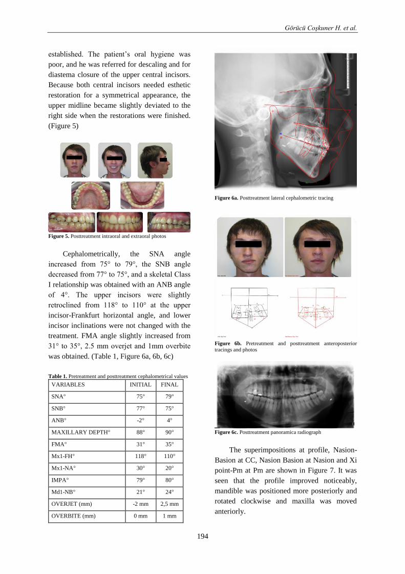
Görücü Coşkuner H. et al.
194
established. The patient’s oral hygiene was
poor, and he was referred for descaling and for
diastema closure of the upper central incisors.
Because both central incisors needed esthetic
restoration for a symmetrical appearance, the
upper midline became slightly deviated to the
right side when the restorations were finished.
(Figure 5)
Figure 5. Posttreatment intraoral and extraoral photos
Cephalometrically, the SNA angle
increased from 75° to 79°, the SNB angle
decreased from 77° to 75°, and a skeletal Class
I relationship was obtained with an ANB angle
of 4°. The upper incisors were slightly
retroclined from 118° to 110° at the upper
incisor-Frankfurt horizontal angle, and lower
incisor inclinations were not changed with the
treatment. FMA angle slightly increased from
31° to 35°, 2.5 mm overjet and 1mm overbite
was obtained. (Table 1, Figure 6a, 6b, 6c)
Table 1. Pretreatment and posttreatment cephalometrical values
Figure 6a. Posttreatment lateral cephalometric tracing
Figure 6b. Pretreatment and posttreatment anteroposterior
tracings and photos
Figure 6c. Posttreatment panoramica radiograph
The superimpositions at profile, Nasion-
Basion at CC, Nasion Basion at Nasion and Xi
point-Pm at Pm are shown in Figure 7. It was
seen that the profile improved noticeably,
mandible was positioned more posteriorly and
rotated clockwise and maxilla was moved
anteriorly.
VARIABLES INITIAL FINAL
SNA° 75° 79°
SNB° 77° 75°
ANB° -2° 4°
MAXILLARY DEPTH° 88° 90°
FMA° 31° 35°
Mx1-FH° 118° 110°
Mx1-NA° 30° 20°
IMPA° 79° 80°
Md1-NB° 21° 24°
OVERJET (mm) -2 mm 2,5 mm
OVERBITE (mm) 0 mm 1 mm
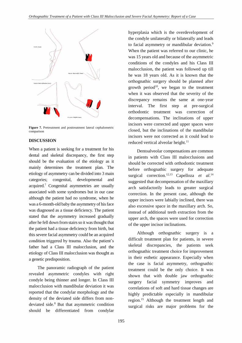
Orthognathic Treatment of a Patient with Class III Malocclusion and Severe Facial Asymmetry: Report of a Case
195
Figure 7. Pretreatment and posttreatment lateral cephalometric
comparison
DISCUSSION
When a patient is seeking for a treatment for his
dental and skeletal discrepancy, the first step
should be the evaluation of the etiology as it
mainly determines the treatment plan. The
etiology of asymmetry can be divided into 3 main
categories; congenital, developmental and
acquired.7 Congenital asymmetries are usually
associated with some syndromes but in our case
although the patient had no syndrome, when he
was a 6-month-old baby the asymmetry of his face
was diagnosed as a tissue deficiency. The patient
stated that the asymmetry increased gradually
after he fell down from stairs so it was thought that
the patient had a tissue deficiency from birth, but
this severe facial asymmetry could be an acquired
condition triggered by trauma. Also the patient’s
father had a Class III malocclusion, and the
etiology of Class III malocclusion was thought as
a genetic predisposition.
The panoramic radiograph of the patient
revealed asymmetric condyles with right
condyle being thinner and longer. In Class III
malocclusion with mandibular deviation it was
reported that the condylar morphology and the
density of the deviated side differs from non-
deviated side.8 But that asymmetric condition
should be differentiated from condylar
hyperplasia which is the overdevelopment of
the condyle unilaterally or bilaterally and leads
to facial asymmetry or mandibular deviation.9
When the patient was referred to our clinic, he
was 15 years old and because of the asymmetric
conditions of the condyles and his Class III
malocclusion, the patient was followed up till
he was 18 years old. As it is known that the
orthognathic surgery should be planned after
growth period10, we began to the treatment
when it was observed that the severity of the
discrepancy remains the same at one-year
interval. The first step at pre-surgical
orthodontic treatment was correction of
decompensations. The inclinations of upper
incisors were corrected and upper spaces were
closed, but the inclinations of the mandibular
incisors were not corrected as it could lead to
reduced vertical alveolar height.11
Dentoalveolar compensations are common
in patients with Class III malocclusions and
should be corrected with orthodontic treatment
before orthognathic surgery for adequate
surgical correction.12,13 Capelloza et al.14
suggested that decompensation of the maxillary
arch satisfactorily leads to greater surgical
correction. In the present case, although the
upper incisors were labially inclined, there was
also excessive space in the maxillary arch. So,
instead of additional teeth extraction from the
upper arch, the spaces were used for correction
of the upper incisor inclinations.
Although orthognathic surgery is a
difficult treatment plan for patients, in severe
skeletal discrepancies, the patients seek
orthognathic treatment choice for improvement
in their esthetic appearance. Especially when
the case is facial asymmetry, orthognathic
treatment could be the only choice. It was
shown that with double jaw orthognathic
surgery facial symmetry improves and
correlations of soft and hard tissue changes are
highly predictable especially in mandibular
region.15 Although the treatment length and
surgical risks are major problems for the
Görücü Coşkuner H. et al.
196
patients, the improvement in the appearance by
orthognathic surgery usually satisfies the
patients16-18 and results in improvement in
psychosocial adjustment.19 In our case, the
patient was unhappy about his facial appearance
and seeking for a treatment for the improvement
in his appearance so after the treatment his self-
esteem increased noticeably.
CONCLUSIONS
The combined orthodontic and orthognathic
treatment of this patient with severe asymmetry
and Class III malocclusion resulted in a Class I
occlusion and esthetically pleasing results.
REFERENCES
1. Bishara SE, Burkey PS, Kharouf JG. Dental
and facial asymmetries: a review. Angle
Orthod. 1994;64(2):89-98.
2. Koehler N, Rhodes G, Simmons LW. Are
human female preferences for symmetrical
male faces enhanced when conception is likely?
Anim Behav. 2002;64:233-8.
3. Rhodes G, Yoshikawa S, Clark A, Lee K,
McKay R, Akamatsu S. Attractiveness of facial
averageness and symmetry in non-western
cultures: in search of biologically based
standards of beauty. Perception.
2001;30(5):611-25.
4. Reyneke JP, Tsakiris P, Kienle F. A simple
classification for surgical treatment planning of
maxillomandibular asymmetry. Br J Oral
Maxillofac Surg. 1997;35(5):349-51.
5. Cheong YW, Lo LJ. Facial asymmetry:
etiology, evaluation, and management. Chang
Gung Med J. 2011;34(4):341-51.
6. Hardy DK, Cubas YP, MF. O. Prevalence of
Angle Class III malocclusion: a systematic
review and meta-analysis. Open J Epidemiol.
2012;2:75-82.
7. Maheswari S, Verma SK, Gaur A, S. D.
Diagnosis and management of facial
asymmetries. J Orthod Res. 2015;3(2):81-7.
8. Wen L, Yan W, Yue Z, Bo D, Xiao Y, Chun-
Ling W. Study of Condylar Asymmetry in Angle
Class III Malocclusion With Mandibular
Deviation. J Craniofac Surg. 2015;26(3):e264-8.
9. Mehrotra D, Dhasmana S, Kamboj M,
Gambhir G. Condylar hyperplasia and facial
asymmetry: report of five cases. J Maxillofac
Oral Surg. 2011;10(1):50-6.
10. Weaver N, Glover K, Major P, Varnhagen
C, Grace M. Age limitation on provision of
orthopedic therapy and orthognathic surgery.
Am J Orthod Dentofacial Orthop.
1998;113(2):156-64.
11. Sun B, Tang J, Xiao P, Ding Y. Presurgical
orthodontic decompensation alters alveolar
bone condition around mandibular incisors in
adults with skeletal Class III malocclusion. Int J
Clin Exp Med. 2015;8(8):12866-73.
12. Troy BA, Shanker S, Fields HW, Vig K,
Johnston W. Comparison of incisor inclination
in patients with Class III malocclusion treated
with orthognathic surgery or orthodontic
camouflage. Am J Orthod Dentofacial Orthop
2009;135(2):146 e1-9.
13. Johnston C, Burden D, Kennedy D,
Harradine N, Stevenson M. Class III surgical
orthodontic treatment: a cephalometric study.
Am J Orthod Dentofacial Orthop
2006;130:300-9.
14. Capelozza Filho L, Martins A, Mazzotini R,
da Silva OG. Effects of dental decompensation
on the surgical treatment of mandibular
prognathism. Int J Adult Orthod Orthognath
Surg 1996;11:165-80.
15. Wermker K, Kleinheinz J, Jung S, Dirksen
D. Soft tissue response and facial symmetry
after orthognathic surgery. J Craniomaxillofac
Surg. 2014;42(6):e339-45.
16. Magro-Filho O, Goiato MC, Oliveira DT,
Martins LP, Salazar M, Medeiros RA, et al.
Evaluation of Patients' Satisfaction after Class
III Orthognathic Surgery. J Clin Diagn Res.
2015;9(10):ZC23-7.
Orthognathic Treatment of a Patient with Class III Malocclusion and Severe Facial Asymmetry: Report of a Case
197
17. Asada K, Motoyoshi M, Tamura T,
Nakajima A, Mayahara K, Shimizu N.
Satisfaction with orthognathic surgery of
skeletal Class III patients. Am J Orthod
Dentofacial Orthop. 2015;148(5):827-37.
18. Pacheco-Pereira C, Abreu LG, Dick BD, De
Luca Canto G, Paiva SM, Flores-Mir C. Patient
satisfaction after orthodontic treatment
combined with orthognathic surgery: A
systematic review. Angle Orthod
2016;86(3):495-508.
19. Lazaridou-Terzoudi T, Kiyak HA, Moore R,
Athanasiou AE, Melsen B. Long-term
assessment of psychologic outcomes of
orthognathic surgery. J Oral Maxillofac Surg
2003;61(5):545-52.
Correspondence Author
Hande GÖRÜCÜ COŞKUNER
Hacettepe University
Faculty of Dentistry
Department of Orthodontics
Sıhhıye, 6100,
Ankara / Turkey
Phone: +90 312 305 22 90
Fax: +90 312 309 11 38
E-mail: [email protected]
Cumhuriyet Dental Journal: 2017; 20(3)
Doi: 10.7126/cumudj.369382 CLINICAL REPORTS
198
1 University of Gaziosmanpaşa, School of Dentistry, Department of Oral and Maxillofacial Surgery, Tokat, Turkey. 2 University of Gaziosmanpaşa, School of Dentistry, Department of Orthodontics, Tokat, Turkey. 3 University of Ankara, School of Dentistry, Department of Oral and Maxillofacial Radiology, Ankara, Turkey. 4 University of Ankara, School of Dentistry, Department of Oral and Maxillofacial Surgery, Ankara, Turkey.
IMPORTANCE OF BIFID MANDIBULAR CANAL IN IMPLANTOLOGY
AND IN ORAL SURGERY: REVIEW OF THE LITERATURE AND
REPORT OF THREE CASES
Bifid Mandibuler Kanalin İmplantoloji ve Oral Cerrahideki Önemi:
Üç Vaka Raporu ile Birlikte Literatürün Gözden Geçirilmesi
Nihat AKBULUT 1, Sibel AKBULUT2, Bengi ÖZTAŞ3
Şebnem KURŞUN3, Emrah SOYLU1, Orhan GÜVEN4
Makale Kodu/Article Code : 129166
Makale Gönderilme Tarihi : 07.06.2015
Kabul Tarihi : 06.03.2017
ABSTRACT
The mandibular canal or the inferior alveolar canal
typically extends from the mandibular foramen to the
mental foramen and includes the inferior alveolar artery,
vein and the inferior alveolar nerve. During embryonic
development three canals fuse to form a single canal.
Failure of these canals to fuse can explain presence of
multiple canals in some individuals. It is important to
localize the course of the mandibular canal at the site of
implant placement as altered sensation of the inferior lip
due to inferior alveolar nerve injury. It is one of the most
serious complication of mandibular implant surgery. Also
the difficulty in performing mandibular anesthesia is the
other problematic issue for patients and the clinicians.
The purpose of this study is to call attention to a rare
anatomical variation. This case report reveals an unusual
variant of the bifid mandibular canals. Three new cases,
two of which are in the same family, have been
presented.
Key words: Bifid mandibular canal, implant surgery,
third molar extraction, inferior alveolar nerve, mandibular
canals
ÖZ
Mandibuler kanal veya inferior alveoler kanal inferior
alveoler arter, ven ve inferior alveoler siniri içerecek
şekilde tipik olarak mandibuler foramenden mental
foramene kadar uzanır. Emriyolojik gelişim sırasında üç
kanal tek bir kanal formu şeklinde füzyona uğrar. Bu
kanalların füzyona uğramasındaki başarısızlık bazı
bireylerdeki çoklu kanalların varlığını açıklayabilir.
İnferior alveoler sinir yaralanması sebebiyle alt dudak
duyu değişikliğinde olduğu gibi implant yerleşim
bölgesindeki mandibuler kanalın seyrinin lokalizasyonu
önemlidir. Bu, mandibuler implant cerrahisinin çok ciddi
komplikasyonundan biridir. Aynı zamanda mandibuler
anestezi uygulamasındaki zorluk, hastalar ve klinisyenler
için başka bir problematik bir konudur.
Bu çalışmanın amacı nadir görülen bir anatomik
varyasyona dikkat çekmektir. Bu vaka raporu bifid
mandibuler kanalların alışılmamış varyantını ortaya
çıkarmaktadır. Üç yeni vaka, ikisi aynı aileden olmak
üzere, sunulmuştur.
Anahtar kelimeler: Bifid mandibuler kanal, implant
cerrahisi, üçüncü molar çekimi, inferior alveoler sinir,
mandibuler kanallar
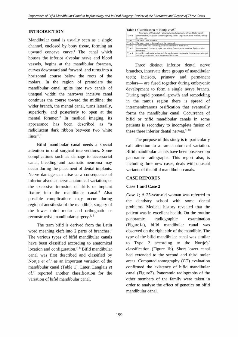
Importance of Bifid Mandibular Canal in Implantology and in Oral Surgery: Review of the Literature and Report of Three Cases
199
INTRODUCTION
Mandibular canal is usually seen as a single
channel, enclosed by bony tissue, forming an
upward concave curve.1 The canal which
houses the inferior alveolar nerve and blood
vessels, begins at the mandibular foramen,
curves downward and forward, and turns into a
horizontal course below the roots of the
molars. In the region of premolars the
mandibular canal splits into two canals of
unequal width: the narrower incisive canal
continues the course toward the midline; the
wider branch, the mental canal, turns laterally,
superiorly, and posteriorly to open at the
mental foramen.2 In medical imaging, its
appearance has been described as “a
radiolucent dark ribbon between two white
lines”.3
Bifid mandibular canal needs a special
attention in oral surgical interventions. Some
complications such as damage to accessorial
canal, bleeding and traumatic neuroma may
occur during the placement of dental implants.
Nerve damage can arise as a consequence of
inferior alveolar nerve anatomical variation; or
the excessive intrusion of drills or implant
fixture into the mandibular canal.4 Also
possible complications may occur during
regional anesthesia of the mandible, surgery of
the lower third molar and orthognatic or
reconstructive mandibular surgery.5, 6
The term bifid is derived from the Latin
word meaning cleft into 2 parts of branches.6
The various types of bifid mandibular canals
have been classified according to anatomical
location and configuration.7, 8 Bifid mandibular
canal was first described and classified by
Nortje et al.7 as an important variation of the
mandibular canal (Table 1). Later, Langlais et
al.8 reported another classification for the
variation of bifid mandibular canal.
Table 1 Classification of Nortje et al.7 Description of Nortjeet al. 7 about patterns of duplication of mandibular canals
Type 1 (most common) Duplicate canals originating from a single mandibular foramen, usually
the same size
Type1a The lower canal is smaller
Type1b The upper canal is the smallest of the two canals
Type 2 A short upper canal extending to the second or third molar areas
Type 3 (less common) 2 canals of equal size, arising from separate foramina, that join in the
molar area
Type 4 A double canal variation in which the supplemental canals arise from the retromolar pad
area and join the main canals in the retromolar areas.
Three distinct inferior dental nerve
branches, innervate three groups of mandibular
teeth; incisors, primary and permanent
molars— are fused together during embryonic
development to form a single nerve branch.
During rapid prenatal growth and remodeling
in the ramus region there is spread of
intramembranous ossification that eventually
forms the mandibular canal. Occurrence of
bifid or trifid mandibular canals in some
patients is secondary to incomplete fusion of
these three inferior dental nerves.9, 10
The purpose of this study is to particularly
call attention to a rare anatomical variation.
Bifid mandibular canals have been observed on
panoramic radiographs. This report also, is
including three new cases, deals with unusual
variants of the bifid mandibular canals.
CASE REPORTS
Case 1 and Case 2
Case 1; A 25-year-old woman was referred to
the dentistry school with some dental
problems. Medical history revealed that the
patient was in excellent health. On the routine
panoramic radiographic examination
(Figure1a), bifid mandibular canal was
observed on the right side of the mandible. The
type of the bifid mandibular canal was similar
to Type 2 according to the Nortje's7
classification (Figure 1b). Short lower canal
had extended to the second and third molar
areas. Computed tomography (CT) evaluation
confirmed the existence of bifid mandibular
canal (Figure2). Panoramic radiographs of the
other members of the family were taken in
order to analyse the effect of genetics on bifid
mandibular canal.
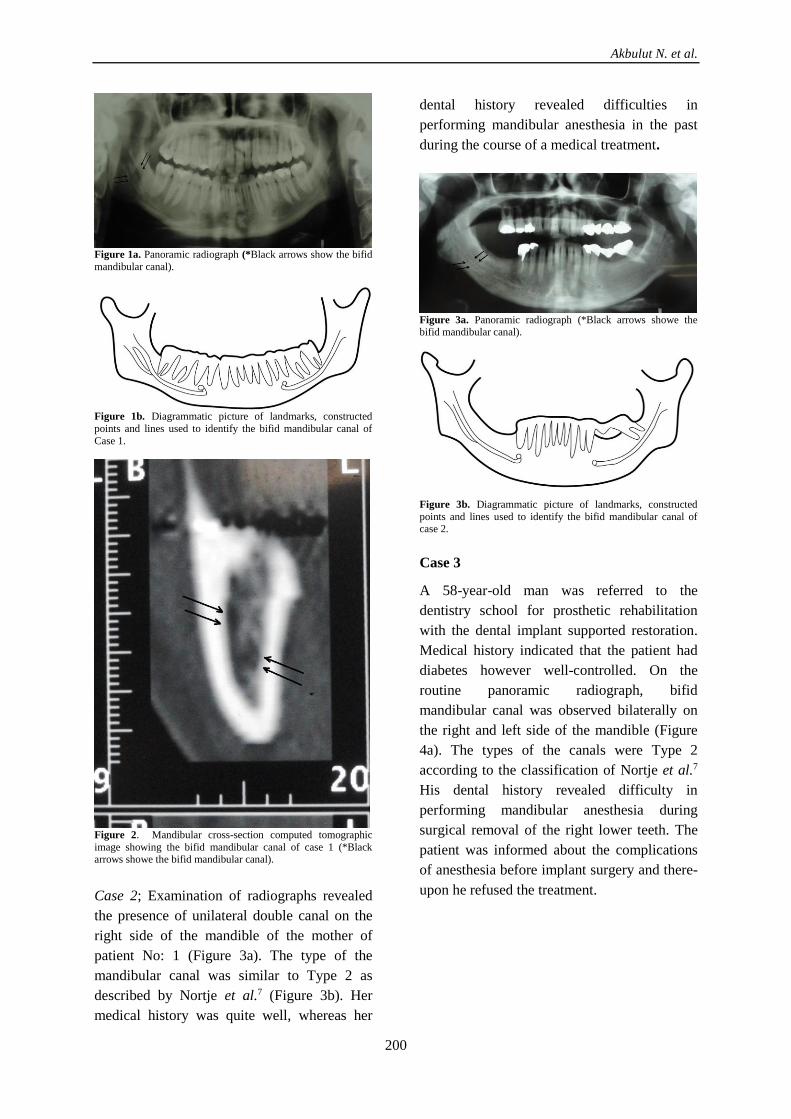
Akbulut N. et al.
200
Figure 1a. Panoramic radiograph (*Black arrows show the bifid
mandibular canal).
Figure 1b. Diagrammatic picture of landmarks, constructed
points and lines used to identify the bifid mandibular canal of
Case 1.
Figure 2. Mandibular cross-section computed tomographic
image showing the bifid mandibular canal of case 1 (*Black arrows showe the bifid mandibular canal).
Case 2; Examination of radiographs revealed
the presence of unilateral double canal on the
right side of the mandible of the mother of
patient No: 1 (Figure 3a). The type of the
mandibular canal was similar to Type 2 as
described by Nortje et al.7 (Figure 3b). Her
medical history was quite well, whereas her
dental history revealed difficulties in
performing mandibular anesthesia in the past
during the course of a medical treatment.
Figure 3a. Panoramic radiograph (*Black arrows showe the bifid mandibular canal).
Figure 3b. Diagrammatic picture of landmarks, constructed
points and lines used to identify the bifid mandibular canal of case 2.
Case 3
A 58-year-old man was referred to the
dentistry school for prosthetic rehabilitation
with the dental implant supported restoration.
Medical history indicated that the patient had
diabetes however well-controlled. On the
routine panoramic radiograph, bifid
mandibular canal was observed bilaterally on
the right and left side of the mandible (Figure
4a). The types of the canals were Type 2
according to the classification of Nortje et al.7
His dental history revealed difficulty in
performing mandibular anesthesia during
surgical removal of the right lower teeth. The
patient was informed about the complications
of anesthesia before implant surgery and there-
upon he refused the treatment.
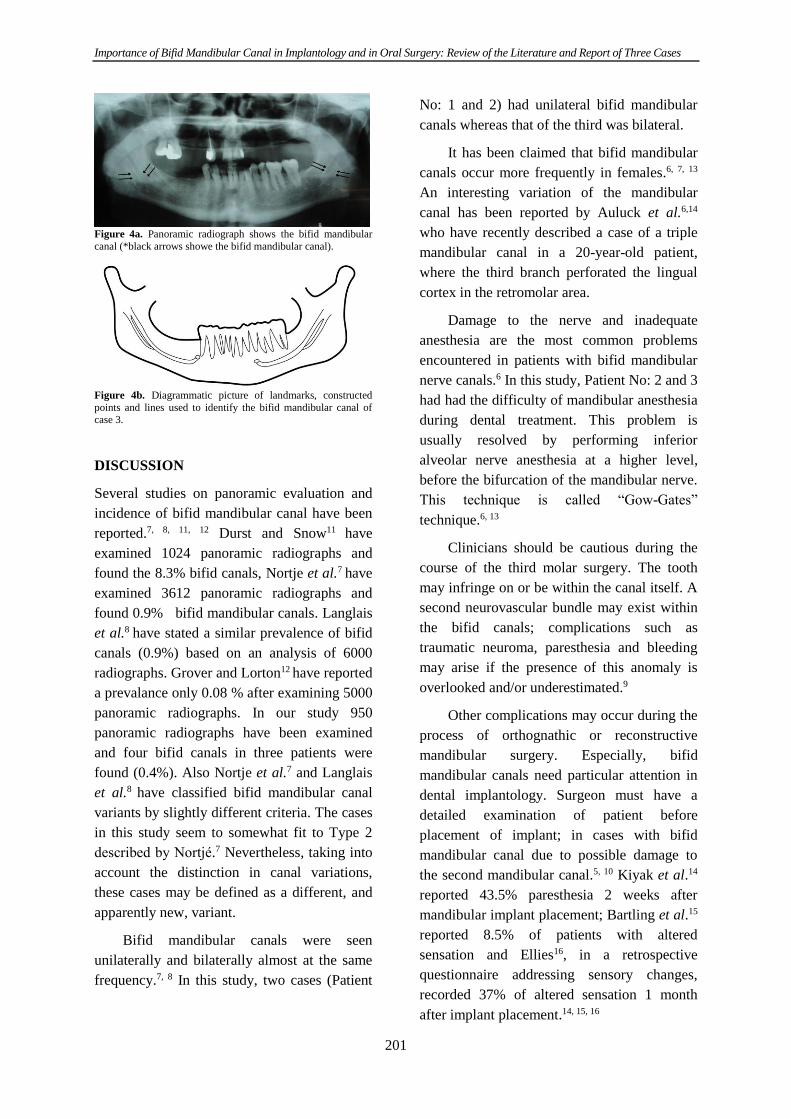
Importance of Bifid Mandibular Canal in Implantology and in Oral Surgery: Review of the Literature and Report of Three Cases
201
Figure 4a. Panoramic radiograph shows the bifid mandibular
canal (*black arrows showe the bifid mandibular canal).
Figure 4b. Diagrammatic picture of landmarks, constructed
points and lines used to identify the bifid mandibular canal of case 3.
DISCUSSION
Several studies on panoramic evaluation and
incidence of bifid mandibular canal have been
reported.7, 8, 11, 12 Durst and Snow11 have
examined 1024 panoramic radiographs and
found the 8.3% bifid canals, Nortje et al.7 have
examined 3612 panoramic radiographs and
found 0.9% bifid mandibular canals. Langlais
et al.8 have stated a similar prevalence of bifid
canals (0.9%) based on an analysis of 6000
radiographs. Grover and Lorton12 have reported
a prevalance only 0.08 % after examining 5000
panoramic radiographs. In our study 950
panoramic radiographs have been examined
and four bifid canals in three patients were
found (0.4%). Also Nortje et al.7 and Langlais
et al.8 have classified bifid mandibular canal
variants by slightly different criteria. The cases
in this study seem to somewhat fit to Type 2
described by Nortjé.7 Nevertheless, taking into
account the distinction in canal variations,
these cases may be defined as a different, and
apparently new, variant.
Bifid mandibular canals were seen
unilaterally and bilaterally almost at the same
frequency.7, 8 In this study, two cases (Patient
No: 1 and 2) had unilateral bifid mandibular
canals whereas that of the third was bilateral.
It has been claimed that bifid mandibular
canals occur more frequently in females.6, 7, 13
An interesting variation of the mandibular
canal has been reported by Auluck et al.6,14
who have recently described a case of a triple
mandibular canal in a 20-year-old patient,
where the third branch perforated the lingual
cortex in the retromolar area.
Damage to the nerve and inadequate
anesthesia are the most common problems
encountered in patients with bifid mandibular
nerve canals.6 In this study, Patient No: 2 and 3
had had the difficulty of mandibular anesthesia
during dental treatment. This problem is
usually resolved by performing inferior
alveolar nerve anesthesia at a higher level,
before the bifurcation of the mandibular nerve.
This technique is called “Gow-Gates”
technique.6, 13
Clinicians should be cautious during the
course of the third molar surgery. The tooth
may infringe on or be within the canal itself. A
second neurovascular bundle may exist within
the bifid canals; complications such as
traumatic neuroma, paresthesia and bleeding
may arise if the presence of this anomaly is
overlooked and/or underestimated.9
Other complications may occur during the
process of orthognathic or reconstructive
mandibular surgery. Especially, bifid
mandibular canals need particular attention in
dental implantology. Surgeon must have a
detailed examination of patient before
placement of implant; in cases with bifid
mandibular canal due to possible damage to
the second mandibular canal.5, 10 Kiyak et al.14
reported 43.5% paresthesia 2 weeks after
mandibular implant placement; Bartling et al.15
reported 8.5% of patients with altered
sensation and Ellies16, in a retrospective
questionnaire addressing sensory changes,
recorded 37% of altered sensation 1 month
after implant placement.14, 15, 16
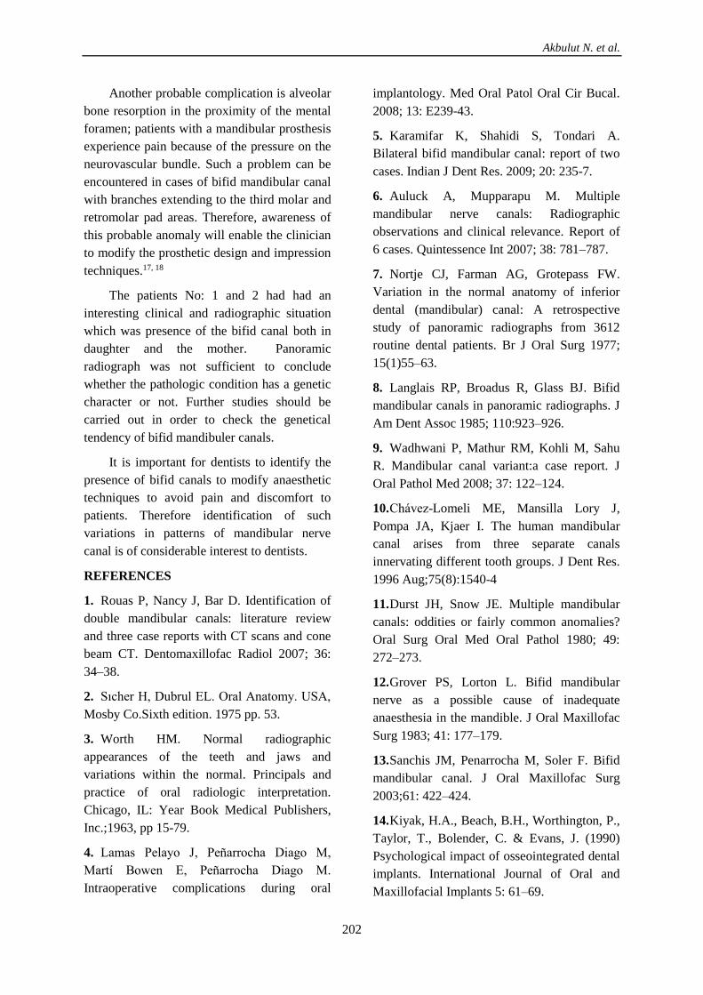
Akbulut N. et al.
202
Another probable complication is alveolar
bone resorption in the proximity of the mental
foramen; patients with a mandibular prosthesis
experience pain because of the pressure on the
neurovascular bundle. Such a problem can be
encountered in cases of bifid mandibular canal
with branches extending to the third molar and
retromolar pad areas. Therefore, awareness of
this probable anomaly will enable the clinician
to modify the prosthetic design and impression
techniques.17, 18
The patients No: 1 and 2 had had an
interesting clinical and radiographic situation
which was presence of the bifid canal both in
daughter and the mother. Panoramic
radiograph was not sufficient to conclude
whether the pathologic condition has a genetic
character or not. Further studies should be
carried out in order to check the genetical
tendency of bifid mandibuler canals.
It is important for dentists to identify the
presence of bifid canals to modify anaesthetic
techniques to avoid pain and discomfort to
patients. Therefore identification of such
variations in patterns of mandibular nerve
canal is of considerable interest to dentists.
REFERENCES
1. Rouas P, Nancy J, Bar D. Identification of
double mandibular canals: literature review
and three case reports with CT scans and cone
beam CT. Dentomaxillofac Radiol 2007; 36:
34–38.
2. Sıcher H, Dubrul EL. Oral Anatomy. USA,
Mosby Co.Sixth edition. 1975 pp. 53.
3. Worth HM. Normal radiographic
appearances of the teeth and jaws and
variations within the normal. Principals and
practice of oral radiologic interpretation.
Chicago, IL: Year Book Medical Publishers,
Inc.;1963, pp 15-79.
4. Lamas Pelayo J, Peñarrocha Diago M,
Martí Bowen E, Peñarrocha Diago M.
Intraoperative complications during oral
implantology. Med Oral Patol Oral Cir Bucal.
2008; 13: E239-43.
5. Karamifar K, Shahidi S, Tondari A.
Bilateral bifid mandibular canal: report of two
cases. Indian J Dent Res. 2009; 20: 235-7.
6. Auluck A, Mupparapu M. Multiple
mandibular nerve canals: Radiographic
observations and clinical relevance. Report of
6 cases. Quintessence Int 2007; 38: 781–787.
7. Nortje CJ, Farman AG, Grotepass FW.
Variation in the normal anatomy of inferior
dental (mandibular) canal: A retrospective
study of panoramic radiographs from 3612
routine dental patients. Br J Oral Surg 1977;
15(1)55–63.
8. Langlais RP, Broadus R, Glass BJ. Bifid
mandibular canals in panoramic radiographs. J
Am Dent Assoc 1985; 110:923–926.
9. Wadhwani P, Mathur RM, Kohli M, Sahu
R. Mandibular canal variant:a case report. J
Oral Pathol Med 2008; 37: 122–124.
10. Chávez-Lomeli ME, Mansilla Lory J,
Pompa JA, Kjaer I. The human mandibular
canal arises from three separate canals
innervating different tooth groups. J Dent Res.
1996 Aug;75(8):1540-4
11. Durst JH, Snow JE. Multiple mandibular
canals: oddities or fairly common anomalies?
Oral Surg Oral Med Oral Pathol 1980; 49:
272–273.
12. Grover PS, Lorton L. Bifid mandibular
nerve as a possible cause of inadequate
anaesthesia in the mandible. J Oral Maxillofac
Surg 1983; 41: 177–179.
13. Sanchis JM, Penarrocha M, Soler F. Bifid
mandibular canal. J Oral Maxillofac Surg
2003;61: 422–424.
14. Kiyak, H.A., Beach, B.H., Worthington, P.,
Taylor, T., Bolender, C. & Evans, J. (1990)
Psychological impact of osseointegrated dental
implants. International Journal of Oral and
Maxillofacial Implants 5: 61–69.

Importance of Bifid Mandibular Canal in Implantology and in Oral Surgery: Review of the Literature and Report of Three Cases
203
15. Bartling, R., Freeman, K. & Kraut, R.A.
(1999) The incidence of altered sensation of
the mental nevre after mandibular implant
placement. Journal of Oral and Maxillofacial
Surgery 57: 1408–1412.
16. Ellies, L.G. (1992) Altered sensation
followingmandibular implant surgery: a
retrospective study. Journal of Prosthetic
Dentistry 68: 664–671.
17. Auluck A, Ahsan A, Pai KM, Shetty C.
Anatomical variations in developing
mandibular nerve canal: a report of three cases.
Neuroanatomy 2005; 4:28-30.
18. Meechan JG. How to over come failed local
anaesthesia. Br. Dent. J. 1999; 186: 15–20.
Correspondence Author
Nihat AKBULUT
Department of Oral and Maxillofacial Surgery
Faculty of Dentistry
University of Gaziosmanpasa
Tokat, Turkey.
Phone: + 905054489263
Fax: + 903562124222
E-mail: [email protected]
Cumhuriyet Dental Journal: 2017; 20(3)
Doi: 10.7126/cumudj.369390 REVIEW
204
Cumhuriyet University, Faculty of Dentistry, Department of Periodontology, Sivas, Turkey.
PARKINSON’S DISEASE IN DENTISTRY AND PERIODONTOLOGY
Diş Hekimliğinde ve Periodontolojide Parkinson Hastalığı
Zeliha MUSLU, Hakan DEVELİOĞLU
Makale Kodu/Article Code : 299338
Makale Gönderilme Tarihi : 22.03.2017
Kabul Tarihi : 01.06.2017
ABSTRACT
Parkinson’s disease (PD) is a progressive
neurological condition which is characterised by
motor (movement) and non-motor symptoms. The
disorder is characterized by hypokinesia, tremor,
muscular rigidity, and a shuffling gait. Oral hygiene
procedures may be adversely affected by tremor,
disturbance of motor skills and depression which are
seen with these patients. It is also known that the
medicines used in the treatment of the disease have
various side effects that affect the oral area. It has
been demonstrated in previous studies that the oral
and dental health status of Parkinson's patients is
worse than healthy individuals, and they have a
larger number and more deep periodontal pockets.
The purpose of this review is to compare the
relationship between Parkinson's disease with dental
and periodontal health and to compile up-to-date
information by scanning the literature to better
understand the possible mechanism.
Key Words: Parkinson’s Disease, Periodontal
Health, Oral hygiene
ÖZ
Parkinson Hastalığı (PH) motor ve motor olmayan
semptomlarla karakterize ilerleyici nörolojik bir
hastalıktır. Hastalık hipokinezi, tremor, kas katılığı
ve yürüme bozukluğu ile karakterizedir. Hastalarda
gözlenen tremor, motor becerilerdeki zayıflama ve
depresyon, oral hijyen uygulamalarını
güçleştirebilmektedir. Ayrıca hastalığın tedavisinde
kullanılan ilaçların da oral bölgeyi etkileyen çeşitli
yan etkileri olduğu bilinmektedir. Parkinson
hastalarının oral ve dental sağlık durumlarının
sağlıklı bireylere göre daha kötü olduğu, ayrıca daha
çok sayıda ve daha derin periodontal ceplere sahip
oldukları geçmiş çalışmalarda gösterilmiştir. Bu
derlemenin amacı, Parkinson hastaları ile ağız diş
sağlığı ve periodontal sağlık arasındaki ilişkiyi, olası
mekanizmayı daha iyi anlamak amacıyla literatürü
tarayarak güncel bilgileri derlemeyi hedeflemektir.
Anahtar kelimeler: Parkinson Hastalığı,
Periodontal Sağlık, Oral hijyen
Muslu Z. et al.
205
GİRİŞ
Parkinson hastalığı; hareket, kas kontrolü,
denge gibi motor fonksiyonları etkilediği kadar,
çeşitli non-motor fonksiyonları da etkileyen
ilerleyici nörodejeneratif bir bozukluktur.1,2
Hastalarda tremor, hareketlerde yavaşlama,
(bradikinezi) kas rijiditesi, postür ve yürüme
bozukluğu karakteristiktir.3 Shaking palsy
olarak da bilinen bu hastalık, ismini hastalığın
semptomlarını ilk kez tanımlayan James
Parkinson’dan almaktadır. Parkinson
hastalığını tanımlayan semptomlardan M.Ö
5000 yılında Hindistan’da bulunan tıbbi
yazılarda ve yaklaşık 2500 yıl öncesinde
Çin’deki yazılarda bahsedildiği görülmüştür.2
Parkinson hastalığı, en sık görülen hareket
bozukluğu ve Alzheimer’dan sonra en sık görülen
ikinci nörodejeneratif bozukluktur. Çalışmalar
erkeklerde insidansın daha yüksek olduğunu
göstermişlerdir.4 Hastalığın başlama yaşı ortalama
57’dir, 40 yaşından önce ve 85 yaşından sonra tanı
konmuş az sayıda vaka da bildirilmiştir, fakat
vakaların sayısı 60 yaşından sonra şiddetli bir artış
göstermektedir. 50 yaşından önce rapor edilmiş
vakalar çoğunlukla genetik ile ilişkili iken, geç
yaşta başlayanlar sıklıkla çevresel faktörler ile
ilişkilendirilmiştir.4,5
Parkinson hastalığı nörotransmiterlerde
veya dopamin üretiminde bir bozulma ile
meydana gelir ve bu da bazal ganglionda beyin
hücreleri arasında iletişim bozukluğuna sebep
olur. Dopamin seviyesinin azalması sinir
hücrelerinin kontrolsüz bir şekilde uyarılmasına
ve kas aktivitesinin kontrol edilememesine
sebep olur. Aynı zamanda substantia nigrada
bulunan dopamin üretici hücrelerin ölümü de
corpus striatumda dopamin seviyesinin
düşmesine sebep olur ve bu da Parkinson
hastalığındaki primer patolojidir. Parkinson
hastalığının motor semptomlar klinik olarak
gözlemlenmeden çok önce başladığı
bilinmektedir. Semptomlar gelişmeye
başladığında substantia nigrada dopamin üreten
hücrelerin en az %60’ı kaybedilmiştir ve corpus
striatumda dopamin kaybı %80 ila 90 dır.2
Ayrıca son on yılda yapılmış çalışmalarda,
hastalığın patogenezinde enflamasyon ve
enfeksiyonun çok önemli bir rolü olduğunu
gösteren kanıtlar mevcuttur.6–8 Periodontal
hastalığın da lokal ve sistemik enflamasyona
yol açabildiği, ayrıca sistemik enflamatuvar
mediatörlerin salınımına sebep olduğu çeşitli
çalışmalarca gösterilmiştir.6 Bu bilgiler ışığında
Parkinson hastalığı patogenezinde Periodontal
hastalıkların da rol oynayabileceği
düşünülmektedir.9
Parkinson hastalığının tanısı; detaylı bir
anamnez, fizik muayene ve bazı durumlarda da
dopaminerjik ilaçlara verilen pozitif cevapla
konur. Laboratuvar testleri ve görüntüleme
çalışmaları rutinde kullanılmamaktadır.10 Kesin
tanı ancak postmortem doğrulama ile
mümkündür.
Parkinson hastaları, motor yetersizlik,
disfaji, apati (kayıtsızlık), depresyon, demans
(bunama), hipersalivasyon, kserostomi ve
medikasyon gibi birkaç faktör sebebiyle oral
hijyen prosedürlerini yerine getirmekte güçlük
çekebilirler.11 Bu hastalarda dental sağlığın kötü
olduğunu gösteren çalışmalar bulunmaktadır.2,
11–17 Ayrıca, Uppoor ve ark. bu iki hastalık
arasındaki ilişkinin iki yönlü olabileceğini
bildirmiştir.9
SİSTEMİK BULGULAR
Parkinson hastalığı motor ve motor olmayan
semptomlar sergiler. Motor bulgular çoğunlukla
tek taraflı tremorlardır ve bir elde para sayma
hareketi şeklinde başlar. Tremorlar daha sonra
bacaklara, yüz, dil ve mandibulaya yayılabilir.
Bunlar genellikle istirahat tremorları olup
istemli hareketlerde şiddeti azalmaktadır.2
Bradikineziye ek olarak hastalarda akinezi
görülür. Akinezi yüzde ifadesizlik, göz kırpma
ve yutkunmada azalma, giyinme, yıkanma ve
sandalyeden kalkmada zorluk, yorgunluk ve
tükenmişlik hissi olarak görülür. İskeletsel
kaslardaki tonus artışı nedeniyle diz ve bilek
gibi eklem etrafındaki pasif ekstremite
hareketlerine karşı direnç oluşur.7,9,17 Bu
hastalarda görülen postür bozukluğu ve yürüme
Muslu Z. et al.
206
güçlüğü onların düşerek yaralanmalarına yol
açar (orofacial yaralanmalar dahil) ve hasta
özgürlüğünü kısıtlar.18–20
Parkinson hastaları aynı zamanda geniş bir
nonmotor semptomlar spektrumuna sahiptir.
Kan basıncındaki değişiklikler, özellikle de
ortostatik hipotansiyon; kardiyak disritmiler;
aşırı terleme ve idrar, dışkılama ve seksüel
disfonksiyonlarla kendini gösteren otonomik
disfonksiyonun bu hastaların yarısından
çoğunda görüldüğü rapor edilmiştir.21
İnsomnia, uyku apnesi ve uyku bölünmelerini
içeren uyku bozuklukları yaygındır ve bu da
gün içinde uykulu olmaya sebep olur.22
Parkinson hastalığında depresyon,
kavramsal zayıflık ve demans gibi primer
olarak hastalıktan kaynaklanan birçok
davranışsal bozukluğun yanı sıra dopaminerjik
ilaçların sebep olduğu psikoz da
görülebilmektedir.23 Depresyon, hastaların
neredeyse yarısını etkilemektedir fakat
hastalarda hareketlerde yavaşlama, yüzde
ifadesizlik, uykusuzluk hali, kilo ve enerji kaybı
gibi bulguların varlığı nedeniyle depresyonun
tanısı zorlaşmaktadır.15 Hastalığın ileri
evrelerinde planlama, sıralama, görsel motor
beceriler, uzaysal algıda bozukluk ve sözlü veya
sözlü olmayan komutlara yanıtta ve akıcı
konuşmada aksamalar gibi demans kriterleri
hastalarda %10-30 oranında gözlenmektedir.24
ORAL BULGULAR
Parkinsonlu bireylerde oral bulgular çeşitli
şekillerde karşımıza çıkabilmektedir.
Orofasiyal bölgede parkinson tremoru dil
ve/veya dudakları etkileyerek; çiğneme,
yutkunma ve konuşma gibi fonksiyonları
bozabilir.1 Ayrıca, hastalığın tedavisinde
sıklıkla kullanılan dopaminin yan etkisi olarak
dilde (fly-catching) ve dudaklarda (pursing)
diskinetik hareketler görülebilir. Parkinson
hastalarının yüzleri tipik olarak ifadesiz "maske
yüz" şeklindedir.25,26
Parkinson hastalarında en sık görülen
oral bulgular; oral hijyen uygulamalarında
karşılaşılan güçlükler, kserostomi, burning
mouth/ mukozit, yutkunma güçlüğü, salya akışı
ve kontrol grubuna göre aynı yaştaki sağlıklı
bireylerden daha az çürük sayısı ve daha fazla
dişe sahip olmalarıdır. Oral bulguların çoğu
Parkinson hastalığının kendisi ile değil
kullandıkları ilaçların tükürük akışında
meydana getirdiği değişiklikler ile ilişkilidir.11
Disfaji
Parkinson hastalığında normal yutkunma
işlemi bile bir sorun oluşturabilir. Bu durum
hastaların %75’ini etkiler ve normal tükürüğün
yutulma işlemi de etkilenmiştir. Hastalığın
ilerlemesiyle birlikte en büyük risk olan “sessiz
aspirasyon” da artmaktadır. Bu durum normal
koruyucu refleks mekanizmaların yokluğunda
küçük miktarda yiyecek veya tükürük
aspirasyonuna yol açar. Parkinson hastalarında
bu şekilde oluşan çok sayıda pnömoni vakası
rapor edilmiştir.2 Japonya’da yapılan bir
çalışmada, 4mm'den derin cebi bulunan on veya
daha fazla dişe sahip bireyler pnömoniye bağlı
mortalite açısından periodontal cebi
bulunmayan bireylere kıyasla 3.9 kat daha fazla
risk taşıdıkları bildirilmiştir.25 Bu nedenle
Parkinson hastalarında periodontal hastalığın
mevcut aspirasyon riskini önemli oranda
artırabileceğini söylemek mümkündür.
Siyalore ve Salya akışı
Parkinson hastalarında normal tükürük
fonksiyonları bozulmuştur ve çeşitli
derecelerde azalmış tükürük akışı veya
kserestomi ile sonuçlanabileceği gibi artmış
tükürük akışı veya siyalore de
görülebilmektedir.26 Sonuç olarak lubrikasyon,
yiyeceklerin çözünmesi, çiğnemeye ve yutmaya
yardım ve tamponlama gibi normal tükürük
fonksiyonları bozulmuştur.27 Parkinson
hastalığı sıklıkla artmış saliva ve tükürük akışı
ve yutkunmada zorluk ile ilişkilidir. Hastaların
%78 kadarında rapor edilmiş siyalore
görülmektedir ve bu da fungal enfeksiyonlara
(angular şelitis) sebep olmaktadır.28
Parkinson’s Disease in Dentistry and Periodontology
207
Kserestomi
İlginç bir şekilde Parkinson hastalığında
siyalorenin yaygın görülmesine rağmen
kserostomi de sık rastlanan bir
komplikasyondur. Çürük ve periodontal
hastalık için hazırlayıcı bir etken olan
kserostominin Parkinsonlu bireylerde en sık
gözlenen oral bulgulardan biri olduğu rapor
edilmiştir. Clifford ve Finnerty' nin yaptıkları
anket çalışmasına göre, Parkinsonlu hastalar
genel populasyona göre en az iki kat daha sık
kserestomiden yakınmaktadırlar. Parkinsonlu
bireylerin yaklaşık %55’inde kserestomi
gözlenmektedir.14,15
Yanan Ağız Sendromu
Yanan ağız sendromu, fiziksel veya
laboratuvar bulguları ile korele olmayan
intraoral dokularda ağrı ve yanma hissi ile
karakterize olan bir durumdur.29,30 Parkinson
hastalarında kserestomi, ilaç medikasyonu,
beslenme bozukluğu ve hatta diş fırçalamayı
zorlaştıran azalmış kas koordinasyonu gibi
etkenlerin varlığı sebebiyle bu sendrom sık
görülebilmektedir.15,17 Diğer yandan, anksiyete,
depresyon, kompulsif hastalıklar, stres gibi
fizyolojik faktörler bu sendromda rol
oynamaktadır.31 Parkinson hastalarında yüksek
oranda gözlenen depresyon ve anksiyetenin de
bu sendrom ile Parkinson hastalığı arasındaki
ilişkiyi açıklayabileceği belirtilmiştir.32
Tat ve Koku Almada Değişiklikler
Parkinson hastaları tat ve koku almadaki
değişikliklere karşı oldukça hassastır. Değişmiş
tükürük fonksiyonları ve kullanılan ilaçlar bu
değişikliklerde major rol oynamaktadır.
Gastrointestinal sistemdeki kaslar da Parkinson
hastalığından etkilenebilir. Bu durum yemek
parçalarının boğaza yapıştığı hissini yaratır ve
mide ekşimesi veya gastroözefajiyal reflüye sebep
olabilir.13,17 Tat duyusu sadece santral sinir sistem
dejenerasyonundan değil; aynı zamanda
depresyon, azalmış tükürük üretimi, zayıf oral
hijyen, gastrointestinal hastalıklar ve kullanılan
ilaçlar gibi fiziksel durumlardan da
etkilenmektedir.33 Bu faktörlerin Parkinson
hastalarında gözlenen tat kaybını açıklayabileceği
bildirilmiştir.32
Çiğneme Bozuklukları
Çiğneme fonksiyonunun, Parkinson
hastalarında birkaç farklı yoldan etkilendiği
düşünülmektedir. Çene hareketliliği ve çene
hareket hızı azalmıştır. Rijidite, çene ve dilin
hareketliliğinin azalması ve çenede tremor
oluşumu, gıdaların ağza yerleştirilmesi, çiğneme
ve yutma işlemlerini güçleştirmektedir. Çiğneme
ve orofasiyal fonksiyonlar hastalığın orta-ileri
evrelerinde daha sık görülmekte ve hastalığın
ilerlemesi ile artmaktadır.12
Konuşma bozuklukları
Konuşma bozukluklarının Parkinson’da
hastalıkla ilişkili gözlenen ilk non-motor
semptom olabileceği bildirilmiştir. Kısık ses,
kelimelerin ağızda yuvarlanması, hızlı
konuşma, konuşmaya başlamadan önce
tereddüt sık görülen bulgulardır.34
Parkinson Hastalarında Periodontal
Sağlık İle İlgili Geçmiş Çalışmalar
Literatür incelendiğinde, Parkinson
hastaları ve oral sağlık ile ilgili yapılan
çalışmaların bir kısmında bu hasta grubunda
dental durumun daha kötü olduğu bildirilirken,
bazı araştırmacılar da farklı sonuçlar ortaya
koymuşlardır.14,35,36 Buna ilaveten, geçmiş
çalışmalarda Parkinsonlu hastalarda daha fazla
periodontal patolojiler gözlendiği
gösterilmiştir.9,15,35 Kısaca bu çalışmalarda;
Parkinsonlu bireylerde kontrol grubuna göre
daha yüksek CPITN değerleri, daha derin
periodontal cep değerleri ve daha sık gözlenen
periodontal cep varlığı rapor edilmiştir.
Parkinson hastaları ile ilgili derlemelerde
hastaların rutin oral hijyen uygulamalarını
yapamamaları, tükürük akışındaki ve
çiğnemedeki disfonksiyonel değişimlerin dental
komplikasyonlara sebep olduğu belirtilmiştir.37,38
Grover ve ark., Parkinson hastalarının
durumlarından dolayı daha komplike hastalar
olduklarının ve dental tedavilerinde; tıbbi ve
Muslu Z. et al.
208
kognitif durumlarına ve ilaç kullanımlarına göre
dental tedavi prosedürlerinin modifiye
edilebileceğini bildirmişlerdir.2 Hanaoka ve
Kenichi' nin 89 Parkinson hastasını dahil ettikleri
çalışmalarında, Parkinsonlu bireylerde kontrol
grubuna göre daha yüksek prevelansta çürük,
periodontal hastalık ve daha çok sayıda diş kaybı
olduğu rapor edilmiştir. Periodontal hastalığın,
henüz günlük aktivitelerinin olumsuz
etkilenmediği Parkinson hastalığının erken
evrelerinde dahi daha sık gözlendiği
bildirilmiştir.17
Müller ve arkadaşlarının çalışmasında,
parkinsonlu hastaların kontrol grubuna göre
daha az sayıda dişe sahip oldukları ve oral
hijyen uygulamalarının daha kötü olduğu
gösterilmiştir.19 Schwartz ve arkadaşları
cinsiyete göre Parkinson hastaları ve sağlıklı
kontrol grubunda periodontal sağlığı
değerlendirdikleri çalışmalarında, kontrol
grubundaki kadınlarda periodontal sağlığın
daha iyi olduğunu bildirmişlerdir. Parkinson
hastalığının şiddeti ile oral hijyenin olumsuz
etkilendiği ancak cinsiyetin etkili olmadığı
görülmüştür.39
Parkinson hastalığı birçok dental, oral ve
maksillofasial problemle ilişkili olabilir.
Periodontal hastalık ile kuron ve kök çürükleri
prevelansı hastalığın ilerleme sürecinde
artabilir. Ayrıca oral sağlığın devam etmesinde;
bakteriyel plağın uzaklaştırılması, tamponlama,
lubrikasyon gibi koruyucu birçok etkisi bulunan
tükürükte yaşanan değişimler bu hasta
grubunda periodontal hastalıklara ve diş
çürüklerine neden olabilmektedir.15
Orta ve ileri derecedeki Parkinson
hastalarında yapılan güncel bir çalışmada,
orofasiyal disfonksiyon ve oral sağlık aynı yaş
ve cinsiyetteki kontrol grubuna göre daha düşük
bulunmuştur ayrıca hastalık şiddetlendikçe bu
fark belirginleşmektedir.12 Hastalarda görülen
artmış plak akümülasyonunun, bu hasta
grubunda gözlenen kas rijiditesi, el ve çene
tremoru ile azalmış motor becerilerden
kaynaklanabileceği belirtilmiştir.
Clifford ve Finnerty parkinsonlu hastaların
%51’inde dental problem olduğunu rapor
etmişlerdir.11 Nakayama ve ark ise parkinsonlu
hastaların kontrol grubuna göre daha az dişe
sahip olduklarını, çürük, periodontal hastalık ve
diş kaybı açısından daha yüksek riske sahip
olduklarını rapor etmişlerdir. Çürük ve
periodontal hastalık görülme sıklığı hastalığın
erken evrenlerinde bile sağlıklı bireylere göre
daha fazladır.40
Bu sonuçların aksine Persson ve ark.
Parkinsonlu hastaların aynı yaş aralığındaki
kontrol grubuna göre daha az diş kaybı ve çürük
insidansı gösterdiğini rapor etmişlerdir.41
Fukayo ve arkadaşları da kontrol grubuna göre
daha az çürük diş ve ilgili değişikliler
bulmuşlardır.16
Parkinsonlu hastalarda motor ve kognitif
bozulmalarla ilişkilendirilen diş çürükleri sık
görülen komplikasyonlar arasındadır. Fukayo
ve ark. tükürük pH’ının 6’dan düşük olmasını,
günlük diş fırçalama sıklığının iki kez veya
daha az sayıda olmasını ve ana öğünler
arasındaki atıştırmanın parkinsonlu hastalarda
çürük için risk faktörü olarak
tanımlamışlardır.16 Ayrıca Merchant ve ark.
artmış fiziksel aktivitenin erkeklerde
peridontitis riskini azalttığını belirtmişlerdir.42
Parkinsonlu bireyler, çoğunlukla ileri yaşta,
yürüme güçlüğü çekebilen, ileri evrelerde
yardıma muhtaç, büyük oranda deprese bireyler
olmalarından dolayı fiziksel aktiviteleri büyük
çoğunluğunda azalmıştır. Bu hastalarda daha
yüksek prevelansta gözlenen periodontitis
olgularını sebeplerinden biri olabileceği
düşünülmektedir. Nakayama ve ark.
parkinsonlu hastaların kontrol grubundakiler
kadar düzenli dişlerini temizlemediklerini
belirtmişlerdir.40 Ayrıca artmış periodontal
patoloji ve çürük insidansı Alzheimer
hastalarında da rapor edilmiştir. Bu da kognitif
yetersizliğin kötü oral hijyene yol açtığını
göstermektedir. Parkinsonlu hastalarda diş
fırçalama alışkanlığı tremor, akinezi, kas
Parkinson’s Disease in Dentistry and Periodontology
209
rijiditesi ve demans gibi motor ve kognitif
disfonksiyonlardan etkilenebilmektedir.14
Bunların yanında, otonomik disfonksiyon
ve/ veya anti- Parkinson ilaçlardan kaynaklanan
disfaji, çiğneme güçlüğü, oral diskinezi,
hipersiyalore veya kserestomi ağzın kendi
kendine temizleme mekanizmasını etkileyerek
oral hijyeni bozabilir. Apati ve depresyon da
demans gibi hastanın dental problemlerinin
farkına varmasına engel olabilir. Ayrıca yürüme
bozukluğu, akinezi ve postural instabilite diş
hekimi ziyaretini ve dolayısıyla oral sağlığın
korunmasını engelleyebilir.
Bazı semptomlar ve kullanılan ilaçlar da
parkinsonlu hastalarda oral dengenin
bozulmasına sebep olarak çürük ve periodontal
hastalıkla ve nihayetinde diş kaybıyla
sonuçlanabilir. Azalmış diş sayısı çiğnemeyi ve
kendi kendine beslenmeyi zorlaştırır, fiziksel ve
mental sorunlara yol açabilir.
SONUÇ
Parkinson hastalarında fonksiyonel bozukluğun
görülmediği erken evrelerde dahi yüksek
prevelansta görülen periodontal patolojiler
dental bakımın önemini vurgulamaktadır.
Hastalar günlük oral hijyen uygulamalarını
düzenli yapsalar bile el hareketlerindeki
bozukluk, tremor ve azalmış hareket ağız
bakımının yetersiz olmasına sebep olacaktır.
Motor ve kognitif yetersizlikler parkinsonlu
hastalarda zayıf periodontal sağlığa neden
olmaktadır. Dahası bu hastalarda gözlenen
yaygın periodontal problemlerin önüne geçmek
üzere bu hastaların daha sık diş hekimi
kontrollerine gitmeleri, periodontal sağlık
açısından farkındalıklarının artırılması ve oral
hijyen prosedürleri açısından motivasyonlarının
yüksek tutulması büyük bir önem arz
etmektedir. Ayrıca hastalığın seyri ve ilişkili
komplikasyonları yönetmek adına hasta, diş
hekimi ve nörolog arasında kurulacak bağlantı
ve iş birliği hastaların yaşam kalitelerini
artıracaktır. Bu konuda yapılacak geniş
katılımlı çalışmalar mevcut bilgileri daha da
artıracaktır.
KAYNAKLAR
1. Lobbezoo F, Naeije M. Dental implications
of some common movement disorders: a
concise review. Arch Oral Biol
2007;52(4):395–8.
2. Grover S, Rhodus NL. Dental management
of Parkinson’s disease. Northwest Dent
2011;Nov-Dec.
3. Pradeep AR, Singh SP, Martande SS, Raju
AP, Rustagi T, Suke DK, et al. Clinical
evaluation of the periodontal health condition
and oral health awareness in Parkinson’s
disease patients. Gerodontology
2015;32(2):100–6.
4. Wirdefeldt K, Adami HO, Cole P,
Trichopoulos D, Mandel J. Epidemiology and
etiology of Parkinson’s disease: a review of the
evidence. Eur J Epidemiol 2011;26(1):1.
5. Elbaz A, Moisan F. Update in the
epidemiology of Parkinson’s disease. Curr
Opin Neurol 2008;21(4):454–60.
6. Gorell JM, Peterson EL, Rybicki BA,
Johnson CC. Multiple risk factors for
Parkinson’s disease. J Neurol Sci
2004;217(2):169–74.
7. Ebersole JL, Cappelli D. Acute-phase
reactants in infections and inflammatory
diseases. Periodontol 2000 2000;23(1):19–49.
8. Konsman JP, Parnet P, Dantzer R. Cytokine-
induced sickness behaviour: mechanisms and
implications. Trends Neurosci 2002;25(3):154–9.
9. Kaur T, Uppoor A, Naik D. Parkinson’s
disease and periodontitis–the missing link? A
review. Gerodontology 2016;33(4):434–8.
10. Tolosa E, Wenning G, Poewe W. The
diagnosis of Parkinson’s disease. Lancet Neurol
2006;5(1):75–86.
11. Clifford T, Finnerty J. The dental awareness
and needs of a Parkinson’s disease population.
Gerodontology 1995;12(2):99–103.
12. Bakke M, Larsen SL, Lautrup C, Karlsborg
M. Orofacial function and oral health in patients
with Parkinson’s disease. Eur J Oral Sci
2011;119(1):27–32.
13. Cicciù Marco, Risitano Giacomo, Lo
Giudice Giuseppe, Bramanti Ennio. Periodontal
Muslu Z. et al.
210
health and caries prevalence evaluation in
patients affected by Parkinson’s disease.
Parkinsons Dis 2012;2012(1):6. Doi:
10.1155/2012/541908.
14. Einarsdóttir, E. R., Gunnsteinsdottir H,
Hallsdóttir MH, Sveinsson S, Jónsdóttir SR,
Olafsson VG, Holbrook WP. Dental health of
patients with Parkinson’s disease in Iceland.
Spec Care Dent 2009;29(3):123–7.
15. Friedlander AH, Mahler M, Norman KM,
Ettinger RL. Parkinson disease: systemic and
orofacial manifestations, medical and dental
management. J Am Dent Assoc
2009;140(6):658–69.
16. Fukayo S, Nonaka K, Shimizu T, Yano E.
Oral health of patients with Parkinson’s disease:
factors related to their better dental status.
Tohoku J Exp Med 2003;201(3):171–9.
17. Hanaoka A, Kashihara K. Increased
frequencies of caries, periodontal disease and
tooth loss in patients with Parkinson’s disease.
J Clin Neurosci 2009;16(10):1279–82.
18. Yoritaka A, Shimo Y, Takanashi M, Fukae
J, Hatano T, Nakahara T, et al. Motor and non-
motor symptoms of 1453 patients with
Parkinson’s disease: prevalence and risks.
Parkinsonism Relat Disord 2013;19(8):725–31.
19. Müller B, Assmus J, Herlofson K, Larsen JP,
Tysnes OB. Importance of motor vs. non-motor
symptoms for health-related quality of life in
early Parkinson’s disease. Parkinsonism Relat
Disord 2013;19(11):1027–32.
20. Dauer W, Przedborski S. Parkinson’s
disease: mechanisms and models. Neuron
2003;39(6):889–909.
21. Garcia-Ruiz PJ, Chaudhuri KR, Martinez-
Martin. Pablo. Non-motor symptoms of
Parkinson’s disease A review from the past. J
Neurol Sci 2014;338(1):30–3.
22. Maass A, Reichmann H. Sleep and non-
motor symptoms in Parkinson’s disease. J
Neural Transm 2013;120(4):565–9.
23. Trojano L, Santangelo G, Conson M, Grossi
D. Towards a deeper comprehension of
relationships among cognitive, behavioral and
psychiatric symptoms in Parkinson’s disease.
Behav Neurol 2013;27(4):463–7.
24. Weintraub D, Comella CL, Horn S.
Parkinson’s disease--Part 1: Pathophysiology,
symptoms, burden, diagnosis, and assessment.
Am J Manag Care 2008;14(2):40–8.
25. Awano S, Ansai T, Takata Y, Soh I, Akifusa
S, Hamasaki T, et al. Oral health and mortality
risk from pneumonia in the elderly. J Dent Res
2008;87(4):334–9.
26. Chou KL, Evatt M, Hinson V, Kompoliti K.
Sialorrhea in Parkinson’s disease: a review.
Mov Disord 2007;22(16):2306–13.
27. Proulx M, Courval FP De, Wiseman MA.
Salivary production in Parkinson’s disease.
Mov Disord 2005;20(2):204–7.
28. Kalf JG, Bloem BR, Munneke M. Diurnal
and nocturnal drooling in Parkinson’s disease. J
Neurol 2012;259(1):119–23.
29. Coon EA, Laughlin RS. Burning mouth
syndrome in Parkinson’s disease: dopamine as
cure or cause? J Headache Pain
2012;13(3):255–7.
30. Jääskeläinen SK. Pathophysiology of
primary burning mouth syndrome. Clin
Neurophysiol 2012;123(1):71–7.
31. López-Jornet P, Camacho-Alonso F,
Andujar-Mateos P, Sánchez-Siles M, Gómez-
García F. Burning mouth syndrome: Update.
Med Oral Patol Oral Cir Bucal
2010;15(4):562–8.
32. Zlotnik Yair, Balash Yacov, Korczyn Amos
D, Giladi Nir, Gurevich Tanya. Disorders of the
Oral Cavity in Parkinson ’ s Disease and
Parkinsonian Syndromes. Parkinsons Dis
2015;2015.
33. Kashihara K, Hanaoka A, Imamura T.
Frequency and characteristics of taste
impairment in patients with Parkinson’s
disease: results of a clinical interview. Intern
Med 2011;50(20):2311–5.
34. Carranza M, Snyder MR, Shaw J Davenport,
Zesiewicz TA. Parkinson ’ s Disease. 2013.
35. Bartold P Mark. Periodontal tissues in health
and disease: introduction. Periodontol 2000
2006;40(1):7–10. Doi: 10.1111/j.1600-
0757.2005.00147.x.
Parkinson’s Disease in Dentistry and Periodontology
211
36. Breen KC, Drutyte G. Non-motor symptoms
of Parkinson’s disease: the patient's perspective.
J Neural Transm 2013;120(4):531–5.
37. Jolly DE E, Paulson RB B, Paulson GW W,
Pike JA. Parkinson’s disease: a review and
recommendations for dental management. Spec
Care Dentist 1989;9(3):74–8.
38. Johnston BT, Li Q, Castell JA. Swallowing
and esophageal function in Parkinson’s disease.
Am J Gastroenterol 1995;90(10):1741.
39. Schwarz J, Heimhilger E, Storch A.
Increased periodontal pathology in Parkinson’s
disease. J Neurol 2006;253(5):608–11.
40. Nakayama Y, Washio M, Mori M. Oral
health conditions in patients with Parkinson’s
disease. J Epidemiol 2004;14(5):143–50.
41. Persson M, Sterberg TÖ, Granérus AK.
Influence of Parkinson’s disease on oral health.
Acta Odontol Scand 1992;50(1):37–42.
42. Merchant AT, Pitiphat W, Rimm EB.
Increased physical activity decreases
periodontitis risk in men. Eur J Epidemiol
2003;18(9):891–8.
İletişim Adresi
Zeliha MUSLU
Cumhuriyet Üniversitesi
Diş Hekimliği Fakültesi
Periodontoloji ABD
Sivas, Türkiye
Tel : +903462191010/2706
E-posta : [email protected]