ct mri urography
TRANSCRIPT
CT AND MRI UROGRAPHY
DR. DEV LAKHERA
UROGRAPHY DEFINITION
Urography is a radiologic technique used for the evaluation of the genitourinary system: specifically, the kidneys, ureters and bladder.

UROGRAPHIC TECHNIQUES
INTRAVENOUS UROGRAPHY
COMPUTERIZED TOMOGRAPHIC UROGRAPHY
MAGNETIC RESONANCE UROGRAPHY
CT urography
Hybrid CTU
CT Urography
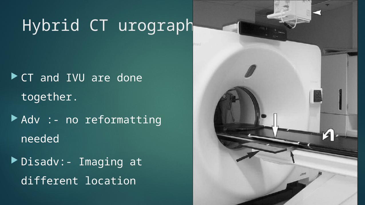
Hybrid CT urography
CT and IVU are done together. Adv :- no reformatting needed Disadv:- Imaging at different
location
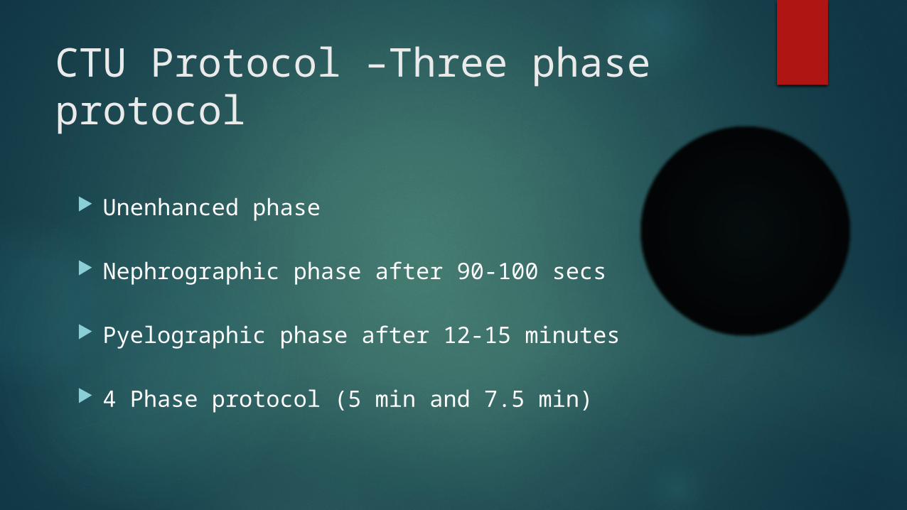
CTU Protocol –Three phase protocol
Unenhanced phase
Nephrographic phase after 90-100 secs
Pyelographic phase after 12-15 minutes
4 Phase protocol (5 min and 7.5 min)

Contrast media
Omnipaque (100-150 ml non ionic contrast media at a rate of 2-4 ml/second)

Split Bolus MDCT Urography with Synchronous Nephrographic and Excretory Phase Enhancement
Unenhanced Phase Nephro-pyelographic phase : 30 ml of nonionic
contrast material is infused and after 10 min another 100 ml of contrast is injected
ADV: Assess tract with low radiation exposure.

Image post processing techniques

Multiplanar Reformation (MPR)
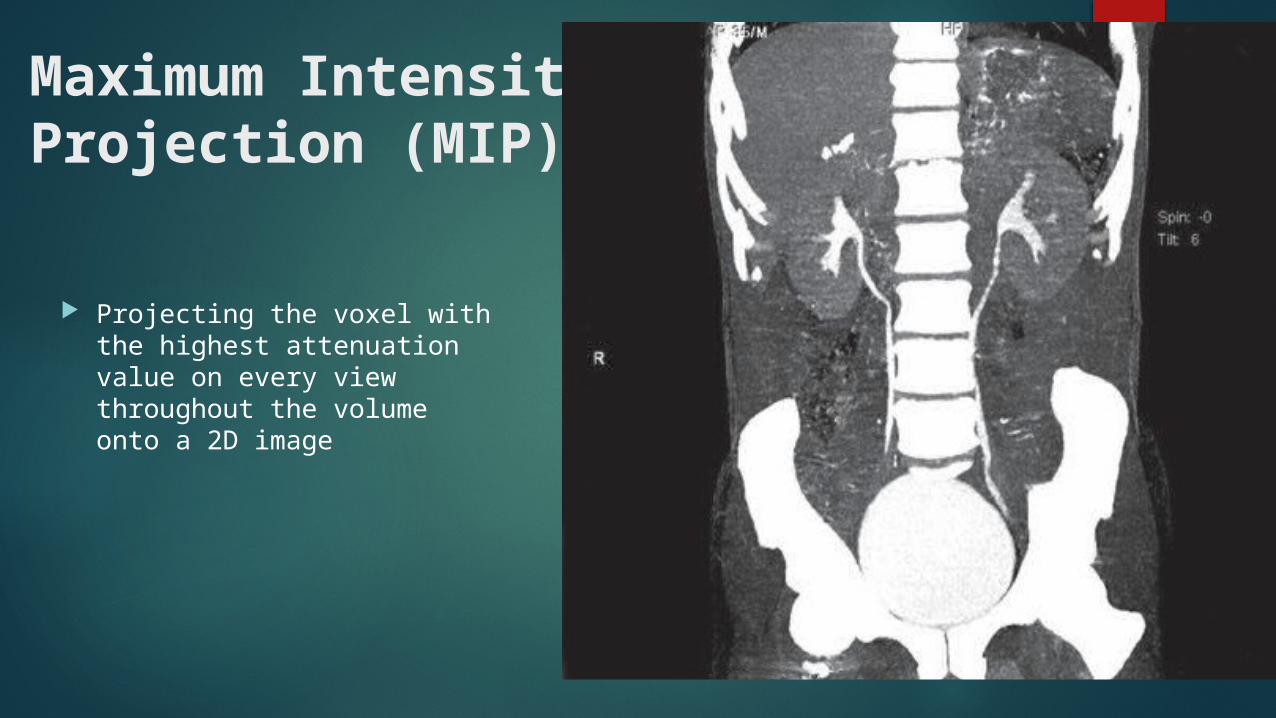
Maximum Intensity Projection (MIP)
Projecting the voxel with the highest attenuation value on every view throughout the volume onto a 2D image
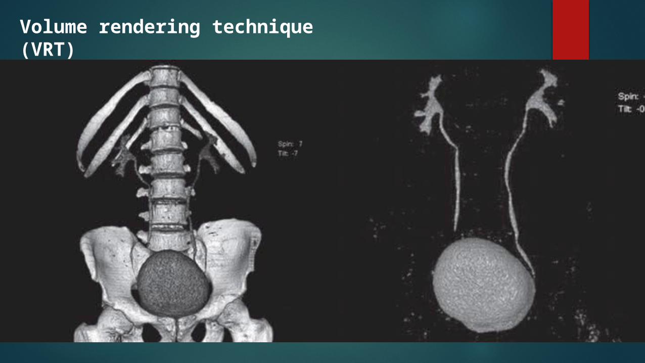
Volume rendering technique (VRT)
Indication of CT Urography
To evaluate patients with hematuria Calculi Renal / Urothelial tumors Congenital anomalies.
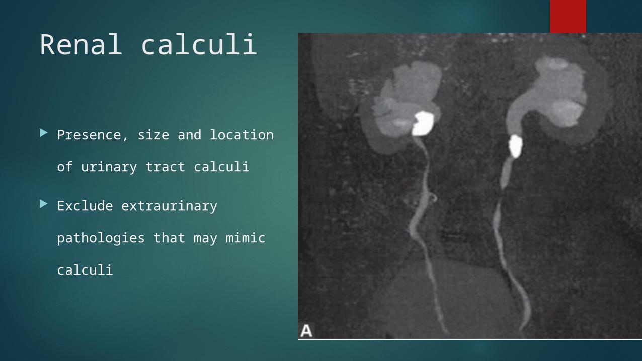
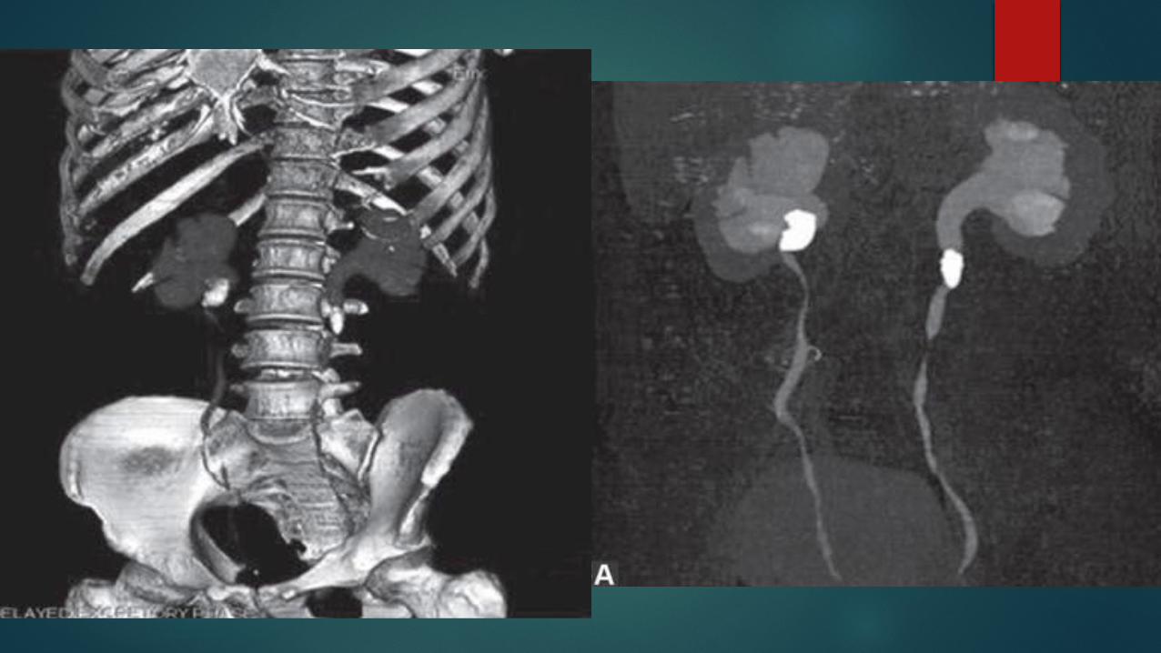
Renal calculi
Presence, size and location of
urinary tract calculi
Exclude extraurinary pathologies
that may mimic calculi
Renal Tumors
Enhancement : Nephrographic phase
Location : Renal cell carcinoma is frequently located at the periphery or near the cortico-medullary junction of the kidney as it originates in the renal cortex
tumors with nephron sparing surgery
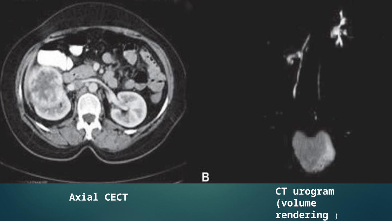
CT urogram (volume rendering )
Axial CECT
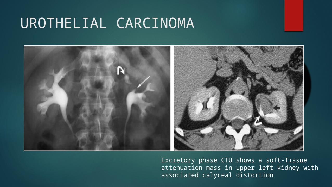
UROTHELIAL CARCINOMA
Excretory phase CTU shows a soft-Tissue attenuation mass in upper left kidney with associated calyceal distortion
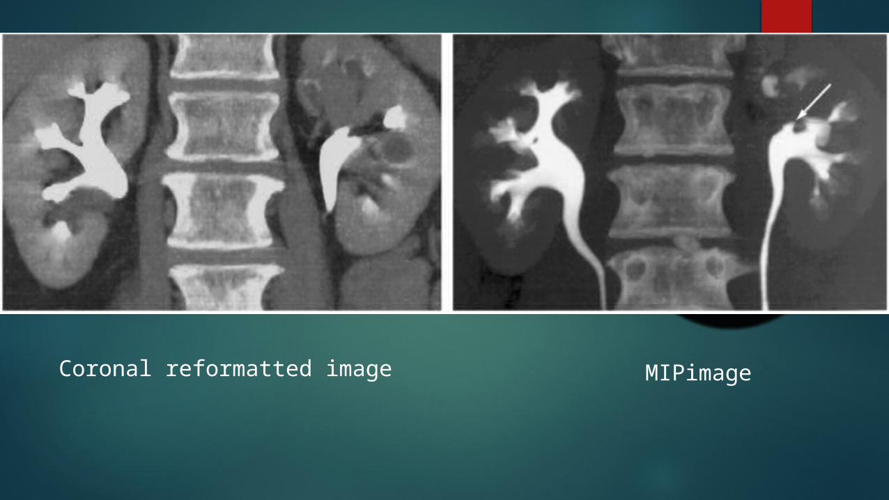
Coronal reformatted image MIPimage
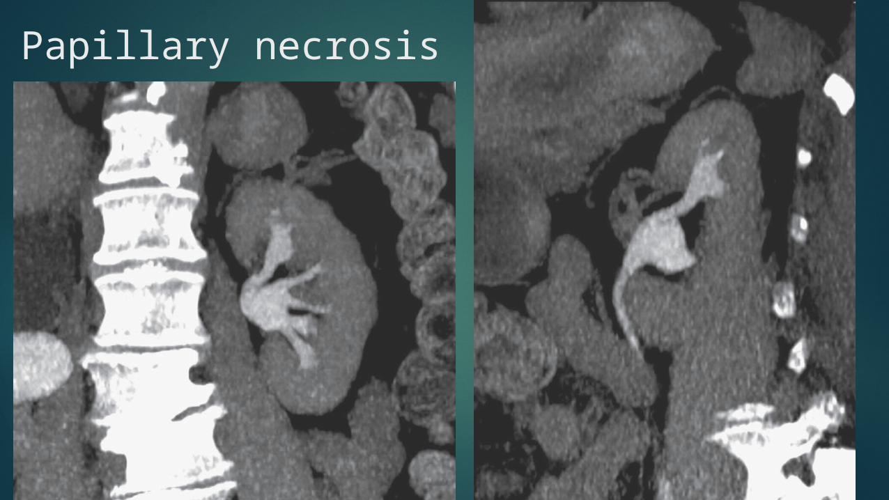
Papillary necrosis

Ureteric injury

Congenital anomalies Retrocaval ureter Ureteral duplication Crossed fused kidneys Ectopic kidneys
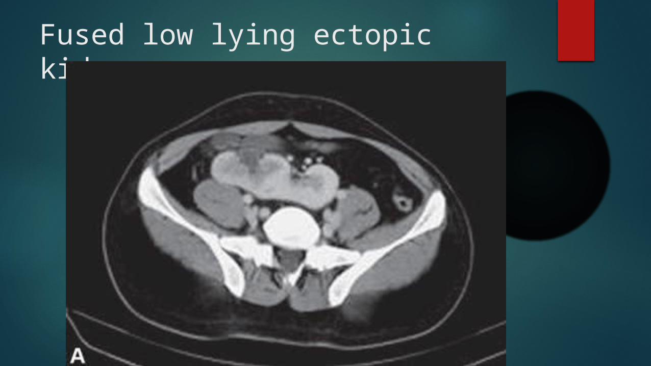
Fused low lying ectopic kidney

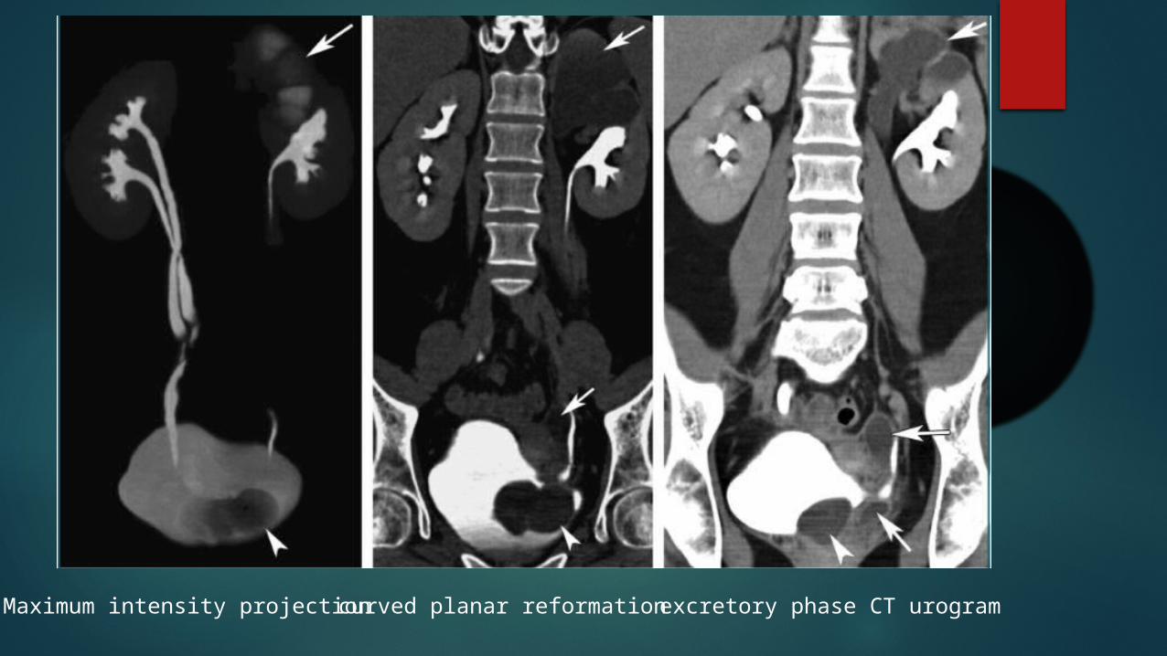
Maximum intensity projection curved planar reformation excretory phase CT urogram
Advantages of IVU over CT urography
Better spatial resolution
Lesser radiation dose
Cheaper
Ct urography: Advantages Multidetector CT : Thin slices in single breath hold allow
optimal anatomic information
Isotropic MPR reconstruction
Better contrast resolution
Image improvement techniques
Compression Saline Infusion Diuretic Administration Patient Positioning
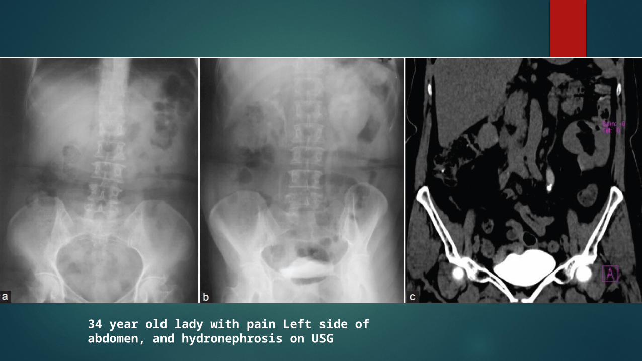
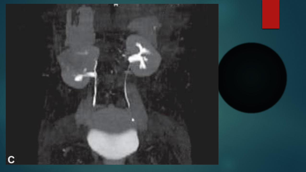
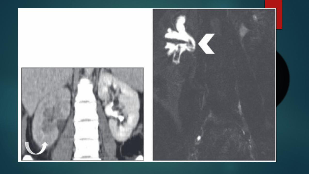
34 year old lady with pain Left side of abdomen, and hydronephrosis on USG
MR Urography
urinary tract obstruction Hematuria congenital anomalies surgically altered anatomy
beneficial in pediatric or pregnant patients or when ionizing radiation is to be avoided
Principle
urine have very long T2-relaxation time
heavily T2-weighted pulse sequence generate images with high signal
intensity from static fluid
Sequences Without contrast administration : Heavily T2-weighted
static-fluid MR urograms Half fourier acquisition single-shot turbo spin-echo(HASTE
sequence)
With contrast administration with excretory T1-weighted sequences (excretory MRU)
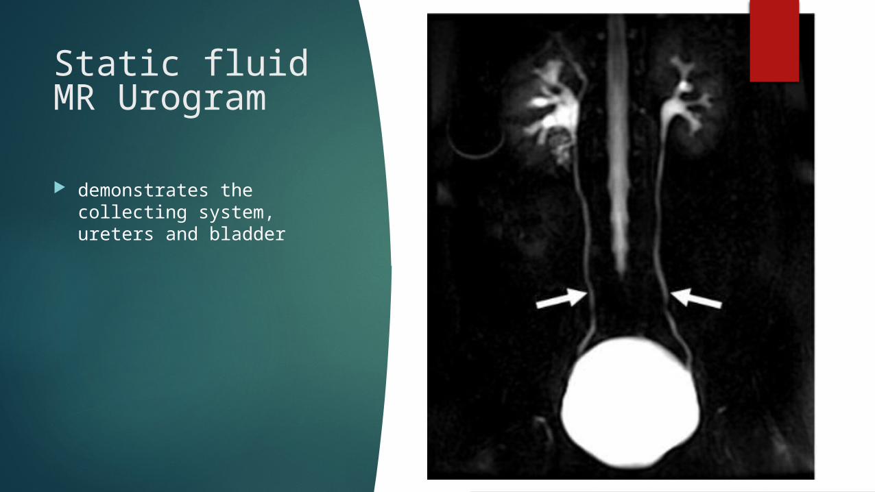
Static fluid MR Urogram
demonstrates the collecting system, ureters and bladder

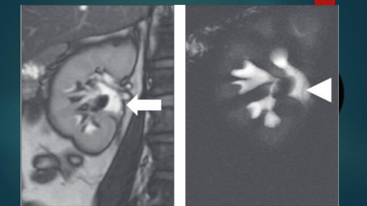
Coronal static-fluid MR urogram shows obstruction of the right distal ureter in a patient with prostrate cancer

Coronal single-shot fast spin-echo image shows a metastatic lymph node

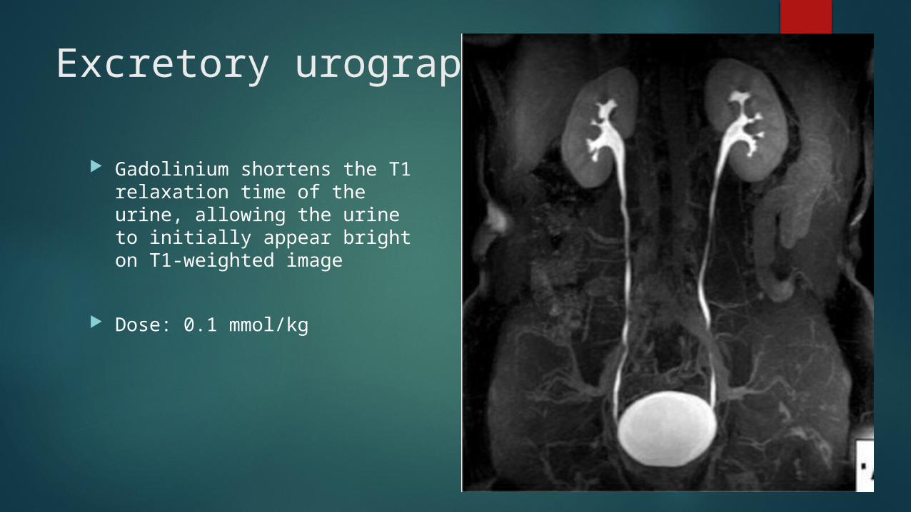
Excretory urography
Gadolinium shortens the T1 relaxation time of the urine, allowing the urine to initially appear bright on T1-weighted image
Dose: 0.1 mmol/kg
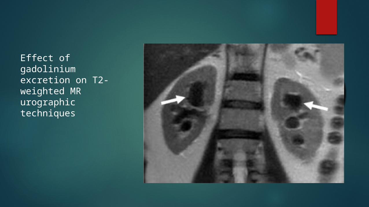
Effect of gadolinium excretion on T2-weighted MR urographic techniques
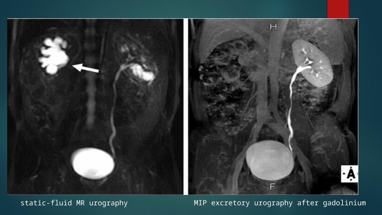
static-fluid MR urography MIP excretory urography after gadolinium
Advantages
MRU has better contrast resolution than CT urography
without radiation exposure and IV contrast administration
Disadvantages over CT Urography
Longer examination times than for CT urography
Decreased spatial resolution
Inability to reliably depict calcifications and calculi
THANK YOU