csf examination : techniques and … of csf examination lumbar puncture cisternal puncture lateral...
TRANSCRIPT

CSF EXAMINATION : TECHNIQUES CSF EXAMINATION : TECHNIQUES AND INTERPRETATIONAND INTERPRETATION
Presented By : DR. PRASAD R G

HISTORICAL BACKGROUNDHISTORICAL BACKGROUND
� 1885 – CORNING – spinal subarachnoid injections of cocaine
� 1891 – QUINCKE – diagnostic LP
� 1903 – FROIN – csf coagulation phenomenaphenomena
� 1916 – QUICKENSTEADT – manometric findings of spinal subarachnoid block
� 1918 – DANDY – ventricular puncture
� 1920 – AYER – cisternal puncture

INDICATIONS FOR CSF ANALYSISINDICATIONS FOR CSF ANALYSIS
� Bacterial , viral, fungal CNS infections� SAH� Demyelinating / degenerative disorders� Primary and metastatic tumors of CNS and
meningitis carcinomatosaPressure recordings – pseudotumor cerebri, � Pressure recordings – pseudotumor cerebri, NPH, head injury
� Suspected cerebral abscess , hemorrhagic infarctions
� Access for neuroradiologic procedures –ventriculography, cisternography, myelography
� Intrathecal administration of drugs

TECHNIQUES OF CSF TECHNIQUES OF CSF EXAMINATIONEXAMINATION� LUMBAR PUNCTURE
� CISTERNAL PUNCTURE
� LATERAL CERVICAL PUNCTURE
� VENTRICULAR PUNCTURE� VENTRICULAR PUNCTURE
� EXTERNAL VENTRICULAR DRAINAGE
� SUBCUTANEOUS CSF RESERVOIR INSTALLATION
LUMBAR PUNCTURELUMBAR PUNCTURE
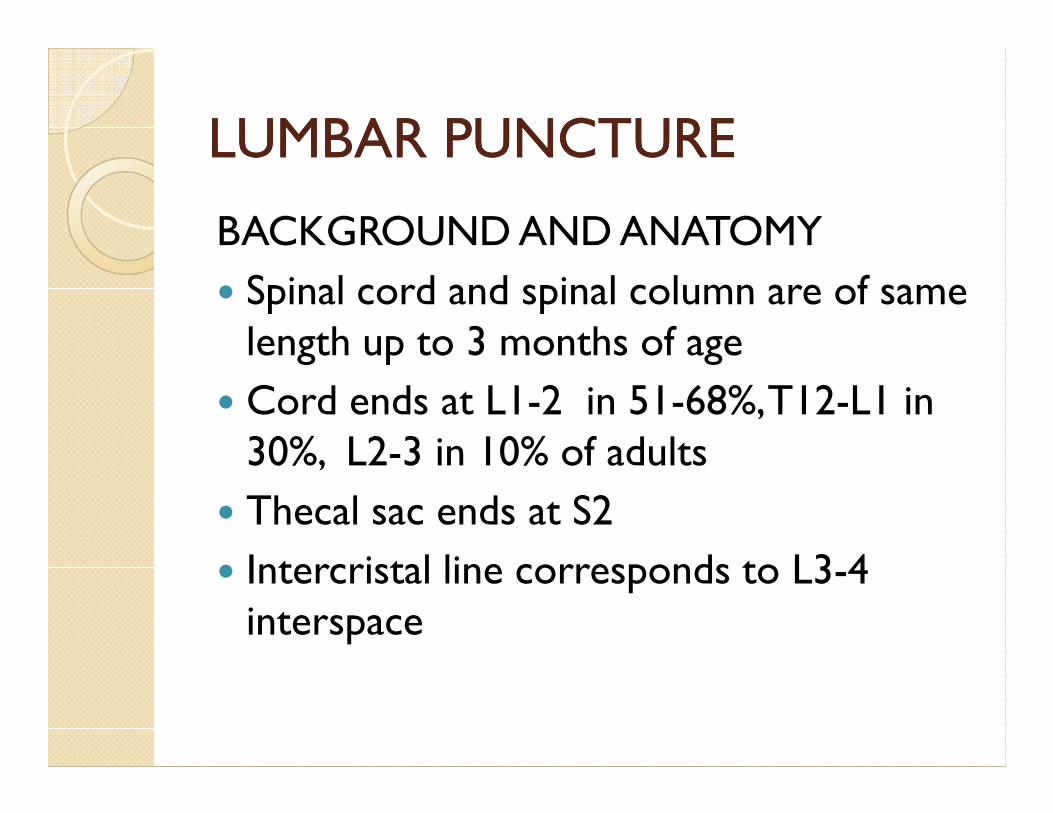
BACKGROUND AND ANATOMY
� Spinal cord and spinal column are of same length up to 3 months of age
� Cord ends at L1-2 in 51-68%, T12-L1 in � Cord ends at L1-2 in 51-68%, T12-L1 in 30%, L2-3 in 10% of adults
� Thecal sac ends at S2
� Intercristal line corresponds to L3-4 interspace
LUMBAR PUNCTURELUMBAR PUNCTURE
POSITIONING
� KNEE CHEST
� SITTING

LUMBAR PUNCTURELUMBAR PUNCTURE

LUMBAR PUNCTURELUMBAR PUNCTURE
SITE
� L3-4 - ADULTS
� L4-5 - CHILDREN
� L5-S1 - INFANTS� L5-S1 - INFANTS

LUMBAR PUNCTURELUMBAR PUNCTURE
LP NEEDLE
� TYPE -
QUINCKE’S
ATRAUMATIC NEEDLEATRAUMATIC NEEDLE
� SIZE -
18-20 Gauge - manometry
22 Gauge - diagnostic tap
14 Gauge tuohy needle / stamey ureteric catheter for spinal drainage

LUMBAR PUNCTURELUMBAR PUNCTURE
STEPS� Cleaning and draping� Infiltration of anesthetic� Bevel parallel to longitudinal dural fibers� Trajectory- directed slightly rostrally � Trajectory- directed slightly rostrally towards umbilicus
� Confirmation of needle patency� Connection to manometer –stop if opening pressure is >240 mm H20
� Quickensteadt test in suspected subarachnoid block

LUMBAR PUNCTURELUMBAR PUNCTURE
COLLECTION OF CSF
� 3 Vials for cell count, protein/glucose, gram stain/culture
� 4 vials in suspected traumatic tap� 4 vials in suspected traumatic tap
� For cyto pathology 5-10 ml CSF should be sent
� CSF should be sent immediately
� CSF can be preserved at 4 degree Celsius

LUMBAR PUNCTURELUMBAR PUNCTURE
CONTRA INDICATIONS� Local infection� Coagulopathy - coagulation disorders, pt on anticoagulant therapy
� Known / suspected increased intracranial � Known / suspected increased intracranial pressure due to mass lesion / non communicating hydrocephalus – 1.2% chance of neurological deterioration
� Complete spinal block – 14% risk of neurological deterioration
� Aneurysmal SAH
LUMBAR PUNCTURELUMBAR PUNCTURE
COMPLICATIONS
� Tonsillar herniation - acute / chronic
� Infection
� Spinal headache� Spinal headache
� Spinal epidural hematoma
� Spinal epidural CSF collection
� Epidermoid tumor
LUMBAR PUNCTURELUMBAR PUNCTURE
COMPLICATIONS� Nerve root injury� Intracranial subdural hygroma / hematoma� Vestibulo cochlear dysfunction
-subclinical-subclinicalsudden hearing loss
due to decreased perilymph pressure with endolymphatic hydrops
� Ocular abnormalities – abducens palsy� Dural sinus thrombosis
Post spinal headachePost spinal headache
� Occurs in up to 20% cases
� Subsides within 2-5 days, but may persist up to 8 weeks
� Factors◦ Age – young age◦ Age – young age
◦ Sex – females
◦ Previous h/o headache
◦ Body size – low BMI
◦ Pregnancy

Post spinal headachePost spinal headache
� Factors
◦ Needle size
◦ Bevel orientation
◦ Replacing stylet before withdrawal◦ Replacing stylet before withdrawal
◦ Number of Dural punctures
◦ Needle type
◦ Position of patient after LP
◦ Volume of fluid drained
◦ hydration

Post spinal headachePost spinal headache
� Treatment
◦ Horizontal position , bed rest
◦ Adequate hydration
◦ Mild analgesics ◦ Mild analgesics
◦ IV caffeine sodium benzoate
◦ Epidural blood / fibrin patch

LATERAL CERVICAL PUNCTURELATERAL CERVICAL PUNCTURE
� INDICATIONS◦ CSF specimen is required but access via LP is difficult / contra indicated
◦ To determine the rostral extent of sub arachnoid blockblock
� CONTRA INDICATIONSCHIARI malformation
� Low incidence of spinal headache
� Safer than cisternal puncture

LATERAL CERVICAL PUNCTURELATERAL CERVICAL PUNCTURE
� STEPS
◦With / without fluoroscopy
◦ 20 gauge spinal needle
◦ Under local anesthetic in co operative ◦ Under local anesthetic in co operative patients
◦ Patient positioned supine without pillow , looking up , avoiding head rotation

LATERAL CERVICAL PUNCTURELATERAL CERVICAL PUNCTURE

LATERAL CERVICAL PUNTURELATERAL CERVICAL PUNTURE

LATERAL CERVICAL PUNCTURELATERAL CERVICAL PUNCTURE
� STEPS
◦ ENTRY POINT lies 1cm below and behind mastoid tip
◦ Trajectory is perpendicular to the neck and Trajectory is perpendicular to the neck and parallel to the bed
◦ Frequent removal of stylet
◦ Subarachnoid space is 5-6 cm deeper
◦ For cervical myelogram 5 ml of 180 mg% IOHEXOL is used
LATERAL CERVICAL PUNCTURELATERAL CERVICAL PUNCTURE
� COMPLICATIONS
◦ Puncture of anomalous vertebral artery
◦ Penetration of spinal cord / medulla
◦ Tonsillar herniation◦ Tonsillar herniation

CISTERNAL TAPCISTERNAL TAP
� Sub occipital access to cisterna magna
� 22 gauge spinal needle with mark at 7.5 cm
� Position – sitting
� Entry point – in midline between inion and C2C2
� Trajectory – towards glabella
� Walking down the occiput
� Distance between – skin to cisterna magna is 4-6 cm , dura to medulla is 2.5 cm
� dural tenting occurs during procedure
CISTERNAL TAPCISTERNAL TAP
� COMPLICATIONS
◦ Hemorrhage
◦ Injury to medulla – vomiting , respiratory arrestarrest
◦ Positioning may compromise blood flow in elderly patients
VENTRICULAR VENTRICULAR CATHETERIZATIONCATHETERIZATIONPOINTS AND TRAJECTORIES OF ACCESS TO VENTRICLES
� Kocher’s point – 3 cm lat to midline and 1cm ant to coronal suture
� Keen’s point 2.5 -3 cm above and 2.5-3cm behind pinnapinna
� Dandy’ s point – 3 cm above inion and 2 cm lateral to midline
� Frazier’s point - 6 cm above inion and 4 cm lat to midline
� Orbital point – 1-2 cm behind superior orbital rim � Supra orbital – 4 cm above orbital rim in midpupillary line

VENTRICULAR VENTRICULAR CATHETERIZATIONCATHETERIZATION

VENTRICULAR VENTRICULAR CATHETERIZATIONCATHETERIZATION
OTHER METHODS OTHER METHODS
� Tapping a ommaya reservoir / shunt chamber
� External ventricular drain

PHYSIOLOGICAL PARAMETERS PHYSIOLOGICAL PARAMETERS OF CSFOF CSF
NEW BORN 1-10 YRS ADULTS
TOTAL VOLUME(ML)
5 150ML(50%CRANIAL,50% SPINAL)VOLUME(ML) L,50% SPINAL)
FORMATION RATE 25 ML / DAY O.3-O.35 ML /MIN
PRESSURE (mm H20)
90-120 <150 70-150
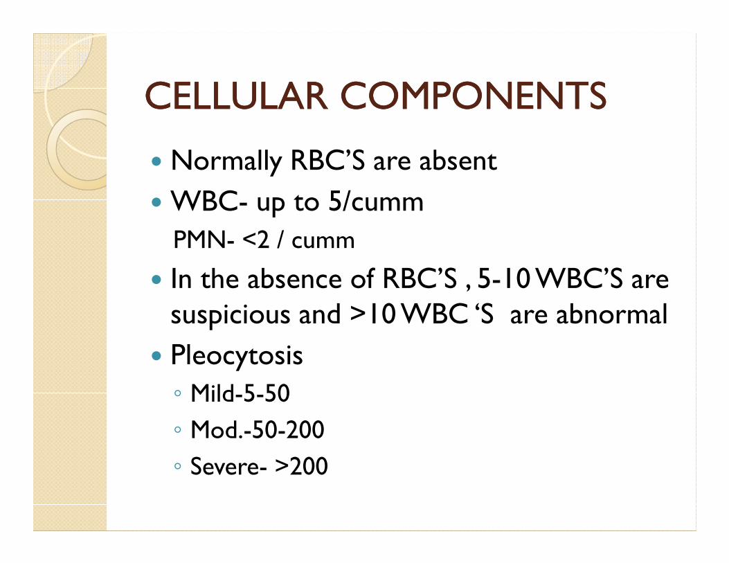
CELLULAR COMPONENTSCELLULAR COMPONENTS
� Normally RBC’S are absent
� WBC- up to 5/cumm
PMN- <2 / cumm
� In the absence of RBC’S , 5-10 WBC’S are � In the absence of RBC’S , 5-10 WBC’S are suspicious and >10 WBC ‘S are abnormal
� Pleocytosis
◦ Mild-5-50
◦ Mod.-50-200
◦ Severe- >200

CELLULAR COMPONENTSCELLULAR COMPONENTS
� TRAUMATIC TAP
◦ Subtract 1 WBC / 700 RBC’S
◦ FISHMAN formula
WBC = WBC (F) - WBC (B) × RBC(F)
--------------------
RBC(B)
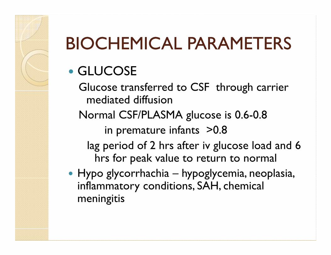
BIOCHEMICAL PARAMETERSBIOCHEMICAL PARAMETERS
� GLUCOSEGlucose transferred to CSF through carrier mediated diffusion
Normal CSF/PLASMA glucose is 0.6-0.8
in premature infants >0.8in premature infants >0.8
lag period of 2 hrs after iv glucose load and 6 hrs for peak value to return to normal
� Hypo glycorrhachia – hypoglycemia, neoplasia, inflammatory conditions, SAH, chemical meningitis
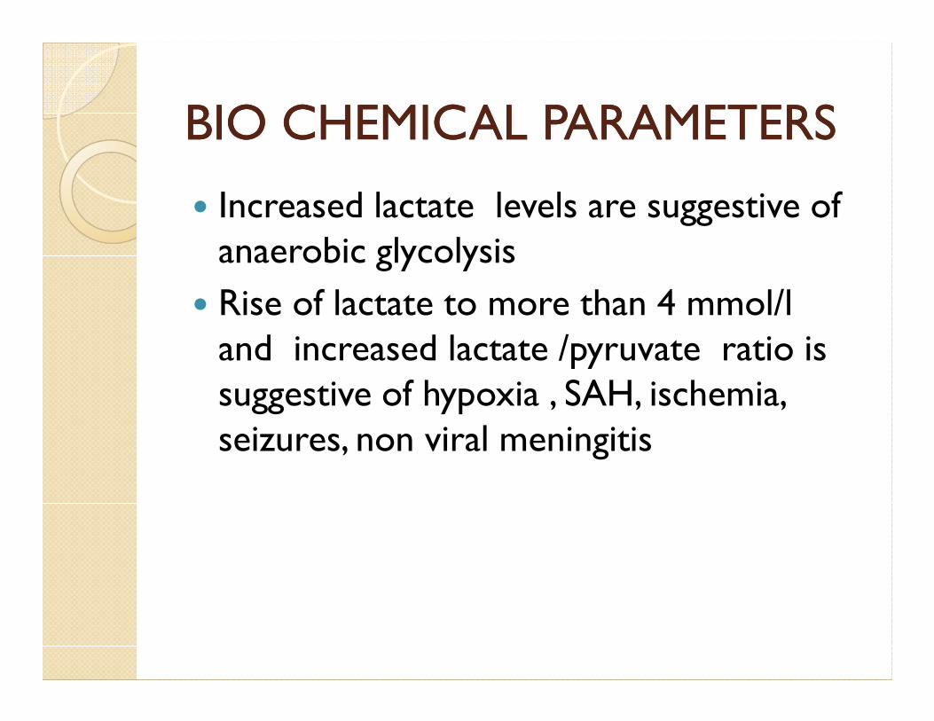
BIO CHEMICAL PARAMETERSBIO CHEMICAL PARAMETERS
� Increased lactate levels are suggestive of anaerobic glycolysis
� Rise of lactate to more than 4 mmol/l and increased lactate /pyruvate ratio is and increased lactate /pyruvate ratio is suggestive of hypoxia , SAH, ischemia, seizures, non viral meningitis
BIOCHEMICAL PARAMETERSBIOCHEMICAL PARAMETERS
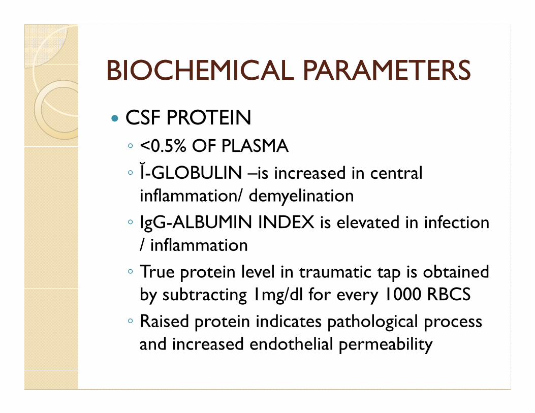
� CSF PROTEIN
◦ <0.5% OF PLASMA
◦ Ĭ-GLOBULIN –is increased in central inflammation/ demyelinationinflammation/ demyelination
◦ IgG-ALBUMIN INDEX is elevated in infection / inflammation
◦ True protein level in traumatic tap is obtained by subtracting 1mg/dl for every 1000 RBCS
◦ Raised protein indicates pathological process and increased endothelial permeability

feature Traumatic tap SAH
RBC count and gross appearance of bloodiness
decreases Little change
WBC/RBC Similar to peripheral blood
leucocytosis
supernatant clear xanthochromic
Clotting of fluid Clots if RBC count >200,000/cumm
Does not clot
Protein conc. Rise 1mg/1000 RBC >1mg/1000RBC
Repeat LP at higher level clear Remains bloody
Opening pressure normal Usually elevated
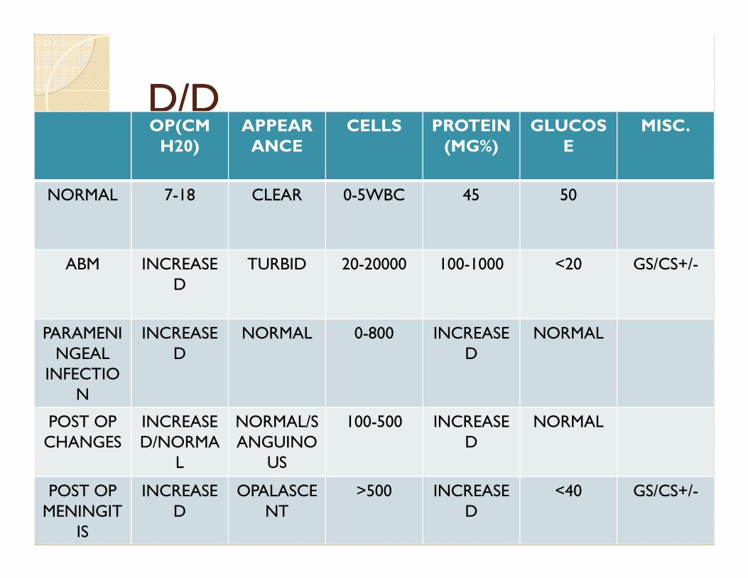
D/DD/DOP(CM H20)
APPEARANCE
CELLS PROTEIN(MG%)
GLUCOSE
MISC.
NORMAL 7-18 CLEAR 0-5WBC 45 50
ABM INCREASED
TURBID 20-20000 100-1000 <20 GS/CS+/-D
PARAMENINGEAL
INFECTION
INCREASED
NORMAL 0-800 INCREASED
NORMAL
POST OP CHANGES
INCREASED/NORMA
L
NORMAL/SANGUINO
US
100-500 INCREASED
NORMAL
POST OP MENINGIT
IS
INCREASED
OPALASCENT
>500 INCREASED
<40 GS/CS+/-

D/DD/D
OP(CM H20)
APPEARANCE
CELLS PROTEIN(MG%)
GLUCOSE
MISC.
FUNGAL MENING
ITIS
INCREASED
OPALASCENT
30-300(LYMPHO)
100-700 <30 +INDIA INK IN CRYPTO.ITIS PHO) CRYPTO.
TB MENING
ITIS
INCREASED
OPALASCENT WITH CLOT
50-500LYMP
HO
60-700 20-40 ZN STAIN+/AFB CS
+
BRAIN ABSCESS
INCREASED
CLEAR/TURBID
INCREASED
INCREASED
NORMAL/DECREASED
LESS SENSITIV
E
POST OP MENINGITISPOST OP MENINGITIS
� Gram stain – 60-90% accurate
� Polymerase chain reaction for bacterial DNA
� C reactive protein levels – strong negative predictive value
� Latex agglutination –sensitive test for partially treated � Latex agglutination –sensitive test for partially treated patients
� Lymulus lysate levels
� Lactate levels >4mmol/l s/o post op meningitis
� Csf pro calcitonin levels
� S-100 protein levels
� TNF –ALFA/IL6 levels

OTHER CONDITIONSOTHER CONDITIONS
� MENINGEAL CARCINOMATOSIS
◦ 25% of CNS malignancy has positive cytology
◦ 60% with lepto meningeal involvement is+
◦ Repeated sampling is necessary◦ Repeated sampling is necessary
◦ Immuno cyto chemical methods improve sensitivity

OTHER MARKERS IN CSFOTHER MARKERS IN CSF
� CSF HCG - Central chorio carcinoma
� CEA - breast, lung bladder mets in CNS
� Alfa feto protein – germ cell tumors, metastatic testicular and hepatic ca.
� Spermidine – meningiomas
� Poly amine in leukemia
� Desmosterol in gliomas
� beta glucuronidase in leptomeningeal involvement
SPINAL CORD TUMORSSPINAL CORD TUMORS
� Increased protein > 100mg/dl
� >100 PMN /cumm
� Froin syndrome

� THANK YOU