crumpled reduced graphene oxide–polyamidoamine dendrimer hybrid nanoparticles for the preparation...
TRANSCRIPT
Journal ofMaterials Chemistry B
PAPER
Publ
ishe
d on
01
Mar
ch 2
013.
Dow
nloa
ded
by W
ashi
ngto
n U
nive
rsity
in S
t. L
ouis
on
23/0
5/20
13 1
0:20
:10.
View Article OnlineView Journal | View Issue
aDepartment of Analytical Chemistry, Facult
Madrid, 28040-Madrid, Spain. E-mail: pin
ucm.es; Fax: +34 913944329; Tel: +34 9139bDepartment of Organic Chemistry I, Faculty
Madrid, 28040-Madrid, Spain
† Electronic supplementary informationhybrid nanomaterial, optimization of10.1039/c3tb20078g
Cite this: J. Mater. Chem. B, 2013, 1,2289
Received 20th January 2013Accepted 1st March 2013
DOI: 10.1039/c3tb20078g
www.rsc.org/MaterialsB
This journal is ª The Royal Society of
Crumpled reduced graphene oxide–polyamidoaminedendrimer hybrid nanoparticles for the preparation ofan electrochemical biosensor†
Elena Araque,a Reynaldo Villalonga,*a Marıa Gamella,a Paloma Martınez-Ruiz,b
Julio Reviejoa and Jose M. Pingarron*a
Reduced graphene nanoparticles were prepared from graphene oxide through a two-step covalent
modification approach. Graphene oxide was first enriched with reactive epoxy groups by anchoring (3-
glycidyloxypropyl)trimethoxysilane at the hydroxyl groups located on the nanocarbon basal plane.
Modified graphene oxide was further cross-linked and partially reduced by treatment with the fourth-
generation ethylenediamine core polyamidoamine G-4 dendrimer producing graphene nanoparticles
with crumpled paper-like morphology. This graphene derivative was employed as a coating material for
glassy carbon electrodes and the nanostructured electrode was tested for the preparation of
electrochemical biosensors by immobilizing the enzyme tyrosinase through cross-linking with
glutaraldehyde. This bioelectrode showed excellent electroanalytical behavior for catechol with a fast
response in about 6 s, linear range of 10 nM to 22 mM, sensitivity of 424 mA M�1, and low detection limit
of 6 nM. The enzyme biosensor also showed high stability when stored at 4 �C under dry andwet conditions.
Introduction
Engineering nanomaterials with well-dened three-dimen-sional morphology has attracted considerable interest in thelast few years due to their envisioned applications in the designof novel electronic devices, drug carrier systems and optical andelectrochemical sensors. These 3D-shaped nanomaterials canalso be employed as building blocks for the construction oforiginal architectures at the nanoscale.1 In this context, thesynthesis of novel hybrid nanomaterials with improved elec-troconductive properties and capacity to recognize/immobilizeanalytical biomolecules is a challenge for biosensor tech-nology.2 In particular, carbon-based nanomaterials havedemonstrated their usefulness in designing hybrid nano-structures for successful application in the preparation ofbiosensors,3 with recent efforts focused on the use of graphenehybrid derivatives for the construction of electrochemicalbiosensors4 due to the relatively low cost, large surface-to-volume ratio and remarkable electrical, mechanical andthermal properties and biocompatibility of this nanomaterial.5
y of Chemistry, Complutense University of
[email protected]; rvillalonga@quim.
44315
of Chemistry, Complutense University of
(ESI) available: Characterization of thebiosensor construction. See DOI:
Chemistry 2013
However, there are some major obstacles for the applicationof graphene in the preparation of electrochemical biosensors.Graphene nanosheets are highly hydrophobic, showing very lowsolubility in water and many organic solvents, and tending toform irreversible agglomerates through strong p–p stackingand van der Waals interactions.6 Moreover, the absence ofchemical functional groups in graphene limits the stable andlarge immobilization of biomolecules through covalent link-ages.2a Chemical derivatization of graphene seems to be arational approach to overcome these disadvantages.7 However,the possibility of generically tailoring the chemical properties ofgraphene is limited by its delicate structure, oen yieldingnanomaterials with poor electroconductive characteristics. Forthis reason, the development of synthetic strategies forpreparing soluble and highly functionalized graphene deriva-tives with low modication of its basal structure receivesconsiderable attention.
Many synthetic approaches commonly use graphene oxide(GO), a water soluble derivative that can be easily prepared byoxidative treatment of graphite, as the starting material.8 GOmainly consists of graphene-like sheets,9 with hydroxyl andepoxide groups on the basal planes and carbonyl and carboxylgroups at the sheet edges.5 These oxygen functionalities can beselectively used as anchoring points for chemical modication.8
GO and its derivatives can be easily reduced by chemical,thermal, photothermal, and electrochemical methods,restoring in high yield the structural and electrical conductivityproperties of graphene.2a,8
J. Mater. Chem. B, 2013, 1, 2289–2296 | 2289
Journal of Materials Chemistry B Paper
Publ
ishe
d on
01
Mar
ch 2
013.
Dow
nloa
ded
by W
ashi
ngto
n U
nive
rsity
in S
t. L
ouis
on
23/0
5/20
13 1
0:20
:10.
View Article Online
In this work we describe the preparation of a novel poly-aminated graphene derivative through the initial modicationof GO with (3-glycidyloxypropyl)trimethoxysilane, using thehydroxyl groups at the basal plane as anchoring points. Thissilanized GO (Sil-GO) was further functionalized and reduced bytreatment with the fourth-generation ethylenediamine-corepolyamidoamine dendrimer (PAMAM). The resulting hybridnanomaterial (PAMAM-Sil-rGO) possessed a crumpled paper-like morphology and was employed as a coating material tomodify glassy carbon electrodes. The nanostructured electrodewas used as a support for the covalent immobilization ofenzyme tyrosinase (Tyr, EC 1.14.18.1), in order to evaluate theperformance of the resulting Tyr-PAMAM-Sil-rGO electrode asan electrochemical biosensor for catechol.
Materials and methodsMaterials
Tyrosinase (5370 U mg�1), (3-glycidyloxypropyl)trimethox-ysilane and glutaraldehyde were acquired from Sigma-AldrichCo. (USA). The ethylenediamine core, PAMAM G-4 dendrimerwas purchased from Dendritech, Inc. (USA). Graphene oxide,prepared according to the Hummer's method,10 was acquiredfrom NanoInnova Technologies (Spain). All other chemicalswere of analytical grade.
Apparatus, electrodes and solutions
Cyclic voltammetry and electrochemical impedance spectros-copy experiments were performed using a FRA2 mAutolab TypeIII potentiostat/galvanostat, and the data were acquired usingGPES Ver. 4.9 and Frequency Response Analyser soware,respectively (Metrohm Autolab B.V., The Netherlands). Amper-ometric measurements in stirred solutions at 300 rpm werecarried out with a dual-channel ultrasensitive Inbea potentio-stat (Inbea Biosensores S.L., Spain). A conventional three-elec-trode system was employed in all electrochemical studies. Theworking electrode was a glassy carbon electrode (GCE, 3.0 mmdiameter) modied with the PAMAM-graed graphene deriva-tive and the immobilized enzyme. An Ag/AgCl/KCl (3M) and a Ptwire were used as the reference and counter electrodes,respectively. The measurements with the biosensor were carriedout at 25 �C in 0.1 M sodium phosphate buffer, pH 6.0 (workingvolume 10 mL). 100 mM catechol solutions in 50 mM sodiumphosphate buffer, pH 6.0, were freshly prepared.
The surface morphology of the nanostructured electrodeswas investigated by high resolution eld emission scanningelectron microscopy (FE-SEM) using a JEOL JSM-6335F micro-scope (JEOL Ltd., Japan). FT-IR spectra were acquired with aNicolet Nexus 670/870 spectrometer (Thermo Fisher ScienticInc., USA).
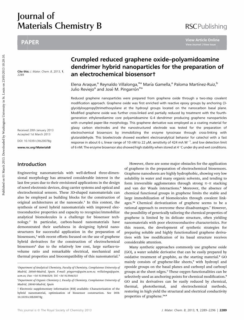
Fig. 1 Schematic display of the steps involved in the preparation of the Tyr/PAMAM-Sil-rGO/GCE enzyme electrode.
Synthesis of PAMAM-Sil-rGO
Silanized graphene oxide was prepared by dispersion, rstly, of50 mg GO in 250 mL ethanol by sonication for 1 h. Thedispersion was then heated at 65 �C, mixed with 25 mL of a 1%(v/v) ethanolic solution of (3-glycidyloxypropyl)trimethoxysilane
2290 | J. Mater. Chem. B, 2013, 1, 2289–2296
and kept under continuous stirring at this temperature for 12 h.The mixture was cooled and ltered and the resulting solid (Sil-GO) was exhaustively washed with ethanol and nally dried in avacuum. To prepare PAMAM-Sil-rGO, 50 mg Sil-GO weredispersed in 200 mL ethanol by sonication and the dispersionwas mixed with 2.5 mL 10% (w/w) ethylenediamine corePAMAM G-4 solution in methanol and stirred at room temper-ature for 12 h. The mixture was ltered and the solid waswashed several times with ethanol and nally dried under avacuum at room temperature.
Preparation of the enzyme electrode
A bare GCE was rst polished to a mirror-like surface withalumina powder (0.3 mM), rinsed thoroughly with doubledistilled water, successively washed with double distilled water,anhydrous ethanol and acetone in an ultrasonic bath, and driedunder N2 before use. Coating of the polished GCE was accom-plished by depositing two 10 mL aliquots of a 0.5 mg mL�1
aqueous dispersion of PAMAM-Sil-rGO on the electrode surfaceand allowing to dry. The modied electrode was then dippedinto a 10 mM sodium phosphate buffer solution, pH 5.0, andthe potential was cycled between 0.0 and �1.5 V for 20 cycles toobtain a more reduced graphene derivative on the electrodesurface.11
The enzyme was further immobilized on the modied elec-trode by dropping 20 mL of 10 mg mL�1 tyrosinase solution in100 mM sodium phosphate buffer, pH 6.0, and then mixingwith 4 mL of 25% (v/v) glutaraldehyde. The electrode was kept at4 �C for 1 h, then washed several times with cold 50 mM sodiumphosphate buffer, pH 6.0, and nally stored in a refrigeratoruntil use.
Results and discussion
The different steps involved in the preparation of the hybridPAMAM-Sil-rGO nanomaterial, as well as the further enzymeimmobilization to construct the nanostructured biosensor, areschematically displayed in Fig. 1.
Firstly graphene nanoparticles were prepared through a two-step synthetic approach as shown in Scheme 1. In a rst step,
This journal is ª The Royal Society of Chemistry 2013
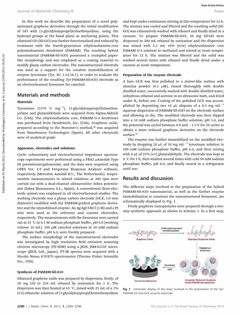
Scheme 1
Fig. 2 Impedance plane diagram (�Z0 0 versus Z0) for the EIS measurements at aglassy carbon electrode before (a) and after coating with Sil-GO (b), PAMAM-Sil-rGO (c) PAMAM-Sil-GO after electrochemical reduction (d), and Tyr/PAMAM-Sil-GO(e, inset) in a 0.1 M KCl solution containing 5 mM K3[Fe(CN)6]/K4[Fe(CN)6] (1 : 1).
Paper Journal of Materials Chemistry B
Publ
ishe
d on
01
Mar
ch 2
013.
Dow
nloa
ded
by W
ashi
ngto
n U
nive
rsity
in S
t. L
ouis
on
23/0
5/20
13 1
0:20
:10.
View Article Online
GO was enriched with reactive epoxy groups by treatment with(3-glycidyloxypropyl)trimethoxysilane, using the hydroxylgroups on the nanomaterial surface as anchoring points.12
Subsequently, silanized graphene oxide (Sil-GO) was modiedwith the polyaminated dendrimer molecules through theformation of stable secondary amine linkages according to thereactions shown below, yielding a dark product that was easilydispersed in aqueous solutions (PAMAM-Sil-rGO).
The PAMAM dendrimer was selected as an attractive sotemplate for GO modication due to its nanosized and mono-disperse characteristics exhibiting a regular and highlybranched three-dimensional architecture, high structuralhomogeneity and chain mobility.13 This so nanomaterialpossesses also a high density of primary amino groups on itssurface, which favoured multipoint cross-linking with activatedGO sheets.
Characterization of the hybrid nanomaterial
Graphene derivatives were characterized by means of differentphysico-chemical techniques. Fig. S1 (see ESI†) shows powderXR diffractograms of GO (a), Sil-GO (b) and PAMAM-Sil-rGO (c).The diffractogram of GO shows an intense peak at 12.35� cor-responding to a d-spacing of about 7.16 A. This interlayerdistance is mainly caused by the presence of oxygen function-alities on GO as well as the water molecules entrapped betweenthe hydrophilic sheets.14 The intensity of this peak decreasedaer covalent modication of GO, and it was shied to 11.50�
(7.69 A) and 10.23� (8.65 A) aer treatment with (3-glycidylox-ypropyl)trimethoxysilane and PAMAM, respectively. Thisincrease in the interlayer distance could be ascribed to the bulkysilane and dendrimer molecules, which should be intercalatedinto the interlayer spacing of the nanomaterial. Diffractogramof PAMAM-Sil-rGO also showed a broad and low intensity peakaround 20.13� (4.41 A), which is close to the d002-spacing ofgraphite (3.35 A). This small interlayer distance might beattributed to the partial reduction of GO with PAMAM allowingthe reduced sheets to pack tighter than the initial nanomaterial.
Fig. S2 (see ESI†) shows the characterization of the graphenederivatives by FT-IR spectroscopy. GO exhibited sharp andbroad peaks centered at 3607 cm�1 and 3090 cm�1 corre-sponding to the stretching of free and intermolecular H-bondedO–H groups, respectively. In addition, the GO spectrumexhibited the peaks at 1353 cm�1 and 1230 cm�1 corresponding
This journal is ª The Royal Society of Chemistry 2013
to the stretching of C–OH and C–O–C groups in unoxidizedgraphitic domains. The peaks at 1730 cm�1 and 1065 cm�1
could be ascribed to the stretching of the C]O and C–O groups,respectively. The peak at 1608 cm�1 could be associated with thevibrations of unoxidized graphitic skeletal domains, as well aswith the adsorbed water.15
The presence of silane moieties on the surface of Sil-GO wasconrmed by the peaks at 2880 cm�1 and 1410 cm�1, which canbe ascribed to the stretching and bending of the methylenegroups from the (3-glycidyloxypropyl)trimethoxysilane mole-cules, respectively. Additionally, the peak at 986 cm�1 could beassociated with the vibration of the Si–O groups. The FT-IRspectrum of PAMAM-Sil-rGO showed that the intensity of thepeaks corresponding to the stretching vibrations of the C]Ogroups at 1730 cm�1, of free O–H groups at 3607 cm�1, of the C–O–C at 1230 cm�1 and the C–O at 1065 cm�1 decreased signif-icantly, and some of them disappeared totally. This fact sug-gested that most oxygen functionalities in the Sil-GO samplewere reduced during the covalent graing of PAMAM mole-cules. The attachment of the dendritic moieties was conrmedby the appearance of the peak at 1541 cm�1, which could beascribed to the bending of the N–H groups. The broad peakfrom 2930 to 3506 cm�1 could be associated with the stretchingvibration of the N–H and O–H groups, as well as with theadsorbed water. However, its multiple peaks pattern also sug-gested the formation of primary ammonium salts between theattached dendrimer and the remaining carboxylate groups atthe graphene sheet edges.
Partial reduction of GO aer derivatization with PAMAM wasconrmed by electrochemical impedance spectroscopy using[Fe(CN)6]
4�/3� as a redox probe (Fig. 2). Coating of a bare glassycarbon electrode with GO and Sil-GO produced a noticeableincrease in the electron transfer resistance (from 256 to 1202 U),in agreement with the poor electro-conducting properties ofthese derivatives. However, the PAMAM-Sil-rGO coated elec-trode showed a very small semicircle diameter at highfrequencies, Rct ¼ 92 U, indicating a fast electron transferprocess. This observation can be justied by the partial recoveryof the characteristic electro-conductive properties of graphene
J. Mater. Chem. B, 2013, 1, 2289–2296 | 2291
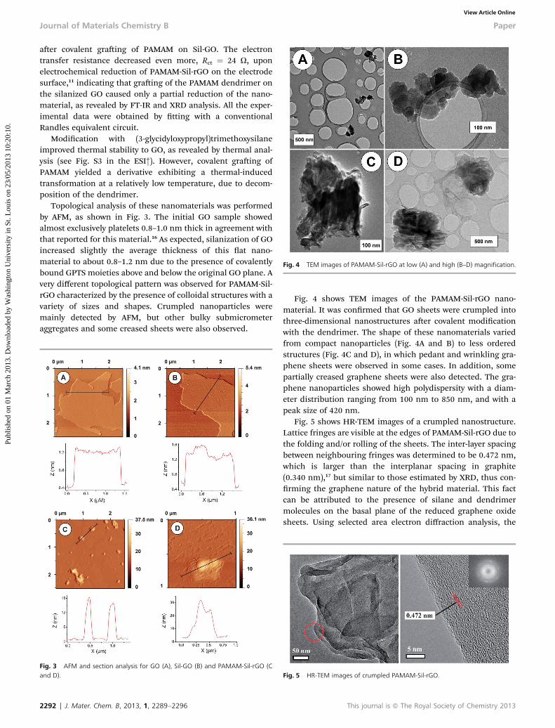
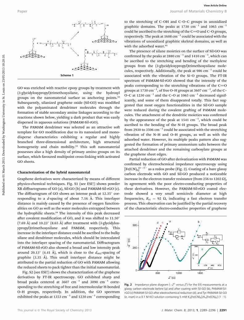
Fig. 4 TEM images of PAMAM-Sil-rGO at low (A) and high (B–D) magnification.
Journal of Materials Chemistry B Paper
Publ
ishe
d on
01
Mar
ch 2
013.
Dow
nloa
ded
by W
ashi
ngto
n U
nive
rsity
in S
t. L
ouis
on
23/0
5/20
13 1
0:20
:10.
View Article Online
aer covalent graing of PAMAM on Sil-GO. The electrontransfer resistance decreased even more, Rct ¼ 24 U, uponelectrochemical reduction of PAMAM-Sil-rGO on the electrodesurface,11 indicating that graing of the PAMAM dendrimer onthe silanized GO caused only a partial reduction of the nano-material, as revealed by FT-IR and XRD analysis. All the exper-imental data were obtained by tting with a conventionalRandles equivalent circuit.
Modication with (3-glycidyloxypropyl)trimethoxysilaneimproved thermal stability to GO, as revealed by thermal anal-ysis (see Fig. S3 in the ESI†). However, covalent graing ofPAMAM yielded a derivative exhibiting a thermal-inducedtransformation at a relatively low temperature, due to decom-position of the dendrimer.
Topological analysis of these nanomaterials was performedby AFM, as shown in Fig. 3. The initial GO sample showedalmost exclusively platelets 0.8–1.0 nm thick in agreement withthat reported for this material.16 As expected, silanization of GOincreased slightly the average thickness of this at nano-material to about 0.8–1.2 nm due to the presence of covalentlybound GPTS moieties above and below the original GO plane. Avery different topological pattern was observed for PAMAM-Sil-rGO characterized by the presence of colloidal structures with avariety of sizes and shapes. Crumpled nanoparticles weremainly detected by AFM, but other bulky submicrometeraggregates and some creased sheets were also observed.
Fig. 3 AFM and section analysis for GO (A), Sil-GO (B) and PAMAM-Sil-rGO (Cand D).
2292 | J. Mater. Chem. B, 2013, 1, 2289–2296
Fig. 4 shows TEM images of the PAMAM-Sil-rGO nano-material. It was conrmed that GO sheets were crumpled intothree-dimensional nanostructures aer covalent modicationwith the dendrimer. The shape of these nanomaterials variedfrom compact nanoparticles (Fig. 4A and B) to less orderedstructures (Fig. 4C and D), in which pedant and wrinkling gra-phene sheets were observed in some cases. In addition, somepartially creased graphene sheets were also detected. The gra-phene nanoparticles showed high polydispersity with a diam-eter distribution ranging from 100 nm to 850 nm, and with apeak size of 420 nm.
Fig. 5 shows HR-TEM images of a crumpled nanostructure.Lattice fringes are visible at the edges of PAMAM-Sil-rGO due tothe folding and/or rolling of the sheets. The inter-layer spacingbetween neighbouring fringes was determined to be 0.472 nm,which is larger than the interplanar spacing in graphite(0.340 nm),17 but similar to those estimated by XRD, thus con-rming the graphene nature of the hybrid material. This factcan be attributed to the presence of silane and dendrimermolecules on the basal plane of the reduced graphene oxidesheets. Using selected area electron diffraction analysis, the
Fig. 5 HR-TEM images of crumpled PAMAM-Sil-rGO.
This journal is ª The Royal Society of Chemistry 2013
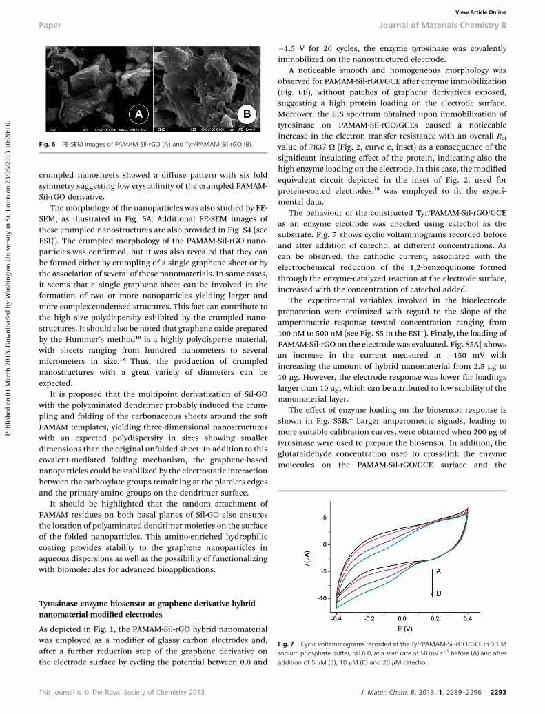
Fig. 6 FE-SEM images of PAMAM-Sil-rGO (A) and Tyr/PAMAM-Sil-rGO (B).
Paper Journal of Materials Chemistry B
Publ
ishe
d on
01
Mar
ch 2
013.
Dow
nloa
ded
by W
ashi
ngto
n U
nive
rsity
in S
t. L
ouis
on
23/0
5/20
13 1
0:20
:10.
View Article Online
crumpled nanosheets showed a diffuse pattern with six foldsymmetry suggesting low crystallinity of the crumpled PAMAM-Sil-rGO derivative.
The morphology of the nanoparticles was also studied by FE-SEM, as illustrated in Fig. 6A. Additional FE-SEM images ofthese crumpled nanostructures are also provided in Fig. S4 (seeESI†). The crumpled morphology of the PAMAM-Sil-rGO nano-particles was conrmed, but it was also revealed that they canbe formed either by crumpling of a single graphene sheet or bythe association of several of these nanomaterials. In some cases,it seems that a single graphene sheet can be involved in theformation of two or more nanoparticles yielding larger andmore complex condensed structures. This fact can contribute tothe high size polydispersity exhibited by the crumpled nano-structures. It should also be noted that graphene oxide preparedby the Hummer's method10 is a highly polydisperse material,with sheets ranging from hundred nanometers to severalmicrometers in size.18 Thus, the production of crumplednanostructures with a great variety of diameters can beexpected.
It is proposed that the multipoint derivatization of Sil-GOwith the polyaminated dendrimer probably induced the crum-pling and folding of the carbonaceous sheets around the soPAMAM templates, yielding three-dimensional nanostructureswith an expected polydispersity in sizes showing smallerdimensions than the original unfolded sheet. In addition to thiscovalent-mediated folding mechanism, the graphene-basednanoparticles could be stabilized by the electrostatic interactionbetween the carboxylate groups remaining at the platelets edgesand the primary amino groups on the dendrimer surface.
It should be highlighted that the random attachment ofPAMAM residues on both basal planes of Sil-GO also ensuresthe location of polyaminated dendrimer moieties on the surfaceof the folded nanoparticles. This amino-enriched hydrophiliccoating provides stability to the graphene nanoparticles inaqueous dispersions as well as the possibility of functionalizingwith biomolecules for advanced bioapplications.
Fig. 7 Cyclic voltammograms recorded at the Tyr/PAMAM-Sil-rGO/GCE in 0.1 Msodium phosphate buffer, pH 6.0, at a scan rate of 50 mV s�1 before (A) and afteraddition of 5 mM (B), 10 mM (C) and 20 mM catechol.
Tyrosinase enzyme biosensor at graphene derivative hybridnanomaterial-modied electrodes
As depicted in Fig. 1, the PAMAM-Sil-rGO hybrid nanomaterialwas employed as a modier of glassy carbon electrodes and,aer a further reduction step of the graphene derivative onthe electrode surface by cycling the potential between 0.0 and
This journal is ª The Royal Society of Chemistry 2013
�1.5 V for 20 cycles, the enzyme tyrosinase was covalentlyimmobilized on the nanostructured electrode.
A noticeable smooth and homogeneous morphology wasobserved for PAMAM-Sil-rGO/GCE aer enzyme immobilization(Fig. 6B), without patches of graphene derivatives exposed,suggesting a high protein loading on the electrode surface.Moreover, the EIS spectrum obtained upon immobilization oftyrosinase on PAMAM-Sil-rGO/GCEs caused a noticeableincrease in the electron transfer resistance with an overall Rct
value of 7837 U (Fig. 2, curve e, inset) as a consequence of thesignicant insulating effect of the protein, indicating also thehigh enzyme loading on the electrode. In this case, the modiedequivalent circuit depicted in the inset of Fig. 2, used forprotein-coated electrodes,19 was employed to t the experi-mental data.
The behaviour of the constructed Tyr/PAMAM-Sil-rGO/GCEas an enzyme electrode was checked using catechol as thesubstrate. Fig. 7 shows cyclic voltammograms recorded beforeand aer addition of catechol at different concentrations. Ascan be observed, the cathodic current, associated with theelectrochemical reduction of the 1,2-benzoquinone formedthrough the enzyme-catalyzed reaction at the electrode surface,increased with the concentration of catechol added.
The experimental variables involved in the bioelectrodepreparation were optimized with regard to the slope of theamperometric response toward concentration ranging from100 nM to 500 nM (see Fig. S5 in the ESI†). Firstly, the loading ofPAMAM-Sil-rGO on the electrode was evaluated. Fig. S5A† showsan increase in the current measured at �150 mV withincreasing the amount of hybrid nanomaterial from 2.5 mg to10 mg. However, the electrode response was lower for loadingslarger than 10 mg, which can be attributed to low stability of thenanomaterial layer.
The effect of enzyme loading on the biosensor response isshown in Fig. S5B.† Larger amperometric signals, leading tomore suitable calibration curves, were obtained when 200 mg oftyrosinase were used to prepare the biosensor. In addition, theglutaraldehyde concentration used to cross-link the enzymemolecules on the PAMAM-Sil-rGO/GCE surface and the
J. Mater. Chem. B, 2013, 1, 2289–2296 | 2293
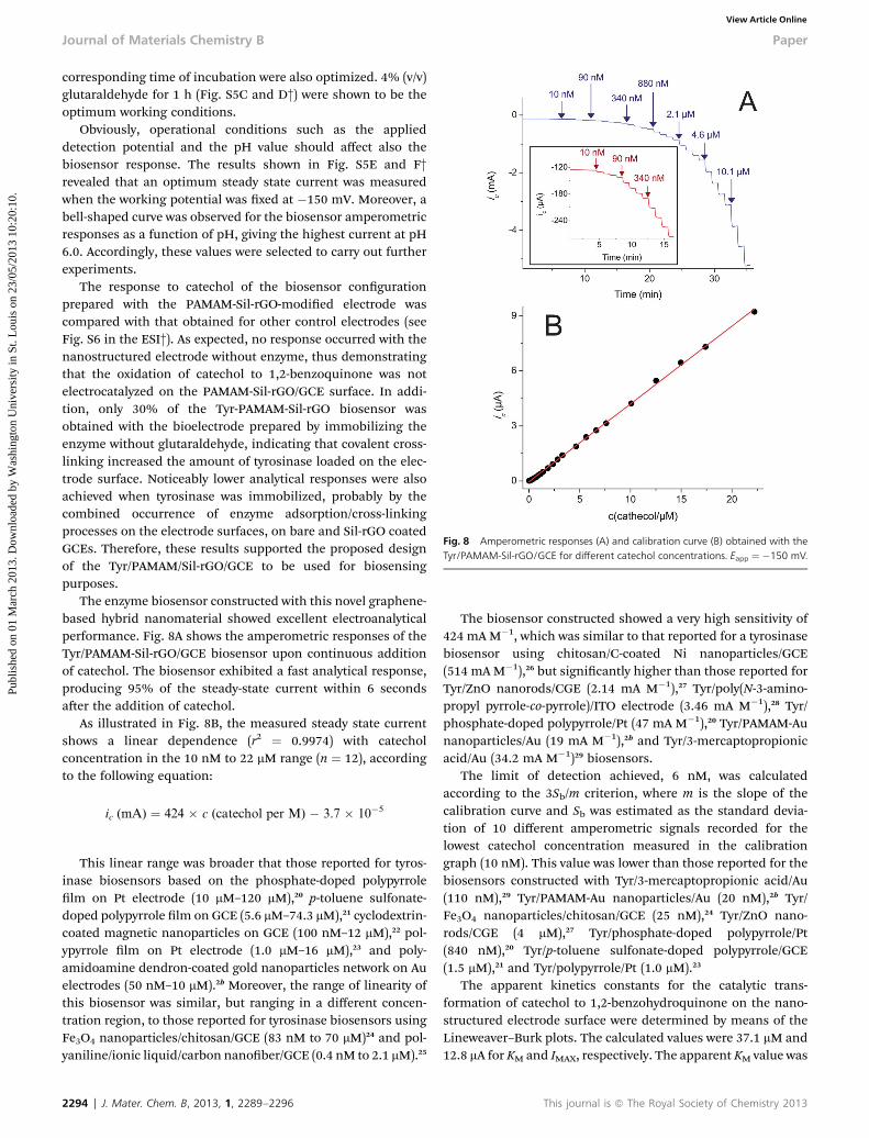
Fig. 8 Amperometric responses (A) and calibration curve (B) obtained with theTyr/PAMAM-Sil-rGO/GCE for different catechol concentrations. Eapp ¼ �150 mV.
Journal of Materials Chemistry B Paper
Publ
ishe
d on
01
Mar
ch 2
013.
Dow
nloa
ded
by W
ashi
ngto
n U
nive
rsity
in S
t. L
ouis
on
23/0
5/20
13 1
0:20
:10.
View Article Online
corresponding time of incubation were also optimized. 4% (v/v)glutaraldehyde for 1 h (Fig. S5C and D†) were shown to be theoptimum working conditions.
Obviously, operational conditions such as the applieddetection potential and the pH value should affect also thebiosensor response. The results shown in Fig. S5E and F†revealed that an optimum steady state current was measuredwhen the working potential was xed at �150 mV. Moreover, abell-shaped curve was observed for the biosensor amperometricresponses as a function of pH, giving the highest current at pH6.0. Accordingly, these values were selected to carry out furtherexperiments.
The response to catechol of the biosensor congurationprepared with the PAMAM-Sil-rGO-modied electrode wascompared with that obtained for other control electrodes (seeFig. S6 in the ESI†). As expected, no response occurred with thenanostructured electrode without enzyme, thus demonstratingthat the oxidation of catechol to 1,2-benzoquinone was notelectrocatalyzed on the PAMAM-Sil-rGO/GCE surface. In addi-tion, only 30% of the Tyr-PAMAM-Sil-rGO biosensor wasobtained with the bioelectrode prepared by immobilizing theenzyme without glutaraldehyde, indicating that covalent cross-linking increased the amount of tyrosinase loaded on the elec-trode surface. Noticeably lower analytical responses were alsoachieved when tyrosinase was immobilized, probably by thecombined occurrence of enzyme adsorption/cross-linkingprocesses on the electrode surfaces, on bare and Sil-rGO coatedGCEs. Therefore, these results supported the proposed designof the Tyr/PAMAM/Sil-rGO/GCE to be used for biosensingpurposes.
The enzyme biosensor constructed with this novel graphene-based hybrid nanomaterial showed excellent electroanalyticalperformance. Fig. 8A shows the amperometric responses of theTyr/PAMAM-Sil-rGO/GCE biosensor upon continuous additionof catechol. The biosensor exhibited a fast analytical response,producing 95% of the steady-state current within 6 secondsaer the addition of catechol.
As illustrated in Fig. 8B, the measured steady state currentshows a linear dependence (r2 ¼ 0.9974) with catecholconcentration in the 10 nM to 22 mM range (n ¼ 12), accordingto the following equation:
ic (mA) ¼ 424 � c (catechol per M) � 3.7 � 10�5
This linear range was broader that those reported for tyros-inase biosensors based on the phosphate-doped polypyrrolelm on Pt electrode (10 mM–120 mM),20 p-toluene sulfonate-doped polypyrrole lm on GCE (5.6 mM–74.3 mM),21 cyclodextrin-coated magnetic nanoparticles on GCE (100 nM–12 mM),22 pol-ypyrrole lm on Pt electrode (1.0 mM–16 mM),23 and poly-amidoamine dendron-coated gold nanoparticles network on Auelectrodes (50 nM–10 mM).2b Moreover, the range of linearity ofthis biosensor was similar, but ranging in a different concen-tration region, to those reported for tyrosinase biosensors usingFe3O4 nanoparticles/chitosan/GCE (83 nM to 70 mM)24 and pol-yaniline/ionic liquid/carbon nanober/GCE (0.4 nM to 2.1 mM).25
2294 | J. Mater. Chem. B, 2013, 1, 2289–2296
The biosensor constructed showed a very high sensitivity of424 mA M�1, which was similar to that reported for a tyrosinasebiosensor using chitosan/C-coated Ni nanoparticles/GCE(514 mA M�1),26 but signicantly higher than those reported forTyr/ZnO nanorods/CGE (2.14 mA M�1),27 Tyr/poly(N-3-amino-propyl pyrrole-co-pyrrole)/ITO electrode (3.46 mA M�1),28 Tyr/phosphate-doped polypyrrole/Pt (47 mA M�1),20 Tyr/PAMAM-Aunanoparticles/Au (19 mA M�1),2b and Tyr/3-mercaptopropionicacid/Au (34.2 mA M�1)29 biosensors.
The limit of detection achieved, 6 nM, was calculatedaccording to the 3Sb/m criterion, where m is the slope of thecalibration curve and Sb was estimated as the standard devia-tion of 10 different amperometric signals recorded for thelowest catechol concentration measured in the calibrationgraph (10 nM). This value was lower than those reported for thebiosensors constructed with Tyr/3-mercaptopropionic acid/Au(110 nM),29 Tyr/PAMAM-Au nanoparticles/Au (20 nM),2b Tyr/Fe3O4 nanoparticles/chitosan/GCE (25 nM),24 Tyr/ZnO nano-rods/CGE (4 mM),27 Tyr/phosphate-doped polypyrrole/Pt(840 nM),20 Tyr/p-toluene sulfonate-doped polypyrrole/GCE(1.5 mM),21 and Tyr/polypyrrole/Pt (1.0 mM).23
The apparent kinetics constants for the catalytic trans-formation of catechol to 1,2-benzohydroquinone on the nano-structured electrode surface were determined by means of theLineweaver–Burk plots. The calculated values were 37.1 mM and12.8 mA for KM and IMAX, respectively. The apparent KM value was
This journal is ª The Royal Society of Chemistry 2013
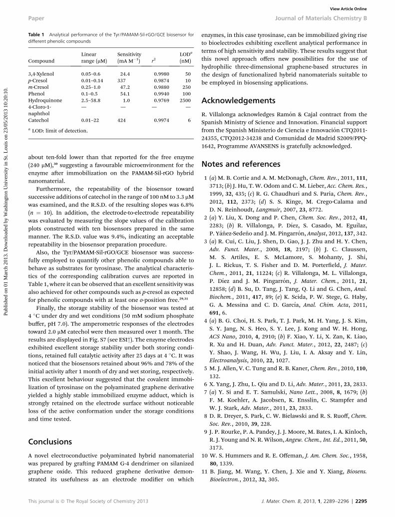
Table 1 Analytical performance of the Tyr/PAMAM-Sil-rGO/GCE biosensor fordifferent phenolic compounds
CompoundLinearrange (mM)
Sensitivity(mA M�1) r2
LODa
(nM)
3,4-Xylenol 0.05–0.6 24.4 0.9980 50p-Cresol 0.01–0.14 337 0.9874 10m-Cresol 0.25–1.0 47.2 0.9880 250Phenol 0.1–0.5 54.1 0.9940 100Hydroquinone 2.5–58.8 1.0 0.9769 25004-Cloro-1-naphthol
— — — —
Catechol 0.01–22 424 0.9974 6
a LOD: limit of detection.
Paper Journal of Materials Chemistry B
Publ
ishe
d on
01
Mar
ch 2
013.
Dow
nloa
ded
by W
ashi
ngto
n U
nive
rsity
in S
t. L
ouis
on
23/0
5/20
13 1
0:20
:10.
View Article Online
about ten-fold lower than that reported for the free enzyme(240 mM),30 suggesting a favourable microenvironment for theenzyme aer immobilization on the PAMAM-Sil-rGO hybridnanomaterial.
Furthermore, the repeatability of the biosensor towardsuccessive additions of catechol in the range of 100 nM to 3.3 mMwas examined, and the R.S.D. of the resulting slopes was 6.8%(n ¼ 10). In addition, the electrode-to-electrode repeatabilitywas evaluated by measuring the slope values of the calibrationplots constructed with ten biosensors prepared in the samemanner. The R.S.D. value was 9.4%, indicating an acceptablerepeatability in the biosensor preparation procedure.
Also, the Tyr/PAMAM-Sil-rGO/GCE biosensor was success-fully employed to quantify other phenolic compounds able tobehave as substrates for tyrosinase. The analytical characteris-tics of the corresponding calibration curves are reported inTable 1, where it can be observed that an excellent sensitivity wasalso achieved for other compounds such as p-cresol as expectedfor phenolic compounds with at least one o-position free.29,31
Finally, the storage stability of the biosensor was tested at4 �C under dry and wet conditions (50 mM sodium phosphatebuffer, pH 7.0). The amperometric responses of the electrodestoward 2.0 mM catechol were then measured over 1 month. Theresults are displayed in Fig. S7 (see ESI†). The enzyme electrodesexhibited excellent storage stability under both storing condi-tions, retained full catalytic activity aer 25 days at 4 �C. It wasnoticed that the biosensors retained about 96% and 78% of theinitial activity aer 1 month of dry and wet storing, respectively.This excellent behaviour suggested that the covalent immobi-lization of tyrosinase on the polyaminated graphene derivativeyielded a highly stable immobilized enzyme adduct, which isstrongly retained on the electrode surface without noticeableloss of the active conformation under the storage conditionsand time tested.
Conclusions
A novel electroconductive polyaminated hybrid nanomaterialwas prepared by graing PAMAM G-4 dendrimer on silanizedgraphene oxide. This reduced graphene derivative demon-strated its usefulness as an electrode modier on which
This journal is ª The Royal Society of Chemistry 2013
enzymes, in this case tyrosinase, can be immobilized giving riseto bioelectrodes exhibiting excellent analytical performance interms of high sensitivity and stability. These results suggest thatthis novel approach offers new possibilities for the use ofhydrophilic three-dimensional graphene-based structures inthe design of functionalized hybrid nanomaterials suitable tobe employed in biosensing applications.
Acknowledgements
R. Villalonga acknowledges Ramon & Cajal contract from theSpanish Ministry of Science and Innovation. Financial supportfrom the Spanish Ministerio de Ciencia e Innovacion CTQ2011-24355, CTQ2012-34238 and Comunidad de Madrid S2009/PPQ-1642, Programme AVANSENS is gratefully acknowledged.
Notes and references
1 (a) M. B. Cortie and A. M. McDonagh, Chem. Rev., 2011, 111,3713; (b) J. Hu, T. W. Odom and C. M. Lieber, Acc. Chem. Res.,1999, 32, 435; (c) R. G. Chaudhuri and S. Paria, Chem. Rev.,2012, 112, 2373; (d) S. S. Kinge, M. Crego-Calama andD. N. Reinhoudt, Langmuir, 2007, 23, 8772.
2 (a) Y. Liu, X. Dong and P. Chen, Chem. Soc. Rev., 2012, 41,2283; (b) R. Villalonga, P. Dıez, S. Casado, M. Eguılaz,P. Ya~nez-Sede~no and J. M. Pingarron, Analyst, 2012, 137, 342.
3 (a) R. Cui, C. Liu, J. Shen, D. Gao, J. J. Zhu and H. Y. Chen,Adv. Funct. Mater., 2008, 18, 2197; (b) J. C. Claussen,M. S. Artiles, E. S. McLamore, S. Mohanty, J. Shi,J. L. Rickus, T. S. Fisher and D. M. Portereld, J. Mater.Chem., 2011, 21, 11224; (c) R. Villalonga, M. L. Villalonga,P. Dıez and J. M. Pingarron, J. Mater. Chem., 2011, 21,12858; (d) B. Su, D. Tang, J. Tang, Q. Li and G. Chen, Anal.Biochem., 2011, 417, 89; (e) K. Scida, P. W. Stege, G. Haby,G. A. Messina and C. D. Garcıa, Anal. Chim. Acta, 2011,691, 6.
4 (a) B. G. Choi, H. S. Park, T. J. Park, M. H. Yang, J. S. Kim,S. Y. Jang, N. S. Heo, S. Y. Lee, J. Kong and W. H. Hong,ACS Nano, 2010, 4, 2910; (b) F. Xiao, Y. Li, X. Zan, K. Liao,R. Xu and H. Duan, Adv. Funct. Mater., 2012, 22, 2487; (c)Y. Shao, J. Wang, H. Wu, J. Liu, I. A. Aksay and Y. Lin,Electroanalysis, 2010, 22, 1027.
5 M. J. Allen, V. C. Tung and R. B. Kaner, Chem. Rev., 2010, 110,132.
6 X. Yang, J. Zhu, L. Qiu and D. Li, Adv. Mater., 2011, 23, 2833.7 (a) Y. Si and E. T. Samulski, Nano Lett., 2008, 8, 1679; (b)F. M. Koehler, A. Jacobsen, K. Ensslin, C. Stampfer andW. J. Stark, Adv. Mater., 2011, 23, 2833.
8 D. R. Dreyer, S. Park, C. W. Bielawski and R. S. Ruoff, Chem.Soc. Rev., 2010, 39, 228.
9 J. P. Rourke, P. A. Pandey, J. J. Moore, M. Bates, I. A. Kinloch,R. J. Young and N. R. Wilson, Angew. Chem., Int. Ed., 2011, 50,3173.
10 W. S. Hummers and R. E. Offeman, J. Am. Chem. Soc., 1958,80, 1339.
11 B. Jiang, M. Wang, Y. Chen, J. Xie and Y. Xiang, Biosens.Bioelectron., 2012, 32, 305.
J. Mater. Chem. B, 2013, 1, 2289–2296 | 2295
Journal of Materials Chemistry B Paper
Publ
ishe
d on
01
Mar
ch 2
013.
Dow
nloa
ded
by W
ashi
ngto
n U
nive
rsity
in S
t. L
ouis
on
23/0
5/20
13 1
0:20
:10.
View Article Online
12 B. Scheibe, E. Borowiak-Palen and R. J. Kalenczuk, J. AlloysCompd., 2010, 500, 117.
13 F. Vogtle, G. Richardt and N. Werner, Dendrimer chemistry:concepts, synthesis, properties, applications, Wiley-VCH,Weinheim, 2009.
14 S. Park, J. An, I. Jung, R. D. Piner, S. J. An, X. Li,A. Velamakanni and R. S. Ruoff, Nano Lett., 2009, 9, 1593.
15 J. F. Shen, Y. Z. Hu, M. Shi, X. Lu, C. Qin, C. Li and M. G. Ye,Chem. Mater., 2009, 21, 3514.
16 S. Stankovich, R. D. Piner, X. Chen, N. Wu, S. T. Nguyen andR. S. Ruoff, J. Mater. Chem., 2005, 16, 155.
17 G. Lu, S. Mao, S. Park, R. S. Ruoff and J. Chen, Nano Res.,2009, 2, 192.
18 J. E. Kim, T. H. Han, S. H. Lee, J. Y. Kim, C. W. Ahn, J. M. Yunand S. O. Kim, Angew. Chem., Int. Ed., 2011, 50, 3043.
19 D. Tang, R. Yuan and Y. Chai, J. Phys. Chem. B, 2006, 110,11640; C. C. Wu, C. H. Lin and W. S. Wang, Talanta, 2009,79, 62.
20 C. Apetrei, M. L. Rodrıguez-Mendez and J. A. De Saja,Electrochim. Acta, 2011, 56, 8919.
2296 | J. Mater. Chem. B, 2013, 1, 2289–2296
21 A. Rajesh, W. Takashima and K. Kaneto, React. Funct. Polym.,2004, 59, 163.
22 P. Dıez, R. Villalonga, M. L. Villalonga and J. M. Pingarron,J. Colloid Interface Sci., 2012, 386, 181.
23 Q. Ameer and S. B. Adeloju, Sens. Actuators, B, 2009, 140, 5.24 S. Wang, Y. Tan, D. Zhao and G. Liu, Biosens. Bioelectron.,
2008, 23, 1781.25 J. Zhang, J. Lei, Y. Liu, J. Zhao and H. Ju, Biosens. Bioelectron.,
2009, 24, 1858.26 L. Yang, H. Xiong, X. Zhang and S.Wang, Bioelectrochemistry,
2012, 84, 44.27 L. Chen, B. Gu, G. Zhu, Y. Wu, S. Liu and C. Xu, J. Electroanal.
Chem., 2008, 617, 7.28 A. Rajesh, W. Takashima and K. Kaneto, Sens. Actuators, B,
2004, 102, 271.29 S. Campuzano, B. Serra, M. Pedrero, F. J. M. Villena and
J. M. Pingarron, Anal. Chim. Acta, 2003, 494, 187.30 J. L. Smith and R. C. Krueger, J. Biol. Chem., 1962, 237, 1121.31 J. C. Espın, P. A. Garcıa-Ruiz, J. Tudela and F. Garcıa-
Canovas, Biochem. J., 1998, 331, 547.
This journal is ª The Royal Society of Chemistry 2013