cross-linking of the proteins in the outer membrane of escherichia coli
TRANSCRIPT

245
Bioehimica et Biophysica Acta, 466 (1977) 245--256 © Elsevier/North-Holland Biomedical Press
BBA 77670
CROSS-LINKING OF THE PROTEINS IN THE OUTER MEMBRANE OF E S C H E R I C H I A C O L I
REINHART A.F. REITHMEIER and PHILIP D. BRAGG
Department of Biochemistry, University of British Columbia, Vancouver, Britisi~ Columbia, V6T 1W5 (Canada)
(Received September 23rd, 1976)
Summary
1. The organization of the proteins in the outer membrane of Escherichia coli was examined by the use of cross-linking agents and two-dimensional sodium dodecyl sulfate-polyacrylamide gel electrophoresis. Treatment of pro- tein A-peptidoglycan complexes with dithiobis(succinimidyl propionate) or glutaraldehyde produced the dimer, trimer, and higher oligomers of protein A. Both forms of this protein, proteins A~ and A2, produced similar cross-linking products. No cross-linking of protein A to the peptidoglycan was detected.
2. The proteins of the isolated outer membrane varied in their ease of cross- linking. The heat-modifiable protein, protein B, was readily cross-linked to give high molecular weight oligomers, while protein A formed mainly the dimer and trimer under the same conditions. The pronase resistant fragment, protein Bp, derived from protein B was not readily cross-linked. No linkage of protein A to protein B was detected.
3. Cross-linking of ce!l wall preparations, consisting of the outer membrane and peptidoglycan, showed that protein B and the free form of the lipoprotein, protein F, could be linked to the peptidoglycan. A dimer of protein F, and protein F linked to protein B, were detected.
4. These results suggest that specific protein-protein interactions occur in the outer membrane.
Introduction
The outer membrane of Eseheriehia coli contains only a few proteins when compared to the cytoplasmic membrane [1]. The properties and arrangement of these proteins in the outer membrane have been studied in this and other laboratories [1--20[. Three proteins are of particular interest because of their presence in large amounts. Protein B [2], called II* by Henning [6,8--11], peak 7 by Inouye [1,14,15], and protein 3 by Schnaitman [21--23], is heat

246
modifiable and changes its shape by unfolding on heating. It is partially exposed at the surface of the outer membrane and may serve as a phage or bacteriocin receptor [2 ,6--9,15,25,26] . Schnaitman [22] has shown that some strains of E. coli may contain two species of this protein, 3a and 3b.
Protein A is tightly associated with the peptidoglycan and has been designated the matrix protein by Rosenbusch [16] , protein I by Henning [6--13] , peak 4 by Inouye [1 ,14,15] , and protein I by Schnaitman [20--24] . This protein can exist in multiple forms that vary with strain and culture conditions [12,13,23,24] . Protein A may be a receptor for bacteriophage [23--27] and so probably spans the outer membrane. This is supported by the finding that this protein can form channels through phospholipid-lipopolysaccharide vesicles [281.
The most abundant protein in the outer membrane of E. coli is a lipoprotein, extensively characterized by Braun [5] and Inouye [1] . One-third of this pro- tein is covalently linked to the peptidoglycan and extends outward into the outer membrane. The remainder of the protein exists in a free from, un- a t tached to the peptidoglycan. This form of the protein is equivalent to our protein F [2] . Inouye has suggested that the two forms of the lipoprotein interact to form channels through the outer membrane [ 1 ].
Cross-linking reagents have been useful in the study of complex structures such as mult isubunit enzymes [29- -31] , r ibosomes [32,33] and membranes [34--36] . Haller et al. [10,11] have used the CuSO4-o-phenanthroline reagent and dimethyl imidoesters to investigate protein interactions in the envelope of E. coli. They found that extensive protein-protein interactions occurred in the outer membrane since t reatment of intact cells or isolated ghosts with dimethyl imidoesters resulted in the cross-linking of all the major proteins to yield structures retaining the cell shape which were resistant to dissolution by 1% sodium dodecyl sulfate at 100°C until after the cross-links had been cleaved [6].
The studies of Haller et al. [10,11] did not provide information on the specific protein-protein interactions of individual outer membrane proteins. Therefore, we have reinvestigated the cross-linking of the outer membranes using dithiobis(succinimidyl propionate), a cross-linking reagent that is readily cleaved by reduction of its disulfide bridge. By combining the use of this reagent with a sensitive two<limensional sodium dodecyl sulfate-polyacryl- amide gel system we have been able to identify interactions between several of the proteins of the outer membrane.
Materials and Methods
Reagents. Dithiobis(succinimidyl propionate) was purchased from Pierce Chemical Co. Histological grade glutaraldehyde as a 24% aqueous solution was obtained from Matheson, Coleman and Bell, Manufacturing Chemists. Acryl- amide and bis(N,N'-methylene-bisacrylamide) were supplied by BioRad Laboratories. Sodium dodecyl sulfate ("specially pure") was a product of B.D.H. Chemicals Ltd.
Growth conditions and preparation o f membranes. E. coli, Strain 482 of the culture collection of the National Research Council of Canada, was used in

247
most of these studies. It was grown with vigorous aeration at 37vC to the mid- exponential phase of growth on a minimal salts medium containing 0.4% (w/v) glucose or 0.5% casein amino acids.
Cells were broken in a French pressure cell (Aminco) at 20 000 lb/inch 2 and the cell envelope and cell wall (Triton-treated envelope) fractions prepared as described previously [2] . Protein A-peptidoglycan complexes were isolated by extracting the cell envelope with 2% (w/v) sodium dodecyl sulfate at 60°C for 1 h [16] . All of these membrane preparations were recovered by centrifugation at 120 000 X g for 1 h and washed twice with the appropriate buffer.
The outer membrane, released from spheroplasts, was prepared according to Mizushima and Yamada [17] . Contaminating lysozyme was removed by washing the membrane four times with 0.2 M triethanolamine buffer, pH 8.5, and recovering it by centrifugation at 120 000 X g for 1 h.
Cross-linking. Membrane samples (0.1 ml} at about 1 mg protein per ml of 0.2 M triethanolamine buffer, pH 8.5, were treated at 22°C with various amounts of dithiobis(succinimidyl propionate) at 20 mg/ml in dimethyl- sulfoxide. After 30 s excess 1 M Tris • HC1 buffer, pH 8.5, was added to destroy unreacted cross-linker. The volume added was equal to that of the cross-linker solution used. Finally, 2--9 volumes of a solution containing 1% sodium dodecyl sulfate, 10% (v/v) glycerol and 0.005% bromophenol blue in 0.0625 M Tris • HC1 buffer, pH 6.8 ("electrophoresis sample buffer") was added and the solution heated at 100°C for 3 min to solubilize the proteins.
Membrane samples in 0.2 M triethanolamine buffer, pH 8.5, were also cross- linked at 22°C by 0.05--0.1% glutaraldehyde.
Sodium dodecyl sulfate-polyacrylamide gel electrophoresis. Slab gels (0.75 mm thick) were prepared according to Ames [37] using the discontinuous buffer system of Laemmli [38] . Gels were run in a BioRad model 220 slab gel apparatus at a constant current of 30 mA/slab for about 1.5 h. Cross-linked products were analyzed by two-dimensional sodium dodecyl sulfate-polyacryl- amide gel electrophoresis. The cross-linked proteins were first resolved on a 7.5% polyacrylamide gel slab, 0.75 mm thick. A 3 mm wide strip from this gel was placed at the top of a previously formed second dimension gel and embedded in warm (45°C) 1% agarose in the stacking gel buffer containing 2% 2-mercaptoethanol [37 ,38] . The agarose was allowed to cool for 5 min before the run was started. The time taken from stopping the first dimension run to this time did not exceed 15 min. The second dimension gel, 1.5 mm thick, was usually composed of a 2 cm stacking gel of 4% polyacrylamide overlaying a 12% polyacrylamide separating gel. The second dimension gel was prepared on the preceding day with the top of the stacking gel kept under a layer of the stacking gel buffer containing 10% 2-mercaptoethanol. With the second dimen- sion gel a constant current of 40 mA/slab was applied for about 2.5 h. The gels were stained in a solution containing 0.05% Coomassie Blue, 25% isopropanol, and 10% acetic acid for 1 or 2 h with 0.75- and 1.5-mm thick slabs, respectively. The gels were destained by immersion in several changes of 10% acetic acid.
Results
Cross-linking of protein A-peptidoglycan complexes Protein At- and protein A~-peptidoglycan complexes prepared from glucose-

2 4 8
and casein amino acid-grown cells, respectively, were treated with increasing amounts of dithiobis(succinimidyl propionate). As shown in Fig. 1, both proteins A1 and A2 cross-linked to form dimer, trimer, and higher oligomers. The trimer was always produced in excess over the tetramer which suggests that protein A might be organized as trimers in the membrane. Although proteins A~ and A2 were not well resolved by the gel system the content of A1 and A2 in the two preparations was confirmed by sodium dodecyl sulfate-gel electro- phoresis at an alkaline pH [ 2 ].
Cross-linking of protein A to oligomers in protein A-peptidoglycan com- plexes was also observed with glutaraldehyde.
Since the protein A-peptidoglycan complexes also contain lipoprotein (protein F) covalently bound to the peptidoglycan [5] , it was possible that protein A could be cross-linked to the peptidoglycan through this protein. This possibility was examined as follows. The protein A-peptidoglycan complex was reacted with dithiobis(succinimidyl propionate) and then extracted with a buffer of 1% sodium dodecyl sulfate, 10% glycerol, and 62.5 mM Tris • HC1, pH 6.8, at 100°C for 3 min. Protein A and its cross-linked oligomers were extracted. The peptidoglycan, and any proteins linked to it, was reextracted as before but in the presence of 1% 2-mercaptoethanol to cleave the cross-linking agent. No further protein A was released indicating that it had not been cross- linked to the bound lipoprotein or directly to the peptidoglycan.
Cross-linking of outer membrane with dithiobis(succinimidyl propionate) Cell wall, composed of outer membrane and peptidoglycan, and isolated
Fig. I . Cross-l inking of prote in A - p e p t i d o g l y c a n c o m p l e x e s w i t h d i t h i o b i s ( s u c c i n i m i d y l prop iona t e ) , Samples (100 /~1; 0.1 m g p ro t e in ) of prote in A - P e p t i d o g l y c a n c o m p l e x e s prepared f r o m g lucose-grown (samples 2 - -5) and casein a m i n o ac id-grown cel ls ( samples 6 - -9) w e r e treated w i t h 0 pg ( samples 2 and 6), 40 ~g (samples 3 and 7), 200 ~zg ( samples 4 and 8), and 600 ~zg ( samples 5 and 9) of d i th iob i s ( suce in imidy l p r o p i o n a t e ) . T he samples w e r e so lubi l i zed w i t h nine vo lumes of e l ec t rophores i s sample b u f f e r and then reso lved on 9% p o l y a c r y l a m i d e gel slabs. Samples 1 and 10 con ta in as m o l e c u l a r we igh t m a r k e r s bovine s e rum a l bum i n (66 000) , o v a l b u m i n (46 000) , and l y s o z y m e (14 400) .

249
outer membrane preparations were treated with different levels of cross-linker (Fig. 2). Proteins A and D2 (molecular weight 18 000) were less readily cross- linked than proteins B and F, the free form of the lipoprotein. The ability of the proteins to be cross-linked is determined by the degree of exposure of suitably disposed amino groups. Thus, protein B, which was most readily cross- linked, was also most reactive with the amino group reagent fluorescamine (un- published results). In contrast, protein A, which was less readily cross-linked in outer membrane preparations, reacted more slowly with fluorescamine.
The effect on cross-linking of pretreating the outer membrane with pronase was studied. Pronase cleaves protein B to a fragment, protein Bp, which remains embedded in the membrane, but does not digest protein A [2] . In the pronase-treated membrane protein A was cross-linked to the dimer and trimer. In contrast to intact protein B, protein Bp was not readily cross-linked (Fig. 3). Thus, either the functional groups responsible for the cross-linking of protein B must be situated on the portion of this protein removed by pronase or proteolyt ic digestion had resulted in alterations in the conformation of the pro- tein such that these groups became inaccessible to the cross-linker.
The formation of the dimer and trimer of protein A occurred when relatively high levels of dithiobis(succinimidyl propionate) (2 mg/ml protein) were reacted with the outer membrane. This was confirmed by two-dimensional sodium dodecyl sulfate-polyacrylamide gel electrophoresis in which the cross- linked products separated in the first dimension were cleaved by 2-mercapto- ethanol prior to entering the separating gel of the second dimension. The
1 2: 3 4 5 6 7 8 9 1 0
Fig. 2. Cross-l inking of cell wall and ou t e r m e m b r a n e wi th d i th iob i s ( succ in imidy l p r o p i o n a t e ) . Samples (100 #1; 0.1 m g p ro t e in ) of cell wall ( samples 2- -5) and o u t e r m e m b r a n e (samples 6 - -9) p r e p a r e d f r o m glucose-grown cells were t r e a t e d wi th 0 #g ( samples 2 and 6), 20 ~zg (samples 3 and 7), 40 ~g ( samples 4 a n d 8) and 60 ~g ( samples 5 and 9) of d i th iob i s ( succ in imidy l p r o p i o n a t e ) . Th e samples were solubi l ized wi th t w o v o l u m e s of e l ec t rophores i s sample b u f f e r a n d resolved on 7.5% p o l y a c r y l a m i d e gel slabs. Samples 1 and 10 c o n t a i n e d as m o l e c u l a r weigh t m a r k e r s bov ine se rum a lb u min (66 000) , o v a l b u m i n (46 000) , and l y s o z y m e (14 400) .

250
Fig. 3. Cross-l inking of p rona s e - t r e a t e d ou t e r m e m b r a n e wi th d i th iob i s ( succ in imidy l p r o p i o n a t e ) . Ou t e r m e m b r a n e was t r ea t ed wi th p ronase at an e n z y m e : p ro t e in ra t io of 1 : 25 for 2 h a t 37°C. The o u t e r m e m b r a n e was r ecove red by cen t r i fuga t i on at 120 000 Xg for 1 h and r e su sp en d ed to a b o u t 1 m g of p r o t e i n / m l in 0.2 M t r i e t h a n o l a m l n e bu f fe r , pH 8.5. Samples (100 pl~ 0.1 m g p ro t e in ) of d iges ted m e m b r a n e were t r e a t ed w i th 0 #g ( sample 1), 200 ttg ( sample 2), and 6 0 0 ttg ( sample 3) of di thiobis- ( succ in imidy l p r o p i o n a t e ) . The samples were solubi l ized wi th nine v o lu mes of e lec t rophores i s sample bu f f e r and then resolved on 9% p o l y a c r y l a m i d e gel slabs. Sample 4 co n t a in ed as m o l e c u l a r we igh t m a r k e r s bov ine se rum a l b u m i n (66 000) , o v a l b u m i n (46 000) , and l y s o z y m e (14 400) .
cleaved products could then be identified from their characteristic rate of migration (Fig. 4). The formation of the dimer and trimer of protein A by cross-linking with both the outer membrane and the protein A-peptidoglycan complex suggests that protein A is arranged in the same way in both prepara- tions. At the higher level of cross-linker, protein B gave a high molecular weight complex that penetrated the 4% stacking gel but did not enter the 7.5% poly- acrylamide gel of the first dimension. Reduct ion of this complex yielded predominantly protein B although a minor amount of protein A was some- times present.
Interaction of proteins B and F with peptidoglycan When the cell wall (outer membrane-peptidoglycan) preparation was treated
with cross-linker under identical conditions to those used in the previous

251
Fig. 4. Cleavage of cross- l inked p r o d u c t s f r o m ou t e r m e m b r a n e . A sample (100 pl; 0.1 m g p ro t e in ) of ou t e r m e m b r a n e was t r ea t ed in the 200 #g of d i th iob i s ( succ in imidy l p r o p i o n a t e ) t hen solubi l ized wi th two v o l u m e s of e l ec t rophores i s sample bu f fe r , and resolved on a first d imen s io n 7.5% p o l y a c r y l a m i d e gel. The cross- l inked p r o d u c t s were c leaved in the second d imen s io n by 2 - m e r c a p t o e t b a n o l and resolved on a 12% p o l y a c r y l a m i d e gel. The pos i t ion of m o n o m e r (A), d i m e r (A)2, an d t r i m e r (A)3, of p ro t e in A, and the high m o l e c u l a r we igh t o l igomer of p ro t e in B, (B)x, are ind ica ted . A s ta ined first d i m e n s i o n gel is p laced along the top of the gel slab.
experiment with the isolated outer membrane, a similar two-dimensional gel pattern was obtained. However, it was consistently observed that a much larger cross-linked complex of protein B was formed which could not enter the 4% stacking gel in the first dimension. In order to determine if this was due to the cross-linking of protein B to the peptidoglycan, either directly or through another protein, the following experiment was performed. The cell wall preparation was cross-linked and then extracted with a buffer containing 1% sodium dodecyl sulfate and 10% glycerol in 62.5 mM Tris • HC1, pH 6.8, at 100°C for 3 min. The insoluble peptidoglycan and attached proteins were reex- tracted with this buffer but containing 1% 2-mercaptoethanol. A control experiment was carried out in which the cross-linker was omitted. As a further control, the isolated outer membrane was taken through the above procedure. The results of this experiment are shown in Fig. 5. In the absence of cross- linker the first extract of the cell wall contained all of the outer membrane protein (sample 1). The first extraxt of the cross-linked cell wall showed the presence of the dimer and trimer of protein A (sample 2) which could be cleaved by the addition of 2-mercaptoethanol {sample 3). The second extract of the non-cross-linked sample contained virtually no proteins (sample 4). How- ever, the second extraction of the cross-linked cell wall with the 2-mercapto- ethanol-containing buffer released proteins B and F from the peptidoglycan
2 5 2
I l l
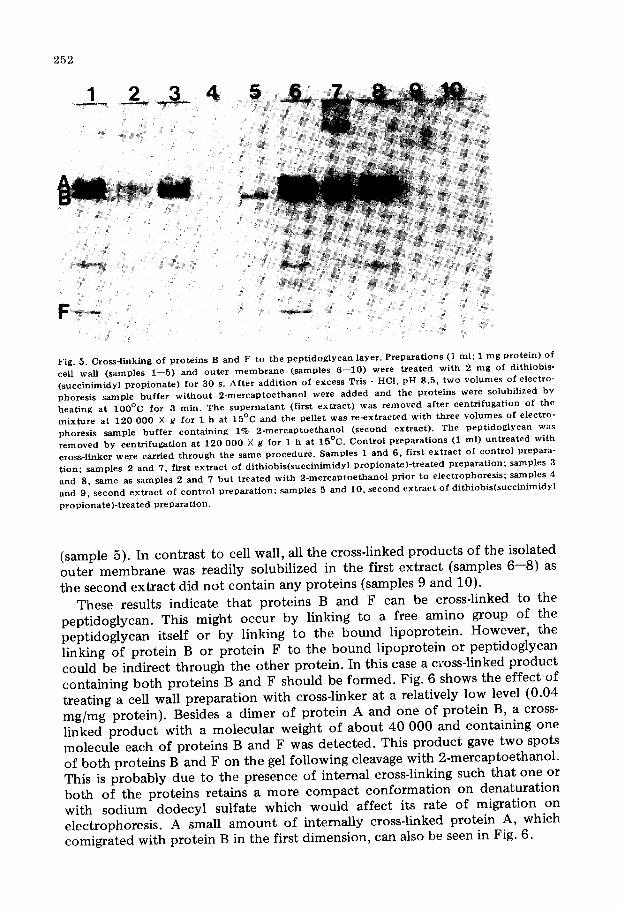
Fig. 5. Cross-l inking of p ro te ins B and F to the p e p t i d o g l y c a n layer . P repa ra t ions (1 ml ; 1 m g p ro te in ) of cell wall ( samples 1- -5) and ou t e r m e m b r a n e ( samples 6 - -10 ) were t r ea t ed w i th 2 m g of di thiobis- ( succ in imidy l p r o p i o n a t e ) for 30 s. Af t e r add i t ion of excess Tris - HCI, pH 8.5, two v o lu mes of e lec t ro- phores is sample bu f f e r w i t h o u t 2 - m e r c a p t o e t h a n o l were a d d e d and the p ro te ins were solubil ized by hea t ing at IO 0°C for 3 rain. The s u p e r n a t a n t (first ex t r ac t ) was r e m o v e d a f t e r cen t r i fuga t ion of the m i x t u r e at 120 000 X g for 1 h at 15°C and the pel let was r e - ex t r ac t ed wi th th ree v o lu mes of e lec t ro- phoresis sample b u f f e r con ta in ing 1% 2 - m e r c a p t o e t h a n o l ( second ex t rac t ) . The p e p t i d o g l y c a n was r e m o v e d by cen t r i fuga t i on a t 120 000 X g for 1 h at 15°C. Con t ro l p r epa ra t i ons (1 ml ) u n t r e a t e d wi th cross- l inker were carr ied t h r o u g h the same p r o c e d u r e . Samples 1 and 6, f irst e x t r a c t of con t ro l p repara- t ion; samples 2 and 7, first e x t r a c t of d i th iob i s ( succ in lmidy l p r o p i o n a t e ) - t r e a t e d p r e p a r a t i o n ; samples 3 and 8, s ame as samples 2 and 7 b u t t r e a t e d wi th 2 - m e r c a p t o e t h a n o l p r io r to e lec t rophores i s ; samples 4 and 9, second e x t r a c t of c on t ro l p r e p a r a t i o n ; samples 5 and 10, second ex t r ac t of d i th iob i s ( succ in imidy l
p r o p i o n a t e ) - t r e a t e d p r epa ra t i on .
(sample 5). In contrast to cell wall, all the cross-linked products of the isolated outer membrane was readily solubilized in the first extract (samples 6--8) as the second extract did not contain any proteins {samples 9 and 10).
These results indicate that proteins B and F can be cross-linked to the peptidoglycan. This might occur by linking to a free amino group of the peptidoglycan itself or by linking to the bound lipoprotein. However, the linking of protein B or protein F to the bound lipoprotein or peptidoglycan could be indirect through the other protein. In this case a cross-linked product containing both proteins B and F should be formed. Fig. 6 shows the effect of treating a cell wall preparation with cross-linker at a relatively low level {0.04 mg/mg protein). Besides a dimer of protein A and one of protein B, a cross- linked product with a molecular weight of about 40 000 and containing one molecule each of proteins B and F was detected. This product gave two spots of both proteins B and F on the gel following cleavage with 2-mercaptoethanol. This is probably due to the presence of internal cross-linking such that one or both of the proteins retains a more compact conformation on denaturation with sodium dodecyl sulfate which would affect its rate of migration on electrophoresis. A small amount of internally cross-linked protein A, which comigrated with protein B in the first dimension, can also be seen in Fig. 6.

2 5 3
Fig . 6. Cleavage o f c ro s s - l i nked p r o d u c t s f r o m cell wal l . A s a m p l e ( 1 0 0 pl ; 0 .1 m g p r o t e i n ) o f cell wal l w a s treated w i t h 4 0 p g o f d i t h i o b i s ( s u c c i n i m i d y l p r o p i o n a t e ) , so lub i l i zed w i t h t w o v o l u m e s o f e l e c t r o p h o r e s i s s a m p l e b u f f e r , a n d r e so lved o n a 7 .5% p o l y a c r y l a m i d e gel. The c ros s - l i nked p r o d u c t s we re c leaved in the s e c o n d d i m e n s i o n w i t h 2 - m e r c a p t o e t h a n o l a n d r e so lved o n a 12% p o l y a c r y l a m i d e gel. T h e p o s i t i o n s o f the c ros s - l i nked p r o d u c t s c o n t a i n i n g b o t h p r o t e i n s B a n d F (B-Fa , m o l e c u l a r w e i g h t , 4 2 2 0 0 ; a n d B-Fb , m o l e c u l a r w e i g h t , 4 0 3 0 0 ) , a n d the d i m e r s o f p r o t e i n A ( (A) 2, m o l e c u l a r w e i g h t , S0 0 0 0 ) a n d p r o t e i n B ((B)2, m o l e c u l a r w e i g h t , 7 0 5 0 0 ) a re i n d i c a t e d . A s t a i n e d f i rs t d i m e n s i o n gel is p l a c e d a l o n g the t o p o f the gel s lab .
The presence of a dimer of protein F was detected when cross-linked material similar to that used in the previous experiment was run on two-dimen- sional gels containing higher concentrations of polyacrylamide (Fig. 7).
These results indicate that at least some of the molecules of protein B and protein F in the outer membrane must be in close proximity to one another, and that there may be groups of molecules of protein F as suggested by Inouye [39] . Our results do not prove that either protein B or F is linked through the other to the peptidoglycan layer but are not inconsistent with this possibility.

2 5 4
Fig. 7. Cleavage of cross- l inked p r o d u c t s f r o m cell wall. A cross- l inked p r e p a r a t i o n of cell wall similax to t h a t desc r ibed in Fig. 6 was s epa ra t ed on a first d i m e n s i o n 12% p o l y a c r y l a m i d e gel to resolve low molec - ular weight cross- l inked p roduc t s . The cross- l inked p ro te ins were c leaved in the second d imen s io n wi th 2- m e r c a p t o e t h a n o l and resolved in a 15% p o l y a c r y l a m i d e gel. The pos i t ion of the cross- l inked d i m e r of pro- te in F, ( (F)2) is ind ica ted .
Discussion
The results presented in this paper confirm the findings of Henning and co- workers [6,10,40] that extensive protein-protein interactions occur in the outer membrane of E. coli. We have extended these observations to determine the specific interactions between some of the major outer membrane proteins.
Protein A is associated with the peptidoglycan [16] and probably interacts with certain bacteriophages [23--27]. It also can form channels through lipo- polysaccharide-phospholipid vesicles [28]. These properties suggest that protein A spans the outer membrane although we could not detect cross-linking to the peptidoglycan. Protein A can be cross-linked to the dimer and trimer but cross-linking to other proteins was not detected. The variable occurrence of minor amounts of protein A together with the major component , protein B, in the large molecular weight cross-linked material not entering the gel during electrophoresis is probably due to the presence of separate large molecular weight complexes of protein A and of protein B. Protein A was never found associated with protein B in cross-linked complexes small enough to have penetrated the polyacrylamide gel. Thus, protein A may be restricted to patches in the membrane and a channel might be formed by the association of three molecules of this protein. This would be consistent with the morphologi-

255
cal results of Rosenbusch [16] who suggested that 1--3 molecules of protein A form a unit.
Protein B can be readily cross-linked to other molecules of protein B to form large molecular weight products suggesting that many molecules of protein B are in close proximity in the membrane. A dimer, and possibly a trimer, of pro- tein F were formed by cross-linking. Inouye [39] has suggested that molecules of protein F, the free form of the lipoprotein which is covalently attached to the peptidoglycan, exist predominantly in an a-helical conformation and are capable of reacting with other molecules of both bound and free protein F to form a superhelical structure containing six molecules of protein per unit. Our cross-linking results are consistent with this suggestion.
Although most of the interactions which we have found are between mole- cules of the same protein, as also seems to be the case with the erythrocyte membrane [34] , an association of protein B with protein F was detected. These proteins can be cross-linked to the peptidoglycan possibly through the lipoprotein which is covalently bound to the peptidoglycan. The association of proteins B and F with the bound lipoprotein would restrict their mobil i ty in the membrane. The non-covalent interaction of protein A with the peptido- glycan [16] might also have the same effect. Thus, the lateral mobil i ty of the outer membrane proteins is likely to be restricted. This could result in the existence of separate regions of protein in the membrane. The absence of detectable interactions between protein A and protein B would support this hypothesis. However, the absence of cross-linking between proteins A and B is not definitive evidence that they are not in proximity. The lack of suitably disposed amino groups required for cross-linking could account for these results.
Acknowledgements
This work was supported by a grant from the Medical Research Council of Canada and by the award of a M.R.C. Studentship to R.A.F.R.
References
1 I n o u y e , M. ( 1 9 7 5 ) in M e m b r a n e Biogenes i s , M i t o c h o n d r i a , C h l o r o p l a s t s , B a c t e r i a ( T z a g o l o f f , A. , ed . ) , pp . 3 5 1 - - 3 9 1 , P l e n u m Press , N e w Y o r k
2 Bragg, P .D. a n d H o u , C. ( 1 9 7 2 ) B i o c h i m . B i o p h y s . A c t a 2 7 4 , 4 7 8 - - 4 8 8 3 R e i t h m e i e r , R . A . F . a n d Bragg , P .D. ( 1 9 7 4 ) F E B S L e t t . 4 1 , 1 9 5 - - 1 9 8 4 R e i t h m e i e r , R . A . F . a n d Bragg , P .D. ( 1 9 7 6 ) A r c h . B i o c h e m . B i o p h y s . , in t he p r e s s 5 B r a u n , V. ( 1 9 7 5 ) B i o c h i m . B i o p h y s . A c t a 4 1 5 , 3 3 5 - - 3 7 7 6 H e n n i n g , U. , H o h n , B. a n d S o n n t a g , I. ( 1 9 7 3 ) Eur . J . B i o c h e m . 3 9 , 2 7 - - 3 6 7 G a r t e n , W. a n d Hemming, U. ( 1 9 7 4 ) Eur . J . B i o c h e m . 47 , 3 4 3 - - 3 5 2 8 H i n d e n n a c h , I. a n d H e n n i n g , U. ( 1 9 7 5 ) Eur . J . B i o c h e m . 5 9 , 2 0 7 - - 2 1 3 9 G a r t e n , W., H i n d e n n a c h , I. a n d H e n n i n g , U. ( 1 9 7 5 ) Eu r . J . B i o c h e m . 59 , 2 1 5 - - 2 2 1
10 Ha l l e r , I. a n d H e n n i n g , U. ( 1 9 7 4 ) P roc . Na t l . A c a d . Sci. U.S. 71 , 2 0 1 8 - - 2 0 2 1 11 Ha l l e r , I., H o h n , B. a n d H e n n i n g , U. ( 1 9 7 5 ) B i o c h e m i s t r y 1 4 , 4 7 8 - - 4 8 4 12 G a r t e n , W., H i n d e n n a c h , I. a n d H e n n i n g , U. ( 1 9 7 5 ) Eur . J . B i o c h e m . 60 , 3 0 3 - - 3 0 7 13 S c h m i t g e s , C .J . a n d H e n n i n g , U. ( 1 9 7 6 ) Eu r . J . B i o c h e m . 6 3 , 4 7 - - 5 2 14 I n o u y e , M. a n d Yee , M.L. ( 1 9 7 3 ) J . Bac te r io l . 1 1 3 , 3 0 4 - - 3 1 2 15 I n o u y e , M. a n d Yee , M.L. ( 1 9 7 2 ) J . Bac t e r io l . 1 1 2 , 5 8 5 - - 5 9 2 16 R o s e n b u s c h , J .P . ( 1 9 7 4 ) J . Biol . C h e m . 2 4 9 , 8 0 1 9 - - 8 0 2 9 17 M i z u s h i m a , S. a n d Y a m a d a , H. ( 1 9 7 5 ) B i o c h i m . B i o p h y s . A c t a 3 7 5 , 4 4 - - 5 3 18 U e m u r a , S. a n d M i z u s h i m a , S. ( 1 9 7 5 ) B i o e h i m . B i o p h y s . A c t a 4 1 3 , 1 7 3 - - 1 7 6 19 N a k a m u r a , K. a n d M i z u s h i m a , S. ( 1 9 7 5 ) B i o c h i m . B i o p h y s . A c t a 4 1 3 , 3 7 1 - - 3 9 3

256
20 Schnaitman, C.A. (1973) Arch. Biochem. Biophys. 157 ,541- -552 21 Schnaitman, C.A. (1973) Arch. Biochem. Biophys. 157 ,553- -560 22 Schnaitman, C.A. (1974) J. Baeteriol. 118, 442--453 23 Schnaitman, C.A. (1974) J. Bacteriol. 118 ,454 - -464 24 Schnaitman, C.A., Smith, D. and Salas, M.F. (1975) J. Virol. 15, 1121--1130 25 Henning, U. and Haller, I. (1975) FEBS Lett. 55, 161--164 26 Chai, T. and Foulds, J. (1974) J. Mol. Biol. 85 ,465 - -474 27 Hancock, R.E.W. and Reeves, P. (1976) J. Bacteriol. 127, 98--108 28 Nakae, T. (1976) Biochem. Biophys. Res. Commun. 71 ,877 - -884 29 Wold, F. (1967) Methods Enzyrnol. 11 ,617 - -640 30 Davies, G.E. and Stark, G.R. (1970) Proc. Natl. Acad. Sci. U.S. 66 ,651 - -656 31 Bragg, P.D. and Hou, C. (1975) Arch. Biochem. Biophys. 167 ,311- -321 32 Sun, T.T., Traut, R.R. and Kahan, L. (1974) J. Mol. Biol. 87 ,509 - -522 33 Sun, T.T., Bollen, A., Kahan, L., Traut, R.R. (1974) Biochemistry 13, 2334--2340 34 Wang, K. and Richards, F.M. (1974) J. Biol. Chem. 249, 8005--8018 35 Wang, K. and Richards, F.M. (1975) J. Biol. Chem. 250, 6622--6626 36 Steck, T.L. (1972) J. Mol. Biol. 6 6 , 2 9 5 - - 3 0 5 37 Ames, G.F.L. (1974) J. Biol. Chem. 249 ,634 - -644 38 Laemmli, U.K. (1970) Nature 227,680---685 39 Inouye, M. (1974) Proc. Natl. Acad. Sci. U.S. 71, 2396--2400 40 Henning, U., Rehn, K. and Hohn, B. (1973) Proc. Natl. Acad. Sci. U.S. 70, 2033--2036