coronoid impingement syndrome: literature review and ... · case report open access coronoid...
TRANSCRIPT
CASE REPORT Open Access
Coronoid impingement syndrome:literature review and clinical managementPriti Acharya1, Andrew Stewart2 and Farhad B. Naini2*
Abstract
Background: This case report discusses the unusual presentation of limited mouth opening as a result of bilateralcoronoid process hyperplasia.
Case presentation: A 14.5-year-old male patient of white Caucasian ethnicity presented with limited mouth opening,mandibular asymmetry, and dental crowding. Investigations confirmed bilateral coronoid process hyperplasia andmanagement involved bilateral intraoral coronoidectomy surgery under general anaesthesia, followed by muscularrehabilitation. Mouth opening was restored to average maximum opening within 4 months of surgery.
Conclusion: Limited mouth opening is a common presentation to medical and dental professionals. The rare butfeasible diagnosis of coronoid impingement syndrome should not be overlooked.
Keywords: Coronoid impingement syndrome, Coronoid process hyperplasia, Coronoidectomy
BackgroundCoronoid process hyperplasia is defined as ‘an abnor-mal elongation of the coronoid process, formed of his-tologically normal bone’ [1]. This unusual condition isrelatively uncommon but well reported in the literature[1–10]. Coronoid hyperplasia was first reported by vonLangenbeck in 1853 [11]. A review of case notes at theQueen Victoria Hospital, East Grinstead, over a 20-yearperiod revealed 31 recorded cases, 23 of which werebilateral [2].
Clinical presentationThe condition typically presents as painless progressivereduction in mouth opening due to contact interferencebetween the elongated coronoid process and the medialsurface of the zygomatic arch or the temporal aspect ofzygomatic bone [2]. It can occur in unilateral and bilat-eral forms [9]. Facial asymmetry may occur if thehyperplasia is unilateral [11]. The condition is alterna-tively termed ‘coronoid impingement syndrome’ (CIS)[3]. Where pathological elongation of the coronoidprocess results in the formation of a new joint with the
zygomatic process, this is also referred to as Jacob’s dis-ease—named after Oscar Jacob in 1899 [11]. Symptomscan present as young as almost 7 years of age [2] andtypically affect young patients with an average age of25 years [7, 11]. A case of Jacob’s disease has been re-ported in a 39-year-old [10] and 52-year-old woman[11]. Male individuals are more commonly affectedwith a reported male to female ratio of 5:1 [12].The aetiology of coronoid hyperplasia is as yet un-
clear [4, 10]. Possible causative factors include previoustrauma to the temporomandibular joint, temporalmuscle hyperactivity, chronic disc displacement, endo-crine anomalies, and genetic alterations [7, 9, 11, 13].Individuals with idiopathic short stature (ISS) whounderwent treatment with growth hormone therapyhave been found to develop trismus caused by bilateralcoronoid process hyperplasia [7]. Familial inheritancepatterns have been postulated [9]. Syndromic associa-tions include trismus pseudocamptodactyly syndro-me—affected individuals are unable to extend theirfingers at the interphalangeal joints with their wrists indorso-flexion and have severely limited mouth openingdue to shortening of the temporalis muscle’s flexormuscle-tendon unit with possible coronoid processelongation. This syndrome is autosomal dominant, withvariable expression [9].* Correspondence: [email protected]
2Kingston and St George’s Hospitals, London, UKFull list of author information is available at the end of the article
Maxillofacial Plastic andReconstructive Surgery
© The Author(s). 2017 Open Access This article is distributed under the terms of the Creative Commons Attribution 4.0International License (http://creativecommons.org/licenses/by/4.0/), which permits unrestricted use, distribution, andreproduction in any medium, provided you give appropriate credit to the original author(s) and the source, provide a link tothe Creative Commons license, and indicate if changes were made.
Acharya et al. Maxillofacial Plastic and Reconstructive Surgery (2017) 39:11 DOI 10.1186/s40902-017-0111-7
Diagnosis can be made with panoramic radiographs[9, 10] and three-dimensional computerized tomog-raphy (CT) scans [5, 10, 14, 15]. Open mouth CTscans are useful for demonstrating direct impinge-ment of the coronoid process on the zygoma [15].Where limited mouth opening ability is observed,coronoid process elongation should always be con-sidered as a possible aetiological factor [8].
HistopathologyHistopathological examination is required to confirma definitive diagnosis and reveals normal bone. Thepresence of cartilage and a synovial capsule indicatesa new joint has formed, as in the case of Jacob’sdisease [11].
Differential diagnosisUnilateral coronoid osteomas and osteochondromasare often mistaken for unilateral coronoid hyperplasia[16]. Histopathological examinations of surgicalcoronoidectomy specimens of cases with Jacob’s
disease have revealed an osteochondroma of theaffected coronoid process [10]. A rare case of bilateralcoronoid process hyperplasia associated with nevoidbasal cell carcinoma syndrome (also known as Gorlin-Goltz syndrome) has been reported [17].
ManagementInitial case management should always include a thor-ough history and clinical examination. Basic dentalradiography including dental panoramic tomographs(DPTs) will reveal the outline of the mandible and rela-tive size of the coronoid processes in relation to thecondyles.Where mouth opening is restricted to the extent that
normal function is compromised, surgical treatment isoften indicated [2, 3]. Surgery involves a coronoidect-omy via an intraoral approach [6, 10], followed byearly post-operative physiotherapy to prevent post-surgical fibrosis and re-establish muscular activity andmaximum opening [1, 3, 6, 7, 10]. The use of dynamiclaser physiotherapy post-surgery has also been sug-gested [10]. In the long term, patients should be moni-tored for possible regrowth of the coronoid process[7]. One case series report showed stable long-termresults at 5 years post-surgery [6].
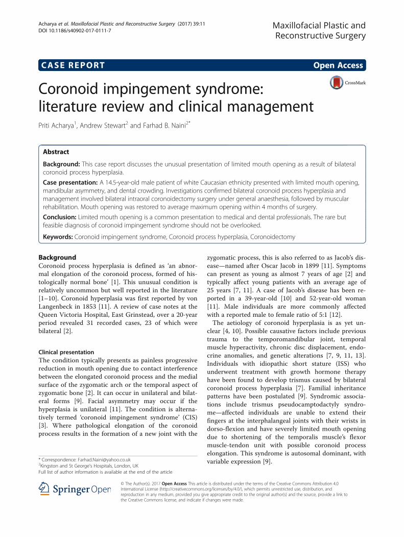
Fig. 1 Preoperative frontal view demonstrating restricted mouth opening
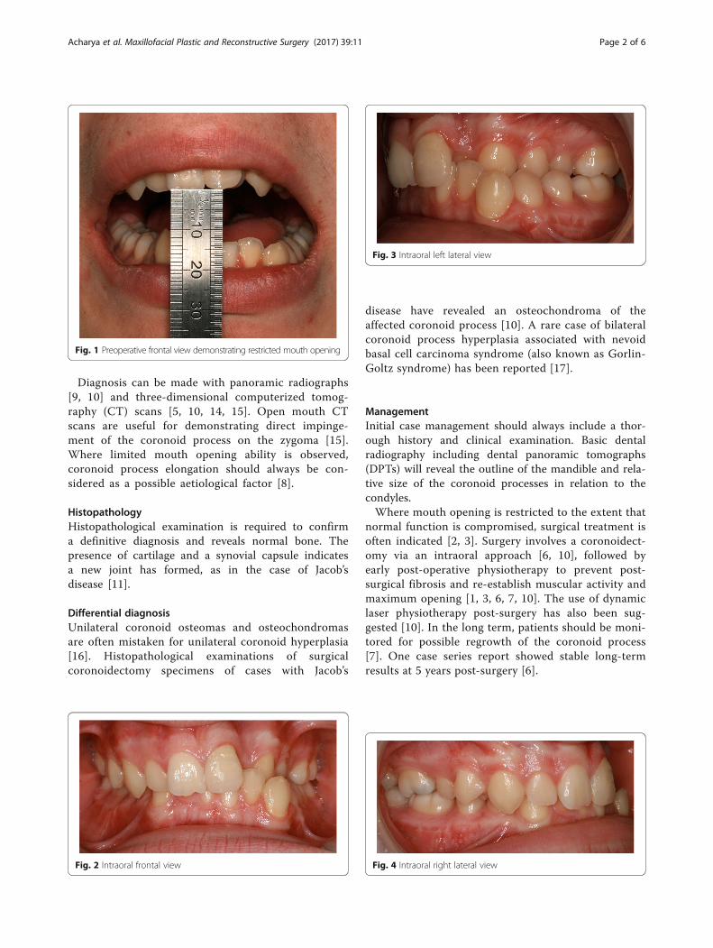
Fig. 2 Intraoral frontal view
Fig. 3 Intraoral left lateral view
Fig. 4 Intraoral right lateral view
Acharya et al. Maxillofacial Plastic and Reconstructive Surgery (2017) 39:11 Page 2 of 6
Case presentationA 14.5-year-old male patient of white Caucasian ethni-city (EP) presented to the Oral and Maxillofacial Sur-gery team, complaining of limited mouth opening anddental crowding. He reported functional and socialdifficulties associated with his limited mouth opening,and he was unable to have orthodontic treatment dueto the same reason. His secondary concerns were anasymmetry of the right side of his lower jaw and con-stant dull headaches, which were interfering with hisschool attendance. He reported a noticeable reductionin his mouth opening from the age of 13 years, whichcoincided with his pubertal growth spurt. His motherand General Dental Practitioner also noticed this.EP presented with a Class II division 2 incisor rela-
tionship on a moderate Class II skeletal base with achin point deviation to the left of his facial midlineand an average lower anterior face height and
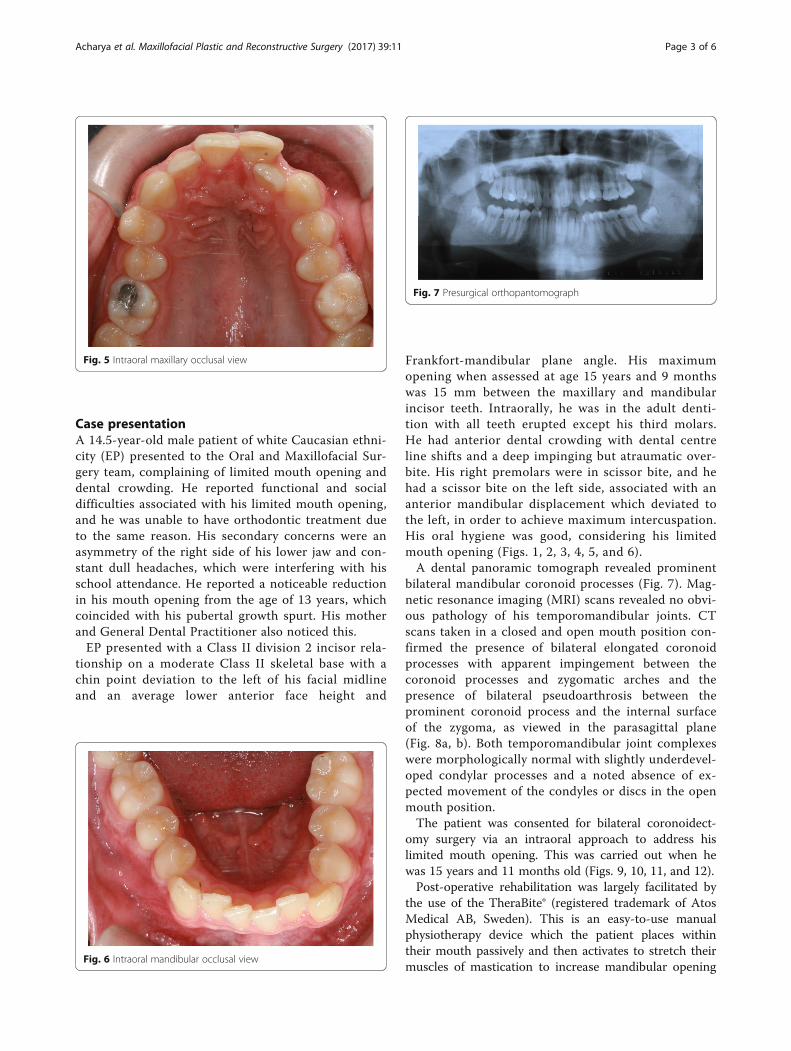
Frankfort-mandibular plane angle. His maximumopening when assessed at age 15 years and 9 monthswas 15 mm between the maxillary and mandibularincisor teeth. Intraorally, he was in the adult denti-tion with all teeth erupted except his third molars.He had anterior dental crowding with dental centreline shifts and a deep impinging but atraumatic over-bite. His right premolars were in scissor bite, and hehad a scissor bite on the left side, associated with ananterior mandibular displacement which deviated tothe left, in order to achieve maximum intercuspation.His oral hygiene was good, considering his limitedmouth opening (Figs. 1, 2, 3, 4, 5, and 6).A dental panoramic tomograph revealed prominent
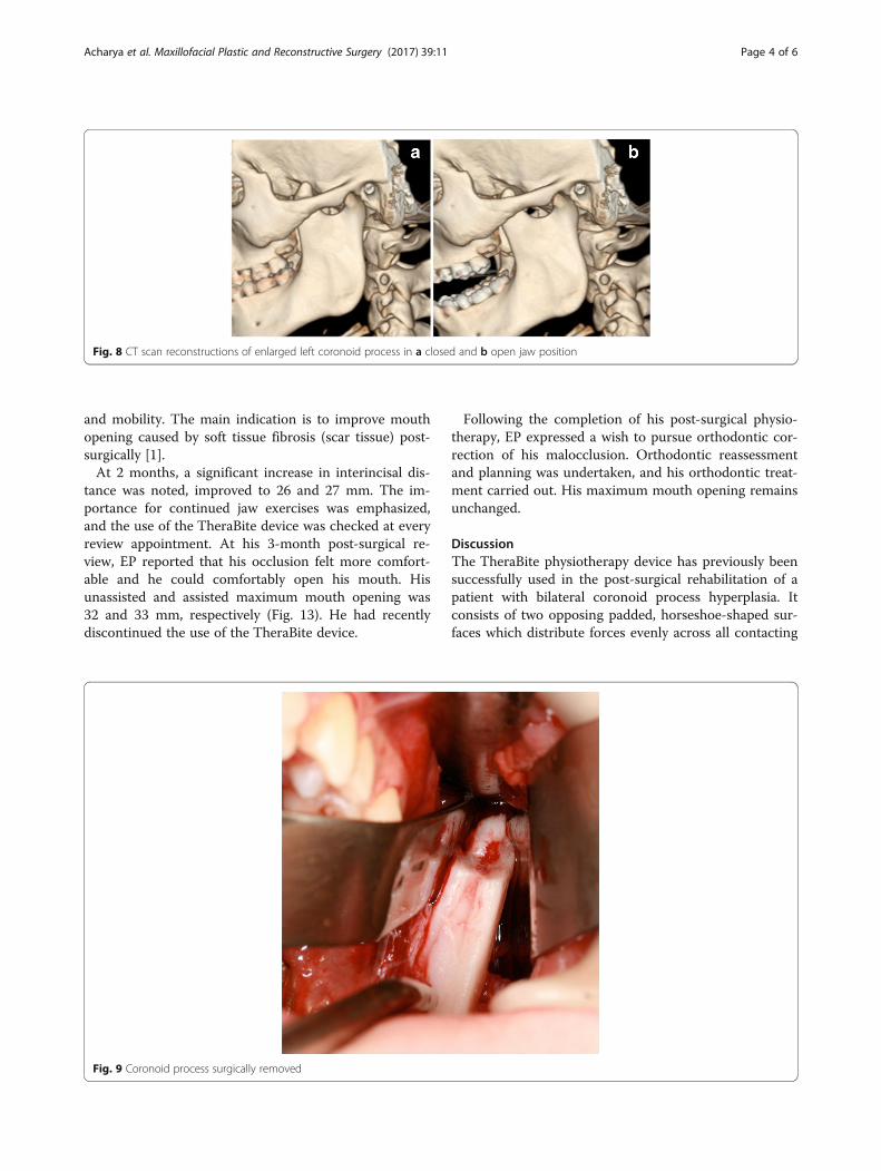
bilateral mandibular coronoid processes (Fig. 7). Mag-netic resonance imaging (MRI) scans revealed no obvi-ous pathology of his temporomandibular joints. CTscans taken in a closed and open mouth position con-firmed the presence of bilateral elongated coronoidprocesses with apparent impingement between thecoronoid processes and zygomatic arches and thepresence of bilateral pseudoarthrosis between theprominent coronoid process and the internal surfaceof the zygoma, as viewed in the parasagittal plane(Fig. 8a, b). Both temporomandibular joint complexeswere morphologically normal with slightly underdevel-oped condylar processes and a noted absence of ex-pected movement of the condyles or discs in the openmouth position.The patient was consented for bilateral coronoidect-
omy surgery via an intraoral approach to address hislimited mouth opening. This was carried out when hewas 15 years and 11 months old (Figs. 9, 10, 11, and 12).Post-operative rehabilitation was largely facilitated by
the use of the TheraBite® (registered trademark of AtosMedical AB, Sweden). This is an easy-to-use manualphysiotherapy device which the patient places withintheir mouth passively and then activates to stretch theirmuscles of mastication to increase mandibular opening
Fig. 5 Intraoral maxillary occlusal view
Fig. 6 Intraoral mandibular occlusal view
Fig. 7 Presurgical orthopantomograph
Acharya et al. Maxillofacial Plastic and Reconstructive Surgery (2017) 39:11 Page 3 of 6
and mobility. The main indication is to improve mouthopening caused by soft tissue fibrosis (scar tissue) post-surgically [1].At 2 months, a significant increase in interincisal dis-
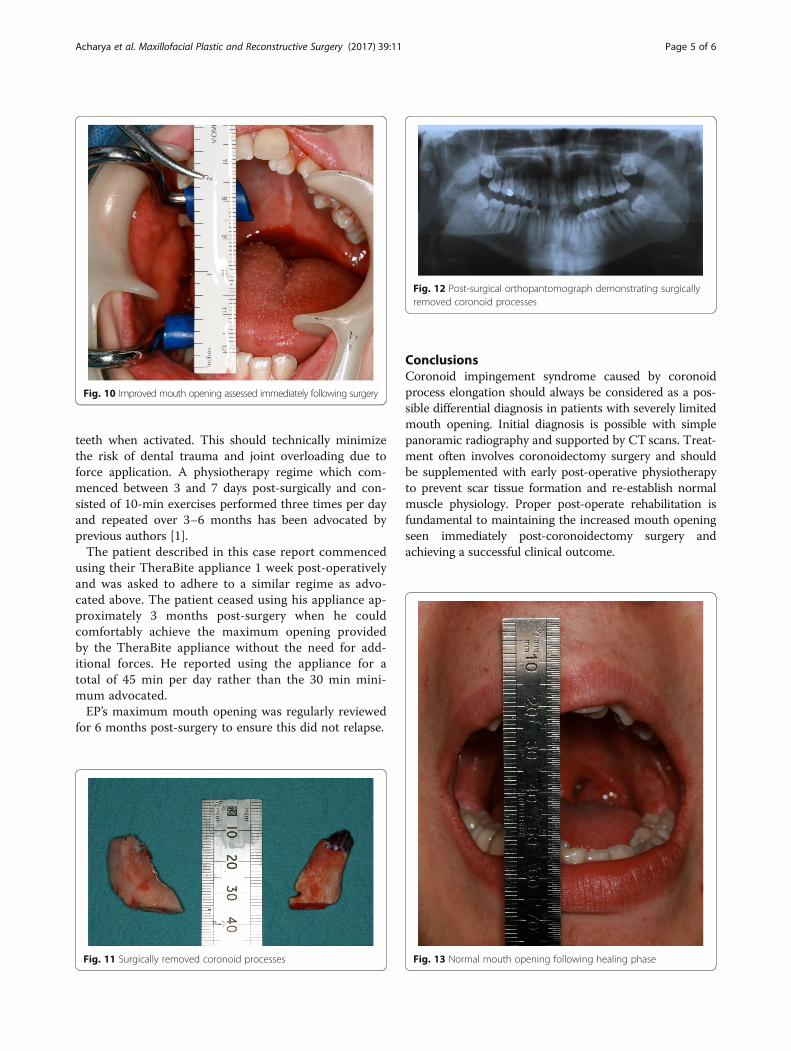
tance was noted, improved to 26 and 27 mm. The im-portance for continued jaw exercises was emphasized,and the use of the TheraBite device was checked at everyreview appointment. At his 3-month post-surgical re-view, EP reported that his occlusion felt more comfort-able and he could comfortably open his mouth. Hisunassisted and assisted maximum mouth opening was32 and 33 mm, respectively (Fig. 13). He had recentlydiscontinued the use of the TheraBite device.
Following the completion of his post-surgical physio-therapy, EP expressed a wish to pursue orthodontic cor-rection of his malocclusion. Orthodontic reassessmentand planning was undertaken, and his orthodontic treat-ment carried out. His maximum mouth opening remainsunchanged.
DiscussionThe TheraBite physiotherapy device has previously beensuccessfully used in the post-surgical rehabilitation of apatient with bilateral coronoid process hyperplasia. Itconsists of two opposing padded, horseshoe-shaped sur-faces which distribute forces evenly across all contacting
Fig. 8 CT scan reconstructions of enlarged left coronoid process in a closed and b open jaw position
Fig. 9 Coronoid process surgically removed
Acharya et al. Maxillofacial Plastic and Reconstructive Surgery (2017) 39:11 Page 4 of 6
teeth when activated. This should technically minimizethe risk of dental trauma and joint overloading due toforce application. A physiotherapy regime which com-menced between 3 and 7 days post-surgically and con-sisted of 10-min exercises performed three times per dayand repeated over 3–6 months has been advocated byprevious authors [1].The patient described in this case report commenced
using their TheraBite appliance 1 week post-operativelyand was asked to adhere to a similar regime as advo-cated above. The patient ceased using his appliance ap-proximately 3 months post-surgery when he couldcomfortably achieve the maximum opening providedby the TheraBite appliance without the need for add-itional forces. He reported using the appliance for atotal of 45 min per day rather than the 30 min mini-mum advocated.EP’s maximum mouth opening was regularly reviewed
for 6 months post-surgery to ensure this did not relapse.
ConclusionsCoronoid impingement syndrome caused by coronoidprocess elongation should always be considered as a pos-sible differential diagnosis in patients with severely limitedmouth opening. Initial diagnosis is possible with simplepanoramic radiography and supported by CT scans. Treat-ment often involves coronoidectomy surgery and shouldbe supplemented with early post-operative physiotherapyto prevent scar tissue formation and re-establish normalmuscle physiology. Proper post-operate rehabilitation isfundamental to maintaining the increased mouth openingseen immediately post-coronoidectomy surgery andachieving a successful clinical outcome.
Fig. 11 Surgically removed coronoid processes
Fig. 12 Post-surgical orthopantomograph demonstrating surgicallyremoved coronoid processes
Fig. 13 Normal mouth opening following healing phase
Fig. 10 Improved mouth opening assessed immediately following surgery
Acharya et al. Maxillofacial Plastic and Reconstructive Surgery (2017) 39:11 Page 5 of 6
FundingNone.
Authors’ contributionsFBN and AS diagnosed, planned, and supervised the treatment. PA carriedout the orthodontic treatment following surgery. PA carried out theliterature review. AS carried out the surgery. All authors helped to completethe manuscript and read and approved the final manuscript.
Competing interestsThe authors declare that they have no competing interests.
Consent for publicationWritten informed consent was obtained from the patient for publication ofthis case report and accompanying images.
Publisher’s NoteSpringer Nature remains neutral with regard to jurisdictional claims inpublished maps and institutional affiliations.
Author details1Eastman Dental Hospital, London, UK. 2Kingston and St George’s Hospitals,London, UK.
Received: 14 March 2017 Accepted: 31 March 2017
References1. Ferro MF, Sanromán JF, Gutierrez JS, López AC, López de Sánchez A, Pérez
AE (2008) Treatment of bilateral hyperplasia of the coronoid process of themandible. Presentation of a case and review of the literature. Med OralPatol Oral Cur Bucal 13:e595–e598
2. McLoughlin PM, Hopper C, Bowley NB (1995) Hyperplasia of the mandibularcoronoid process: an analysis of 31 cases and a review of the literature. JOral Maxillofac Surg 53:250–255
3. Chauhan P, Dixit SG (2011) Bilateral elongated coronoid process of themandible. Int J Anat Var 4:25–27
4. Maurer RM, Wildin RE (1964) Hypertrophy of the coronoid process of themandible: a cause of restricted opening of the mouth. Report of four cases.Radiology 83:1060–1063
5. Gibbons AJ (1995) Computed tomography in the investigation of bilateralmandibular coronoid hyperplasia. Br J Radiol 68:531–533
6. Gerbino G, Bianchi SD, Berrone BS (1997) Hyperplasia of the mandibularcoronoid process: long-term follow-up after coronoidotomy. JCraniomaxillofac Surg 25:69–73
7. Lee ST, Chung IK (2012) Severe trismus due to bilateral coronoid processhyperplasia in growth hormone therapy patient: a case report. J KoreanAssoc Oral Maxillofac Surg 38:249–254
8. Isberg A, Isacsson G, Nah KS (1987) Mandibular coronoid process locking: aprospective study of frequency and association with internal derangementof the temporomandibular joint. Oral Surg Oral Med Oral Pathol 63:275–279
9. Colquhoun A, Cathro I, Kumara R, Ferguson MM, Doyle TCA (2002) Bilateralcoronoid hyperplasia in two brothers. Dentomaxillofac Radiol 31:142–146
10. Zhong SC, Xu ZJ, Zhang ZG, Zheng YH, Li TX, Su K (2009) Bilateral coronoidhyperplasia (Jacob disease on right and elongation on left): report of a caseand literature review. Oral Surg Oral Med Oral Pathol 107:e64–e67
11. Coll-Anglada M, Acero-Sanz J, Vila-Masana I, Navarro-Cuéllar C, Ochandiano-Caycoia S, López de-Atalaya J, Navarro-Vila C (2011) Jacob’s diseasesecondary to coronoid process osteochondroma. A case report. Med OralPatol Oral Cur Bucal 16:e708–710.
12. Blanchard P, Henry JF, Souchere B, Breton P, Freidel M (1992) Permanentconstriction of the jaw due to idiopathic bilateral hyperplasia of thecoronoid process. Rev Stomatol Chir Maxillofac 93:46–50, French
13. Jaskolka MS, Eppley BL, van Aalst JA (2007) Mandibular coronoid hyperplasiain pediatric patients. J Craniofac Surg 18:849–854
14. De Bont LGM, van der Kuijl B, Stegenga B, Vencken LM, Boering G (1993)Computer tomography in differential diagnosis of temporomandibular jointdisorders. Int J Oral Maxillofac Surg 22:200–209
15. Baik JS, Huh KH, Park KS, Park MS, Heo MS, Lee SS et al (2005) The diagnosisof coronoid impingement using computer tomography. Korean J OralMaxillofac Radiol 35:231–234
16. Smyth AG, Wake MJC (1994) Recurrent bilateral coronoid hyperplasia: anunusual case. Br J Oral Maxillofac Surg 32:100–104
17. Leonardi R, Sorge G, Caltabiano M (2001) Bilateral hyperplasia of themandibular coronoid processes associated with the nevoid basal cellcarcinoma syndrome in an Italian boy. Br Dent J 190:349–350
Submit your manuscript to a journal and benefi t from:
7 Convenient online submission
7 Rigorous peer review
7 Immediate publication on acceptance
7 Open access: articles freely available online
7 High visibility within the fi eld
7 Retaining the copyright to your article
Submit your next manuscript at 7 springeropen.com
Acharya et al. Maxillofacial Plastic and Reconstructive Surgery (2017) 39:11 Page 6 of 6