conservation and variation in enamel protein distribution...
TRANSCRIPT
Conservation and Variation in Enamel ProteinDistribution During Vertebrate Tooth Development
PAUL G. SATCHELL,1 XOCHITL ANDERTON,1 OKHEE H. RYU,2
XIANGHONG LUAN,1,7 ADAM J. ORTEGA,3 RENE OPAMEN,1
BRETT J. BERMAN,4,6 DAVID E. WITHERSPOON,1
JAMES L. GUTMANN,1 AKIRA YAMANE,5 MARGERITA ZEICHNER-DAVID,6
JAMES P. SIMMER,2 CHARLES F. SHULER,6 AND
THOMAS G.H. DIEKWISCH1,6,7*1Baylor College of Dentistry/Texas A&M University System, Dallas, Texas2Department of Pediatric Dentistry, The University of Texas Health SciencesCenter at San Antonio, Texas3Harvard School of Dental Medicine, Harvard University, Boston,Massachusetts4The Department of Medicine, The University of California, San Diego,California5Department of Pharmacology, School of Dental Medicine, Tsurumi University,Japan6Center for Craniofacial Molecular Biology, University of Southern CaliforniaSchool of Dentistry, Los Angeles, California7Allan G. Brodie Laboratory for Craniofacial Genetics, University of IllinoisCollege of Dentistry, Chicago, Illinois
ABSTRACT Vertebrate enamel formation is a unique synthesis of the function of highlyspecialized enamel proteins and their effect on the growth and organization of apatite crystals.Among tetrapods, the physical structure of enamel is highly conserved, while there is a greatervariety of enameloid tooth coverings in fish. In the present study, we postulated that in enamelmicrostructures of similar organization, the principle components of the enamel protein matrixwould have to be highly conserved. In order to identify the enamel proteins that might be mosthighly conserved and thus potentially most essential to the process of mammalian enamel formation,we used immunoscreening with enamel protein antibodies as a means to assay for degrees ofhomology to mammalian enamel proteins. Enamel preparations from mouse, gecko, frog, lungfish,and shark were screened with mammalian enamel protein antibodies, including amelogenin,enamelin, tuftelin, MMP20, and EMSP1. Our results demonstrated that amelogenin was the mosthighly conserved enamel protein associated with the enamel organ, enamelin featured a distinctpresence in shark enameloid but was also present in the enamel organ of other species, while theother enamel proteins, tuftelin, MMP20, and EMSP1, were detected in both in the enamel organ andin other tissues of all species investigated. We thus conclude that the investigated enamel proteins,amelogenin, enamelin, tuftelin, MMP20, and EMSP1, were highly conserved in a variety ofvertebrate species. We speculate that there might be a unique correlation between amelogenin-richtetrapod and lungfish enamel with long and parallel crystals and enamelin-rich basal vertebrateenameloid with diverse patterns of crystal organization. J. Exp. Zool. (Mol. Dev. Evol. ) 294:91–106,2002. r 2002 Wiley-Liss, Inc.
Many biominerals such as enamel, dentin, orbone grow within a preformed protein matrix thatdetermines the dimension and shape of theinorganic mineral component (Lowenstam, ’81;Heuer et al., ’92). This protein matrix organizesinto subunit compartments, which determine thedimension and orientation of organizing mineral
Grant sponsor: NIDCR; Grant number: R01 DE13378; Grantsponsor: NIH; Grant number DE13237
*Correspondence to: Dr. Thomas G.H. Diekwisch, Director, Allan G.Brodie Laboratory for Craniofacial Genetics, University of IllinoisCollege of Dentistry M/C 841, 801 South Paulina, Chicago, IL 60612.E-mail: [email protected]
Received 10 August 2001; Accepted 2 January 2002Published online in Wiley InterScience (www.interscience.wiley.
com). DOI: 10.1002/jez.10148
r 2002 WILEY-LISS, INC.
JOURNAL OF EXPERIMENTAL ZOOLOGY (MOL DEV EVOL) 294:91–106 (2002)
crystals (Heuer et al., ’92). Among vertebratebiominerals, there are fundamental differencesbetween the collagenous protein matrix that con-tributes to the assembly of bone, cementum, anddentin crystallites and the noncollagenous proteinmatrix that is associated with the formation ofenamel crystals (Lowenstam, ’81). In tooth enamel,a highly organized matrix of enamel proteins isclosely associated with the formation of enamelhydroxyapatite crystals (Diekwisch et al., ’93).In many vertebrates, the basic organization of
the tooth enamel mineral phase is remarkablysimilar and includes long and parallel-organizedhydroxyapatite crystals organized into enamelprisms (Slavkin and Diekwisch, ’96). Even thoughone would expect similarities in crystal organiza-tion to go along with similarities in proteincomposition and organization, as of yet there isno proof for this assumption. With the exception ofisolated amelogenin sequences from reptilian andamphibian teeth, little is known about the enamelprotein composition of nonmammalian verte-brates (Ishiyama et al., ’98; Toyosawa et al., ’98;Sire, in press). In mammals, however, a number ofnovel tooth enamel proteins have been discoveredrecently, including ameloblastin, enamelin,tuftelin, and enamel proteases (Deutsch, ’89;Krebsbach et al., ’96; Bartlett et al., ’96; Huet al., ’97; Simmer et al., ’98). Although some ofthese novel enamel proteins have also been loca-lized in tissues outside of teeth, they might be ofrelevance to the mechanisms of enamel formation(Deutsch et al., ’91; Zeichner-David et al., ’95, ’97;MacDougall et al., ’98). While the nonamelogeninenamel proteins amount to 10% of the enamelprotein matrix, the major protein component (90%)of the mammalian enamel protein matrix isamelogenin (Termine et al., ’80a, b), a protein thatis believed to be of significant functional relevancefor all stages of enamel formation (Simmer andFincham, ’95; Diekwisch, ’98).A number of studies have shown amelogenin
protein localization in ameloblasts and in theenamel layer of all vertebrate classes (Herold et al.,’80; Slavkin et al., ’82, ’83; Slavkin and Diekwisch,’96, ’97). Subsequently, several authors havepublished on the immunohistochemical localiza-tion of amelogenins in agnathans, fish, andurodeles, including hagfish agnathan teeth (Slav-kin and Diekwisch, ’96, ’97), Calamoichthysactinopterygian scales (Zylberberg et al., ’97),Lepisosteus actinopterygian teeth (Ishiyamaet al., ’99), lungfish sarcopterygian teeth (Satchellet al., 2000), and Triturus urodelian teeth
(Kogaya, ’99). A related finding of amelogeninexon 4 conservation in hagfish agnathan verte-brates based on RT-PCR amplification (Slavkinand Diekwisch, ’96, ’97) might be consideredquestionable at this point because exon 4 isthe least conserved of all amelogenin epitopes(Girondot et al., ’98). However, recent advancesin the cloning and sequencing of nonmammalianenamel genes (Ishiyama et al., ’98; Toyosawa et al.,’98) have strongly supported the case of awide evolutionary conservation of amelogeninproteins.
The similarity of enamel crystal structurein many vertebrates and the conservation ofamelogenins in many vertebrates might suggestthat the protein matrix composition and organiza-tion would be similar and consequently enamelproteins were highly conserved between verte-brate classes. In order to test this hypothesis, wehave decided to determine the presence andlocalization of the enamel proteins, amelogenin,enamelin and tuftelin, and the enamel proteases,MMP20 and EMSP1, in vertebrates from all fourvertebrate classes via peroxidase immunohisto-chemistry. In order to screen a small butrepresentative variety of vertebrates, the follow-ing species were chosen: a mammal (mouse,Mus musculus), a reptile (gecko, Hemidactylusturcicus), an anuran amphibian (green tree frog,Hyla cinerea), a sarcopterygian fish (lungfish,Neoceratodus forsteri), and a chondrychthian fish(shark, Heterodontus francisci). Following immu-nohistochemical analysis, we determined thatenamel protein epitopes were distributed withinall species investigated and thus highly conservedamong vertebrates. Though mainly distributed inthe enamel layer and enamel organ, enamelproteins other than amelogenin were also foundin tissues surrounding the tooth organ and inlayers outside of the enamel organ. Enamelinfeatured a distinct association with shark amelo-blasts and enameloid. In contrast, amelogenin wasmore or less exclusively distributed in the enamel/ameloblast complex of the species investigated inthis study.
MATERIALS AND METHODS
Tissue preparation
The following experimental animals wereused in the present study: a 6-day and a12-day postnatal mouse (Mus musculus), a gecko(Hemidactylus turcia; 43mm total length), ajuvenile green tree frog (Hyla cinerea), a larval
P.G. SATCHELL ET AL.92
lung-fish (Neoceratodus forsteri; stage 46; Kemp,’81), and a young hornshark (Heterodontus fran-ciscus; 22cm total length). Animals were sacrificedby decapitation according to Baylor College ofDentistry animal care regulations. Mandibleswere dissected and fixed immediately. For immu-nohistochemistry, tissues were fixed with 10%buffered formalin, decalcified in 4% EDTA and de-hydrated in a graded series of ethanols. Specimenwere embedded in paraffin and cut at 5mmthickness. Sections were mounted on coated glassslides.
Immunohistochemistry
Immunoreactions were performed followingthe instructions of the Zymed Histostain SP kit(San Francisco, CA). All reactions were carried outin a humidified chamber at room temperature.Briefly, sections were treated against endogenousperoxidase using methanol and 3% hydrogenperoxide and then blocked using in 10%goat serum for 10min. Sections were incubatedwith primary antibody for two hours. Primaryantibodies were diluted in phosphate bufferedsaline (PBS). The dilution of the primary antibodywas determined in preliminary experiments. As amethodological control, the primary antibody wasreplaced with normal serum. Sections werewashed three times in PBS and subsequentlyincubated for 10 min with biotinylated IgGs(either anti-chicken (MMP20) or anti-rabbit) assecondary antibodies. After washing in PBS (threetimes), sections were exposed to the streptavidin-peroxidase conjugate for 10 min and then washedagain in PBS (three times). Signals were detectedusing the AEC Substrate-Chromogen mixture ofthe Zymed Histostain kit. Sections were counter-stained using hematoxylin and mounted withGVA-mount.
List of primary antibodies and dilutions.
The following five primary antibodies were usedin this study. Western blotting analyses of theantibodies used in this study have establishedspecificity to a single protein epitope; referencepublications are quoted below.
A. Polyclonal antibody against a recombinantmouse amelogenin (M179), IgG preparation(Simmer et al., ’94). Dilution 1:100.
B. Polyclonal antibody against the full-lengthMMP20 (enamelysin) amino acid sequencegenerated in chicken. Dilution 1:100.
C. Polyclonal rabbit antibody generated againsta recombinant pig EMSP1 from E. coli that wasexcised from SDS-PAGE gels (Hu et al., 2000).Dilution 1:50.D. Polyclonal peptide antibody against theN-terminal enamelin portion. The antigenwas a modified hexadecapeptide (MPMQMPRMPGFSSKSE) corresponding to the N-terminalenamelin amino acids 1–16 (Fukae et al., ’96;Hu et al., ’97; Dohi et al., ’98). Dilution 1:100.
E. Polyclonal rabbit antibody against a syn-thetic polypeptide derived from the tuftelinsequence (QSKDTTIQELKEKIA) (Diekwischet al., ’97). Dilution 1:50.
Controls
The following controls were performed totest for antibody specificity: tissue controlsFthe specificity of the antibody was evaluatedin nondental tissues; antibody controls byusing a dilution series; controls with pre-adsorbed antibody to exclude unspecificbinding; controls with pre-immune serumto control for binding to serum components; andomission of primary antibody as a systematiccontrol.
RESULTS
Mouse, gecko, frog, lungfish, and sharkfeatured characteristic tooth organsat the onset of tooth development.
In all five species investigated, the developingtooth enamel/enameloid was immediately sur-rounded by one or more distinct layers ofepithelial cells. Mice teeth featured a fully devel-oped enamel organ with highly polarized andprismatic ameloblasts immediately adjacent tothe developing enamel (Figs. 1A–1F). The amelo-blast cell layer was lined by a thin layer ofperpendicular oriented stratum intermedium cells(Figs. 1A–F). The coronal center of the enamelorgan was filled with a seemingly irregular net-work of star-shaped cells, the stellate reticulum(Figs. 1A–1F). The enamel organ was enclosed byan outer enamel epithelium (Figs. 1A–1F). In thecase of the gecko (Figs. 1G–1L) and frog (Figs. 1M–1R) enamel organ, the ameloblast cell layerappeared to have gained prominence while theother three cell layers were less distinct. Never-theless, also in gecko and frog stratum interme-dium, stellate reticulum, and outer enamel
ENAMEL PROTEINS AND EVOLUTION 93
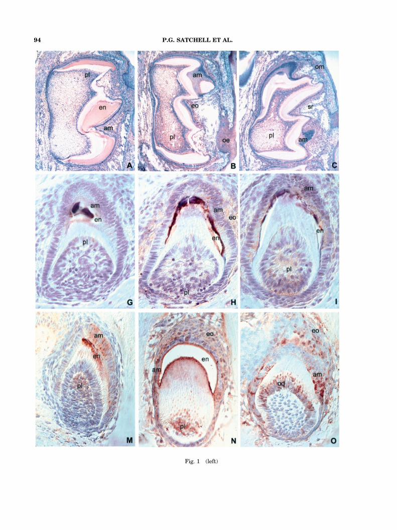
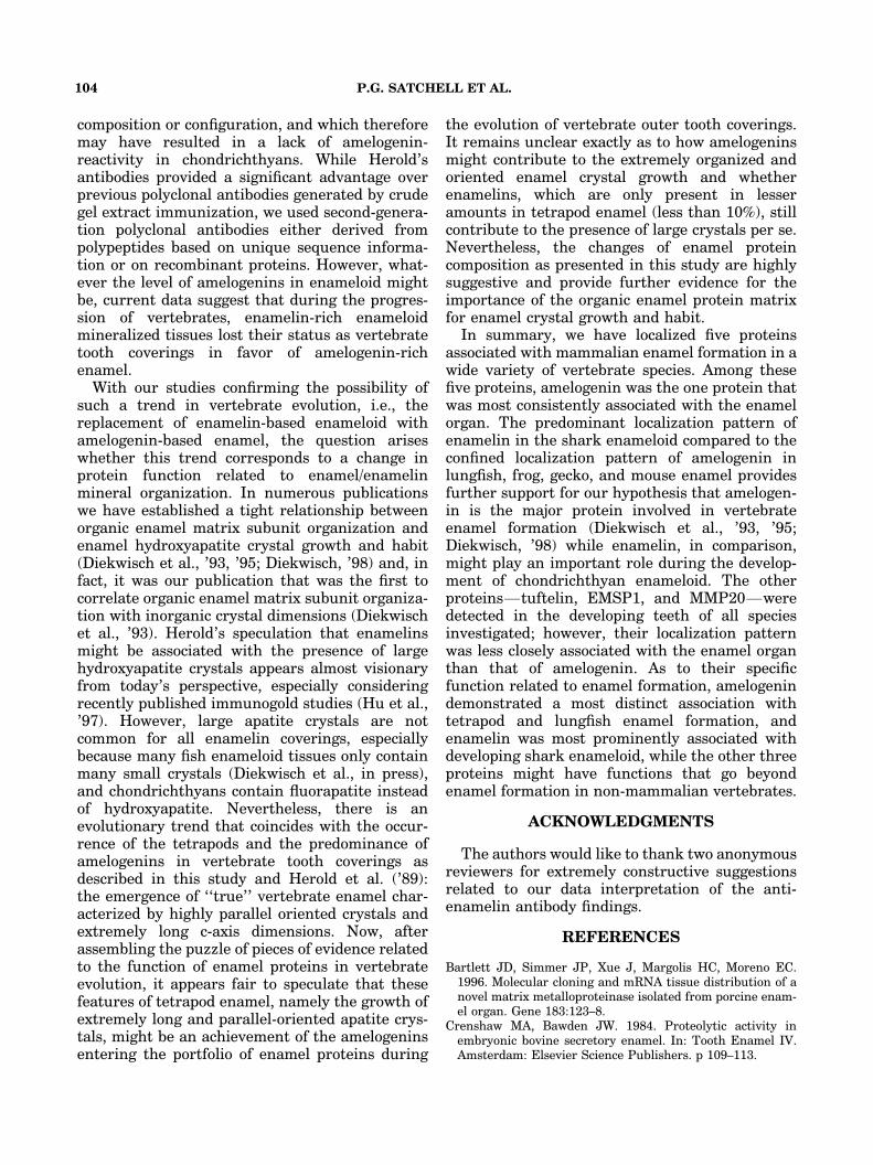
Fig. 1 (left)
P.G. SATCHELL ET AL.94
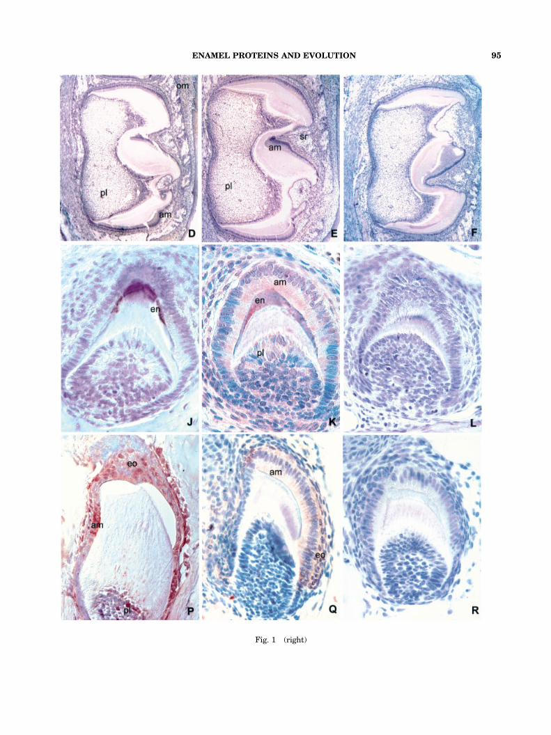
Fig. 1 (right)
ENAMEL PROTEINS AND EVOLUTION 95
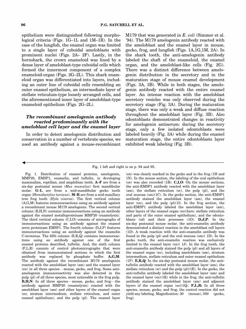
epithelium were distinguished following morpho-logical criteria (Figs. 1G–1L and 1M–1R). In thecase of the lungfish, the enamel organ was limitedto a single layer of cuboidal ameloblasts withprominent nuclei (Figs. 2A– 2F). Lastly, in thehornshark, the crown enameloid was lined by adense layer of ameloblast-type cuboidal cells whichformed the innermost component of a complexenameloid-organ (Figs. 2G–2L). This shark enam-eloid organ was differentiated into layers, includ-ing an outer line of cuboidal cells resembling anouter enamel epithelium, an intermediate layer ofstellate reticulum-type loosely arranged cells, andthe aforementioned inner layer of ameloblast-typeenameloid epithelium (Figs. 2G–2L).
The recombinant amelogenin antibodyreacted predominantly with the
ameloblast cell layer and the enamel layer
In order to detect amelogenin distribution andconservation in a number of vertebrate species, weused an antibody against a mouse-recombinant
M179 that was generated in E. coli (Simmer et al.’94). The M179 amelogenin antibody reacted withthe ameloblast and the enamel layer in mouse,gecko, frog, and lungfish (Figs. 1A,1G,1M, 2A). Inthe shark tooth, the anti-amelogenin antibodylabeled the shaft of the enameloid, the enamelorgan, and the ameloblast-like cells (Fig. 2G).There was a distinct difference between amelo-genin distribution in the secretory and in thematuration stage of mouse enamel development(Figs. 3A, 3B). While in both stages, the amelo-genin antibody reacted with the entire enamellayer. An intense reaction with the ameloblastsecretory vesicles was only observed during thesecretory stage (Fig. 3A). During the maturationstage, there was only a weak and diffuse reactionthroughout the ameloblast layer (Fig. 3B). Alsoodontoblasts demonstrated changes in reactivityfor amelogenin antiserum: during the secretorystage, only a few isolated odontoblasts werelabeled heavily (Fig. 3A) while during the enamelmaturation stage, the entire odontoblasts layerexhibited weak labeling (Fig. 3B).
Fig. 1. Distribution of enamel proteins, amelogenin,MMP20, EMSP1, enamelin, and tuftelin, in developingmammalian, reptilian, and amphibian teeth. A–F are from asix-day postnatal mouse (Mus musculus) first mandibularmolar. G–L are from a mid-mandibular gecko toothorgan (Hemidactylus turcicus). M–R are from a mid-maxillarytree frog tooth (Hyla cinerea). The first vertical column(A,G,M) features immunoreactions using an antibody againsta recombinant mouse M179 amelogenin. The second verticalcolumn (B,H,N) contains immunoreactions using an antibodyagainst the enamel metalloproteinase MMP20 (enamelysin).The third vertical column (C,I,O) consists of micrographs ofimmunoreactions using an antibody against the enamelserin proteinase EMSP1. The fourth column (D,J,P) featuresimmunoreactions using an antibody against the enamelinC-terminus. The fifth column (E,K,Q) contains immunoreac-tions using an antibody against one of the firstenamel proteins described, tuftelin. And, the sixth column(F,L,R) consists of control photomicrographs that wereobtained from immunostained sections in which the firstantibody was replaced by phosphate buffer. A,G,M:The antibody against the recombinant M179 amelogeninreacted with the ameloblast layer (am) and the enamel layer(en) in all three speciesFmouse, gecko, and frog. Some anti-amelogenin immunoreactivity was also detected in thepulp (pl) of all three species, most noticeable in the frog pulp.B,H,N: In all three speciesFmouse, gecko, and frogFtheantibody against MMP20 (enamelysin) reacted with theameloblast layer (am) and other layers of the enamel organ(eo; stratum intermedium, stellate reticulum, and outerenamel epithelium), and the pulp (pl). The enamel layer
(en) was clearly marked in the gecko and in the frog (1H and1N). In the mouse section, the labeling of the oral epithelium(oe) was also recorded (1B). C,I,O: On the mouse sections,the anti-EMSP1 antibody reacted with the ameloblast layer(am), the stellate reticulum (sr), the pulp (pl), and theoral mucosa (om)(1C). In the gecko section, the anti-EMSP1antibody stained the ameloblast layer (am), the enamellayer (en), and the pulp (pl)(1I). In the frog section, theanti-EMSP1 antibody labeled the ameloblast layer (am),other layers of the enamel organ (eo)(here: stellate reticulumand parts of the outer enamel epithelium), and the odonto-blasts (od) and their processes (1O). D,J,P: In thesix-day postnatal mouse molar, the anti-enamelin antibodydemonstrated a distinct reaction in the ameloblast cell layers(1D). A weak reaction with the anti-enamelin antibody wasfound in the pulp (pl) and the oral mucosa (om)( 1D). In thegecko tooth, the anti-enamelin reaction was exclusivelylimited to the enamel layer (en)( 1J). In the frog tooth, theanti-enamelin antibody stained the pulp (pl) and all layers ofthe enamel organ (eo), including ameloblasts (am), stratumintermedium, stellate reticulum and outer enamel epithelium(1P). E,K,Q: In the six-day postnatal mouse molar, the anti-tuftelin antibody reacted with the ameloblast layer (am), thestellate reticulum (sr) and the pulp (pl)(1E). In the gecko, theanti-tuftelin antibody labeled the ameloblast layer (am) andthe enamel layer (en)(1K) while in the frog, the anti-tuftelinantibody stained the ameloblast layer (am) and adjacentlayers of the enamel organ (eo)(1Q). F,L,R: In all threespecies, mouse, gecko, and frog, the control reaction did notyield any labeling. Magnification: 30� (mouse), 500� (gecko,frog).
3——————————————————————————————————————————————Fig. 1 left and right is on p. 94 and 95.
P.G. SATCHELL ET AL.96
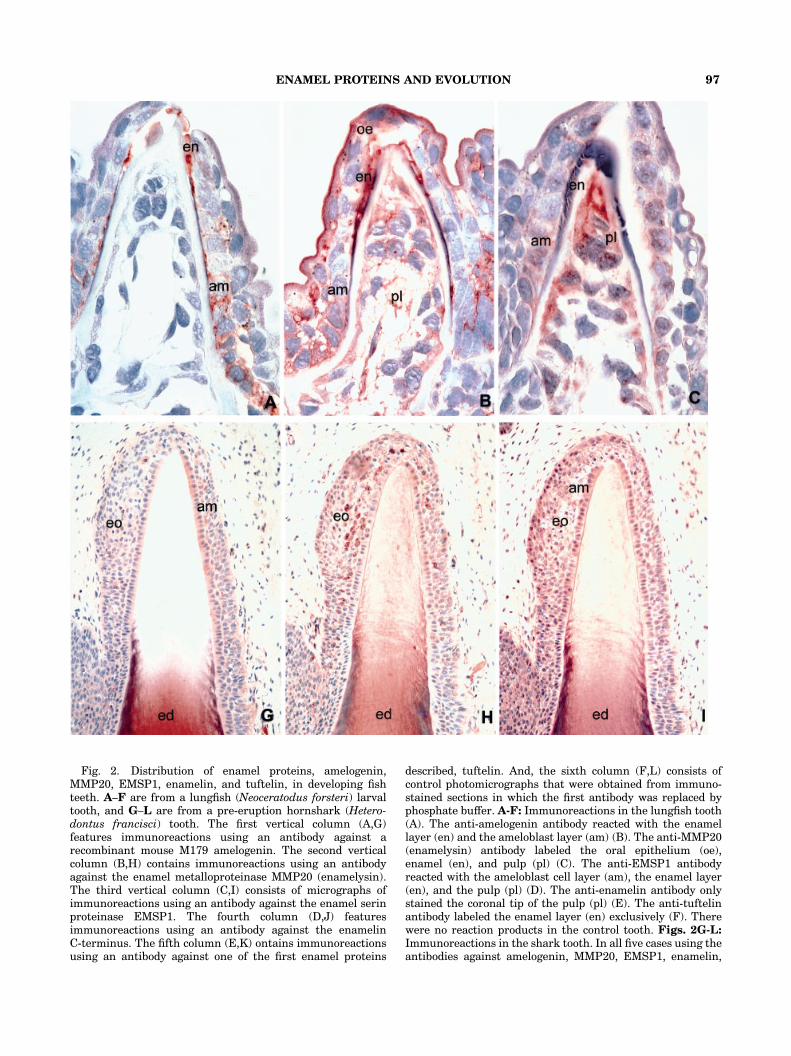
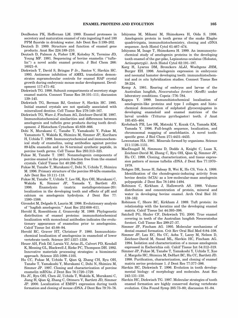
Fig. 2. Distribution of enamel proteins, amelogenin,MMP20, EMSP1, enamelin, and tuftelin, in developing fishteeth. A–F are from a lungfish (Neoceratodus forsteri) larvaltooth, and G–L are from a pre-eruption hornshark (Hetero-dontus francisci) tooth. The first vertical column (A,G)features immunoreactions using an antibody against arecombinant mouse M179 amelogenin. The second verticalcolumn (B,H) contains immunoreactions using an antibodyagainst the enamel metalloproteinase MMP20 (enamelysin).The third vertical column (C,I) consists of micrographs ofimmunoreactions using an antibody against the enamel serinproteinase EMSP1. The fourth column (D,J) featuresimmunoreactions using an antibody against the enamelinC-terminus. The fifth column (E,K) ontains immunoreactionsusing an antibody against one of the first enamel proteins
described, tuftelin. And, the sixth column (F,L) consists ofcontrol photomicrographs that were obtained from immuno-stained sections in which the first antibody was replaced byphosphate buffer. A-F: Immunoreactions in the lungfish tooth(A). The anti-amelogenin antibody reacted with the enamellayer (en) and the ameloblast layer (am) (B). The anti-MMP20(enamelysin) antibody labeled the oral epithelium (oe),enamel (en), and pulp (pl) (C). The anti-EMSP1 antibodyreacted with the ameloblast cell layer (am), the enamel layer(en), and the pulp (pl) (D). The anti-enamelin antibody onlystained the coronal tip of the pulp (pl) (E). The anti-tuftelinantibody labeled the enamel layer (en) exclusively (F). Therewere no reaction products in the control tooth. Figs. 2G-L:Immunoreactions in the shark tooth. In all five cases using theantibodies against amelogenin, MMP20, EMSP1, enamelin,
ENAMEL PROTEINS AND EVOLUTION 97
The MMP20 (enamelysin) enamelproteinase was detected predominantlyin the enamel organ and in the pulp
For MMP20 (enamelysin) detection, we used apolyclonal antibody against the full-length enam-
elysin (Uchida et al., ’91; Bartlett et al., ’96). Inmouse, gecko, frog, and lungfish, the anti-enamel-ysin antibody recognized the enamel organ includ-ing the ameloblast layer, the enamel layer, andthe pulp (Figs. 1B,H,N; 2B). Higher magnifi-cation images revealed discrete staining of the
and tuftelin, the antibodies reacted with theenameloid (ed)-containing shaft of the shark tooth (G–K).In the case of amelogenin and tuftelin, the tip of thetooth enameloid was free of reaction products, while theother three antibodies, MMP20, EMSP1, and enamelin,also exhibited a week reaction at the tip of the tooth. All
five antibodiesFamelogenin, MMP20, EMSP1, enamelin, andtuftelinFreacted with the shark enamel organ (eo) (G–K)with enamelin (J), demonstrating the strongest reaction inthe shark ameloblast layer (am). Also in the shark, the controlwas without reaction products (L). Magnification: 1,000�(lungfish), 200� (shark).
P.G. SATCHELL ET AL.98
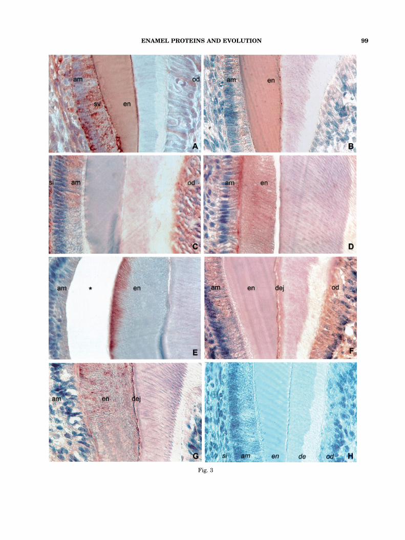
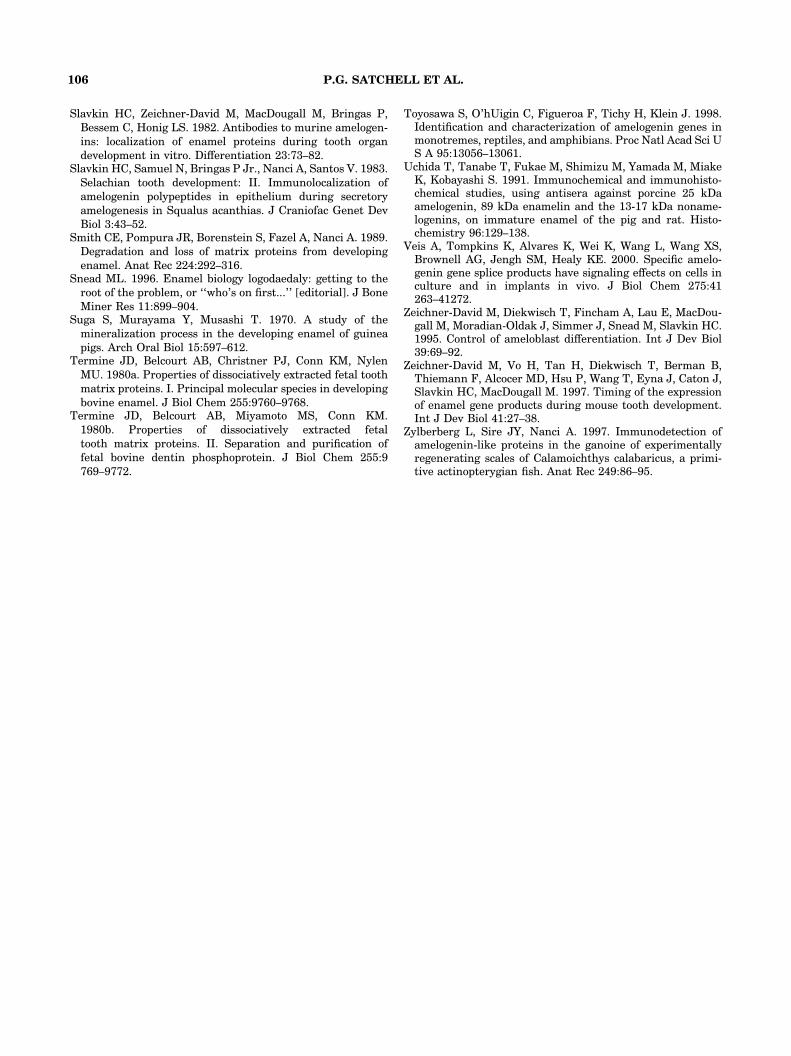
Fig. 3
ENAMEL PROTEINS AND EVOLUTION 99
ameloblast secretory pole, dentin-enamel junction,and odontoblasts in the mouse (Fig. 3F). On themouse section, a labeling of the oral epitheliumwas also recorded (Fig. 1B). In the shark tooth, theanti-MMP20 antibody reacted with the enameloidof the mineralizing shark tooth and the sharkenamel organ (Fig. 1H).
The EMSP1 enamel serin proteinase wasdetected predominantly in the enamelorgan and in the pulp and exhibited
stage-dependent changes in mouse enamel
The antibody used for EMSP1 detectionwas a polyclonal rabbit antibody generated againsta recombinant pig EMSP1 from E. coli thatwas excised from SDS-PAGE gels (Hu et al.,2000). The anti-EMSP1 antibody reactedwith the enamel/enameloid layer in the gecko, inthe lungfish, and in the shark (Figs. 1I; 2C,I);with the ameloblast cell layer in mouse, gecko,frog, lungfish, and shark (Figs. 1C, 1I, 1O, 2C, 2I);with other cell layers of the enamel organincluding stellate reticulum in mouse, frog, andshark (Figs. 1C, 1O, 2I); and with the pulp inmouse, gecko, frog (restricted to odontoblasts),and lungfish (Figs. 1C, 1I, 1O, 2C). On mousemolar sections, we detected a stage-dependentdistinct difference in protein distribution usingthe anti-EMSP1 antibody (Figs. 3C, 3D). Duringthe secretory stage, the EMSP1 antibody reactedwith stratum intermedium, ameloblasts, andodontoblasts, but not in the enamel layer (Fig.3C), while during the maturation stage, the
EMSP1 antibody caused an intense stain in theenamel layer and at the ameloblast secretory pole(Fig. 3D).
The enamelin antibody featured a distinctlocalization pattern in the ameloblasts
and enameloid of the developingshark enameloid organ
For preparing N-terminal specific anti-enamelinantibodies, a modified hexadecapeptide(MPMQMPRMPGFSSKSE) corresponding tothe N-terminal enamelin amino acids 1–16was synthesized and used for anti-peptideantibody production in rabbit (Fukae et al., ’96;Hu et al., ’97; Dohi et al., ’98). In six-day postnatalmouse molars, the anti-enamelin antibodyreacted distinctly with the ameloblast celllayer, but also demonstrated staining in thepulp and the oral mucosa (Fig. 1D). In12-day postnatal molars, labeling in the superficialenamel layer and at the ameloblast secretorypole was detected (Fig. 3E). In the gecko,the anti-enamelin antibody labeling was restrictedto the enamel layer (Fig. 1J). In the frog tooth,all four layers of the enamel organ, ameloblasts,outer enamel epithelium, stellate reticulum,and stratum intermedium were stained usingthe anti-enamelin immunoreaction (Fig. 1P).A positive signal was also detected in the odonto-blasts (Fig. 1P). In the lungfish, only the tipof the pulp reacted with the anti-enamelinantibody (Fig. 2D). Lastly, in the shark, the anti-enamelin antibody recognized the entire enamel
Fig. 3. High magnification images of enamelprotein localization in mouse molars at selected stagesof development. Figs. 3D and 3E were from 12-day postnatalmouse molars; all other figures from six-day postnatalmouse molars. 3A and 3B: Amelogenin localization insecretory stage ameloblasts (A) and maturation stageameloblasts (B) using an antibody against a recombinantM179 amelogenin. (A) Positive amelogenin immunoreac-tions were in ameloblast secretory vesicles (sv), atthe proximal ameloblast pole (am), in the enamel layer (en),and in isolated odontoblasts (od). (B) The amelogenin anti-body demonstrated a distinct reaction in the enamel layer(en). A less intense reaction was also recorded in theameloblast layer (am) and, even less intense, inthe odontoblasts layer (od). 3C and 3D: EMSP1 immunolo-calization in secretory stage ameloblasts (1C, six-daypostnatal) and maturation stage ameloblasts (1D,12-day postnatal). During the secretory stage (C),EMSP1 epitopes were in ameloblasts (am), in the stratum
intermedium (si) and in odontoblasts (od). Duringthe maturation stage (D), EMSP1 epitopes were concentratedat the ameloblast secretory pole (am) and in the enamellayer (en). 3E: Immunolabeling of the superficial enamel layer(en) and of the ameloblast secretory pole (am) usingan antibody against the enamelin C-terminus on12-day postnatal mouse molar sections. The asterisk (*)indicates a preparation artifact between enamel layer andameloblasts. 3F: MMP20 immunohistochemistry demonstrat-ing a color reaction at the ameloblast secretory pole (am), thedentin-enamel junction (dej) and in odontoblasts (od) as wellas a weak reaction in the enamel layer (en). 3G: Tuftelinlocalization in ameloblasts (am) and at the dentin-enameljunction (dej). Tuftelin epitopes were also detected in theinterprismatic framework of the enamel layer (en). 3H: Thecontrol section indicates the localization of the stratumintermedium (si), the ameloblast layer (am), the enamel layer(en), the dentin layer (de), and the odontoblasts layer (od).Magnification: 500� .
3——————————————————————————————————————————————
P.G. SATCHELL ET AL.100
organ including a distinctly stained ameloblastlayer as well as the mineralizing portion of theenameloid (Fig. 2J).
The tuftelin antibody had preferredreaction sites in the frog, gecko, and
lungfish ameloblast and enamel layersbut was also detected in other parts
of the developing tooth organ
The anti-tuftelin antibody was a polyclonalrabbit antibody against a synthetic polypeptidederived from the tuftelin sequence (QSKDTTI-QELKEKIA)(Zeichner-David et al., ’97). The anti-tuftelin antibody reacted with the ameloblast celllayer in mouse, gecko, and frog (Figs. 1E, 1K, 1Q,3G). Other parts of the enamel organ, includingstellate reticulum, were labeled in mouse, frog,and shark (Figs. 1E, 1Q, 2K, 3G). The enamel/enameloid layer was detected with the anti-tuftelin antibody in mouse, gecko, lungfish, andshark (Figs. 1E, 1K, 2E, 2K, 3G).
Using the indirect immunoperoxidasemethod, the control sections did not
exhibit any labeling
Controls were performed in a number of ways asdescribed in the Materials and Methods section.We photographed the controls in which theprimary antibody was replaced with normalserum. All control sections from mouse, gecko,frog, lungfish, and shark did not show any labeling(Figs. 1F, 1L, 1R, 2F, 2L, 3H).
DISCUSSION
In the present study, immunohistochemicalmethods were applied to ask the question whetherenamel proteins were widely and specificallydistributed throughout the vertebrate subphylum.To address this question, we assayed the localiza-tion and distribution of five proteins that havebeen classified at some point as enamel proteins,amelogenin, enamelin, and tuftelin and the pro-teinases MMP20 (enamelysin) and EMSP1. Inorder to screen a small but representative varietyof vertebrates, we selected a mammal (mouse,Musmusculus), a reptile (gecko, Hemidactylus turci-cus), an anuran amphibian (Hyla cinerea), asarcopterygian fish (lungfish, Neoceratodus for-steri), and a chondrichthyan fish (shark, Hetero-dontus francisci). Our immunohistochemical dataindicated that enamel proteins were widely dis-tributed within the vertebrate subphylum. Whileoften associated with the enamel layer or the
enamel-forming cells (ameloblasts), four of the fiveproteins investigated were also found in othertissues of the tooth organ and beyond. Among thefive enamel proteins investigated, amelogenin wasthe only protein that was more or less exclusivelydistributed in the enamel/ameloblast complex inthe species investigated in this study.
In the present study, we have applied immunor-eactions using mammalian enamel protein anti-bodies as a strategy to detect whether and to whatdegree these enamel proteins were conserved inother vertebrate classes. Basically, we have beenusing these antibodies as a bait to detect whetherproteins with a similar epitope constellation werepresent in nonmammalian species. While thisstrategy has been used in previous studies as well(Herold et al., ’80, ’89; Slavkin et al., ’82, ’83;Zylberberg et al., ’97; Ishiyama et al., ’99; Kogaya,’99), it is not free from systematic errors. As theprinciple antigen-antibody reaction is based on thereaction between an antibody and prominentepitopes of an antigenic protein, only slightchanges in amino acid composition at the crucialbinding site might cause the antibody not to bindwith the antigen. In this situation, the detection ofevolutionary conservation of a given protein byimmunological similarity might yield negativeresults even in case of an overall sequencehomology. False-positive results in which a specificsite of a nonenamel protein would yield a reactionwith the enamel protein antibody are also possiblebut less likely to occur. Like many other proteins,enamel protein antibodies have a high affinity tothe hydroxyapatite present in enamel and dentin,but our control sections indicated that the indirectimmunoperoxidase strategy was free from thistype of unspecific reaction. Another indicator forthe validity of our strategy was the consistency indistribution pattern that we found within eachsingle species.
The ultimate strategy to determine homologiesand presence of enamel proteins in severalvertebrate species, the cloning and sequencing ofnon-mammalian enamel genes, has significantlyadvanced in the case of amelogenin (Ishiyamaet al., ’98; Toyosawa et al., ’98). Even though thisstrategy does not provide any quantitative dataon the prevalence of specific enamel proteins, thepresence of a particular gene product can bedetermined with a higher degree of certainty.Meanwhile, sequences of snake amelogenin(Ishiyama et al., ’98), caiman amelogenin(Toyosawa et al., ’98), and Xenopus amelogenin(Toyosawa et al., ’98) have been identified.
ENAMEL PROTEINS AND EVOLUTION 101

Nonmammalian sequences of enamel proteinsother than amelogenin have not yet been identi-fied because this strategy is time-consuming andrather costly. As a consequence, our screeningprocedure using homology to mammalian anti-bodies as a detection method for the presence ofenamel proteins was the strategy of choice at thispoint.Using our immunohistochemical screening pro-
cedure and M179 recombinant amelogenin anti-bodies, we detected a distinct and intenseamelogenin immunolabeling in the enamel andthe ameloblast layer of all species investigated.This finding confirms previous reports on a highconservation of the amelogenin gene in manyvertebrates (Herold et al., ’80; Slavkin et al., ’82,’83; Slavkin and Diekwisch, ’96, ’97; Kogaya, ’99).Changes in mouse amelogenin distribution fromthe secretory stage to the maturation stage, asdemonstrated in this study, are reported for thefirst time but have been observed in previousstudies already (Diekwisch et al., ’97). Moreover,in the present study, we clearly document discreteamelogenin labeling in isolated odontoblasts, con-firming earlier reports on amelogenin spliceproducts in odontoblasts and pulp (Nebgen et al.,’99; Veis et al., 2000). In the past, the presence ofamelogenin-related products in mesenchymal tis-sues such as pulp and odontoblasts has often beenrefuted (e.g., Karg et al., ’98), but our discretelocalization data as well as recent in situ hybridi-zation data (Veis, personal communication) in-dicate that odontoblasts do express and containamelogenins. While the function of these mesench-ymal amelogenins is not well understood, a role inepithelial-mesenchymal signaling or duringmineral induction has been proposed (Nebgenet al., ’99; Veis et al., 2000).Apart from amelogenin, which with a few
exceptions was exclusively limited to the amelo-blast/enamel region, all other ‘‘enamel’’ proteinswere also found in other parts of the tooth organand the head as well. Thus, these proteins cannotbe termed as ‘‘enamel-specific’’ proteins in a strictsense. On the other hand, most of them feature adistinct and conserved expression pattern inenamel, suggesting they play some defined roleduring enamel formation. This discussion hasbecome most applicable in the case of tuftelin,one of the first nonamelogenin enamel proteinsdescribed (Deutsch, ’89). The term ‘‘tuftelin’’ wascreated after its presumed localization in theenamel tufts (Robinson et al., ’89), a protein-richstructure at the dentin-enamel junction resem-
bling grass tufts in their visual appearance.Following its sequence determination (Deutschet al., ’91), tuftelin has been defined as anindependent gene product and most recentlylocalized in a number of nontooth-specific tissuesincluding kidney, lung, liver, and testis (MacDou-gall et al., ’98). In the present study, we foundtuftelin to be conserved in tooth-related tissuesFnotably enamel, ameloblasts, and other cells of theenamel organ in all species investigated. In high-magnification images of mouse sections, tuftelinwas distinctly localized at the dentin-enameljunction and in the enamel prism sheaths, indicat-ing that tuftelin may have a significant role duringenamel formation. Besides in its tooth-specificlocalization, tuftelin was also found in otherorgans (data not shown) and in the oral mucosa.Thus, our findings suggest a distinct function oftuftelin in enamel formation and in the develop-ment of other tissues as well.
As in the case of tuftelin, the nomenclature ofnonamelogenin enamel proteins has been non-straightforward at best. One of the nonamelogeninenamel proteins has been synchronously termedameloblastin, amelin, and sheathlin, causing anenamel biology logodaedaly or at least an editorialthereof (Snead, ’96). The case of enamelin, one ofthe other nonamelogenin enamel proteins, has notbeen less confusing. Initially used as a termto describe all nonamelogenin enamel proteins(Termine et al., ’80a, b), the word ‘‘enamelin’’ hasonly more recently become associated with aspecific gene product found in enamel and else-where (Fukae and Tanabe, ’87; Uchida et al., ’91;Hu et al., ’97; Dohi et al., ’98). Previous investiga-tions in porcine tooth germs (Hu et al., ’97; Dohiet al., ’98) have localized enamelins in the outer-most enamel layer, a finding that was confirmed inthe present study using mouse as an experimentalmodel. As such, enamelin might function duringthe spacing and nucleation of initial enamelcrystals. We further detected enamelin to behighly conserved in the enamel layer and in cellsof the enamel organ including ameloblasts in allspecies investigated, suggesting a defined andimportant function of enamelins during enamelformation in all vertebrates.
Several authors have postulated the presence ofspecific proteases in the developing enamel matrix(Suga, ’70; Crenshaw and Bawden, ’84; DenBestenand Heffernan, ’89; Smith et al., ’89). Recently,two distinct enamel proteinases have been clonedand sequenced: enamelysin or MMP20, a metallo-proteinase (Bartlett et al., ’96) and EMSP1, an
P.G. SATCHELL ET AL.102
enamel matrix serine proteinase (Simmer et al.,’98). In previous studies, MMP20 has beenassociated with ameloblasts and odontoblasts bymeans of in situ hybridization (Fukae et al., ’98),while EMSP1 was detected in transition and earlymaturation stage ameloblasts (Simmer et al., ’98;Hu et al., 2000). In the present study, we haveconfirmed these previous findings using our mousemodel system. In addition, we have documented adistinct change in EMSP1 localization betweensecretory-stage tooth organs and early maturationstage tooth organs. While EMSP1 was localized insecretory-stage ameloblasts and stratum interme-dium throughout the entire cell layer, EMSP1 washeavily concentrated at the ameloblast secretorypole and in the enamel layer during the earlymaturation stage. This finding corroborates apresumed and significant role of EMSP1 duringmaturation stage enamel matrix degradation(Simmer et al., ’98). Furthermore, both enamelproteases, MMP20 and EMSP1, were localized inthe enamel organ including enamel and amelo-blasts as well as in the dental papilla/pulp in allspecies investigated, suggesting that both enzymesmight play significant roles in several tissuesduring tooth development, including enamel for-mation, and that its role in enamel developmentmight be highly conserved. Both enzymes alsoreacted in other tissues (data not shown) indicat-ing that these are ubiquitous enzymes withimportant functions during enamel formation.Based on their known function related to enamelmatrix processing and degradation we speculatethat the enamel proteases might have beengradually recruited to the process of enameldevelopment throughout the evolution of verte-brate enamel formation.While all five proteins were associated with the
enamel organ in all species investigated, therewere also significant differences in the localizationand distribution of these proteins in variousspecies. As mentioned, amelogenin was the oneprotein most specifically associated with theameloblast layer and the enamel organ. However,in frog, gecko, and mouse, distinct amelogeninepitopes were also localized in the dental papillaand pulp and in odontoblasts. The implications ofthese findings have been discussed. Tuftelins werequite specifically associated with the ameloblastlayer in frog as well as gecko and lungfish enamel.They might play a role during ameloblast differ-entiation and enamel formation of these species,but our observations provide no further cluestoward a general evolutionary trend. In contrast
to the extremely confined amelogenin reactionproducts, the two enamel proteases EMSP1 andMMP20 were almost ubiquitous in all speciesinvestigated. While playing a distinct role duringenamel maturation, they appear to have manyother functions in other tissues as well.
Interestingly, enamelin featured a highly con-fined distribution in shark and frog ameloblasts,while in other species it either exhibited a weaklocalization pattern or was distributed in othertissues than the enamel organ. This findingsuggests that enamelin might play a distinct roleduring shark enameloid formation while this rolemight have become rudimentary or lost in mam-malian teeth. Our speculation of an amelogenin/enamel and enamelin/enameloid relationship isfurther supported by our amelogenin immunor-eactions, which were rather less specific in shark,compared to the highly distinct distributionpattern in all other species investigated. Together,these observations lend themselves to the hypoth-esis that enamelins might play a major role inshark enameloid formation while only playing arudimentary role in more derived vertebrates. Incontrast, functional and morphological studieshave clearly established amelogenin as a majorplayer during mouse enamel formation (Diekwischet al., ’93; Diekwisch, ’98), while its function insharks and other basal vertebrates might be lessimportant as the less than distinct immunoreac-tions might suggest.
Our findings of a predominance of amelogeninin amphibian, reptilian, and mouse enamel intandem with a predominance of enamelin insharks are confirmed by previous observations byHerold et al. (’89) using monoclonal antibodies.Herold and his colleagues isolated a 27kDa proteinfraction to generate a monoclonal amelogeninantibody and an acidic 60kDa–70kDa proteinfraction for a monoclonal enamelin antibody. Theyconcluded from their studies that enamelinsappeared prior to amelogenins in evolution be-cause enamelins were found in all species includ-ing fish and larval amphibians, while amelogeninswere restricted to vertebrates with true enamelsuch as adult amphibians, reptiles, and mammals.Our studies concur with these results but suggestthat lesser amounts of amelogenin-related epi-topes are present in enamelin-bearing chon-drichthyan teeth and that the difference betweenamelogenin/enamelin content in enamel and en-ameloid is gradual rather than absolute. Heroldet al. (’89) were using monoclonal antibodies,which are sensitive for minute changes in epitope
ENAMEL PROTEINS AND EVOLUTION 103
composition or configuration, and which thereforemay have resulted in a lack of amelogenin-reactivity in chondrichthyans. While Herold’santibodies provided a significant advantage overprevious polyclonal antibodies generated by crudegel extract immunization, we used second-genera-tion polyclonal antibodies either derived frompolypeptides based on unique sequence informa-tion or on recombinant proteins. However, what-ever the level of amelogenins in enameloid mightbe, current data suggest that during the progres-sion of vertebrates, enamelin-rich enameloidmineralized tissues lost their status as vertebratetooth coverings in favor of amelogenin-richenamel.With our studies confirming the possibility of
such a trend in vertebrate evolution, i.e., thereplacement of enamelin-based enameloid withamelogenin-based enamel, the question ariseswhether this trend corresponds to a change inprotein function related to enamel/enamelinmineral organization. In numerous publicationswe have established a tight relationship betweenorganic enamel matrix subunit organization andenamel hydroxyapatite crystal growth and habit(Diekwisch et al., ’93, ’95; Diekwisch, ’98) and, infact, it was our publication that was the first tocorrelate organic enamel matrix subunit organiza-tion with inorganic crystal dimensions (Diekwischet al., ’93). Herold’s speculation that enamelinsmight be associated with the presence of largehydroxyapatite crystals appears almost visionaryfrom today’s perspective, especially consideringrecently published immunogold studies (Hu et al.,’97). However, large apatite crystals are notcommon for all enamelin coverings, especiallybecause many fish enameloid tissues only containmany small crystals (Diekwisch et al., in press),and chondrichthyans contain fluorapatite insteadof hydroxyapatite. Nevertheless, there is anevolutionary trend that coincides with the occur-rence of the tetrapods and the predominance ofamelogenins in vertebrate tooth coverings asdescribed in this study and Herold et al. (’89):the emergence of ‘‘true’’ vertebrate enamel char-acterized by highly parallel oriented crystals andextremely long c-axis dimensions. Now, afterassembling the puzzle of pieces of evidence relatedto the function of enamel proteins in vertebrateevolution, it appears fair to speculate that thesefeatures of tetrapod enamel, namely the growth ofextremely long and parallel-oriented apatite crys-tals, might be an achievement of the amelogeninsentering the portfolio of enamel proteins during
the evolution of vertebrate outer tooth coverings.It remains unclear exactly as to how amelogeninsmight contribute to the extremely organized andoriented enamel crystal growth and whetherenamelins, which are only present in lesseramounts in tetrapod enamel (less than 10%), stillcontribute to the presence of large crystals per se.Nevertheless, the changes of enamel proteincomposition as presented in this study are highlysuggestive and provide further evidence for theimportance of the organic enamel protein matrixfor enamel crystal growth and habit.
In summary, we have localized five proteinsassociated with mammalian enamel formation in awide variety of vertebrate species. Among thesefive proteins, amelogenin was the one protein thatwas most consistently associated with the enamelorgan. The predominant localization pattern ofenamelin in the shark enameloid compared to theconfined localization pattern of amelogenin inlungfish, frog, gecko, and mouse enamel providesfurther support for our hypothesis that amelogen-in is the major protein involved in vertebrateenamel formation (Diekwisch et al., ’93, ’95;Diekwisch, ’98) while enamelin, in comparison,might play an important role during the develop-ment of chondrichthyan enameloid. The otherproteinsFtuftelin, EMSP1, and MMP20Fweredetected in the developing teeth of all speciesinvestigated; however, their localization patternwas less closely associated with the enamel organthan that of amelogenin. As to their specificfunction related to enamel formation, amelogenindemonstrated a most distinct association withtetrapod and lungfish enamel formation, andenamelin was most prominently associated withdeveloping shark enameloid, while the other threeproteins might have functions that go beyondenamel formation in non-mammalian vertebrates.
ACKNOWLEDGMENTS
The authors would like to thank two anonymousreviewers for extremely constructive suggestionsrelated to our data interpretation of the anti-enamelin antibody findings.
REFERENCES
Bartlett JD, Simmer JP, Xue J, Margolis HC, Moreno EC.1996. Molecular cloning and mRNA tissue distribution of anovel matrix metalloproteinase isolated from porcine enam-el organ. Gene 183:123–8.
Crenshaw MA, Bawden JW. 1984. Proteolytic activity inembryonic bovine secretory enamel. In: Tooth Enamel IV.Amsterdam: Elsevier Science Publishers. p 109–113.
P.G. SATCHELL ET AL.104
DenBesten PK, Heffernan LM. 1989. Enamel proteases insecretory and maturation enamel of rats ingesting 0 and 100PPM fluoride in drinking water. Adv Dent Res 3:199–202.
Deutsch D. 1989. Structure and function of enamel geneproducts. Anat Rec 224:189–210.
Deutsch D, Palmon A, Fisher LW, Kolodny N, Termine JD,Young MF. 1991. Sequencing of bovine enamelin (‘‘tufte-lin’’) a novel acidic enamel protein. J Biol Chem 266:16021–8.
Diekwisch T, David S, Bringas P Jr., Santos V, Slavkin HC.1993. Antisense inhibition of AMEL translation demon-strates supramolecular controls for enamel HAP crystalgrowth during embryonic mouse molar development. Devel-opment 117:471–82.
Diekwisch TG. 1998. Subunit compartments of secretory stageenamel matrix. Connect Tissue Res 38:101–111; discussion139–145.
Diekwisch TG, Berman BJ, Gentner S, Slavkin HC. 1995.Initial enamel crystals are not spatially associated withmineralized dentine. Cell Tissue Res 279:149–167.
Diekwisch TG, Ware J, Fincham AG, Zeichner-David M. 1997.Immunohistochemical similarities and differences betweenamelogenin and tuftelin gene products during tooth devel-opment. J Histochem Cytochem 45:859–866.
Dohi N, Murakami C, Tanabe T, Yamakoshi Y, Fukae M,Yamamoto Y, Wakida K, Shimizu M, Simmer JP, KuriharaH, Uchida T. 1998. Immunocytochemical and immunochem-ical study of enamelins, using antibodies against porcine89-kDa enamelin and its N-terminal synthetic peptide, inporcine tooth germs. Cell Tissue Res 293:313–325.
Fukae M, Tanabe T. 1987. Nonamelogenin components ofporcine enamel in the protein fraction free from the enamelcrystals. Calcif Tissue Int 40:286–293.
Fukae M, Tanabe T, Murakami C, Dohi N, Uchida T, ShimizuM. 1996. Primary structure of the porcine 89-kDa enamelin.Adv Dent Res 10:111–118.
Fukae M, Tanabe T, Uchida T, Lee SK, Ryu OH, Murakami C,Wakida K, Simmer JP, Yamada Y, Bartlett JD.1998. Enamelysin (matrix metalloproteinase-20):localization in the developing tooth and effects of pH andcalcium on amelogenin hydrolysis. J Dent Res 77:1580–1588.
Girondot M, Delgado S, Laurin M. 1998. Evolutionary analysisof ‘‘hagfish amelogenin.’’ Anat Rec 252:608–611.
Herold R, Rosenbloom J, Granovsky M. 1989. Phylogeneticdistribution of enamel proteins: immunohistochemicallocalization with monoclonal antibodies indicates the evolu-tionary appearance of enamelins prior to amelogenins.Calcif Tissue Int 45:88–94.
Herold RC, Graver HT, Christner P. 1980. Immunohisto-chemical localization of amelogenins in enameloid of lowervertebrate teeth. Science 207:1357–1358.
Heuer AH, Fink DJ, Laraia VJ, Arias JL, Calvert PD, KendallK, Messing GL, Blackwell J, Rieke PC, Thompson DH. 1992.Innovative materials processing strategies: a biomimeticapproach. Science 255:1098–1105.
Hu CC, Fukae M, Uchida T, Qian Q, Zhang CH, Ryu OH,Tanabe T, Yamakoshi Y, Murakami C, Dohi N, Shimizu M,Simmer JP. 1997. Cloning and characterization of porcineenamelin mRNAs. J Dent Res 76:1720–1729.
Hu JC, Ryu OH, Chen JJ, Uchida T, Wakida K, Murakami C,Jiang H, Qian Q, Zhang C, Ottmers V, Bartlett JD, SimmerJP. 2000. Localization of EMSP1 expression during toothformation and cloning of mouse cDNA. J Dent Res 79:70–76.
Ishiyama M, Mikami M, Shimokawa H, Oida S. 1998.Amelogenin protein in tooth germs of the snake Elaphequadrivirgata, immunohistochemistry, cloning and cDNAsequence. Arch Histol Cytol 61:467–474.
Ishiyama M, Inage T, Shimokawa H. 1999. An immunocyto-chemical study of amelogenin proteins in the developingtooth enamel of the gar-pike, Lepisosteus oculatus (Holostei,Actinopterygii). Arch Histol Cytol 62:191–197.
Karg H, Lyaruu DM, Bronckers ALJJ, Woeltgens JHM,Burger EH. 1998. Amelogenin expression in embryonicand neonatal hamster developing teeth: immunohistochem-ical and in situ hybridization studies. Connect Tissue Res38:224.
Kemp A. 1981. Rearing of embryos and larvae of theAustralian lungfish, Neoceratodus forsteri (Krefft) underlaboratory conditions. Copeia :776–784.
Kogaya Y. 1999. Immunohistochemical localisation ofamelogenin-like proteins and type I collagen and histo-chemical demonstration of sulphated glycoconjugates indeveloping enameloid and enamel matrices of thelarval urodele (Triturus pyrrhogaster) teeth. J Anat195:455–464.
Krebsbach PH, Lee SK, Matsuki Y, Kozak CA, Yamada KM,Yamada Y. 1996. Full-length sequence, localization, andchromosomal mapping of ameloblastin. A novel tooth-specific gene. J Biol Chem 271:4431–4435.
Lowenstam HA. 1981. Minerals formed by organisms. Science211:1126–1131.
MacDougall M, Simmons D, Dodds A, Knight C, Luan X,Zeichner-David M, Zhang C, Ryu OH, Qian Q, Simmer JP,Hu CC. 1998. Cloning, characterization, and tissue expres-sion pattern of mouse tuftelin cDNA. J Dent Res 77:1970–1978.
Nebgen DR, Inoue H, Sabsay B, Wei K, Ho CS, Veis A. 1999.Identification of the chondrogenic-inducing activity frombovine dentin (bCIA) as a low-molecular-mass amelogeninpolypeptide. J Dent Res 78:1484–1494.
Robinson C, Kirkham J, Hallsworth AS. 1988. Volumedistribution and concentration of protein, mineral andwater in developing bovine enamel. Arch Oral Biol 33:159–162.
Robinson C, Shore RC, Kirkham J. 1989. Tuft protein: itsrelationship with the keratins and the developing enamelmatrix. Calcif Tissue Int 44:393–398.
Satchell PG, Shuler CF, Diekwisch TG. 2000. True enamelcovering in teeth of the Australian lungfish Neoceratodusforsteri. Cell Tissue Res 299:27–37.
Simmer JP, Fincham AG. 1995. Molecular mechanisms ofdental enamel formation. Crit Rev Oral Biol Med 6:84–108.
Simmer JP, Lau EC, Hu CC, Aoba T, Lacey M, Nelson D,Zeichner-David M, Snead ML, Slavkin HC, Fincham AG.1994. Isolation and characterization of a mouse amelogeninexpressed in Escherichia coli. Calcif Tissue Int 54:312–319.
Simmer JP, Fukae M, Tanabe T, Yamakoshi Y, Uchida T, XueJ, Margolis HC, Shimizu M, DeHart BC, Hu CC, Bartlett JD.1998. Purification, characterization, and cloning of enamelmatrix serine proteinase 1. J Dent Res 77:377–386.
Slavkin HC, Diekwisch T. 1996. Evolution in tooth develop-mental biology: of morphology and molecules. Anat Rec245:131–150.
Slavkin HC, Diekwisch TG. 1997. Molecular strategies of toothenamel formation are highly conserved during vertebrateevolution. Ciba Found Symp 205:73–80; discussion 81–84.
ENAMEL PROTEINS AND EVOLUTION 105
Slavkin HC, Zeichner-David M, MacDougall M, Bringas P,Bessem C, Honig LS. 1982. Antibodies to murine amelogen-ins: localization of enamel proteins during tooth organdevelopment in vitro. Differentiation 23:73–82.
Slavkin HC, Samuel N, Bringas P Jr., Nanci A, Santos V. 1983.Selachian tooth development: II. Immunolocalization ofamelogenin polypeptides in epithelium during secretoryamelogenesis in Squalus acanthias. J Craniofac Genet DevBiol 3:43–52.
Smith CE, Pompura JR, Borenstein S, Fazel A, Nanci A. 1989.Degradation and loss of matrix proteins from developingenamel. Anat Rec 224:292–316.
Snead ML. 1996. Enamel biology logodaedaly: getting to theroot of the problem, or ‘‘who’s on first...’’ [editorial]. J BoneMiner Res 11:899–904.
Suga S, Murayama Y, Musashi T. 1970. A study of themineralization process in the developing enamel of guineapigs. Arch Oral Biol 15:597–612.
Termine JD, Belcourt AB, Christner PJ, Conn KM, NylenMU. 1980a. Properties of dissociatively extracted fetal toothmatrix proteins. I. Principal molecular species in developingbovine enamel. J Biol Chem 255:9760–9768.
Termine JD, Belcourt AB, Miyamoto MS, Conn KM.1980b. Properties of dissociatively extracted fetaltooth matrix proteins. II. Separation and purification offetal bovine dentin phosphoprotein. J Biol Chem 255:9769–9772.
Toyosawa S, O’hUigin C, Figueroa F, Tichy H, Klein J. 1998.Identification and characterization of amelogenin genes inmonotremes, reptiles, and amphibians. Proc Natl Acad Sci US A 95:13056–13061.
Uchida T, Tanabe T, Fukae M, Shimizu M, Yamada M, MiakeK, Kobayashi S. 1991. Immunochemical and immunohisto-chemical studies, using antisera against porcine 25 kDaamelogenin, 89 kDa enamelin and the 13-17 kDa noname-logenins, on immature enamel of the pig and rat. Histo-chemistry 96:129–138.
Veis A, Tompkins K, Alvares K, Wei K, Wang L, Wang XS,Brownell AG, Jengh SM, Healy KE. 2000. Specific amelo-genin gene splice products have signaling effects on cells inculture and in implants in vivo. J Biol Chem 275:41263–41272.
Zeichner-David M, Diekwisch T, Fincham A, Lau E, MacDou-gall M, Moradian-Oldak J, Simmer J, Snead M, Slavkin HC.1995. Control of ameloblast differentiation. Int J Dev Biol39:69–92.
Zeichner-David M, Vo H, Tan H, Diekwisch T, Berman B,Thiemann F, Alcocer MD, Hsu P, Wang T, Eyna J, Caton J,Slavkin HC, MacDougall M. 1997. Timing of the expressionof enamel gene products during mouse tooth development.Int J Dev Biol 41:27–38.
Zylberberg L, Sire JY, Nanci A. 1997. Immunodetection ofamelogenin-like proteins in the ganoine of experimentallyregenerating scales of Calamoichthys calabaricus, a primi-tive actinopterygian fish. Anat Rec 249:86–95.
P.G. SATCHELL ET AL.106