connective tissue
TRANSCRIPT
Connective Tissue
DR Mubashar Iqbal


• Connective & supportive tissue connect other tissues, provide a frame work, & support the entire body by mean of cartilage & bones.
OR• Connective tissue binds other tissues, vascular,
having abundant intercellular substance & relatively few cells.
• Connective tissue develops from mesenchyme, an embryonic type of tissue. Embryonic connective tissue is present in the umbilical cord and in the pulp of the developing teeth

• Connective tissue provide the mechanical support, the connective tissue perform other function as well.
• The ground substance part of the connective tissue matrix serves as a medium through which nutrients & waste products are exchanges b/w the cells (epithelial & muscular) & their blood supply.
• Connective tissue also plays important role ion the defense of the body against injurious agents; this is accomplished in the three ways .
1.The intercellular substances (matrix) of connective tissue acts as a physical barrier to those bacteria which manage to penetrate the epithelial membranes.
2.Connective tissue cells (MAC) have ability to engulf bacteria & other unwanted matter.
3.Some CN-tissue (plasma) produce antibodies which react with & inactivate antigens.
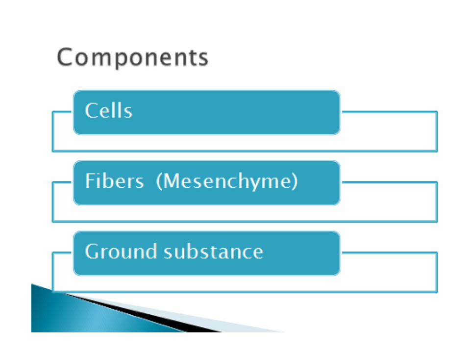
Structure of the CN-tissue• Connective tissue consists of cells &
intercellular substance or matrix. The matrix is further composed of two components.
1.Amorphous ground substance
2. Thread-like formed elements called fibers.• Different type of connective tissue differ from
each other in cell variety, chemical composition of ground substance, & number & kind of fibers.
• The connective tissue is classified as either loose connective tissue or dense connective tissue, depending on the amount, type, arrangement, and abundance of cells, fibers, and ground substance.
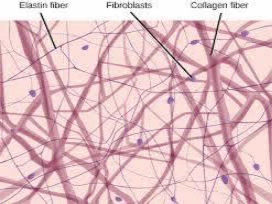
• Loose connective tissue. It is characterized by a loose, irregular arrangement of connective tissue fibers and abundant ground substance. Numerous connective tissue cells and fibers are found in the matrix. Collagen fibers, fibroblasts, adipose cells, mast cells, and macrophages predominate in loose connective tissue, with fibroblasts being the most common cell types.

• Dense connective tissue contains thicker and more densely packed collagen fibers, with fewer cell types and less ground substance. The collagen fibers in dense irregular connective tissue exhibit a random and irregular orientation. Dense connective tissue is present in the dermis of skin, in capsules of different organs, and in areas that need strong support.
• In contrast, dense regular connective tissue contains densely packed collagen fibers that exhibit a regular and parallel arrangement. This type of tissue is found in the tendons and ligaments. In both connective tissue types, fibroblasts are the most abundant cells, which are located between the dense collagen bundles.
Cells of the Connective Tissue• Many types of cells are found in different
varieties of connective tissue. Cell found in the connective tissue proper will discussed here .
Fibroblasts & Fibrocytes: Most common cell types in the connective
tissue. Fibrocytes are elongated & spindle-shaped with processes that contact adjacent cells & fibers .Nucleus is surrounded by scant amount pale cytoplasm. at the TEM level, the cytoplasm has sparse RER & a small golgi complex.
• Free ribosomes,mitochondria, lysosomes & vesicles are also presents.
• Actin filaments occur as bundles in the cell processes.
• Fibrocytes maintain the CN-tissue matrix by forming the fibers & constantly renewing the ground substances.
Fibroblasts:• These cells constitute the most abundant variety
of CN-tissue cells. the fibroblast appear as large somewhat flattened, roughly ovoid cells with branching processes.

• At the EM level, abundant rER & a prominent Golgi complex are present in the cytoplasm. There structural characteristics indicate more active CN-tissue matrix production in comparision to the fibrocyte. Fibroblast may arise directly from undifferentiated mesenchymal cells or are transformed from fibrocytes under the influence of microenviromentsal factors(cytokines).
• In certain situation fibroblast may differentiate into adipose cells, chondroblasts, or osteoblasts.
Histocytes (Macrophages):Are phagocytic cells and are most numerous in loose connective tissue. They are difficult to distinguish from fibroblasts, unless they are performing phagocytic activity and contain ingested material in their cytoplasm.
• They occur most frequently in the richly vasularized areas of the body.
• Histocytes are imp agents of defence b/c of their mobility & phagocytic properties, they are able to act as scavenger cells. They engulf dead cells extravasated blood cells, bacteria & foreign bodies
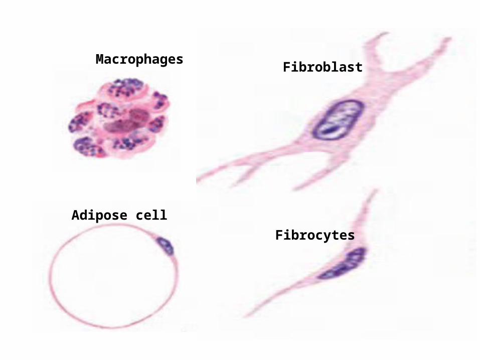
Fibroblast
Fibrocytes
Adipose cell
Macrophages
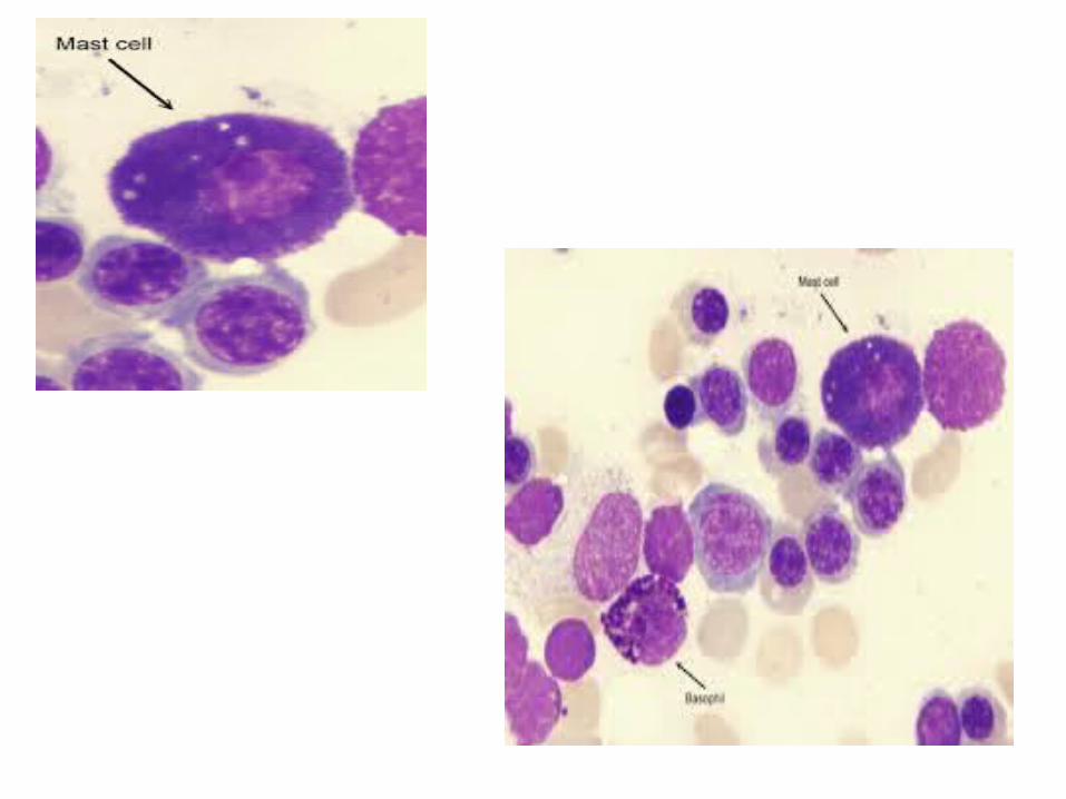

Mast cells:
Mast cells, usually closely associated with blood vessels, are widely distributed in the connective tissue of the skin and in the digestive and respiratory organs. Mast cells are spherical cells filled with fine, regular dark-staining and basophilic granules with centrally placed pale-staining nucleus.
• Mast cells secrete heparin (a powerful anticoagulant) & histamine ( a potent vasodilator)

Plasma cells:
These cells are rare in CN-tissue but are numerous in sites subject to the penetration of bacteria & foreign proteins (e.g., the intestinal mucosa) & in area where chronic inflammation is present.
• Plasma cells are large, ovoid cells, having a basophilic cytoplasm. This basophilia is due to abundance of RER. Nucleus of plasma cell is spherical in shape & eccentric in position. With the nucleus, chromatin occurs as course granules arranged in a regular manner against the nuclear membrane due to which the nucleus is said to exhibit a cart-wheel appearance.

• Plasma cells are most numerous in lymphatic tissue , especially in the center medullary cord of lymph nodes. they are also particularly abundant in bone marrow, the loose CN-tissue underlying the epithelium of the G.I.T, the respiratory system, & the female reproductive system.
• Plasma cells do not originate in loose CN-tissue but develop from B lymphocytes that immigrate into CN-tissue from the blood; they produce circulating or humoral antibodies
Adipocytes:
Adipocytes are also referred to as adipose cells. individual adipocytes or clusters containing multiple cells are normal components of loose CN-Tissue, but when the far cells out number other cells types, the tissue is called adipose tissue.
• The fat cells are large in size 50-150um in diameter & have an ovoid or spherical shape.
• The cytoplasm is displaced to the peripheral region of the cell by a single large fat droplet.
• Nucleus is flattened & found pressed against the cell membrane
Wandering cells:
These cells are not normally present in the connective tissue but they are temporary
visitors from the blood & lymp stream.e.g
lymphocytes, eosinophils and neutrophils.
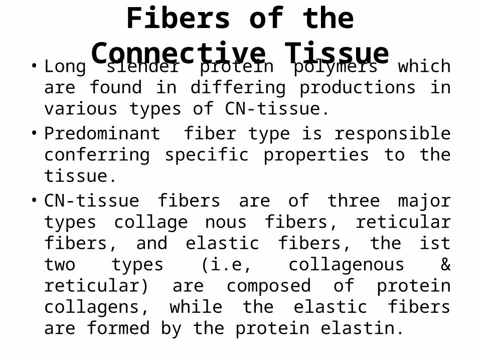
Fibers of the Connective Tissue• Long slender protein polymers which are
found in differing productions in various types of CN-tissue.
• Predominant fiber type is responsible conferring specific properties to the tissue.
• CN-tissue fibers are of three major types collage nous fibers, reticular fibers, and elastic fibers, the ist two types (i.e, collagenous & reticular) are composed of protein collagens, while the elastic fibers are formed by the protein elastin.
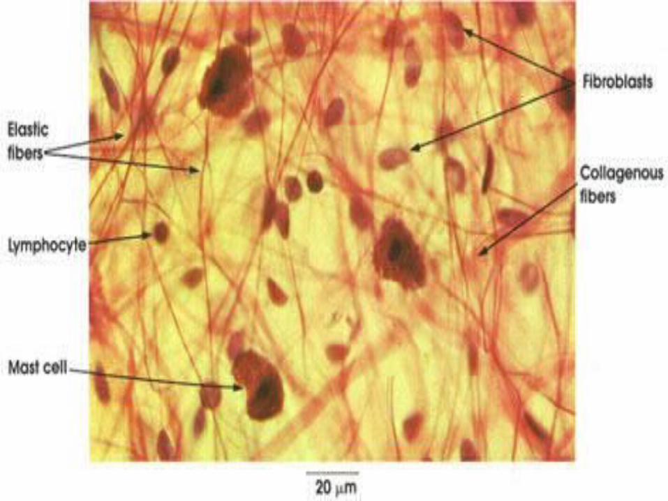
• Collagen FibersThe collagenous fibers are the most commonly occurring CN-tissue fibers. These are made up of the protein collagens type 1 . Collagen fibers are tough, thick, fibrous proteins that do not branch.
The collagenous fibers are flexible but inelastic(i.e, non-extensible) and, thus they provide a unique combination of flexibility & strength to the structure in which they are present.
In H&E preparation examined under L/M, collagen fibers stain acidophilic, taking a pink colour with Eosin.
The most frequently recognized fibers in histologic slides are the following:
1.Type I collagen fibers. (most abundant occur)These are found in the dermis of skin, tendons, ligaments, and bone & organ capsules. They are very strong and offer great resistance to tensil stresses.
2. Type II collagen fibers. These are present in hyaline cartilage and elastic cartilage and in vitreous body of the eye. The fibers provide resistance to pressure.

3. Type III collagen fibers. These are the thin, branching reticular fibers that form the delicate supporting meshwork in such organs as the lymph nodes, spleen, and bone marrow.
4. Type IV collagen fibers. These are present in the basal lamina of the basement membrane, to which the basal regions of the cells attach.
5.Type V collagen fibers. It is mainly present in fetal membrane and placenta.

• Reticular Fibers: (a very thin, branched fibers, which form extensive network in certain organs) .Reticular fibers, consist mainly of type III collagen, are thin and form a delicate net like framework in the liver, lymph nodes, spleen, hemopoietic organs, and other locations where blood and lymph are filtered. Reticular fibers also support capillaries, nerves, and muscle cells.

• Elastic Fibers these fibers are highly elastic. They can be stretched easily even by weak traction forces but return to their original length when these forces are removed.
Elastic fibers are found in abundance in the lungs, bladder, skin,ligamenta flava, ligamentum nuchae,pinna of ear, vocal cords, epiglottis, musculars arteries.
Ground Substance of the Connective Tissue
• Cell & fibers of the CN-tissue are embedded in a highly hydrated gel which is called ground substances. The water bound by ground substance serves as the medium through which all nutrients & waste products must pass in transit between the blood and the parenchymal cells of the organs.
• Ground Substance of the CN-tissue consist mainly of proteoglycans.in addition it contain adhesive glycoprotein, fibronectin & laminin.
• Several types of proteoglycans have been isolated from the ground substance of the connective tissue in different location with in the body, these include hyaluronic acid, chondroition sulfate, keratan sulfates 1 & 11, heparan sulfate & dermatan sulfate .
• The adhesive glycoprotein fibronectin & laminin play very important role in adhesion of cells to the extracellular matrix.
BASEMENT MEMBRANE• The extensive interface b/w CN-tissue &
various epithelia is characterized by the presence of thin layers of extracellular material which is called basement membrane(or basal lamina).
• Composed of two layers
(1)Basal lamina-directly beneath the basal plasmalemma of the epithelial cells.
(2) Reticular lamina—the thick layer, rich in reticular fibers, merging into underlying CN-tissue

• Under the EM basal lamina is seen to consist of three layers or laminae, there is a central electron-dense layer(called lamina densa) having on either side an electron-lucent layers.
• There are two locations in the body where a single basal lamina is found b/w two adjacent epithelial layers
(1)Alveoli of the lung,
(2)Glomeruli of the kidney
Functions of the Basement Membrane• bind the cells to the underlying or surrounding
CN-tissue• Provide the epithelial cells a flexible support
capable of stretching & recoiling• To serve as a molecular sieve or ultrafilter,
impeding the passage of macromolecules
Classification of CN-tissue• Classification into various types depending on the
following four criteria.
(1)Relative proportion of the various fibers presents
(2) Compactness & arrangement of these fibers
(3)Nature of the matrix
(4)types of cells present
Above mention criteria CN-tissue are the following two groups
(A)Embroyonal CN-tissue
(B)Adult CN-tissue

EMBROYONAL CN-TISSUE• Developmentally, the CN-tissue are derived
from mesoderm which is one of the three primary embryonic layers.
• The immature CN-tissue of the embryo derived from the mesoderm is known as mesenchyme. It is composed of roughly star-shaped cells which lie in an abundant, relatively homogenous intercellular substance.
• As the development proceeds, the mesenchyme gradually assumes characteristics of Adult CN-tiisue.

• The first changes appears of fibers in the intercellular substance which thus becomes more viscous .
• The embryonic CN-tissue of this stages is known as mucous tissue. Widely distributed in the body of fetus. The umbilical cord also contain a considerable amount of mucous tissue .
ADULT CN-TISSUE• Divide into mail three varities
1. CN-tisue proper
2. Cartilage
3. Bone
1. CN-tissue proper discuss earlier as loose & dense CN-tisse
CARTILAGE• Cartilage is a special form of connective tissue
that also develops from the mesenchyme. Similar to the connective tissue, cartilage consists of cells and extracellular matrix composed of connective tissue fibers and ground substance. In contrast to connective tissue, cartilage is nonvascular(avascular) and receives its nutrition via diffusion through the extracellular matrix(cappilaries located in adjacent CN-tissue) or synovial fluid present in joint cavities. there is no lymphatic vessels or nerves in the cartilage.
Perichondrium• It is a special capsule like structure , composed
of dense irregular connective tissue that surrounds cartilages in most places. the perichondrium contain blood vessels which is responsible for the supply of nutrients & oxygen to the perichondrium is seen to be composed of two layers.
1.Outer fibrous layer which contains collagenous & elastic fibers, fibroblast & large blood vessels.
2. Inner cellular layers which lodges cartilages forming cells known as chondroblasts. In addition, this layer contains fine blood vessels and a few collagenous fibers.

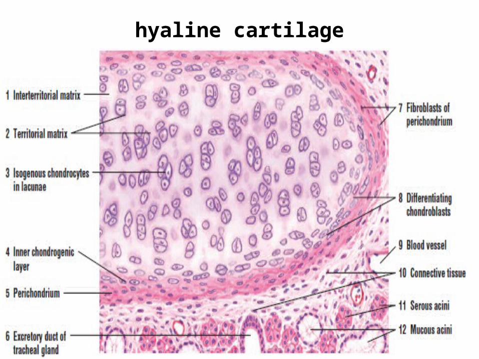
Cartilage Types• Hyaline Cartilage (glass like):
its intercellular substance appears clear & uniform & the fibers, although present, are not visible under the LM stained sections.
this cartilage is mostly widely distributed cartilage in the body. it is very flexible & some what elastic and is covered by perichondrium except for articular cartilages of the synovial; joints, which obtain their nutrients & oxygen, by diffusion from the synovial fluid.
Cells Of Hyaline Cartilage • Cells are chondrocytes. These are large
roughly spherical cells, each containing a bid centrally- placed nucleus one are more nuclei.
• The cytoplasm is finely granular & moderatly basophilic.
• The chondrocytes lie with in small cavities in the matrix. These cavities are called lacunae.
• In the lacunae are mature cartilage cells called chondrocytes. The main function of chondrocytes is to maintain the cartilage matrix.

Arrangement of chondrocytes• In the central region of the hyaline cartilage,
the cells are generally arranged in groups known as isogenous groups.the cells of a group are spherical or ovoid in shape and are flattened on adjacent sides. All the members of an isogenous groups occupy a single lacuna

Hyaline Cartilage Matrix• It is imp to know that the apparently
homogenous matrix of hyaline cartilage contains numerous fibrils composed of collagen types 11.these fibrils are masked by ground substances because of the following two reasons.
1.The fibrils are very fine being 10-25nm in diameter .hence they are beyond the resolving power of the LM
2.The fibrils & ground substance have nearly the same refractive index. resulting in a lack of contrast between these two components.

• The type 11collagen fibrils can easily be seen E/M. they are not arranged into bundles but form a fine network.
• Due to the abundance of chondroitin sulfate. The hyaline cartilage matrix appears basophilic in routine H&E preparation.
• The region immediately around an isogenous cell group stain intensely basophilic b/c it contain large amounts of chondroitin sulfate but relatively small numbers of collagenous fibrils .such regions are known as territorial matrix or capsule (of the lacunae)
• That matrix which lies b/w the cells groups contains relatively smaller amount of chondroitin sulfate & thus stain lightly basophilic .thus matrix is known as interterrittorial matrix.
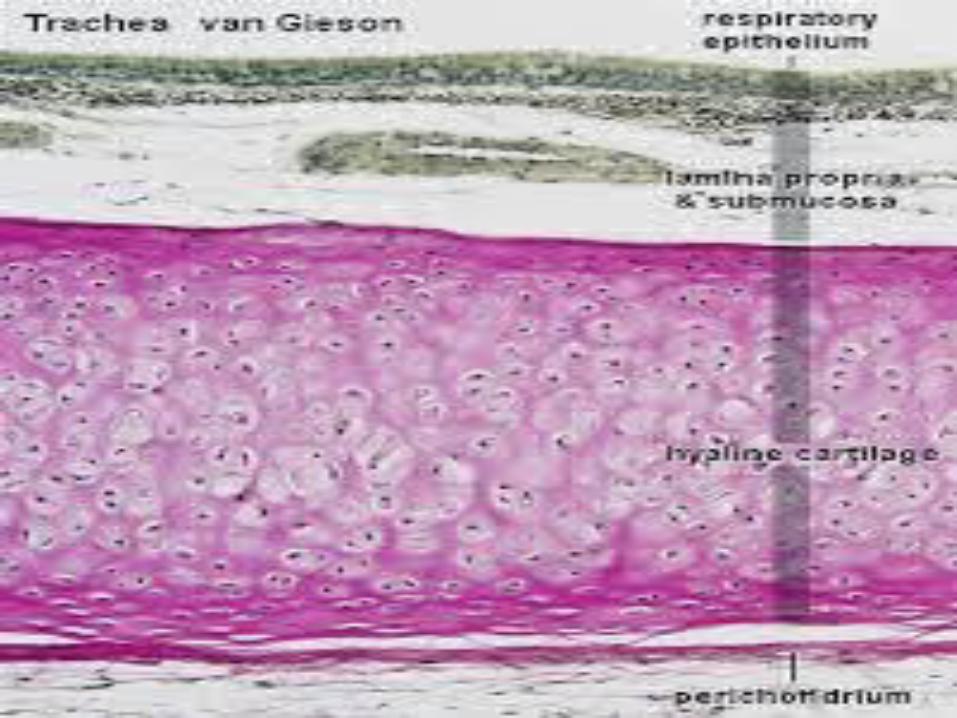
Distribution of hyaline cartilage :• ribs (costal cartilage), nose, larynx, trachea,
and in bronchi. Here, the hyaline cartilage persists through out life and does not calcify.
hyaline cartilage


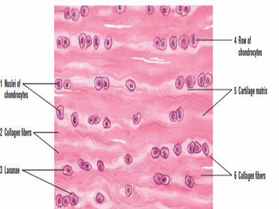
Fibrocartilage• Fibrocartilage is characterized by large
amounts of irregular and dense bundles of coarse collagen fibers in its matrix. In contrast to hyaline and elastic cartilage, fibrocartilage consists of alternating layers of cartilage matrix and thick dense layers of type I collagen fibers.
• In fibrocartilage the cells are not arranged as groups but instead they form shorts rows. abundance of collagens fibers gives a general acidophilic reaction to the matrix .
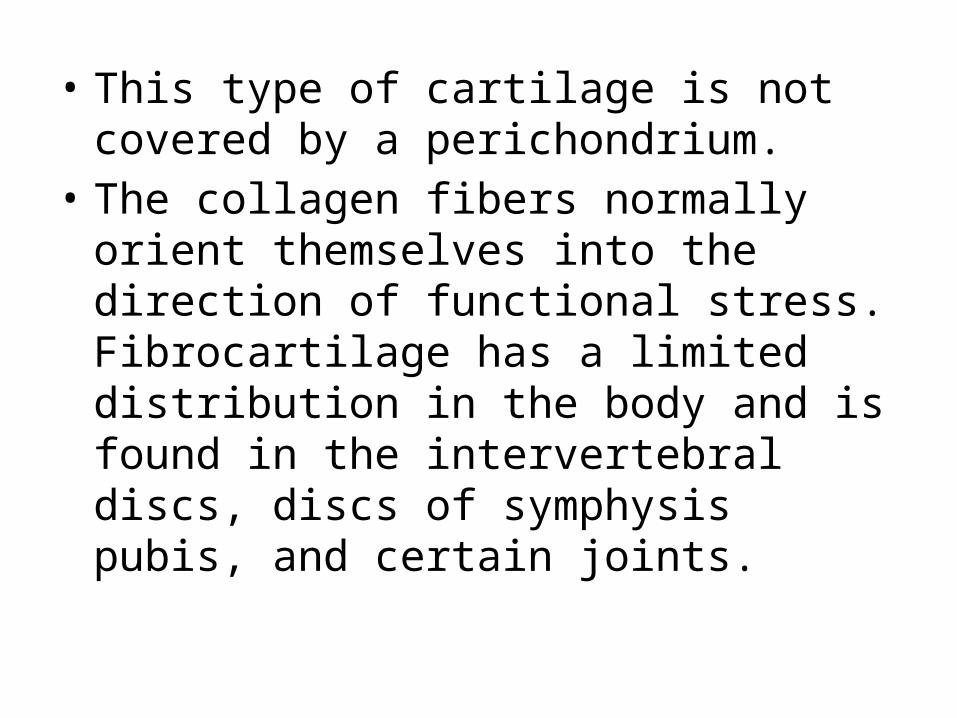
• This type of cartilage is not covered by a perichondrium.
• The collagen fibers normally orient themselves into the direction of functional stress. Fibrocartilage has a limited distribution in the body and is found in the intervertebral discs, discs of symphysis pubis, and certain joints.

Elastic Cartilage• Elastic cartilage is similar in appearance to
hyaline cartilage, except for the presence of numerous branching elastic fibers within its matrix which branched and interlace with each other to form a closely woven network.
• The elastic fibers, the matrix also contains fibrils composed of collagens type 11.
• The elastic fiber covered by perichondrium.• Elastic cartilage is highly flexible and occurs
in the external ear, walls of the auditory tube, epiglottis, and larynx.

Elastic cartilage

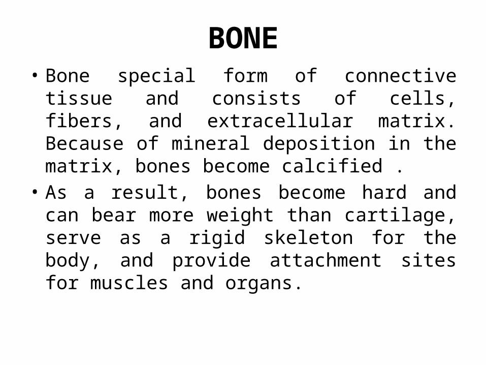
BONE• Bone special form of connective tissue and
consists of cells, fibers, and extracellular matrix. Because of mineral deposition in the matrix, bones become calcified .
• As a result, bones become hard and can bear more weight than cartilage, serve as a rigid skeleton for the body, and provide attachment sites for muscles and organs.

• Bone also protects the brain in the skull, heart and lungs in the thorax, and urinary and reproductive organs between the pelvic bones. In addition, bones function in hemopoiesis (blood cell formation), and serve as crucial reservoirs for calcium, phosphate, and other minerals.
• Bone function metabolically providing a source of calcium to maintain proper calcium levels and various growth factors.
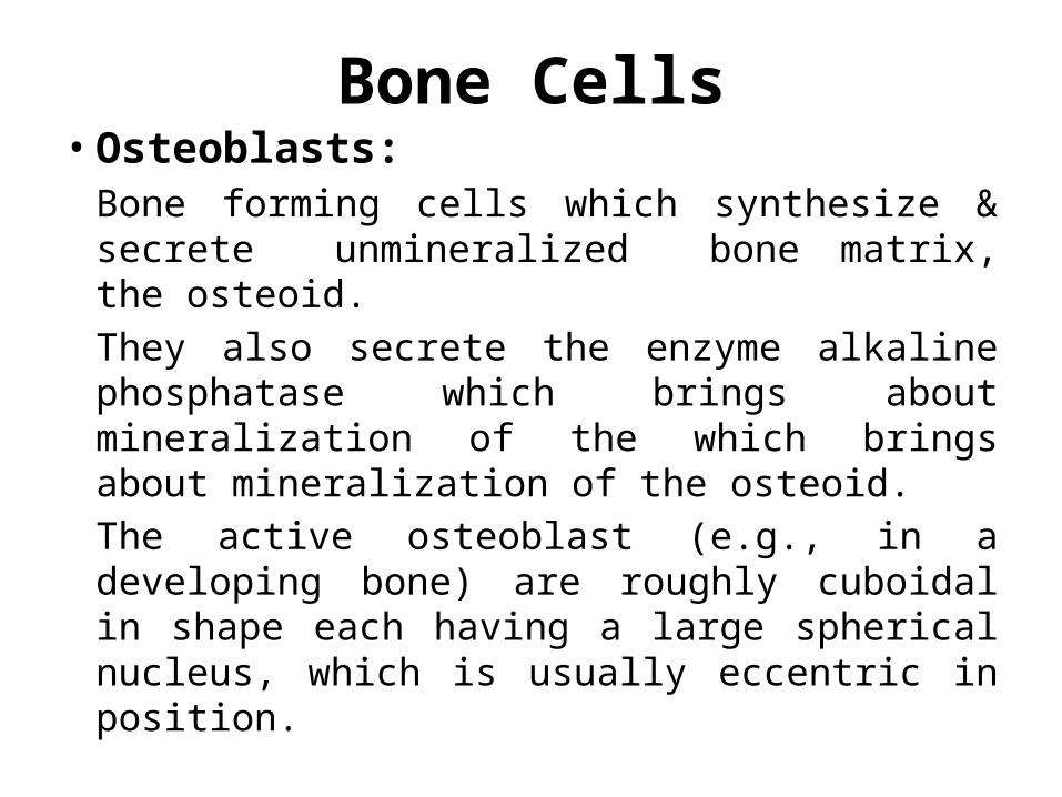
Bone Cells• Osteoblasts:
Bone forming cells which synthesize & secrete unmineralized bone matrix, the osteoid.
They also secrete the enzyme alkaline phosphatase which brings about mineralization of the which brings about mineralization of the osteoid.
The active osteoblast (e.g., in a developing bone) are roughly cuboidal in shape each having a large spherical nucleus, which is usually eccentric in position.

• The cytoplasm appears markedly basophilic in the routine H&E stained sections. E/M reveals but the basophilia is due the presence of a large quantity of rough endoplasmic reticulm.
• The inactive or resting osteoblast (also called bone linning cells) appear as fusiform (spindle-shaped) cells having very slightly basophilic cytoplasm and a small darkly staining nucleus.
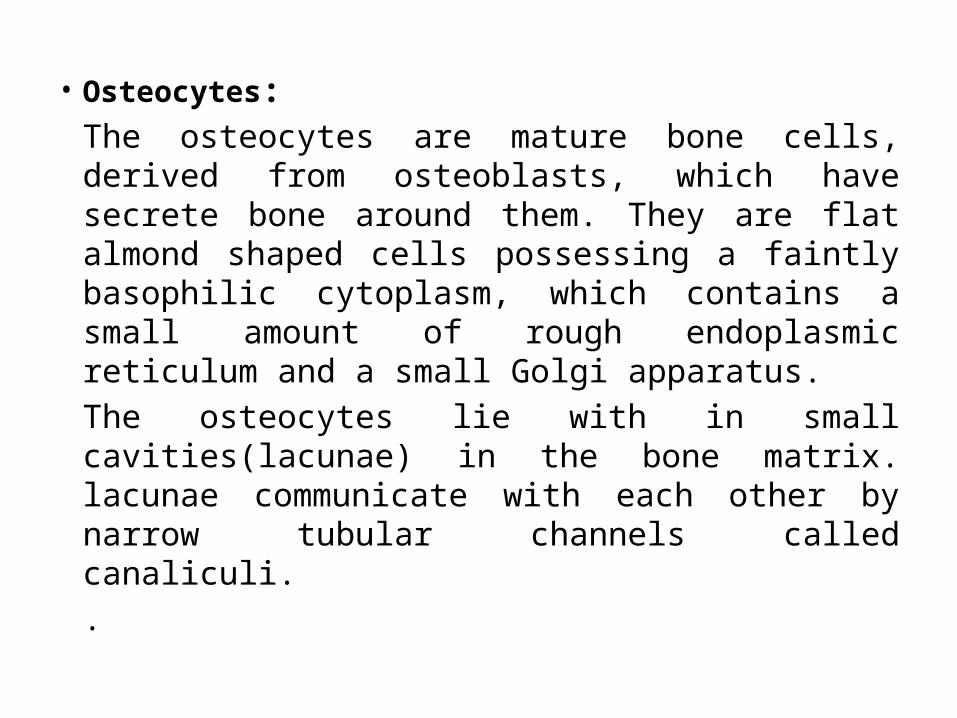
• Osteocytes:The osteocytes are mature bone cells, derived from osteoblasts, which have secrete bone around them. They are flat almond shaped cells possessing a faintly basophilic cytoplasm, which contains a small amount of rough endoplasmic reticulum and a small Golgi apparatus.
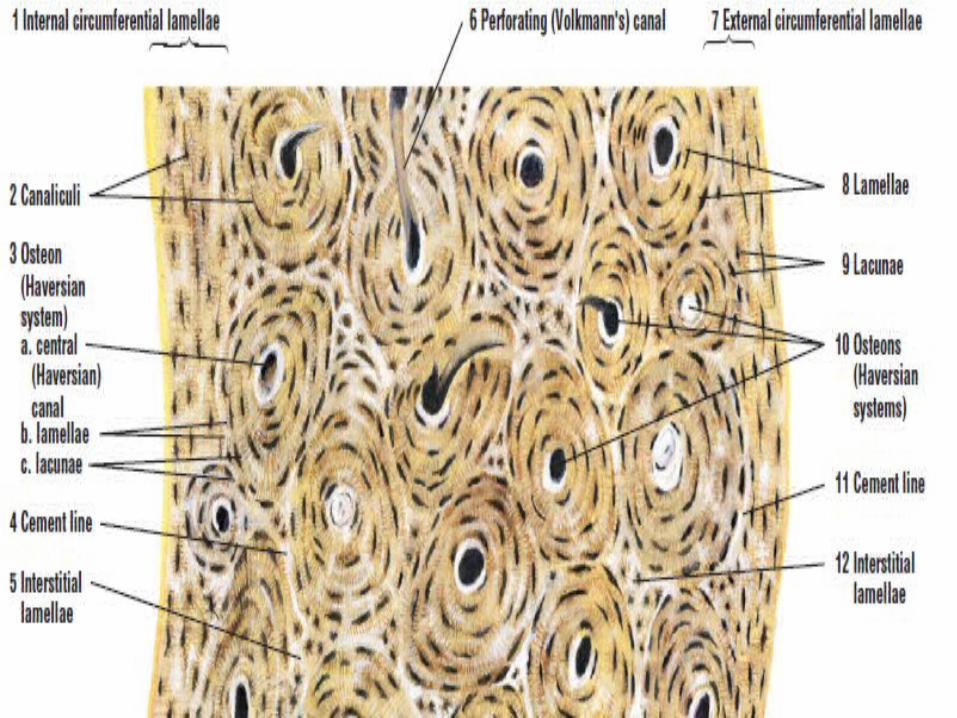
The osteocytes lie with in small cavities(lacunae) in the bone matrix. lacunae communicate with each other by narrow tubular channels called canaliculi.
.
• fine cytoplasmic processes from each oosteocyte extend for some distance into the canalculi & make contac with similar processes from neighbouring cells.
• Gap junction are present where the cytoplasmic processess contac each other .these junctions provide for intercelluar flow of ions & small molecules .
• The intercellular flow provides a mechanism by which nutrients & metabolites can be exchanged b/w the blood vessels(present in the periosteum or haversion canals) & distant osteocytes which lie imprisoned with in the impermeable bone matrix
• Osteoclasts: large multinucleated cells found along bone
surfaces where resorption(removal of bone),
remodeling, and repair of bone take place.
The main function of osteoclasts is bone resorption during remodeling (renewal or restructuring) and calcium haemostasis. Osteoclasts are often located on the resorbed surfaces or in shallow depressions in the bone matrix called Howship’s lacunae. Lysosomal enzymes released by osteoclasts erode these depressions.

Bone Matrix• The bone matrix consists of living cells and
extracellular material. Because the bone matrix is calcified or mineralized, it is harder than cartilage.
• Bone matrix contains both organic and inorganic components. The organic components enable bones to resist tension, while the mineral components resist compression.

• The major organic components (35% of the dry weight of the bone) of bone matrix are the coarse type I collagen fibers, which are the predominant proteins & amorphous ground substance(proteoglycan of bone is chondroitin sulphate ).
• The inorganic component (65% of the dry weight of bone) of bone matrix consists of the minerals calcium and phosphate in the form of hydroxyapatite crystals. The association of coarse collagen fibers with hydroxyapatite crystals provides the bone with its hardness, durability, and strength.
Bone Formation (Ossification)• Bone formation process starts in the early
intrauterine life & continuous into adult hood of the person. the bony tissue ,like all other types of CN-tissue, is derived from mesenchyme .
• Bone developed from mesenchyme by two method.
1.Intra membranous ossification
2.Endochondral ossification
Intramembranous Ossification• The process of intramembranous ossification
begins in the second month of intrauterine life.• In this type of ossification mesenchyme is
directly converted into bone with out intervening stage of cartilage formation.
• Some mesenchyme cells differentiate directly into osteoblasts that produce the surrounding osteoid matrix, which quickly calcifies. Numerous ossification centers are formed, anastomose, and produce a network of spongy bone that consists of thin rods, plates, and spines called trabeculae.
• The osteoblasts then become surrounded by bone in the cavelike lacunae and become osteocytes. As in endochondral ossification, once osteocytes are in the lacunae, they establish a complex cell-to-cell connection through the canaliculi. The mandible, maxilla, clavicles, and most of the flat bones of the skull are formed by the intramembranous method.
Endochondral Ossification• In this type of bone formation, the
mesenchyme is the first converted into cartilage which serves as temporary supportive framework. this cartilage is then replace by bone. this processes may be called indirect method of bone formation
• Most bones in the body develop by the process of endochondral ossification, in which a temporary hyaline cartilage model precedes bone formation.
• The chondrocytes divide, hypertrophy (enlarge), and mature, and the hyaline cartilage model begins to calcify.
• Calcification of the cartilage model continues, diffusion of nutrients and gases through the calcified matrix decreases
• Chondrocytes die and the fragmented calcified matrix serves as structural framework for the deposition of bony material
• layer of bony material is deposited around the calcifying cartilage, the inner perichondrial cells exhibit their osteogenic potential
• Thin periosteal collar of bone forms around the midpoint of the shaft of the bone.
• This external surrounding connective tissue is now called the periosteum. Mesenchyme cells differentiate into osteoprogenitor cells from the inner layer of periosteum, and blood vessels from the periosteum invade the calcified and degenerating cartilage model.
• Osteoprogenitor cells proliferate and differentiate into osteoblasts that secrete the osteoid matrix, a soft initially collagenous tissue that lacks minerals but is quickly mineralized into the bone.

• The osteoblasts are then surrounded by bone in the cavelike lacunae and are now called osteocytes;there is one osteocyte per lacuna. Osteocytes establish a complex cell-to-cell connection through tiny canals in the bone called canaliculi; these eventually open into channels that house the blood vessels. Osteoprogenitor cells also arise from the inner surface of bone called endosteum.
• Endosteum lines all internal cavities in the bone and consists of a single layer of osteoprogenitor cells.
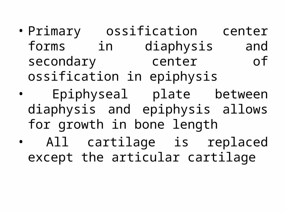
• Primary ossification center forms in diaphysis and secondary center of ossification in epiphysis
• Epiphyseal plate between diaphysis and epiphysis allows for growth in bone length
• All cartilage is replaced except the articular cartilage
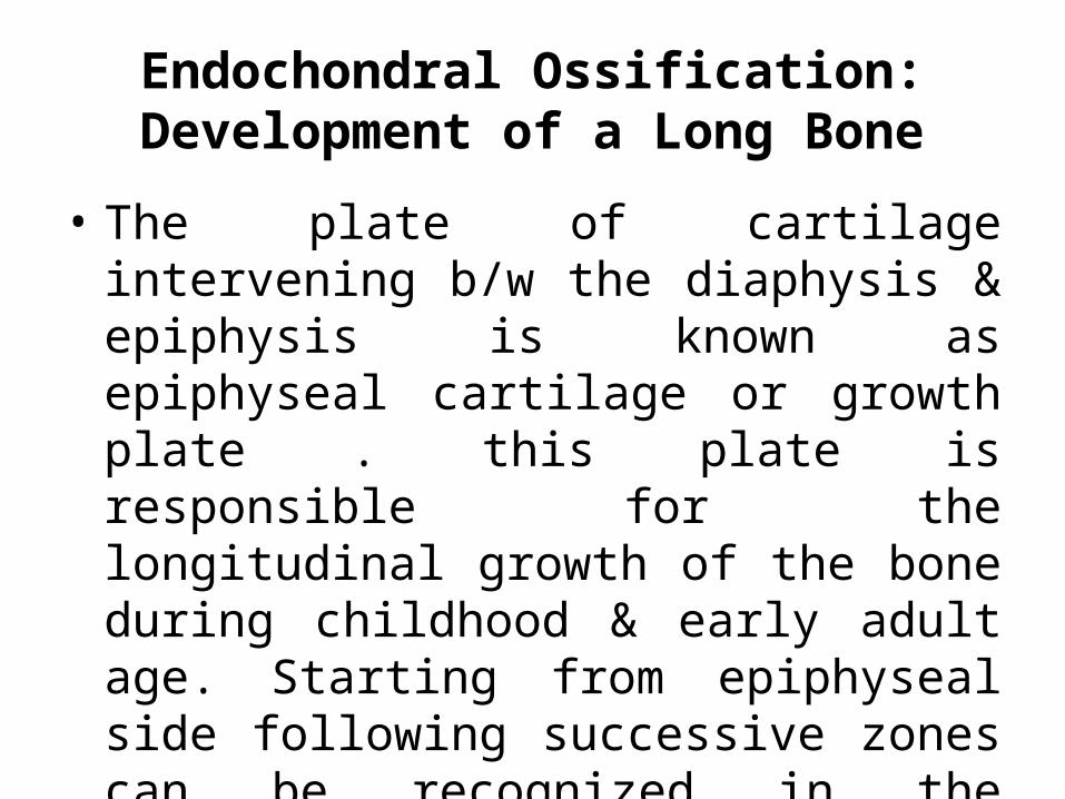
Endochondral Ossification: Development of a Long Bone
• The plate of cartilage intervening b/w the diaphysis & epiphysis is known as epiphyseal cartilage or growth plate . this plate is responsible for the longitudinal growth of the bone during childhood & early adult age. Starting from epiphyseal side following successive zones can be recognized in the epiphyseal cartilage.
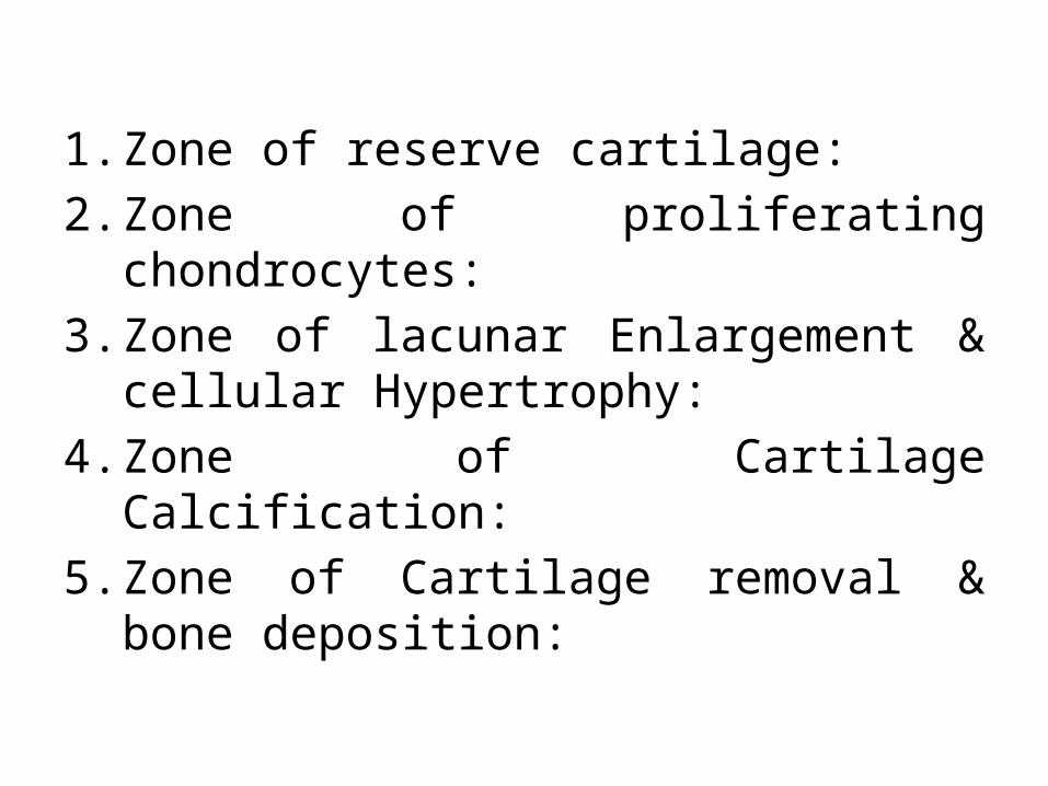
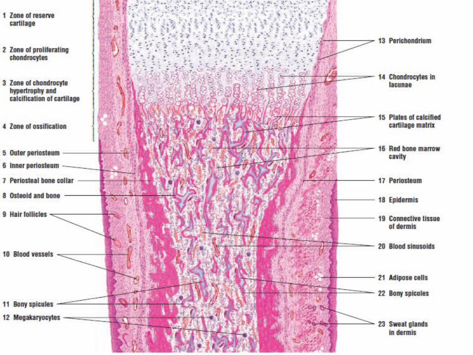
1. Zone of reserve cartilage:
2. Zone of proliferating chondrocytes:
3. Zone of lacunar Enlargement & cellular Hypertrophy:
4. Zone of Cartilage Calcification:
5. Zone of Cartilage removal & bone deposition:

Microscopic Structure of the Bone Tissue
Osteon
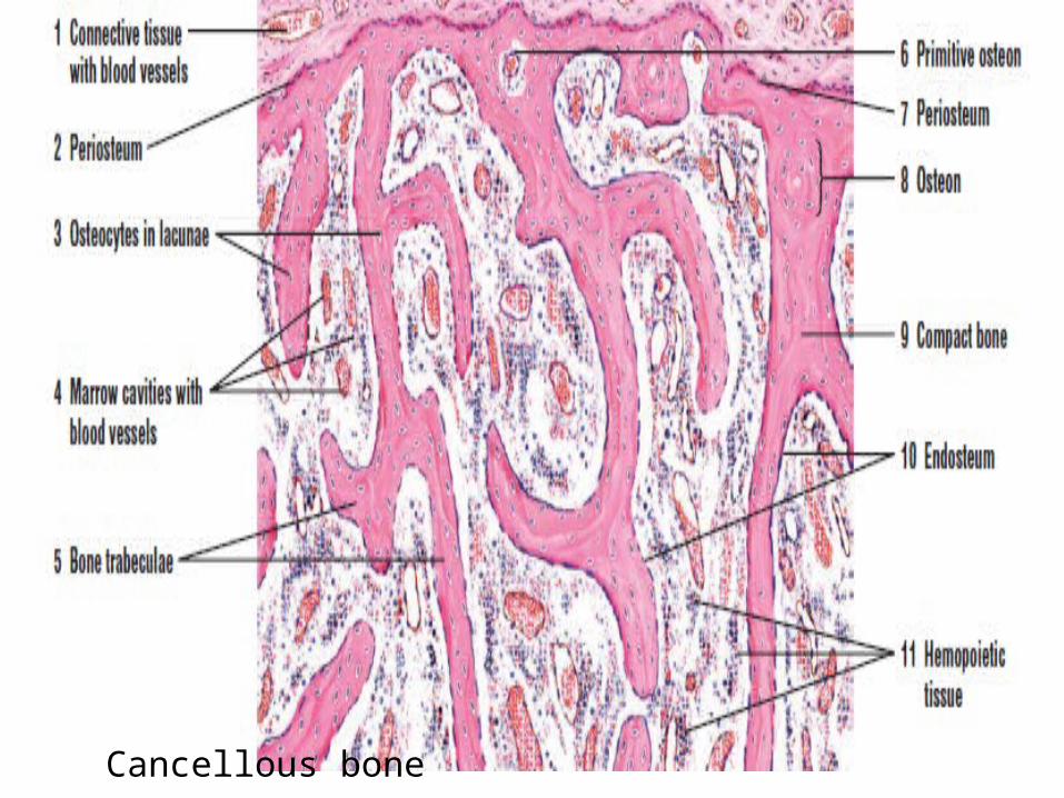
Cancellous bone