complete blood count - sinoe medical association...
TRANSCRIPT
The Complete Blood Count (Cartesian Thinking at Its Best)
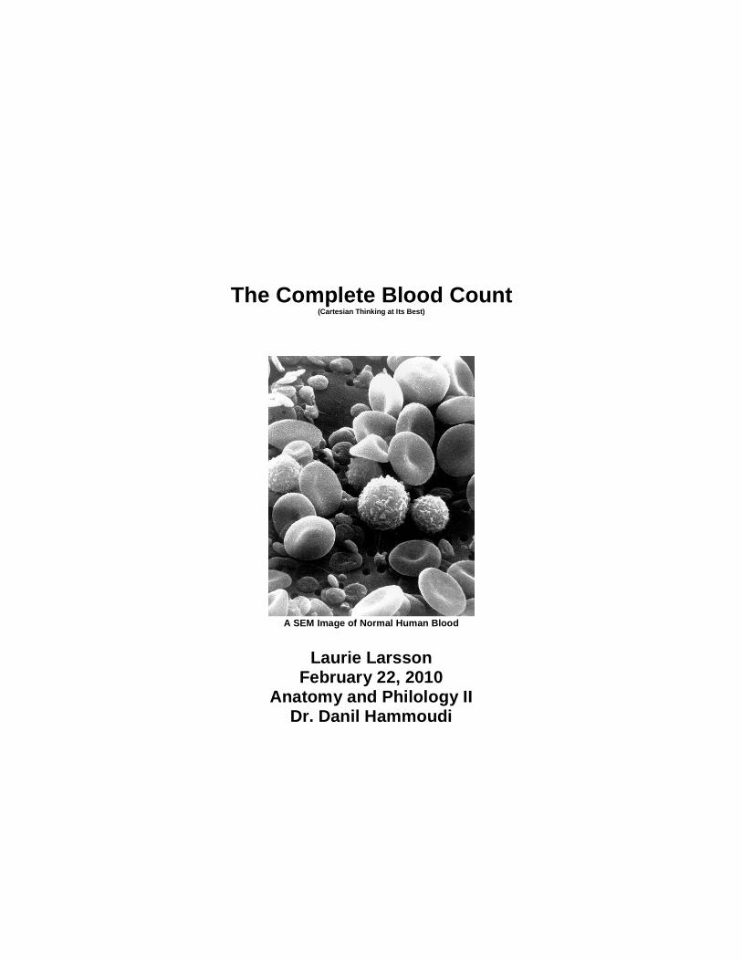
A SEM Image of Normal Human Blood
Laurie Larsson
February 22, 2010 Anatomy and Philology II
Dr. Danil Hammoudi
Introduction A complete blood count (CBC) is a common screening tool. It can be used to establish a baseline in a healthy patient during a routine physical or diagnose disorders such as anemia, infection and disease in a sick patient. A CBC is a panel of tests that includes the following:
Red blood cell (RBC) count Hematocrit Hemoglobin Mean corpuscular volume (MCV) Mean corpuscular hemoglobin (MCH) Mean corpuscular hemoglobin concentration (MCHC) Red cell distribution width (RDW) White blood cell count (WBC) White blood cell differential Platelet count Mean platelet volume (MPV)
In the subsequent sections, each test is described. Its normal range of results is presented. Deviations from the standard values are interpreted and typical related diseases and conditions are presented. Tests That Study Red Blood Cells
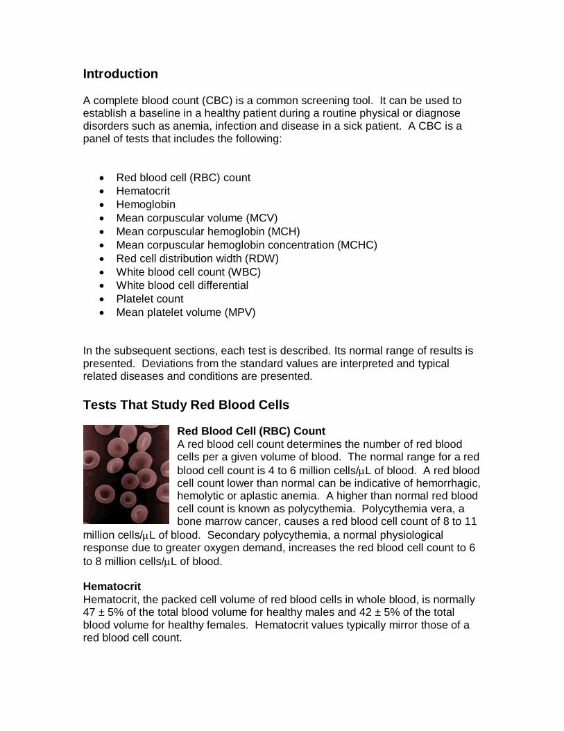
Red Blood Cell (RBC) Count A red blood cell count determines the number of red blood cells per a given volume of blood. The normal range for a red blood cell count is 4 to 6 million cells/L of blood. A red blood cell count lower than normal can be indicative of hemorrhagic, hemolytic or aplastic anemia. A higher than normal red blood cell count is known as polycythemia. Polycythemia vera, a bone marrow cancer, causes a red blood cell count of 8 to 11
million cells/L of blood. Secondary polycythemia, a normal physiological response due to greater oxygen demand, increases the red blood cell count to 6 to 8 million cells/L of blood. Hematocrit Hematocrit, the packed cell volume of red blood cells in whole blood, is normally 47 ± 5% of the total blood volume for healthy males and 42 ± 5% of the total blood volume for healthy females. Hematocrit values typically mirror those of a red blood cell count.
Hemoglobin The amount of hemoglobin, the iron-containing, oxygen-transport protein found in red blood cells, is measured in g/dL. Normal values are as follows: Men: 13.5 to 16.5 g/dL Women: 12.1 to 15.1 g/dL Children: 11 to 16 g/dL Pregnant Women: 11 to 12 g/dL When hemoglobin molecules are normal, but the amount of hemoglobin in the blood is low, iron-deficiency anemia is suspected. Iron-deficiency anemia is generally a secondary consequence of a hemorrhage. However, it can also be caused by inadequate intake of iron-containing foods and impaired iron absorption. Mean Corpuscular Volume (MCV) The mean corpuscular volume is the average red blood cell volume. It is calculated by dividing the hematocrit by the red blood cell count. The normal range is 80 to 100 fL (10-18m3). A high MCV is typical in the macrocytes associated with pernicious anemia, a vitamin B12 deficiency. A low MCV is common with the microcytes of iron-deficient anemia. Mean Corpuscular Hemoglobin (MCH) Mean corpuscular hemoglobin is the average mass of hemoglobin per red blood cell. It is calculated by dividing the mass of hemoglobin by the red blood cell count. The normal value is 27 to 31 pg/cell. MCH values track MCV values. Mean Corpuscular Hemoglobin Concentration (MCHC) Mean corpuscular hemoglobin concentration is a measure of the concentration of hemoglobin in a given volume of packed red blood cells. It is calculated by dividing the hemoglobin value by the hematocrit value. The normal range is 32 to 36 g/dL. MCHC may decrease as MCV decreases; the increase is limited by the maximum amount of hemoglobin that fits inside a red blood cell. Red Cell Distribution Width (RDW) Red cell distribution width is a measure of the variation of the red blood cell width. “Width” is somewhat misleading. The calculation is actually of volume and not diameter. RDW = (the standard deviation of MCV ÷ MCV x 100). RDW is useful in determining anemias of mixed cause rather than of a single cause.

Tests That Study White Blood Cells White Blood Cell Count
A white blood cell count determines the number of white blood cells, leukocytes, per a given volume of blood. The normal value is 4,800 to 10,800 cells/L of blood. Leukocytosis is defined as a white blood cell count greater than 11,000 cells /L of blood and is common in acutely ill patients. Some causes are bacterial, viral,
fungal or parasitic infections, cancer, hemorrhage or exposure to chemicals or medications. A differential white count gives more specific, targeted information as to which leukocytes levels are elevated. Leukopenia is a lower than normal white blood cell count. Common causes are leukemia, chemotherapy, radiation therapy and aplastic anemia. White Blood Cell Differential The white blood cell differential looks at the number of each type of white blood cell for a given blood volume. Because each type of cell has a specific function, an increase or decrease in the number of that cell type can indicate the class of disease or condition that may be occuring. Neutrophils: The normal neutrophil count is 3000 to 7000 cells /L of blood. Neutrophilia, an increase in the number of neutrophils above the normal range, may be caused by a bacterial infection, acute inflammation such as a heart attack or other infarct, an appendicitis, or chronic myelogenous leukemia. A decrease in the number of neutrophils, neutropenia, may indicate a viral infection, decreased bone marrow production or an autoimmune condition. Eosinophils: The eosinophil count is typically 100 to 400 cells/L of blood. A high concentration of eosinophils, eosinophilia, is caused by allergic reactions and parasitic infections. Eosinopenia, a lower than normal count, can signify a bacterial infection. Basophils: The normal basophil count is 20 to 50 cells/L of blood. An elevated levels of basophils, known as basophilia, is uncommon as an isolated finding. However, it is a common feature of myeloproliferative disorders and prominent in chronic granulocytic leukemia. Low levels of basophils, basopenia, are difficult to detect. However, one known cause is urticaria. Lymphocytes: The typical lymphocyte count is 1500 to 3000 cells/L of blood. High concentrations of lymphocytes, lymphocytosis, are associated with acute viral infections such as mononucleosis, Epstein-Barr virus and hepatitis. They are also consistent with both chronic lymphocytic leukemia and acute lymphoblastic leukemia. Lymphocytopenia, a lower than normal lymphocyte count, is usually a temporary condition caused by a recent infection such as the common cold.
Monocytes: The normal monocyte count is 100 to 700 cells/L of blood. Monocytosis, an elevated monocyte count, is indicative of chronic inflammation, necrosis, infection mononucleosis, and stress response. A lower than normal number of monocytes is not typically an isolated finding. Tests That Study Platelets
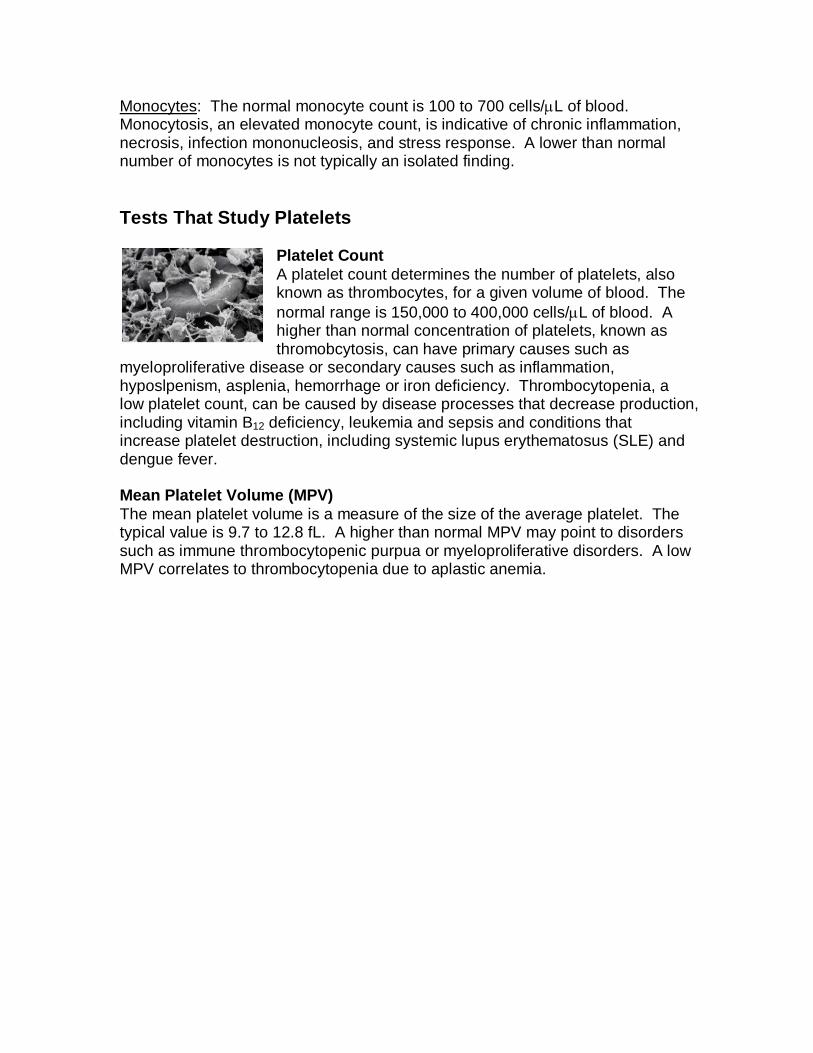
Platelet Count A platelet count determines the number of platelets, also known as thrombocytes, for a given volume of blood. The normal range is 150,000 to 400,000 cells/L of blood. A higher than normal concentration of platelets, known as thromobcytosis, can have primary causes such as
myeloproliferative disease or secondary causes such as inflammation, hyposlpenism, asplenia, hemorrhage or iron deficiency. Thrombocytopenia, a low platelet count, can be caused by disease processes that decrease production, including vitamin B12 deficiency, leukemia and sepsis and conditions that increase platelet destruction, including systemic lupus erythematosus (SLE) and dengue fever. Mean Platelet Volume (MPV) The mean platelet volume is a measure of the size of the average platelet. The typical value is 9.7 to 12.8 fL. A higher than normal MPV may point to disorders such as immune thrombocytopenic purpua or myeloproliferative disorders. A low MPV correlates to thrombocytopenia due to aplastic anemia.