comparison of acute respiratory tract infections caused by ...ijhbr.com/pdf/january 2017...
TRANSCRIPT
International J. of Healthcare and Biomedical Research, Volume: 05, Issue: 02, January 2017, 127-138
127 www.ijhbr.com ISSN: 2319-7072
Original article:
Comparison of Acute respiratory tract infections caused by Respiratory
syncytial virusand Human Rhinovirus in hospitalized patients of Children's
Hospital of Soochow University
PROFESSOR HAO CHUANG LI, DR.ANJUMUNNISA
Departments of Respiratory Medicine, and Clinical Laboratory, Children’s Hospital of Soochow University, Suzhou 215003,
China
Address for correspondence: Dr. Hao Chuang Li, Department of Respiratory Medicine, Children’s Hospital of Soochow University,
No. 303, Jing De Road, Suzhou 215003
Abstract
Objectives: It is not clearly established if co-infections are more severe than single viral respiratory infections. The objective of this study was
to study and to compare single infections and viral co-infections of respiratory syncytial virus (RSV) and Human Rhinovirus (HRV) in
hospitalized children in Suzhou.
Patients and Methods: From December 2012 to December 2015, a prospective study was conducted for patients admitted with respiratory
infection to the Pediatric Respiratory Department of the Children’s Hospital of Soochow University. Specimens of nasopharyngeal aspirate
were taken for virological study by using polymerase chain reaction, and clinical data was recorded. Single RSV and HRV infections were
selected and compared with mixed infections of RSV with HRV. Season of occurrence, gender, age distribution, duration of hospitalization,
clinical signs; symptoms, diagnosis, blood culture, laboratory findings and other supplementary examinations were evaluated and compared
among the three study groups
Results: In this study 1177 cases were analyzed. Single infections (587 RSV and 560 HRV) were compared with 30 RSV-HRV mixed
infections.
Conclusion: Co-infections between RSV-HRV are not very frequent. Overall viral co-infections do not present greater severity, but have
mixed clinical features. No significant differences in the admission diagnosis, laboratory parameters, patient demographics and treatment
measures between the two viral causes of respiratory illness were found. No correlation between viral load and disease severity was observed.
Keywords: Pneumonia, Co-infection, Children, Respiratory syncytial virus and Human rhinovirus.
INTRODUCTION
Acute respiratory tract infections (ARTIs) are common
diseases among children and a major cause of
hospitalization, mainly in infants. Viral pathogens play an
important role in infants who present ARIs, respiratory
syncytial virus (RSV) being the most important virus
associated [1]. In addition, children with RSV infections are
also exposed to a variety of other respiratory viruses with a
similar seasonal pattern, mainly during winter months, such
as influenza, human rhinovirus (HRV/ RV), human
metapneumovirus (hMPV), and Human Boca virus
(HBoV)[2-4]
. Despite the fact that numerous studies have
revealed that an important number of ARTI pediatric
patients become simultaneously infected with multiple
respiratory viruses, there are few studies focused on
analyzing viral co-infections. This issue usually becomes a
marginal part of the studies.
Over the past few years, several groups, including ours, have
described various viral co-infection combinations compared
with single ones, with different methodologies, and some of
them observed worse prognosis with multiple viral
infections, whereas others revealed a very similar prognosis
for single virus infections.
Thus, there is a need of a carefully designed study to shed
some light on this issue. We aimed to compare, in a
prospective study, clinical characteristics and severity of
International J. of Healthcare and Biomedical Research, Volume: 05, Issue: 02, January 2017, 127-138
128 www.ijhbr.com ISSN: 2319-7072
single versus viral co-infections, defined as simultaneous
detection of RSV with HRV, in a large cohort of
hospitalized children.
METHODS AND PATIENTS
Study site
The study was conducted at the Children’s Hospitalof
Soochow University in Suzhou City, Jiangsu, China.
Suzhouformerly Romanized as Soochow, is a major city
located in southeastern Jiangsu Province of East China,
about 100 km (62 mi) northwest of Shanghai. Its urban
population grew at an unprecedented rate of 6.5% between
2000 and 2014, which is the highest among cities with more
than 5 million people. Suzhou has a four-season, monsoon-
influenced humid subtropical climate with hot, humid
summers and cool, cloudy, damp winters with occasional
snowfall. Northwesterly winds blowing from Siberia during
winter can cause temperatures to fall below freezing at
night, while southerly or southwesterly winds during the
summer can push temperatures above 35 °C (95 °F). The
hottest temperature recorded since 1951 was at 41.0 °C
(106 °F) on 7 August 2013 and the lowest at −9.8 °C (14 °F)
on 16 January 1958. These different seasons affect the
activity of different bacterial and viral infections. The
Children’s hospital affiliated to Soochow University has
400 beds with an average yearly admission of about 10,000
children in the age group of 0-14 years. This hospital is also
a pediatric referral center for the Greater Suzhou Area and
the surrounding region. There are 5.26 million people in
Suzhou itself with a pediatric population (0-14 years) of 0.9
million[5]
.
Study patients and case definitions
This is a prospective observational study. Ethics Committee
of Soochow University approved the study and the parents
of all children gave informed consent. The study population
comprised of all children less than 14 years of age with
ARTI admitted to the Children’s hospital of Soochow
University between December 2012 and December
2015.Patients assessed to have ARTIs requiring
hospitalization were recruited for participation in the study.
“Upper respiratory tract infection” (URTI) was diagnosed in
patients with rhinorrhea and/or cough and no signs of
wheezing, dyspnea, crackles, or bronchodilator use, with or
without fever. “Asthma” was diagnosed on the basis of the
National Asthma Education and Prevention Program
guidelines [6]
. All other episodes of acute expiratory
wheezing were considered to be “recurrent wheezing.”
Acute expiratory wheezing was considered to be
“bronchiolitis” when it occurred for the first time in children
aged less than 2 years. Cases with both focal infiltrates and
consolidation in chest radiographs were, in the absence of
wheezing, classified as “pneumonia.” However, if wheezing
were present, even though there was a radiographic
infiltrate, the patient was classified as having episodes of
wheezing.
Guidelines for diagnosis of severe pneumonia: Diagnosis of
severe pneumonia was made with reference to British
Thoracic Society Guidelines for the Management of
Community Acquired Pneumonia in Childhood [7]
. The
guidelines for infants included temperature >38.5°C, >70
breaths/min, moderate to the severe recession, nasal flaring,
cyanosis, intermittent apnea, grunting respiration, and not
feeding. For older children guidelines included temperature
>38.5°C, >50 breaths/min, severe difficulty in breathing,
nasal flaring, cyanosis, and grunting respiration. Indications
for admission to the ICU included the patient failing to
maintain a SaO2 of >92% in FiO2 of >0.6, the patient in
shock, rising respiratory and pulse rates with clinical
evidence of severe respiratory distress and exhaustion with
or without a raised arterial carbon dioxide tension (PaCO2),
and recurrent apnea or slow irregular breathing. The patients
with the following conditions should be excluded: preterm
children, those with heart diseases, chronic pulmonary
diseases, congenital airway malformations, known immune-
deficiencies and children with missing data were excluded
from the study. Asthma or recurrent wheezing was not
considered an underlying chronic disease
Clinical Assessment
Clinical characteristics of patients were analyzed. During the
hospital stay, and as part of the hospital’s medical record, a
physician filled out a study questionnaire with the following
variables: age, sex, date of admission, duration of stay, chief

International J. of Healthcare and Biomedical Research, Volume: 05, Issue: 02, January 2017, 127-138
129 www.ijhbr.com ISSN: 2319-7072
complain; presence of cough, wheezing, nasal discharge,
fever, duration of fever, peak temperature, clinical diagnosis,
birth history, history of prematurity and underlying chronic
diseases, need for oxygen therapy, evaluated via
transcutaneous oxygen saturation, axillary temperature
(≥38°C), presence of infiltrates and/or atelectasis in chest
radiographs, administration of antibiotic therapy, length of
hospital stay, total white blood cell (WBC) count, C-reactive
protein (CRP) serum levels, and blood culture results (for
those cases in which such tests had been performed).
Radiological assessment
Antero-posterior and lateral chest radiographs were taken
upon admission. Interpretation was performed by a
designated radiologist trained in the standard interpretation
of chest radiographs for the diagnosis of childhood
pneumonia.
Sample collection
Nasal aspirate samples were obtained from each patient
within 24 hours of admission using a sterile plastic catheter
that was introduced into the lower part of the pharynx via
the nasal cavity. The samples were then divided into two
aliquots for the detection of common viruses.
Detection of virus
Diagnosis of virus in the nasopharyngealsecretionsof
thevirus7 common respiratory viruses detected by direct
immunofluorescence ,that is respiratory syncytial virus
(respiratory syncytial virus, RSV),Adenovirus(adenovirus,
ADV),Influenza virus A, B type (influenza virus type A and
B, Inf-AandInf-B),Parainfluenza virus and 1, 2, 3-type
(Parainfluenza virus type 1 ~ 3). A sensitive quantitative
PCR method targeted a 210-bp region from the conserved 5’
untranslated region (5’ UTR) was used to screen for HRV
modified from a previous study [8]
.
Laboratory tests
For each patient white blood cells (WBC) count was
measured in an automated cell counter and the erythrocyte
sedimentation rate (ESR) was measured in a period of one
hour; C-reactive protein (CRP) was estimated by
nephelometry (Image Beckmann). ESR <20 mm/hour and
CRP < 0.5 mg/dl were considered at normal levels, while
normal WBC range in the various age groups was
considered to be in a range of 4-10 x10^9/L.
Statistical analysis
The continuous variables were compared using the Student t
test if the data were abnormal in distribution. Three samples
of continuous variables were compared using the ANOVA
test. Categorical data were analyzed using the chi-squared
(χ2). Statistical analysis was performed using SPSS 17.0
statistical software. P < 0.05 was considered statistically
significant
RESULTS
Season of occurrence
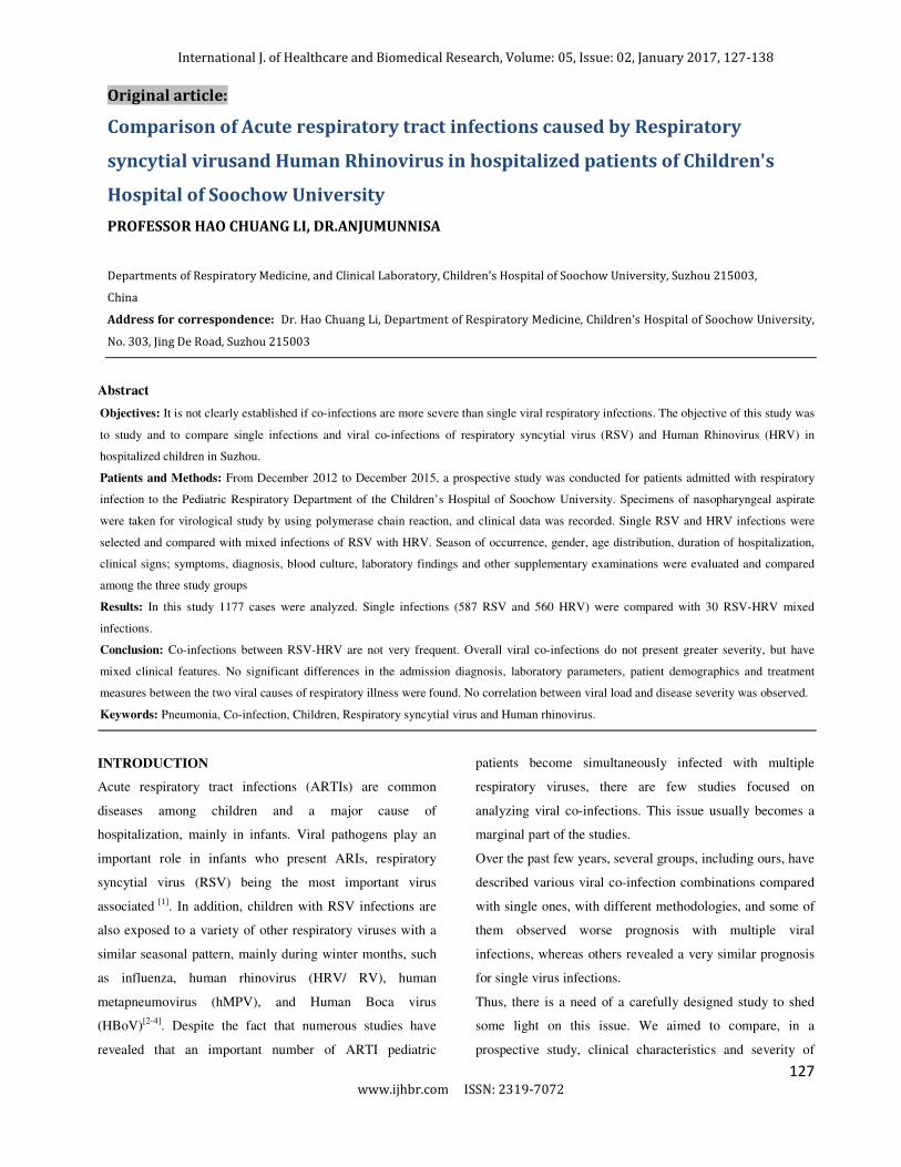
From the 587 patients with RSV infection; 325 cases, 55.3%
were seen during winter; 88 cases, 14.99% were seen during
spring; 16 cases,2.72% were seen during summer and 158
cases, 26.91% were seen during autumn. While in the 560
patient with HRV infection 64 cases, 11.42% were seen in
winter; 149 cases, 25.38%were seen in spring; 168 cases,
30.00%were seen in summer and 179 cases, 31.96% were
seen in autumn, And in the 30 patients with co-infections of
RSV + HRV 15 cases, 50.00% were seen in winter; 3 cases
10.00% were seen in spring; 1case, 3.33% was seen in
summer and 11 cases, 36.6% were seen in autumn. This
shows that RSV is more predominant in winter and autumn
where as HRV is more predominantly seen in autumn,
spring and summer.
International J. of Healthcare and Biomedical Research, Volume: 05, Issue:
Figure1: Season of occurrence
Baseline characteristics of the study population
A total of 1177 children with severe pneumonia were
screened in this study. Three groups were created as 587
patients with RSV infection, 560 patients with
patients with viral co-infection of RSV+HRV.
proportion of male: female subjects with RSV infection are
382:205, with 382 male 65.07%and 205
34.9%. The proportion of male: female subjects with
infection are 346:214, with 346 male 61.78%and 2
cases 38.2%. The proportion of male: female subjects with
co-infection of RSV and HRV are 22:08, with 22 male
73.33% and 8 female cases 26.66%.The youngest was
month-old, while the oldest was 11.5-year-old.
were stratified in four age groups <1 year old
International J. of Healthcare and Biomedical Research, Volume: 05, Issue: 02, January 2017, 12
www.ijhbr.com ISSN: 2319-7072
Baseline characteristics of the study population
A total of 1177 children with severe pneumonia were
Three groups were created as 587
infection, 560 patients with HRV and 30
RSV+HRV. The
female subjects with RSV infection are
female cases
The proportion of male: female subjects with HRV
%and 214 female
The proportion of male: female subjects with
infection of RSV and HRV are 22:08, with 22 male
The youngest was <1-
old. The patients
1 year olds, 1-<3 years
olds, 3-<5 years olds and ≥5-year
hospitalization of patients with RSV, HRV and co
infections of RSV + HRV infections was from3~30
1~42 days and 4~20 days respectively
length of stay was 8.3±3.1 days, 7.9
days respectively.
RSV infection was found to be significantly higher in
patients in the age group < 1 year olds while HRV infection
was found to be significantly higher in
groups older than 1 year old (P<0.05, respectively).
was no significant difference found in the duration of
hospitalization between the three study groups. (P>0.05)
02, January 2017, 127-138
127
year-olds. The duration of
patients with RSV, HRV and co-
HRV infections was from3~30 days,
20 days respectively and the average
7.9±3.3 days and 8.2±3.3
RSV infection was found to be significantly higher in
the age group < 1 year olds while HRV infection
was found to be significantly higher in patients in the age
groups older than 1 year old (P<0.05, respectively). There
was no significant difference found in the duration of
hospitalization between the three study groups. (P>0.05)
130

International J. of Healthcare and Biomedical Research, Volume: 05, Issue: 02, January 2017, 127-138
127 www.ijhbr.com ISSN: 2319-7072
Figure 2: Age distribution
Table 1: Duration of hospitalization, Gender and Age distribution
Variables RSV*
(n=587)
HRV*
(n=560)
RSV+HRV
(n=30)
P value
Gender
Male/ Female, n
Age, n
<1 year old
1- <3 years old
3-<5 years old
≥5-year-old
Duration of hospitalization,
d, �� � ��**
382/ 205
447
104
31
5
8.3±3.1
346/214
277
163
72
48
7.9±3.3
22/08
21
6
1
2
8.2±3.3
0.276
<0.001
<0.001
<0.001
<0.001
0.067
*RSV denotes respiratory syncytial virus and HRV denotes human rhinovirus.
**SD denotes standard deviation
Clinical manifestations
Clinical symptom:
From the 587 patients with RSVinfection in children;
583cases, 99.3%had cough; 387cases,
65.90%hadwheezing; 263cases, 44.80%had nasal
congestion; 201cases, 34.2% had fever; 31cases 5.30% had
dyspnea; 22 cases, 3.6% had feeding difficulties and 55
cases, 9.40% had gastrointestinal symptoms. While from
the 560 patients with HRV in children 543 cases,
97.0%had cough; 258 cases, 46.10%had wheezing; 229
cases, 40.90%had nasal congestion; 242 cases, 43.20% had
fever; 17cases, 30.40% had dyspnea; 16 cases, 2.7%had
feeding difficulties and 54 cases, 9.60% had
gastrointestinal symptoms. In the 30 patients with co-
infection of RSV + HRV 28 cases, 98.00% had cough; 30
cases, 100% had wheezing; 10 cases, 33.30% had nasal
congestion; 10cases, 33.30% had fever; 1 case,3.30% had
dyspnea; 1 case,3.125% had feeding difficulties and 6
cases, 20.00% had gastrointestinal symptoms.
< 1 year old 1-<3 years old 3-<5 years old ≥5 years old
RSV 447 104 31 5
HRV 277 163 72 48
MIX 21 6 1 2
447
104
31
5
277
163
72
48
216 1 2
0
50
100
150
200
250
300
350
400
450
500
Age Group
Number of Patients
Age Distribution
RSV HRV MIX
131

International J. of Healthcare and Biomedical Research, Volume: 05, Issue: 02, January 2017, 127-138
128 www.ijhbr.com ISSN: 2319-7072
Clinical signs:
From the 587 patients with RSVinfection in children 115
cases, 19.60% had tachypnea and 25 cases, 4.30% had
cyanosis.In the 560 patients with HRV infection in
children 70 cases, 12.50% had tachypnea and 13 cases,
2.3% had cyanosis. While in the 30 patients with co-
infection of RSV+ HRV 4 cases, 13.30% had tachypnea
and 0 cases, 0% had cyanosis.
Wheezing, nasal congestion and tachypnea were found to
be significantly higher in patients with RSV infection as
compared to those with HRV infection and co-infection of
RSV+ HRV (P<0.05, respectively). While fever and
dyspnea were found to be significantly higher in patients
with HRV infection as compared to those with RSV
infection and co- infections of RSV +HRV (P<0.05,
respectively). Cough was found to show no significant
difference between the three study groups (P>0.05).
Table 2: Clinical signs and symptoms
Clinical symptoms (%) RSV*
(n=587)
HRV*
(n=560)
RSV+HRV
(n=30)
P value
Cough 583(99.3) 543(97.00) 28(98.00) 0.1
Wheezing 387(65.90) 258(46.10) 30(100) <0.001
Nasal Congestion/Runny Nose 263(44.80) 229(40.90) 10(33.30) 0.235
Presence of Fever 201(34.2) 242(43.20) 10(33.30) 0.006
Dyspnea 31(5.30) 17(30.40) 1(3.30) <0.001
Feeding Difficulties 22(3.6) 16(2.7) 1(3.125) 0.71
Gastrointestinal symptoms 55(9.40) 54(9.60) 6(20.00) 0.159
Signs, n (%)
Tachypnea 115(19.60) 70(12.50) 4(13.30) 0.004
Cyanosis 25(4.30) 13(2.3) 0(0) 0.107
*RSV denotes respiratory syncytial virus and HRV denotes human rhinovirus.
132

International J. of Healthcare and Biomedical Research, Volume: 05, Issue: 02, January 2017, 127-138
128 www.ijhbr.com ISSN: 2319-7072
Laboratory tests
In the 587 patients with RSVinfection, the mean peripheral
white blood cell count was 9.4±4.0x10^9/L, the mean
percentage of granulocytes was 36.4±27.0%, lymphocytes
was 55.4±16.6%, mean platelets level was385.7±118.0%,
eosinophils was 4.9±14.1 x10^9/L and Creatine Kinase
Myocardial Band (CKMB) was 6.1±2.8 ng/mL. While in
the 560 patients with HRVinfection, the mean peripheral
white blood cell count was 12.2±6.4 x10^9/L, the mean
percentage of granulocytes was 45.0±21.3%, lymphocytes
was 46.4±20.0%, mean platelets level was 383.7±129.8%,
eosinophils was 13.4±27.4 x10^9/L and CKMB was
16.7±15.3 ng/mL. Finally for the 30 patients withco-
infections ofRSV and HRV, the mean peripheral white
blood cell count was 9.3±3.9x10^9/Lthe mean percentage of
granulocytes was 35.7±15.8%, lymphocytes was
55.7±16.2%, mean platelets level was 390.6±123.8%,
eosinophils was 6.6±19.3 x10^9/L and CKMB
was11.9±12.6ng/mL. No significant difference was found in
CRP values because CRP is not so elevated in viral
infections. In this study, the mean alanine aminotransferase
(ALT) level of 587 patients with RSVinfection was
32.19±29.6 U/L;the mean aspartate aminotransferase (AST)
level was 51.2±28.3 U/L. Where as the mean ALT level of
560 patients with HRV was 27.8±53.0 U/L and the mean
AST level was 43.3±30.1 U/L. Finally the mean ALT level
of 30 patients with RSV+ HRV infection was 28.0±16.2
U/L and the mean AST level was 43.9±13.3 U/L. Statistical
differences in the levels of IgA, IgG and IgM were observed
between the threegroups.
In patients with HRV infection WBC, GRN% and
eosinophils were significantly higher compared to those
with RSV infection and co-infection of RSV and HRV
(P<0.05, respectively). While in patients with RSV
infection the percentage of lymphocytes, CKMBand AST
levels were significantly higher compared to those with
HRV infection and co-infection of RSV and HRV.
(P<0.05,respectively).
Table 3: Laboratory tests- Blood analysis
Laboratory tests
(Blood Analysis, mean ±SD*)
RSV* (n=587) HRV*(n=560) RSV+HRV((((n=30)))) P value
WBC*, x10^9/L 9.4±4.0 12.2±6.4 9.3±3.9 <0.001
GRN*% 36.4±27.0 45.0±21.3 35.7±15.8 <0.001
L*, x10^9/L % 55.4±16.6 46.4±20.0 55.7±16.2 <0.001
PLT*, x10^9/L 385.7±118.0 383.7±129.8 390.6±123.8 0.068
Eosinophils, x10^9/L 4.9±14.1 13.4±27.4 6.6±19.3 <0.001
AST*, U/L 51.2±28.3 43.3±30.1 43.9±13.3 <0.001
ALT*, U/L 32.19±29.6 27.8±53.0 28.0±16.2 0.215
CKMB*, ng/mL 22.25±21.9 16.7±15.3 11.9±12.6 <0.001
IgG*, mg/dL 6.1±2.8 7.3±2.8 7.5±3.6 <0.001
133

International J. of Healthcare and Biomedical Research, Volume: 05, Issue: 02, January 2017, 127-138
128 www.ijhbr.com ISSN: 2319-7072
IgA*, mg/dL 0.7±0.7 1.0±0.6 0.7±0.6 <0.001
IgM*, mg/dL 1.0±0.6 1.3±0.7 1.0±1.2 <0.001
*RSV denotes respiratory syncytial virus, HRV human
rhinovirus, WBC white blood cell, GRN granulocytes, L
lymphocytes, PLT platelets, AST aspartate
aminotransferase, ALT alanine aminotransferase, CKMB
Creatine Kinase Myocardial Band, and IgG, IgA, IgM
denotes Immunoglobulin G, A and M respectively.
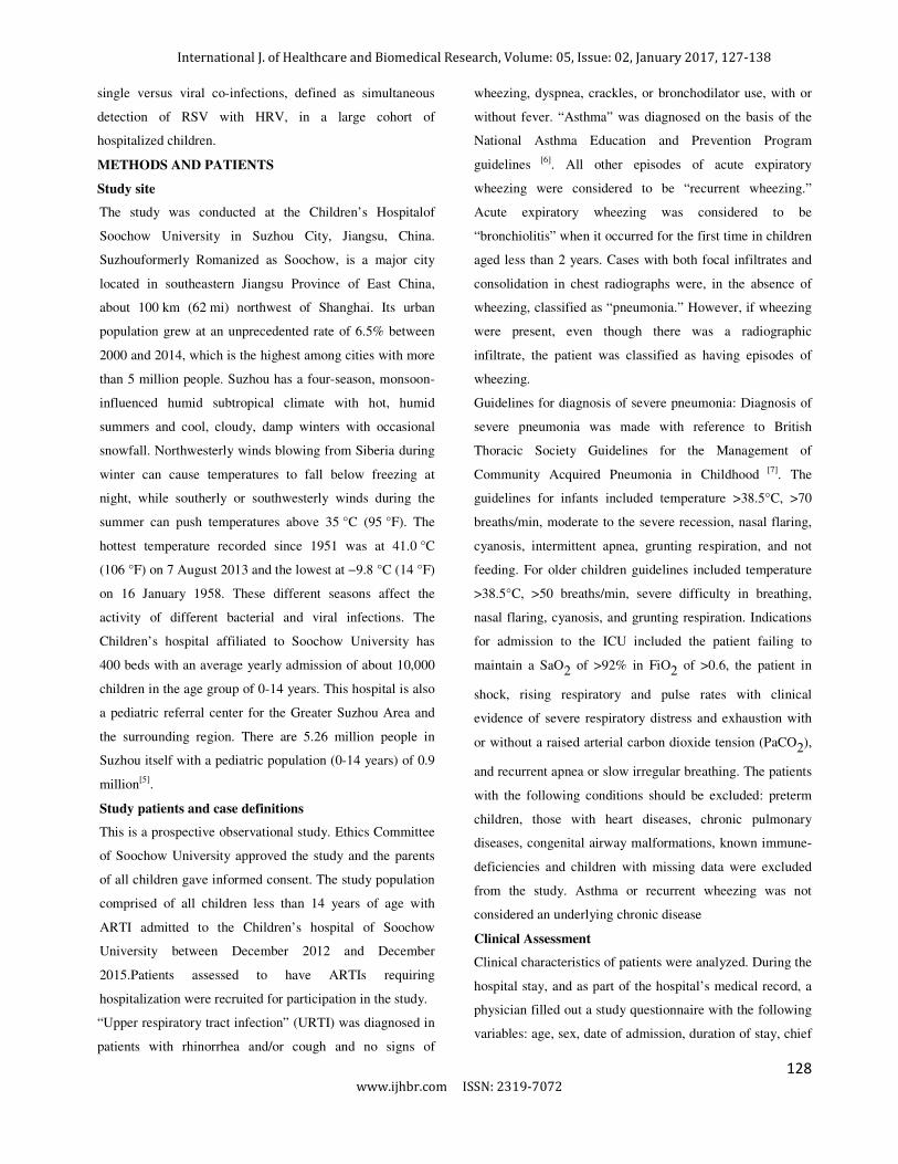
Diagnosis:
From the 587 patients with RSV infection, 560 patients with
HRV infection and 30 patients with RSV+HRV co-
infections; 477 cases, 81.30%; 431, 77.00% and 25, 83.30%
had bronchial pneumonia; 11, 1.90%, 5, 0.90% and 0, 0
.00% had bronchiolitis; 13, 2.20%; 15, 2.70% and 0, 0.00%
had bronchitis; 12, 2.00%, 62, 11.10% and 0, 0.00% had
asthma; 13, 2.1%), 379 cases, 67.70% and 5, 16.70% had
upper respiratory tract infections respectively. The duration
of days before hospitalization for patients with RSV, HRV
and co-infections of RSV + HRV infections was from 0~20
days, 0~28 days and 0~15 days respectively and the average
number of days was 2.2±2.7 days, 2.7±3.6 days and 3.3±3.6
days respectively.
In patients with bronchial pneumonia HRV is significantly
less detected compared to RSV and co-infections of
RSV+HRV (P<0.05). In patients with asthma, and upper
respiratory tract infection HRV is significantly more
detected compared to RSV and co-infections of RSV +HRV
(P<0.05,respectively).
Table 4: Diagnosis.
Diagnosis RSV* (n=587) HRV*(n=560) RSV+HRV
((((n=30))))
P value
Bronchial Pneumonia 477(81.30%) 431(77.0%) 25(83.30%) <0.001
Bronchiolitis 11(1.90%) 5(0.90%) 0(0%) <0.001
Bronchitis 13(2.20%) 15(2.70%) 0(0%) 0.003
Asthma 12(2.00%) 62(11.10%) 0(0%) <0.001
Upper respiratory tract
infection
13(2.1%) 379(67.70%) 5(16.70%) <0.001
Days before admission, d, X ±
SD
2.2±2.7 2.7±3.6 3.3±3.6 0.015
*RSV denotes respiratory syncytial virus and HRV denotes human rhinovirus.
134
International J. of Healthcare and Biomedical Research, Volume: 05, Issue:
** SD denotes standard deviation
DISCUSSION
The United Nations Children’s Fund (UNICEF) and
Health Organization (WHO) have identified pneumonia as
the major “forgotten killer of children”[9]
. According to the
WHO, about 1.9 million children worldwide die each year
from respiratory tract infection and its complications
Respiratory tract infection has been listed as the second
leading cause of death in children under five
leading cause of mortality among children less than five
years of age, despite effective vaccines and nutritional and
environmental interventions [12]. Viruses are key pathogens
of respiratory infections [13,14]
. Characterization of
respiratory viruses and understanding its relationships with
gender, age, and season will help to carry out the prevention
and treatment of childhood respiratory tract infections and
indirectly reduce the antibiotic abuse in clinical settings.
Pneumonia is an illness, usually caused by bacterial, viral or
more rarely fungal organisms. Common symptoms in
children and infants include difficulty breathing, cough, and
wheezing. Diagnosis involves confirmatory
radiography and laboratory tests. Antibiotics are the
preferred choice for treatment and management. Risks
factors include low paternal education, low birth weight,
International J. of Healthcare and Biomedical Research, Volume: 05, Issue: 02, January 2017, 12
www.ijhbr.com ISSN: 2319-7072
The United Nations Children’s Fund (UNICEF) and World
have identified pneumonia as
According to the
1.9 million children worldwide die each year
ection and its complications [10].
as the second
death in children under five [11]
. It’s the
leading cause of mortality among children less than five
years of age, despite effective vaccines and nutritional and
Viruses are key pathogens
. Characterization of
respiratory viruses and understanding its relationships with
gender, age, and season will help to carry out the prevention
and treatment of childhood respiratory tract infections and
e antibiotic abuse in clinical settings.
Pneumonia is an illness, usually caused by bacterial, viral or
more rarely fungal organisms. Common symptoms in
children and infants include difficulty breathing, cough, and
wheezing. Diagnosis involves confirmatory chest
radiography and laboratory tests. Antibiotics are the
preferred choice for treatment and management. Risks
factors include low paternal education, low birth weight,
and lack of breastfeeding. Key strategies for the prevention
of childhood pneumonia are community
management, adequate nutrition and zinc intake
We choose to study about RSV and HRV infections, as
these are the most frequent viruses detected in pediatric
outpatients and inpatients not just in China but also around
the world as was found during a 12
done in Turkey by Candan Çiçek
were found to be the two most predominant respiratory viral
pathogens in our hospital. This result is consistent with the
findings in most studies conducted in
Kunming, Beijing and Chongqing [16]
United States [21]
, Brazil [22]
, and the United Kingdom
Thus we know pneumonia is one of the leading causes of
illness and death in children younger than 5 years
worldwide, but there seems to be little information on the
viral etiology of severe pneumonia in the developing
countries, where the disease burden is particularly high.
this study we aimed at underscoring the importance of viral
etiology in pediatric patients.
In Suzhou, one or more viruses could be identified i
all the children suffering from
finding in this study was that RSV could be detected all
02, January 2017, 127-138
128
and lack of breastfeeding. Key strategies for the prevention
are community –based case
management, adequate nutrition and zinc intake8.
We choose to study about RSV and HRV infections, as
these are the most frequent viruses detected in pediatric
outpatients and inpatients not just in China but also around
world as was found during a 12-year period research
Çiçek [15].
These two viruses
predominant respiratory viral
This result is consistent with the
nducted in China such as in
[16]and also abroad
[17-20] in
, and the United Kingdom [23].
pneumonia is one of the leading causes of
illness and death in children younger than 5 years of age
worldwide, but there seems to be little information on the
viral etiology of severe pneumonia in the developing
countries, where the disease burden is particularly high. In
this study we aimed at underscoring the importance of viral
In Suzhou, one or more viruses could be identified in almost
all the children suffering from pneumonia.One of the
RSV could be detected all year
135
International J. of Healthcare and Biomedical Research, Volume: 05, Issue: 02, January 2017, 127-138
128 www.ijhbr.com ISSN: 2319-7072
round, peaked in November [Fig1]; this result was inline
with the reports from Changsha, China [24]
. The
epidemiology of RSV and HRV varied by areas and seasons
of the year[25]
. One study showed no seasonality[26,27]
,
whereas others found HRV peaking in spring and autumn [28,
29], which might have been, related to different rhinoviral
species in different regions and seasons. On the other hand,
this three-year study showed HRV peaking in autumn,
spring and summer. In contrast, RSV presented with a
seasonal pattern, predominantly in the winter months, which
contributed to an increase in hospitalization during the
winter. In our study we found only 30 cases positive for
both RSV and HRV co-infection in a span of 3 years
suggesting that these viruses are active during different
seasons and hence it is an infrequent combination of
pathogens causing severe pneumonia in children in Suzhou,
China.
Our study has estimated the incidence of respiratory viruses
among the hospitalized children with the diagnosis of ARTI
that may be considered as viral etiology. It has also
demonstrated the importance of RSV and HRV among
ARTIs. In our studyco-infection of RSV and HRV was the
most significant viral infection associated with pediatric
hospitalizations for Bronchial pneumonia (83.3%), followed
by RSV single viral infection (81.3%)as compared to single
HRV infection, especially in patients under one year of age,
similar to findings in other studies [4, 30,31].The possible
reason is that younger children are more prone to be sick
than older children due to their developing immune system
and that parents are more likely to take their younger
children to the doctor if they are sick. However HRV was
detected in a larger proportion of hospitalized children older
than one year of age [Fig 2]. From the above discussion we
can say that in patients with bronchial pneumonia HRV is
significantly less detected compared to RSV and co-
infection of RSV and HRV (P<0.05). And in patients with
asthma, and upper respiratory tract infection HRV is
significantly more detected compared to RSV and co-
infections of RSV and HRV (P<0.05,respectively)[Table 4].
No significant difference was found in the duration of
hospitalization between the three study groups.
(P>0.05)[Table 1] which also has support from the
literature[32,33]
.
As expected, RSV was the main cause of wheezing; 387 of
587, 65.9% and also wheezing caused due to HRV was
identified in a considerable number of patients; 258 of 560,
46.10% a finding comparable with the report of Miller [28]
.
Similar to other studies, RSV was associated with high
severity of diseases. Cough, 99.3%; wheezing, 65.9%; Nasal
congestion, 44.8%; fever, 34.2%; dyspnea, 5.3%; feeding
difficulties, 3.6%; gastrointestinal symptoms, 9.4%;
tachypnea, 19.5%and cyanosis, 4.3% were the most
commonly encountered symptoms in hospitalized children
with RSV [Table 2]. Wheezing, nasal congestion and
tachypnea were found to be significantly higher in patients
with RSV infection as compared to those with HRV
infection and co-infection of RSV+ HRV (P<0.05,
respectively). While fever and dyspnea were found to be
significantly higher in patients with HRV infection as
compared to those with RSV infection and co- infections of
RSV +HRV (P<0.05, respectively). Cough was found to
show no significant difference between the three study
groups (P>0.05), consistent with other studies.
Leukocytosis may also occur in viral infections, as more
than half of the RV-infected patients presented with
leukocytosis, notably higher than that of RSV-infected
patients [Table 3]. Previous studies had similar findings [2,4]
.
Whether these RV infections were accompanied by bacterial
infection was uncertain. As suggested in other studies [2, 4,
34,35], in our study patients with HRV infection the GRN%
and eosinophils counts were significantly higher compared
to those with RSV infection and co-infection of RSV and
HRV (P<0.05, respectively). While in patients with RSV
infection the percentage of lymphocytes, CKMB and AST
levels were significantly higher compared to those with
HRV infection and co-infection of RSV and HRV (P<0.05,
respectively).
LIMITATIONS
This study was not without shortcomings. It should be noted
that a major limitation of this study is that RSV diagnosis
was performed by antigen detection method while HRV
diagnosis was by PCR, which might have underestimated
136
International J. of Healthcare and Biomedical Research, Volume: 05, Issue: 02, January 2017, 127-138
129 www.ijhbr.com ISSN: 2319-7072
the prevalence of RSV. Second, the phylogenetic analysis of
HRV was not performed, as the monthly distribution and
clinical manifestations might differ among different HRV
species. Thus, more work is essential for a better
understanding of the seasonal epidemiology of different RV
species. Only 30 patients with co-infection were studied,
however, a larger study group is needed to learn more about
virus co-infections.
CONCLUSION
Three important observations made in our study are: (1) no
significant differences in clinical severity were observed
between children with virus co-infection compared to those
with single virus infection; (2) Single RSV infection was
more predominate in children less than 1 year of age as
compared to HRV which was more predominant in older
children. (3) RSV presented with a seasonal pattern, peaking
in the winter months in contrast to HRV infection, which
was predominant in autumn, spring and summer.
In summary, our study suggests co-infections between RSV-
HRV are not very frequent due to the difference in seasonal
occurrence. Overall viral co-infections do not present
greater severity, but have mixed clinical features. No
significant differences in the admission diagnosis,
laboratory parameters, patient demographics and treatment
measures between the two viral causes of respiratory illness
were found. No correlation between viral load and disease
severity was observed.
ACKNOWLEDGMENTS
We thank all the physicians and nurses who helped enroll
patients in the study and the staff that helped provide the
necessary patient files from the medical archives of the
Hospital. We also thank the staff members of the
parasitology, physiology, and genetics laboratories for
allowing us to use their facilities.
REFERENCES
1. Dong, W., Chen, Q., Hu, Y. et al. Epidemiological and clinical characteristics of respiratory viral infections in children in Shanghai, China. Arch
Virol (2016) 161: 1907.
2. Jacobs SE, Lamson DM, St George K, Walsh TJ. Human rhinoviruses. Clin Microbiol Rev. 2013; 26:135–62. doi: 10.1128/CMR.00077-12.
3. Wardlaw, Tessa et al. Pneumonia: the leading killer of childrenThe Lancet, Volume 368, Issue 9541, 1048 - 1050.
4. Yongdong Yan, Li Huang, Meijuan Wang, Yuqing Wang, Wei Ji, Canhong Zhu, and Zhengrong Chen, Clinical and epidemiological profiles
including meteorological factors of low respiratory tract infection due to human rhinovirus in hospitalized children, Ital J Pediatr. 2017; 43: 23.
5. Suzhou, From Wikipedia, the free encyclopedia
6. Susan M. Pollart, MD, MS and Kurtis S. Elward, Overview of Changes to Asthma Guidelines: Diagnosis and Screening, Am Fam
Physician. 2009 May 1; 79(9): 761-767.
7. Michael Harris,1 Julia Clark,2 Nicky Coote,3 Penny Fletcher,4 Anthony Harnden,5 Michael McKean,6 Anne Thomson,1 On behalf of the British
Thoracic Society Standards of Care Committee , British Thoracic Society guidelines for the management of community acquired pneumonia in
children: update 2011 , BMJ publications April 6, 2017
8. Granados A, Luinstra K, Chong S, Goodall E, Banh L, Mubareka S, et al. Use of an improved quantitative polymerase chain reaction assay to
determine differences in human rhinovirus viral loads in different populations. Diagn Microbiol Infect Dis. 2012;74:384–7.
9. Mulholland K. Global burden of acute respiratory infections in children: implications for interventions. Pediatr Pulmonol 2003; 36:469-74. [PubMed]
10. Bryce J, Boschi-Pinto C, Shibuya K, et al. WHO estimates of the causes of death in children. Lancet 2005; 365:1147-52. [PubMed]
11. Scott et al. Samuel Mbugua Penumonia in preschool children. 2008, 1291-1299.
12. Khamrin P, Thongprachum A, Shimizu H, et al. Detection of human bocavirus 1 and 2 from children with acute gastroenteritis in Japan. J Med Virol
2012; 84:901-5. [PubMed]
13. Moriyama Y, Hamada H, Okada M, et al. Distinctive clinical features of human bocavirus in children younger than 2 years. Eur J Pediatr 2010;
169:1087-92. [PubMed]
14. Achten NB et al,Interference Between Respiratory Syncytial Virus and Human Rhinovirus Infection in Infancy.J Infect Dis. 2017 Mar 24
15. Candan Çiçeket al, Prevalence and Seasonal Distribution of Respiratory Viruses in Patients with Acute Respiratory Tract Infections, 2002-2014.
16. Jia L. Detection of viruses causing lower respiratory tract infection in children. Chinese Journal of Nosocomiology 2011; 21:3386-8.
17. Bezerra PG, Britto MC, Correia JB, et al. Viral and atypical bacterial detection in acute respiratory infection in children under five years. PLoS One
2011; 6:e18928. [PubMed]
18. Kim MR, Lee HR, Lee GM. Epidemiology of acute viral respiratory tract infections in Korean children. J Infect 2000; 41:152-8. [PubMed]
137

International J. of Healthcare and Biomedical Research, Volume: 05, Issue: 02, January 2017, 127-138
128 www.ijhbr.com ISSN: 2319-7072
19. Lin TY, Huang YC, Ning HC, et al. Surveillance of respiratory viral infections among pediatric outpatients in northern Taiwan. J Clin Virol 2004;
30:81-5. [PubMed]
20. Wu X, Ni L, Xian FM, et al. Clinical and Epidemiological Characteristics Of Pathogenic Analysis in children with acute lower respiratory infection.
Contemporary Medicine 2010;16:57-8.
21. Murata Y. Respiratory syncytial virus infection in adults. Curr Opin Pulm Med 2008;14:235-40. [PubMed]
22. Bellei N, Carraro E, Perosa A, et al. Acute respiratory infection and influenza-like illness viral etiologies in Brazilian adults. J Med Virol
2008;80:1824-7. [PubMed]
23. Lees EA, Carrol ED, Gerrard C, et al. Characterisation of acute respiratory infections at a United Kingdom paediatric teaching hospital: observational
study assessing the impact of influenza A (2009 pdmH1N1) on predominant viral pathogens. BMC Infect Dis 2014;14:343. [PubMed].
24. Zeng SZ, Xiao NG, Xie ZP, et al. Prevalence of human rhinovirus in children admitted to hospital with acute lower respiratory tract infections in
Changsha, China. J Med Virol. 2014;86:1983–9.
25. Monto AS. The seasonality of rhinovirus infections and its implications for clinical recognition. Clin Ther. 2002;24:1987–97.
26. Bicer S, Giray T, Col D, et al. Virological and clinical characterizations of respiratory infections in hospitalized children. Ital J Pediatr. 2013;39:22.
27. Ozcan C, Toyran M, Civelek E, et al. Evaluation of respiratory viral pathogens in acute asthma exacerbations during childhood. J Asthma.
2011;48:888–93.
28. Miller EK, Gebretsadik T, Carroll KN, et al. Viral etiologies of infant bronchiolitis, croup and upper respiratory illness during 4 consecutive years.
Pediatr Infect Dis J. 2013;32:950–5.
29. Wurzel DF, Marchant JM, Clark JE, et al. Respiratory virus detection in nasopharyngeal aspirate versus bronchoalveolar lavage is dependent on virus
type in children with chronic respiratory symptoms. J Clin Virol. 2013;58:683–8.
30. Wang Y, Ji W, Chen Z, Yan YD, Shao X, Xu J. Comparison of severe pneumonia caused by Human metapneumovirus and respiratory syncytial
virus in hospitalized children. Indian J Pathol Microbiol 2014;57:413-7.
31. Huiquan Suna,1, Qiufeng Sunb,1, Wunjun Jianga, Zhengrong Chena, Li Huanga, Meijuan Wanga, WeiJia, Xuejun Shaoa, Yongdong Yan , “
Prevalence of rhinovirus in wheezing children: a comparison with respiratory syncytial virus wheezing” braz j infect dis. 2016;20(2):179–183
32. Sandra A. Asner, Astrid Petrich, Jemila S. Hamid, Dominik Mertz, Susan E. Richardson, Marek Smieja Clinical severity of
rhinovirus/enteroviruscompared to other respiratory viruses in children. 6 May 2014
33. Xiaoli Ge1*, Zhijun Han2*, Hongmin Chen1, Juanjuan Cheng1, Mingzhu Gao2, Haibin Sun3Characterization of acute respiratory infections among
340 infants in Wuxi, Jiangsu Province, Journal of Clinical and Pathological Research 2015;35(1):31-35
34. Zhu Runan, Song Qinwei, Qian Yuan, Zhao Linqing, Deng Jie, Wang Fang and Sun Yu, Virus profile in children with acute respiratory infections
with various severities in Beijing, China, Chinese Medical Journal 2014;127(21):3706-3711:10.3760/cma.j.issn.0366-6999.20141556
35. S.A. Asner, W. Rose, A. Petrich, S. Richardson, D.J. Tran ‘Is virus coinfection a predictor of severity in children with viral respiratory
infections?’March 2015Volume 21, Issue 3, Pages 264.e1–264.e6
138