clostridia: sporeforming anaerobic bacilli -...
TRANSCRIPT
Clostridia: Sporeforming Anaerobic Bacilli
Carol L. Wells
Tracy D. Wilkins
General Concepts Clostridia are strictly anaerobic to aerotolerant sporeforming bacilli found in soil as well as in normal intestinal flora of man and animals. There are both gram-positive and gram-negative species, although the majority of isolates are gram-positive. Exotoxin(s) play an important role in disease pathogenesis.
Gas Gangrene and Related Clostridial Wound Infections
Clinical Manifestations
Patients may present with a wound infection. Severity varies from invasion of live tissue with systemic toxemia to relatively benign superficial contamination of already necrotic tissue.
Structure
The clostridia that cause gas gangrene are anaerobic, spore-forming bacilli, but some species may not readily sporulate, e.g., C perfringens.
Classification and Antigenic Types
Clostridial wound infections are typically polymicrobic. The primary pathogens are various clostridial species, including C perfringens, C novyi, C septicum, and others.
Pathogenesis
Wounds are contaminated by clostridia from the environment or the host's normal flora. The anaerobic tissue environment facilitates replication of clostridia and secretion of toxins.
Host Defenses
Host defenses are essentially absent. There is little, if any, innate immunity.
Epidemiology
Clostridial wound infections are found worldwide. Clostridia are ubiquitous in the soil and in the normal microbial flora of humans and animals.
Diagnosis
These infections are diagnosed by recognition of a characteristic lesion coupled with tissue Gram stains and bacterial culture.
Control
Wound infections are controlled by administration of antimicrobial agents (e.g., penicillin, chloramphenicol) coupled with tissue debridement (for more severe forms of clostridial wound infections).
Tetanus and Clostridium Tetani
Clinical Manifestations
Tetanus is characterized by twitching of muscles around a wound, pain in neck and jaw muscles (trismus), and around the wound. Patients have no fever, but sweat profusely and exhibit muscle rigidity and spasms.
Structure
These organisms are bacilli with terminal spores.
Classification and Antigenic Types
C tetani is the only species. There are no serotypes.
Pathogenesis
The infection is initiated as a result of contamination of a wound with C tetani. The anaerobic tissue environment facilitates C tetani replication and secretion of exotoxins. A spasmogenic toxin, tetanospasmin, fixes to inhibitory neurons and blocks the release of neurotransmitters, glycine and gamma-aminobutyric acid.
Host Defenses
Host defenses are essentially absent. There is little, if any, inate immunity and the disease does not produce immunity in the patient. Active immunity follows vaccination with tetanus toxoid.
Epidemiology
C tetani is found worldwide. Ubiquitous in soil, it is occasionally found in intestinal flora of humans and animals.
Diagnosis
Diagnosis is primarily by the clinical symptoms (above). The wound may not be obvious. Furthermore, C tetani is recovered from only one-third of all implicated wounds.
Control
The administration of tetanus toxoid is a preventive measure. C tetani infection is treated with antimicrobial agents (metronidazole or penicillin) and by local wound debridement. Other measures include tetanus immunoglobulin and supportive therapy.
Botulism and Clostridium Botulinum
Clinical Manifestations
These infections may have early gastrointestinal symptoms. The cranial nerves are initially affected, followed by descending, symmetric paralysis of motor nerves, with critical involvement of the respiratory tree. Muscle paralysis may occur.
Structure
These organisms are bacilli with oval, subterminal spores.
Classification and Antigenic Types
C botulinum consists of several biochemically distinct groups of organisms that produce botulinum toxin. Seven types of neurotoxins are designated A, B, C, D, E, F, and G, some of which have been shown to be encoded on bacteriophage DNA.
Pathogenesis
There are three forms: (1) adult botulism, caused by ingestion of preformed toxin in food; (2) infant botulism, in which the organism replicates and secretes toxin in the intestinal tract; and (3) wound botulism, in which the organism replicates in the wound and secretes toxin. Toxin binds to neuromuscular junctions of parasympathetic nerves and interferes with acetylcholine release, causing flaccid muscle paralysis.
Host Defenses
No host defenses are known.
Epidemiology
C botulinum is distributed worldwide, and is ubiquitous in soil. Improper heating of canned foods is a major factor in botulism food poisoning.
Diagnosis
Diagnosis is from the clinical symptoms (above), especially gastrointestinal and neurological symptoms, coupled with laboratory confirmation. A finding of normal spinal fluid helps to eliminate the possible diagnosis of numerous other central nervous system disorders.
Control
The best means of control is to eliminate the toxin source via proper food handling. Once the food poisoning is diagnosed, treatment measures should include an attempt to neutralize unbound toxin. Supportive care is of primary importance.
Antibotic-Associated Diarrhea, Pseudomembranous Colitis, and Clostridium Difficile
Clinical Manifestations
Patients can present with a spectrum of disease that varies from uncomplicated antibiotic-associated diarrhea to antibiotic-associated pseudomembranous colitis that may be fatal.
Structure
This species consists of bacilli with large, oval, subterminal spores.
Classification and Antigenic Types
C difficile is the only species. There are no defined serotypes. Toxigenic and nontoxigenic strains exist. The former produce varying amounts of toxin A (enterotoxin) and toxin B (cytotoxin).
Pathogenesis
Broad spectrum antibiotic therapy eliminates much competing normal flora, permitting intestinal overgrowth of toxigenic C difficile.
Host Defenses
There are no defined host defenses.
Epidemiology
C difficile is a component of the normal intestinal flora of a small percentage of healthy adults and of a relatively large percentage of healthy neonates. It also may be found in the environment, especially in hospitals.
Diagnosis
The presence of antibiotic therapy, diarrhea, and pseudomembranes by colonoscopy help establish the severity of disease, coupled with the demonstration of organisms and/or toxin in feces.
Control
Metronidazole and vancomycin should be used therapeutically. However, relapses can occur. Supportive therapy may be needed.
Other Pathogenic Clostridia
Clostridium perfringens causes food poisoning and necrotizing enteritis. C sordellii causes bacteremia, endometritis and nonbacteremic infections. C septicum is correlated with the presence of cancer. C tertium is associated with bacteremia.
INTRODUCTION Of the anaerobes that infect humans, the clostridia are the most widely studied. They are involved in a variety of human diseases, the most important of which are gas gangrene, tetanus, botulism, pseudomembranous colitis and food poisoning. In most cases, clostridia are opportunistic pathogens; that is, one or more species establishes a nidus of infection in a particular site in a compromised host. All pathogenic clostridial species produce protein exotoxins (such as botulinum and tetanus toxins) that play an important role in pathogenesis.
Most generalizations about Clostridium have exceptions. The clostridia are classically anaerobic rods, but some species can become aerotolerant on subculture; a few species (C carnis, C histolyticum, and C tertium) can grow under aerobic conditions. Most species are Gram-positive, but a few are Gram-negative. Also, many Gram-positive species easily lose the Gram reaction, resulting in Gram-negative cultures.
The clostridia form characteristic spores, the position of which is useful in species identification; however, s ome species do not sporulate unless exposed to exacting cultural conditions. Many clostridia are transient or permanent members of the normal flora of the human skin and the gastrointestinal tracts of humans and animals. Unlike typical members of the human bacterial flora, most clostridia can also be found worldwide in the soil.
Because clostridia are ubiquitous saprophytes, many isolated from clinical specimens are accidental contaminants and not involved in a disease process. Because these organisms are normally found on the skin, even a pure culture of clostridia isolated from blood may have no clinical significance. In determining the importance of a clinical isolate of clostridia, the clinician should consider the frequency of isolation of the species, the presence of other microbes of pathogenic potential, and the clinical
symptoms of the patient. Many clostridial infections can be controlled by antibiotic therapy (e.g., penicillin, chloramphenicol, vancomycin, metronidazole) accompanied, in some cases, by tissue debridement. Antitoxin therapy and toxoid immunization are clearly useful in some clostridial infections, such as tetanus.
Gas Gangrene and Related Clostridial Wound Infections
Clinical Manifestations
Clostridial wound infections may be divided into three categories: gas gangrene or clostridial myonecrosis, anaerobic cellulitis, and superficial contamination. Gas gangrene can have a rapidly fatal outcome and requires prompt, often severe, treatment. The more common clostridial wound infections are much less acute and require much less radical treatment; however, they may share some characteristics with gas gangrene and must be included in the differential diagnosis.
Gas gangrene is an acute disease with a poor prognosis and often fatal outcome (Fig. 18-1). Initial trauma to host tissue damages muscle and impairs blood supply. This lack of oxygenation causes the oxidation-reduction potential to decrease and allows the growth of anaerobic clostridia. Initial symptoms are generalized fever and pain in the infected tissue. As the clostridia multiply, various exotoxins (including hemolysins, collagenases, proteases, and lipases) are liberated into the surrounding tissue, causing more local tissue necrosis and systemic toxemia. Infected muscle is discolored (purple mottling) and edematous and produces a foul-smelling exudate; gas bubbles form from the products of anaerobic fermentation. As capillary permeability increases, the accumulation of fluid increases, and venous return eventually is curtailed. As more tissue becomes involved, the clostridia multiply within the increasing area of dead tissue, releasing more toxins into the local tissue and the systemic circulation. Because ischemia plays a significant role in the pathogenesis of gas gangrene, the muscle groups most frequently involved are those in the extremities served by one or two major blood vessels.
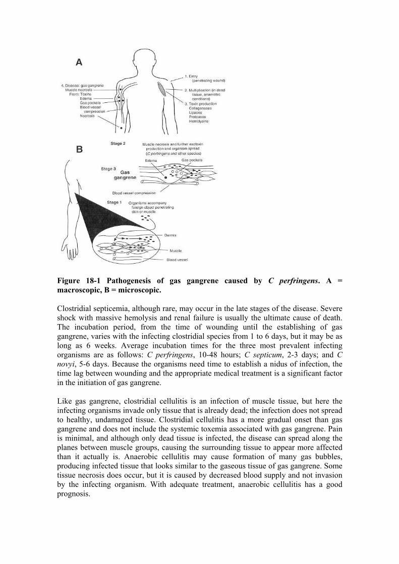
Figure 18-1 Pathogenesis of gas gangrene caused by C perfringens. A = macroscopic, B = microscopic.
Clostridial septicemia, although rare, may occur in the late stages of the disease. Severe shock with massive hemolysis and renal failure is usually the ultimate cause of death. The incubation period, from the time of wounding until the establishing of gas gangrene, varies with the infecting clostridial species from 1 to 6 days, but it may be as long as 6 weeks. Average incubation times for the three most prevalent infecting organisms are as follows: C perfringens, 10-48 hours; C septicum, 2-3 days; and C novyi, 5-6 days. Because the organisms need time to establish a nidus of infection, the time lag between wounding and the appropriate medical treatment is a significant factor in the initiation of gas gangrene.
Like gas gangrene, clostridial cellulitis is an infection of muscle tissue, but here the infecting organisms invade only tissue that is already dead; the infection does not spread to healthy, undamaged tissue. Clostridial cellulitis has a more gradual onset than gas gangrene and does not include the systemic toxemia associated with gas gangrene. Pain is minimal, and although only dead tissue is infected, the disease can spread along the planes between muscle groups, causing the surrounding tissue to appear more affected than it actually is. Anaerobic cellulitis may cause formation of many gas bubbles, producing infected tissue that looks similar to the gaseous tissue of gas gangrene. Some tissue necrosis does occur, but it is caused by decreased blood supply and not invasion by the infecting organism. With adequate treatment, anaerobic cellulitis has a good prognosis.
Superficial contamination, the least serious of the clostridial wound infections, involves infection of only necrotic tissue. Usually, the patient experiences little pain, and the process of wound healing proceeds normally; however, occasionally an exudate may form and the infection may interfere with wound healing. Superficial wound contamination caused by clostridia usually involves C perfringens, with staphylococci or streptococci, or both, as frequent co-isolates.
Structure
The clostridia that cause gas gangrene are anaerobic, spore-forming bacilli, but some species may not readily sporulate, e.g., C perfringens.
Classification and Antigenic Types
Clostridial wound infections usually are polymicrobic because the source of wound contamination (feces, soil) is polymicrobic. In gas gangrene and anaerobic cellulitis, the primary pathogen can be any one of various clostridial species including C perfringens (80%), C novyi (40%), C septicum (20%), and, occasionally, C bifermentans, C histolyticum, or C fallax. Other bacterial isolates may be any of a wide number and variety of organisms (for example, Proteus, Bacillus, Escherichia, Bacteroides, Staphylococcus). The distinctive or unique properties of the causative agents of gas gangrene are difficult to list; morphologic characteristics and biochemical reactions vary among these species, and a reliable laboratory manual should be consulted for their proper identification. Isolation of 107 or more clostridia per milliliter of wound exudate is strong evidence for a clostridial wound infection.
The most frequently isolated pathogen, C perfringens, has five types, designated A, B, C, D, and E. Each of these types produces a semi-unique spectrum of protein toxins. Alpha-toxin (a lecithinase, also called phospholipase-C) and theta-toxin (oxygen-labile cytolysin) are both considered important in the disease pathology. Alpha-toxin is lethal and necrotizing; it lyses cell membrane lecithins, disrupting cell membranes and causing cell death. Theta-toxin also contributes to rapid tissue destruction by several mechanisms. At the site of infection, theta-toxin acts as a cytolysin, promoting direct vascular injury; lower toxin concentrations activate polymorphonuclear leukocytes and endothelial cells, promoting distal vascular injury by stimulating leukocyte adherence to the endothelium. The result is leukostasis, thrombosis, decreased perfusion, and tissue hypoxia. Theta-toxin also mediates the production of shock through induction of inflammatory mediators such as platelet activating factor, tumor necrosis factor, interleukin 1 and interleukin 6.
Pathogenesis
All clostridial wound infections occur in an anaerobic tissue environment caused by an impaired blood supply secondary to trauma, surgery, foreign bodies, or malignancy. Contamination of the wound by clostridia from the external environment or from the host's normal flora produces the infection. The detailed pathogenesis of the disease is intimately associated with the clinical presentation as described above (Fig. 18-1).
Host Defenses
Host defenses against gas gangrene and other clostridial wound infections are mostly ineffective. Even repeated episodes of clostridial wound infection do not seem to produce effective immunity.
Epidemiology
Clostridial spores are ubiquitous in the soil, on human skin, and in the gastrointestinal tracts of humans and animals. Thus, the causative agents of clostridial wound infections are not environmentally restricted. Even operating theaters can be habitats for infecting clostridial organisms and spores. The incidence of clostridial wound infections has declined with the advance of prompt, adequate medical treatment. Historically, war casualties have had the greatest incidence of gas gangrene; however, the prompt evacuation and medical attention given United States casualties in the Vietnam war greatly decreased the incidence of gas gangrene in these soldiers, emphasizing the importance of prompt medical treatment.
Diagnosis
Diagnosis of clostridial wound infections is based on clinical symptoms coupled with Gram stains and bacterial culture of clinical specimens. Gas gangrene, once initiated, may spread and cause death within hours. By the time the typical lesions of gas gangrene are evident, the disease usually is firmly established and the physician must treat the patient on a clinical basis without waiting for laboratory confirmation. Characteristic lesions and the presence of large numbers of Gram-positive bacilli (with or without spores) in a wound exudate provide strong presumptive evidence. In contrast to tissue infections caused by Staphylococcus aureus, there is typically an absence of polymorphonuclear leukocytes at the site of infection, likely due to the presence of clostridial toxins. Spores are rare in cultures of C perfringens, the most common etiologic agent of these diseases. A commonly used laboratory test for presumptive identification of C perfringens is the Nagler reaction which detects the presence of alpha-toxin (phospholipase-C), one of the most prominent toxins produced by C perfringens. However, several other species of clostridia also have a positive Nagler reaction, and thus this test is not entirely specific for C perfringens.
Discussion of the differential diagnosis of clostridial wound infections appropriately includes streptococcal myositis, as this disease can be characterized by an edematous, necrotizing, often gaseous lesion. Like anaerobic cellulitis and superficial contamination with clostridia, streptococcal myositis is a relatively localized disease, but its later stages may include some systemic toxicity that mimics the toxemia of gas gangrene.
Control
Correction of the anaerobic conditions combined with antibiotic treatment form the basis for therapy. Penicillin is the drug of choice for all clostridial wound infections; chloramphenicol is a second-choice antibiotic. Successful treatment of the less severe forms of clostridial wound infections includes local debridement and antibiotic therapy; after these measures are taken, patient recovery usually proceeds along a steady, positive course. Treatment of gas gangrene includes radical surgical debridement coupled with high doses of antibiotics. Blood transfusions and supportive therapy for shock and renal failure also may be indicated.
The usefulness of gas gangrene antitoxin is currently a disputed matter. Some physicians maintain that the efficacy of this polyvalent antitoxin has been proved in the past, but better medical care now may have eliminated the need for its use. Others believe that because of insufficient data, antitoxin should be administered systemically as early as possible after diagnosis, and that the antitoxin should be injected locally into tissue that cannot be excised.
Obviously, prevention of wound contamination is the single most important factor in controlling clostridial wound infections. In the past, immunization has been considered a possible preventive measure for gas gangrene; however, several factors have discouraged the use of active immunization, including difficulty in preparing a suitable antigenic toxoid, availability of prompt wound treatment, and accessibility of effective therapeutic agents.
Tetanus and Clostridium Tetani
Clinical Manifestations
Tetanus is a severe disease caused by the toxin of C tetani (Fig. 18-2). This organism grows in a wound and secretes a toxin that invades systemically and causes muscle spasms. The initial symptom is cramping and twitching of muscles around a wound. The patient usually has no fever but sweats profusely and begins to experience pain, especially in the area of the wound and around the neck and jaw muscles (trismus). Portions of the body may become extremely rigid, and opisthotonos (a spasm in which the head and heels are bent backward and the body bowed forward) is common. Complications include fractures, bowel impaction, intramuscular hematoma, muscle ruptures, and pulmonary, renal, and cardiac problems.
Structure and Classification
C tetani is an anaerobic gram-positive rod that forms terminal spores, giving it a characteristic tennis racquet appearance. Some strains do not sporulate readily, and spores may not appear until the third or fourth day of culture. Most strains are motile with peritrichous flagella; colonies often swarm on agar plates, but some strains are nonflagellated and nonmotile. The presence of C tetani should be suspected on isolation of a swarming rod that produces indole and has terminal spherical spores, but does not produce acid from glucose. Toxigenic C tetani contains a plasmid that produces a toxin called tetanospasmin, but nontoxigenic strains also exist. Tetanospasmin is responsible for the infamous toxemia called tetanus. The two animal species most susceptible to this toxemia are horses and humans.
Pathogenesis
As with all clostridial wound infections, the initial event in tetanus is trauma to host tissue, followed by accidental contamination of the wound with C tetani (Fig. 18-2). Tissue damage is needed to lower the oxidation-reduction potential and provide an environment suitable for anaerobic growth. Once growth is initiated, the organism itself is not invasive and remains confined to the necrotic tissue, where the vegetative cells of C tetani elaborate the lethal toxin. The incubation period from the time of wounding to the appearance of symptoms varies from a few days to several weeks, depending on the
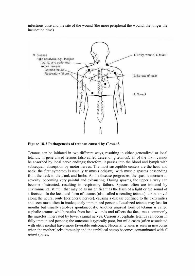
infectious dose and the site of the wound (the more peripheral the wound, the longer the incubation time).
Figure 18-2 Pathogenesis of tetanus caused by C tetani.
Tetanus can be initiated in two different ways, resulting in either generalized or local tetanus. In generalized tetanus (also called descending tetanus), all of the toxin cannot be absorbed by local nerve endings; therefore, it passes into the blood and lymph with subsequent absorption by motor nerves. The most susceptible centers are the head and neck; the first symptom is usually trismus (lockjaw), with muscle spasms descending from the neck to the trunk and limbs. As the disease progresses, the spasms increase in severity, becoming very painful and exhausting. During spasms, the upper airway can become obstructed, resulting in respiratory failure. Spasms often are initiated by environmental stimuli that may be as insignificant as the flash of a light or the sound of a footstep. In the localized form of tetanus (also called ascending tetanus), toxins travel along the neural route (peripheral nerves), causing a disease confined to the extremities and seen most often in inadequately immunized persons. Localized tetanus may last for months but usually resolves spontaneously. Another unusual form of tetanus is called cephalic tetanus which results from head wounds and affects the face, most commonly the muscles innervated by lower cranial nerves. Curiously, cephalic tetanus can occur in fully immunized persons; the outcome is typically poor, but mild cases (often associated with otitis media) have more favorable outcomes. Neonatal tetanus is seen in newborns when the mother lacks immunity and the umbilical stump becomes contaminated with C tetani spores.
C tetani actually produces two toxins: tetanolysin, a hemolysin that is inactivated by cholesterol and has no role in pathogenesis, and tetanospasmin, a spasmogenic toxin responsible for the classical symptoms of the disease.
The actions of tetanospasmin are complex and involve three components of the nervous system: central motor control, autonomic function, and the neuromuscular junction. Toxin enters the nervous system primarily through the neuromuscular junction of alpha motor neurons. The toxin is then transported to the other neurons, most importantly presynaptic inhibitory cells, where it is no longer accessible to be neutralized by antitoxin. (This retrograde transport to the central nervous system is similar to that utilized by some viruses, such as herpes virus and rabies.) The toxin also spreads hematogenously, but it still must enter the central nervous system via retrograde transport from peripheral neuronal processes. Once the toxin gains access to inhibitory neurons, it blocks the release of the neurotransmitters glycine and gamma-aminobutyric acid. The absence of this inhibition permits the simultaneous spasms of both agonist and antagonist muscles, producing muscle rigidity and convulsions. Tetanospasmin also acts on the autonomic nervous system and is associated with elevated plasma catacholamine levels; respiratory failure is a frequent complication of the disease. Peripherally, there is a failure of transmission at the neuromuscular junction, involving defective release of acetylcholine in a manner similar to that seen with C botulinum. Tetanospasmin may be as potent as the toxin of C botulinum; as little as 130 µg constitutes a lethal dose for humans. In untreated tetanus, the fatality rate is 90% for the newborn and 40% for adults. However, with aggressive hospital care, these fatality rates can be substantially reduced. The ultimate cause of death is usually pulmonary or cardiac failure.
Host Defenses
Although there are scattered reports that tetanus antibodies can be acquired by natural, presumably enteric, infection with C tetani, innate immunity to tetanus toxin does not typically exist. In addition, one or more episodes of tetanus do not produce immunity to future attacks. The reason for the lack of immune response may be twofold: the toxin is potent, and the amount released may be too small to trigger immune mechanisms but still be enough to cause symptoms and, because the toxin binds firmly to neural tissue, it may not interact effectively with the immune system.
Epidemiology
C tetani can be isolated from the soil in almost every environment throughout the world. The organism can be found in the gastrointestinal flora of humans, horses, and other animals. Isolation of C tetani from the intestinal flora of horses, coupled with the high frequency of equine tetanus, led to the erroneous assumption that the horse was the animal reservoir of C tetani.
Generalized outbreaks of tetanus do not occur, but certain populations can be considered at risk. Historically, wounded soldiers have had a high incidence of tetanus, but this phenomenon declined with widespread use of immunizations. Umbilical tetanus (tetanus neonatorum) usually is a generalized, fulminating, fatal disease that occurs with the neonates of unimmunized mothers who have given birth under unsanitary conditions. In the United States, intravenous drug abusers have become another population with an increasing incidence of clinical tetanus. One million cases of tetanus
occur annually in the world. Tetanus is rare in most developed countries. The United States has about one case per million per year, most often seen in the elderly with declining immune status due to failure to receive timely tetanus booster vaccinations. In some less developed countries, tetanus is still one of the ten leading causes of death, and neonatal tetanus accounts for approximately one-half of the cases worldwide. In less developed countries, approximate mortality rates remain 85% for neonatal tetanus and 50% for nonneonatal tetanus. This is an unfortunate situation because with adequate immunization, tetanus is a completely preventable disease.
Diagnosis
Diagnosis of tetanus is obvious in advanced cases; however, successful treatment depends on early diagnosis before a lethal amount of toxin becomes fixed to neural tissue. The patient should be treated on a clinical basis without waiting for laboratory data. C tetani can be recovered from the wound in only about one-third of the cases, and a wound is not even evident in 10-20% of cases. It is important for the clinician to be aware that toxigenic strains of C tetani can grow actively in the wound of an immunized person, but the presence of antitoxin antibodies prevents initiation of tetanus. Also, because tetanus is commonly found in the soil, the mere presence of tetanus in a wound does not imply that the organism is actively replicating and secreting toxin.
Numerous syndromes, including rabies and meningitis, have symptoms similar to those of tetanus and must be considered in the differential diagnosis. Ingestion of strychnine (found in rat poison) can cause symptoms that closely resemble those of generalized tetanus. Trismus can occur in encephalitis, phenothiazine reactions, and diseases involving the jaw.
Control
Injections of tetanus toxoid are prophylactic. Currently, booster doses are recommended only every 10 years by the CDC. More frequent boosters are unnecessary and may cause local reactions resembling the Arthus phenomenon or a delayed hypersensitivity reaction. It has been noted that, because of their immunodeficiency state, AIDS patients may not respond to prophylactic injections of tetanus toxoid. An antibody titer above 0.01 international units (IU) per ml is usually considered protective. Human tetanus immunoglobulin (HTIG) in a dose of 250 IU intramuscularly should be considered for those with questionable immune status.
Treatment of diagnosed tetanus has a number of aspects. The offending organism must be removed by local debridement, after the patient's spasms are controlled by benzodiazepines. Penicillin or metronidazole is usually administered to kill the bacteria, but may not be a necessary adjunct in therapy. Although penicillin has been historically considered to be the drug of choice, it has been speculated that penicillin could have an adverse effect by acting synergistically with tetanospasmin. Metronidazole is currently recommended, and there is some evidence that it is associated with an improved prognosis. HTIG is injected intramuscularly: dosage recommendations vary from 500 IU in a single intramuscular injection to 3000-6000 IU injected intramuscularly in several sites. Supportive measures, such as respiratory assistance and intravenous fluids, are often critical to patient survival. Recommended treatment includes benzodiazepines, such as diazepam (Valium). Analgesics that will not cause respiratory depression should
be used, and include codeine, meperidine (Demerol), and morphine. Adequate nutritional support should be provided and should consider that the patient's nutritional needs are extraordinarily great.
In cases of clean, minor wounds, tetanus toxoid should be administered only if the patient has not had a booster dose within the past 10 years. For more serious wounds, toxoid should be administered if the patient has not had a booster dose within the past 5 years. All patients who have a reasonable potential for contracting tetanus should receive injections of tetanus immunoglobulin, including those recovering from diagnosed cases of tetanus.
Botulism and Clostridium Botulinum
Clinical Manifestations
Botulism is a disease caused by the toxin of a diverse group of clostridia called C botulinum. This neurotoxin characteristically causes a symmetrical, descending paralysis (Fig. 18-3). The symptoms of botulism can occur in both the nervous system and the alimentary tract of the patient. Therefore, many diseases enter into the differential diagnosis, including pharyngitis, gastroenteritis, sepsis, intestinal obstruction, myasthenia gravis, encephalitis, muscular dystrophy, electrolyte imbalance, meningitis, poliomyelitis, cerebrovascular accident, Guillain-Barré syndrome, chemical food poisoning, tick paralysis, Reye syndrome, hypothyroidism, heavy metal ingestion, carbon monoxide poisoning, and snake bite. For infant botulism, additional syndromes enter into the differential diagnosis: failure to thrive, acute infantile polyneuropathy, dehydration, and various hereditary and metabolic disorders. Infant botulism often is missed by physicians, but it always should be considered if any of the typical symptoms are present.
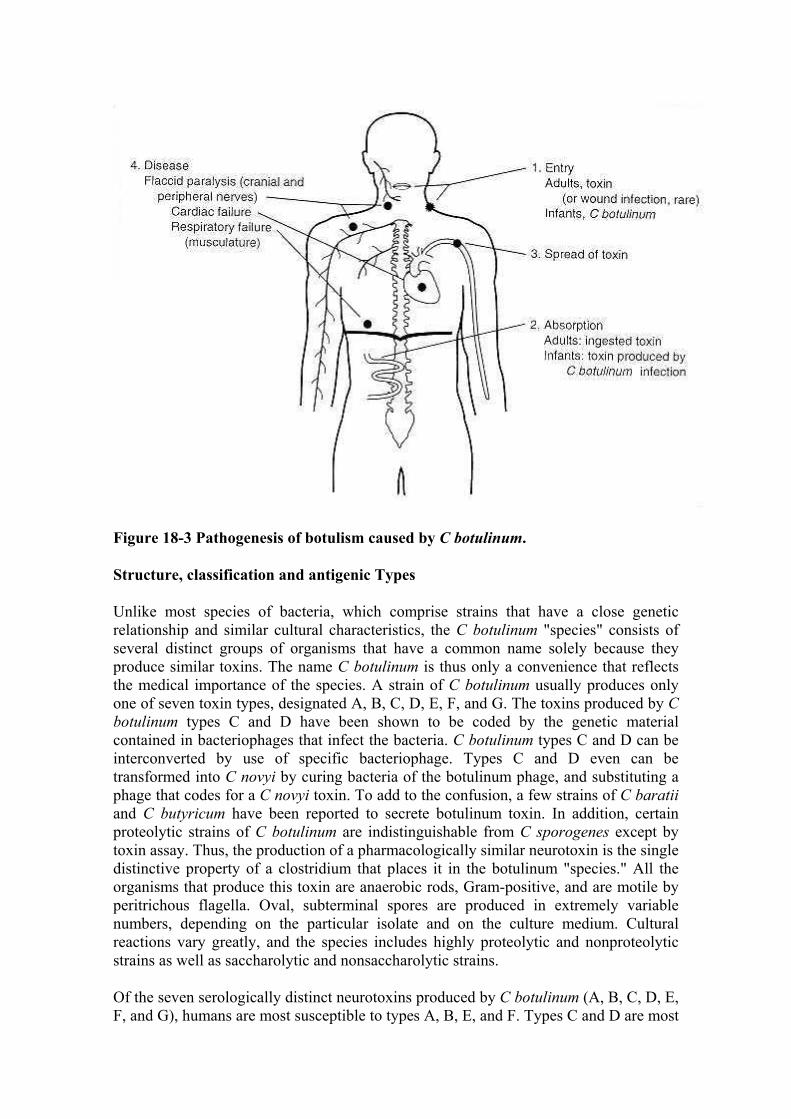
Figure 18-3 Pathogenesis of botulism caused by C botulinum.
Structure, classification and antigenic Types
Unlike most species of bacteria, which comprise strains that have a close genetic relationship and similar cultural characteristics, the C botulinum "species" consists of several distinct groups of organisms that have a common name solely because they produce similar toxins. The name C botulinum is thus only a convenience that reflects the medical importance of the species. A strain of C botulinum usually produces only one of seven toxin types, designated A, B, C, D, E, F, and G. The toxins produced by C botulinum types C and D have been shown to be coded by the genetic material contained in bacteriophages that infect the bacteria. C botulinum types C and D can be interconverted by use of specific bacteriophage. Types C and D even can be transformed into C novyi by curing bacteria of the botulinum phage, and substituting a phage that codes for a C novyi toxin. To add to the confusion, a few strains of C baratii and C butyricum have been reported to secrete botulinum toxin. In addition, certain proteolytic strains of C botulinum are indistinguishable from C sporogenes except by toxin assay. Thus, the production of a pharmacologically similar neurotoxin is the single distinctive property of a clostridium that places it in the botulinum "species." All the organisms that produce this toxin are anaerobic rods, Gram-positive, and are motile by peritrichous flagella. Oval, subterminal spores are produced in extremely variable numbers, depending on the particular isolate and on the culture medium. Cultural reactions vary greatly, and the species includes highly proteolytic and nonproteolytic strains as well as saccharolytic and nonsaccharolytic strains.
Of the seven serologically distinct neurotoxins produced by C botulinum (A, B, C, D, E, F, and G), humans are most susceptible to types A, B, E, and F. Types C and D are most
toxic for animals. Type G is rare, with only a few reported human cases. The toxins often are released from the bacteria as inactive proteins that must be cleaved by a protease to expose the active site. These proteases may be produced by the cell itself or may be in the body fluids of the infected host. Type A toxin is the most potent poison known; ingestion of only 10-8 grams of this toxin can kill a human. Put another way, the amount of toxin that could be held on the tip of a dissecting probe could kill 40 medical students.
Pathogenesis
The pathogenicity of C botulinum depends entirely on neurotoxin production (Fig. 18-3). In humans, these toxins cause disease in three ways: the well-known form of food poisoning results from ingestion of toxin in improperly preserved food; wound botulism, a rare disease, results from C botulinum growing in the necrotic tissue of a wound; and infant botulism is caused when the organism grows and produces toxin in the intestines of infants.
From its site of entry into the body, the toxin travels through the blood and lymphatic systems (and possibly the nervous system). It then becomes fixed to cranial and peripheral nerves, but exerts almost all of its action on the peripheral nervous system. The toxin appears to bind to receptor sites at the neuromuscular junctions of parasympathetic nerves, and inhibits the release of acetylcholine at peripheral cholinergic synapses. The result is flaccid muscular paralysis.
The cranial nerves are affected first, followed by a descending, symmetric paralysis of motor nerves. The early involvement of cranial nerves causes problems with eyesight, hearing, and speech. Double or blurred vision, dilated pupils, and slurred speech are common symptoms. Decreased saliva production causes a dryness of the mouth and throat, and swallowing may be painful. An overall weakness ensues, followed by descending paralysis with critical involvement of the respiratory tree. Death usually is caused by respiratory failure, but cardiac failure also can be the primary cause. Mortality is highest for type A, followed by type E, and then type B, possibly reflecting the affinities of the toxins for neural tissue: type A binds most firmly, followed by type E, then type B. Fatality rates are directly proportional to the infectious dose and inversely proportional to the incubation time of the disease.
Type A toxin is used therapeutically to treat a variety of conditions involving involuntary muscle spasms, including strabismus and certain focal dystonias. This therapy takes advantage the effect of the toxin as a specific muscle relaxant. The therapeutic toxin is a neurotoxin-hemagglutinin complex isolated from C botulinum cultures. The extreme potency of type A botulinum toxin requires extreme caution in using this compound as a therapeutic agent.
Food poisoning. In botulism food poisoning, the toxin is produced by the vegetative cells of C botulinum in contaminated food, and preformed toxin then is ingested with the contaminated food. The incubation time can vary from a few hours to 10 days, but most commonly is 18-36 hours. Only a small, but effective, percentage of the ingested toxin is absorbed through the intestinal mucosa; the remainder being eliminated in the feces. Gastrointestinal disturbances are early symptoms of the disease in about one-third of the patients with toxin types A or B, and in almost all of the cases involving type E
toxin. These symptoms include nausea, vomiting, and abdominal pain. Diarrhea often is present, but constipation also may occur. Symptoms of toxemia then become apparent. No fever occurs in the absence of complicating infections.
Wound botulism. Wound botulism is a rare disease. The initial event is contamination of a wound by C botulinum. The organisms are not invasive and are confined to the necrotic tissue, where they replicate and elaborate the lethal neurotoxin. The incubation time varies from a few days to as long as 2 weeks. The only differences in the symptoms of wound botulism and food poisoning (in addition to a possibly longer incubation time) are that wound botulism lacks gastrointestinal symptoms, and a wound exudate or a fever, or both may be present. C botulinum may be present in a wound but creates no symptoms of botulism. There have been several recent reports of wound botulism in intravenous drug abusers, who are now considered a population at risk.
Infant botulism. In contrast to food poisoning with toxemia caused by ingestion of preformed toxin, infant botulism results from germination of spores in the gastrointestinal tract. Here vegetative cells replicate and release the botulinum toxin. It is unclear as to why spores can germinate and bacteria replicate in the infant intestine, but phenomenon apears to be related to the composition of the intestinal flora of infants. Almost all reported cases have occurred in infants between 2 weeks and 6 months of age, with the median age of onset being 2 to 4 months. Toxins A or B are most frequently implicated. In infant botulism, the usual first indication of illness, constipation, is often overlooked. The infant then becomes lethargic and sleeps more than normally. Suck and gag reflexes diminish, and dysphagia often becomes evident as drooling. Later, head control may be lost, and the infant becomes flaccid. In the most severely affected babies, respiratory arrest can occur. Infant botulism can be lethal and is the likely etiologic agent in 4 to 15% of the cases of sudden infant death.
There are scattered reports that C botulinum can occasionally multiply and secrete toxin in the intestinal tracts of adults with an altered intestinal flora due to antibiotic therapy or achlorhydria.
Host Defenses
Host defenses against C botulinum are undefined. Some people can tolerate ingestion of botulinum toxin better than others. The reason for this phenomenon is obscure, but could be due to differences in the efficiency of uptake of the toxin from the intestine or in transporting the toxin to neural tissue. An attack of botulism does not produce effective immunity. The small amount of toxin in the circulation and its affinity for neural tissue probably prevent adequate amounts of toxin from interacting with the immune system.
Epidemiology
C botulinum spores are found worldwide in the soil (including in sea sediments) and in low numbers in the gastrointestinal tracts of some birds, fish, and mammals. In the United States, the most frequent isolate is type A, followed by B and E, with an occasional isolate of type F. In Europe, B is the most frequent isolate, whereas A is comparatively rare. Despite the worldwide occurrence of C botulinum in the environment, wound botulism is a comparatively rare disease.
Originally, botulism food poisoning was thought to be associated only with contaminated meat, especially sausage; however, it is now known that C botulinum can grow equally well in many types of food including vegetables, fish, fruits, and condiments. Home canning using inadequate sterilization techniques has been responsible for most cases of botulism during this century. The spores are heat resistant and can survive 100° C for hours, but the toxin is relatively heat labile. The toxin is usually produced at pH 4.8-8.5. However, even acid foods such as canned tomatoes have been responsible for several recent cases of botulism food poisoning. In addition, certain culture conditions have been shown to cause toxin production at pH values lower than 4.6. In general, germination of botulinum spores is favored in food kept at warm temperatures under anaerobic conditions for a long period of time.
C botulinum spores exist throughout the environment; all adults have likely ingested these spores with no ill effects. Because spores can cause poisoning in infants, obvious sources should be eliminated from the infant's environment and especially the infant's diet. Honey is the only dietary ingredient that has been implicated, and honey is no longer recommended for infants under 1 year of age. Most cases are not caused by ingesting honey, however, so this will not eliminate the disease. The other more common environmental sources of spores, such as soil and dust, are not so easily controlled.
Diagnosis
Although all forms of botulism are difficult to diagnose, prompt diagnosis and treatment are crucial to patient survival. Laboratory tests offer little in establishing an initial diagnosis of botulism, and accordingly, the finding of a normal cerebrospinal fluid can help to eliminate many of the diseases concerned with central nervous system disorders. Differential diagnoses are myriad and include neurological as well as gastrointestinal disorders. The infant with botulism is typically afebrile with generalized weakness, a weak cry, pooling of oral secretions, and poor sucking ability. Constipation may precede the illness by several weeks. An electromyogram pattern of brief, small-amplitude overabundant motor reaction potentials often is seen.
Confirmation of the initial diagnosis rests on demonstrating toxin in the patient's feces, serum, or vomitus. In adult botulism, serum samples rarely yield type A toxin because of the strong affinity of this toxin for neural tissue. In infant botulism, circulating toxin can occasionally be found in the serum. Fecal samples are the best specimens for detecting toxin in botulism food poisoning or infant botulism because only a small percentage of ingested or in situ formed toxin is absorbed through the intestinal mucosa. Toxin may be excreted for days or even weeks following botulism food poisoning. Toxin is usually detected by its lethal effect in mice coupled with neutralization of this effect by specific antisera. In infants, the organism can usually be cultured from the stool.
Control
The best way to control botulism food poisoning is to use adequate food preservation methods and to heat all canned food before eating. Because botulinum toxin is heat-labile, boiling food for a few minutes will eliminate toxin contamination; however, the

spores themselves are not destroyed by boiling, and proper canning procedures must be followed to kill clostridial spores.
Once a case of wound botulism or food poisoning has been diagnosed, therapy has four objectives: to eliminate the source of the toxin, to eliminate any unabsorbed toxin, to neutralize any unbound toxin with specific antitoxin, and to provide general supportive care.
Food Poisoning in Adults and Wound Botulism. In food poisoning, the unabsorbed toxin may be eliminated by stomach lavage and high enemas. Although cathartics may be used to eliminate residual toxin, they may have adverse effects in patients with bowel paralysis. In wound botulism, debridement and antibiotic therapy with penicillin are used to eliminate the offending organism. Antibiotic therapy is of questionable value in food poisoning, but is advocated by those who believe the organism can replicate in the intestinal tract of adults.
For both food poisoning and wound botulism, antitoxin therapy is most effective if administered early; however, clear-cut evidence for the efficacy of antitoxin therapy exists for only type E toxin. Antitoxin is available from the Centers for Disease Control (Atlanta, GA) through State Health Departments; trivalent ABE botulinum antitoxin is currently recommended. Unfortunately, all antitoxins are equine preparations, so a significant percentage of patients experience reactions typical of anaphylaxis and serum sickness. Thus, before they receive antitoxin, all patients should be tested for sensitivity to horse serum. The most important aspect of treatment in botulism is close observation of the patient and availability of adequate facilities for immediate respiratory support. Respiratory failure may occur within minutes, and immediate respiratory assistance often saves the lives of patients with botulism toxemia. Due to improvements in supportive care, the mortality rate for botulism has been dramatically reduced from approximately 60% (in the 1940s) to 10%.
All cases of botulism food poisoning should be reported immediately to local, state, or federal authorities, who will then take steps to minimize the chance of an outbreak. All persons suspected of ingesting contaminated food should be closely observed. Antitoxin should be administered both to those with overt symptoms and to those who have definitely ingested contaminated food.
Infant Botulism. The most important aspect of treatment of infant botulism is meticulous supportive care. Oral antibiotic therapy is not indicated because it may unpredictably alter the intestinal microecology and allow accidental overgrowth of C botulinum. Cathartics and enemas are also potentially dangerous. The value of human botulinum antitoxin is disputed, and there is not firm evidence to support its efficacy. The most significant aspect of therapy for infant botulism is supportive care. The infant should be kept under close supervision, with facilities for respiratory support immediately available. The fatality rate for infant botulism is surprisingly less than 5%.
Antibiotic-Associated Diarrhea, Pseudomembranous Colitis, and Clostridium Difficile
Clinical Manifestations

Clostridium difficile is a major nosocomial pathogen that causes a spectrum of intestinal disease from uncomplicated antibiotic-associated diarrhea to severe, possibly fatal, antibiotic-associated colitis. Diarrhea has come to be accepted as a natural accompaniment of treatment with many antibiotics. Although this diarrhea usually causes only minor concern, it can evolve into a life-threatening enterocolitis.
Many antibiotics have been associated with diarrhea and with pseudomembranous colitis, including ampicillin, cephalosporins, clindamycin, and amoxicillin. Patients treated with clindamycin have a higher incidence of C difficile disease, but most cases are found in patients treated with other antibiotics because of the more widespread use of these agents. Occasionally, antineoplastic agents that alter the normal intestinal flora may also induce pseudomembranous colitis, with methotrexate most commonly implicated. Chemotherapy-associated C difficile disease may not be easily recognized due either to an absence of antibiotic therapy or due to the frequent concomitant use of antibiotics, obscuring true incidence of chemotherapy-associated C difficile disease.
Clinical symptoms of C difficile disease vary widely from mild diarrhea to severe abdominal pain accompanied by fever (typically >101°F) and severe weakness. Diarrhea is watery and usually nonbloody (Fig. 18-4), but approximately 5 to 10% of patients have bloody diarrhea. Fecal material typically contains excess mucus, and pus or blood may also be noted. Hypoalbuminemia and leukocytosis are common findings. Pathology involves only the colon where there may be disruption of brush border membranes followed by extensive damage to the mucosa. The disease may progress to a pseudomembranous colitis, possibly including intestinal perforation and toxic megacolon. There is a leukocytic infiltrate into the lamina propria accompanied by elaboration of a mixture of fibrin, mucus, and leukocytes, which can form gray, white, or yellow patches on the mucosa. These areas are called pseudomembranes; hence the common term pseudomembranous colitis. Pseudomembranes usually develop after 2-10 days of antibiotic treatment, but they may appear 1-2 weeks after all antibiotic therapy has stopped. Mortality varies, but may be as high as 10% in patients with pseudomembranous colitis. The ultimate cause of death often is difficult to determine, as most patients show a nonspecific deterioration over a period of weeks.
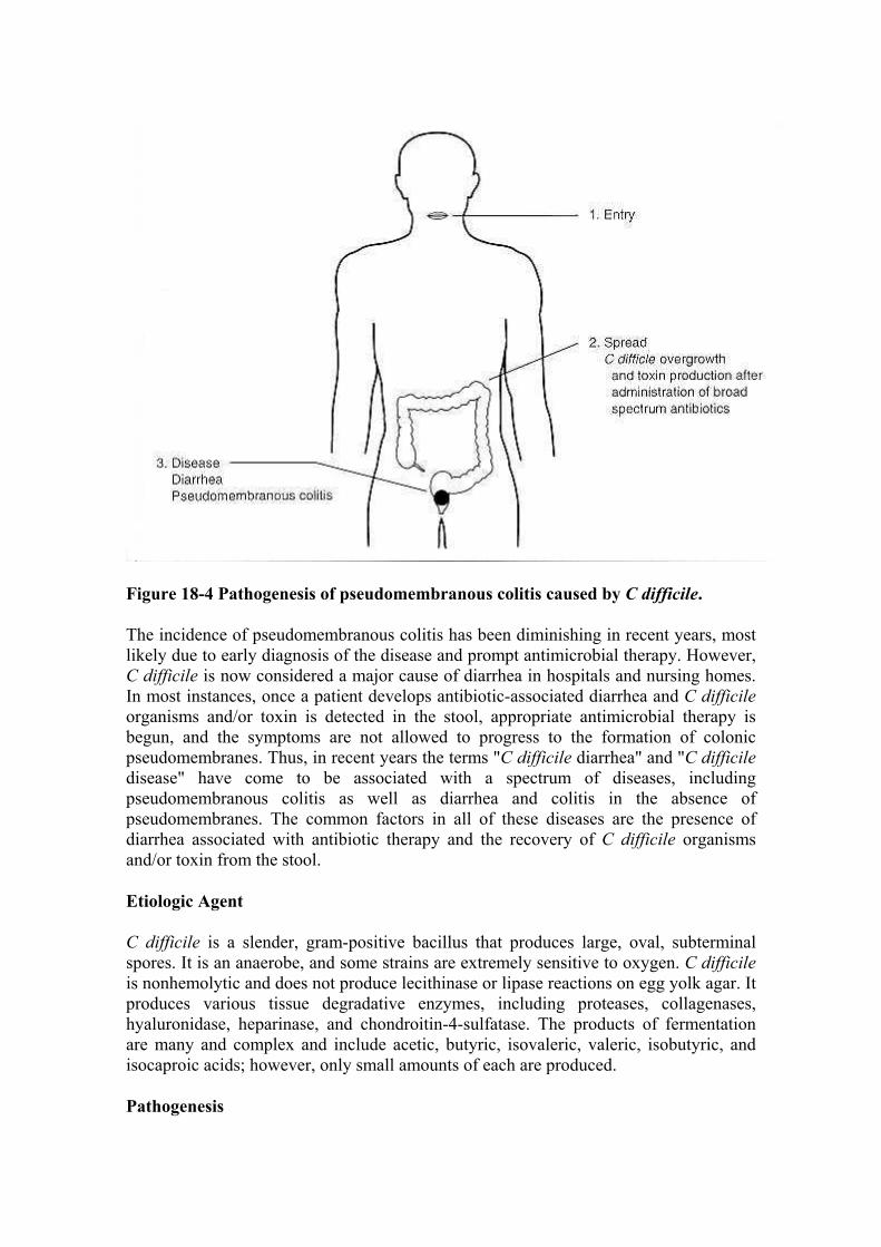
Figure 18-4 Pathogenesis of pseudomembranous colitis caused by C difficile.
The incidence of pseudomembranous colitis has been diminishing in recent years, most likely due to early diagnosis of the disease and prompt antimicrobial therapy. However, C difficile is now considered a major cause of diarrhea in hospitals and nursing homes. In most instances, once a patient develops antibiotic-associated diarrhea and C difficile organisms and/or toxin is detected in the stool, appropriate antimicrobial therapy is begun, and the symptoms are not allowed to progress to the formation of colonic pseudomembranes. Thus, in recent years the terms "C difficile diarrhea" and "C difficile disease" have come to be associated with a spectrum of diseases, including pseudomembranous colitis as well as diarrhea and colitis in the absence of pseudomembranes. The common factors in all of these diseases are the presence of diarrhea associated with antibiotic therapy and the recovery of C difficile organisms and/or toxin from the stool.
Etiologic Agent
C difficile is a slender, gram-positive bacillus that produces large, oval, subterminal spores. It is an anaerobe, and some strains are extremely sensitive to oxygen. C difficile is nonhemolytic and does not produce lecithinase or lipase reactions on egg yolk agar. It produces various tissue degradative enzymes, including proteases, collagenases, hyaluronidase, heparinase, and chondroitin-4-sulfatase. The products of fermentation are many and complex and include acetic, butyric, isovaleric, valeric, isobutyric, and isocaproic acids; however, only small amounts of each are produced.
Pathogenesis
C difficile disease is caused by the overgrowth of the organism in the intestinal tract, primarily in the colon (Fig. 18-4). The organism appears unable to compete successfully in the normal intestinal ecosystem, but can compete when normal flora are disturbed by antibiotics, allowing overgrowth of C difficile. This organism then replicates and secretes two toxins. Toxin A is an enterotoxin that causes fluid accumulation in the bowel, and it is a weak cytotoxin for most mammalian cells; toxin B is a potent cytotoxin. Nearly all toxigenic strains produce both toxins A and B. Highly toxigenic strains produce high levels of both toxins, while weakly toxigenic strains produce low levels of both toxins. Results from in vitro studies using cultured intestinal epithelial cells have indicated that toxin A causes necrosis, increased intestinal permeability, and inhibition of protein synthesis. Toxin A somehow affects phospholipase A2, resulting in the production of several arachidonic acid metabolites including prostaglandins and leukotrienes. Although the exact mechanism of endocytosis is unclear, both toxins A and B are internalized by host cells, resulting in alterations in the actin-containing cytoskeleton. Toxin A is a chemotactic factor for granulocytes; both toxins A and B have effects on leukocytes that include alterations in actin cytoskeletal microfilaments, and induction of tumor necrosis factor, interleukin 1, and interleukin 6. These latter effects contribute to the inflammatory response associated with C difficile disease.
Both toxins A and B kill experimental animals, and both probably are involved in the pathology of disease. Toxin production causes diarrhea that may progress to pseudomembranous colitis, where the characteristic pseudomembranes are largely limited to the colon. In the intestinal tract, toxin A damages villous tips and brush border membranes, and may result complete in erosion of the mucosa. This tissue damage causes a viscous hemorrhagic fluid response. In contrast, toxin B does not have noticeable enterotoxic activity, but it is lethal when injected into experimental animals. Thus, it seems reasonable to speculate that, in humans, toxin B exerts its pathogenic effect following dissemination through a damaged gut wall to extraintestinal organs. It has been speculated that infants harboring high levels of intestinal toxins A and B are at risk for the systemic toxicity of toxin B if their intestinal barrier is compromised.
Host Defenses
Host defenses for C difficile disease are not completely understood, but it seems reasonable to assume that the best host defense against C difficile disease is maintenance of the stability of the normal intestinal flora. Certain prostaglandins are known to protect the stomach and small intestine from mucosal necrosis caused by harsh chemicals; and, in experimental animals, prostaglandins have been shown to prevent extensive mucosal damage from C difficile toxin. These prostaglandins are also produced by toxin A-induced activation of the arachidonic cascade by phospholipase A2. Production of specific neutralizing antibodies to toxins A and B may participate in host defense, and a specific intestinal secretory IgA response to toxin A is more evident in the colon than the upper intestinal tract, compatible with the colon as the primary site of intestinal disease. The intestinal tract responds to C difficile toxins by increased fluid production, by secretory IgA neutralization of toxin, and by mucus production, which may inhibit the attachment of the toxins to their putative receptor sites on intestinal epithelial cells.
Epidemiology

C difficile is a member of the normal intestinal flora of <3% of adults. The organism can be acquired as a nosocomial pathogen and a variable incidence of disease is noted in hospitals and nursing homes. This seems to be due in part to environmental contamination with C difficile spores, and in part to different patient populations in various institutions. Patients with C difficile diarrhea excrete large numbers of C difficile spores, and epidemiological studies have shown that the organism can reside on environmental surfaces as well as on the hands of health care workers. Healthy adults do not carry significant numbers of the organism in their intestinal tracts, but healthy infants may have large numbers of these organisms in their feces. Most studies report a high carriage rate of approximately 50% in neonates, although some studies report a carriage rate of 0 to 6%, likely due to differences in environmental exposure to the organism. The toxins also are present in these infants' stools, and the same amounts of toxins are associated with disease in adults. The toxins typically have no adverse effect in infants, but confound the diagnosis of C difficile disease. There is circumstantial evidence supporting the theory that infants do not develop disease because they lack specific intestinal receptors for C difficile toxins. In recent years, C difficile as also emerged as one of the causes of chronic diarrhea in AIDS patients.
Diagnosis
It is often difficult to distinguish C difficile disease from other intestinal diseases, including ulcerative colitis and Crohn's disease. Diagnosis of C difficile disease includes the presence of diarrhea associated with antibiotic therapy in the preceding 4 to 6 weeks, and the recovery of C difficile organisms and/or toxin from the stool. However, the isolation of toxigenic C difficile from patients is often not a definitive diagnosis because other enteric pathogens are usually not excluded. Many cases of severe diarrhea are caused not by C difficile, but are caused by other enteric pathogens such as Campylobacter spp, Salmonella spp, Shigella spp, toxigenic strains of Escherichia coli, etc. Moreover, antimicrobial therapy increases the likelihood of isolating C difficile from the fecal flora: C difficile can be isolated from the feces of approximately 20 to 40% of asymptomatic hospitalized patients who are receiving antimicrobial therapy. Despite these caveats, C difficile is likely responsible for 25% of cases of antibiotic-associated diarrhea and colitis. Diagnosis of pseudomembranous colitis requires demonstration of pseudomembranes by colonoscopy, and C difficile can be isolated from the stools of almost all patients with this disease.
A good selective agar medium is used for the isolation of C difficile from stool. Toxin detection is also used for diagnosis. Although the most appropriate test for toxin detection remains controversial, a cellular cytotoxicity test remains the "gold standard"; here, filter sterilized fecal extract (or filter-sterilized broth containing a pure culture of C difficile) is added to a monolayer of cultured mammalian cells resulting in a cytopathic effect that is neutralized with specific antiserum. Unfortunately, this test is time-consuming and cumbersome. A rapid latex agglutination test is widely used, but this test is not specific, does not detect toxin A or B, and often results in false negative or false positive reactions. Enzyme-linked immunoassays can be used to detect both toxin A and toxin B, and these tests are useful for diagnosis of C difficile disease.
Control
In many cases, symptoms resolve 1-14 days after the offending antibiotic is discontinued, and antibiotic treatment is not needed. Vancomycin or metronidazole are the antibiotics of choice to treat active disease. Oral vancomycin is the "gold standard," and metronidazole is most often used to treat milder infections. Some claim that metronidazole should be considered the drug of choice in all but the most severe cases, based on relative cost of the two drugs and based on prevention of development of vancomycin resistance in enteric bacteria. C difficile is susceptible to both of these antimicrobial agents, but relapses occur in 15 to 20% of patients. Some patients have had many repeated relapses. Constipating agents, such as atropine diphenoxylate (Lomotil) or codeine, should not be used. Supportive therapy is needed to compensate for the often severe fluid and electrolyte loss. Health care workers caring for patients infected with C. difficile should wear gloves and strictly adhere to proper hand washing procedures.
Other Pathogenic Clostridia Food Poisoning and Clostridium perfringens
C perfringens is a major cause of food poisoning in the United States. The disease results from ingestion of a large number of organisms in contaminated food, usually meat or meat products. Food poisoning usually does not occur unless the food contains at least 106-107 organisms per gram. The spores are ubiquitous and, if present in food, can be triggered to germinate when the food is heated. Some heat-sensitive strains do not need heating to germinate. After germination, the number of organisms quickly increases in warm food because the generation time can be extremely short (minutes) and bacterial multiplication occurs over a wide temperature range. The location of C perfringens enterotoxin within the bacterial cell is controversial; some investigators claim that the enterotoxin is localized in the bacterial cytoplasm and others claim that it is associated with the spore coat. Regardless, food poisoning results from the ingestion food contaminated with enterotoxin-producing C perfringens. The enterotoxin directly affects the permeability of the plasma membrane of mammalian cells.
C perfringens type A is the usual causative agent, and serotyping is necessary and available for epidemiologic studies. Incubation time is 8-22 hours after ingestion of contaminated food, with a mean of 14 hours. Symptoms include diarrhea, cramps, and abdominal pain. Fever, nausea, and vomiting are rare, and the disease lasts only about 24 hours. The organism and its enterotoxin usually can be isolated from the feces of infected persons. The mortality rate is essentially zero, but elderly and immunologically compromised patients should be closely supervised.
Necrotizing Enteritis and Clostridium perfringens
Necrotic enteritis in humans has not been well documented. In adults, the disease appears to result from ingesting large amounts of food contaminated with C perfringens, usually type C. It generally follows ingestion of a large meal, implicating bowel distention and bacterial stasis as contributing factors. The intestinal pathology varies considerably, and may include sloughing of intestinal mucosa, submucosa, and mesenteric lymph nodes. Intestinal perforations occur frequently. The best-documented cases of this disease involve the natives of New Guinea, who develop necrotic enteritis ("pig-bel") after eating large quantities of improperly cooked pork that has been contaminated with the bowel contents of the animal. The course of the disease is fulminate, and the mortality rate is high. Scattered cases of necrotizing enteritis with C
perfringens as the prominent bacterial isolate have been reported in Western countries. In these cases, controversy exists concerning whether C perfringens is a primary invader, an accidental contaminant, or an opportunistic pathogen.
Some evidence suggests that acute necrotizing enterocolitis of infants may be caused by a clostridium, but definitive evidence is lacking. The theory is supported by the fact that pneumatosis cystoides intestinalis, a syndrome that can be caused by C perfringens, often is present in cases of acute necrotizing enterocolitis of infants. In addition, C perfringens, C butyricum, C difficile and other clostridial species are often isolated in cases of neonatal necrotizing enterocolitis, but a clear pathogenic role for clostridia is yet to be elucidated.
Bacteremia, endometritis and nonbacteremic infections with Clostridium sordellii
C sordellii is part of the normal intestinal flora of humans. The organism produces several exotoxins including toxins serologically related to the toxins of C difficile. There are scattered reports in the literature of C sordellii wound infections, most of which involve significant trauma. C sordellii has been occasionally implicated in bone and joint infections, in pulmonary infections, in bacteremia, and in fulminate endometritis. Because many clinical laboratories fail to speciate clostridial pathogens, the pathogenic potential of C sordellii is likely underestimated.
Malignancy and Clostridium septicum
C septicum is a spindle-shaped rod that is motile in young cultures. The organism produces toxins designated alpha, beta, gamma, and delta; the alpha toxin is necrotizing and lethal for mice. Whether C septicum is a member of the host's normal flora or whether it takes advantage of a compromised host is uncertain. The organism is not strongly invasive, but has been associated with gas gangrene. Fewer than 200 cases of invasive disease have been reported, but the majority have a malignancy somewhere in the body. The most frequent association is with colorectal cancer, but other types of malignancies have been noted, including leukemia, lymphoma, and sarcoma. In one survey of C septicum bacteremia, 49 of 59 (83%) cases had an underlying malignancy and, in 28 of these cases, the portal of entry appeared to be the distal ileum or the colon. Diabetes mellitus is seen in about 20% of cases. In collective review of 162 cases of nontraumatic C septicum infection, 81% of the patients had malignant disease; in contrast, other clostridial species are associated with malignancy in approximately 10% of cases. Thus, in the absence of an overt infection, isolation of C septicum should alert the physician to the possible presence of a malignancy, most likely in the ileum or the colon. Immediate antibiotic therapy is indicated because most patients die quickly of the infection if not treated. Penicillin is the antibiotic of choice, but chloramphenicol, carbenicillin, and cephalothin also have been used successfully.
Bacteremia and Clostridium tertium
C tertium is an aerotolerant clostridium that is usually considered nonpathogenic. However, there are scattered reports of this organism causing bacteremia. Most cases have involved neutropenic patients, and the gastrointestinal tract appears to be the source of the infection. It is possible that this organism causes many more cases of
bacteremia than is currently appreciated. The aerotolerant nature of C tertium may result in its misidentification as a Bacillus species.
Other clostridia with potential clinical significance
Clostridium butyricum, C clostrdioforme, C innocuum, and C ramosum are isolated with some frequency from clinical specimens and may have an unrecognized clinical significance. These species are often resistant to clindamycin and cephalosporins. C ramosum is usually listed with the ten anaerobic species most frequently isolated from clinical specimens. C ramosum frequently is misidentified, as the Gram reaction is lost easily, and spores are difficult to detect. C clostridioforme stains Gram-negative and forms characteristic football-shaped cells, rarely sporulates, and may be easily misidentified as Bacteroides sp or Fusobacterium sp.
REFERENCES Anand A, Glatt AE. Clostridium difficile infection associated with antineoplastic chemotherapy: a review. Clin Infect Dis 17:109, 1993
Bartlett JG. Clostridium difficile: History of its role as an enteric pathogen and the current state of knowledge about the organism. Clin Infect Dis 18: S265, 1994
Bongaerts GPA, Lyerly DM. Role of toxins A and B in the pathogenesis of Clostridium difficile disease. Microbial Pathogenesis 17: 1, 1994
Bleck TP. Tetanus: pathophysiology, management, and prophylaxis. Disease-A-Month 37: 545, 1991.
Coffield JA, Considine RV, Simpson, LL: Clostridial neurotoxins in the age of molecular medicine. Trends in Microbiology 67: 67, 1994
Finegold SM, George WL (eds): Anaerobic infections in humans. Academic Press, New York, 1989
Hambleton P. Clostridium botulinum toxins: a general review of involvement in disease, structure, mode of action and preparation for clinical use. J Neurol. 239: 16, 1992
Holdeman LV, Cato EP, Moore WEC (ed): Anaerobic laboratory manual, 4th ed. Blacksburg: V.P.I. Anaerobic Laboratory, Virginia Polytechnic Institute and State University, 1977.
Knoop FC, Owens M, Crocker IC. Clostridium difficile: clinical disease and diagnosis. Clin Microbiol Rev 6: 251, 1993
Lorimer JW, Eidus LB. Invasive Clostridium septicum infection in association with colorectal carcinoma. Can J Surg 37: 245, 1994
Lyerly DM, Krivan HC, Wilkins TD: Clostridium difficile: Its disease and toxins. Clin Microbiol Rev 1:1, 1988
Morbidity and Mortality Weekly Report: Tetanus - United States, 1985-1986. 36: 477-481, 1987
Rood JI, Cole ST. Molecular genetics and pathogenesis of Clostridium perfringens. Microbiol Rev 55: 621, 1991
Schofield F: Selective primary health care: strategies for control of disease in a developing world. XXII. Tetanus: a preventable problem. Rev Infect Dis 8: 144, 1986
Speirs G, Warren RE, Rampling A: Clostridium tertium septicemia in patients with neutropenia. J Infect Dis 158: 1336, 1988
Spera RV, Kaplan MH, Allen SL. Clostridium sordellii bacteremia: case report and review. Clin Infect Dis 15: 950, 1992
Stevens DL, Bryant AE. Role of theta toxin, a sulfhydryl-activated cytolysin, in the pathogenesis of clostridial gas gangrene. Clin Infect Dis 16: S195, 1993
Wigginton JM, Thill P. Infant botulism: A review of the literature. Clin Pediatr. 32: 669, 1993