clostridia infection
TRANSCRIPT

Clostridia Infection

Plan Of Talk
Introduction Etiology Problem Pathogenesis Economic importance Predisposing factors Clinical signs Post mortem lesions Diagnosis Treatment Prevention
Plan Of Talk
Introduction Etiology Problem Pathogenesis Economic importance Predisposing factors Clinical signs Post mortem lesions Diagnosis Treatment Prevention
Introduction
Necrotic enteritis (NE) is an enteric bacterial disease of chickens, turkeys, and a few other avian species caused by Clostridium perfringens.
Necrotic enteritis in poultry is caused by Clostridium perfringens type A.
The disease is characterized by damage to the intestinal mucosa by toxins produced by Clostridium perfringens.
Mild or subclinical form of NE is associated with poor growth and feed utilization.
Cont. …
NE occurs worldwide and causes considerable financial losses to broiler producers due to mortality and treatment cost.
NE is estimated to cost the worldwide poultry industry approximately a 2 billion dollars a year, due to:1. Increased mortality. 2. Increased feed conversion ratio. 3. Decreased weight gain.

Plan Of Talk
Introduction Etiology Problem Pathogenesis Economic importance Predisposing factors Clinical signs Post mortem lesions Diagnosis Treatment Prevention
Etiology
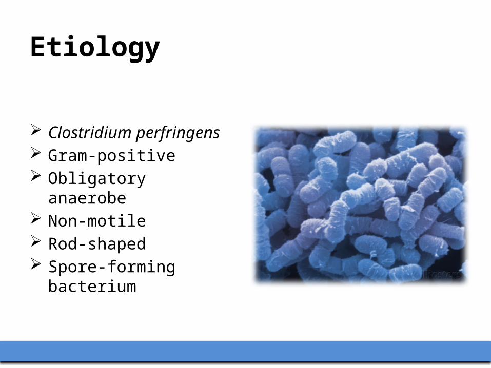
Clostridium perfringens Gram-positive Obligatory anaerobe Non-motile Rod-shaped Spore-forming bacterium

Natural Hosts
Necrotic enteritis is most common in broiler chickens and young broiler breeder pullets.
In commercially reared broiler chickens, clinical disease usually occurs between 2 - 5 weeks of age.
The disease is also seen in turkeys (young meat-type) and table-egg layers (pullets kept on litter).

Cont. …
Clostridium perfringens;– Grows at a temperature 15-50°C.– Optimum growth at 45°C for most strains.
Generation time for most strains;– Less than 20 minutes at 33°C to 49°C.– Generation time of 8 minutes has been reported.
Spores can withstand 100°C for two hours.

Clostridium perfringensNormal Inhabitant Pathogen
Clostridium perfringens is a natural inhabitant of:1. The environment
– Used litter – Soil– Marine sediments– Decaying vegetation– Sewage
2. Intestinal tracts of :– Healthy humans, animals and insects.
Cont. …
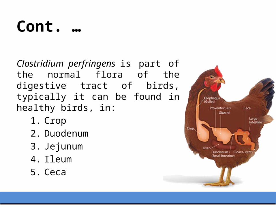
Clostridium perfringens is part of the normal flora of the digestive tract of birds, typically it can be found in healthy birds, in:
1. Crop2. Duodenum3. Jejunum4. Ileum5. Ceca

Cont. …
Clostridium perfringens colonizes the intestines of broiler chickens within few hours after hatching.
The numbers of Clostridium perfringens increase gradually after initial colonization.
The intestine of healthy birds contains large numbers of C. perfringens, up to 10 log 5 colony forming units CFU per gram of intestinal content.

Cont. …
The population of C. perfringens in the intestine is affected by:1. Nutritional factors
– Feed and litter contaminated with large numbers of C. Perfringens have been convincingly implicated as a source of infection.
2. Environmental factors3. Health status of the gut of the bird.

Cont. …
It is difficult to prepare spore-free poultry feed by standard feed preparation procedures.

Role Of MDA
Transfer of maternal antibodies [IgY, IgM, and IgA] from hen to chick in the yolk mass are important in protecting newborn chicks from disease.
Yolk contains > 100 mg of IgY, and IgA and IgM are incorporated into egg albumin during formation.
However, yolk IgY has a short half-life. Chicks are immunologically-immature until 3-4 weeks of age,
and maternal antibodies persist for < 3 weeks. This may explain why NE typically occurs seen in chickens 2 - 6
weeks of age.

Plan Of Talk
Introduction Etiology Problem Pathogenesis Economic importance Predisposing factors Clinical signs Post mortem lesions Diagnosis Treatment Prevention
Problem
Over the last few decades, necrotic enteritis (NE) in poultry has been controlled and treated by addition of antimicrobials to feed or water.

Cont. …
Due to rising consumer concerns, many countries are banning routine use of antimicrobials in feed.
The European Union has already banned antimicrobials, including many used to prevent NE, and a complete ban on the use of antimicrobials in animal feed is planned.

Cont. …
The ban has been accompanied by an increased number of European outbreaks of NE, and similar scenarios are anticipated elsewhere.
Thus, alternative prevention methods, such as vaccines, are needed.
Plan Of Talk
Introduction Etiology Problem Pathogenesis Economic importance Predisposing factors Clinical signs Post mortem lesions Diagnosis Treatment Prevention

Pathogenesis
NE occurs when C. perfringens; 1. Proliferates in the intestinal tract. 2. Produces potent toxins that severely damage the
intestinal mucosa. 3. Toxins absorbed from the intestinal tract resulting in
toxemia, which is responsible for death of the bird. Thus, NE is a type of “enterotoxemia”.
Cont. …
Clostridium perfringens is divided into five toxinotypes (A, B, C, D, and E) based on four major toxins (alpha, beta, epsilon, and iota).
The majority of isolates from NE cases are type A, with a few cases caused by type C. – Alpha toxin produced by types A and C– Beta toxin produced by type C
Cont. …
There is convincing evidence that:– C. perfringens strains vary in virulence.– NE is caused by certain strains of the organism. • Why these strains are capable of inducing NE remains
unknown.
Toxins/Virulence Factors
Alpha toxin, produced by C. perfringens toxinotype type A, as well as 4 other toxinotypes, are considered to be important virulence factors in the pathogenicity of the organism.
Alpha toxin is a zinc metalloenzyme with phospholipase and sphingolipase activities; – It hydrolyzes phospholipids in membranes of red and white
blood cells, thrombocytes, endothelial cells and muscle cells.
– Thus, the toxin is hemolytic, cytotoxic, necrotizing and potentially lethal.

Plan Of Talk
Introduction Etiology Problem Pathogenesis Economic importance Predisposing factors Clinical signs Post mortem lesions Diagnosis Treatment Prevention

Economical Importance
Lovland and Kaldhusdal (2001) studied the association between Clostridium perfringens infection and production performance
Commercial broiler flocks 2.5 years time period Clinical and subclinical NE were
frequently seen.
33%Comparing flocks with high and low levels of the disease
Profit

Cont. …
Major causes of production losses associated with clostridium perfringens infection;
1. Impaired feed conversion2. Reduced live weight at slaughter and3. Increased condemnation percentage
Subclinical clostridial enteritis has also been associated with impaired feed conversion and retarded growth.

Plan Of Talk
Introduction Etiology Problem Pathogenesis Economic importance Predisposing factors Clinical signs Post mortem lesions Diagnosis Treatment Prevention

Predisposing Factors
It is presumed that these factors promote excess growth of C. perfringens through mechanisms that are yet to be clarified.

Predisposing Factors
1. Small intestinal coccidiosis NE cases are commonly associated with varying degrees of
Eimeria maxima infection. Protein-rich exudate leaking from the damaged mucosa
may provide necessary nutrients for the growth of C. perfringens.
The minimum growth requirement of C. perfringens includes several amino acids and many growth factors and vitamins.
In modern, intensive poultry production, coccidiosis is probably the most important predisposing factor to NE under field conditions.
Cont. …
2. Diets high in cereal grains Such as rye, wheat and barley. They contain high levels of non-digestible, water soluble,
non-starch polysaccharides. Higher viscosity of wheat and barley ingesta may
contribute to greater bacterial numbers in the intestines, probably because of a slower intestinal transit time associated with these diets.
Cont. …
3. High amounts of animal protein Fish meal and bone meal, in the diet, increase the risk of
NE compared to feeds formulated with plant sources of protein.
The increased risk associated with animal sources of protein has been attributed to high glycine and methionine levels in animal protein; both of which enhance C. perfringens growth in vitro.

Cont. …
4. Animal fat It increases the numbers of C. perfringens in the ileum,
compared with soy oil. 5. Infection of chickens with immunosuppressive viruses
Infection with infectious bursal disease IBD Chicken anemia virus CAV Marek's disease virus MDV
Cont. …
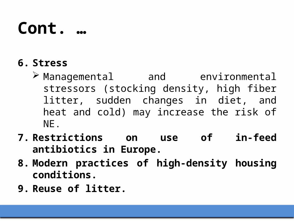
6. Stress Managemental and environmental stressors (stocking
density, high fiber litter, sudden changes in diet, and heat and cold) may increase the risk of NE.
7. Restrictions on use of in-feed antibiotics in Europe.8. Modern practices of high-density housing conditions.9. Reuse of litter.
Plan Of Talk
Introduction Etiology Problem Pathogenesis Economic importance Predisposing factors Clinical signs Post mortem lesions Diagnosis Treatment Prevention

Clinical Signs
Typical clinical signs of NE in chickens include:1. Depression2. Reluctance to move3. Diarrhea4. Ruffled feathers 5. Somnolence 6. Decreased appetite/anorexia 7. Dehydration 8. Huddling

Cont. …
NE has a short clinical course. Birds in the flock are found dead without premonitory clinical
signs. Some birds may appear listless and lethargic for a few hours
before death. Mortality in broiler flocks due to NE is usually below 10%, but
can be as high as 50%.

Cont. …
Mild and Subclinical NE Birds may not die but show;– Reduced weight gains – Higher feed conversion ratios– Increased condemnations at the processing due to liver
lesions (cholangiohepatitis).

Plan Of Talk
Introduction Etiology Problem Pathogenesis Economic importance Predisposing factors Clinical signs Post mortem lesions Diagnosis Treatment Prevention

Gross Lesions
Lesions in the intestinal tract is usually confined to the jejunum and ileum.
It varies in appearance from bird to bird depending on:1. Severity of infection2. Stage of development3. Presence or absence of coccidiosis4. Freshness of the carcass

Cont. …
When birds have NE it is best to examine euthanized or fresh dead birds for lesions;
1. Once the intestine starts to decompose after death, NE lesions tend to be less obvious and, in some cases, difficult to recognize.
2. Carcasses of birds with necrotic enteritis undergo postmortem autolysis quickly, probably because of toxemia.

Jejunum & Ileum
Jejunum and ileum may appear dilated, have a thin friable wall, and be filled with gas.
Wall of the affected segment of the small intestine is firm giving the gut the feel of a piece of hose.

• Broiler chicken, 31 days, jejunum/ileum, necrotic enteritis. • Jejunum and ileum are dilated and thin-walled. • Only birds that have been euthanized or died recently can be evaluated, as the changes
seen here can result from postmortem decomposition because of gas that increases in the gut after the bird dies.

• Broiler chicken, 35 days, jejunum/ileum, necrotic enteritis. • Jejunum and ileum are dilated, have firm walls, and are distended with fluid contents.
Mucosa
Granular or somewhat finely roughened appearance. The mucosa is either coarsely roughend and has a velvety
(“Turkish towel”) appearance. Markedly thickened and irregular. Green, yellow-brown, or orange-brown discoloration.
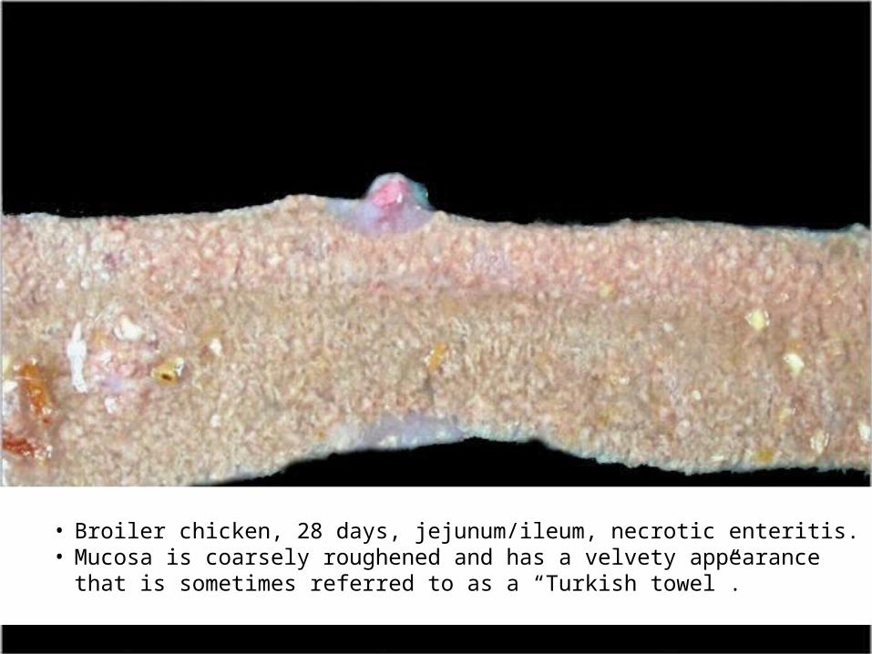
• Broiler chicken, 28 days, jejunum/ileum, necrotic enteritis. • Mucosa is coarsely roughened and has a velvety appearance that is sometimes referred
to as a “Turkish towel”.

• Broiler chicken, 17 days, jejunum/ileum, necrotic enteritis. • Mucosa is pale, thickened, and irregular because of diffuse necrosis.
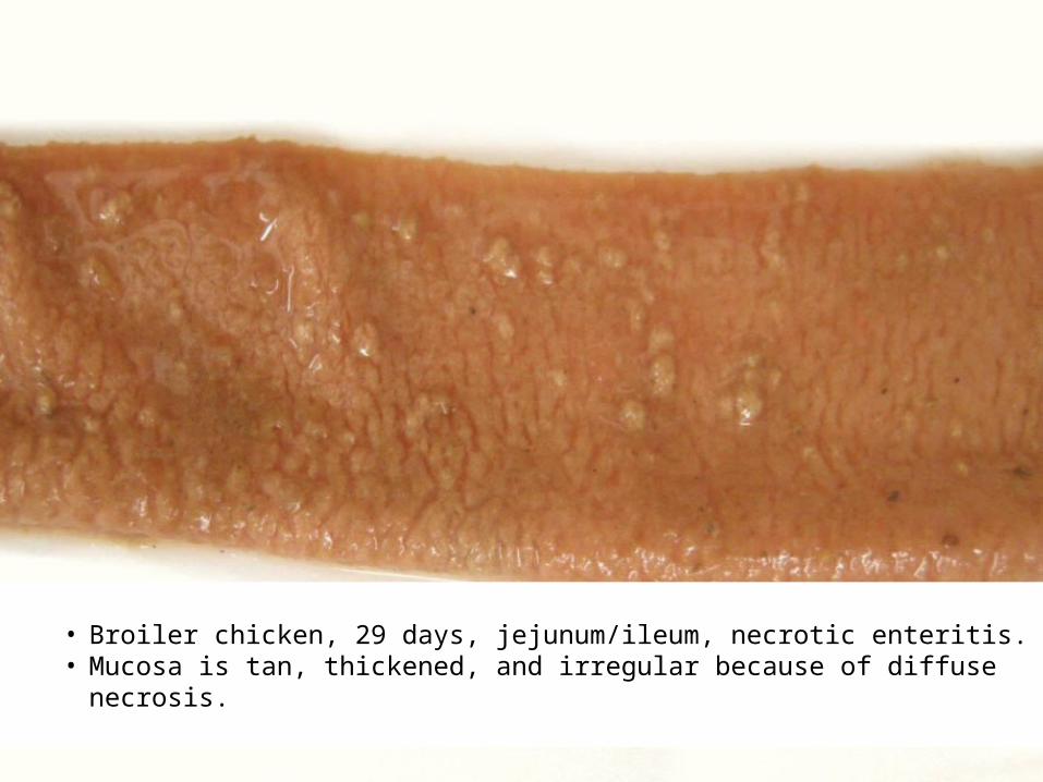
• Broiler chicken, 29 days, jejunum/ileum, necrotic enteritis. • Mucosa is tan, thickened, and irregular because of diffuse necrosis.

• Broiler chicken, 31 days, jejunum/ileum, necrotic enteritis. • Mucosa is yellow-brown, thickened, and irregular because of diffuse necrosis.
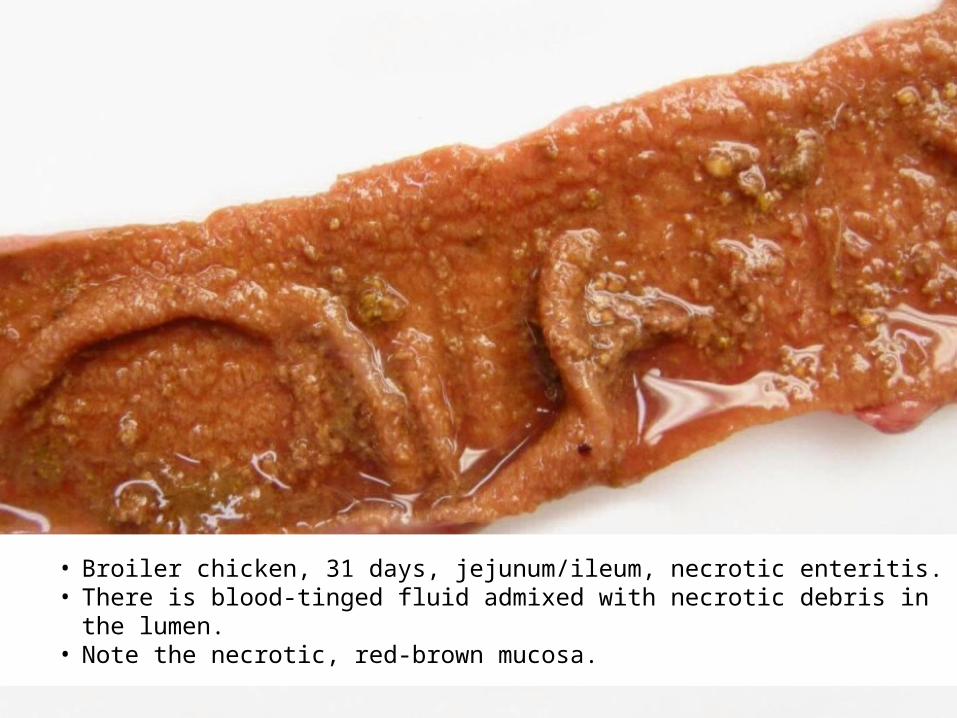
• Broiler chicken, 31 days, jejunum/ileum, necrotic enteritis. • There is blood-tinged fluid admixed with necrotic debris in the lumen. • Note the necrotic, red-brown mucosa.

• Broiler chicken, 38 days, jejunum/ileum, necrotic enteritis. • Mucosa is tan with a roughened mucosa due to diffuse necrosis.

• Broiler chicken, 45 days, jejunum/ileum, necrotic enteritis. • Mucosa is discolored green and necrotic.

• Broiler chicken, 53 days, jejunum/ileum, necrotic enteritis and ascaridiasis. • Mucosa is discolored green and necrotic. • Note the roundworms (Ascaridia) in the lumen.
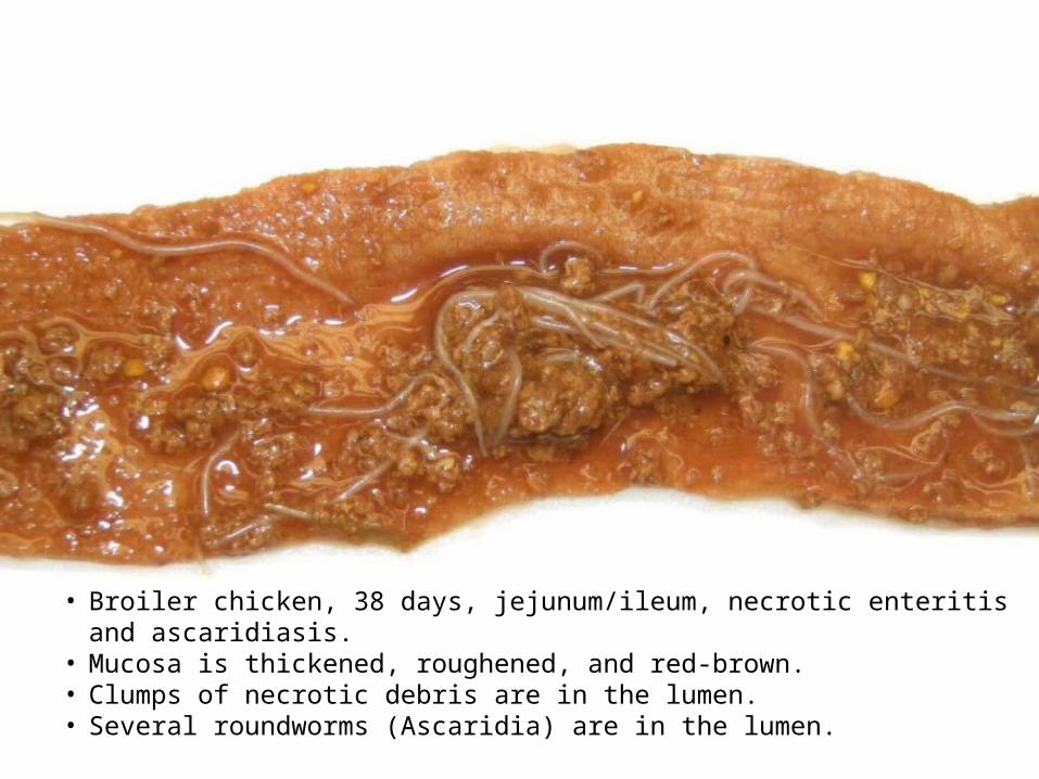
• Broiler chicken, 38 days, jejunum/ileum, necrotic enteritis and ascaridiasis. • Mucosa is thickened, roughened, and red-brown. • Clumps of necrotic debris are in the lumen. • Several roundworms (Ascaridia) are in the lumen.

• Broiler chicken, 45 days, jejunum/ileum, necrotic enteritis. • Mucosa is discolored orange-brown, thickened, and irregular.
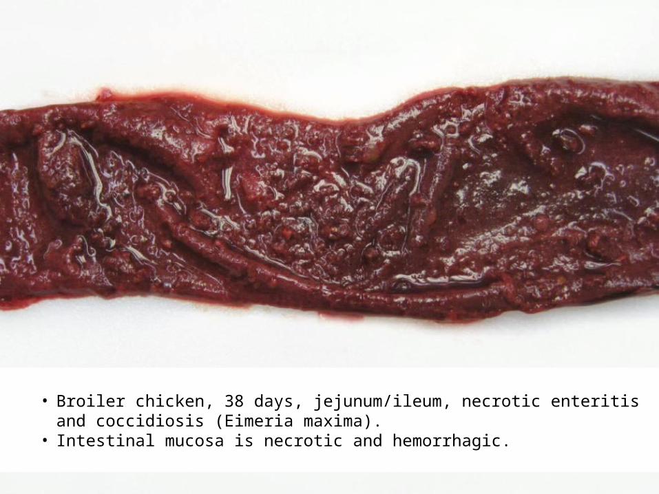
• Broiler chicken, 38 days, jejunum/ileum, necrotic enteritis and coccidiosis (Eimeria maxima).
• Intestinal mucosa is necrotic and hemorrhagic.
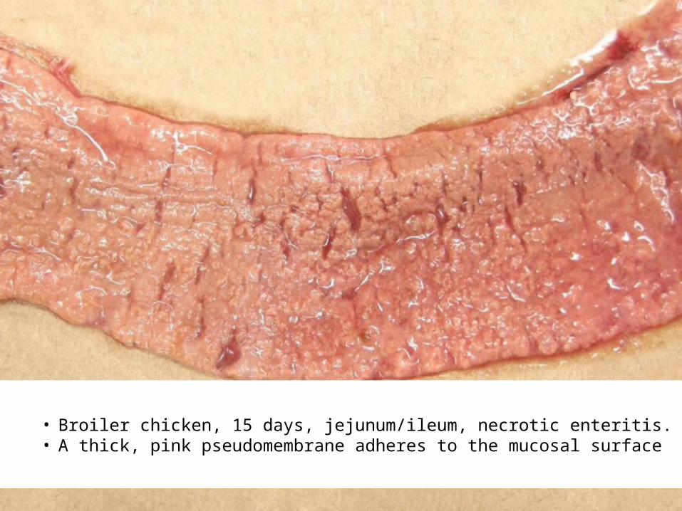
• Broiler chicken, 15 days, jejunum/ileum, necrotic enteritis. • A thick, pink pseudomembrane adheres to the mucosal surface
Lumen
The lumen may be empty or filled with green or red-tinged fluid that may contain pieces of debris.
In a few cases, a green or yellow-brown pseudomembrane may cover and loosely or firmly adhere to the mucosa.
Pieces of the membrane may detach into the lumen. Rarely the dilated small intestine has a hemorrhagic
appearance.

• Broiler chicken, 15 days, jejunum/ileum, necrotic enteritis. • Jejunum and ileum are dilated and markedly reddened. • Such an appearance for the intestine is unusual in birds with necrotic enteritis. • The bird was negative for coccidiosis.

• Broiler chicken, 38 days, jejunum/ileum.• Necrotic enteritis and coccidiosis (Eimeria maxima). • Jejunum and ileum are dilated, markedly hemorrhagic, and filled with bloody contents.

Liver
Lesions uncommonly occur in the liver, gallbladder, and/or extrahepatic bile duct.
Both liver lobes may be enlarged, firm, and have randomly scattered pale foci that represent necrotic lesions.
In rare cases, a small or large part of a liver lobe (most commonly the right lobe) appears discolored and has a firm texture due to extensive necrosis.
With cholecystitis, the gallbladder is distended and its wall is thickened and opaque. White to yellow exudate may be on the surface. Bile is often pale yellow, opaque, or inspissated.
The extrahepatic bile duct may be swollen, discolored, and filled with thick material.

• Broiler chicken, 28 days, gallbladder, cholecystitis. • Chicken had necrotic enteritis. • Gallbladder is distended, wall is thickened and opaque, and there is exudate on the
serosal surface. • Bile is discolored. • Culture of the bile yielded a pure growth Clostridium perfringens. • Note the yellow (necrotic) areas in the liver.

• Broiler chicken, 28 days, gallbladder, cholecystitis. • Chicken had necrotic enteritis.• Gallbladder is distended, wall is thickened and opaque, and there is exudate on the
serosal surface. • Bile is discolored. • Culture of the bile yielded a pure growth Clostridium perfringens. • Note the yellow (necrotic) areas in the liver.

Kidney
Kidneys are often pale and have a prominent lobular pattern.
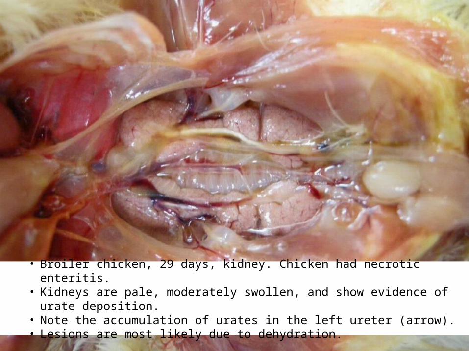
• Broiler chicken, 29 days, kidney. Chicken had necrotic enteritis. • Kidneys are pale, moderately swollen, and show evidence of urate deposition. • Note the accumulation of urates in the left ureter (arrow). • Lesions are most likely due to dehydration.

• Broiler chicken, 15 days, kidney. Chicken had necrotic enteritis. • Kidneys are swollen, discolored, and have a prominent lobular pattern. • Note the accumulation of urate in the ureters (arrows).
Plan Of Talk
Introduction Etiology Problem Pathogenesis Economic importance Predisposing factors Clinical signs Post mortem lesions Diagnosis Treatment Prevention

Diagnosis
Post mortem lesions NE lesions tend to become less obvious or even
unrecognizable in birds in an advanced state of autolysis, so freshly dead birds or euthanized birds are preferable for postmortem examination.
Gross lesions of NE are usually characteristic and allow preliminary diagnosis of the disease.

Plan Of Talk
Introduction Etiology Problem Pathogenesis Economic importance Predisposing factors Clinical signs Post mortem lesions Diagnosis Treatment Prevention
Treatment
Flocks affected with NE are usually treated with antibiotics administered via drinking water.
Penicillin, tetracycline, lincomycin, and erythromycin are the drugs of choice for treating NE. – Dose and duration of treatment should be as
recommended by manufacturer ensure efficacy of treatment.
Cont. …
Usually, the flock responds to treatment within 24-48 hours. If mortality does not drop in response to treatment, the
following possibilities need to be considered: 1. Incorrect diagnosis of NE.2. Resistance of C. Perfringens to the antibiotic.3. Incorrect dose of medication.4. Presence of a concurrent disease, such as severe
coccidiosis.
Cont. …
NE has been treated with lincomcyin, bacitracin, oxytetracycline, penicillin, and tylosin in water or bacitracin, lincomycin, virginiamycin, penicillin avoparcin, and nitrovin in feed.

Cont. …
Lincomycin:– 20 g per ton of feed significantly reduces mortality due to NE. – 16.9 mg/L in water was effective in treatment of broilers.
Virginiamycin – 5 - 40 g per ton of feed is also effective in treating NE.
Bacitracin– 100 mg per gallon in water for prevention.– 200 – 400 mg per gallon for treatment.
Penicillin– 100,000 IU per L prevented mortality in experimentally induced NE.– 110 mg per L in water delayed and reduced NE mortality, but did not
completely prevent disease .

Cont. …
The ionophore narasin, used primarily as a coccidiostat,– 70 ppm reduces mortality due to NE and also decreases numbers of C.
perfringens in ceca. – Narasin also increases feed efficiency and terminal body weight.
Plan Of Talk
Introduction Etiology Problem Pathogenesis Economic importance Predisposing factors Clinical signs Post mortem lesions Diagnosis Treatment Prevention

Prevention
Preventing NE can be accomplished through;1. In feed antibiotics2. Ionophorous coccidiostats3. Competitive exclusion products4. Preventing coccidiosis

PreventionIn Feed Antibiotics
C. perfringens is sensitive to several in-feed antibiotic growth promoters.
Use of these antibiotics as feed additives suppresses the number of C. perfringens in the intestinal tract and reduces the incidence of NE.

Cont. …
Since 1997 the European Union has banned the use of several growth promoting antibiotics (which has been associated with increased occurrence of NE), these antibiotics are; including
1. Avoparcin2. Ardamycin3. Bacitracin4. Virginiamycin5. Tylosin6. Spiramycin

Prevention Ionophorous Coccidiostats
C. perfringens is sensitive to the ionophorous coccidiostats monensin, salinomycin and narasin.
Incorporating these coccidiostats into the feed is effective in reducing the number of C. perfringens in the intestine and protecting against experimental NE.

Prevention Competitive Exclusion Products
The intestinal tract of birds contains several bacteria that compete with each other in the intestinal environment.
NE occurs when C. perfringens overgrows other bacteria in the intestinal tract.
Keeping a healthy balance of intestinal microflora is a key element in NE prevention.
Cont. …
Giving certain beneficial bacteria (particularly Lactobacillus spp.) to birds is effective in preventing, or at least reducing, the severity of NE “Competitive exclusion”.
Competitive exclusion products need to be given to the birds as soon as possible after hatching.
Beneficial bacteria in these products may reduce colonization of C. perfringens in young chicks.

PreventionPreventing Coccidiosis Preventing coccidiosis, whether by coccidiostats or
vaccination, is probably the most important measure to prevent NE.
When a coccidial vaccine is administered, follow the instructions of the vaccine manufacturer to avoid excessive exposure of the birds to Eimeria oocysts.
Key factors are:1. Vaccine dose.2. Maintenance and calibration of vaccination equipments.3. Management of the flock during the first two weeks post-
vaccination.

Cont. …
Cases of concurrent NE and coccidiosis have been encountered in 2-3 week old chickens in which a coccidial vaccine had been administered in the hatchery.

Finally
The prevention of NE involves an integrated intervention strategy that takes into consideration all of the factors that may increase the risk of the disease.