clinical reasoning - s3.amazonaws.com · what is clinical reasoning? integration of best clinical...
TRANSCRIPT

Clinical
Reasoning DPTI Mentoring Program

What is Clinical Reasoning?
Interaction of individuals in a
collaborative exchange to achieve a
mutual understanding of the problem and
negotiate a plan
Patient-centered
Deductive and inductive reasoning
Complex, non-linear, CYCLICAL
Critical role in reflective learning

Clinical Reasoning or
Evidence Based Practice?
Clinical Reasoning is a process that is used
and guides the clinicians where and
when good quality evidence does not
exist or cannot be applied due to a
variety of factors (time, resources, patient
values, etc.)

What is Clinical Reasoning?
Integration of best clinical research,
knowledge gained through clinical
practice, knowledge gained through
personal or like experience.

Clinical Reasoning and Expert Practice
Collaborative/ Patient centered
Clinical Pattern Development
Hypothetico-deductive reasoning
Diagnosis reasoning
Treatment reasoning

Errors in Clinical Reasoning
Failure to generate initial concept of the
problem
Wrong hypothesis/ inadequate testing
Confirmation Bias (tendency to look,
notice, remember what fits with pre-
existing expectations)
Outcome Bias- over reliance of the
outcome of the treatment intervention

Limiting Error
Awareness of clinical errors
Include screens/ examination questions
that would disprove your hypothesis
Clinical pattern recognition

Differential Diagnosis of
Soft Tissue Pathology: Muscle
History
Unaccustomed
activity
Repetitive
eccentric activity
Sudden/
unexpected strain
Direct trauma
Physical Examination Pain with
contraction Pain at endrange
stretch
Tender to palpation over muscle belly or trigger point
Imbalance length and strength
Swelling may be present

Differential Diagnosis of
Soft Tissue Pathology: Tendon
History
Recent repetitive activity
Midrange arc of pain with ROM
Direct trauma
Physical Examination Pain with isometric
contraction while joint is maintained in mid/ endrange or repetitive contraction
Weakness with mod-major pathology
Palpable tenderness
Muscle imbalance of length/ strength

Differential Diagnosis of
Soft Tissue Pathology: Bursa
History
Recent overuse
Unaccustomed
pressure
Acute:
Pain at rest
Pain with all
motions
Physical
Examination
Reproduces with
direct palpation

Differential Diagnosis of
Soft Tissue Pathology: Capsule
History
Acute
Trauma
Sudden,
unguarded mvtmt
Swelling
Pain with motion
Chronic
Stiffness
Physical Exam:
*Capsular Pattern
Pain at endrange
of motion
Worse in one
direction

Differential Diagnosis of
Soft Tissue Pathology: Ligament
History
Trauma
Postural: sustained
position
Physical Exam:
Acute
Swelling, compensatory
Partial tear: pn testing
Complete: Laxity
Postural: Pn with OP in one direction
Eased with movement in opposite direction

Differential Diagnosis of
Soft Tissue Pathology: Disc
History
Recurring episodes
Worse with flexion
25-45
Referred pain
Physical Exam
Location changes
with movement
Loss of lordosis
Peripheralization/
centralization
Lateral shift?
Reduced with
unloading

Differential Diagnosis of
Soft Tissue Pathology: Nerve
History
Paresthesia/
numbness
Lancinating pain
Pulling
Physical Exam
Positive Neuro
Screen
Sensation, reflex or
myotomal loss
Position nerve
tension test
Palpable nerve
tenderness

Differential Diagnosis of
Soft Tissue Pathology: Dura
History
Multisegmental
Pain
Pain/ Paresthesia
assoc
Headache,
“entire” spine,
extremities, ANR
Physical Exam
Positive Slump Test
Abnormal tension
testing

Differential Diagnosis of
Hard Tissue Pathology: Fracture History
Trauma
Minor if osteoporotic
Aching/ throbbing
Pain at rest
Pain worse with movement
Pain with stress
Unusual location of pain
Stress fracture: rapid increase in activity
Physical Exam
Deformity may be
present
Grinding or grating
Point tenderness
Confirm with
diagnostics

Differential Diagnosis of
Hard Tissue Pathology: Avulsion,
Tendon Rupture, Ligament Tear
History
Trauma
Pain, swelling
Loss of function
Bony deformation
Instability
Joint Locking
Physical Exam
Special tests
confirm
Surgical consult
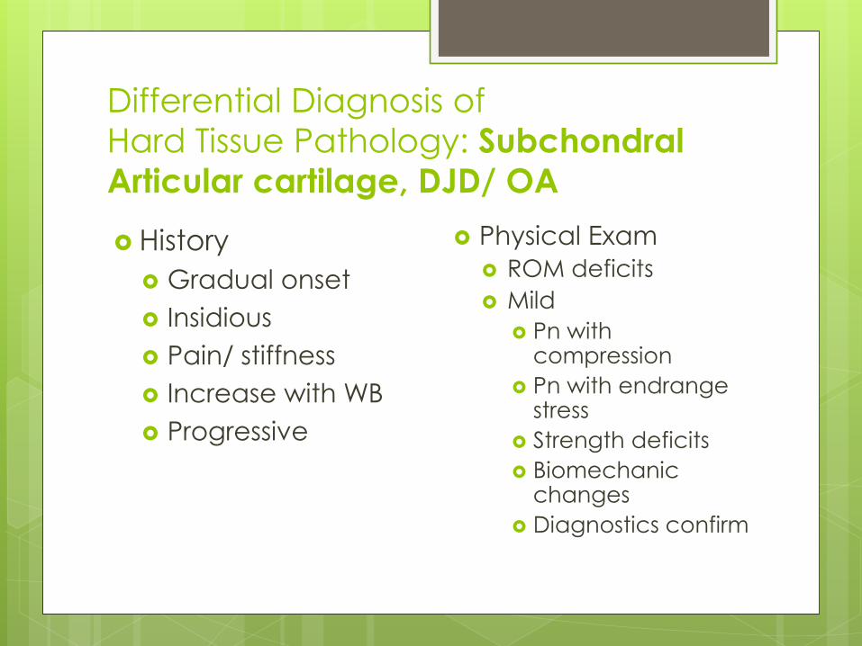
Differential Diagnosis of
Hard Tissue Pathology: Subchondral
Articular cartilage, DJD/ OA
History
Gradual onset
Insidious
Pain/ stiffness
Increase with WB
Progressive
Physical Exam
ROM deficits
Mild
Pn with compression
Pn with endrange stress
Strength deficits
Biomechanic changes
Diagnostics confirm
Subjective Exam
“ The patient is the most valuable source
of information and our ability to extract
that information will determine our depth
of understanding, and subsequently our
ability to manage the patient’s problem.”

Subjective Exam
However, the patient will not know what is not important and cannot be expected to know what we do, and do not need to know. This is important as we need to be skilled in helping patients through accounts of their problems and virtually teach patients how to listen to their own bodies and inform us of relevant information.”
-Jones and Butler 1991

Subjective Exam
Nonverbal communication
Spontaneous information
Use patient’s words
Active listener
Never assume anything
Assume responsibility
Subjective Examination:
Inquiry Strategies
Open-ended questions
Directed questions
Forced choices
Repetition of the story
Subjective Examination:
Self Assessment
Communication
Clear and Concise?
Non-verbal behaviors interacting?
Rapport established?
Effectively perceiving, interpreting?
Spontaneous information?
Using patient’s words?
Assumptions?
Open ended questions?
Relevant observation?
Subjective Examination:
Self Assessment
Collaboration
Patient’s initial concept of problem?
Drawing information from patient’s frame of reference?
Learning promoted?
Explanations?
Self management?
Negotiation?
Goals?

Subjective Examination:
data gathering
Is all information useful?
Is all of the necessary information
gathered?
Was a search and scan method used?
Any confirmation bias?
Was an initial impressions/ concept of the
patient used?

Subjective Examination:
data gathering
Establish ‘kind’ of disorder
Site/ Area of symptoms
Behavior of symptoms
Special questions/ med history
Present episode
Related past history

Subjective Examination:
data gathering- body chart
Map out each component separately
Depth
Varying/ Non-varying
Constant/ Intermittent
Relate different areas
Paresthesias

Subjective Examination:
data gathering- body chart
Identify all structures in each location of pain that could be “possible” sources of pain
Remember….
somatic (local)
referred- any pain felt away from source
radicular- must have neurological loss
peripheral
vascular
visceral
latent- pain that does not occur immediately

Subjective Examination:
Completion- Can you answer?
Mechanical vs chemical
“Kind of disorder”
Body region primarily to be examined
Body regions to be cleared
Neurological exam
Degree of provocation
Severity, irritability, nature, stage, stability
Hypothesis

Subjective Examination: Completion- Have you developed?
Hypothesis
Nocioceptive
Central vs. Autonomic
Source
Precautions/ Contraindications
Contributing Factors
Management
Prognosis

Contraindications/ Precautions
Malignancy Cauda Equina Cord Lesion Active Inflammatory Process Recent Fracture Osteoporosis
Neurological Signs RA Spondylolisthesis Hypermobility Dizziness

Subjective Examination:
Completion- Can you answer?
Patient Severity: Clinician’s assessment of the intensity of patient’s complaint and influence on functional activities
Patient Irritability: Amount of aggravating activity provoke symptoms, intensity of symptoms provoked, amount of time for symptoms to settle to previous level, problems with sleeping
Nature of Problem: Clinicians assessment/ hypothesis of the structures or pathology responsible for producing the patient’s symptoms and any relevant contributing factors.

Subjective Examination:
Completion- Can you answer?
Nature: Progression of Symptoms,
progression overall of repeated episodes
Stability: Predictability, consistency of
symptoms, consistency of movement or
resting

Physical Examination: Purpose
Identify Potential Sources
Identify Contributing Factors
Goals:
Determine structure that is involved
Reproduce symptoms
Pattern of movement comparable with history
Refine, support or rule out hypothesis
Establish baseline

Physical Examination
Posture and Observation
Neurologic Screen
Active Range of Motion
Passive and Physiologic Motion
Palpation
Flexibility
Motor Function

Assessment
“Although a clinician must possess a skill in
the area of examination and treatment,
the most important skill of all is that of
assessment, and the skilled use of
assessment in the overall process of
analytical assessment and clinical
reasoning” -Maitland
Assessment Assessment at end of initial examination
Assessment during performance of technique
Assessment after application of forces
Assessment at the end of treatment session
Assessment at the beginning of the next treatment session
Retrospective assessment
Assessment at completion of treatment

Assessment: Communication
Perspective of Patient
Assume Nothing
Comparative/ Baseline
Spontaneous/ Open ended Questioning
