clinical practice guidelines on postmenopausal ... clinical practice guidelines on postmenopausal...
TRANSCRIPT
GUIDELINES
Clinical practice guidelines on
postmenopausal osteoporosis: *An
executive summary and recommendations Lead Author - Meeta, Authors - C. V. Harinarayan, Raman Marwah, Rakesh Sahay, Sanjay Kalra, Sushrut Babhulkar Indian Menopause Society, Hyderabad, India
For review on vitamin D: Michael F Holick, Professor of Medicine, Physiology and Biophysics and Molecular Medicine. Director, Vitamin D, Skin, and Bone Research Laboratory. Programme Director General Clinical Research Unit. Director, Biologic Effects of Light Research Centre, Director, Bone Healthcare, Boston. External Review Board: A Muruganathan, Anil K Jain, Dinesh K Dhanwal, G R Sridhar, Hema Divakar, K V
Radha Krishna, Prof Nihal Thomas, N S Neki, P K Shah, S K S Marya, Sandhya Kamath, Sarita Bajaj, Thomas Paul. Advisory Board: Asha Kapadia, Atul Munshi, Duru Shah, Rama Vaidya, Saroj Srivastava, Sonia Malik, Sunila Khandelwal, Urvashi Prasad Jha. External Review Board: A Muruganathan, Anil K Jain, Dinesh K Dhanwal, G R Sridhar, Hema Divakar, K V Radha Krishna, Prof Nihal Thomas, N S Neki, P K Shah, S K S Marya, Sandhya Kamath, Sarita Bajaj, Thomas Paul. Resource Faculty: Alap Shah, Amita Pandey, Anil Mahajan, Ashok Vaidya, Beena Bansal, Bharti Kalra Prof. Dr C.V. Harinarayan, Dilip Mehta, Hemant Tiwari, I.V. Reddy, Jyothi Unni, Ketan Mehta, Manisha Sahay, Meeta, Nagamani, Neelam Agarwal, Rabindera Nath Mehrotra, Raghava Dutt Mulukutla, Rakesh Sahay, Major General (Dr) Raman Kumar Marwaha, Ram Prabhoo, Rama Vaidya, Ranu Patni, Rashmi Shah, Sanjay Bhadada, Sanjay Kalra, Sailesh. B, Seema Puri, Sharad Kumar, Shashank Joshi, Shushrut Babhulkar, Siddharth Sarkar, Sudha Sharma, Sunila Khandelwal, Sushil Gupta, Vishal R. Tandon, Vivek Arya, U.R.K. Rao, Yatan Pal Singh Balhara.
INTRODUCTION Guidelines are a method of translating the best available evidence into clinical, communicable, organizational, and policy making statements in the hope of improving health care and or policies. Do we need country specific guidelines? Yes, we do. Given the fact that the model of
health‑care delivery system and the prevailing environment of one country may not be extrapolated to that of another. “Working with what you have, where you are and not with what you
wish for” – is the principle each one of us follow in the
clinical practice to give the best to our patients. This
guideline hopes to bridge the gap between evidence based
practice, backed by scientific evidence and experience based
practice based on the published and unpublished Indian
Address for Correspondence: Dr. Meeta, Tanvir Hospital, Plot No. 100, Phase‑I, Kamalapuri Colony, Hyderabad ‑ 500 073, India. E‑mail: [email protected]
data and expert opinions. Unlike protocols, guidelines are meant to aid the clinician in decision making. The target readers of this guideline are the adult women, members of the Indian Menopause Society (IMS), allied
professionals, health‑care providers, and policy makers. India is a land of rich and diverse cultural heritage. It is a
land of diversity in terms of, socioeconomic, religion,
culture, beliefs, education, nutrition urban, rural, and
*This is a summary and recommendations from the detailed document on
Clinical Practice Guidelines on Menopause published by Jaypees. (R‑indicates Recommendation with Grading, the detailed references is listed in the main document. The text of the unpublished references can be procured from Dr. Meeta at [email protected])
Meeta, et al.: Guidelines on postmenopausal osteoporosis
geographical regions. The dilemmas and challenges are unique to different regions and solutions need to be planned accordingly. The specific issues pertaining to Indian women are an early age of natural menopause, genetic and environmental influences, nutritional deficiencies, and excesses resulting in physiologic differences. These factors contribute significantly to an increased incidence of diabetes, cardiovascular disease, osteoporosis, and thyroid dysfunction. Genetic components are likely to play a prominent role in these disorders for example, polymorphisms in estrogen receptors alpha and vitamin D receptor has been implicated in the pathogenesis of osteoporosis. The burden of morbidity from osteoporosis has significant medical, social, and financial implications. Osteoporotic fractures are preventable, yet diagnosed only
after the event; a situation similar to the diagnosis of
hypertension after myocardial infarction or stroke. It has a
long incubation period and cost‑ effective treatment
strategies currently available for this disease mandate that
osteoporosis be diagnosed and treated early. OBJECTIVES • To recognize post‑menopausal osteoporosis(PMO)
as a major health issue among health‑care professionals, policy makers, and the public.
• To assist health‑care practitioners in providing optimal care to post‑menopausal women with the available resources. Osteoporosis is a costly debilitating disease, hence it is important to instill preventive measures, diagnose early, encourage modifications of risk factors associated with osteoporosis. Counseling on nutritional factors, abuse of tobacco, heavy alcohol consumption, and on life‑style should be mandatory. Treat with pharmacologic agents only when indicated.
• To fill the lacunae of medical care after managing fragility fracture.
• To aid primary care physicians to decide when to refer patients with difficult problems to the relevant specialists.
• To stimulate interest in research on osteoporosis. METHODS The planning to publishing of the document took 24 months. The core committee was formed and a broad
based multi‑disciplinary list of experts were invited to write
on the topic of their expertise. Majority of the reviews and
deliberations wear by E‑mail. A two day intensive contact
program of the contributors was convened at Hyderabad
on December 8th and 9th 2012. Each topic was presented
and deliberated upon, and the consensus obtained by an automated response system. Later one day contact meeting
of the Editorial Board was convened on January 11th, 2013. Finally, the document was validated by an External Review Board. Data were sourced from the electronic database PubMed, MEDLINE, Cochrane Database of Systematic Reviews and published guidelines on PMO management.
The appraisal of Guidelines Research and Evaluation,[1] instrument was used to appraise published guidelines. Abstracts from papers and posters presented at the National Indian Menopause Society Meetings, published and unpublished studies, expert opinion was considered.
Cost‑effectiveness of diagnosis and treatment is based on the available market value. SYSTEM FOR GRADING: EVIDENCE USED
IN THE DOCUMENT The quality of evidence and the level of recommendation was
carried out using the grades of recommendation, assessment,
development, and evaluation (GRADE),[2] system. Recommendations are based on strong evidence, suggestions
on experience based evidence, this method is adapted to unite
the diverse conditions of India with the best available data and
the rich experience based evidence from the experts. A. GRADE: Grades of evidence:
• High quality– GRADE A: Further research is very unlikely to change our confidence in the estimate of effect.
• Moderate quality – GRADE B: Further research is likely to have an important impact on our confidence in the estimate of effect and may change the estimate.
• Low quality – GRADE C: Further research is very likely to have an important impact on our confidence in the estimate of effect and is likely to change the estimate.
• Very low quality – GRADE D: We are very
uncertain about the estimate. B. In terms of the strength of the recommendation, strong
recommendations use the phrase “recommend,” and weak recommendations use the phrase “suggest.”
Research questions are placed at the end of each chapter
in the monogram of the book. BENEFITS OF USING THE GUIDELINE Benefits of using these guidelines are: (i) Improved early
identification and better management of women at risk
for fragility fractures; (ii) down grading the disease
Meeta, et al.: Guidelines on postmenopausal osteoporosis
burden after an episode of fragility fracture by improving
the assessment, management and follow‑up of these
women; (iii) understanding the urgent need of conducting
preventive health programs by all stake holders related to
women’s health; and (iv) in addition, in view of the paucity
of Indian data it is hoped that this guideline will help
stimulate interest in research in various aspects of PMO. CONCLUSIONS Osteoporosis has significant medical, social, and
financial implications. The onus is on the Government and Non‑Government
Organizations to develop specialty menopause and
osteoporosis clinics akin to antenatal clinics in the private and
public sectors besides developing management of menopause
as a medical specialty within obstetrics and gynecology care.
The aim of the guideline is to provide a resource documentss
to aid the busy clinician to give optimal care to the ageing
woman. Limitations are the paucity of robust research
evidence in India. This is one of the endeavors of the Indian
Menopause Society to work toward the slogan “Fit @ Forty, Strong @ Sixty, Independent @ Eighty”. ACKNOWLEDGMENTS We thank the experts who took time out of their busy family life,
academics, and work to contribute to the document on PMO
in India. A special thanks to Dr. Hemant Zaveri for sourcing
the data. DISSEMINATION OF THE GUIDELINES A free copy of the guideline is for the members of the IMS and Jaypee Publishers are making the monogram
available widely for purchase by the health‑care
providers and policy makers. The Guideline is available on the IMS website www.indianmenopausesociety.org.com and is published in the Journal of Midlife, official publication of the IMS. REVISION OF THE GUIDELINES It is recommended that the guidelines are upgraded
every 2 years. EDITORIAL INDEPENENCE The views expressed are independent of any extraneous
influences. REFERENCES 1. AGREE Next Steps Consortium, 2009. The AGREE II
Instrument [Electronic version]. Available from: http://www. agreetrust.org. [Last accessed on 2012 Feb 10].
2. Atkins D, Best D, Briss PA, Eccles M, Falck‑Ytter Y, Flottorp S, et al. Grading quality of evidence and strength of recommendations. BMJ 2004;328:1490.
INTRODUCTION Among the several challenges faced by the growing elderly
population with increasing longevity in India,
post‑menopausal osteoporosis (PMO) is emerging as one of
the major public health issues. Osteoporosis is an
asymptomatic or “silent” disease and generally presents as a fragility fracture. Typical osteoporotic fractures are those of
the hip, spine and wrist. Global data indicates that 20% of
women with hip fracture die within 1 year of the fracture and
50% of them never regain their functional independence.[1] Vertebral fractures can also have significant morbidity and
are associated with increased long‑term mortality.[2] World
Health Organization (WHO) has identified osteoporosis as
an important non‑communicable disease. Osteoporotic
fractures impose great financial, medical, and social burden on society. These guidelines are intended to be used as a
resource document by the health‑care providers involved in
post‑menopausal women’s health at all levels of health‑care
with specific reference to India. Though framed for India, it is hoped that these guidelines will be useful for menopause practitioners across the globe.
BASICS CONCEPTS Definition 1. WHO defines osteoporosis as “a systemic skeletal disease
characterized by low bone mass (measured as bone
mineral density– [BMD]) and micro architectural
deterioration of bone tissue with a consequent increase in
bone fragility and susceptibility to fractures involving the
wrist, spine, hip, pelvis, ribs or humerus.[3] The National Institute of Health definition is “a disease characterized by decreased bone strength and
propensity to fall.”[4] Diagnosis 2. The diagnosis of an osteoporotic fracture, the clinical
end point of osteoporosis is by the presence of fragility fracture (clinical or radiological) and or by BMD.
3. The “gold standard” method of BMD testing is by dual X‑ray absorptiometry (DXA). Its value is
expressed in standard deviation (SD) units from the
population mean in young adults (T‑score) or from
the mean in an age‑matched population (Z score). Journal of Mid-life Health ¦ Apr-Jun 2013 ¦ Vol 4 ¦ Issue 109
Meeta, et al.: Guidelines on postmenopausal osteoporosis
4. The reference range recommended by the International
Osteoporosis Foundation (IOF), International
Society of Clinical Densitometry WHO and National Osteoporosis Foundation (NOF) for calculating the
T‑score in post‑menopausal women is the National Health and Nutrition Examination Survey (NHANES) III reference database in Caucasian
women aged 20‑29 years.[4‑6] 5. BMD based criteria for diagnosis of osteoporosis by
different organizations is based on a common principle: • In 1994, WHO definition was based on the T‑score
of the lumbar spine, total hip, or femoral neck.
• Currently WHO/IOF define osteoporosis as a value for BMD 2.5 SD or more assessed at the
femoral neck and is below the young female adult
mean (T‑score ≤ −2.5 SD).[4,5] Population based
sampling and expressing fracture risk as a function
of BMD with age adjustment have shown that the
risk of hip/vertebral fracture is similar in men and women for a given absolute value of BMD and
therefore the diagnostic criteria for defining
osteoporosis in men is the same as in women.[7,8]
Hence, the femoral neck BMD is used for
diagnosis of osteoporosis. It needs to understand
that the T‑score cannot be used interchangeably with different techniques and at different sites
since the prevalence of osteoporosis and
proportion of individuals allocated to any
diagnostic category would vary as does the risk of
fracture. Due to the above reasons, the WHO uses
the femoral neck as the reference site, but this does not mean that other techniques or other sites
cannot be used in the clinical management.
Further, it should be recognized that the
information derived from the T‑score may differ
from that provided by BMD at the femoral neck. • The NOF diagnosis is based on T‑score of
femoral neck and spine.[5] • The International Society for Clinical
Densitometry diagnostic criteria for
osteoporosis in post‑menopausal women and in
men age 50 and older is if the T‑score of the lumbar spine, total hip, or femoral neck is −2.5 or less. In certain circumstances, the 33% radius
(also called 1/3 radius) may be utilized.[9]
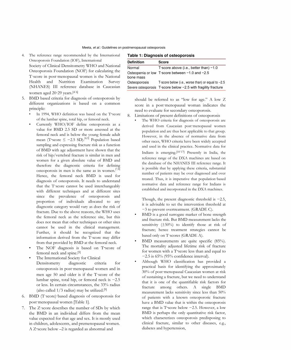
6. BMD (T‑score)‑based diagnosis of osteoporosis for
post‑menopausal women [Table 1]. 7. The Z‑score describes the number of SDs by which
the BMD in an individual differs from the mean value expected for that age and sex. It is mostly used in children, adolescents, and premenopausal women. A Z‑score below –2 is regarded as abnormal and
Table 1: Diagnosis of osteoporosis Definition Score
Normal T‑score above (i.e., better than) −1.0 Osteopenia or low T‑score between −1.0 and −2.5 bone mass
Osteoporosis T‑score below (i.e., worse than) or equal to –2.5 Severe osteoporosis T‑score below −2.5 with fragility fracture
should be referred to as “low for age.” A low Z score in a post‑menopausal woman indicates the need to evaluate for secondary osteoporosis.
8. Limitations of present definitions of osteoporosis • The WHO criteria for diagnosis of osteoporosis are
derived from Caucasian post‑menopausal women
population and are thus best applicable to that group.
However, in the absence of normative data from
other races, WHO criteria have been widely accepted
and used in the clinical practice. Normative data for
Indians is emerging.[10‑17] Presently in India, the
reference range of the DXA machines are based on
the database of the NHANES III reference range. It
is possible that by applying these criteria, substantial
number of patients may be over diagnosed and over
treated. Thus, it is imperative that population‑based
normative data and reference range for Indians is
established and incorporated in the DXA machines.
Though, the present diagnostic threshold is −2.5, it is advisable to set the intervention threshold at −3 to prevent overtreatment. (GRADE C).
• BMD is a good surrogate marker of bone strength and fracture risk. But BMD measurement lacks the sensitivity (±50%) to identify those at risk of fracture; hence treatment strategies cannot be
based only on T‑scores (GRADE A).
• BMD measurements are quite specific (85%). The mortality adjusted lifetime risk of fracture for women with a T‑score less than and equal to −2.5 is 65% (95% confidence interval).
• Although WHO classification has provided a practical basis for identifying the approximately
30% of post‑menopausal Caucasian women at risk of sustaining a fracture, but we need to understand
that it is one of the quantifiable risk factors for fracture among others. A single BMD measurement lacks sensitivity since less than 50%
of patients with a known osteoporotic fracture have a BMD value that is within the osteoporosis
range that is T‑score below −2.5. However, a low BMD is perhaps the only quantitative risk factor, which characterizes osteoporosis predisposing to
clinical fracture, similar to other diseases, e.g., diabetes and hypertension,
110 Journal of Mid-life Health ¦ Apr-Jun 2013 ¦ Vol 4 ¦ Issue 2
Meeta, et al.: Guidelines on postmenopausal osteoporosis
which are characterized by quantitative risk factors (GRADE A).
• The WHO diagnostic criteria have been validated
using DXA of the axial skeleton, that is, the spine
and hip, extrapolation of these criteria to other
techniques (e.g., ultrasound, computed tomography)
used to measure BMD, including the peripheral
skeleton may yield incorrect information regarding
fracture risk. T‑scores cannot, therefore, be used
interchangeably between the different techniques
available to measure BMD. • The exclusively BMD‑based diagnostic approach
of the WHO classification does not include
extra‑skeletal risk factors like the propensity to fall.
• The emergence of largely BMD‑independent risk
factors contributing to fragility fractures, the
BMD‑based criteria alone will not be sensitive
enough to identify those at risk of a fracture. This has underscored the need for fracture risk assessment tools or algorithms to manage patients with osteopenia and osteoporosis. It is important that we understand that we should not be treating
T‑scores only (GRADE C).
• Bone quality, an important component of bone strength is not considered in the WHO definition.
A low bone mass is most commonly the result of osteoporosis, it may be reflected secondary to osteomalacia and primary hyperparathyroidism, which are simpler to treat than advanced osteoporosis.
• Falsely high BMD may be due to flurosis, which
is prevalent in some parts of the country.[18] TYPES OF OSTEOPOROSIS 9. Osteoporosis was earlier classified as primary and
secondary.(experts feel that this classification is no longer valid): • Primary osteoporosis is seen in post‑menopausal
women in whom there is no specific pathogenetic mechanism other than age.
• There is an accelerated bone loss at the rate of 2‑5%/year due to declining estrogens levels and is seen in the first 5‑7 years after menopause.[19]
• Secondary osteoporosis is due to specific causes. 10. Osteoporosis and osteomalacia
Bone is a dynamic tissue with a continuous remodeling leading to the formation of new bone and resorption of old bone. A mismatch of this process forms the basis for osteoporosis while defective mineralization of the newly formed osteoid is called osteomalacia.
11. Fragility fracture • A fragility fracture has been defined by the WHO
as “a fracture caused by injury, which would be
insufficient to fracture normal bone: The result of reduced compressive and/or torsional strength of bone”.
• Clinically, a fragility fracture can be defined as one, which occurs as a result of minimal trauma, such as a fall from a standing height or less, or no identifiable trauma.
• The most common sites of fragility fracture are the hip, spine, and forearm. The other sites are pelvis, proximal femur, proximal humerus, proximal tibia, and fractures involving three ribs simultaneously.
• Fragility fractures account for 80% of fractures in post‑menopausal women.[20]
• Risk of recurrence of a hip fracture is 5‑10%, and with vertebral fracture, it is 20% within a year following a primary fracture.[21,22]
• Fragility fracture leads to significant impairment of physical, social, and mental health, with a drain on emotions and economics.
12. Fragility Fried et al.,[23] have standardized the definition as three or more of the following five criteria:
Unintentional weight loss, self‑reported exhaustion, weakness (grip strength), slow motor performance (walking speed), and low physical activity.
EPIDEMIOLOGY 13. Preliminary data from India (published and
unpublished) indicate a high prevalence rate of PMO making it a major public health problem. This
also underscores the need for population‑based studies for prevalence, and incidence of new hip fractures and related mortality.
14. Though the prevalence of PMO in population above the age of 50 years varies widely across the globe
5.8‑50.1%,[24] the limited data from India reveal the prevalence to be ranging from 25.8 to 62. This reflects the need for using a standard site, method and reference standards for diagnosis of PMO based on BMD and is prudent to use the current WHO based criteria. Further, stratification of the risk based on age shows that the prevalence of low bone mass is more than 40% from the age of 40 years and increases to more than 62% by age 60 and 80% by
the age of 65 years.[25‑46]
15. Community‑based epidemiological data on fractures are
lacking. Hospital‑based studies show that hip fractures are
more common in women and with an average age
between 60 years and 70 years.[42] The exact hip fracture
incidence remains a challenge to investigators, but data
from expatriate Indians, places their incidence somewhat
lower than white Caucasians and Chinese, and higher
than Malays.[42,44,45] A longitudinal follow‑up
Journal of Mid-life Health ¦ Apr-Jun 2013 ¦ Vol 4 ¦ Issue 2 111
Meeta, et al.: Guidelines on postmenopausal osteoporosis
study of urban women (n = 450) over a 3 year period revealed that 7 women who had sustained an atraumatic fracture had osteoporosis at either the hip or the spine (unpublished data, Shah and Savardekar).
16. X‑rays revealed vertebral fractures in 17% (urban) 23.8% (rural) of women between 55 years and 59
years and 22% (urban)‑38% (rural) in women above
65 years.[38] Recent data suggest that Indians have similar vertebral fracture risk as western and other Asian populations Delhi Vertebral Osteoporosis Study.
(DeVOS).[46] 17. High prevalence of PMO in Indian women may be due to
inadequate nutrition, i.e., protein, vitamin D3,[47‑50] and
calcium, sedentary life‑style, and early menopause.[51] 18. There is wide prevalence of low dietary calcium
intake in Indians of all age groups with the majority
of post‑menopausal women consuming < 400
mg/day.[10,28,48] This extends to all the other age
groups (infancy, adult hood, post‑menopausal women, pregnancy, and lactation).
19. Studies on bone mineral health from different parts of India indicate wide prevalence of vitamin D deficiency in all age groups, including neonates, infants, school children, pregnant/lactating women, adults, and
post‑menopausal women.[40,42,44,47‑50,52‑60]
20. The result of Indian multi‑centric study in middle aged
health‑care professional found that 79% of subjects were
vitamin D deficient, 15% had insufficient levels of 25‑hydroxy vitamin D (25(OH) D), and just 6% of health
professionals were adequate in vitamin D status. The mean concentration of 25(OH) D was 14.65±10.32ng/
mL (median 11.93 ng/mL)[61] This study confirms the
results from a single center smaller studies carried out
earlier amongst the health professionals in India.[62,63] A
review of the global vitamin D status by the IOF in 2009 underscores the fact that South Asia may be one of the
worst affected regions in the world.[43] 21. Recent data indicate that Indians have lower bone
density than their North American and European
counterparts.[15,16,28,64‑69] 22. It is reported that osteoporotic fractures occur 10‑20
years earlier in Indians compared to Caucasians.[28,64] The probable reasons cited are genetic, environmental, and nutritional.
23. Nutritional factors probably play a major role as shown in the ICMR (Indian Council of Medical Research)
studies on three socio‑economic groups at the National Institute of Nutrition. They showed that after the age of 50 years, osteoporosis of the spine was only 16% in the high income group (with calcium intake of 1000 mg) compared to the low income group with
65% osteoporosis (calcium intake around 400 mg).[10] The major limitation of this study was it was confined
only to four major cities in India and only up to
Hyderabad (latitude 17°22’N) (latitudinal extent of India 8°4’N to 37°6’N). India has a vast cultural, ethnic, dietary, and dress code variations and it is imperative to include all sections of the population in length and breadth of the country.
24. Parallel to a decrease in the calcium intake, bone densities were also lower with a decreasing income. Those above 50 years suffered from much worse bone densities than those less than 50 years in the same
group.[10] The fracture rate at the neck of the femur
was shown to occur 12‑15 years earlier in women from low income group as compared to that in high income
group.[70]
Dhanwal et al., have reported that hip fracture patients in India have vitamin D deficiency
and secondary hyperparathyroidism.[71]
25. Marwaha et al., have evaluated the impact of life‑style on
BMD and osteoporosis in Indian Jawans and Indian
sportswomen, and highlighted that good nutrition, better
bone biochemical parameters, adequate sun exposure, and
physical activity from younger age helped to attain better
peak bone mass (PBM) when compared to their age
matched sedentary controls.[14,72,73] 26. Early menopause in Indian women in comparison
with their Caucasian counterparts may assume significance in causation of PMO in Indian context since there is an inverse correlation between and number of years since menopause and BMD.
27. This brings to attention the urgent need for the
Government and Non‑Government Organizations to
promote community and school‑based educational
programs on nutrition, physical, and outdoor activities,
to tackle these modifiable risk factors for osteoporosis. PEAK BONE MASS 28. PBM is the highest level of bone mass achieved as a result
of normal growth and is important as it determines
resistance or susceptibility to osteoporosis and fractures.
PBM is the result of the interaction of various factors:
Genetic, hormonal, racial, nutritional, life‑style, and
physical exercise. Environmental factors modulate the
expression of the genetic potential to achieve PBM. 29. Age, sex, and genetic predisposition are important
non‑modifiable risk factors for osteoporosis. 30. Acquisition of PBM and the rate of subsequent bone
loss and formation are modulated by important modifiable environmental risk factors. Poor nutritional intake of protein, calcium and vitamin D from infancy to late adult life leads to low PBM and risk of
osteoporosis and early post‑menopausal fractures.[74] 31. Maternal nutrition and in utero influences have the
potential to influence skeletal development through in
utero programming of several hormones. Recent data
on vitamin D status in pregnant and lactating Indian
Meeta, et al.: Guidelines on postmenopausal osteoporosis
women from Delhi, Lucknow, and Mumbai reveal a
very high prevalence of hypovitaminosis D (84‑93%). One study suggested that supplementation with vitamin D during the pregnancy could result in better anthropometric indices in the newborns up to 9
months of follow‑up.[75] Hence, optimization of maternal nutrition and
intrauterine growth should ideally be included in the
preventive strategies for osteoporotic fracture.[76‑78] There
is an evidence to support the role of poor maternal
nutrition (calcium and vitamin D) on the neonatal bone
mineral content and its consequences on adult bone mass,
skeletal size, and fracture risk. It is suggested to initiate
the management of PMO from in utero.[75,79‑81] 32. Although PBM is achieved by 25‑30 years, 40‑50% of
bone mass is accumulated during pubertal years and 80%
is achieved by the age of 18 years. At skeletal maturity
women have 10‑15% lower bone mass than men. Asian Indians have a significantly lower PBM than Caucasians.
33. Calcium intake seems to be a main nutritional determinant in bone mass acquisition. In children and adolescents in whom calcium intake is deficient, calcium supplementation according to adequate intake for age is recommended (GRADE B).
34. Vitamin D has a major role in stimulating intestinal calcium absorption. In children and adolescents having an insufficient vitamin D status, vitamin D supplementation according to adequate intake for age is recommended (GRADE B).
35. Physical exercise (mainly regular weight bearing
activity) is a key factor for the acquisition and
maintenance of bone mass. Children and adolescents
need good nutrition and encouragement to undertake
outdoor physical activities (GRADE B). SCREENING AND DIAGNOSIS 36. Osteoporosis is asymptomatic unless a fracture occurs.
Early diagnosis in the asymptomatic period is essential, and timely management of osteoporosis will prevent the associated morbidity and mortality. Screening is the
early diagnosis of a pre‑symptomatic disease among
well individuals in the general population. Osteoporosis screening of large scale whole population groups is not
likely to be cost‑effective, so more selective
approaches, i.e., targeted screening for disease detection is advocated. In the absence of a validated population screening tool for PMO in India, a case finding strategy utilizing clinical risk factors with the addition of DXA as needed is suggested (GRADE C).
37. Asymptomatic women: Opportunistic screening for women above 40 years is suggested: • Risk assessment factors for fractures are derived
by history and clinical examination. It is important
to distinguish between those risk factors, which lead to reduced bone mass from those which predispose to osteoporotic fractures with a BMD not in the osteoporotic range.
Major risk factors defined by WHO are (GRADE A): • Age: Advancing age is a single most significant
risk factor. • Low body mass index (BMI). Although low BMI
is a significant risk factor for hip fracture, but the value of BMI in predicting other fractures is much
reduced when adjusted for BMD. • Prior history of a fracture: Doubles the risk of
for further fracture. This is observed more markedly for a vertebral fracture following a previous spine fracture. The risks are in part independent of BMD.
• Parental history of hip fracture: Is a significant risk factor that is largely independent of BMD.
• Smoking: Is a risk factor that is in part dependent on BMD.
• Glucocorticoid: The fracture risk by the use of glucocorticoid is not only dependent upon bone loss, but other BMD independent risks have been identified.
• Alcohol: Alcohol intake and fracture risk is
dose‑dependent. Consumption of 3 or more
Units daily are associated with a dose‑dependent increase in fracture risk.
• Rheumatoid arthritis. There are many secondary
causes of osteoporosis (e.g., inflammatory bowel
disease, endocrine disorders), but in most instances it is uncertain to what extent this is
dependent on low BMD or other risk factors such
as the use of glucocorticoids. Rheumatoid arthritis
increases fracture risk independently of BMD. • Environmental factors: include nutrition (calcium
intake using the quick dietary calculator, protein), physical activity, and sunlight exposure, which are important modifiable risk factors in India. Relevance
of risk of falling increases with ageing (GRADE B). 38. Secondary osteoporosis: Case finding for secondary
osteoporosis is practiced in high‑risk disease
subgroups, such as chronic glucocorticoid users and
patients with rheumatoid arthritis, collagen vascular
disease, or inflammatory bowel disease, hypogonadism, thyroid dysfunction, type 2 diabetes (GRADE A).
39. Symptomatic woman • Women presenting with fracture complain of
severe pain, which is sudden in onset with minimal trauma, or chronic pain localized to the mid back, may radiate to the abdomen.
• Generalized bone pain indicates osteomalacia or
metastasis.
Meeta, et al.: Guidelines on postmenopausal osteoporosis
• If needed a multifactorial fall assessment is
recommended. • In vitamin D deficiency, proximal muscle is
affected more than the distal so activity, such as
using a squatting toilet, climbing stairs, and getting out of low chair can be particularly difficult. Tenderness on the pretibial and sternum can be elicited.
40. Physical examination • Should include recording the height and weight
annually, checking for balance and gait, get up, and go test by asking the women to get up from the chair without using their arms.
• The occiput to wall distance in a standing position
is ideally zero: Inability to touch the occiput to wall while standing implies a thoracic fracture.
• Inability to insinuate the four fingers of the hand between the lower rib cage and anterior superior iliac crest implies a lumbar fracture.
• Kyphosis and Dowager’s hump are seen in the late stage of osteoporosis (GRADE A).
41. Laboratory studies • Essential (GRADE A). • Complete blood picture, (ESR) Erythrocte
Sedimentation Rate. • Random blood sugar. • Serum calcium. • Preferably fasting serum phosphorus. • Serum creatinine. • Serum albumin. • Alkaline phosphatase. • Serum (TSH) Thyroid Stimulating Hormone. • 25‑Hydroxy vitamin D. • X‑ray of thoracolumbar spine (lateral view). • PTH (based on clinical judgment). Parathyroid
Hormone. 42. The FRAX (WHO fracture risk assessment tool):
(this is the expanded one). For online use is available for India (http: www.
shef.ac.uk/FRAX). FRAX is a validated and widely
accepted tool used worldwide to identify patients in the
osteopenia group most likely to benefit from treatment. It
predicts the 10‑year absolute risk for a fracture in an
individual and the cost‑effective analysis determines the
interventional threshold above which treatment is
cost‑effective. All this is possible and valid when adequate
data on the prevalence of osteoporotic fractures, mortality
rates, and health economics data are available for the
country. FRAX is country specific, and until more Indian
data is available on the prevalence of osteoporotic
fractures and mortality rates, the usage of FRAX in the
Indian context for uniform guidance on intervention
threshold is to be applied cautiously.
Having said that an enormous advantage of FRAX is that it can be used without BMD also to identify cases at risk for fractures. In view of the limited availability
of DXA dual X‑ray absorptiometry machines in India,
it will be helpful to use FRAX without BMD in Indian context. Given the heterogenecity of Indian scenario, intervention thresholds, and management may need to be individualized (GRADE C).
43. Heterogeneity in different regions of the country and the prevalence of nutritional and other risk factors unique to the Indian population has not been considered in the calculation of FRAX. Moreover, the diversity of the Indian population, make it difficult to develop a universally acceptable FRAX model for India (GRADE B).
44. It is suggested to conduct central DXA of the spine and hip in all women 5 years beyond the natural age of menopause and in women less than 5 years since menopause with one high clinical risk or more than two clinical risk factors. This suggestion is based on the following (GRADE C):
• Early age of natural menopause that is 46.7 years in Indian women.[51]
• Average life expectancy of a woman in India is 68 years (WHO statistics 2011).
• Accrual of low PBM.[10,14,28,64‑67] • Early age of presentation of fracture.[28,64,70] • Accelerated bone loss in the immediate 5 years of
menopause and the trabecular bone is affected more.[4,42,40,68]
• Stratification by age shows that the prevalence
of low bone mass is more than 40% from the
age of 40 years and increases to more than 80%
by the age of 65 years.[25‑46] DXA 45. Indications for DXA (GRADE B) • All post‑menopausal women more than 5 years
of menopause. • Post‑menopausal women less than 5 years of
menopause with risk factors. • Women in menopause transition with secondary
causes. • Radiological evidence of osteopenia and
presence of vertebral compression fracture. • Women with fragility fractures. • Ideally before initiating pharmacotherapy for
osteoporosis. • Emerging indications are to measure total body
fat and lean tissue mass. 46. To monitor therapy the interval to the next DXA should
depend on the calculated individual risk and would mostly
be scheduled between 1 years and 5 years later.
114 Journal of Mid-life Health ¦ Apr-Jun 2013 ¦ Vol 4 ¦ Issue 2
Meeta, et al.: Guidelines on postmenopausal osteoporosis
47. The diagnosis is based on central DXA of the spine,
total hip, and neck of femur. If this is not feasible
lower one‑third of the radius (33%) is measured. The Caucasian female normative database is used as
a reference for T‑scores (GRADE A). 48. The lowest BMD score obtained from all sites is
used for diagnosis (GRADE A). 49. Screen post‑menopausal women for secondary
osteoporosis if history or examination shows systemic disease or low Z scores on DXA (GRADE A).
50. Peripheral DXA (X‑ray based) may be used as a
mass screening tool because of its high negative
predictive value (GRADE C). RADIOGRAPHY 51. X‑ray abnormality is a feature of advanced bone
disease. We recommend X‑rays in all the diagnostic protocols for osteoporosis (GRADE A).
52. Vertebral Fractures are mostly clinically silent, but their
presence pre‑disposes a person to further fragility
fractures of vertebra or even hip. Diagnosis of a
vertebral fracture; therefore, on lateral X‑ray of the
spine is clinically important, ideally using a standard
semi‑quantitative method. Radiographic techniques are
easily available and we recommended their usefulness in epidemiological screening (GRADE B).
53. Radiographic techniques cannot be used in monitoring drug therapy (GRADE A).
54. Adequate care should be taken while utilizing
radiographic techniques since numerous physical
factors, such as inconsistencies in beam quality,
instability of the X‑ray source, film response,
processing conditions, radiation scattering conditions,
and beam hardening effects may influence the
radiographic image and have an adverse effect on
precision and accuracy of the method (GRADE A). QUANTITATIVE ULTRASOUND 55. WHO’s diagnostic criteria of − 2.5 SD below
PBM does not apply for non‑DXA techniques (GRADE A).
56. In the absence of calibrated ultrasound machines, it is not recommended for population screening (GRADE A). In areas of endemic fluorosis, (rampant in several parts of India) it is not advisable to follow ultrasound based bone densitometry for
diagnosis of osteoporosis.[18] (QCT) Quantitative Computed Tomography AND MAGNETIC RESONANCE IMAGING (MRI)
57. QCT and magnetic resonance imaging (MRI) give
an additional advantage of 3D structural assessment
of bone tissue (GRADE A).
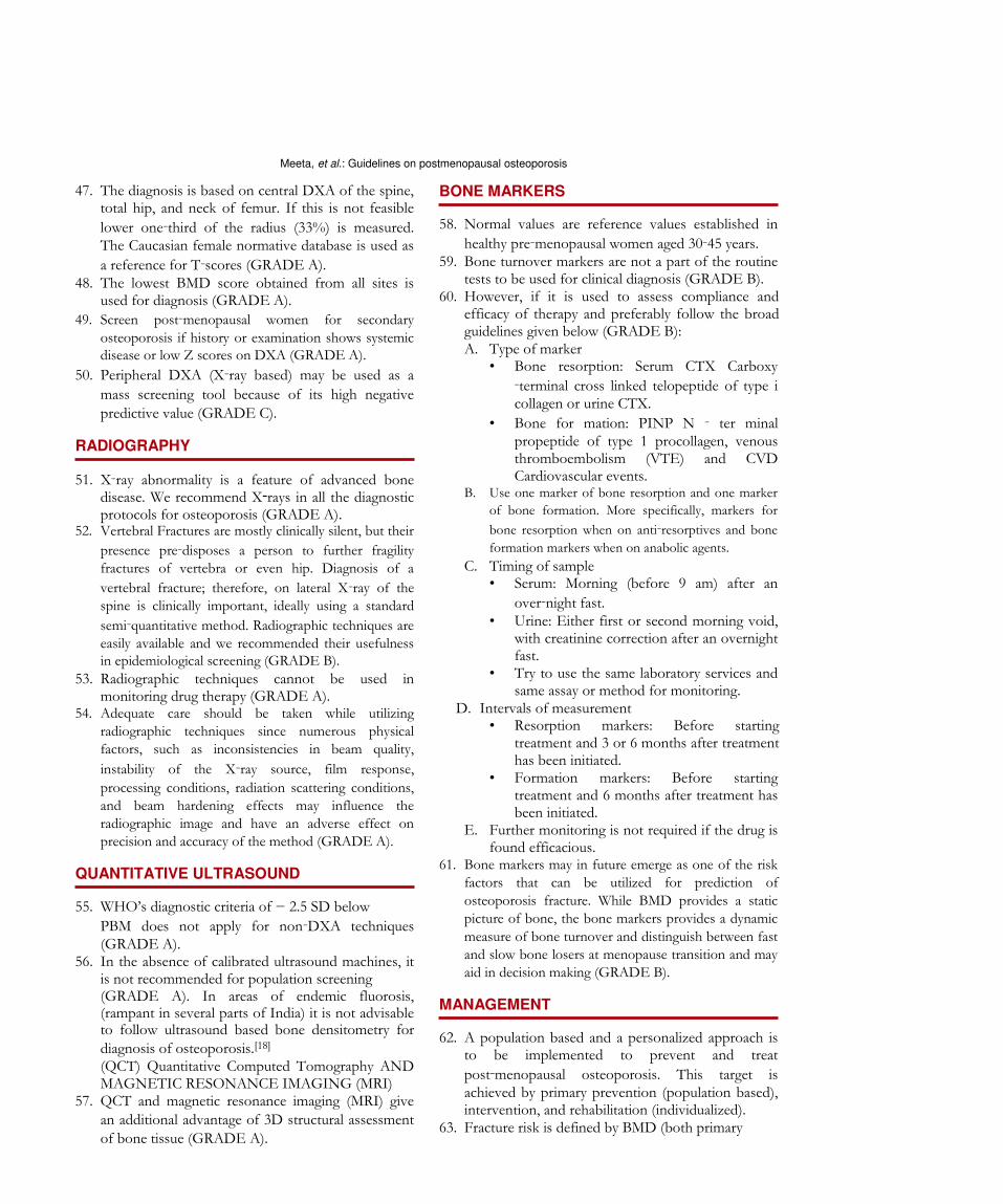
BONE MARKERS 58. Normal values are reference values established in
healthy pre‑menopausal women aged 30‑45 years. 59. Bone turnover markers are not a part of the routine
tests to be used for clinical diagnosis (GRADE B). 60. However, if it is used to assess compliance and
efficacy of therapy and preferably follow the broad guidelines given below (GRADE B): A. Type of marker
• Bone resorption: Serum CTX Carboxy
‑terminal cross linked telopeptide of type i collagen or urine CTX.
• Bone for mation: PINP N ‑ ter minal propeptide of type 1 procollagen, venous thromboembolism (VTE) and CVD Cardiovascular events.
B. Use one marker of bone resorption and one marker
of bone formation. More specifically, markers for
bone resorption when on anti‑resorptives and bone
formation markers when on anabolic agents.
C. Timing of sample • Serum: Morning (before 9 am) after an
over‑night fast. • Urine: Either first or second morning void,
with creatinine correction after an overnight fast.
• Try to use the same laboratory services and same assay or method for monitoring.
D. Intervals of measurement • Resorption markers: Before starting
treatment and 3 or 6 months after treatment has been initiated.
• Formation markers: Before starting treatment and 6 months after treatment has been initiated.
E. Further monitoring is not required if the drug is found efficacious.
61. Bone markers may in future emerge as one of the risk
factors that can be utilized for prediction of
osteoporosis fracture. While BMD provides a static
picture of bone, the bone markers provides a dynamic
measure of bone turnover and distinguish between fast
and slow bone losers at menopause transition and may
aid in decision making (GRADE B). MANAGEMENT 62. A population based and a personalized approach is
to be implemented to prevent and treat
post‑menopausal osteoporosis. This target is achieved by primary prevention (population based), intervention, and rehabilitation (individualized).
63. Fracture risk is defined by BMD (both primary
Meeta, et al.: Guidelines on postmenopausal osteoporosis
and secondary causes) and clinical risk factors for
osteoporotic fracture. For treatment purpose, combining
BMD with clinical risk factors provides a better estimate
of fracture risk. Hence, it will be easier to use a risk
assessment tool like FRAX with or without BMD for risk assessment. We simply should not treat T‑scores, but must take a patient’s full clinical status into account when we make therapeutic decisions.
64. It is good to understand the term prevention and
treatment in the context of osteoporosis. The term
prevention is used to denote the prevention of bone
loss in post‑menopausal women with osteopenia
(T‑score between −1 and −2.5) and increased fracture risk. Treatment is defined as a reduction in fracture risk
in post‑menopausal women with osteoporosis. UNIVERSAL RECOMMENDATIONS 65. Life‑style management
Balanced diet, adequate physical activity, and exposure to sunlight avoidance of bone depleting agents such as tobacco and alcohol.
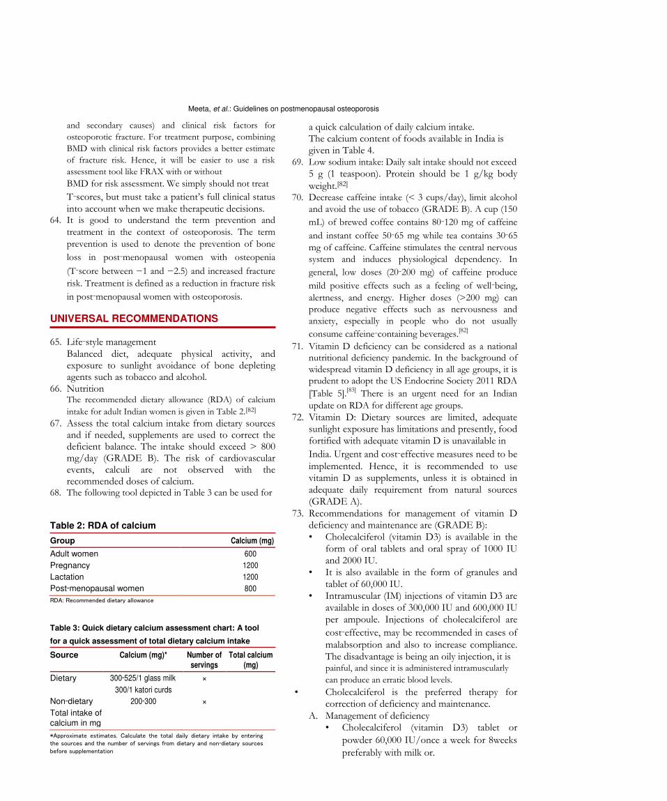
66. Nutrition The recommended dietary allowance (RDA) of calcium
intake for adult Indian women is given in Table 2.[82] 67. Assess the total calcium intake from dietary sources
and if needed, supplements are used to correct the deficient balance. The intake should exceed > 800 mg/day (GRADE B). The risk of cardiovascular events, calculi are not observed with the recommended doses of calcium.
68. The following tool depicted in Table 3 can be used for Table 2: RDA of calcium Group Calcium (mg)
Adult women 600 Pregnancy 1200 Lactation 1200 Post‑menopausal women 800 RDA: Recommended dietary allowance Table 3: Quick dietary calcium assessment chart: A tool
for a quick assessment of total dietary calcium intake Source Calcium (mg)* Number of Total calcium servings (mg)
Dietary 300‑525/1 glass milk ×
300/1 katori curds Non‑dietary 200‑300 × Total intake of calcium in mg *Approximate estimates. Calculate the total daily dietary intake by entering
the sources and the number of servings from dietary and non‑dietary sources
before supplementation
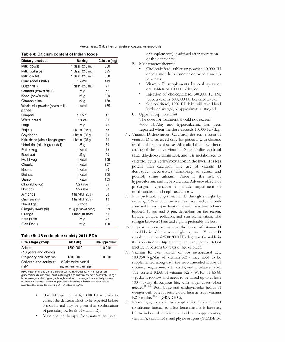
a quick calculation of daily calcium intake. The calcium content of foods available in India is given in Table 4.
69. Low sodium intake: Daily salt intake should not exceed 5 g (1 teaspoon). Protein should be 1 g/kg body weight.[82]
70. Decrease caffeine intake (< 3 cups/day), limit alcohol and avoid the use of tobacco (GRADE B). A cup (150
mL) of brewed coffee contains 80‑120 mg of caffeine
and instant coffee 50‑65 mg while tea contains 30‑65
mg of caffeine. Caffeine stimulates the central nervous system and induces physiological dependency. In
general, low doses (20‑200 mg) of caffeine produce
mild positive effects such as a feeling of well‑being,
alertness, and energy. Higher doses (>200 mg) can produce negative effects such as nervousness and anxiety, especially in people who do not usually
consume caffeine‑containing beverages.[82] 71. Vitamin D deficiency can be considered as a national
nutritional deficiency pandemic. In the background of widespread vitamin D deficiency in all age groups, it is prudent to adopt the US Endocrine Society 2011 RDA
[Table 5].[83] There is an urgent need for an Indian
update on RDA for different age groups. 72. Vitamin D: Dietary sources are limited, adequate
sunlight exposure has limitations and presently, food fortified with adequate vitamin D is unavailable in India. Urgent and cost‑effective measures need to be implemented. Hence, it is recommended to use vitamin D as supplements, unless it is obtained in adequate daily requirement from natural sources (GRADE A).
73. Recommendations for management of vitamin D deficiency and maintenance are (GRADE B): • Cholecalciferol (vitamin D3) is available in the
form of oral tablets and oral spray of 1000 IU and 2000 IU.
• It is also available in the form of granules and tablet of 60,000 IU.
• Intramuscular (IM) injections of vitamin D3 are available in doses of 300,000 IU and 600,000 IU per ampoule. Injections of cholecalciferol are
cost‑effective, may be recommended in cases of malabsorption and also to increase compliance. The disadvantage is being an oily injection, it is painful, and since it is administered intramuscularly
can produce an erratic blood levels. • Cholecalciferol is the preferred therapy for
correction of deficiency and maintenance. A. Management of deficiency
• Cholecalciferol (vitamin D3) tablet or
powder 60,000 IU/once a week for 8weeks
preferably with milk or.
116 Journal of Mid-life Health ¦ Apr-Jun 2013 ¦ Vol 4 ¦ Issue 2
Meeta, et al.: Guidelines on postmenopausal osteoporosis
Table 4: Calcium content of Indian foods Dietary product Serving Calcium (mg)
Milk (cows) 1 glass (250 mL) 300 Milk (buffalos) 1 glass (250 mL) 525 Milk low fat 1 glass (250 mL) 300 Curd (cow’s milk) 1 katori 149 Butter milk 1 glass (250 mL) 75 Channa (cow’s milk) 25 g 52 Khoa (cow’s milk) 25 g 239 Cheese slice 20 g 158 Whole milk powder (cow’s milk) 1 katori 155 paneer
Chapati 1 (25 g) 12 White bread 1 slice 30 Ragi 25 g 75 Rajma 1 katori (25 g) 65 Soyabean 1 katori (25 g) 60 Kale chane (whole bengal gram) 1 katori (25 g) 72 Udad dal (black gram dal) 25 g 50 Palak veg 1 katori 73 Beetroot 25 g 50 Methi veg 1 katori 395 Chaulai 1 katori 397 Beans 1 katori 90 Bathua 1 katori 150 Sarso 1 katori 155 Okra (bhendi) 1/2 katori 65 Broccoli 1/2 katori 50 Almonds 1 handful (25 g) 58 Cashew nut 1 handful (25 g) 13 Dried figs 5 whole 95 Gingelly seed (til) 25 g (1 tablespoon) 363 Orange 1 medium sized 50 Fish Hilsa 25 g 45 Fish Rohu 25 g 160
Table 5: US endocrine society 2011 RDA Life stage group RDA (IU) The upper limit
Adults 1500‑2000 10,000 (18 years and above)
Pregnancy and lactation 1500‑2000 10,000 Children and adults at 2‑3 times the normal risk* requirement for their age RDA: Recommended dietary allowance; *At risk: Obesity, HIV infection, on
glucocorticoids, anticonvulsant, antifungal, and antiviral therapy. A desirable range
is between 30 and 60 ng/mL, although levels up to 100 ng/mL are unlikely to result in vitamin D toxicity. Except in granuloma disorders, wherein it is advisable to
maintain the serum levels of 25(OH) D upto >30 ng/mL
• One IM injection of 6,00,000 IU is given to
correct the deficiency.(not to be repeated before
3 months and may be given after confirmation
of persisting low levels of vitamin D).
• Maintenance therapy (from natural sources
or supplements) is advised after correction of the deficiency.
B. Maintenance therapy • Cholecalciferol tablet or powder 60,000 IU
once a month in summer or twice a month in winter.
• Vitamin D supplements by oral spray or oral tablets of 1000 IU/day, or.
• Injection of cholecalciferol 300,000 IU IM, twice a year or 600,000 IU IM once a year.
• Cholecalciferol, 1000 IU daily, will raise blood
levels, on average, by approximately 10ng/mL. C. Upper acceptable limit
The dose for treatment should not exceed 4000 IU/day and hypercalcemia has been reported when the dose exceeds 10,000 IU/day.
74. Vitamin D derivatives: Calcitriol, the active form of vitamin D is reserved only for patients with chronic renal and hepatic disease. Alfacalcidol is a synthetic analog of the active vitamin D metabolite calcitriol
(1,25‑dihydroxyvitamin D3), and it is metabolized to
calcitriol by its 25‑hydroxylation in the liver. It is less potent than calcitriol. The use of vitamin D derivatives necessitates monitoring of serum and possibly urine calcium. There is the risk of hypercalcemia and hypercalciuria. Adverse effects of prolonged hypercalcemia include impairment of renal function and nephrocalcinosis.
75. It is preferable to get vitamin D through sunlight by
exposing 20% of body surface area (face, neck, and both
arms and forearms) without sunscreen for at least 30 min
between 10 am and 3 pm, depending on the season,
latitude, altitude, pollution, and skin pigmentation. The
sunlight between 11 am and 2 pm is preferably the best. 76. In post‑menopausal women, the intake of vitamin D
should be in addition to sunlight exposure. Vitamin D
supplementation (≥500‑2000 IU/day) was favorable in
the reduction of hip fracture and any non‑vertebral
fracture in persons 65 years of age or older. 77. Vitamin K: For women of post‑menopausal age,
180‑350 g/day of vitamin K2‑7 may need to be
supplemented along with the recommended intake of calcium, magnesium, vitamin D, and a balanced diet.
The current RDA of vitamin K2‑7 WHO of 65‑80
g/day is too low and needs to be raised up to at least
100 g/day throughout life, with larger doses when
needed.[84,85] Both bone and cardiovascular health of
women with osteoporosis would benefit from vitamin K2‑7 intake.[86‑97] (GRADE C).
78. Interestingly, exposure to complex nutrients and food
constituents interact to affect bone mass, it is however,
left to individual clinician to decide on supplementing
vitamin A, vitamin B12, and phytoestrogens (GRADE B).
Journal of Mid-life Health ¦ Apr-Jun 2013 ¦ Vol 4 ¦ Issue 1
Meeta, et al.: Guidelines on postmenopausal osteoporosis
PHYSICAL ACTIVITY 79. Adequate physical activity is needed to maintain
bone health. Brisk walking 4‑5 times a week for 30 min for hip, back strengthening exercises for spine, and resistances exercises for the upper arm is specific to maintain bone health (GRADE B).
80. Patients with severe osteoporosis should avoid engaging in motions, such as forward flexion exercises, using heavy weights or even performing
side‑bending exercises, because pushing, pulling,
lifting, and bending exert compressive forces on the spine that may lead to fracture (GRADE A).
PREVENTION OF FALLS 81. Patients should receive a multifactorial risk assessment
and intervention because; it is the most consistently effective strategy to prevent falls (GRADE A).
82. A fall evaluation includes an assessment of the following: History of fall, circumstances, medications, acute or chronic medical problems, and mobility levels; an examination of vision, gait and balance, and lower extremity joint function; an examination of basic neurological function, including mental status, muscle strength, lower extremity peripheral nerves, proprioception, reflexes, tests of cortical, extrapyramidal, and cerebellar function; assessment of basic cardiovascular status, including heart rhythm, and postural blood pressure.
83. Home hazard assessment and modification, exercise and physical therapy are recommended to prevent falls and injuries from falls. Biomechanics of posture and safe movements are a vital component of counseling (GRADE A).
84. Evaluation of medications (sedatives, antidepressants, antihypertensive, and hypoglycemic) and withdrawal of medications that increase the risk of falling is recommended (GRADE B).
85. Frailty related falls and fractures have been reported with
OR of 1.38‑2.4 for falls and recurrent falls, 1.40 and 1.7
for hip fracture in old women. Planning of individualized
exercise program, for preventing vertebral fractures and
strengthening for fall prevention, takes into consideration
patients cardiovascular condition, muscular strength,
flexibility, core stability, bone’s biochemical competence, etc., The program addresses balance and strengthening for
fall and fracture prevention. Women’s health‑care
programs targeting post‑menopausal women’s comprehensive care can contribute a lot by educating
women as to take care of their musculoskeletal health
through lifelong commitment to proper nutrition,
exercise, and understanding about issues related to
prevention of falls.[98‑100]
EMOTIONAL AND PSYCHOLOGICAL FACTORS 86. Education and psychosocial support for various
emotional and psychological aspects during treatment for PMO is needed.
87. Recommend smoking cessation and reduced alcohol consumption (GRADE B).
88. Psychiatric evaluation of patients with post‑menopausal
osteoporosis, structured unstructured, should be carried
out whenever indicated, especially in patients with
depression, psychosis, suicidal or cognitive impairment. Use brief screening instruments like Whooley’s 2‑question screening test for depression (GRADE B).
89. Treatment for osteoporosis has been associated with improved psychological functioning in patients with
post‑menopausal osteoporosis. This has been seen
with pharmacotherapeutic agents such as raloxifene, ibandronate, hormone replacement therapy,
exercise‑based interventions, etc., Strontium ranelate
has been associated with memory impairment. Avoid/ minimize the bone depleting action of medications.
90. For all patients having risk factors for secondary osteoporosis, universal recommendations need to be followed.
91. Glucocorticoid‑induced osteoporosis (GIOP) • It is the most common secondary cause of
osteoporosis. • For all patients receiving any dose of chronic oral
glucocorticoid therapy or initiating glucocorticoids (> 7.5 mg prednisolone or equivalent) with an anticipated duration of ≥ 3 months.
• Low bone mass is seen with the use of high potency
or prolonged low dose inhaled glucocorticoid. • Inhaled glucocorticoid below 400 mcg/day and
usage of budesonide or fluticasone seemed to have minimal systemic effects compared to beclomethasone. Use of spacer device significantly reduces the effect of inhaled glucocorticoid on bone formation.
• Calcitriol is used for preventing glucocorticoid- induced bone loss and post-transplant-related bone loss (GRADE A).
• A baseline BMD is necessary and treatment initiated in cases of osteopenia associated with clinical factors.
• Duration of anti‑resorptive therapy is as long as the steroids are prescribed. Bisphosphonates are the first‑line option for GIOP (GRADE A).
• Teriparatide counteracts many aspects of the pathophysiology of GIOP, hence better than
anti‑resorptive therapy (Ref: Indian Rheumatology Association Guidelines).
92. Women need to be supplemented with calcium and
vitamin D while on anticonvulsant therapy,[101]
118 Journal of Mid-life Health ¦Apr-Jun 2013 ¦ Vol 4 ¦ Issue 2
Meeta, et al.: Guidelines on postmenopausal osteoporosis
retroviral therapy, and drugs for thyroid dysfunction. to determine whether the change is real and not
93. We suggest that depot medroxy progesterone (DMP) simply random fluctuation or artefact.
may be us as a method of contraception (GRADE C). • Each center should determine its precision
There is no need to limit DMP use to 2 years in error in order to estimate the least significant
most patients; women should be instructed to change (i.e. the change in BMD required to have
follow universal recommendations for bone health: 95% confidence that the change is real).
Age‑appropriate calcium and vitamin D intake, regular • However, most osteoporosis therapies do not
weight‑bearing exercise, and smoking cessation. DMP cause large increases in BMD, and the anti‑fracture
should be used with caution in women with major risk effect of treatment is only partly explained by the
factors for low bone density, such as a BMI < 17.5, relatively small changes in BMD.
previous amenorrhea, rheumatoid arthritis, and • Stable BMD is consistent with successful
chronic oral glucocorticoid therapy[101‑104] treatment.
94. Low dose (OCPs) oral contraceptive pills is a good 100. Non‑responders to PMO therapy may be due to
choice of hormonal contraception in amenoheric poor adherence, poor calcium/vitamin D health, and
women, women on (GnRH) Gonadotrophin Releasing untreated secondary osteoporosis, concomitant therapy
Hormone analogs and women in the reproductive with skeletotropic drugs, inappropriate choice of drugs
period (GRADE B). or wrong choice of monitoring strategies (GRADE C).
PHARMACOTHERAPY 101. Duration of therapy has to be individualized depending
on the patient’s profile, drug used, and response to
therapy.
95. Treatment should be considered in women
102. There is no recommendation on combination
• Presenting with fragility fractures. therapies, sequential therapies and drug holidays; these
• Radiographic diagnosis of incidental vertebral should be planned as per individual patient’s need.
fracture and osteopenia.
• Diagnosis of osteoporosis based on DXA, i.e., BISPHOSPHONATES
T‑score of less than −2.5 at hip or spine.
• With secondary causes and high‑risk of fractures. 103. Bisphosphonates are recommended as first‑line drugs
• In the absence of BMD measurements by DXA, for treating post‑menopausal women, with proven
intervention is individualized, understanding efficacy in the prevention of vertebral and non‑vertebral
and considering the cost benefit and risk benefit fractures, including hip fractures (GRADE A).
outcome of the intervention. 104. Fracture reduction is seen after 1 year of treatment.
96. Health‑care professionals managing PMO should 105. Alendronate 70 mg weekly has similar efficacy
be aware of the concepts of compliance, adherence, to alendronate 10 mg daily in the treatment of
concordance, and persistence, and work to improve post‑menopausal osteoporosis. Continuous use of
adherence in patients of PMO, in order to optimize alendronate, for up to 10 years, if clinically indicated,
therapeutic outcomes. produces a sustained increase in BMD and 55%
97. In choosing therapy, drug‑related (risk‑benefit), significant reduction in spine fracture with a good
patient profile (age, years since menopause, symptoms, safety profile (GRADE A).
comorbidities) and environment‑related factors 106. Risedronate: This is associated with up to 49%
(economics and social) should be identified. Patients reduction in new vertebral fracture in women with prior
should be educated in PMO and its treatment and vertebral fractures and 39% reduction in non‑vertebral
empowered to take part in shared decision making to fractures. Vertebral fracture risk reduction is seen
improve adherence. after 6 months of therapy. Reduction of hip fracture
98. Patients should be monitored initially, every 3‑6 months risk after 3 years was 40% in women with confirmed
for 2‑3 contacts, then annually for clinical assessment. osteoporosis and 60% in women with at least one
We suggest that markers of bone resorption and coexisting vertebral fracture. Currently, the use of
formation may be tested at baseline and after 3‑6 risedronate for up to 7 years is safe and efficacious.
months of therapy in certain situations and research Risedronate 35 mg once weekly has similar efficacy to
settings (GRADE C). the 5 mg daily dosing (GRADE A).
99. We suggest that DXA should be performed every 107. Ibandronate: Oral ibandronate 2.5 mg daily for 3 years
2 years on the same machine in order to monitor reduces vertebral fracture by 62% in post‑menopausal
osteoporosis therapy (GRADE B). women with prevalent vertebral fracture.
• Measurement error must be considered when 108. Zoledronic acid: Treatment with zoledronic acid (5 mg
interpreting serial BMD assessments in order by intravenous infusion over at least 15 min once yearly)

Meeta, et al.: Guidelines on postmenopausal osteoporosis
in osteoporotic post‑menopausal women reduces the
incidence of vertebral fracture by 70% over 3 years with
significant reduction seen by 1 year. Hip fracture is
reduced by 41% and non‑vertebral fracture by 25% over
3 years (GRADE A). Zoledronic acid yearly infusion is
also indicated for the prevention of new clinical fractures
in patients who recently (within 90 days) have had a low
trauma hip fracture. It has also been shown to be
associated with a reduction in mortality. 109. Adverse effects of bisphosphonates: An association
has been suggested between atypical femur shaft fractures and over suppression of bone turnover in patients exposed to bisphosphonates for longer than
3‑5 years, particularly in younger Asian women.[105] 110. Bisphosphonate‑related osteonecrosis of the jaw is a
rare complication. Routine dental care is recommended
for all patients and postponing the therapy until the
dental treatment has been carried out. Patients who are
vitamin D insufficient may be at higher risk for this
complication. Calcium and vitamin D deficiency should
be corrected before initiating the therapy (GRADE A). HORMONE THERAPY 111. Estrogen progesterone therapy/estrogen therapy
(EPT/ET) may be used for prevention and treatment of
osteoporosis in the early post‑menopause in symptomatic
women unless there is a contra‑indication. ET/EPT prevents all osteoporotic fractures even in low risk population, it increases lumbar spine BMD up to 7.6% and femoral neck BMD up to 4.5% over 3 years. It reduces the risk of spine, hip, and other
osteoporotic fractures by 33‑40% (GRADE A). 112. Pre‑hormone therapy (HT) work‑up and an annual
follow‑up are essential when prescribing HT. The dose and duration of HT should be individualized, and a risk‑benefit assessment carried out annually. A full gynecological assessment is mandatory prior to starting HT and at regular intervals thereafter. Self ‑breast examination is advised monthly and clinical breast examination at least annually. Mammogram where available, should be carried out 1‑3 yearly if the initial mammogram is normal (GRADE C).
113. All preparations, including low dose, non‑oral
routes of estrogen are effective in preserving bone mass. In women with hypertriglyceridemia, obesity, glucose intolerance, history of deep vein thrombosis,
and tobacco users, non‑oral route should be preferred (GRADE B).
114. HT should not be started solely for bone protection after
10 years of menopause. Extended use of HT in women
with reduced bone mass is an option after considering the
risk‑benefit analysis compared to the other available
therapies for osteoporosis. The bone protective effect is
lost after stopping HT (GRADE B). 115. HT is indicated as primary therapy to prevent bone
loss in women with premature menopause and secondary amenorrhea (GRADE C).
116. Progestogens should be added to ET in women with uterus (GRADE A).
117. If menopausal hormone therapy is given to women below the age of 60 or within 10 years of menopause, the risks are rare. Tables 6 and 7 elaborate the risks and benefits in terms that can be used during counseling for easy and understandable communication.
118. Classification of Frequency of Drug Reactions (according to WHO and Council for International Organizations of Medical Sciences) • Very common > 1/10. • Common (frequent) >1/100 and < 1/10. • Uncommon (infrequent) >1/1000 and < 1/100, • Rare > 1/10,000 and < 1/1000. • Very rare<1/10,000.
119. Harms: Based on WHI, number of excess events on HT versus placebo per 10,000 women/year of HT. Use between the age group of 50 years and 59 years (GRADE A) [Table 6].
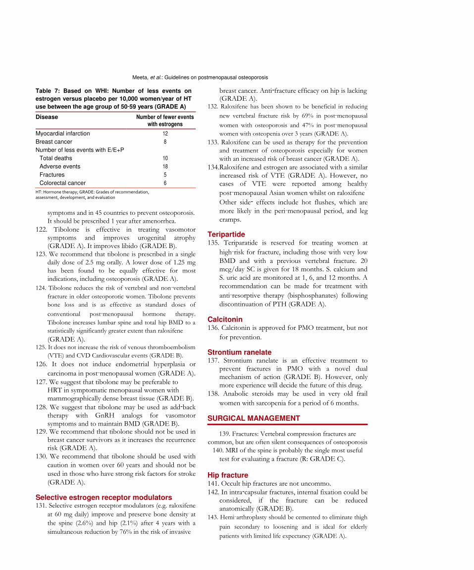
120. Benefits: Benefit of hormone therapy are shown in Table 7.
TIBOLONE 121. Tibolone is a selective tissue estrogenic activity regulator.
It is a synthetic steroid compound, which has estrogenic,
progestogenic and androgenic properties. It has an
estrogenic effect on bone, inhibiting bone resorption by
reducing osteoclastic activity. Tibolone is approved in 90
countries to treat menopausal
Table 6: Based on WHI: Number of excess events on HT versus placebo per 10,000 women/year of HT use between the age group of 50‑59 years Disease Estrogen WHO/CIOMS definition of risk Estrogen+progesterone WHO/CIOMS definition of risk
VTE 4 Rare <1/10,000 and <1/100 11 Rare >1/10,000 and <1/1000 Stroke 1 Rare >1/10,000 and <1/1,000 4 Rare >1/10,000 and <1/1,000 Breast cancer 5 Rare >1/10,000 and <1/1,000 CVD 5 Rare >1/10,000 and <1/1,000 WHO: World health organization; CIOMS: Council for international organizations of medical sciences; VTE: Venous thromboembolism; CVD: Cardiovascular disease; T: Hormone therapy
120 Journal of Mid-life Health ¦ Apr-Jun 2013 ¦ Vol 4 ¦ Issue 2
Meeta, et al.: Guidelines on postmenopausal osteoporosis
Table 7: Based on WHI: Number of less events on
estrogen versus placebo per 10,000 women/year of HT
use between the age group of 50‑59 years (GRADE A) Disease Number of fewer events with estrogens
Myocardial infarction 12 Breast cancer 8 Number of less events with E/E+P
Total deaths 10 Adverse events 18 Fractures 5 Colorectal cancer 6
HT: Hormone therapy; GRADE: Grades of recommendation, assessment, development, and evaluation
symptoms and in 45 countries to prevent osteoporosis. It should be prescribed 1 year after amenorrhea.
122. Tibolone is effective in treating vasomotor symptoms and improves urogenital atrophy (GRADE A). It improves libido (GRADE B).
123. We recommend that tibolone is prescribed in a single daily dose of 2.5 mg orally. A lower dose of 1.25 mg has been found to be equally effective for most indications, including osteoporosis (GRADE A).
124. Tibolone reduces the risk of vertebral and non‑vertebral
fracture in older osteoporotic women. Tibolone prevents
bone loss and is as effective as standard doses of
conventional post‑menopausal hormone therapy.
Tibolone increases lumbar spine and total hip BMD to a
statistically significantly greater extent than raloxifene (GRADE A).
125. It does not increase the risk of venous thromboembolism
(VTE) and CVD Cardiovascular events (GRADE B). 126. It does not induce endometrial hyperplasia or
carcinoma in post‑menopausal women (GRADE A). 127. We suggest that tibolone may be preferable to
HRT in symptomatic menopausal women with mammographically dense breast tissue (GRADE B).
128. We suggest that tibolone may be used as add‑back therapy with GnRH analogs for vasomotor symptoms and to maintain BMD (GRADE B).
129. We recommend that tibolone should not be used in breast cancer survivors as it increases the recurrence risk (GRADE A).
130. We recommend that tibolone should be used with caution in women over 60 years and should not be used in those who have strong risk factors for stroke (GRADE A).
Selective estrogen receptor modulators 131. Selective estrogen receptor modulators (e.g. raloxifene
at 60 mg daily) improve and preserve bone density at
the spine (2.6%) and hip (2.1%) after 4 years with a
simultaneous reduction by 76% in the risk of invasive
breast cancer. Anti‑fracture efficacy on hip is lacking (GRADE A).
132. Raloxifene has been shown to be beneficial in reducing
new vertebral fracture risk by 69% in post‑menopausal
women with osteoporosis and 47% in post‑menopausal
women with osteopenia over 3 years (GRADE A). 133. Raloxifene can be used as therapy for the prevention
and treatment of osteoporosis especially for women with an increased risk of breast cancer (GRADE A).
134.Raloxifene and estrogen are associated with a similar increased risk of VTE (GRADE A). However, no cases of VTE were reported among healthy
post‑menopausal Asian women whilst on raloxifene Other side‑ effects include hot flushes, which are more likely in the peri‑menopausal period, and leg cramps.
Teripartide 135. Teriparatide is reserved for treating women at
high‑risk for fracture, including those with very low BMD and with a previous vertebral fracture. 20 mcg/day SC is given for 18 months. S. calcium and S. uric acid are monitored at 1, 6, and 12 months. A recommendation can be made for treatment with
anti‑resorptive therapy (bisphosphanates) following discontinuation of PTH (GRADE A).
Calcitonin 136. Calcitonin is approved for PMO treatment, but not
for prevention. Strontium ranelate 137. Strontium ranelate is an effective treatment to
prevent fractures in PMO with a novel dual mechanism of action (GRADE B). However, only more experience will decide the future of this drug.
138. Anabolic steroids may be used in very old frail
women with sarcopenia for a period of 6 months. SURGICAL MANAGEMENT
139. Fractures: Vertebral compression fractures are common, but are often silent consequences of osteoporosis
140. MRI of the spine is probably the single most useful test for evaluating a fracture (R: GRADE C).
Hip fracture 141. Occult hip fractures are not uncommo. 142. In intra‑capsular fractures, internal fixation could be
considered, if the fracture can be reduced anatomically (GRADE B).
143. Hemi‑arthroplasty should be cemented to eliminate thigh
pain secondary to loosening and is ideal for elderly
patients with limited life expectancy (GRADE A).
Journal of Mid-life Health ¦ Apr-Jun 2013 ¦ Vol 4 ¦ Issue 2 121

Meeta, et al.: Guidelines on postmenopausal osteoporosis
Flowchart 1: Post menopausal woman Asymptomatic 144. Total hip replacement should be considered when
internal fixation is inappropriate or contraindicated in physiologically younger patients for improved quality of life (GRADE B).
145. All patients who suffer from a hip fracture should be subjected to BMD after surgery where possible and appropriate treatment for osteoporosis initiated (GRADE A).
146. Long bone fractures – preferred fixation – close
reduction and intramedullary nailing (GRADE A).
147. Juxtra‑articular fractures – preferred fixation – close reduction and anatomical locking plates with variable angle screws (GRADE A).
148. Articular fractures– stable/reducible – internal fixation and augmentation (GRADE A).
149. Articular fractures –unstable/comminuted replacement
may be an option in elderly patients (GRADE B). 150. Post‑fracture fixation – patient specific osteoporosis
122 Journal of Mid-life Health ¦ Apr-Jun 2013 ¦ Vol 4 ¦ Issue 2
Meeta, et al.: Guidelines on postmenopausal osteoporosis
Flowchart 2: Postmenopausal woman- with fragility fracture
related medical management to avoid subsequent fractures (GRADE A).
151. Post‑operatively start patients on: • Calcium and vitamin D as needed and start
appropriate treatment for osteoporosis. • When indicated drugs like teriparatide, which
facilitate osteoblastic bone formation can be started (GRADE A).
definite role in the management of those vertebral
compression fractures that do not respond to
non‑operative treatment (GRADE A). REHABILITATION 154. Use of phase wise physiotherapy based on severity
of osteoporosis, hip/spine affection. • Anti‑resorptive drugs like bisphosphonates are 155. Use of strengthening, resistance, weight‑bearing
started 4‑6 weeks later (GRADE B). Spine fracture 152. All vertebral compression fractures without
neurological deficit are treated conservatively for 3weeks as majority get better during this period.
153. Percutaneous vertebroplasty and kyphoplasty have a
exercise depending on age and severity of osteoporosis.
156. Combined approach using occupational and diet therapy along.
ECONOMICS OF OSTEOPOROSIS 157. Advising life‑style measures to all post‑menopausal
Journal of Mid-life Health ¦ Apr-Jun 2013 ¦ Vol 4 ¦ Issue
Meeta, et al.: Guidelines on postmenopausal osteoporosis
women, improving calcium intake in diet, and increasing physical activity improves bone health
and is cost‑effective (GRADE B). 158. Prescription of calcium and vitamin D to all
post‑menopausal women with a view to improve bone
health and prevent osteoporosis and its consequent
fractures is the cost‑effective strategy (GRADE A). 159. Screening post‑menopausal women for osteoporosis/
osteopenia by DXA at hip/spine and biochemical test is a cost‑effective strategy (GRADE A).
160. Treatment of post‑ menopausal women with
diagnosed osteoporosis is the cost‑effective
treatment strategy (GRADE A). Flowcharts 1 and 2 show an approach to management of
asymptomatic post‑menopausal woman and
post‑menopausal woman with fragility fracture,
respectively. Decision making in the management of PMO is depicted
in Flowcharts 1 and 2. REFERENCES 1. Cooper C, Campion G, Melton LJ 3
rd. Hip fractures in the
elderly: A world‑wide projection. Osteoporos Int 1992;2:285‑9. 2. Kado DM, Browner WS, Palermo L, Nevitt MC, Genant HK,
Cummings SR. Vertebral fractures and mortality in older women: A prospective study. Study of Osteoporotic Fractures Research Group. Arch Intern Med 1999;159:1215‑20.
3. Consensus development conference: Prophylaxis and treatment of osteoporosis. Am J Med 1991;90:107‑10.
4. NIH consensus development panel on osteoporosis prevention, diagnosis, and therapy, March 7‑29, 2000: Highlights of the conference. South Med J 2001;94:569‑73.
5. Kanis JA. Assessment of Osteoporosis at the Primary Health Care Level 2008 World Health Organisation Scientific Group. WHO Collaboration Centre for Metabolic Bone Diseases, UK: University of Shiffield; 2007.
6. Dawson‑Hughes B, National Osteoporosis Foundation Guide Committee. A revised clinician’s guide to the prevention and treatment of osteoporosis. J Clin Endocrinol Metab 2008;93:2463‑5.
7. Johnell O, Kanis JA, Oden A, Johansson H, De Laet C, Delmas P, et al. Predictive value of BMD for hip and other fractures. J Bone Miner Res 2005;20:1185‑94.
8. Kanis JA, Johnell O, Oden A, De Laet C, Mellstrom D. Diagnosis of osteoporosis and fracture threshold in men. Calcif Tissue Int 2001;69:218‑21.
9. Lewiecki EM, Gordon GB, Baim S, Leonard MB, Nicholas J. International Society for Clinical Densitometry 2007 Adult and Pediatric, Vol. 43. BishopBone: Elseiver; 2008. p. 1115‑21.
10. ICMR Taskforce Study. Population based reference standards of peak bone mineral density of Indian males and female. An ICMR multicentre task force study. New Delhi: Indian Council of Medical Research; 2010.
11. Marwaha RK, Tandon N, Kaur P, Sastry A, Bhadra K, Narang A, et al. Establishment of age‑specified bone mineral density reference range for Indian females using dual‑energy X‑ray absorptiometry. J Clin Densitom 2012;15:241‑9.
12. Makker A, Mishra G, Singh BP, Tripathy A, Singh MM. Normative bone mineral density data at multiple skeletal sites in Indian subjects. Arch Osteoporos 2008;3:25‑37.
13. Patni R. Normal BMD values for Indian females aged 20‑80
years. J Midlife Health 2010;1:70‑3. 14. Marwaha RK, Tandon N, Shivaprasad C, Kanwar R, Mani K,
Aggarwal R, et al. Peak bone mineral density of physically active healthy Indian men with adequate nutrition and no known current constraints to bone mineralization. J Clin Densitom 2009;12:314‑21.
15. Nangia S, Arya V, Gujral Ratni B, Mithal A. Spinal bone mineral
density in normal Indian females. Presented at 27th
Annual Meeting of The Endocrine Society of India, Lucknow; 1997.
16. Arya V, Nangia S, Gujral RB, Mithal A. Femoral bone mineral
density in normal Indian females. Presented at 27th
Annual Meeting of The Endocrine Society of India, Lucknow.
17. Pande KC, Johansen KB, Helboe AB. Digital X‑ray radiogrammetry: Establishment and comparison of Indian female and male normative reference data. J Bone Miner Res 2001;16 Suppl 1:M0871997.
18. Harinarayan CV, Ramalakshmi T, Prasad UV, Kumar EG, Srinivasa Rao PV. Ultrasound bone mineral density of os calcis‑its relationship with bone mineral markers and 25(OH) vitamin D in endemic fluorotic and non‑fluorotic villages. J Clin Sci Res 2012;1:157‑62.
19. Parfitt AM. The coupling of bone formation to bone resorption: A critical analysis of the concept and of its relevance to the pathogenesis of osteoporosis. Metab Bone Dis Relat Res 1982;4:1‑6.
20. Bessette L, Ste‑Marie LG, Jean S, Davison KS, Beaulieu M, Baranci M, et al. The care gap in diagnosis and treatment of women with a fragility fracture. Osteoporos Int 2008;19:79‑86.
21. Papaioannou A, Wiktorowicz ME, Adachi JD, Goeree R, Papadimitropoulos E, Bedard M, et al. Mortality, independence in living and re‑fracture, one year following hip fracture in Canadians. J Soc Obstet Gynaecol Can. 2000;22:591‑7.
22. Colón‑Emeric C, Kuchibhatla M, Pieper C, Hawkes W, Fredman L, Magaziner J, et al. The contribution of hip fracture to risk of subsequent fractures: Data from two longitudinal studies. Osteoporos Int 2003;14:879‑83.
23. Fried LP, Tangen CM, Walston J, Newman AB, Hirsch C, Gottdiener J, et al. Frailty in older adults: Evidence for a phenotype. J Gerontol A Biol Sci Med Sci 2001;56:M146‑56.
24. Cole ZA, Dennison EM, Cooper C. Osteoporosis epidemiology update. Curr Rheumatol Rep 2008;10:92‑6.
25. Joshi VR, Mangat G, Balakrishnan C, Mittal G. Osteoporosis – Approach in Indian scenario. J Assoc Physicians India 1998;46:965‑7.
26. Makkar A, Mishra G, Singh BP, Peak bone density at multiple skeletal sites in Indian subjects. Arch Osteoporos 2008;3:25‑3.
27. Chibber G, Roy R, Eunice M, Srivastava M, Ammini AC. Prevalence of osteoporosis among elderly women living in Delhi and rural Haryana. IJEM 2007;1:11‑4.
28. Shatrugna V, Kulkarni B, Kumar PA, Rani KU, Balakrishna N. Bone status of Indian women from a low‑income group and its relationship to the nutritional status. Osteoporos Int 2005;16:1827‑35.
29. Gandhi A, Shukla AR. Evaluation of BMD of women above 40 years of age. J Obstet Gynecol India 2005;55:265.
30. Meeta. Evaluation of Risk Factors for Dexa Referral in Indian Women Poster Presented at International Menopause Meeting, Spain; 2008. Abstract book
31. Krishna U, Mehta RU. Osteoporosis–Incidence and implications. J Obstet Gynecol India 2000;50:150‑6.
32. Babu AS, Ikbal FM, Noone MS, Joseph AN, Samuel P. Osteoporosis and osteopenia in India: A few more observations. Indian J Med Sci 2009;63:76‑7.
33. Sharma S, Tandon VR, Mahajan A, Kour A, Kumar D. Preliminary screening of osteoporosis and osteopenia in urban women from Jammu using calcaneal QUS. Indian J Med Sci 2006;60:183‑9.
34. Pande KC. Prevalence of low bone mass in healthy Indian population. J Indian Med Assoc 2002;100:598‑600, 602.
124 Journal of Mid-life Health ¦ Apr-Jun 2013 ¦ Vol 4 ¦ Issue 2
Meeta, et al.: Guidelines on postmenopausal osteoporosis
35. Meeta. Osteoporosis Revisted. Osteoporosis Alert. Issue 1. 36. Paul TV, Thomas N, Seshadri MS, Oommen R, Jose A,
Mahendri NV. Prevalence of osteoporosis in ambulatory postmenopausal women from a semiurban region in Southern India: Relationship to calcium nutrition and vitamin D status. Endocr Pract 2008;14:665‑71.
37. Aggrawal N, Bathla S, Juneja S. Measurement of bone mineral density by dexa scan in postmenopausal women. Obs Gynae Today 2004;9:768‑71.
38. Meeta. Two hundred and six reasons to be informed about osteoporosis. Poster at the Annual Indian Menopause Society meeting at Chandigarh; 2006, Feb 4‑5.
39. Unni J, Garg R, Pawar R. Bone mineral density in women above 40 years. J Midlife Health 2010;1:19‑22.
40. Savardekar LS, Shah RS, Iddya U, Balaiah D, Parihar A, Jaknkaria B. Bone density in normal Indian women: Assessment by USG and DEXA. Obs Abd Gynae Today 2004;9:772‑6.
41. Meeta, Shaantanu, Tanvir. Assessment of risk factors for osteoporosis in Indian women submitted for publication ‑yet to be and published, ‑details from [email protected]
42. Dhanwal DK, Siwach R, Dixit V, Mithal A, Jameson K, Cooper C. Incidence of hip fracture in Rohtak district, North India. Arch Osteoporos 2013;8:135.
43. International Osteoporosis Foundation. The Asian Audit: Epidemiology, costs and burden of osteoporosis in Asia 2009, 2009. Available from: http://www.iofbonehealth. org. [Last accessed on 2013 Jan 2].
44. Sankaran B. Clinical studies: Incidence of fracture neck of femur and intertrochanteric fractures in three Delhi hospitals. In: Sankaran
B, editor. Osteoporosis. New Delhi: South East Asia Regional Office, World Health Organisation; 2000. p. 9‑18.
45. Jha RM, Mithal A, Malhotra N, Brown EM. Pilot case‑control investigation of risk factors for hip fractures in the urban Indian population. BMC Musculoskelet Disord 2010;11:49.
46. Marwaha RK, Tandon N, Gupta Y, Bhadra K, Narang A, Mani K, et al. The prevalence of and risk factors for radiographic vertebral fractures in older Indian women and men: Delhi Vertebral Osteoporosis Study (DeVOS). Arch Osteoporos 2012;7:201‑7.
47. Balasubramanian K, Rajeswari J, Gulab, Govil YC, Agarwal AK, Kumar A, et al. Varying role of vitamin D deficiency in the etiology of rickets in young children vs. adolescents in northern India. J Trop Pediatr 2003;49:201‑6.
48. Harinarayan CV, Ramalakshmi T, Venkataprasad U. High prevalence of low dietary calcium and low vitamin D status in healthy south Indians. Asia Pac J Clin Nutr 2004;13:359‑64.
49. Kalra S, Kalra B, Khandelwal SK. Vitamin D deficiency in healthy postmenopausal women in Haryana. World J Life Sci Med Res 2011;1:11.
50. Harinarayan CV. Prevalence of vitamin D insufficiency in postmenopausal south Indian women. Osteoporos Int 2005;16:397‑402.
51. Singh M. Early age of natural menopause in India, a biological marker for early preventive health programs. Climacteric 2012;15:581‑6.
52. Harinarayan CV, Joshi SR. Vitamin D status in India – Its implications and remedial measures. J Assoc Physicians India 2009;57:40‑8.
53. Sachan A, Gupta R, Das V, Agarwal A, Awasthi PK, Bhatia V. High prevalence of vitamin D deficiency among pregnant women and their newborns in northern India. Am J Clin Nutr 2005;81:1060‑4.
54. Marwaha RK, Tandon N, Reddy DR, Aggarwal R, Singh R, Sawhney RC, et al. Vitamin D and bone mineral density status of healthy schoolchildren in northern India. Am J Clin Nutr 2005;82:477‑82.
55. Harinarayan CV, Appicatlaa L, Nalini BA, Joshi S. Efficacy and safety of cholecalciferol supplementation in Vitamin D deficient subjects based on endocrine society clinical practice
guidelines. Endocrinol Metab Syndr 2012;S4:004.
56. Goswami R, Gupta N, Ray D, Singh N, Tomar N. Pattern of 25‑hydroxy vitamin D response at short (2 month) and long (1 year) interval after 8 weeks of oral supplementation with cholecalciferol in Asian Indians with chronic hypovitaminosis D. Br J Nutr 2008;100:526‑9.
57. Agarwal N, Faridi MM, Aggarwal A, Singh O. Vitamin D Status of term exclusively breastfed infants and their mothers from India. Acta Paediatr 2010;99:1671‑4.
58. Harinarayan CV, Sachan A, Reddy PA, Satish KM, Prasad UV, Srivani P. Vitamin D status and bone mineral density in women of reproductive and postmenopausal age groups: A cross‑sectional study from south India. J Assoc Physicians India 2011;59:698‑704.
59. Marwaha RK, Tandon N, Garg MK, Kanwar R, Narang A, Sastry A, et al. Vitamin D status in healthy Indians aged 50 years and above. J Assoc Physicians India 2011;59:706‑9.
60. Mithal A, Arya V. Vitamin D status in urban North India adults. J Bone Miner Res 1999;14(suppl) Abstract.
61. Beloyartseva M, Mithal A, Kaur P, Kalra S, Baruah MP, Mukhopadhyay S, et al. Widespread vitamin D deficiency among Indian health care professionals. Arch Osteoporos 2012;7:187‑92.
62. Goswami R, Gupta N, Goswami D, Marwaha RK, Tandon N, Kochupillai N. Prevalence and significance of low 25‑hydroxyvitamin D concentrations in healthy subjects in Delhi. Am J Clin Nutr 2000;72:472‑5.
63. Zargar AH, Ahmad S, Masoodi SR, Wani AI, Bashir MI, Laway BA, et al. Vitamin D status in apparently healthy adults in Kashmir Valley of Indian subcontinent. Postgrad Med J 2007;83:713‑6.
64. Gupta A. Osteoporosis in India— The nutritional hypothesis. In: Mithal A, Rao DS, Zaidi M, editors. Metabolic Bone Disorders. Lucknow: Hindustani Book Depot; 1998. p. 115.
65. Teotia SP, Teotia M. Nutritional bone disease: The continuing challenge to neonatal bone health. Postgrad Med 2009;23:30‑8.
66. Nangia S, Arya V, Gujral RB, Mithal A. Spinal bone mineral density in normal Indian females In: Mithal A, Rao DS, Zaidi M, editors. Metabolic Bone Disorders. Lucknow: Hindustani Book Depot; 1998. p. 213.
67. Nangia S, Arya V, Gujral RB, Mithal A. Femoral bone mineral density in normal Indian females. In: Mittal A, Rao DS, Zaidi M, editors. Metabolic bone disorders Indian Society for Bone and Mineral Research; 1998. p. 215.
68. Singh M, Magon N, Singh T. Major and minor discordance in the diagnosis of postmenopausal osteoporosis among Indian women using hip and spine dual‑energy X‑ray absorptiometry. J Midlife Health 2012;3:76‑80.
69. Reddy PG, Mithal A, Rao DS. Bone mineral density in healthy Asian Indian women: Development of a reference database and implications for diagnosis of osteoporosis in Indian women living in the United States [abstract]. J Bone Miner Res 2002;17 Suppl 1:SA270.
70. Shatrugna V. Osteoporosis in Asian region: Newer questions. In: Shetty P, Gopalan C, editors. Diet, Nutrition and Chronic diseases: An Asian Perspective. London: Smith Gordon Co; 1998. p. 81‑3.
71. Dhanwal DK, Sahoo S, Gautam VK, Saha R. Hip fracture patients in India have vitamin D deficiency and secondary hyperparathyroidism. Osteoporos Int 2013;24:553‑7.
72. Alekel DL.Mortillaro E, Hussain EA,1999 Lifestyle and biologic contributors to proximal femur bone mineral density and hip axis length in two distinct etnic groups of premenopausal women.Osteoporos Int 9:327‑338.
73. Marwaha RK, Puri S, Tandon N, Dhir S, Agarwal N, Bhadra K, et al. Effects of sports training and nutrition on bone mineral density in young Indian healthy females. Indian J Med Res 2011;134:307‑13.
74. Davies JH, Evans BA, Gregory JW. Bone mass acquisition in
Journal of Mid-life Health ¦ Apr-Jun 2013 ¦ Vol 4 ¦ Issue 2 125
Meeta, et al.: Guidelines on postmenopausal osteoporosis
healthy children. Arch Dis Child 2005;90:373‑8.
75. Ganpule A, Yajnik CS, Fall CH, Rao S, Fisher DJ, Kanade A, et al.
Bone mass in Indian children – Relationships to maternal nutritional
status and diet during pregnancy: The Pune maternal nutrition
study. J Clin Endocrinol Metab 2006;91:2994‑3001.
76. Langley‑Evans SC. Developmental programming of health and disease. Proc Nutr Soc 2006;65:97‑105.
77. Cooper C, Cawley M, Bhalla A, Egger P, Ring F, Morton L, et
al. Childhood growth, physical activity, and peak bone mass in women. J Bone Miner Res 1995;10:940‑7.
78. Cooper C, Fall C, Egger P, Hobbs R, Eastell R, Barker D. Growth in infancy and bone mass in later life. Ann Rheum Dis 1997;56:17‑21.
79. Sayer AA, Cooper C. Fetal programming of body composition and musculoskeletal development. Early Hum Dev 2005;81:735‑44.
80. Cooper C, Eriksson JG, Forsén T, Osmond C, Tuomilehto J, Barker DJ. Maternal height, childhood growth and risk of hip fracture in later life: A longitudinal study. Osteoporos Int 2001;12:623‑9.
81. Dennison EM, Syddall HE, Sayer AA, Gilbody HJ, Cooper C. Birth weight and weight at 1 year are independent determinants of bone mass in the seventh decade: The Hertfordshire cohort study. Pediatr Res 2005;57:582‑6.
82. National Institute of Nutrition, ICMR. Dietary Guidelines for Indians: A Manual. 2011. Annexure 3. page 89‑90
83. Holick MF, Binkley NC, Bischoff‑Ferrari HA, Gordon CM, Hanley DA, Heaney RP, et al. Evaluation, treatment, and prevention of vitamin D deficiency: An Endocrine Society clinical practice guideline. J Clin Endocrinol Metab 2011;96:1911‑30.
84. Subcommittee on the Tenth Edition of the Recommended Dietary Allowances, Food and Nutrition Board. Recommended
Dietary Allowances. 10th
ed., Washington, DC: National Academy Press; 1989.
85. Kaneki M, Hodges SJ, Hosoi T, Fujiwara S, Lyons A, Crean SJ, et al. Japanese fermented soybean food as the major determinant of the large geographic difference in circulating levels of vitamin K2: Possible implications for hip‑fracture risk. Nutrition 2001;17:315‑21.
86. Geleijnse JM, Vermeer C, Grobbee DE, Schurgers LJ, Knapen MH, van der Meer IM, et al. Dietary intake of menaquinone is associated with a reduced risk of coronary heart disease: The Rotterdam Study. J Nutr 2004;134:3100‑5.
87. Gast GC, de Roos NM, Sluijs I, Bots ML, Beulens JW, Geleijnse JM, et al. A high menaquinone intake reduces the incidence of coronary heart disease. Nutr Metab Cardiovasc Dis 2009;19:504‑10.
88. Ikeda Y, Iki M, Morita A, Kajita E, Kagamimori S, Kagawa Y, et
al. Intake of fermented soybeans, natto, is associated with
reduced bone loss in postmenopausal women: Japanese Population‑Based Osteoporosis (JPOS) Study. J Nutr 2006;136:1323‑8.
89. Kawana K, Takahashi M, Hoshino H, Kushida K. Circulating levels of vitamin K1, menaquinone‑4, and menaquinone‑7 in healthy elderly Japanese women and patients with vertebral fractures and patients with hip fractures. Endocr Res 2001;27:337‑43.
90. Amizuka N, Li M, Guo Y, Liu Z, Suzuki R, Yamamoto T. Biological effects of vitamin K2 on bone quality. Clin Calcium 2009;19:1788‑96.
91. Sokoll LJ, Booth SL, O’Brien ME, Davidson KW, Tsaioun KI,
Sadowski JA. Changes in serum osteocalcin, plasma phylloquinone, and urinary gamma‑carboxyglutamic acid in response to altered intakes of dietary phylloquinone in human subjects. Am J Clin Nutr 1997;65:779‑84.
92. Yasui T, Uemura H, Umino Y, Yamada M, Kuwahara A, Matsuzaki T, et al. Undercarboxylated osteocalcin concentration in postmenopausal women receiving hormone therapy daily and on alternate days. Menopause 2006;13:314‑22.
93. Kanellakis S, Moschonis G, Tenta R, Schaafsma A, van den Heuvel EG, Papaioannou N, et al. Changes in parameters of bone metabolism in postmenopausal women following a 12‑month intervention period using dairy products enriched with calcium, vitamin D, and phylloquinone (vitamin K(1)) or menaquinone‑7 (vitamin K(2)): The Postmenopausal Health Study II. Calcif Tissue Int 2012;90:251‑62.
94. Emaus N, Gjesdal CG, Almås B, Christensen M, Grimsgaard AS, Berntsen GK, et al. Vitamin K2 supplementation does not influence bone loss in early menopausal women: A randomised double‑blind placebo‑controlled trial. Osteoporos Int 2010;21:1731‑40.
95. Knapen MH, Drummen NE, Smit E, Vermeer C, Theuwissen E. Three‑year low‑dose menaquinone‑7 supplementation helps decrease bone loss in healthy postmenopausal women. Osteoporos Int 2013. [Epub ahead of print]
96. Mehta DS, Vaidya RA, Dound YA, Nabar NS, Pandey SN, Vaidya AD. Therapeutic Activity and Safety of Vitamin K2‑7 in Muscle Cramps. Indian Pract 2010;63:287‑91.
97. Ensrud KE, Ewing SK, Taylor BC, Fink HA, Stone KL, Cauley JA, et al. Frailty and risk of falls, fracture, and mortality in older women: The study of osteoporotic fractures. J Gerontol A Biol Sci Med Sci 2007;62:744‑51.
98. Sinaki M. Exercise for patients with osteoporosis: Management of vertebral compression fractures and trunk strengthening for fall prevention. PM R 2012;4:882‑8.
99. Sakuma K, Yamaguchi A. Sarcopenia and age‑related endocrine function. Int J Endocrinol 2012;2012:127362.
100. Menon B, Harinarayan CV. The effect of anti epileptic drug therapy on serum 25‑hydroxyvitamin D and parameters of calcium and bone metabolism – A longitudinal study. Seizure 2010;19:153‑8.
101. American College of Obstetricians and Gynecologists Committee on Gynecologic Practice. ACOG Committee Opinion No. 415: Depot medroxyprogesterone acetate and bone effects. Obstet Gynecol 2008;112:727‑30.
102. Cromer BA, Scholes D, Berenson A, Cundy T, Clark MK, Kaunitz AM, et al. Depot medroxyprogesterone acetate and bone mineral density in adolescents – The black box warning: A position paper of the society for adolescent medicine. J Adolesc Health 2006;39:296‑301.
103. Cundy T, Ames R, Horne A, Clearwater J, Roberts H, Gamble G, et al. A randomized controlled trial of estrogen replacement therapy in long term users of depot medroxyprogesterone acetate. J Clin Endocrinol Metab 2003,88:78‑81.
104. Khandelwal S, Chandra M, Lo JC. Clinical characteristics, bone mineral density and non‑vertebral osteoporotic fracture outcomes among post‑menopausal U.S. South Asian Women. Bone 2012;51:1025‑8.
How to cite this article: Meeta, Harinarayan CV, Marwah R, Sahay R,
Kalra S, Babhulkar S. Clinical practice guidelines on postmenopausal
osteoporosis: *An executive summary and recommendations.
126 Journal of Mid-life Health ¦ Apr-Jun 2013 ¦ Vol 4 ¦ Issue 2