clinical guidelines chest drainage with the thopaz
TRANSCRIPT
Clinical guidelines
Chest drainage with the Thopaz +digital chest drainage system
Precious life, progressive care
Medela Healthcare is a global manufacturer of medical vacuum solutions which are respected and trusted by doctors and healthcare professionals around the world.
Medela Healthcare aims to improve the lives of patients, doctors, and hospital staff through constant innovation and our passion to understand the challenges that patients and clinicians face in their daily lives. Life is precious and it needs passionate people like the people at Medela to provide progressive care.
Medela Healthcare provides pioneering solutions for airway and surgical suction, vacuum-assisted delivery, fluid collection, chest drainage management and Negative Pressure Wound Therapy (NPWT).
Michael Larsson Chairman of the Board, Medela AG
Editorial Review Board
Dr Stephen J Cole MBChB BSc(Hons) FRCA FFICM FRCPConsultant in Anaesthesia & Intensive Care MedicineDept Anaesthesia, Pain & Critical Care, Level 6Ninewells Hospital & Medical SchoolDundee DD1 9SY, Scotland
Dr. med. Thomas KieferHead of Thoracic SurgeryKlinikum KonstanzMainaustr. 3578464 Konstanz, Germany
Ian Naldrett RN, MSc Lecturer Practitioner, Adult Intensive Care NursingRoyal Brompton & Harefield NHS Foundation TrustUniversity of West London, England
Context . . . . . . . . . . . . . . . . . . . . . . . . . . . . . . . . . . . . . . . . . . . . . . . . . . . . . . . . . . . . . . . . . . . . . . . . . . . . . . . . . . . . . . . . . . . . . . . . . . . . . . . . . . . . . . . . 4Historical perspective . . . . . . . . . . . . . . . . . . . . . . . . . . . . . . . . . . . . . . . . . . . . . . . . . . . . . . . . . . . . . . . . . . . . . . . . . . . . . . . . . . . . . . . . . . . . . . . . . 4Clinical rationale behind chest drainage . . . . . . . . . . . . . . . . . . . . . . . . . . . . . . . . . . . . . . . . . . . . . . . . . . . . . . . . . . . . . . . . . . . . . . . . . . . . . 6 Removal of air, blood or other fluid from the pleural cavity . . . . . . . . . . . . . . . . . . . . . . . . . . . . . . . . . . . . . . . . . . . . . . . . . . . . . . . . . . 6 Pneumothorax . . . . . . . . . . . . . . . . . . . . . . . . . . . . . . . . . . . . . . . . . . . . . . . . . . . . . . . . . . . . . . . . . . . . . . . . . . . . . . . . . . . . . . . . . . . . . . . . . . . . . . 6 Pleural effusion . . . . . . . . . . . . . . . . . . . . . . . . . . . . . . . . . . . . . . . . . . . . . . . . . . . . . . . . . . . . . . . . . . . . . . . . . . . . . . . . . . . . . . . . . . . . . . . . . . . . . 7 Removal of fluid from the pericardial space . . . . . . . . . . . . . . . . . . . . . . . . . . . . . . . . . . . . . . . . . . . . . . . . . . . . . . . . . . . . . . . . . . . . . . . . . 7 Cardiac tamponade . . . . . . . . . . . . . . . . . . . . . . . . . . . . . . . . . . . . . . . . . . . . . . . . . . . . . . . . . . . . . . . . . . . . . . . . . . . . . . . . . . . . . . . . . . . . . . . . 7 Post-operative atrial fibrillation (POAF) . . . . . . . . . . . . . . . . . . . . . . . . . . . . . . . . . . . . . . . . . . . . . . . . . . . . . . . . . . . . . . . . . . . . . . . . . . . . . . 7General principles for the management of chest drains . . . . . . . . . . . . . . . . . . . . . . . . . . . . . . . . . . . . . . . . . . . . . . . . . . . . . . . . . . . . . . 8 Pulmonary procedures. . . . . . . . . . . . . . . . . . . . . . . . . . . . . . . . . . . . . . . . . . . . . . . . . . . . . . . . . . . . . . . . . . . . . . . . . . . . . . . . . . . . . . . . . . . . . . 8 Air leaks . . . . . . . . . . . . . . . . . . . . . . . . . . . . . . . . . . . . . . . . . . . . . . . . . . . . . . . . . . . . . . . . . . . . . . . . . . . . . . . . . . . . . . . . . . . . . . . . . . . . . . . . . . . . 8 Fluid drainage . . . . . . . . . . . . . . . . . . . . . . . . . . . . . . . . . . . . . . . . . . . . . . . . . . . . . . . . . . . . . . . . . . . . . . . . . . . . . . . . . . . . . . . . . . . . . . . . . . . . . . 8 Cardiac procedures . . . . . . . . . . . . . . . . . . . . . . . . . . . . . . . . . . . . . . . . . . . . . . . . . . . . . . . . . . . . . . . . . . . . . . . . . . . . . . . . . . . . . . . . . . . . . . . . 8 Avoidance of complications is critical . . . . . . . . . . . . . . . . . . . . . . . . . . . . . . . . . . . . . . . . . . . . . . . . . . . . . . . . . . . . . . . . . . . . . . . . . . . . . . . 9 Should negative pressure be applied to chest drains? The debate . . . . . . . . . . . . . . . . . . . . . . . . . . . . . . . . . . . . . . . . . . . . . . . . . . 9Benefits of Thopaz+ . . . . . . . . . . . . . . . . . . . . . . . . . . . . . . . . . . . . . . . . . . . . . . . . . . . . . . . . . . . . . . . . . . . . . . . . . . . . . . . . . . . . . . . . . . . . . . . . . . 10 Reduced drainage time . . . . . . . . . . . . . . . . . . . . . . . . . . . . . . . . . . . . . . . . . . . . . . . . . . . . . . . . . . . . . . . . . . . . . . . . . . . . . . . . . . . . . . . . . . . . 11 Reduced length of stay in hospital. . . . . . . . . . . . . . . . . . . . . . . . . . . . . . . . . . . . . . . . . . . . . . . . . . . . . . . . . . . . . . . . . . . . . . . . . . . . . . . . . . 12 Improved safety . . . . . . . . . . . . . . . . . . . . . . . . . . . . . . . . . . . . . . . . . . . . . . . . . . . . . . . . . . . . . . . . . . . . . . . . . . . . . . . . . . . . . . . . . . . . . . . . . . . 12 Improved clinical decision-making . . . . . . . . . . . . . . . . . . . . . . . . . . . . . . . . . . . . . . . . . . . . . . . . . . . . . . . . . . . . . . . . . . . . . . . . . . . . . . . . . 12 Improved mobility . . . . . . . . . . . . . . . . . . . . . . . . . . . . . . . . . . . . . . . . . . . . . . . . . . . . . . . . . . . . . . . . . . . . . . . . . . . . . . . . . . . . . . . . . . . . . . . . . 12 Cost saving . . . . . . . . . . . . . . . . . . . . . . . . . . . . . . . . . . . . . . . . . . . . . . . . . . . . . . . . . . . . . . . . . . . . . . . . . . . . . . . . . . . . . . . . . . . . . . . . . . . . . . . . 13 Easy to manage . . . . . . . . . . . . . . . . . . . . . . . . . . . . . . . . . . . . . . . . . . . . . . . . . . . . . . . . . . . . . . . . . . . . . . . . . . . . . . . . . . . . . . . . . . . . . . . . . . . 13 Higher rates of patient satisfaction . . . . . . . . . . . . . . . . . . . . . . . . . . . . . . . . . . . . . . . . . . . . . . . . . . . . . . . . . . . . . . . . . . . . . . . . . . . . . . . . . 13Thopaz+ system . . . . . . . . . . . . . . . . . . . . . . . . . . . . . . . . . . . . . . . . . . . . . . . . . . . . . . . . . . . . . . . . . . . . . . . . . . . . . . . . . . . . . . . . . . . . . . . . . . . . . . 14 Product details. . . . . . . . . . . . . . . . . . . . . . . . . . . . . . . . . . . . . . . . . . . . . . . . . . . . . . . . . . . . . . . . . . . . . . . . . . . . . . . . . . . . . . . . . . . . . . . . . . . . . 14 Indications . . . . . . . . . . . . . . . . . . . . . . . . . . . . . . . . . . . . . . . . . . . . . . . . . . . . . . . . . . . . . . . . . . . . . . . . . . . . . . . . . . . . . . . . . . . . . . . . . . . . . . . . . 15 Contraindications. . . . . . . . . . . . . . . . . . . . . . . . . . . . . . . . . . . . . . . . . . . . . . . . . . . . . . . . . . . . . . . . . . . . . . . . . . . . . . . . . . . . . . . . . . . . . . . . . . 15 Application . . . . . . . . . . . . . . . . . . . . . . . . . . . . . . . . . . . . . . . . . . . . . . . . . . . . . . . . . . . . . . . . . . . . . . . . . . . . . . . . . . . . . . . . . . . . . . . . . . . . . . . . 15 Data collection . . . . . . . . . . . . . . . . . . . . . . . . . . . . . . . . . . . . . . . . . . . . . . . . . . . . . . . . . . . . . . . . . . . . . . . . . . . . . . . . . . . . . . . . . . . . . . . . . . . . 17 During treatment with Thopaz+ . . . . . . . . . . . . . . . . . . . . . . . . . . . . . . . . . . . . . . . . . . . . . . . . . . . . . . . . . . . . . . . . . . . . . . . . . . . . . . . . . . . . 18Quick reference guide . . . . . . . . . . . . . . . . . . . . . . . . . . . . . . . . . . . . . . . . . . . . . . . . . . . . . . . . . . . . . . . . . . . . . . . . . . . . . . . . . . . . . . . . . . . . . . . 20 Intended use/indications . . . . . . . . . . . . . . . . . . . . . . . . . . . . . . . . . . . . . . . . . . . . . . . . . . . . . . . . . . . . . . . . . . . . . . . . . . . . . . . . . . . . . . . . . . . 20 Contraindications. . . . . . . . . . . . . . . . . . . . . . . . . . . . . . . . . . . . . . . . . . . . . . . . . . . . . . . . . . . . . . . . . . . . . . . . . . . . . . . . . . . . . . . . . . . . . . . . . . 20 Instructions . . . . . . . . . . . . . . . . . . . . . . . . . . . . . . . . . . . . . . . . . . . . . . . . . . . . . . . . . . . . . . . . . . . . . . . . . . . . . . . . . . . . . . . . . . . . . . . . . . . . . . . . 20 Warnings . . . . . . . . . . . . . . . . . . . . . . . . . . . . . . . . . . . . . . . . . . . . . . . . . . . . . . . . . . . . . . . . . . . . . . . . . . . . . . . . . . . . . . . . . . . . . . . . . . . . . . . . . . 20 Other points relating to use . . . . . . . . . . . . . . . . . . . . . . . . . . . . . . . . . . . . . . . . . . . . . . . . . . . . . . . . . . . . . . . . . . . . . . . . . . . . . . . . . . . . . . . . 21Abbreviations/Definitions . . . . . . . . . . . . . . . . . . . . . . . . . . . . . . . . . . . . . . . . . . . . . . . . . . . . . . . . . . . . . . . . . . . . . . . . . . . . . . . . . . . . . . . . . . . . 21References . . . . . . . . . . . . . . . . . . . . . . . . . . . . . . . . . . . . . . . . . . . . . . . . . . . . . . . . . . . . . . . . . . . . . . . . . . . . . . . . . . . . . . . . . . . . . . . . . . . . . . . . . . . 22
Contents
4
Context
The Thopaz+ system is designed to be used in conjunction with chest drains. This document will: l Provide an overview of chest drainage after lung and
heart surgeryl Describe the advantages and disadvantages of
the different options currently availablel Describe the Thopaz+ digital chest drainage and
monitoring system
Historical perspective
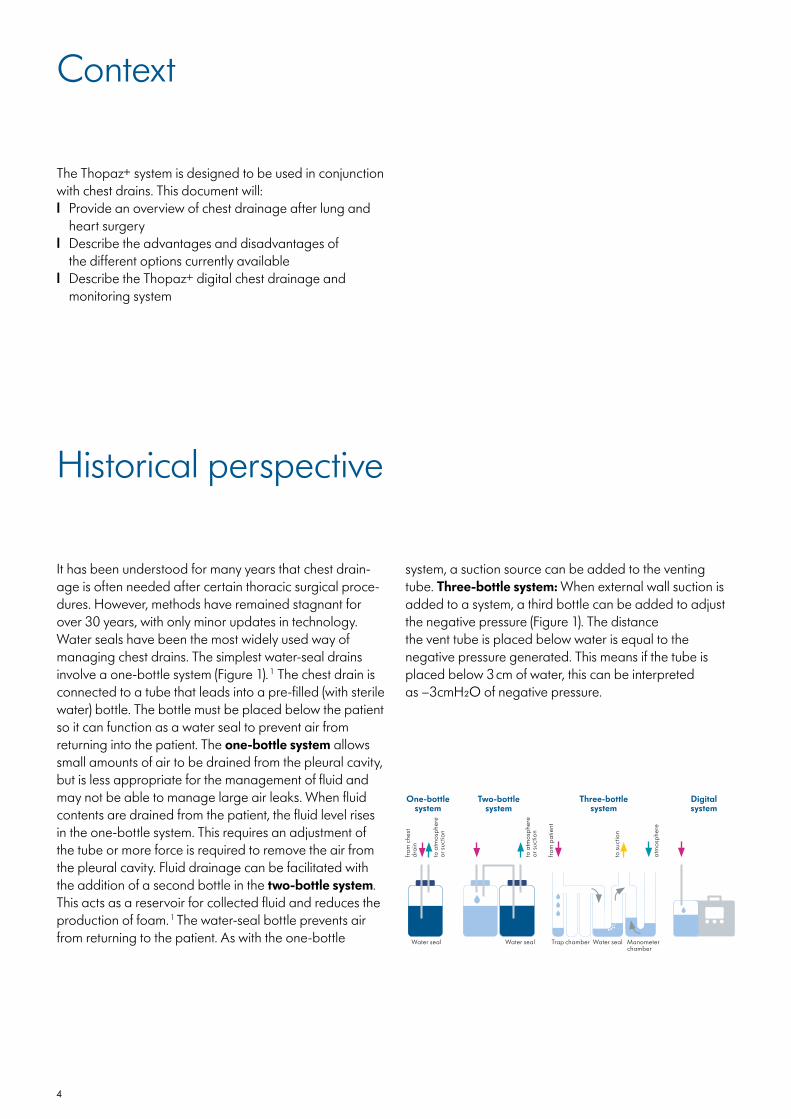
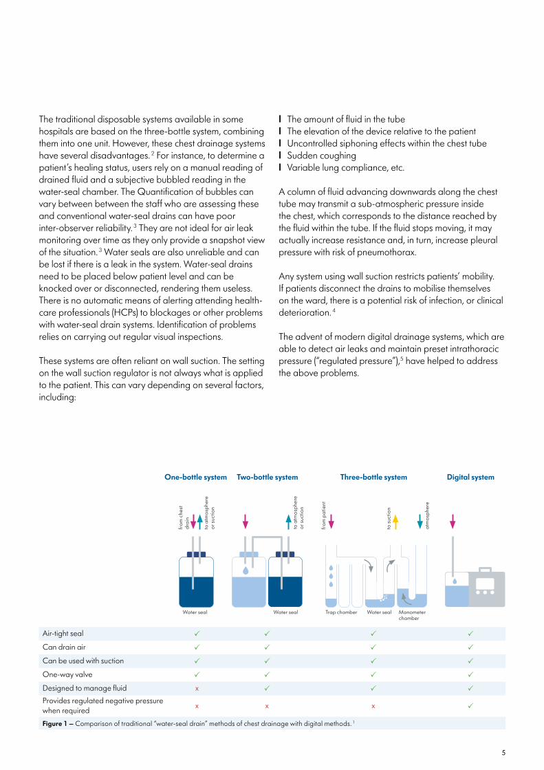
It has been understood for many years that chest drain-age is often needed after certain thoracic surgical proce-dures. However, methods have remained stagnant for over 30 years, with only minor updates in technology. Water seals have been the most widely used way of managing chest drains. The simplest water-seal drains involve a one-bottle system (Figure 1). 1 The chest drain is connected to a tube that leads into a pre-filled (with sterile water) bottle. The bottle must be placed below the patient so it can function as a water seal to prevent air from returning into the patient. The one-bottle system allows small amounts of air to be drained from the pleural cavity, but is less appropriate for the management of fluid and may not be able to manage large air leaks. When fluid contents are drained from the patient, the fluid level rises in the one-bottle system. This requires an adjustment of the tube or more force is required to remove the air from the pleural cavity. Fluid drainage can be facilitated with the addition of a second bottle in the two-bottle system. This acts as a reservoir for collected fluid and reduces the production of foam. 1 The water-seal bottle prevents air from returning to the patient. As with the one-bottle
system, a suction source can be added to the venting tube. Three-bottle system: When external wall suction is added to a system, a third bottle can be added to adjust the negative pressure (Figure 1). The distance the vent tube is placed below water is equal to the negative pressure generated. This means if the tube is placed below 3 cm of water, this can be interpreted as –3cmH2O of negative pressure.
Digital system
from
che
st
drai
n
to a
tmos
pher
e
or s
uctio
n
One-bottle system
Water seal
Two-bottle system
to a
tmos
pher
e or
suc
tion
Water seal
Three-bottle system
atm
osph
ere
to s
uctio
n
from
pat
ient
Trap chamber Water seal Manometer chamber
5
The traditional disposable systems available in some hospitals are based on the three-bottle system, combining them into one unit. However, these chest drainage systems have several disadvantages. 2 For instance, to determine a patient’s healing status, users rely on a manual reading of drained fluid and a subjective bubbled reading in the water-seal chamber. The Quantification of bubbles can vary between between the staff who are assessing these and conventional water-seal drains can have poor inter-observer reliability. 3 They are not ideal for air leak monitoring over time as they only provide a snapshot view of the situation. 3 Water seals are also unreliable and can be lost if there is a leak in the system. Water-seal drains need to be placed below patient level and can be knocked over or disconnected, rendering them useless. There is no automatic means of alerting attending health-care professionals (HCPs) to blockages or other problems with water-seal drain systems. Identification of problems relies on carrying out regular visual inspections.
These systems are often reliant on wall suction. The setting on the wall suction regulator is not always what is applied to the patient. This can vary depending on several factors, including:
l The amount of fluid in the tube l The elevation of the device relative to the patientl Uncontrolled siphoning effects within the chest tubel Sudden coughingl Variable lung compliance, etc.
A column of fluid advancing downwards along the chest tube may transmit a sub-atmospheric pressure inside the chest, which corresponds to the distance reached by the fluid within the tube. If the fluid stops moving, it may actually increase resistance and, in turn, increase pleural pressure with risk of pneumothorax.
Any system using wall suction restricts patients’ mobility. If patients disconnect the drains to mobilise themselves on the ward, there is a potential risk of infection, or clinical deterioration. 4
The advent of modern digital drainage systems, which are able to detect air leaks and maintain preset intrathoracic pressure (“regulated pressure”),5 have helped to address the above problems.
Air-tight seal P P P P
Can drain air P P P P
Can be used with suction P P P P
One-way valve P P P P
Designed to manage fluid x P P P
Provides regulated negative pressure when required
x x x P
Figure 1 – Comparison of traditional “water-seal drain” methods of chest drainage with digital methods. 1
from
che
st
drai
n
to a
tmos
pher
e
or s
uctio
n
One-bottle system Three-bottle system
atm
osph
ere
to s
uctio
n
Digital systemTwo-bottle system
to a
tmos
pher
e
or s
uctio
n
from
pat
ient
Trap chamber Water seal Manometer chamber
Water seal Water seal

6
Clinical rationale behind chest drainage
PneumothoraxA pneumothorax occurs in the event of an injury to the thoracic cavity or the lung that allows air to enter the pleural cavity. As this air cannot evacuate the chest, the lung collapses, creating a gap of air and separation of the pleural layers (Figure 2). This is informally known as a “collapsed lung”. Removal of air from the pleural cavity is mediated via a drain inserted into the pleural space.
There are many causes of a pneumothorax including trauma (e.g. from a fracture penetrating the lung or a gunshot wound), post-surgical complication (e.g. after pulmonary resection), oesophageal perforation, respira-tory failure or a bronchopleural fistula. Iatrogenic causes can include damage caused during central-line place-ment. A pneumothorax can also occur spontaneously.
A tension pneumothorax describes an accumulation of air in the pleural space under positive pressure. This com-presses the lung and decreases venous return to the heart. The contents of the thorax are forced to the opposite side of the chest putting extra pressure on the heart and the uninvolved lung. This is an acute life-threatening emergen-cy situation that requires immediate attention, such as needle decompression followed by the placement of a chest drain to remove the air from the pleural cavity.
Chest drains are commonly used to manage accumula-tions of air or fluid, including blood and pus. They can be inserted into the pleural space surrounding the lungs (Figure 2) or into the mediastinum surrounding the heart (Figure 3).
Removal of air, blood or other fluid from the pleural cavity The visceral pleura covers the outside of the lungs and the parietal pleura adheres to the thoracic wall, the mediastinum and the diaphragm. The space between the lungs and thoracic wall, between these two pleura, is called the pleural space (Figure 2). The pressure inside this space is usually negative.
The pleural space is normally filled with between 2–4 ml of pleural fluid which lubricates the layers, allowing them to slide smoothly across each other during breathing. The pleural fluid also forms a seal that holds the outer surface of the lungs against the inner surface of the thoracic wall. This ensures that when the chest cavity expands with breathing, the lungs are kept in contact with the thoracic wall, allowing expansion of the lungs and therefore air can be drawn in. The negative pressure inside the pleural space also facilitates this close contact.
When the pleural cavity is contaminated with air, blood or too much fluid, patients have difficulty breathing (dyspnoea). Consequently, they may be unable to mobilise and find that they have to sit upright. Using their accessory muscles, breathing feels exhausting and requires more effort, which can lead to hyperventilation. Affected patients may require extra oxygen, and possible advanced ventilatory support.
Figure 2 – Use of pleural drains to manage collections of air (pneumothorax), fluid (pleural effusion) including collections of serous fluid or blood (haemothorax) in the pleural cavity.
7
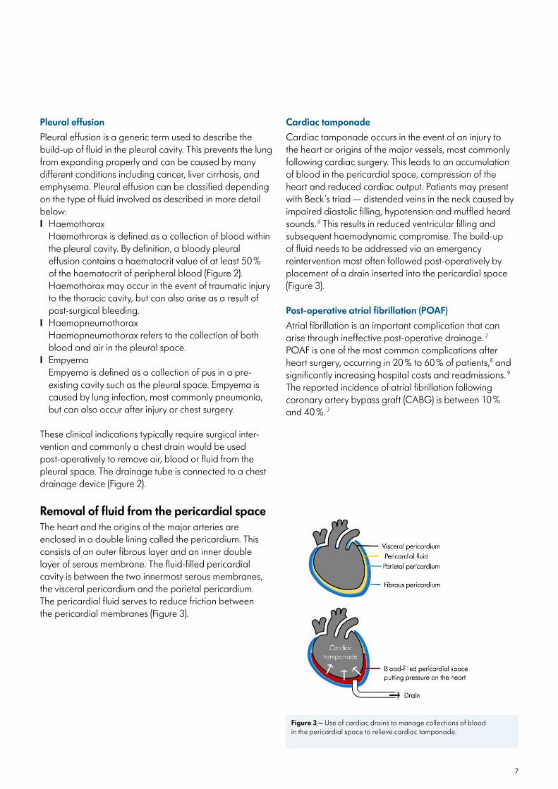
Figure 3 – Use of cardiac drains to manage collections of blood in the pericardial space to relieve cardiac tamponade.
Pleural effusionPleural effusion is a generic term used to describe the build-up of fluid in the pleural cavity. This prevents the lung from expanding properly and can be caused by many different conditions including cancer, liver cirrhosis, and emphysema. Pleural effusion can be classified depending on the type of fluid involved as described in more detail below:I Haemothorax Haemothrorax is defined as a collection of blood within
the pleural cavity. By definition, a bloody pleural effusion contains a haematocrit value of at least 50 % of the haematocrit of peripheral blood (Figure 2). Haemothorax may occur in the event of traumatic injury to the thoracic cavity, but can also arise as a result of post-surgical bleeding.
I Haemopneumothorax Haemopneumothorax refers to the collection of both
blood and air in the pleural space.I Empyema Empyema is defined as a collection of pus in a pre-
existing cavity such as the pleural space. Empyema is caused by lung infection, most commonly pneumonia, but can also occur after injury or chest surgery.
These clinical indications typically require surgical inter-vention and commonly a chest drain would be used post-operatively to remove air, blood or fluid from the pleural space. The drainage tube is connected to a chest drainage device (Figure 2).
Removal of fluid from the pericardial spaceThe heart and the origins of the major arteries are enclosed in a double lining called the pericardium. This consists of an outer fibrous layer and an inner double layer of serous membrane. The fluid-filled pericardial cavity is between the two innermost serous membranes, the visceral pericardium and the parietal pericardium. The pericardial fluid serves to reduce friction between the pericardial membranes (Figure 3).
Cardiac tamponadeCardiac tamponade occurs in the event of an injury to the heart or origins of the major vessels, most commonly following cardiac surgery. This leads to an accumulation of blood in the pericardial space, compression of the heart and reduced cardiac output. Patients may present with Beck’s triad — distended veins in the neck caused by impaired diastolic filling, hypotension and muffled heard sounds. 6 This results in reduced ventricular filling and subsequent haemodynamic compromise. The build-up of fluid needs to be addressed via an emergency reintervention most often followed post-operatively by placement of a drain inserted into the pericardial space (Figure 3).
Post-operative atrial fibrillation (POAF)Atrial fibrillation is an important complication that can arise through ineffective post-operative drainage. 7 POAF is one of the most common complications after heart surgery, occurring in 20 % to 60 % of patients,8 and significantly increasing hospital costs and readmissions. 9 The reported incidence of atrial fibrillation following coronary artery bypass graft (CABG) is between 10 % and 40 %. 7

8
General principles for the management of chest drainsPulmonary proceduresAir leaksAir leaks are the most common complication after pulmonary resection. 1 They can be caused by a tear in the visceral pleura and peripheral lacerations of the lung. 10 While around 50 % of all patients experience some form of air leak,11 in as many as 25 %, a prolonged air leak can occur. 12 This can be defined as an air leak that lasts more than 5 days. 1
There has been much debate around definitions,10 and little published information describing the correct timing for chest drain removal. 13 Ultimately, the responsibility for the decision is in the hands of the attending physician. Historically, the situation has been compounded by use of traditional chest drainage methods because these are unable to objectively measure air leaks. The use of digital systems that can quantify air leaks is the basis to develop standardised criteria to help decide when to remove chest drains. 14 Research has shown that data from digital systems have the potential to allow prediction of air-leak resolution, which may ultimately guide effective clinical decision-making. 12 The ability to predict fast-resolving air leaks may speed up chest drain removal and hospital discharge. 12 Indeed, consideration of early removal of chest drains in such patients has been a recent focus of interest among thoracic surgeons. 15
Fluid drainageThe volume of fluid obtained from a chest drain is closely monitored. While, drainage needs to reduce before a chest drain can be removed, in practice, the cut-off point ranges from 250 ml/day to 450 ml/day, with little consen-sus. 1,3 Physiologically, daily pleural fluid filtration is estimated to be 350 ml, hence, many authors suggest removal when daily recorded drainage volume is less than 300 ml. 5 Guidelines for enhanced recovery after lung surgery, produced by the Enhanced Recovery After Surgery (ERAS®) Society and the European Society of Thoracic Surgeons (ESTS), and published in 2018,16 recommend a relatively high pleural fluid output for chest drain removal (up to 450 ml/day).
A randomised controlled trial suggests the protein content of the drained fluid helps determine the timing of chest tube removal 15. Accordingly their data suggests that a pleural fluid-to-blood protein ratio of less than 0.5 is a
good indicator of safe chest drain removal. 15 Lymph fluid and blood need to be substantially absent from the fluid before the drain is removed (i.e. non-chylous, non-hae-matic). 5,16
Use of this criterion may facilitate removal, reducing post-operative hospital stays (to the benefit of both patients and clinicians) while decreasing treatment costs for healthcare providers. 15
Table 1 summarises key questions that may be used to determine whether a pleural chest drain is ready for removal.
Cardiac proceduresMultiple chest drains are often required following cardiac surgery. During surgery, lungs may have been deflated to enable the cardiac surgery to proceed and are reinflated towards the end of the procedure. Post-operatively, a chest drain may need to be inserted into the pleural cavity to keep the lungs reinflated. Patients undergoing coronary artery bypass grafting (CABG) may have the pleura opened and if both internal mammary arteries are used, there will be both left and right pleural drains. More commonly, the left internal mammary artery is used. Dissection of this may involve opening the left pleura, which will necessitate an appropriate drain.
Valve operations typically require a mediastinal and pericardial drain. The mediastinal drain lies in front of the heart whilst the pericardial drain is positioned under the diaphragmatic surface of the heart. Sometimes the mediastinal drain acts as a pleural drain as well; the side holes are in front of the heart whilst the tip of the tube lies high in the pleural space.
Recent ERAS® guidelines describing perioperative care in cardiac surgery state that while there are no standard criteria for the timing of mediastinal drain removal, evidence suggests that these can be safely removed as soon as the drainage becomes macroscopically serous. 18 Criteria for the removal of pericardial drains typically follow local practice, varying between countries and also on a hospital level. In some institutions, pericardial drains may be routinely removed at 6 a.m. when this is deemed appropriate, but in other instances, physicians may be guided by X-ray results.
9
Table 1. Key questions to determine whether a chest drain is ready for removal. Local protocols may differ and should take precedence.
Is the drained fluid non-chylous? 16
Does the drained fluid contain an acceptably low level of blood?
Has the air leak dropped to an acceptable level?
Do other factors support the decision to remove the chest drain?e.g. lung expansion on chest X-ray 17
Avoidance of complications is criticalPain, drain blockage and accidental dislodgement are common complications of chest drains. More serious complications include organ injury, haemothorax, infec-tions, and re-expansion pulmonary oedema. Protocols that reduce the duration of chest drain placement may reduce the risk of infection. Also, once chest drains are removed, associated pain recedes.
Should negative pressure be applied to chest drains? The debateThere has been conflicting evidence as to the value of negative pressure in chest drains. Suction promotes pleura-pleural apposition helping to seal air leaks and drain large air leaks. Lack of suction may compromise drainage of large air leaks and has been associated with an increased risk of other complications, such as pneumo-nia and arrhythmia. 16 However, in some circumstances, not applying suction may reduce the duration of air leaks,19 presumably as a consequence of decreased air flow. In relation to this, suction can increase the flow through the chest drain proportional to the level of suction applied 20, and as previously mentioned, patient mobilisa-tion is reduced if wall suction is used.
Given the contested benefits of applying wall suction to water-seal systems compared to a water seal alone, three meta-analyses have investigated a variety of outcomes in
patients with these drains. 21–23 In all three meta-analyses, the incidence of post-operative pneumothorax was found to be significantly reduced following suction compared with water-seal alone. 21–23 However, in two of these analyses, no differences in length of hospital stay or the duration of the drains were observed with suction and no-suction water-seal protocols 21,22 while one study favoured the water seal alone. 23
These underwhelming clinical outcomes, coupled with the fact that the patient is immobilised when external (unregulated) wall suction is used, have led recent guide-lines produced by ERAS® and the European Society of Thoracic Surgeons to recommend that external wall suction is NOT used following lung surgery. 16
It is important to recognise that digital chest drainage systems do not provide suction in the same way as wall suction. Their use is widely believed to provide benefits not provided by water-seal drains with or without suction. 16,24,25 The Thopaz+ digital chest drainage and monitoring system regulates pressure, providing suction only when necessary.
The evidence base describing the benefits of Thopaz+ is outlined on the following pages.
10
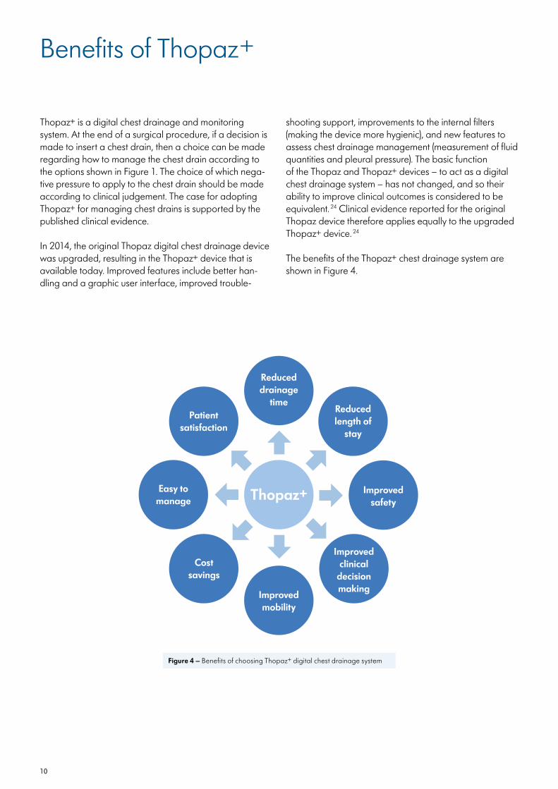
Benefits of Thopaz +
Thopaz+ is a digital chest drainage and monitoring system. At the end of a surgical procedure, if a decision is made to insert a chest drain, then a choice can be made regarding how to manage the chest drain according to the options shown in Figure 1. The choice of which nega-tive pressure to apply to the chest drain should be made according to clinical judgement. The case for adopting Thopaz+ for managing chest drains is supported by the published clinical evidence.
In 2014, the original Thopaz digital chest drainage device was upgraded, resulting in the Thopaz+ device that is available today. Improved features include better han-dling and a graphic user interface, improved trouble-
shooting support, improvements to the internal filters (making the device more hygienic), and new features to assess chest drainage management (measurement of fluid quantities and pleural pressure). The basic function of the Thopaz and Thopaz+ devices – to act as a digital chest drainage system – has not changed, and so their ability to improve clinical outcomes is considered to be equivalent. 24 Clinical evidence reported for the original Thopaz device therefore applies equally to the upgraded Thopaz+ device. 24
The benefits of the Thopaz+ chest drainage system are shown in Figure 4.
Figure 4 – Benefits of choosing Thopaz+ digital chest drainage system
Easy tomanage
Improvedclinical
decisionmaking
Reduceddrainage
timeReducedlength of
stay
Improvedsafety
Costsavings
Improvedmobility
Thopaz+
Patientsatisfaction
11
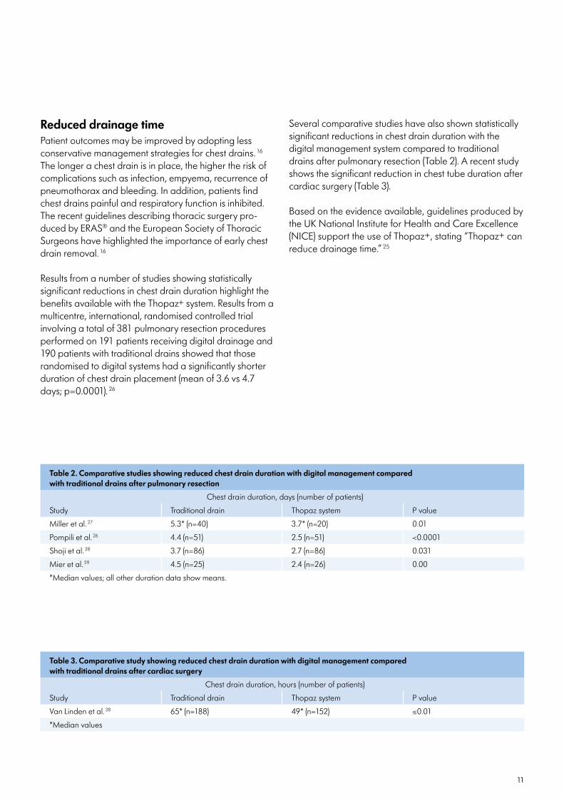
Table 2. Comparative studies showing reduced chest drain duration with digital management compared with traditional drains after pulmonary resection
Chest drain duration, days (number of patients)
Study Traditional drain Thopaz system P value
Miller et al. 27 5.3* (n=40) 3.7* (n=20) 0.01
Pompili et al. 26 4.4 (n=51) 2.5 (n=51) <0.0001
Shoji et al. 28 3.7 (n=86) 2.7 (n=86) 0.031
Mier et al. 29 4.5 (n=25) 2.4 (n=26) 0.00
*Median values; all other duration data show means.
Reduced drainage timePatient outcomes may be improved by adopting less conservative management strategies for chest drains. 16 The longer a chest drain is in place, the higher the risk of complications such as infection, empyema, recurrence of pneumothorax and bleeding. In addition, patients find chest drains painful and respiratory function is inhibited. The recent guidelines describing thoracic surgery pro-duced by ERAS® and the European Society of Thoracic Surgeons have highlighted the importance of early chest drain removal. 16
Results from a number of studies showing statistically significant reductions in chest drain duration highlight the benefits available with the Thopaz+ system. Results from a multicentre, international, randomised controlled trial involving a total of 381 pulmonary resection procedures performed on 191 patients receiving digital drainage and 190 patients with traditional drains showed that those randomised to digital systems had a significantly shorter duration of chest drain placement (mean of 3.6 vs 4.7 days; p=0.0001). 26
Several comparative studies have also shown statistically significant reductions in chest drain duration with the digital management system compared to traditional drains after pulmonary resection (Table 2). A recent study shows the significant reduction in chest tube duration after cardiac surgery (Table 3).
Based on the evidence available, guidelines produced by the UK National Institute for Health and Care Excellence (NICE) support the use of Thopaz+, stating “Thopaz+ can reduce drainage time.” 25
Table 3. Comparative study showing reduced chest drain duration with digital management compared with traditional drains after cardiac surgery
Chest drain duration, hours (number of patients)
Study Traditional drain Thopaz system P value
Van Linden et al. 38 65* (n=188) 49* (n=152) ≤0.01
*Median values

12
Reduced length of stay in hospitalAs well as enabling shorter chest drain times, three separate studies have shown that patients treated with Thopaz had shorter hospital stays than those receiving conventional/analogue drainage. 26,27,30 Results from the previously mentioned randomised controlled trial involv-ing pulmonary resection procedures showed a mean length of stay of 4.6 (digital) and 5.6 (traditional) days respectively (p<0.0001). 26 The Benefits of digital drainage have also been quantified for patients experiencing a spontaneous pneumothorax with air leak: In a randomised controlled trial involving 60 affected individuals ran-domised to either a digital drainage device or traditional drainage, those receiving the digital device had a mean hospital stay length of 5.1 days, vs 7.0 days for those receiving traditional drainage. (p<0.001). The correspond-ing values for mean drainage duration were 48 and 85 hours respectively (p<0.001). 31 In addition, results from two of the comparative studies mentioned above also de-scribed reduced length of hospital stay with the digital drainage compared with conventional method, with medians of 4.1 and 5.6 days (p=0.05), respectively, in one,27 and means of 4.5 and 6 days (p<0.0003), respectively, in the other. 26
On the basis of available evidence, the NICE guidelines state that “Thopaz+ can reduce… length of stay in hospi-tal.” 25 These guidelines also recommend that “Thopaz+ should be considered for people who need chest drainage after pulmonary resection or because of a pneumothorax.” 25
Improved safety Studies have documented fewer adverse events in patients treated with Thopaz compared with alternative ways of managing chest drains. In a randomised con-trolled trial involving a total of 64 patients who underwent pulmonary resection, Thopaz significantly reduced complication rates compared with traditional drainage (25% vs 50% [p=0.039]). 20 In addition, one of the compar-ative studies mentioned above also reported fewer overall complications with the digital system compared with conventional drainage: 22% vs 35% (p=0.01). 27,32 The NICE guidelines state that “Thopaz+… improves safety for people with chest drains.” 25 The system has built-in alarms. 25 Users can be warned of potential problems, such as patients losing large amounts of fluid, as well as system leakage, tube blockage, a full canister or a low battery.
Improved clinical decision makingWhen using traditional systems, it can be a subjective decision to understand when an air leak has resolved. The decision to remove the drain may therefore be delayed, in turn delaying hospital discharge in many cases. The recent series of guidelines for thoracic surgery produced by the ERAS® and the European Society of Thoracic Surgeons stated that “Digital drainage systems reduce variability in decision-making and should be used.” 16
The Thopaz+ system may improve clinical decision making through continuous, objective monitoring of air leaks and fluid loss. 25 The ability to store information and display trends in air leaks over time allows more informed deci-sion-making about chest drain removal. 16 Inter-observer and clinical practice variability is reduced. 3,16,33 Scientific monitoring of patient progress provides a more scientific rationale for drain management and removal. 2 An article describing a single-centre randomised controlled trial involving Thopaz+ concluded that “the technology holds promise for leading to a deeper understanding of pleural space mechanics after pulmonary resection, and to refine-ment of evidence-based practice in chest drain manage-ment.” 34 Furthermore, in patients with spontaneous pneumothorax, digital air leak measurements early in the treatment course have the potential to predict future treatment failure. 35
Improved mobilityIn the multicentre, randomised controlled study mentioned above, patients perceived an improved ability to get out of bed following surgery when using Thopaz compared with traditional chest drainage (p=0.008). 26 The light weight of the Thopaz+ system and its ability to offer portable suction provide benefits in this regard. 2 Indeed in another study, evaluation of patient and staff feedback following the use of Thopaz in 120 surgical cases found that “patients appreciated that it was portable and light which meant they could mobilise on suction, giving them more independence.” 4 This study also reported that, as patients could be mobilised earlier, nursing and physiotherapy were easier. 4
13
On the basis of available evidence, the NICE guidelines state that Thopaz+ allows “increased mobility which aids recovery….” 25 Use of Thopaz has been reported as part of a regimen employing early mobilisation in an attempt to improve outcomes after lung cancer surgery. 36 However, Thopaz+ is also relevant for patients needing chest drainage after cardiac surgery and trauma, as well as after pulmonary resection. 25
Cost savingsThopaz+ is more cost-effective than traditional chest drainage. 24,25 Studies have shown that using the Thopaz+ system saves up to £550 per patient treated for pneumo-thorax, and £111 per patient per hospital stay after pulmonary resection. 24,25
Cost savings are driven mainly by reduced length of stay. 24 However, NICE modelling confirming the cost savings included a range of parameters to reflect both evidence and practice: 25
l A hospital stay of 5.4 days for Thopaz+ and 5.8 days for conventional chest drainage
l A drainage time of 3.5 days for Thopaz+ l Costs for consumables and training associated with
standard drainagel Cost of chest drain reinsertion and complications l Consumer and training costs for Thopaz+
Easy to manageDigital drainage systems may improve and unify hospital practice. 5 Their simplicity enables relatively easy introduc-tion into hospital wards and theatres. 2 Thopaz+ has a user-friendly set up, and nurses report being pleased that with such technology, which contains sealed, dry-unit canisters, there is no need for priming and the risk of spillage/infection is reduced. 4 On-screen displays and alarms increase the safety of patient management and facilitate accurate assessment of air leaks. 4 Greater consensus with regard to the severity of air leaks may translate into increased cooperation among multidisci-plinary care teams. The importance of such cooperation, and communication to execute patient care algorithms for air-leak management, cannot be overemphasised. 33 As a consequence of this objective evaluation and clear definition of algorithms, chest drain removal may be delegated to staff nurses. 37 Furthermore, use of digital chest drainage technology may help to streamline communication and agreement in the clinical research setting, allowing air leaks to be objectively assessed following procedural interventions. 33
Higher rates of patient satisfactionIncreased patient satisfaction is apparent with Thopaz+. 25 In the multicentre, randomised controlled study mentioned above, patients treated with Thopaz reported that the system was more convenient than conventional technolo-gy. 26 Fewer patients felt they would want to change the system compared with those treated with a traditional drainage device. 26
In another study, where patients appreciated the portabil-ity and light weight of the system enabling them greater mobility as described above,4 patients had more indepen-dence. They preferred the lack of bubbling noise and the compactness over conventional drains and suction. This feedback was particularly prominent in individuals with pneumothoraces who had previously experienced standard water-seal bottles on continuous wall suction. 4 Greater mobility may reduce complications associated with decreased mobility, as well as benefitting patients’ privacy and dignity. 2
14
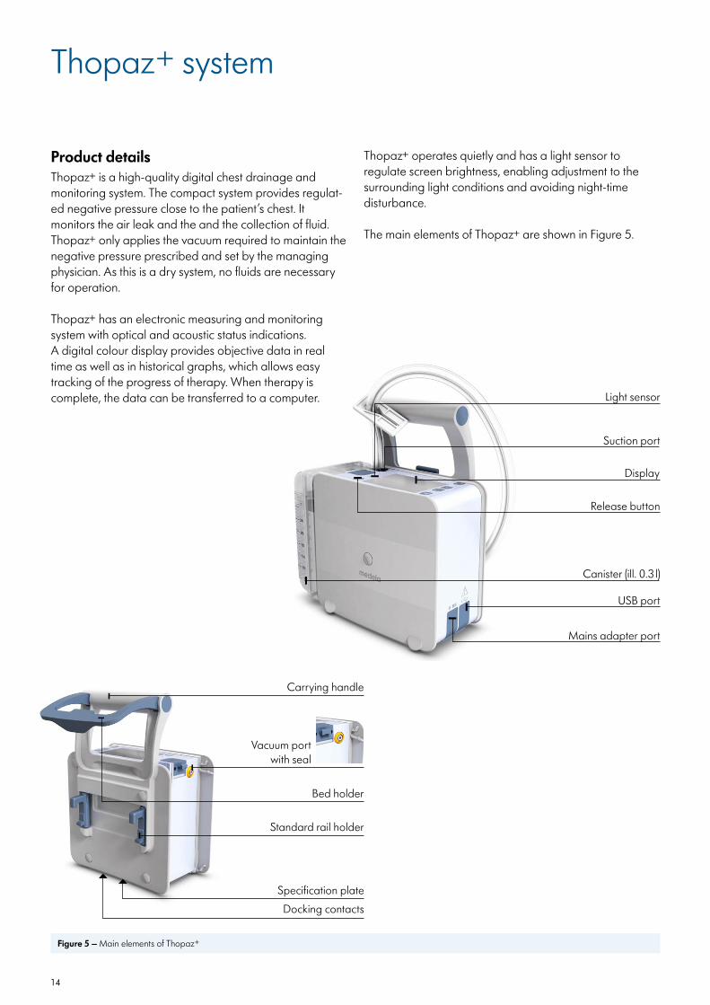
Thopaz + system
Figure 5 – Main elements of Thopaz+
Thopaz+ operates quietly and has a light sensor to regulate screen brightness, enabling adjustment to the surrounding light conditions and avoiding night-time disturbance.
The main elements of Thopaz+ are shown in Figure 5.
Bed holder
Vacuum port with seal
Standard rail holder
Specification plate
Carrying handle
Docking contacts
Mains adapter port
Canister (ill. 0.3 l)
USB port
Suction port
Light sensor
Display
Release button
Product details Thopaz+ is a high-quality digital chest drainage and monitoring system. The compact system provides regulat-ed negative pressure close to the patient’s chest. It monitors the air leak and the and the collection of fluid. Thopaz+ only applies the vacuum required to maintain the negative pressure prescribed and set by the managing physician. As this is a dry system, no fluids are necessary for operation.
Thopaz+ has an electronic measuring and monitoring system with optical and acoustic status indications. A digital colour display provides objective data in real time as well as in historical graphs, which allows easy tracking of the progress of therapy. When therapy is complete, the data can be transferred to a computer.
15
IndicationsThopaz+ is used for chest drainage in the pleural and mediastinum. It can be used after:l Pneumothorax l Cardiac or thoracic surgeryl Thorax injury l Pleural effusion l Pleural empyemal Or other related conditions
Thopaz+ is indicated for all situations where chest drains are applied.
Thopaz+ is intended to be used for aspiration and removal of surgical fluids, tissues, gases, bodily fluids or infectious materials and for use on patients in appropriate care settings.
ContraindicationsThere are no known contraindications.
ApplicationPlease refer to the product Instructions for Use to obtain detailed guidance on the application of Thopaz+. Additional safety information is provided in the Quick Reference Guide at the end of this booklet.
IMPORTANT: Please review all the available safety information before using Thopaz+.
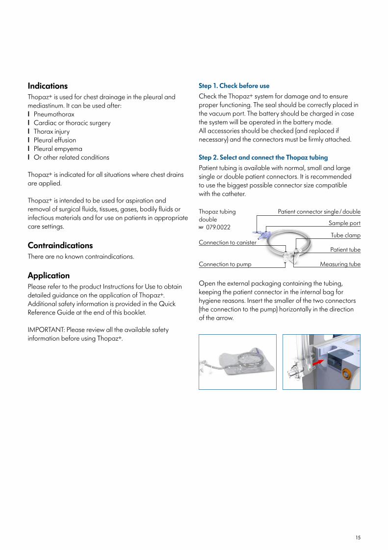
Open the external packaging containing the tubing, keeping the patient connector in the internal bag for hygiene reasons. Insert the smaller of the two connectors (the connection to the pump) horizontally in the direction of the arrow.
Step 2. Select and connect the Thopaz tubingPatient tubing is available with normal, small and large single or double patient connectors. It is recommended to use the biggest possible connector size compatible with the catheter.
Step 1. Check before useCheck the Thopaz+ system for damage and to ensure proper functioning. The seal should be correctly placed in the vacuum port. The battery should be charged in case the system will be operated in the battery mode. All accessories should be checked (and replaced if necessary) and the connectors must be firmly attached.
Thopaz tubing double
079.0022Sample port
Measuring tubeConnection to pump
Tube clampConnection to canister
Patient connector single / double
Patient tube
16
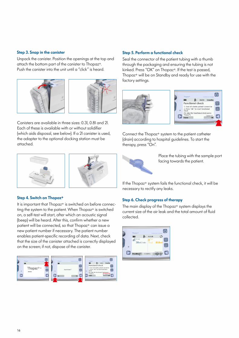
Step 6. Check progress of therapyThe main display of the Thopaz+ system displays the current size of the air leak and the total amount of fluid collected.
Step 5. Perform a functional check Seal the connector of the patient tubing with a thumb through the packaging and ensuring the tubing is not kinked. Press “OK” on Thopaz+. If the test is passed, Thopaz+ will be on Standby and ready for use with the factory settings.
Connect the Thopaz+ system to the patient catheter (drain) according to hospital guidelines. To start the therapy, press “On”.
Step 4. Switch on Thopaz+ It is important that Thopaz+ is switched on before connec-ting the system to the patient. When Thopaz+ is switched on, a self-test will start, after which an acoustic signal (beep) will be heard. After this, confirm whether a new patient will be connected, so that Thopaz+ can issue a new patient number if necessary. The patient number enables patient-specific recording of data. Next, check that the size of the canister attached is correctly displayed on the screen; if not, dispose of the canister.
Canisters are available in three sizes: 0.3l, 0.8I and 2l. Each of these is available with or without solidifier (which aids disposal, see below). If a 2l canister is used, the adapter to the optional docking station must be attached.
Step 3. Snap in the canister Unpack the canister. Position the openings at the top and attach the bottom part of the canister to Thopaz+. Push the canister into the unit until a “click” is heard.
Place the tubing with the sample port facing towards the patient.
If the Thopaz+ system fails the functional check, it will be necessary to rectify any leaks.
17
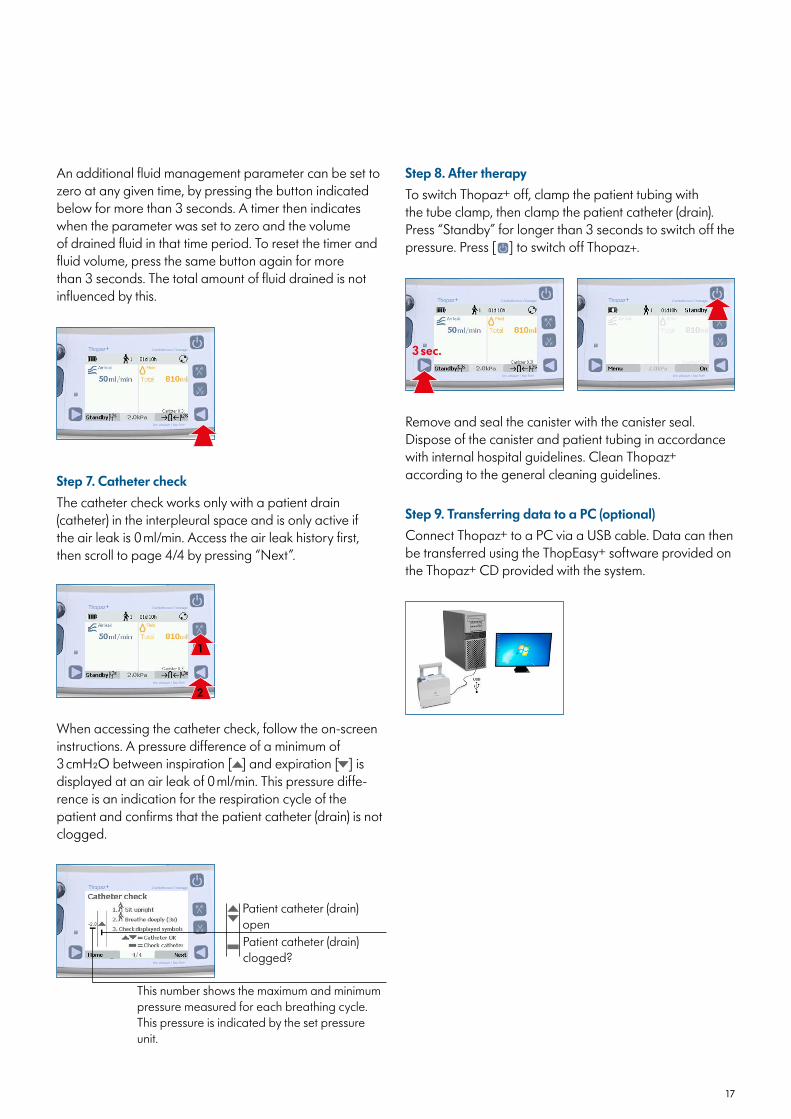
An additional fluid management parameter can be set to zero at any given time, by pressing the button indicated below for more than 3 seconds. A timer then indicates when the parameter was set to zero and the volume of drained fluid in that time period. To reset the timer and fluid volume, press the same button again for more than 3 seconds. The total amount of fluid drained is not influenced by this.
Step 9. Transferring data to a PC (optional)Connect Thopaz+ to a PC via a USB cable. Data can then be transferred using the ThopEasy+ software provided on the Thopaz+ CD provided with the system.
Remove and seal the canister with the canister seal. Dispose of the canister and patient tubing in accordance with internal hospital guidelines. Clean Thopaz+ according to the general cleaning guidelines.
Step 8. After therapyTo switch Thopaz+ off, clamp the patient tubing with the tube clamp, then clamp the patient catheter (drain). Press “Standby” for longer than 3 seconds to switch off the pressure. Press [ ] to switch off Thopaz+.
Step 7. Catheter checkThe catheter check works only with a patient drain (catheter) in the interpleural space and is only active if the air leak is 0 ml/min. Access the air leak history first, then scroll to page 4/4 by pressing “Next”.
1
2
3 sec.
When accessing the catheter check, follow the on-screen instructions. A pressure difference of a minimum of 3 cmH2O between inspiration [ ] and expiration [ ] is displayed at an air leak of 0 ml/min. This pressure diffe-rence is an indication for the respiration cycle of the patient and confirms that the patient catheter (drain) is not clogged.
This number shows the maximum and minimum pressure measured for each breathing cycle. This pressure is indicated by the set pressure unit.
Patient catheter (drain) openPatient catheter (drain) clogged?
18
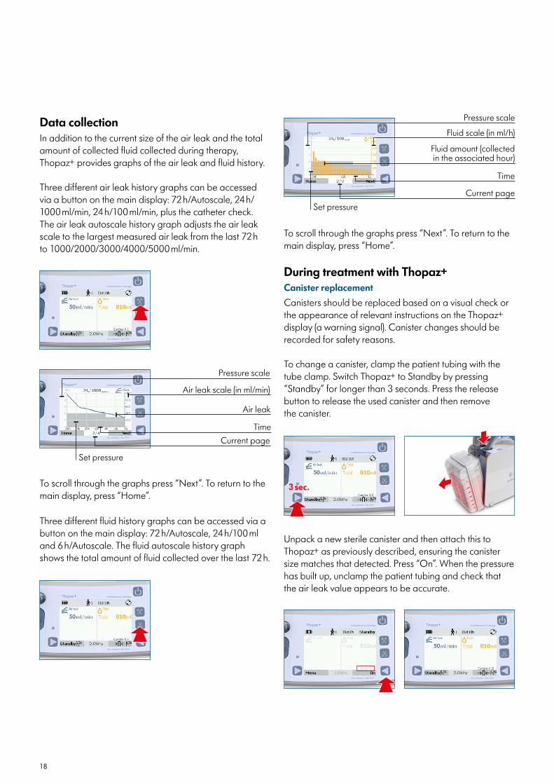
During treatment with Thopaz+Canister replacementCanisters should be replaced based on a visual check or the appearance of relevant instructions on the Thopaz+ display (a warning signal). Canister changes should be recorded for safety reasons.
To change a canister, clamp the patient tubing with the tube clamp. Switch Thopaz+ to Standby by pressing “Standby” for longer than 3 seconds. Press the release button to release the used canister and then remove the canister.
Unpack a new sterile canister and then attach this to Thopaz+ as previously described, ensuring the canister size matches that detected. Press “On”. When the pressure has built up, unclamp the patient tubing and check that the air leak value appears to be accurate.
3 sec.
Three different fluid history graphs can be accessed via a button on the main display: 72 h/Autoscale, 24 h/100 ml and 6 h/Autoscale. The fluid autoscale history graph shows the total amount of fluid collected over the last 72 h.
To scroll through the graphs press “Next”. To return to the main display, press “Home”.
Pressure scale
Fluid scale (in ml/h)
Fluid amount (collected in the associated hour)
Current page
Time
Set pressure
Data collectionIn addition to the current size of the air leak and the total amount of collected fluid collected during therapy, Thopaz+ provides graphs of the air leak and fluid history.
Three different air leak history graphs can be accessed via a button on the main display: 72 h/Autoscale, 24 h/ 1000 ml/min, 24 h/100 ml/min, plus the catheter check. The air leak autoscale history graph adjusts the air leak scale to the largest measured air leak from the last 72 h to 1000/2000/3000/4000/5000 ml/min.
To scroll through the graphs press “Next”. To return to the main display, press “Home”.
Pressure scale
Air leak scale (in ml/min)
Air leak
TimeCurrent page
Set pressure

19
Changing pressure during operationThe negative pressure may only be changed by a physi-cian or under medical direction. Press [ ] and [ ] simulta-neously, then change the pressure by pressing [ ] or [ ] and confirm with “OK” [ ].
For used canisters with solidifier and “press & shake™” technology, check the canister is sealed. Then press the middle of the solidifier chamber to open and shake (to activate the solidification process) before disposal.
Canister disposalUsed canisters should be sealed with the canister seal and disposed of according to internal hospital guidelines.
Press simultaneously
Physiological pressure can be activated for patients who are to be treated by gravity drainage (equivalent to a water seal). To do this, press “Physio” [ ] and confirm with “OK” [ ]. This mode corresponds to a pressure of -0.8 kPa/-6 mmHg/-8 cmH2O/-8 mbar.
Sample port
Patient tube
Taking a drainage sampleTo take a drainage sample, ensure that the patient tube contains fluid, then clamp the catheter (drain). Switch Thopaz+ to Standby by pressing “Standby” for longer than 3 seconds. Disinfect the sample port (using hospital approved wipes) and remove air from the patient tubing with a syringe (17 G [1.4 mm] or thinner). Repeat the process until fluid has gathered at the sample port and a sample can be taken. Switch Thopaz+ on by pressing “On”. To ensure Thopaz+ can reduce the pressure, it is essential to wait 30 seconds before unclamping the patient catheter (drain).
3 sec.
20
Quick reference guide
Intended use/indicationsThopaz+ is intended to be used on patients in appropriate care settings for aspiration and removal of surgical fluids, tissue, gases, bodily fluids or infectious materials. It is indicated for all situations where chest drains are applied, particularly for thoracic drainage in the pleural and mediastinum in situations such as:l Pneumothorax l Cardiac or thoracic surgeryl Thorax injury l Pleural effusion l Pleural empyemal Or other related conditions
Thopaz+ should only be operated by properly instructed users who should have adequate visual faculty and must not be hearing impaired or deaf. Physicians should evaluate the appropriateness of the treatment based on their knowledge and experience, complying with proper surgical procedures and techniques.
ContraindicationsThere are no known contraindications. However, Thopaz+ should only be used on patients exhibiting conditions as described above. The use of Thopaz+ for any other indication than intended is neither desired nor authorised. The Thopaz+ system is not designed for retransfusion.
InstructionsThopaz+ should only be used by medically trained and qualified persons who have been adequately trained to use the device. The Instructions for Use are a general guide for the product, and medical matters must be addressed by a physician.
WarningsBefore operating Thopaz+ please read and observe the warning and safety points in the Instructions for Use. Key points are given below:l Safe functioning of the system can only be guaranteed
when Thopaz+ is used in combination with the original Thopaz+ accessories (canisters, tubing etc)
l Gloves should be worn for all operations – Do not touch the patient and the docking contacts
simultaneouslyl Do not use Thopaz+ in MRT (magnetic resonance
tomography)
l The pressure range to be set must be determined by a physician in accordance with the age and weight of the patient
– For paediatric patients adapt pressure settings according to hospital guidelines
l Patients should be regularly monitored according to internal hospital guidelines
l Do not use Thopaz+ if the drainage therapy indicates: – A pressure greater than the maximum pressure range
of -10 kPa – A flow rate greater than the maximum flow capacity
of 5 l/min – No pressure should be applied to a patientl Do not connect bilateral thoracic drains to one Thopaz+
unit. In such cases, the use of two Thopaz+ units is recommended
l The catheter/connector interface is a location where clotting may occur. Regular monitoring of this interface is recommended and an appropriate removal proce-dure should be in place in case this occurs
l Thopaz+ is not suitable for use while bathing, showering or in a hazardous explosive environment
l Wireless communications equipment can affect Thopaz+ and should be kept at least 30 cm away
l If an internal fault occurs (e.g. broken cable, defective battery), Thopaz+ turns off and an acoustic warning is sounded for at least 3 minutes (powered by a backup battery)
– Under these conditions Thopaz+ functions as a one-way valve. Replace Thopaz+ immediately
l A replacement must always be available for patients for whom the breakdown of Thopaz+ may lead to a critical situation
Other points relating to useAs with the warnings above, full details are available in the Instructions for Use. Key points are given below:l Single use, sterile products are not intended to be
reprocessed – Reprocessing could cause loss of mechanical,
chemical and/or biological characteristics – Reuse could cause cross-contaminationl If a persistent air leak is indicated, check the system is
assembled correctly before taking further corrective action. Ensure the system is airtight by clamping the catheter and observing the air leak decrease to zero
l Thopaz+ 2 l canisters are not for portable use (either by hand or using the carrying strap)
21
CABGCoronary artery bypass graft
Cardiac tamponadeCompression of the heart resulting from an accumulation of fluid, pus, blood or gas in the sac in which the heart is enclosed.
Chest drain/chest tubeA flexible tube inserted through the chest wall and into the pleural space or mediastinum. Note that for the purposes of this article, the term chest drain has been used through-out.
ERAS®
Enhanced Recovery After Surgery
HCPsHealthcare professionals
MRTMagnetic resonance tomography
NICENational Institute for Health and Care Excellence
Abbreviations/Definitions
l All canisters should be replaced on the basis of a visual check or in line with the relevant instructions on the Thopaz+ display (a warning signal)
l When attaching a new canister, verify that the canister size displayed on the screen is the same size as the canister attached
l When taking a drainage sample, to ensure Thopaz+ can reduce the pressure, it is essential to wait 30 seconds between taking the sample collection and unclamping the patient catheter (drain)
Pleural effusionAbnormal accumulation of fluid in the pleural space.
Pleural empyemaOne kind of pleural effusion, with an accumulation of pus caused by microorganisms, often occurring in the context of pneumonia, injury or chest surgery.
Post-operative atrial fibrillation (POAF)An irregular heartbeat caused by uncoordinated contrac-tion of the top two chambers of the heart, and occurring in patients after surgery.
Pulmonary resectionA surgical procedure to remove part, or all, of the lung, performed on patients with lungs that are diseased or damaged. These resections can be categorised as anatomical (also known as typical) resections (e.g. seg-mentectomy, lobectomy, pneumonectomy) and extra-ana-tomical (or atypical) resections such as wedge resections.
l Data transfer via USB is not permitted during therapyl After each use of Thopaz+, the parts that have been in
contact with aspirated secretions must be cleaned and disinfected or disposed of
l Before cleaning Thopaz+, disconnect the plug from the mains socket
22
1 Cerfolio, R. J. & Bryant, A. S. The management of chest tubes after pulmonary resection. Thorac Surg Clin 20, 399-405, doi:10.1016/j.thorsurg.2010.04.001 (2010).
2 Danitsch, D. Benefits of digital thoracic drainage systems. Nursing times 108, 16-17 (2012).
3 French, D. G. et al. Optimizing postoperative care protocols in thoracic surgery: best evidence and new technology. J Thorac Dis 8, S3-s11, doi:10.3978/j.issn.2072-1439.2015.10.67 (2016).
4 Rathinam, S., Bradley, A., Cantlin, T. & Rajesh, P. B. Thopaz Portable Suction Systems in Thoracic Surgery: an end user assessment and feedback in a tertiary unit. Journal of cardiothoracic surgery 6, 59, doi:10.1186/1749-8090-6-59 (2011).
5 Gao, S., Zhang, Z., Aragón, J., Brunelli, A. & Cassivi, S. The Society for Translational Medicine: clinical practice guidelines for the postoperative manage-ment of chest tube for patients undergoing lobectomy. J Thorac Dis. 2017 Sep; 9(9): 3255–3264. 9, 3255-3264 (2017).
6 Sternbach, G. Claude Beck: cardiac compression triads. The Journal of emergency medicine 6, 417-419, doi:10.1016/0736-4679(88)90017-0 (1988).
7 Ege, T. et al. The importance of intrapericardial drain selection in cardiac surgery. Chest 126, 1559-1562, doi:10.1378/chest.126.5.1559 (2004).
8 Kosuma, P., Wachirasrisirikul, S. & Jedsadayanmata, A. Attributable Costs of Postoperative Atrial Fibrillati-on among Patients Undergoing Cardiac Surgery. Cardiology research and practice 2018, 3759238, doi:10.1155/2018/3759238 (2018).
9 LaPar, D. J. et al. Postoperative atrial fibrillation significantly increases mortality, hospital readmission, and hospital costs. Ann Thorac Surg 98, 527-533; discussion 533, doi:10.1016/j.athoracsur.2014.03.039 (2014).
10 Jablonski, S. et al. Outcome of pleurodesis using different agents in management prolonged air leakage following lung resection. The clinical respira-tory journal 12, 183-192, doi:10.1111/crj.12509 (2018).
11 Mueller, M. R. & Marzluf, B. A. The anticipation and management of air leaks and residual spaces post lung resection. J Thorac Dis 6, 271-284, doi:10.3978/j.issn.2072-1439.2013.11.29 (2014).
12 Yeung, C. et al. Forecasting pulmonary air leak duration following lung surgery using transpleural airflow data from a digital pleural drainage device. J Thorac Dis 10, S3747-s3754, doi:10.21037/jtd.2018.08.11 (2018).
References
13 Kiefer, T. Removal of the chest drain: How to do it. Chapter 12 in Chest Drains in Daily Clinical Practice. pp189-193. Springer International Publishing Switzer-land. 2017. (2017).
14 Porcel, J. M. Chest Tube Drainage of the Pleural Space: A Concise Review for Pulmonologists. Tuberculosis and respiratory diseases 81, 106-115, doi:10.4046/trd.2017.0107 (2018).
15 Olgac, G., Cosgun, T., Vayvada, M., Ozdemir, A. & Kutlu, C. A. Low protein content of drainage fluid is a good predictor for earlier chest tube removal after lobectomy. Interactive cardiovascular and thoracic surgery 19, 650-655, doi:10.1093/icvts/ivu207 (2014).
16 Batchelor, T. J. P. et al. Guidelines for enhanced recovery after lung surgery: recommendations of the Enhanced Recovery After Surgery (ERAS®) Society and the European Society of Thoracic Surgeons (ESTS). Eur J Cardiothorac Surg 55, 91-115, doi:10.1093/ejcts/ezy301 (2019).
17 De Waele, M. et al. Does the usage of digital chest drainage systems reduce pleural inflammation and volume of pleural effusion following oncologic pulmonary resection?-A prospective randomized trial. J Thorac Dis 9, 1598-1606, doi:10.21037/jtd.2017.05.78 (2017).
18 Engelman, D. T. et al. Guidelines for Perioperative Care in Cardiac Surgery: Enhanced Recovery After Surgery Society Recommendations. JAMA surgery, doi:10.1001/jamasurg.2019.1153 (2019).
19 Cerfolio, R. J., Bass, C. & Katholi, C. R. Prospective randomized trial compares suction versus water seal for air leaks. The Annals of Thoracic Surgery 71, 1613-1617, doi:10.1016/s0003-4975(01)02474-2 (2001).
20 Manzanet, G. et al. A hydrodynamic study of pleural drainage systems: some practical consequences. Chest 127, 2211-2221, doi:10.1378/chest.127.6.2211 (2005).
21 Deng, B., Tan, Q. Y., Zhao, Y. P., Wang, R. W. & Jiang, Y. G. Suction or non-suction to the underwater seal drains following pulmonary operation: meta-analysis of randomised controlled trials. Eur J Cardiothorac Surg 38, 210-215, doi:10.1016/j.ejcts.2010.01.050 (2010).
22 Coughlin, S. M., Emmerton-Coughlin, H. M. & Maltha-ner, R. Management of chest tubes after pulmonary resection: a systematic review and meta-analysis. Canadian journal of surgery. Journal canadien de chirurgie 55, 264-270, doi:10.1503/cjs.001411 (2012).
23
23 Lang, P., Manickavasagar, M., Burdett, C., Treasure, T. & Fiorentino, F. Suction on chest drains following lung resection: evidence and practice are not aligned. Eur J Cardiothorac Surg 49, 611-616, doi:10.1093/ejcts/ezv133 (2016).
24 Evans, J. M. et al. Thopaz+ Portable Digital System for Managing Chest Drains: A NICE Medical Technology Guidance. Applied health economics and health policy 17, 285-294, doi:10.1007/s40258-019-00461-y (2019).
25 NICE. Thopaz+ portable digital system for managing chest drains: Medical technologies guidance. (2018).
26 Pompili, C. et al. Multicenter international randomized comparison of objective and subjective outcomes between electronic and traditional chest drainage systems. Ann Thorac Surg 98, 490-496; discussion 496-497, doi:10.1016/j.athoracsur.2014.03.043 (2014).
27 Miller, D. L., Helms, G. A. & Mayfield, W. R. Digital Drainage System Reduces Hospitalization After Video-Assisted Thoracoscopic Surgery Lung Resec-tion. Ann Thorac Surg 102, 955-961, doi:10.1016/j.athoracsur.2016.03.089 (2016).
28 Shoji, F. et al. Clinical Evaluation and Outcomes of Digital Chest Drainage after Lung Resection. Annals of thoracic and cardiovascular surgery : official journal of the Association of Thoracic and Cardiovas-cular Surgeons of Asia 22, 354-358, doi:10.5761/atcs.oa.16-00179 (2016).
29 Mier, J. M., Molins, L. & Fibla, J. J. [The benefits of digital air leak assessment after pulmonary resection: prospective and comparative study]. Cirugia espano-la 87, 385-389, doi:10.1016/j.ciresp.2010.03.012 (2010).
30 Pompili, C., Brunelli, A., Salati, M., Refai, M. & Sabba-tini, A. Impact of the learning curve in the use of a novel electronic chest drainage system after pulmo-nary lobectomy: a case-matched analysis on the duration of chest tube usage. Interactive cardiovascu-lar and thoracic surgery 13, 490-493; discussion 493, doi:10.1510/icvts.2011.280941 (2011).
31 Jablonski, S., Brocki, M., Wawrzycki, M., Smigielski, J. A. & Kozakiewicz, M. Efficacy assessment of the drainage with permanent airflow measurement in the treatment of pneumothorax with air leak. The Thoracic and cardiovascular surgeon 62, 509-515, doi:10.1055/s-0033-1359714 (2014).
32 Marjański, T., Sternau, A. & Rzyman, W. THORACIC SURGERY The implementation of a digital chest drainage system significantly reduces complication rates after lobectomy – a randomized clinical trial. Polish Journal of Cardio-Thoracic Surgery 2, 133-138, doi:10.5114/kitp.2013.36133 (2013).
33 McGuire, A. L. et al. Digital versus analogue pleural drainage phase 1: prospective evaluation of interob-server reliability in the assessment of pulmonary air leaks. Interactive cardiovascular and thoracic surgery 21, 403-407, doi:10.1093/icvts/ivv128 (2015).
34 Gilbert, S. et al. Randomized trial of digital versus analog pleural drainage in patients with or without a pulmonary air leak after lung resection. J Thorac Cardiovasc Surg 150, 1243-1249, doi:10.1016/j.jtcvs.2015.08.051 (2015).
35 Hallifax, R. J., Laskawiec-Szkonter, M. & Rahman, N. M. Predicting outcomes in primary spontaneous pneumothorax using air leak measurements. Thorax 74, 410-412, doi:10.1136/thoraxjnl-2018-212116 (2019).
36 Brocki, B. C., Andreasen, J. J., Langer, D., Souza, D. S. & Westerdahl, E. Postoperative inspiratory muscle training in addition to breathing exercises and early mobilization improves oxygenation in high-risk patients after lung cancer surgery: a randomized controlled trial. Eur J Cardiothorac Surg 49, 1483-1491, doi:10.1093/ejcts/ezv359 (2016).
37 Lijkendijk, M., Licht, P. B. & Neckelmann, K. Electronic versus traditional chest tube drainage following lobectomy: a randomized trial. Eur J Cardiothorac Surg 48, 893-898; discussion 898, doi:10.1093/ejcts/ezu535 (2015).
38 Van Linden A, Hecker F, Courvoisier DS, Arsalan M, Köhne J, Brei C, Holubec T, Walther T. Reduction of drainage-associated complications in cardiac surgery with a digital drainage system: a randomized cont-rolled trial. J Thorac Dis 2019;11(12):5177-5186.
© M
edel
a A
G/1
0104
2155
/202
0-0
5/A
Medical Vacuum Technology for Healthcare Professionals Please contact us or your local Medela representative for details.
Medela AGLättichstrasse 4b6340 Baar, Switzerlandwww.medelahealthcare.com
CAUTION: U.S. Federal law restricts this device to sale by or on the order of a physician.
CanadaMedela Inc.4160 Sladeview Cres., Unit #8Mississauga, Ontario L5L 0A1CanadaPhone +1 800 435 8316Fax +1 800 995 [email protected]
International SalesMedela AGLättichstrasse 4b6340 BaarSwitzerlandPhone +41 41 562 51 51Fax +41 41 562 51 [email protected]
UKMedela UK Ltd.Huntsman DriveNorthbank Industrial ParkIrlam, Manchester M44 5EGUKPhone +44 161 776 0400Fax +44 161 776 [email protected]
GermanyMedela MedizintechnikGmbH & Co. Handels KGPostfach 114885378 EchingGermanyPhone +49 89 31 97 59-0Fax +49 89 31 97 59 [email protected]
USAMedela LLC1101 Corporate DriveMcHenry, IL 60050USAPhone +1 877 735 1626Fax +1 815 307 [email protected]