classification of key attributes of soft material gradients · pdf fileclassification of key...
TRANSCRIPT
2CLASSIFICATION OF KEYATTRIBUTES OF SOFTMATERIAL GRADIENTS
Jan GenzerDepartment of Chemical and Biomolecular Engineering, North Carolina StateUniversity, Raleigh, North Carolina, USA
Rajendra R. BhatDepartment of Chemical and Biomolecular Engineering, North Carolina StateUniversity, Raleigh, North Carolina, USA; Becton Dickinson Technologies, Durham,North Carolina, USA
2.1 Introduction 20
2.2 Gradient attributes 222.2.1 Gradient type 222.2.2 Gradient dimensionality 262.2.3 Gradient directionality 272.2.4 Gradient length scale 282.2.5 Gradient temporal dependency 29
2.3 Gradient functionality 292.3.1 Driving a phenomenon 302.3.2 Recording a phenomenon 312.3.3 Screening a phenomenon 32
2.4 Conclusions 33
References 34
Soft Matter Gradient Surfaces: Methods and Applications, First Edition. Edited by Jan Genzer.© 2012 John Wiley & Sons, Inc. Published 2012 by John Wiley & Sons, Inc.
19
20 CLASSIFICATION OF KEY ATTRIBUTES OF SOFT MATERIAL GRADIENTS
2.1 INTRODUCTION
The Merriam–Webster dictionary identifies a gradient as (i) the rate of regularor graded ascent or descent; (ii) change in the value of a quantity (i.e., temper-ature, pressure, or concentration) with change in a given variable and especiallyper unit distance in a specified direction; (iii) a graded difference in physio-logical activity along an axis (as of the body or an embryonic field); and (iv)change in response with distance from the stimulus. While all these definitionsfit the general description of gradients, we have to be more descriptive in orderto introduce the general topic of this book. Specifically, by “soft matter gra-dient surfaces” we understand assemblies made of soft condensed matter, thatis, liquids (including small molecules, molecular clusters, macromolecules), liq-uid crystals, colloids, gels, or foams, at interfaces and surfaces, where at leastone of the attributes of such an assembly varies gradually as a function of theposition on the substrate and/or in time between two extremes. The discussionthat follows has been purposely broadened beyond the above definition; it alsoincludes selected examples of structures that, although are not made of softmaterials, facilitate the transport and partitioning of soft materials on surfaces.While the most obvious parameter to vary in a gradual manner is the concen-tration of species in question, we demonstrate below that other physicochemicalcharacteristics of the material can also be altered gradually across the substrate.We discuss that one of the chief attributes of the gradient methods developedover the past four decades is that they can be combined to form complex gra-dient assemblies and geometries exhibiting gradual variation of two (or more)properties of the newly generated surface in two (or more) independent direc-tions.
In our recent review, we discussed that soft matter gradient substrates can befabricated by employing one of the two general class methods: (i) direct deposi-tion and (ii) post-deposition modification.1 In the former category of techniques,gradients are built on a parent (typically flat) substrate by gradually putting downthe gradient-building blocks (monomers, oligomers, polymers, etc.) via either nat-urally occurring processes (i.e., diffusion, propagating front, etc.) or man-madetechniques (i.e., controlled sample dipping into a solution, position-dependentevaporation, or external field assisted deposition methods). In the post-depositionmodification methodologies, a parent material, typically a flat substrate bearing anatural or predefined surface containing a functional “pre-coat,” is progressivelymodified either chemically or physically. All methods result in surface gradientassemblies comprising either a chemical (in most cases) or/and a physical (inselected cases) variation.
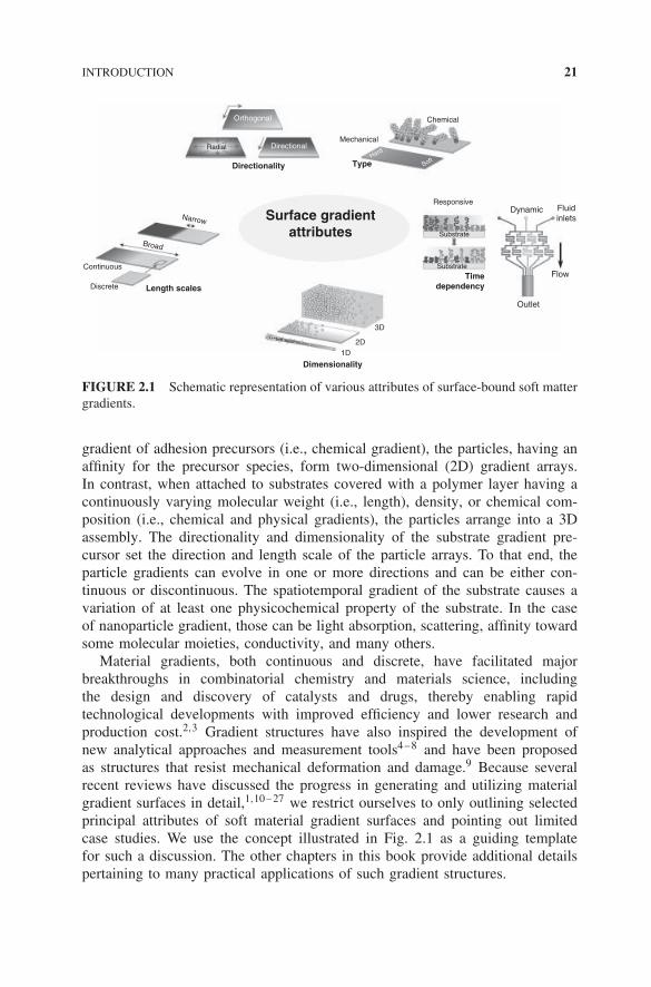
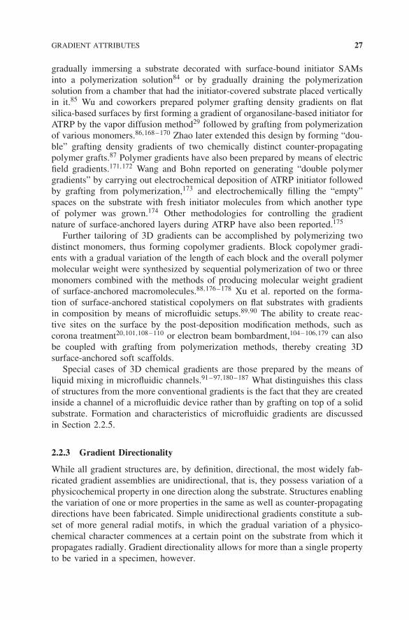
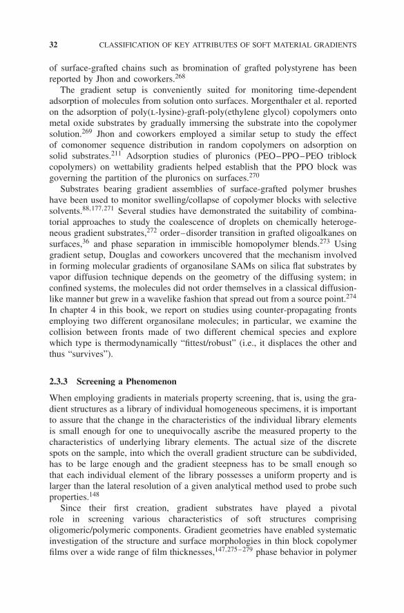
In order to facilitate the discussion, we classify gradient structures based on afew key attributes (Fig. 2.1). We note that any gradient substrate can belong tomore than one category depending on its attributes. For instance, let us considera case involving a gradient comprising assemblies of nanoparticles arranged ina gradual manner on a support. If deposited onto a flat substrate comprising a
INTRODUCTION 21
Orthogonal
Mechanical
Chemical
Responsive
3D
2D
Discrete
Continuous
Broad
Narrow
1D
Dimensionality
Length scales
Directionality Type
Timedependency
Surface gradientattributes
Dynamic
Substrate
Fluidinlets
Flow
Outlet
Substrate
SoftHard
DirectionalRadial
FIGURE 2.1 Schematic representation of various attributes of surface-bound soft mattergradients.
gradient of adhesion precursors (i.e., chemical gradient), the particles, having anaffinity for the precursor species, form two-dimensional (2D) gradient arrays.In contrast, when attached to substrates covered with a polymer layer having acontinuously varying molecular weight (i.e., length), density, or chemical com-position (i.e., chemical and physical gradients), the particles arrange into a 3Dassembly. The directionality and dimensionality of the substrate gradient pre-cursor set the direction and length scale of the particle arrays. To that end, theparticle gradients can evolve in one or more directions and can be either con-tinuous or discontinuous. The spatiotemporal gradient of the substrate causes avariation of at least one physicochemical property of the substrate. In the caseof nanoparticle gradient, those can be light absorption, scattering, affinity towardsome molecular moieties, conductivity, and many others.
Material gradients, both continuous and discrete, have facilitated majorbreakthroughs in combinatorial chemistry and materials science, includingthe design and discovery of catalysts and drugs, thereby enabling rapidtechnological developments with improved efficiency and lower research andproduction cost.2,3 Gradient structures have also inspired the development ofnew analytical approaches and measurement tools4–8 and have been proposedas structures that resist mechanical deformation and damage.9 Because severalrecent reviews have discussed the progress in generating and utilizing materialgradient surfaces in detail,1,10–27 we restrict ourselves to only outlining selectedprincipal attributes of soft material gradient surfaces and pointing out limitedcase studies. We use the concept illustrated in Fig. 2.1 as a guiding templatefor such a discussion. The other chapters in this book provide additional detailspertaining to many practical applications of such gradient structures.
22 CLASSIFICATION OF KEY ATTRIBUTES OF SOFT MATERIAL GRADIENTS
2.2 GRADIENT ATTRIBUTES
Gradients can be classified into many categories depending on their physico-chemical nature. In most instances, the resultant structures exhibit typically morethan just a single attribute identified, as in Fig. 2.1. Since a detailed discussionof the gradient attributes has been presented earlier,1 we restrict ourselves to justa succinct summary.
2.2.1 Gradient Type
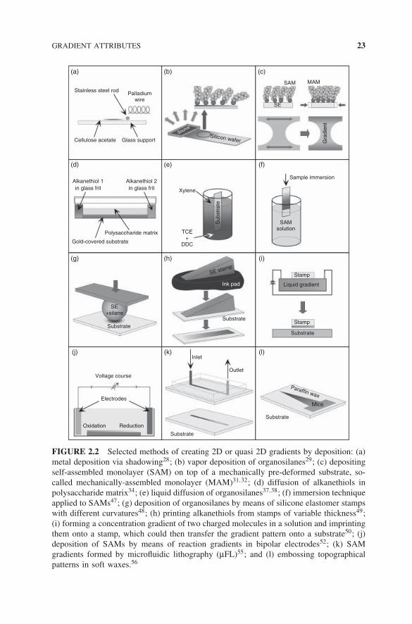
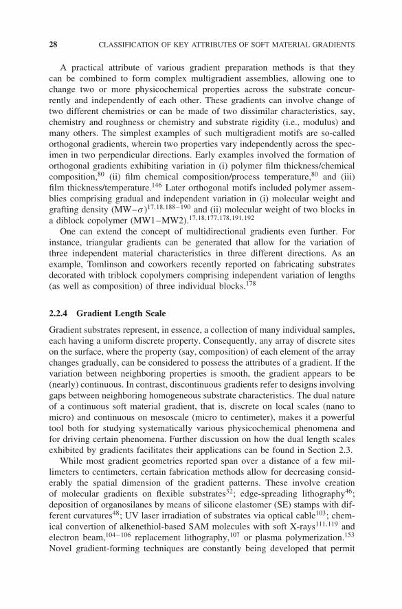
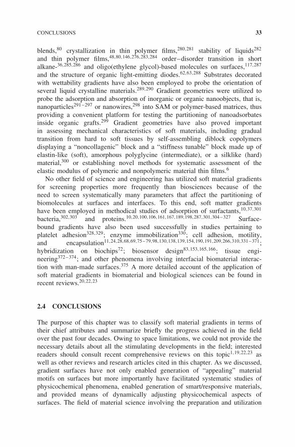
Substrates can be made, which possess gradual variation of any physicochem-ical property. Here we concentrate on chemical, physical, and mechanical gra-dients because they are among the most widely studied and used structures.Figures 2.2–2.4 show a pictorial representation of selected methods that lead tothe formation of such gradient patterns on substrates.
Chemical gradients have been formed by various techniques involving thedirect deposition (see Fig. 2.2 and Fig. 2.3) and post-deposition modificationmethodologies (see Fig. 2.4). Various methods have been developed that uti-lize directed deposition of atomic (i.e., metals) or molecular clusters (i.e., smallsynthetic precursors, peptides, nanoparticles) to fabricate 2D material gradients(Fig. 2.2).28–61 In addition to depositing metals or small organic moieties,62,63
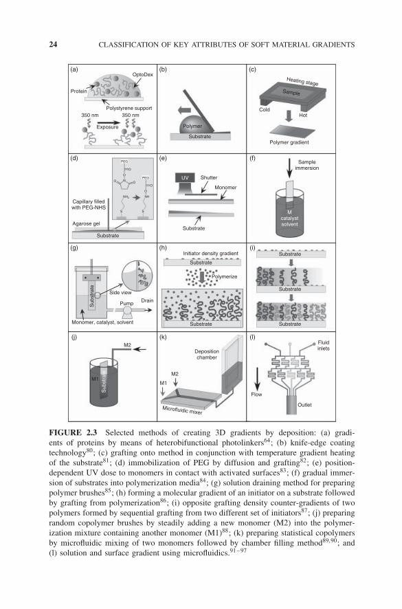
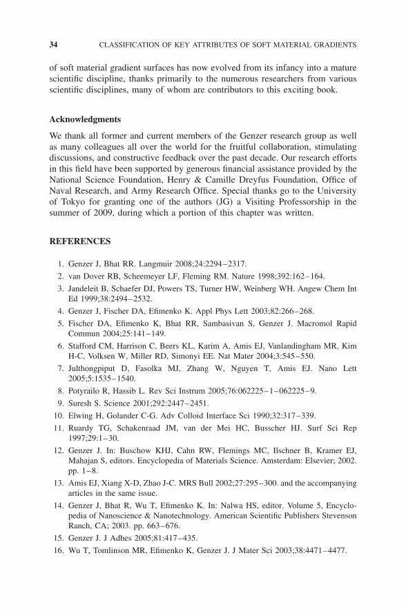
techniques have been introduced that enable the formation of 3D gradient lay-ers (Fig. 2.3) by either (i) laying down larger organic clusters (i.e., proteins) ornanoparticles or (ii) preparing polymer layers. While the former class of methodsleads to the formation of “quasi 3D” soft material structures with a gradual densityvariation across the substrate,64–77 thicker 3D gradients have almost exclusivelybeen formed by coating flat substrates with a relatively thick (a few nanometersto micrometers) polymer layer. In some instances, gradient structures have alsobeen prepared by using external fields78 or pH modulation during material depo-sition on the surface.79 We discuss the formation of 3D gradients in more detailin Section 2.2.2.
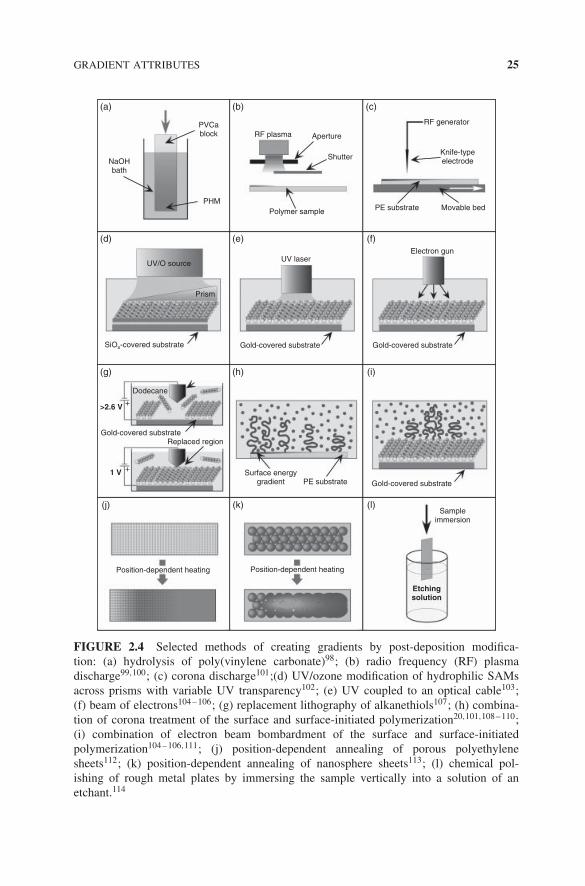
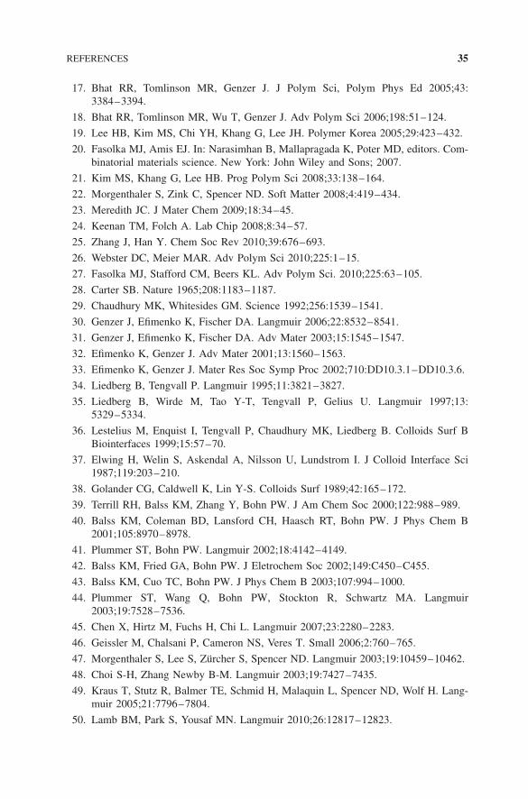
Earlier, we mentioned that gradient structures can also be formed bypost-deposition modification methods (see Fig. 2.4) that encompass exposinghomogeneous substrates to a gradual dose of either a chemical or a physicalmodifier.98–101,108–110,115–117 Other post-deposition modification techniques ofpreparing surface-bound chemical gradients involve preparing self-assembledmonolayer (SAM) films and either (i) altering the chemical functional groups inthe original molecules (typically photooxidation of end groups) to another groupby an external beam102–106,111,118,119 or (ii) selectively removing certain siteson the substrate and filling the empty sites with another molecule.107,120
Physical gradients possess a gradual variation of some physical property;the two most widely explored types of physical gradients involve structuresthat exhibit a gradual variation of substrate rigidity (i.e., Young’s modulus),roughness, or porosity.121 Biologists have long been interested in preparing andutilizing supports with gradients in modulus because it impacts cell motility(so-called durotaxis).122 Ingenious approaches leading to fabrication of such
GRADIENT ATTRIBUTES 23
(a) (b) (c)
(d) (e) (f)
(g) (h) (i)
(j) (k) (l)
Stainless steel rod Palladiumwire
Glass supportSource
Silicon waferCellulose acetate
Alkanethiol 1in glass frit in glass frit
Polysaccharide matrix TCE+
DDC
Xylene
Sample immersion
Stamp
Stamp
Liquid gradient
SAM MAM
SE
Gra
dien
t
Sub
stra
teSAM
solution
Gold-covered substrate
Substrate
Substrate
Substrate
Mica
Substrate
Substrate
Paraffin wax
SE stamp
Ink pad
Outlet
Inlet
Voltage course
Electrodes
Oxidation Reduction
SE+silane
Alkanethiol 2
FIGURE 2.2 Selected methods of creating 2D or quasi 2D gradients by deposition: (a)metal deposition via shadowing28; (b) vapor deposition of organosilanes29; (c) depositingself-assembled monolayer (SAM) on top of a mechanically pre-deformed substrate, so-called mechanically-assembled monolayer (MAM)31,32; (d) diffusion of alkanethiols inpolysaccharide matrix34; (e) liquid diffusion of organosilanes37,38; (f) immersion techniqueapplied to SAMs47; (g) deposition of organosilanes by means of silicone elastomer stampswith different curvatures48; (h) printing alkanethiols from stamps of variable thickness49;(i) forming a concentration gradient of two charged molecules in a solution and imprintingthem onto a stamp, which could then transfer the gradient pattern onto a substrate50; (j)deposition of SAMs by means of reaction gradients in bipolar electrodes52; (k) SAMgradients formed by microfluidic lithography (μFL)55; and (l) embossing topographicalpatterns in soft waxes.56
24 CLASSIFICATION OF KEY ATTRIBUTES OF SOFT MATERIAL GRADIENTS
(a) (b)OptoDex
Protein
Polystyrene support
Exposure
Capillary filledwith PEG-NHS
Agarose gel
Substrate
Substrate
Substrate
Polymerize
Initiator density gradient
Substrate
Substrate
Substrate
Substrate
Substrate
Shutter
Monomer
Sampleimmersion
Polymer gradient
Sample
Heating stage
ColdHot
Polymer
UV
Mcatalystsolvent
Depositionchamber
Microfluidic mixer
Flow
Outlet
Fluidinlets
Side view
DrainPump
Monomer, catalyst, solvent
M2
M1
M2M1
Sub
stra
te
Sub
stra
te
350 nm 350 nm
(c)
(d) (e) (f)
(g) (h) (i)
(j) (k) (l)
FIGURE 2.3 Selected methods of creating 3D gradients by deposition: (a) gradi-ents of proteins by means of heterobifunctional photolinkers64; (b) knife-edge coatingtechnology80; (c) grafting onto method in conjunction with temperature gradient heatingof the substrate81; (d) immobilization of PEG by diffusion and grafting82; (e) position-dependent UV dose to monomers in contact with activated surfaces83; (f) gradual immer-sion of substrates into polymerization media84; (g) solution draining method for preparingpolymer brushes85; (h) forming a molecular gradient of an initiator on a substrate followedby grafting from polymerization86; (i) opposite grafting density counter-gradients of twopolymers formed by sequential grafting from two different set of initiators87; (j) preparingrandom copolymer brushes by steadily adding a new monomer (M2) into the polymer-ization mixture containing another monomer (M1)88; (k) preparing statistical copolymersby microfluidic mixing of two monomers followed by chamber filling method89,90; and(l) solution and surface gradient using microfluidics.91–97
GRADIENT ATTRIBUTES 25
(a) (b)
PVCablock
PHM
NaOHbath
UV/O source
Prism
+
+
Position-dependent heating Position-dependent heating
Etchingsolution
>2.6 V
1 V
SiOx-covered substrate
Dodecane
Gold-covered substrateReplaced region
Surface energygradient PE substrate
Sampleimmersion
Gold-covered substrate
Gold-covered substrate
UV laserElectron gun
Polymer sample
RF plasma Aperture
Shutter
RF generator
Knife-typeelectrode
PE substrate Movable bed
Gold-covered substrate
(c)
(d) (e) (f)
(g) (h) (i)
(j) (k) (l)
FIGURE 2.4 Selected methods of creating gradients by post-deposition modifica-tion: (a) hydrolysis of poly(vinylene carbonate)98; (b) radio frequency (RF) plasmadischarge99,100; (c) corona discharge101;(d) UV/ozone modification of hydrophilic SAMsacross prisms with variable UV transparency102; (e) UV coupled to an optical cable103;(f) beam of electrons104–106; (g) replacement lithography of alkanethiols107; (h) combina-tion of corona treatment of the surface and surface-initiated polymerization20,101,108–110;(i) combination of electron beam bombardment of the surface and surface-initiatedpolymerization104–106,111; (j) position-dependent annealing of porous polyethylenesheets112; (k) position-dependent annealing of nanosphere sheets113; (l) chemical pol-ishing of rough metal plates by immersing the sample vertically into a solution of anetchant.114
26 CLASSIFICATION OF KEY ATTRIBUTES OF SOFT MATERIAL GRADIENTS
unique structures have been developed by Wang et al.,123,124 Wong et al.,125–127
and others.128,129 The utilization of such structures in biological applications isdiscussed later in Section 2.3 of this chapter. Techniques facilitating the fabri-cation of surfaces with position-dependent variation of topography/roughnesshave also been developed that employed (i) selective removal of one chemicalcomponent from the multicomponent chemically modulated surfaces130; (ii)sintering of the resultant porous substrates112 or close-packed sheets ofnanospheres113,131–134 using position-dependent heating; or (iii) “chemicalpolishing” of rough metal plates by immersing the sample vertically into asolution of an etchant,114 (iv) phase separation of immiscible polymer films castonto surface gradient substrates,135 (v) photolithography-assisted molding ofpillars and holes,136 and other methods.137 Such substrates were then used toinvestigate the effect of substrate topography on cell adhesion.138,139
A special type of physicochemical gradient involves the generation of othergradients in liquids that can be transferred onto substrates,140,141 gradients ofpH,142–144 and refractive index.145
2.2.2 Gradient Dimensionality
Section 2.2.1 discussed the methods that lead nearly exclusively to 2D gradi-ents. True 3D soft matter gradient structures can be built by deposition tech-niques, whereby the 3D hierarchy is achieved either in a single step or bysequentially building layers on the substrate. Early work included the applica-tion of the “knife-edge coating” method,80,146–148 which enabled the formationof polymer layer with gradual variation of composition and thickness. Otherefforts included plasma polymerization on surfaces,149–155 electrodeposition,156
and chemical vapor deposition.157 A very popular class of technologies involveschemical grafting of polymer chains to the substrate by employing either theso-called grafting onto or grafting from approaches.158
“Grafting onto” techniques, which are based on anchoring the chemicallyfunctionalized polymer chains onto reactive sites on the substrate, result ingradients in grafting density (i.e., number of polymer chains per unit area) ofpolymer chains, as demonstrated in a series of papers by Luzinov, Minko, andcoworkers,81,159–162 and others.82,163 The “grafting from” methods involve build-ing the polymer layer by carrying out polymerization directly from surface-boundpolymerization initiator centers chemisorbed on the substrate. Numerous variantsof this methodology have been reported.164 For instance, Liedberg and coworkersdeveloped a versatile way of producing gradient assemblies of polymers byexposing activated surfaces to the solution of a monomer and carrying out freeradical polymerization via exposure to ultraviolet (UV) light. They controlledthe length of the grafted chain on the substrate by modulating the UV dosageby means of a movable shutter.83,165–167 Controlled radical polymerizationsfor instance, atom transfer radical polymerization (ATRP), have been employedwidely for generating 3D gradient assemblies. For instance, Tomlinson and Gen-zer reported on gradients in molecular weight of the anchored polymer by either
GRADIENT ATTRIBUTES 27
gradually immersing a substrate decorated with surface-bound initiator SAMsinto a polymerization solution84 or by gradually draining the polymerizationsolution from a chamber that had the initiator-covered substrate placed verticallyin it.85 Wu and coworkers prepared polymer grafting density gradients on flatsilica-based surfaces by first forming a gradient of organosilane-based initiator forATRP by the vapor diffusion method29 followed by grafting from polymerizationof various monomers.86,168–170 Zhao later extended this design by forming “dou-ble” grafting density gradients of two chemically distinct counter-propagatingpolymer grafts.87 Polymer gradients have also been prepared by means of electricfield gradients.171,172 Wang and Bohn reported on generating “double polymergradients” by carrying out electrochemical deposition of ATRP initiator followedby grafting from polymerization,173 and electrochemically filling the “empty”spaces on the substrate with fresh initiator molecules from which another typeof polymer was grown.174 Other methodologies for controlling the gradientnature of surface-anchored layers during ATRP have also been reported.175
Further tailoring of 3D gradients can be accomplished by polymerizing twodistinct monomers, thus forming copolymer gradients. Block copolymer gradi-ents with a gradual variation of the length of each block and the overall polymermolecular weight were synthesized by sequential polymerization of two or threemonomers combined with the methods of producing molecular weight gradientof surface-anchored macromolecules.88,176–178 Xu et al. reported on the forma-tion of surface-anchored statistical copolymers on flat substrates with gradientsin composition by means of microfluidic setups.89,90 The ability to create reac-tive sites on the surface by the post-deposition modification methods, such ascorona treatment20,101,108–110 or electron beam bombardment,104–106,179 can alsobe coupled with grafting from polymerization methods, thereby creating 3Dsurface-anchored soft scaffolds.
Special cases of 3D chemical gradients are those prepared by the means ofliquid mixing in microfluidic channels.91–97,180–187 What distinguishes this classof structures from the more conventional gradients is the fact that they are createdinside a channel of a microfluidic device rather than by grafting on top of a solidsubstrate. Formation and characteristics of microfluidic gradients are discussedin Section 2.2.5.
2.2.3 Gradient Directionality
While all gradient structures are, by definition, directional, the most widely fab-ricated gradient assemblies are unidirectional, that is, they possess variation of aphysicochemical property in one direction along the substrate. Structures enablingthe variation of one or more properties in the same as well as counter-propagatingdirections have been fabricated. Simple unidirectional gradients constitute a sub-set of more general radial motifs, in which the gradual variation of a physico-chemical character commences at a certain point on the substrate from which itpropagates radially. Gradient directionality allows for more than a single propertyto be varied in a specimen, however.
28 CLASSIFICATION OF KEY ATTRIBUTES OF SOFT MATERIAL GRADIENTS
A practical attribute of various gradient preparation methods is that theycan be combined to form complex multigradient assemblies, allowing one tochange two or more physicochemical properties across the substrate concur-rently and independently of each other. These gradients can involve change oftwo different chemistries or can be made of two dissimilar characteristics, say,chemistry and roughness or chemistry and substrate rigidity (i.e., modulus) andmany others. The simplest examples of such multigradient motifs are so-calledorthogonal gradients, wherein two properties vary independently across the spec-imen in two perpendicular directions. Early examples involved the formation oforthogonal gradients exhibiting variation in (i) polymer film thickness/chemicalcomposition,80 (ii) film chemical composition/process temperature,80 and (iii)film thickness/temperature.146 Later orthogonal motifs included polymer assem-blies comprising gradual and independent variation in (i) molecular weight andgrafting density (MW–σ )17,18,188–190 and (ii) molecular weight of two blocks ina diblock copolymer (MW1–MW2).17,18,177,178,191,192
One can extend the concept of multidirectional gradients even further. Forinstance, triangular gradients can be generated that allow for the variation ofthree independent material characteristics in three different directions. As anexample, Tomlinson and coworkers recently reported on fabricating substratesdecorated with triblock copolymers comprising independent variation of lengths(as well as composition) of three individual blocks.178
2.2.4 Gradient Length Scale
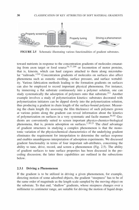
Gradient substrates represent, in essence, a collection of many individual samples,each having a uniform discrete property. Consequently, any array of discrete siteson the surface, where the property (say, composition) of each element of the arraychanges gradually, can be considered to possess the attributes of a gradient. If thevariation between neighboring properties is smooth, the gradient appears to be(nearly) continuous. In contrast, discontinuous gradients refer to designs involvinggaps between neighboring homogeneous substrate characteristics. The dual natureof a continuous soft material gradient, that is, discrete on local scales (nano tomicro) and continuous on mesoscale (micro to centimeter), makes it a powerfultool both for studying systematically various physicochemical phenomena andfor driving certain phenomena. Further discussion on how the dual length scalesexhibited by gradients facilitates their applications can be found in Section 2.3.
While most gradient geometries reported span over a distance of a few mil-limeters to centimeters, certain fabrication methods allow for decreasing consid-erably the spatial dimension of the gradient patterns. These involve creationof molecular gradients on flexible substrates32; edge-spreading lithography46;deposition of organosilanes by means of silicone elastomer (SE) stamps with dif-ferent curvatures48; UV laser irradiation of substrates via optical cable103; chem-ical convertion of alkenethiol-based SAM molecules with soft X-rays111,119 andelectron beam,104–106 replacement lithography,107 or plasma polymerization.153
Novel gradient-forming techniques are constantly being developed that permit

GRADIENT FUNCTIONALITY 29
the generation of chemical and/or physical gradients on nanometer-to-micrometerscale.193,194
2.2.5 Gradient Temporal Dependency
Time dependency constitutes yet another important attribute of gradient sub-strates. Most gradient motifs are static so that their physicochemical propertiescorrespond to the state present at the time of their creation. However, some softmaterial designs are capable of responding to a variation of external stimulus (i.e.,solvent quality, pH, temperature, electric or magnetic field, ion concentration) ormay be varied by adjusting the gradient attributes in real time.
The first category of gradient structures involves polymer assemblies thatalter their properties as a function of solvent quality, pH, charge, or an externalpotential. Ionov and coworkers created responsive polymer surfaces195 compris-ing mixed brushes made of poly(acrylic acid) (PAA) and poly(2-vinyl pyridine)(P2VP),196 whose density changed gradually across the substrate in two oppo-site directions. These mixed brush gradients responded to the variation of pH byswelling the P2VP brushes at low pH and swelling the PAA brushes at high pH.
The second category of dynamic gradients includes structures that are pro-duced by mixing liquids inside a microfluidic device. In these gradients, thetime-dependent variation of the concentration of the various liquids is achievedin a flowing liquid.91 Here, multiple solutions are infused simultaneously into anetwork through various inlets. As the fluid streams travel down the network,they are repeatedly split, mixed, and recombined; and after several generationsof branched systems, each branch contains different proportions of the infusedsolutions. As a result, a gradient is established—perpendicular to the flow—ina single large channel that combines all individual branches of fluids. Multiplestudies demonstrated the versatility of this method in probing numerous biolog-ical phenomena,197–200 as well as in achieving spatial control of surfactants andmicelles in solution.201
A few nontraditional gradient types were created by varying externalfields such as temperature,202–205 pH,206 and electrochemical potential. Forinstance, Isaksson and coworkers recently presented a method for producing awettability gradient by an external voltage.207 Yamada and Tada reported onthe formation of dynamic wettability gradients by first decorating substrateswith ferrocenyl alkanethiols and then applying in-plane gradients in theelectrochemical potential between the ends of the substrate. Reversibility innitrobenzene and dichloromethane drop motion on wettability gradients has alsobeen reported.199,208
2.3 GRADIENT FUNCTIONALITY
The very nature of gradient surfaces makes them functional structures. Chem-ical and physical gradients are responsible for driving many important biolog-ical and physical processes, such as the directed movement of certain bacteria
30 CLASSIFICATION OF KEY ATTRIBUTES OF SOFT MATERIAL GRADIENTS
Driving a phenomenonProperty tuning
Phenomenon recording
Property screening
FIGURE 2.5 Schematic illustrating various functionalities of gradient substrates.
toward nutrients in response to the concentration gradients of molecules emanat-ing from axon target or food source76,77,209 or locomotion of motor proteins,that is, kinesin, which can haul cargoes attached to them along microtubu-lar “railroads.”210 Concentration gradients of molecules on surfaces also affectphenomena such as osmotic swelling, surface pressure, and surface wettabil-ity. Various fabrication methods leading to the formation gradients on surfacescan also be employed to record important physical phenomena. For instance,by immersing a flat substrate continuously into a polymer solution, one canstudy systematically the adsorption of polymers onto that substrate.211 Anotherexample involves a study of polymerization kinetics: substrates decorated withpolymerization initiators can be dipped slowly into the polymerization solution,thus producing a gradient in chain length of the surface-bound polymer. Measur-ing the chain length (by assessing the film thickness) of such polymers grownat various points along the gradient can reveal information about the kineticsof polymerization on surfaces in a very systematic and facile manner.84,85 Gra-dients are conveniently suited to screen important physico-chemico-biologicalphenomena, that is, protein adsorption on surfaces.1,20,23 The chief advantageof gradient structures in studying a complex phenomenon is that the mono-tonic variation of the physicochemical characteristics of the underlying gradienteliminates the requirement for interpolation to determine the surface responseand enables unambiguous interpretation of adsorption experiments. We discussedgradient functionality in terms of four important sub-attributes, concerning theability to tune, drive, record, and screen a phenomenon (Fig. 2.5). The abilityof gradient surfaces to tune surface properties has been discussed in the pre-ceding discussion; the latter three capabilities are outlined in the subsectionsbelow.
2.3.1 Driving a Phenomenon
If the gradient is to be utilized in driving a given phenomenon, for example,directing motion of some adsorbed objects, the gradient “steepness” has to be ofthe same order of magnitude as the length scale sampled by the moving object onthe substrate. To that end, “shallow” gradients, whose steepness changes over amillimeter to centimeter range, are suitable for driving the motion of liquid drops
GRADIENT FUNCTIONALITY 31
while “sharp” gradients have to be employed for studying the mobility of muchsmaller objects. For example, cell locomotion can only be probed with gradientswhose inherent length scale is on the order of a few tens to hundreds of microns.
Traditionally, soft material gradient surfaces have been employed in trans-porting liquids across surfaces by implementing the gradient in the interfacialtension at the front and back edge of the drop acting at the droplet/substrate/airinterface.29 While relatively slow motion was observed in the initial studies,much higher drop speeds have been observed for small water droplets formed bycondensation of steam onto a gradient surface,212 by vibrating the surface,213,214
by rolling the drop on a rough substrate decorated with a chemical gradient madeof hydrophobic organosilane modifiers,215 or on wettability gradients prepared byembossing topographical patterns in soft waxes.56 A large body of work pertain-ing to probing the liquid motion due to “static gradients” as well as “dynamicgradients” has been presented recently.202–205,207,216–236 It is important to notethat chemical reactions on gradient surfaces can also govern the movement oflarger molecules, that is, dendrimers237 or nanoparticles.238
Surface-bound gradients have been employed as “directional engines”capable of driving synthetic239 and biological macromolecules along surfaces.Over the past few decades, multiple experimental and theoretical studies havebeen published that reported on the response of living cells (orientation andmigration)240–246 to the variation of chemistry (chemotaxis, haptotaxis),28,247–249
light intensity (phototaxis),250 electrostatic potential (galvanotaxis),251,252 gravi-tational field (geotaxis),253 mechanical properties (durotaxis),123–128 as well assubstrate topography,254,255 or concurrent combination of several cues.256 Manyof those studies employed either static or dynamic gradients in physicochemicalproperties or micropatterned arrays of asymmetric regions of sticky groupson the substrate257 that governed the locomotion of cells,258–262 kinesin oractin,161,263–265 and axon growth.266
2.3.2 Recording a Phenomenon
Processes leading to the formation of soft material gradients on surfaces can beperceived as a convenient means of “recording” time-dependent physicochem-ical phenomena. The resultant gradient substrate then constitutes a convenientrecoding medium, which further facilitates detailed and expedient “post-process”analytical platform.
Tomlinson and coworkers used the gradient geometry to study the kineticsof the surface-initiated controlled radical polymerization of poly(methylmethacrylate)84,85; they confirmed that the reaction obeyed the predicteddependence on the concentration of the activator and deactivator species in thepolymerization mixture and established the “living” nature of the macroinitiatorin surface-initiated ATRP. Shovsky and Schonherr employed gradient geometryto monitor reaction kinetics, rate constants, and activation energies anddetermined entropies associated with the surface reactions of alkaline hydrolysisof model SAMs on surfaces.267 The kinetics of post-polymerization reaction
32 CLASSIFICATION OF KEY ATTRIBUTES OF SOFT MATERIAL GRADIENTS
of surface-grafted chains such as bromination of grafted polystyrene has beenreported by Jhon and coworkers.268
The gradient setup is conveniently suited for monitoring time-dependentadsorption of molecules from solution onto surfaces. Morgenthaler et al. reportedon the adsorption of poly(l-lysine)-graft-poly(ethylene glycol) copolymers ontometal oxide substrates by gradually immersing the substrate into the copolymersolution.269 Jhon and coworkers employed a similar setup to study the effectof comonomer sequence distribution in random copolymers on adsorption onsolid substrates.211 Adsorption studies of pluronics (PEO–PPO–PEO triblockcopolymers) on wettability gradients helped establish that the PPO block wasgoverning the partition of the pluronics on surfaces.270
Substrates bearing gradient assemblies of surface-grafted polymer brusheshave been used to monitor swelling/collapse of copolymer blocks with selectivesolvents.88,177,271 Several studies have demonstrated the suitability of combina-torial approaches to study the coalescence of droplets on chemically heteroge-neous gradient substrates,272 order–disorder transition in grafted oligoalkanes onsurfaces,36 and phase separation in immiscible homopolymer blends.273 Usinggradient setup, Douglas and coworkers uncovered that the mechanism involvedin forming molecular gradients of organosilane SAMs on silica flat substrates byvapor diffusion technique depends on the geometry of the diffusing system; inconfined systems, the molecules did not order themselves in a classical diffusion-like manner but grew in a wavelike fashion that spread out from a source point.274
In chapter 4 in this book, we report on studies using counter-propagating frontsemploying two different organosilane molecules; in particular, we examine thecollision between fronts made of two different chemical species and explorewhich type is thermodynamically “fittest/robust” (i.e., it displaces the other andthus “survives”).
2.3.3 Screening a Phenomenon
When employing gradients in materials property screening, that is, using the gra-dient structures as a library of individual homogeneous specimens, it is importantto assure that the change in the characteristics of the individual library elementsis small enough for one to unequivocally ascribe the measured property to thecharacteristics of underlying library elements. The actual size of the discretespots on the sample, into which the overall gradient structure can be subdivided,has to be large enough and the gradient steepness has to be small enough sothat each individual element of the library possesses a uniform property and islarger than the lateral resolution of a given analytical method used to probe suchproperties.148
Since their first creation, gradient substrates have played a pivotalrole in screening various characteristics of soft structures comprisingoligomeric/polymeric components. Gradient geometries have enabled systematicinvestigation of the structure and surface morphologies in thin block copolymerfilms over a wide range of film thicknesses,147,275–279 phase behavior in polymer
CONCLUSIONS 33
blends,80 crystallization in thin polymer films,280,281 stability of liquids282
and thin polymer films,48,80,146,276,283,284 order–disorder transition in shortalkane-36,285,286 and oligo(ethylene glycol)-based molecules on surfaces,117,287
and the structure of organic light-emitting diodes.62,63,288 Substrates decoratedwith wettability gradients have also been employed to probe the orientation ofseveral liquid crystalline materials.289,290 Gradient geometries were utilized toprobe the adsorption and absorption of inorganic or organic nanoobjects, that is,nanoparticles291–297 or nanowires,298 into SAM or polymer-based matrices, thusproviding a convenient platform for testing the partitioning of nanoadsorbatesinside organic grafts.299 Gradient geometries have also proved importantin assessing mechanical characteristics of soft materials, including gradualtransition from hard to soft tissues by self-assembling diblock copolymersdisplaying a “noncollagenic” block and a “stiffness tunable” block made up ofelastin-like (soft), amorphous polyglycine (intermediate), or a silklike (hard)material,300 or establishing novel methods for systematic assessment of theelastic modulus of polymeric and nonpolymeric material thin films.6
No other field of science and engineering has utilized soft material gradientsfor screening properties more frequently than biosciences because of theneed to screen systematically many parameters that affect the partitioning ofbiomolecules at surfaces and interfaces. To this end, soft matter gradientshave been employed in methodical studies of adsorption of surfactants,10,37,301
bacteria,302,303 and proteins.10,20,100,106,161,167,189,198,287,301,304–327 Surface-bound gradients have also been used successfully in studies pertaining toplatelet adhesion328,329; enzyme immobilization330; cell adhesion, motility,and encapsulation11,24,28,68,69,75–79,98,130,138,139,154,190,191,209,266,310,331–371;hybridization on biochips72; biosensor design83,153,165,166; tissue engi-neering372–374; and other phenomena involving interfacial biomaterial interac-tion with man-made surfaces.375 A more detailed account of the application ofsoft material gradients in biomaterial and biological sciences can be found inrecent reviews.20,22,23
2.4 CONCLUSIONS
The purpose of this chapter was to classify soft material gradients in terms oftheir chief attributes and summarize briefly the progress achieved in the fieldover the past four decades. Owing to space limitations, we could not provide thenecessary details about all the stimulating developments in the field; interestedreaders should consult recent comprehensive reviews on this topic1,19,22,23 aswell as other reviews and research articles cited in this chapter. As we discussed,gradient surfaces have not only enabled generation of “appealing” materialmotifs on surfaces but more importantly have facilitated systematic studies ofphysicochemical phenomena, enabled generation of smart/responsive materials,and provided means of dynamically adjusting physicochemical aspects ofsurfaces. The field of material science involving the preparation and utilization
34 CLASSIFICATION OF KEY ATTRIBUTES OF SOFT MATERIAL GRADIENTS
of soft material gradient surfaces has now evolved from its infancy into a maturescientific discipline, thanks primarily to the numerous researchers from variousscientific disciplines, many of whom are contributors to this exciting book.
Acknowledgments
We thank all former and current members of the Genzer research group as wellas many colleagues all over the world for the fruitful collaboration, stimulatingdiscussions, and constructive feedback over the past decade. Our research effortsin this field have been supported by generous financial assistance provided by theNational Science Foundation, Henry & Camille Dreyfus Foundation, Office ofNaval Research, and Army Research Office. Special thanks go to the Universityof Tokyo for granting one of the authors (JG) a Visiting Professorship in thesummer of 2009, during which a portion of this chapter was written.
REFERENCES
1. Genzer J, Bhat RR. Langmuir 2008;24:2294–2317.
2. van Dover RB, Scheemeyer LF, Fleming RM. Nature 1998;392:162–164.
3. Jandeleit B, Schaefer DJ, Powers TS, Turner HW, Weinberg WH. Angew Chem IntEd 1999;38:2494–2532.
4. Genzer J, Fischer DA, Efimenko K. Appl Phys Lett 2003;82:266–268.
5. Fischer DA, Efimenko K, Bhat RR, Sambasivan S, Genzer J. Macromol RapidCommun 2004;25:141–149.
6. Stafford CM, Harrison C, Beers KL, Karim A, Amis EJ, Vanlandingham MR, KimH-C, Volksen W, Miller RD, Simonyi EE. Nat Mater 2004;3:545–550.
7. Julthongpiput D, Fasolka MJ, Zhang W, Nguyen T, Amis EJ. Nano Lett2005;5:1535–1540.
8. Potyrailo R, Hassib L. Rev Sci Instrum 2005;76:062225–1–062225–9.
9. Suresh S. Science 2001;292:2447–2451.
10. Elwing H, Golander C-G. Adv Colloid Interface Sci 1990;32:317–339.
11. Ruardy TG, Schakenraad JM, van der Mei HC, Busscher HJ. Surf Sci Rep1997;29:1–30.
12. Genzer J. In: Buschow KHJ, Cahn RW, Flemings MC, Ilschner B, Kramer EJ,Mahajan S, editors. Encyclopedia of Materials Science. Amsterdam: Elsevier; 2002.pp. 1–8.
13. Amis EJ, Xiang X-D, Zhao J-C. MRS Bull 2002;27:295–300. and the accompanyingarticles in the same issue.
14. Genzer J, Bhat R, Wu T, Efimenko K. In: Nalwa HS, editor. Volume 5, Encyclo-pedia of Nanoscience & Nanotechnology. American Scientific Publishers StevensonRanch, CA; 2003. pp. 663–676.
15. Genzer J. J Adhes 2005;81:417–435.
16. Wu T, Tomlinson MR, Efimenko K, Genzer J. J Mater Sci 2003;38:4471–4477.
REFERENCES 35
17. Bhat RR, Tomlinson MR, Genzer J. J Polym Sci, Polym Phys Ed 2005;43:3384–3394.
18. Bhat RR, Tomlinson MR, Wu T, Genzer J. Adv Polym Sci 2006;198:51–124.
19. Lee HB, Kim MS, Chi YH, Khang G, Lee JH. Polymer Korea 2005;29:423–432.
20. Fasolka MJ, Amis EJ. In: Narasimhan B, Mallapragada K, Poter MD, editors. Com-binatorial materials science. New York: John Wiley and Sons; 2007.
21. Kim MS, Khang G, Lee HB. Prog Polym Sci 2008;33:138–164.
22. Morgenthaler S, Zink C, Spencer ND. Soft Matter 2008;4:419–434.
23. Meredith JC. J Mater Chem 2009;18:34–45.
24. Keenan TM, Folch A. Lab Chip 2008;8:34–57.
25. Zhang J, Han Y. Chem Soc Rev 2010;39:676–693.
26. Webster DC, Meier MAR. Adv Polym Sci 2010;225:1–15.
27. Fasolka MJ, Stafford CM, Beers KL. Adv Polym Sci. 2010;225:63–105.
28. Carter SB. Nature 1965;208:1183–1187.
29. Chaudhury MK, Whitesides GM. Science 1992;256:1539–1541.
30. Genzer J, Efimenko K, Fischer DA. Langmuir 2006;22:8532–8541.
31. Genzer J, Efimenko K, Fischer DA. Adv Mater 2003;15:1545–1547.
32. Efimenko K, Genzer J. Adv Mater 2001;13:1560–1563.
33. Efimenko K, Genzer J. Mater Res Soc Symp Proc 2002;710:DD10.3.1–DD10.3.6.
34. Liedberg B, Tengvall P. Langmuir 1995;11:3821–3827.
35. Liedberg B, Wirde M, Tao Y-T, Tengvall P, Gelius U. Langmuir 1997;13:5329–5334.
36. Lestelius M, Enquist I, Tengvall P, Chaudhury MK, Liedberg B. Colloids Surf BBiointerfaces 1999;15:57–70.
37. Elwing H, Welin S, Askendal A, Nilsson U, Lundstrom I. J Colloid Interface Sci1987;119:203–210.
38. Golander CG, Caldwell K, Lin Y-S. Colloids Surf 1989;42:165–172.
39. Terrill RH, Balss KM, Zhang Y, Bohn PW. J Am Chem Soc 2000;122:988–989.
40. Balss KM, Coleman BD, Lansford CH, Haasch RT, Bohn PW. J Phys Chem B2001;105:8970–8978.
41. Plummer ST, Bohn PW. Langmuir 2002;18:4142–4149.
42. Balss KM, Fried GA, Bohn PW. J Eletrochem Soc 2002;149:C450–C455.
43. Balss KM, Cuo TC, Bohn PW. J Phys Chem B 2003;107:994–1000.
44. Plummer ST, Wang Q, Bohn PW, Stockton R, Schwartz MA. Langmuir2003;19:7528–7536.
45. Chen X, Hirtz M, Fuchs H, Chi L. Langmuir 2007;23:2280–2283.
46. Geissler M, Chalsani P, Cameron NS, Veres T. Small 2006;2:760–765.
47. Morgenthaler S, Lee S, Zurcher S, Spencer ND. Langmuir 2003;19:10459–10462.
48. Choi S-H, Zhang Newby B-M. Langmuir 2003;19:7427–7435.
49. Kraus T, Stutz R, Balmer TE, Schmid H, Malaquin L, Spencer ND, Wolf H. Lang-muir 2005;21:7796–7804.
50. Lamb BM, Park S, Yousaf MN. Langmuir 2010;26:12817–12823.
36 CLASSIFICATION OF KEY ATTRIBUTES OF SOFT MATERIAL GRADIENTS
51. Venkateswar RA, Branch DW, Wheeler BC. Biomed Microdevices 2000;2:255–264.
52. Ulrich C, Andersson O, Nyholm L, Bjorefors F. Anal Chem 2009;81:453–459.
53. Yu X, Wang Z, Kiang Y, Zhang X. Langmuir 2006;22:4483–4486.
54. Han JT, Kim S, Karim A. Langmuir 2007;23:2608–2614.
55. Lamb BM, Westcott NP, Yousaf MN. ChemBioChem 2008;9:2628–2632.
56. Zhang J, Han Y. Langmuir 2007;23:6136–6141.
57. Venkataraman NV, Zurcher S, Rossi A, Lee S, Naujoks N, Spencer ND. J PhysChem C 2009;113:5620–5628.
58. Morf P, Ballav N, Nolting F, von Wrochem F, Nothofer H-G, Yasuda A, WesselsJM, Jung TA. ChemPhysChem 2009;10:2212–2216.
59. Ye F, Cui C, Kirkeminde A, Dong D, Collinson MM, Higgins DA. Chem Mater2010;22:2970–2977.
60. Bauer E, Venkataraman NV, Rossi A, Bauchman F, Engeli R, Spencer ND. Langmuir2010;26:8392–8399.
61. Acharya AP, Dolgova NV, Moore NM, Xia C-Q, Clare-Salzler MJ, Becker ML,Gallant ND, Keselowsky BG. Biomaterials 2010;31:7444–7454.
62. Schmitz C, Thelakkat M, Schmidt HW. Adv Mater 1999;11:821–826.
63. Schmitz C, Posch P, Thelakkat M, Schmidt HW. Phys Chem Chem Phys1999;1:1777–1781.
64. Hypolite CL, McLernon TL, Adams DN, Chapman KE, Herbert CB, Huang CC,Distefano MD, Hu W-S. Bioconjug Chem 1997;8:658–663.
65. Caelen I, Gao H, Sigrist H. Langmuir 2002;18:2463–2467.
66. Kipper MJ, Kleinma HK, Wang FW. Anal Biochem 2007;363:175–184.
67. Kramer S, Xie H, Gaff J, Williamson JR, Tkachenko AG, Nouri N, Feldheim DA,Feldheim DL. J Am Chem Soc 2004;126:5388–5395.
68. Kang CE, Gemeinhart EJ, Gemeinhart RA. J Biomed Mater Res A2004;71:403–411.
69. Dillmore WS, Yousaf MN, Mrksich M. Langmuir 2004; 20:7223–7231.
70. Bhangale SM, Tjong V, Wu L, Yakovlev N, Moran PM. Adv Mater2005;17:809–813.
71. Liu H, Xu J, Li Y, Li B, Ma J, Zhang X. Macromol Rapid Commun 2006;27:1603–1607.
72. Park SH, Krull U. Anal Chim Acta 2006;564:133–140.
73. Wang Q, Bohn PW. Thin Solid Films 2006;513:338–346.
74. Gallant ND, Lavery CA, Amis EJ, Becker ML. Adv Mater 2007;19:965–969.
75. Brandley BL, Weisz OA, Schnaar RL. J Biol Chem 1987;262:6431–6437.
76. Baier H, Bohnoeffer F. Science 1992;255:472–475.
77. Rosentreter S, Davenport RW, Loschinger J, Huf J, Jung J, Bohnoeffer R. J Neuro-biol 1998;37:541–562.
78. Bronstein LM, Ivanovskaya A, Mates T, Holten-Andersen N, Stucky GD. J PhysChem B 2009;113:647–655.
79. Tauk L, Schroder AP, Decher G, Guiseppone N. Nat Chem 2009;1:649–656.
80. Meredith JC, Karim A, Amis EJ. Macromolecules 2000;33:5760–5762.
REFERENCES 37
81. Ionov L, Zdyrko B, Sidorenko A, Minko S, Klep V, Luzinov I, Stamm M. MacromolRapid Commun 2004;25:360–365.
82. Mougin K, Ham AS, Lawrence MB, Fernandez EJ, Hillier AC. Langmuir2005;21:4809–4812.
83. Larsson A, Erblad T, Andersson O, Liedberg B. Biomacromolecules 2007;8:287–295.
84. Tomlinson MR, Efimenko K, Genzer J. Macromolecules 2006;39:9049–9056.
85. Tomlinson MR, Genzer J. Macromolecules 2003;36:3449–3451.
86. Wu T, Efimenko K, Genzer J. J Am Chem Soc 2002;124:9394–9395.
87. Zhao B. Langmuir 2004;20:11748–11755.
88. Xu C, Wu T, Batteas JD, Drain CM, Beers KL, Fasolka MJ. Appl Surf Sci2006;252:2529–2534.
89. Xu C, Wu T, Mei Y, Drain CM, Batteas JD, Beers KL. Langmuir 2005;21:11136–11140.
90. Xu C, Barnes SE, Wu T, Fischer DA, DeLongchamp DM, Batteas JD, Beers KL.Adv Mater 2006;18:1427–1430.
91. Jeon NL, Dertinger SKW, Chiu DT, Choi IS, Stroock A, Whitesides GM. Langmuir2000;16:8311–8316.
92. Caelen I, Bernard A, Juncker D, Michel B, Heinzelmann H, Delamarche E. Langmuir2000;16:9125–9130.
93. Dertinger SKW, Chiu DT, Jeon NL, Whitesides GM. Anal Chem 2001;73:1240–1246.
94. Dertinger SKW, Jiang XY, Li ZY, Murthy VN, Whitesides GM. Proc Natl AcadSci USA 2002;99:12542–12547.
95. Fosser KA, Nuzzo RG. Anal Chem 2003;75:5775–5782.
96. Rhoads DS, Nadkarni SM, Song L, Voeltz C, Bodenschatz E, Guan J-L. In: Guan J-L, editor. Volume 294, Methods in molecular biology, cell migration: developmentalmethods and protocols. Totowa (NJ): Humana Press, Inc.; 2004.
97. Jiang X, Xu Q, Dertinger SKW, Stroock AD, Fu T-M, Whitesides GM. Anal Chem2005;77:2338–2347.
98. Ueda-Yukoshi T, Matsuda T. Langmuir 1995;11:4135–4140.
99. Pitt WG. J Colloid Interface Sci 1989;133:223–227.
100. Spijker HT, Bos R, van Oeveren W, de Vries J, Busscher HJ. Colloids Surf BBiointerfaces 1999;15:89–97.
101. Lee JH, Kim HG, Khang GS, Lee HB, Jhon MS. J Colloid Interface Sci1992;151:563–570.
102. Roberson S, Sehgal A, Fahley A, Karim A. Appl Surf Sci 2003;203–204:855–858.
103. Burgos P, Geoghegan M, Leggett GJ. Nano Lett 2007;7:3747–3752.
104. Steenackers M, Kuller A, Balav N, Zharnikov M, Grunze M, Jordan R. Small2007;3:1764–1773.
105. Schilp S, Balav N, Zharnikov M. Angew Chem Int Ed 2008;47:6786–6789.
106. Winkler T, Balav N, Thomas H, Zharnikov M, Terfort A. Angew Chem Int Ed2008;47:7238–7241.
107. Fuierer RR, Carroll L, Feldheim DL, Gorman CB. Adv Mater 2002;14:154–157.
38 CLASSIFICATION OF KEY ATTRIBUTES OF SOFT MATERIAL GRADIENTS
108. Lee JH, Kim HW, Pak PK, Lee HB. J Polym Sci, Part A: Polym Chem1994;32:1569–1579.
109. Kim MS, Cho YH, Lee SY, Khang G, Lee TG, Moon DW, Lee HB. Chem Lett2006;35:728–729.
110. Lee TG, Shon HK, Kim MS, Lee HB, Moon DW. Appl Surf Sci 2006;252:6754–6756.
111. Ballav N, Shaporenko A, Terfort A, Zharnikov M. Adv Mater 2007;19:998–1000.112. Lu X, Zhang J, Zhang C, Han Y. Macromol Rapid Commun 2005;26:637–642.113. Zhang J, Xue L, Han Y. Langmuir 2005;21:5–8.114. Kunzler TP, Drobek T, Sprecher CM, Schuler M, Spencer ND. Appl Surf Sci
2006;253:2148–2153.115. Wijesundara MB, Fuoco E, Hanley L. Langmuir 2001;17:5721–5726.116. Kim HG, Lee JH, Lee HB, Jhon MS. J Colloid Interface Sci 1993;157:82–87.117. Jeong BJ, Lee JH, Lee HB. J Colloid Interface Sci 1996;178:757–763.118. Ito Y, Heydari M, Hashimoto A, Cono T, Hirasawa A, Hori S, Kurita K, Nakajima
A. Langmuir 2007;23:1845–1850.119. Klauser R, Chen C-H, Huang M-L, Wang S-C, Chuang TJ, Zharnikov M. J Electron
Spectrosc Relat Phenom 2005;144–147:393–396.120. Blondiaux N, Zurcher S, Liley M, Spencer MD. Langmuir 2007;23:3489–3494.121. Oh SH, Kim TH, Im GI, Lee JH. Biomacromolecules 2010;11:1948–1955.122. Pelham RJ Jr, Wang Y-L. Proc Natl Acad Sci USA 1997;94:13661–13665.123. Lo C-M, Wang H-B, Dembo M, Wang Y-L. Biophys J 2000;79:144–152.124. Frey MT, Wang Y-L. Soft Matter 2009;5:1918–1924.125. Wong JY, Velasco A, Rajagopalan P, Pham Q. Langmuir 2003;19:1908–1913.126. Zaari N, Rajagopalan P, Kim SK, Engler AJ, Wong JY. Adv Mater 2004;16:
2133–2137.127. Isenberg BC, DiMilla PA, Walker M, Kim S, Wong JY. Biophys J 2009;97:
1313–1322.128. Kloxin AM, Benton JA, Anseth KS. Biomaterials 2010;31:1–8.129. Guvendiren M, Burdick JA, Yang S. Soft Matter 2010;6:2044–2049.130. Tsai IY, Kimura M, Russell TP. Langmuir 2004;20:5952–5957.131. Zhang J, Xue L, Han Y. Langmuir 2005;21:5667–5671.132. Li J, Han Y. Langmuir 2006;22:1885–1890.133. Huwiler C, Kunzler TP, Textor M, Voros J, Spencer ND. Langmuir 2007;23:
5929–5935.134. Zhang S, You B, Gu G, Wu L. Polymer 2009;50:6235–6244.135. Blondiaux N, Morgenthaler S, Pugin R, Spencer ND, Liley M. Appl Surf Sci
2008;254:6820–6825.136. Spori D, Drobek T, Zurcher S, Spener ND. Langmuir 2010;26:9465–9473.137. Li X, Dai H, Tan S, Zhang X, Liu H, Wang Y, Zhao N, Xu J. J Colloid Interface
Sci 2009;340:93–97.138. Kunzler TP, Drobek T, Schuler M, Spencer ND. Biomaterials 2007;28:2175–2182.139. Kunzler TP, Huwiler C, Drobek T, Voros J, Spencer ND. Biomaterials
2007;33:5000–5006.
REFERENCES 39
140. Domingo A. Anal Biochem 1990;190:88–90.
141. Shearer G Jr. Anal Biochem 1994;221:397–400.
142. Ogawa K, Kokufuta E. Langmuir 2002;18:5661–5667.
143. May EL, Hillier AC. Anal Chem 2005;21:6487–6493.
144. Jayaraman S, May EL, Hillier AC. Langmuir 2006;22:10322–10328.
145. Dobashi T, Nobe M, Yoshihara H, Yamamoto T, Cono A. Langmuir 2004;20:6530–6534.
146. Meredith JC, Smith AP, Karim A, Amis EJ. Macromolecules 2000;33:9747–9756.
147. Smith AP, Douglas J, Meredith JC, Karim A, Amis EJ. Phys Rev Lett 2001;87:15503–1–15503–4.
148. Meredith JC, Karim A, Amis EJ. MRS Bull 2002;27:330–335.
149. Ogumi Z, Abe T, Nakamura S, Inaba M. Solid State Ionics 1999;121:289–293.
150. Whittle JD, Barton D, Alexander MR, Short RD. Chem Commun 2003;(14):1766–1767.
151. Alexander MR, Whittle JD, Barton D, Short RD. J Mater Chem 2004;14:408–412.
152. Parry KL, Shard AG, Short RD, White RG, Whittle JD, Wright A. Surf InterfaceAnal 2006;38:1497–1504.
153. Walker RA, Cunliffe VT, Whittle JD, Steele DA, Short RD. Langmuir 2009;25:4243–4246.
154. Wells N, Baxter MA, Turnbull JE, Murray P, Edgar D, Parry KL, Steele DA, ShortRD. Biomaterials 2009;30:1066–1070.
155. Zetler M, Scurr D, Abdullah B, Urquhart AJ, Gadegaard N, Bradley JW, AlexanderMR. J Phys Chem B 2009;113:8487–8494.
156. Sehayek T, Meisel D, Vaskevich A, Rubinstein I. Isr J Chem 2008;48:359–366.
157. Elkasabi Y, Lahann J. Macromol Rapid Commun 2009;30:57–63.
158. Brittain W, Advincula R, Ruhe J, Caster K, editors. Polymer brushes. New York:John Wiley and Sons; 2004.
159. Ionov L, Sidorenko A, Stamm M, Minko S, Zdyrko B, Klep V, Luzinov I. Macro-molecules 2004;37:7421–7423.
160. Ionov L, Sidorenko A, Eichhorn K-J, Stamm M, Minko S, Hinrichs K. Langmuir2005;21:8711–8716.
161. Ionov L, Stamm M, Diez S. Nano Lett 2005;5:1910–1914.
162. Liu Y, Klep V, Zdyrko B, Luzinov I. Langmuir 2005;21:11806–11813.
163. Halfter W. J Neurosci 1996;16:4389–4401.
164. Enright TP, Hagaman D, Kokorus M, Coleman N, Sidorenko A. J Polym Sci, PartB: Polym Phys 2010;48:1616–1622.
165. Larsson A, Du C-X, Liedberg B. Biomacromolecules 2007;8:3511–3518.
166. Larsson A, Liedberg B. Langmuir 2007;23:11319–11325.
167. Ekblad T, Andersson O, Tai F-I, Ederth T, Liedberg B. Langmuir 2009;25:3755–3762.
168. Wu T, Efimenko K, Vlcek P, Subr V, Genzer J. Macromolecules 2003;36:2448–2453.
40 CLASSIFICATION OF KEY ATTRIBUTES OF SOFT MATERIAL GRADIENTS
169. Wu T, Genzer J, Gong P, Szleifer I, Vlcek P, Subr V. In: Brittain W, Advincula R,Ruhe J, Caster K, editors. Polymer brushes. New York: John Wiley and Sons; 2004.pp. 287–316.
170. Wu T, Gong P, Szleifer I, Vlcek P, Subr V, Genzer J. Macromolecules 2007;40:8756–8764.
171. Wang X, Bohn PW. J Am Chem Soc 2004;126:6825–6832.
172. Ratcliff E, Hillier AC. Langmuir 2007;23:9905–9910.
173. Wang X, Tu H, Braun PV, Bohn PW. Langmuir 2006;22:817–823.
174. Wang X, Bohn PW. Adv Mater 2007;19:515–520.
175. Jiang D, Huang X, Qiu F, Luo C, Huang LL. Macromolecules 2010;43:71–76.
176. Tomlinson MR, Genzer J. Chem Commun 2003;(12):1350–1351.
177. Tomlinson MR, Genzer J. Langmuir 2005;21:11552–11555.
178. Tomlinson MR, Genzer J. Polymer 2008;49:4837–4845.
179. Steenackers M, Kuller A, Stoycheva S, Grunze M, Jordan R. Langmuir 2009;25:2225–2231.
180. Sun K, Wang Z, Jiang X. Lab Chip 2008;8:1536–1543.
181. Kang T, Han J, Lee KS. Lab Chip 2008;8:1220–1222.
182. Cooksey GA, Sip CG, Folch A. Lab Chip 2009;9:417–426.
183. Lee K, Kim C, Ahn B, Panchapakesan R, Full AR, Nordee L, Kang JY, Oh KW.Lab Chip 2009;9:709–717.
184. Du Y, Shim J, Vidula M, Hancock MJ, Lo E, Chung BG, Borenstein JT, KhabiryM, Cropek DM, Khademhosseini A. Lab Chip 2009;9:761–767.
185. Atencia J, Morrow J, Locascio LE. Lab Chip 2009;9:2707–2714.
186. Du Y, Hancock MJ, He J, Villa-Uribe JL, Wang B, Cropek DM, KhademhosseiniA. Biomaterials 2010;31:2686–2694.
187. Ahmed T, Shimizu TS, Stocker R. Nano Lett 2010;10:3379–3385.
188. Bhat RR, Tomlinson MR, Genzer J. Macromol Rapid Commun 2004;25:270–274.
189. Bhat RR, Genzer J. Mater Res Soc Symp Proc 2004;804:JJ.5.8.1–JJ.5.8.9.
190. Bhat RR, Chaney BN, Rowley J, Liebmann-Vinson A, Genzer J. Adv Mater2005;17:2802–2807.
191. Khire VS, Benoit DWS, Anseth KS, Bowman CN. J Polym Sci, Part A: PolymChem 2006;44:7027–7039.
192. Harris BP, Metters AT. Macromolecules 2006;39:2764–2772.
193. Schuh C, Santer S, Prucker O, Ruhe J. Adv Mater 2009;21:4706–4710.
194. Kumar TA, Bardea A, Shai Y, Yoffe A, Naaman R. Nano Lett 2010;10:2262–2267.
195. Minko S, editor. Responsive polymer materials, design and applications. BlackwellPublishing, Oxford 2006.
196. Ionov L, Houbenov N, Sidorenko A, Stamm M, Luzinov I, Minko S. Langmuir2004;20:9916–9919.
197. Paliwal S, Iglesias PA, Campbell K, Hilioti Z, Groisman A, Levchenko A. Nature2007;446:46–51.
198. Gunawan RC, Choban ER, Concour JE, Silverstre J, Schook LB, Gaskins HR,Leckband DE, Kenis PJA. Langmuir 2005;21:3061–3068.
REFERENCES 41
199. Gunawan RC, Silvestre J, Gaskins HR, Kenis PJA, Leckband DE. Langmuir2006;22:4250–4258.
200. Koster S, Leach JB, Struth B, Pfohl T, Wong JY. Langmuir 2007;23:357–359.
201. Liu X, Abbott NL. Anal Chem 2009;81:772–781.
202. Brochard F. Langmuir 1989;5:432–438.
203. Brzoska JB, Brochard-Wyart F, Rondelez F. Langmuir 1993;9:2220–2224.
204. Schneemilch M, Cazabat AM. Langmuir 2000;16:8796–8801.
205. Schneemilch M, Cazabat AM. Langmuir 2000;16:9850–9856.
206. Phayre AN, Vanegas Farfano HM, Hayes MA. Langmuir 2002;18:6499–6503.
207. Isaksson J, Robinson ND, Berggren M. Thin Solid Films 2006;515:2003–2008.
208. Yamada R, Tada H. Langmuir 2005;21:4254–4256.
209. Harris A. Exp Cell Res 1973;77:285–297.
210. Limberis L, Stewart RJ. Nanotechnology 2000;11:47–51.
211. Jhon JY, Semler JJ, Genzer J, Beevers M, Gus’kova O, Khalatur PG, Khokhlov AR.Macromolecules 2009;42:2843–2853.
212. Daniel S, Chaudhury MK, Chen JC. Science 2001;291:633–636.
213. Daniel S, Chaudhury MK. Langmuir 2002;18:3404–3407.
214. Daniel S, Sircar S, Gliem J, Chaudhury MK. Langmuir 2004;20:4085–4092.
215. Petrie RJ, Bailey TN, Gorman CB, Genzer J. Langmuir 2004;20:9893–9896.
216. Subramanian RS, Moumen N, McLaughlin JB. Langmuir 2005;21:11844–11849.
217. Moumen N, Subramanian RS, McLaughlin JB. Langmuir 2006;22:2682–2690.
218. Bain CD. ChemPhysChem 2001;2:580–582.
219. Suda H, Yamada S. Langmuir 2003;19:529–531.
220. Shastry A, Case MJ, Bohringer KF. Langmuir 2006;22:6161–6167.
221. Pismen LM, Thiele U. Phys Fluids 2006;18:042104–1–042104–10.
222. Kay ER, Leigh DA, Zerbetto F. Angew Chem Int Ed 2007;46:72–191.
223. Bain CD, Burnett-Hall G, Montgomerie R. Nature 1994;372:414–415.
224. Dos Santos FD, Ondarcuhu T. Phys Rev Lett 1995;75:2972–2975.
225. John K, Bar M, Thiele U. Eur Phys J E 2005;18:183–199.
226. Nagi K, Sumino Y, Yoshikawa K. Colloids Surf B Biointerfaces 2007;56:197–200.
227. Gallardo BS, Gupta VK, Eagerton FD, Jong LI, Craig VS, Shah RR, Abbott AL.Science 1999;283:57–60.
228. Ichimura K, Oh SK, Nakagawa M. Science 2000;288:1624–1626.
229. Shin JY, Abbott NL. Langmuir 1999;15:4404–4410.
230. Berna J, Leigh DA, Lubomska M, Mendoza SM, Perez EM, Rudolf P, Teobaldi G,Zerbetto F. Nat Mater 2005;4:704–710.
231. Fang G, Li W, Wang X, Qiao G. Langmuir 2008;24:11651–11660.
232. Zheng X-P, Zhao H-P, Gao L-T, Liu J-L, Yu S-W, Feng X-Q. J Colloid InterfaceSci 2008;323:133–140.
233. Khoo HS, Tseng F-G. Appl Phys Lett 2009;95:063108-1–063108-3.
234. Reyssat M, Pardo F, Quere D. Europhys Lett 2009;87:36003-p1–36003-p5.
235. Song W, Veiga DD, Custodio CA, Mano J. Adv Mater 2009;21:1830–1834.
42 CLASSIFICATION OF KEY ATTRIBUTES OF SOFT MATERIAL GRADIENTS
236. Lai Y-H, Yang J-T, Shieh D-B. Lab Chip 2010;10:499–504.
237. Chang T, Rozkiewicz DI, Ravoo BJ, Meijer EW, Reinhouldt DN. Nano Lett2007;7:978–980.
238. Walder R, Honciuc A, Schwartz DK. Langmuir 2009;26:1501–1503.
239. Buros P, Zhang Z, Golestanian R, Leggett GJ, Geoghegan M. ACS Nano2009;3:3235–3243.
240. Lamalice L, Le Boeuf F, Huot J. Circ Res 2007;100:782–794.
241. Sarvestani AS, Jabbari E. Biotechnol Bioeng 2009;103:424–429.
242. Reinhart-King CA. Methods Enzymol 2008;443:45–64.
243. Simon CG Jr, Yang Y, Thomas V, Dorsey SM, Morgan AW. Comb Chem HighThroughput Screen 2009;12:544–553.
244. Smith JT, Kim DH, Reichert WM. Comb Chem High Throughput Screen2009;12:598–603.
245. Petrie RJ, Doyle AD, Yamada KM. Nat Rev Mol Cell Biol 2009;10:538–549.
246. Hjortø GM, Hansen M, Larsen NB, Kledal TN. Biomaterials 2009;30:5305–5311.
247. Carter SB. Nature 1967;213:256–260.
248. Pettit EJ, Fay FS. Physiol Rev 1998;78:949–967.
249. Kay RR, Langridge P, Traynor D, Hoeller O. Nat Rev Mol Biol 2008;9:455–463.
250. Saranak J, Foster KW. Nature 1997;387:465–466.
251. Erickson CA, Nuccitelli R. J Cell Biol 1984;98:296–307.
252. Brown MJ, Loew LM. J Cell Biol 1994;127:117–128.
253. Lowe B. J Exp Biol 1997;200:1593–1606.
254. Doyle AD, Wang FW, Matsumoto K, Yamada KM. J Cell Biol 2009;184:481–490.
255. Kim D-H, Han K, Gupta K, Kwon KW, Sun K-Y, Levchenko A. Biomaterials2009;30:5433–5444.
256. Hale NA, Yang Y, Rajagopalan P. ACS Appl Mater Interfaces 2010;2:2317–2324.
257. Kumar G, Ho C-C, Co CC. Adv Mater 2007;19:1084–1090.
258. Smith JT, Elkin JE, Reichert WM. Exp Cell Res 2006;312:2424–2432.
259. Rhoads DS, Guan J-L. Exp Cell Res 2007;313:3859–3867.
260. Hirschfeld-Warneken VC, Arnold M, Cavalcanti-Adam A, Lopez-Garcıa M, KesslerH, Spatz JP. Eur J Cell Biol 2008;87:743–750.
261. Arnold M, Hirschfeld-Warneken VC, Lohmuller T, Heil P, Blummel J, Cavalcanti-Adam EA, Lopez-Garcıa M, Walther P, Kessler H, Geiger B, Spatz JP. Nano Lett2008;8:2063–2069.
262. Cai K, Dong H, Chen C, Yang L, Jandt KD, Deng L. Colloids Surf B Biointerfaces2009;72:230–235.
263. Ionov L, Stamm M, Diez S. Nano Lett 2006;6:1982–1987.
264. Hess H, Clemmens J, Qin D, Howard J, Vogel V. Nano Lett 2001;1:235–239.
265. Vikhorev PG, Vikhoreva NV, Sundberg M, Balaz M, Albert-Torres N, Bunk R,Kvennefors A, Liljesson K, Nicholls IA, Nillsson L, Omling P, Tagerud S, MonteliusL, Mansson A. Langmuir 2008;24:13509–13517.
266. Mai J, Fok L, Gao H, Zhang X, Poo M-M. J Neurosci 2009;29:7450–7458.
267. Shovsky A, Schonherr H. Langmuir 2005;21:4393–4399.
REFERENCES 43
268. Jhon YK, Semler JJ, Genzer J. Macromolecules 2008;41:6719–6727.
269. Morgenthaler S, Zink C, Stadler B, Voros J, Lee S, Spencer ND, Tosatti SGP.Biointerphases 2006;1:156–165.
270. Van de Steeg LMA, Golander C-G. Colloids Surf 1991;55:105–119.
271. Xu C, Wu T, Drain CM, Batteas JD, Fasolka MJ, Beers KL. Macromolecules2006;39:3359–3364.
272. Zhao H, Beysens D. Langmuir 1995;11:627–634.
273. Genzer J, Kramer EJ. Erophys Lett 1998;44:180–185.
274. Douglas JF, Efimenko K, Fischer DA, Phelan FR, Genzer J. Proc Natl Acad SciUSA 2007;104:10324–10329.
275. Smith AP, Douglas J, Meredith JC, Karim A, Amis EJ. J Polym Sci, Polym Phys2001;87:2141–2158.
276. Smith AP, Sehgal A, Douglas JF, Karim A, Amis EJ. Macromol Rapid Commun2003;24:131–135.
277. Epps TH III, DeLongchamp DM, Fasolka MJ, Fischer DA, Jablonski EL. Langmuir2007;23:3355–3362.
278. Albert JNL, Baney MJ, Stafford CM, Kelly JY, Epps TH III. ACS Nano2009;3:3977–3986.
279. Albert JNL, Epps TH III. Mater Today 2010;13(6): 24–33.
280. Beers KL, Douglas JF, Amis EJ, Karim A. Langmuir 2003;19:3935–3940.
281. Walker ML, Smith AP, Karim A. Langmuir 2003;19:6582–6585.
282. Smith JT, Viglianti BL, Reichert WM. Langmuir 2002;18:6289–6293.
283. Choi S-H, Zhang Newby B-M. Langmuir 2003;19:1419–1428.
284. Ashley KM, Ragavan D, Douglas JF, Karim A. Langmuir 2005;21:9518–9523.
285. Morgenthaler SM, Lee S, Spencer ND. Langmuir 2006;22:2706–2711.
286. Venkataraman NV, Zurcher S, Spencer ND. Langmuir 2006;22:4184–4189.
287. Riepl M, ostblom M, Lundstrom I, Svensson SCT, van der Gon AWD, SchaferlingM, Liedberg B. Langmuir 2005;21:1042–1050.
288. Gross M, Muller DC, Nothofer H-G, Scherf U, Neher D, Brauchle C, Meerholz K.Nature 2000;405:661–665.
289. Clare BH, Efimenko K, Fischer DA, Genzer J, Abbott NL. Chem Mater2006;18:2357–2363.
290. Price AD, Schwartz DK. Langmuir 2006;22:9753–9759.
291. Bhat RR, Fischer DA, Genzer J. Langmuir 2002;18:5640–6543.
292. Bhat RR, Genzer J, Chaney BN, Sugg HW, Liebmann-Vinson A. Nanotechnology2003;14:1145–1152.
293. Bhat RR, Genzer J. Appl Surf Sci 2005;252:2549–2554.
294. Bhat RR, Genzer J. Surf Sci 2005;596:187–196.
295. Bhat RR, Genzer J. Nanotechnology 2007;18:025301–1–025301–6.
296. Song F, Cai Y, Zhang Newby B-M. Appl Surf Sci 2006;253:2393–2398.
297. Wang X, Haasch RT, Bohn PW. Langmuir 2005;21:8452–8459.
298. Myung S, Im J, Huang L, Rao SG, Kim T, Lee DJ, Hong S. J Phys Chem B2006;110:10217–10219.
44 CLASSIFICATION OF KEY ATTRIBUTES OF SOFT MATERIAL GRADIENTS
299. Kim JU, O’Shaughnessy B. Phys Rev Lett 2002;89:238301–238301–4.
300. Waite JH, Lichtenegger HC, Stucky GD, Hansma P. Biochemistry 2004;43:7653–7662.
301. Welin-Klinstrom S, Askendal A, Elwing H. J Colloid Interface Sci 1993;158:188–194.
302. Otto K, Elwing H, Hermansson M. Colloids Surf B Biointerfaces 1999;15:99–111.
303. Bos R, de Jonge JH, van de Belt-Gritter B, de Vries J, Busscher HJ. Langmuir2000;16:2845–2850.
304. Elwing H, Askendal A, Lundstrom I. Prog Colloid Polym Sci 1987;74:103–107.
305. Elwing H, Welin S, Askendal A, Lundstrom I. J Colloid Interface Sci 1988;123:306–308.
306. Elwing H, Askendal A, Lundstrom I. J Colloid Interface Sci 1989;128:296–300.
307. Golander C-G, Lin Y-S, Hlady V, Andrade JD. Colloids Surf 1990;49:289–302.
308. Hlady V. Appl Spectrosc 1991;45:246–252.
309. Welin-Klintstrom S, Lestelius M, Liedberg B, Tegvall P. Colloids Surf B Biointer-faces 1999;15:81–87.
310. Iwasaki Y, Sawada SI, Nakabayashi N, Khang G, Lee HB, Ishihara K. Biomaterials1999;20:2185–2191.
311. Choee J-H, Lee SJ, Lee YM, Rhee JM, Lee HB. J Appl Polym Sci 2004;92:599–606.
312. Smith JT, Tomfohr JK, Wells MC, Beebe TP, Kepler TB, Reichert WM. Langmuir2004;20:8279–8286.
313. Kim MS, Seo KS, Khang G, Lee HB. Langmuir 2005;21:4066–4070.
314. Kim MS, Seo KS, Khang G, Lee HB. Bioconjug Chem 2005;16:245–249.
315. Elliot JT, Woodward JT, Umarji A, Mei Y, Tona A. Biomaterials 2007;28:576–585.
316. Xu L-C, Siedlecki CA. Biomaterials 2007;28:3273–3283.
317. Robinson DE, Marson A, Short RD, Buttle DJ, Day AJ, Parry KL, Highfield P,Mistry A, Whittle JD. Adv Mater 2008;20:1166–1169.
318. Ionov L, Houbenov N, Sidorenko A, Stamm M, Minko S. Biointerphases2009;4:F45–F49.
319. Berglin M, Pinori E, Sellborn A, Andersson M, Hulander M, Elwing H. Langmuir2009;25:5602–5608.
320. Ballav N, Thomas H, Winkles T, Terfort A, Zharnikov M. Angw Chem Int Ed2009;48:5833–5836.
321. Cosson S, Kobel SA, Lutolf MP. Adv Funct Mater 2009;19:3411–3419.
322. Zhang K, Sugawara A, Tirell DA. ChemBioChem 2009;10:2617–2619.
323. Ding Y-X, Streitmatter S, Wright BE, Hlady V. Langmuir 2010;26:12140–12146.
324. Vasilev K, Mierczynska A, Hook AL, Chan J, Voelcker NH, Short RD. Biomaterials2010;31:392–397.
325. Yamamoto M, Yanase K, Tabata Y. Materials 2010;3:2393–2404.
326. Shi J, Wang L, Zhang F, Li H, Lei L, Liu L, Chen Y. ACS Appl Mater Interfaces2010;2:1025–1030.
327. Menzies DJ, Cowie B, Fong C, Forsythe JS, Gengenbach TR, McLean KM, PuskarL, Textor M, Thomsen L, Tobin M, Muir BW. Langmuir 2010;26:13987–13994
REFERENCES 45
328. Lee JH, Jeong BJ, Lee HB. J Biomed Mater Res 1997;34:105–114.329. Lee JH, Lee HB. J Biomed Mater Res 1998;41:304–311.330. Loos K, Kennedy SB, Eidelman N, Tai Y, Zharnikov M, Amis EJ, Ulman A, Gross
RA. Langmuir 2005;21:5237–5241.331. Herbert CB, McLernon TL, Hypolite CL, Adams DN, Pikus L, Huang C-C, Fields
GF, Letourneau PC, Distefano MD, Hu W-S. Chem Biol 1997;4:731–737.332. Higashi J, Nakayama Y, Marchant RE, Matsuda T. Langmuir 1999;15:2080–2088.333. Harris BP, Kutty JK, Fritz EW, Webb CK, Burg KJL, Metters AT. Langmuir
2006;22:4467–4471.334. McKenna MP, Raper JA. Dev Biol 1988;130:232–236.335. Brandley BK, Schnaar RL. Dev Biol 1989;135:74–86.336. Brandley BK, Shaper JH, Schnaar RL. Dev Biol 1990;140:161–171.337. Khang G, Lee SJ, Lee JH, Kim YS, Lee HB. Biomed Mater Eng 1999;9:179–187.338. Wigglesworth-Cooksey B, van der Mei H, Busscher HJ, Cooksey KE. Colloids Surf
B Biointerfaces 1999;15:71–79.339. Lee JH, Khang G, Lee JW, Lee HB. J Colloid Interface Sci 1998;205:323–330.340. Khang G, Rhee JM, Lee JH, Lee I, Lee HB. Korea Polym J 2000;8:276–284.341. Chen G, Ito Y. Biomaterials 2001;22:2453–2457.342. Khang G, Choee J-H, Rhee JM, Lee HB. J Appl Polym Sci 2002;85:1253–1262.343. Lee SJ, Khang G, Lee YM, Lee HB. J Colloid Interface Sci 2003;259:228–235.344. Spijker HT, Bos R, Busscher HJ, van Kooten TG, van Oeveren W. Biomaterials
2002;23:757–766.345. Kapur TA, Schoichet MS. J Biomed Mater Res 2004;68A:235–243.346. Wang Q, Jakubowski JA, Sweedler JV, Bohn PW. Anal Chem 2004;76:1–8.347. Mei Y, Wu T, Xu C, Langenbach KJ, Elliot JT, Vogt BD, Beers KL, Amis EJ,
Washburn NR. Langmuir 2005;21:12309–12314.348. Simon CG Jr, Eidelman N, Kennedy SB, Sehgal A, Khatri CA, Washburn NR.
Biomaterials 2005;26:6906–6915.349. Mitchell SA, Poulsson AHC, Davidson MR, Bradley RH. Colloids Surf B Biointer-
faces 2005;46:108–116.350. DeLong SA, Gobin AS, West JL. J Controlled Release 2005;109:139–148.351. Li B, Ma X, Wang S, Moran PM. Biomaterials 2005;26:1487–1495.352. DeLong SA, Moon JJ, West JL. Biomaterials 2005;26:3227–3234.353. Li B, Ma X, Wang S, Moran PM. Biomaterials 2005;26:4956–4963.354. Vepari CP, Kaplan DL. Biotechnol Bioeng 2005;93:1130–1137.355. Kennedy SB, Washburn NR, Simon CG Jr, Amis EJ. Biomaterials 2006;27:
3817–3824.356. von Phillipsborn AC, Lang S, Loeschinger J, Bernard A, David C, Lehnert D,
Bonhoeffer F, Mastmeyer M. Development 2006;133:2487–2495.357. Mei Y, Elliot JT, Smith JR, Langenbach KJ, Wu T, Xu C, Beers KL, Amis EJ,
Henderson L. J Biomed Mater Res A 2006;79:974–988.358. Zapata P, Su J, Garcia AJ, Meredith JC. Biomacromolecules 2007;8:1907–1917.359. Kim SH, Ha HJ, Ko YK, Yoon SJ, Rhee JM, Kim MS, Lee HB, Khang G. J Biomater
Sci Polym Ed 2007;18:609–622.
46 CLASSIFICATION OF KEY ATTRIBUTES OF SOFT MATERIAL GRADIENTS
360. Kim MS, Shin YN, Cho MH, Kim SH, Kim SK, Chi YH, Khang G, Lee IW, LeeHB. Tissue Eng 2007;13:2095–2103.
361. Petty RT, Li HW, Maduram JH, Ismagilov R, Mrksich M. J Am Chem Soc2007;129:8966–8967.
362. Cavalcanti-Adam EA, Aydin D, Hirschfield-Warneken VC, Spatz JP. HFSP J2008;2:276–284.
363. Cai Y, Yun YH, Zhang-Newby B-. Colloids Surf B Biointerfaces 2010;75:115–122.
364. Zelzer M, Majani R, Bradley JW, Rose FRAJ, Davies MC, Alexander MR. Bioma-terials 2008;29:172–184.
365. Shin YN, Kim BS, Ahn HH, Lee JH, Kim KS, Lee JY, Kim MS, Khang G, LeeHB. Appl Surf Sci 2008;255:293–296.
366. Corum LE, Hlady V. Biomaterials 2010;31:3148–3155.
367. Yang J, Rose FRAJ, Gadegaard N, Alexander MR. Adv Mater 2009;21:300–304.
368. Lamour G, Eftekhari-Bafrooei A, Borguet E, Soues S, Hamraoui A. Biomaterials2010;31:3762–3771.
369. Cai K, Kong T, Wang L, Yang W, Chen C. Colloids Surf B Biointerfaces2010;79:291–297.
370. Wang S, Foo CWP, Warrier A, Poo M-m, Heilshrn SC, Zhang X. Biomed Microde-vices 2009;11:1127–1134.
371. Chatterjee K, Lin-Gibson S, Wallace WE, Parekh SH, Lee YJ, Cicerone MT, YoungMF, Simon CG Jr. Biomaterials 2010;31:5051–5062.
372. Singh M, Berkland C, Detamore MS. Tissue Eng B 2008;14:341–366.
373. Singh M, Morris CP, Ellis RJ, Detamore MS, Berkland C. Tissue Eng C2008;14:299–309.
374. Kim T, Pinelis M, Maharbiz MM. Biomed Microdevices 2009;11:65–73.
375. Khan MS, Fon D, Li X, Tian J, Forsythe J, Garnier G, Shen Y. Colloids Surf BBiointerfaces 2010;75:441–447.