classification of inflammation
TRANSCRIPT

Classification of inflammation
• 1. Exudative inflammation : • a. Serous inflammation
• b. Catarrhal inflammation
• c. Fibrinous inflammation
• d. Purulent (suppurative) inflammation
• e. Haemorrhagic inflammation
2. Necrotic (alterative) inflammation
3. Productive (Granulomatous) inflammation
1. Exudative inflammation
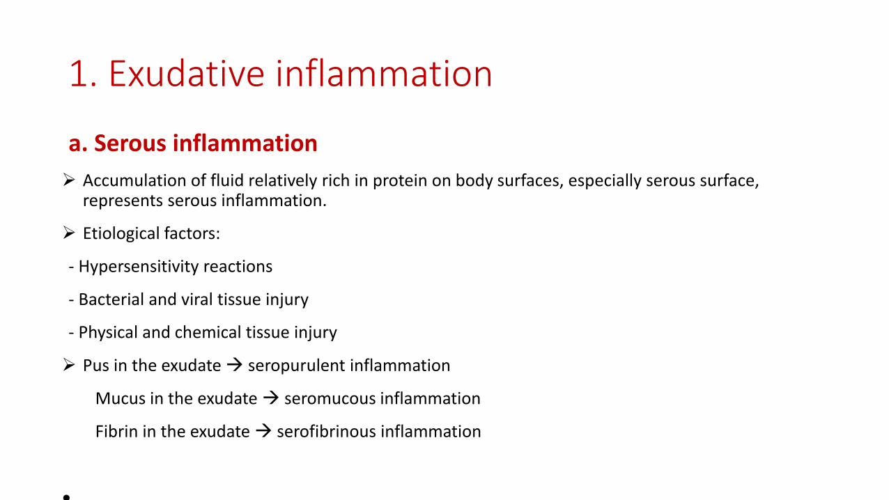
a. Serous inflammation
Accumulation of fluid relatively rich in protein on body surfaces, especially serous surface, represents serous inflammation.
Etiological factors:
- Hypersensitivity reactions
- Bacterial and viral tissue injury
- Physical and chemical tissue injury
Pus in the exudate seropurulent inflammation
Mucus in the exudate seromucous inflammation
Fibrin in the exudate serofibrinous inflammation
•

b. Catarrhal inflammation• Exudative inflammation occurring on the mucous membranes of the
respiratory and gastrointestinal tracts and producing a watery exudate ofserum and mucus.
• Etiological factors:- Bacteria and viruses- Chemical substances like phenol and cresol
• Grossly the surface appears reddened and swollen and may be coveredwith or contain, a clear to slightly opaque, thick fluid.
• Microscopically, the vessels of the Lamina Propria and the submucosa arehyperemic and desquamated epithelial cells and neutrophils can be seen.

c. Purulent inflammation
Inflammation with exudate consisting primarily of died neutrophils and cellular debris.
The predominant feature of the exudate is the formation of pus, a creamy liquid.
Etiological factors: - Pyogenic bacteria: Staphylococci, Streptococci,…- Chemical substances like silver nitrate and turpentine
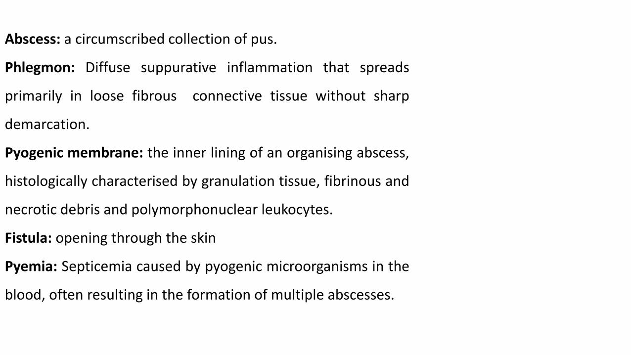
Abscess: a circumscribed collection of pus.
Phlegmon: Diffuse suppurative inflammation that spreads
primarily in loose fibrous connective tissue without sharp
demarcation.
Pyogenic membrane: the inner lining of an organising abscess,
histologically characterised by granulation tissue, fibrinous and
necrotic debris and polymorphonuclear leukocytes.
Fistula: opening through the skin
Pyemia: Septicemia caused by pyogenic microorganisms in the
blood, often resulting in the formation of multiple abscesses.

Metastatic abscess: a secondary abscess formed, at a distance from the primary focus, as a
result of the transportation of pyogenic bacteria by the lymph or bloodstream.
Cellulitis: bacterial infection involving the inner layers of the skin. It specifically affects the
dermis and subcutaneous fat.
Pyorrhea: the purulent inflammation of the tissues surrounding the teeth.
Pustule : A small inflamed elevation of the skin that is filled with pus.
Folliculitis: The purulent inflammation of the hair follicles of the skin.
Furuncle: the purulent inflammation of the hair follicles and the sebaceous glands of the
skin.
Acne: inflammation of the hair follicles and accompanying sebaceous glands of the skin and
subcutaneous connective tissue.
Carbuncle: Carbuncles are clusters of furuncles connected subcutaneously.

d. Haemorrhagic inflammation
Hemorrhagic inflammation is characterized by large numbers of erythrocytes in the exudate.
Etiologic factors:
Microorganisms like bacillus anthracis, hemolytic streptococci, clostridium species etc.
Viruses like Infectious canine hepatitis and Infectious laryngotracheitis (ILT)
Pathogenic Leptospira spp
Some chemical substances that cause acute poisoning like phenol arsenic and phosphorus, etc.
Some protozoa
This type of inflammation arises quickly and is often fatal. There is massive damage to endothelium.
The inflamed area is usually necrotic and filled with blood.

e. Fibrinous inflammation
Exudative inflammation with exudation of fibrinogen containing serumthat polymerizes to fibrin outside the blood vessels.
Fibrinous inflammation occurs in more severe conditions.
When fibrin forms a distinct layer covering an ulcer, it is referred to as afibrinous pseudomembrane.
If there is extensive necrosis of underlying areas so that the fibrin istightly adhered to the tissue and is harder to peel away, it is called adiphtheritic membrane. This term diphtheritic membrane came fromhuman diphtheria, caused by Corynebacterium diphtheria.

2. Necrotic (alterative) inflammation
Necrotic inflammation is characterized largely by necrosis and degeneration.
Inflammations characterized by tissue loss (alteration= tissue loss) are examined into two
groups:
o Necrotic inflammation of epithelial surfaces: (such as the trachea, intestine, nasal passages).
Examples: necrobacillosis in cattle, Rinderpest, ecthyma disease
o Necrotic inflammation of organs: characterized by the formation of necrosis in organs and, in
some cases, the formation of caverns resulting of necrosis melting.
Examples: necrobasillosis, pulmonary tuberculosis, Camplilobacteriosis in sheep, etc.

3. Productive (Granulomatous) inflammation
Granulomatous inflammation is a distinct type of chronic inflammation.
• It is marked by the formation of granulomas, which are small collections of modifiedmacrophages called epithelioid cells and are usually surrounded by lymphocytes.Granulomas often contain giant, or Langhans, cells that form from the coalescence ofepithelioid cells.
Granulomas are seen in a wide variety of diseases, both infectious and non-infectious.
• Examples of infections characterized by granulomas include tuberculosis, paratuberculosis,glanders, brucellosis,...
• Examples of non-infectious agents causing granulomas formation are Cholesterine crystals,uric acid crystals, splinters of wood or iron, operation residues.

Formation of a Granuloma. Circulatingmonocytes that become attracted bychemokines and inflammatory mediators to theextravascular lesion adhere to the vascular walland transmigrate between endothelial cells intothe perivascular extracellular matrix stroma andmigrate to form the granuloma.

Classification of inflammationaccording to the duration
Acute inflammation − has a short duration, ranging from a few hours to a few days.Vascular and exudative processes predominate.Marked clinically by the signs of heat, redness, swelling, pain, and loss of function.Neutrophils are often predominant, lymphocytes may be present.
Chronic inflammation −inflammation of prolonged duration, usually weeks to months andeven years.
The response is characterized predominantly by lymphocytes and macrophages, tissuenecrosis, and accompanied by tissue repair, such as healing, fibrosis, and granulation tissueformation, all of which may occur simultaneously.
Subacute inflammation − a condition intermediate between chronic and acuteinflammation.