cisplatin-functionalized silica nanoparticles for cancer chemotherapy
TRANSCRIPT
ORIGINAL PAPER
Cisplatin-functionalized silica nanoparticlesfor cancer chemotherapy
Chandrababu Rejeeth & Tapas C. Nag &
Soundarapandian Kannan
Received: 27 May 2013 /Accepted: 1 July 2013# Springer-Verlag Wien 2013
Abstract Cisplatin is used to treat a variety of tumors, butdose-limiting toxicities or intrinsic and acquired resistancelimit its application in many types of cancer including breast.Cisplatin was attached to silica nanoparticles usingaminopropyltriethoxy silane as a linker molecule and charac-terized in terms of size, shape, as well as the dissolution ofcisplatin from the silica surface. The primary particle diameterof the as received silica nanoparticles ranged from 20 to 90 nm.The results show that adverse effects on cell function, asevidenced by reduced metabolic activity measured by theMTT assay and increased membrane permeability observedusing the live/dead stain, can be correlated with surface areaof the silica. Cisplatin-functionalized silica nanoparticles withthe highest surface area incited the greatest response, whichwas almost equivalent to that induced by free cisplatin. More-over, if verified by further studies, would indicate that cisplatinwas attached to silica nanoparticles might prove to be useful insite-specific drug delivery.
Keywords Silica .Cisplatin .TEM .Nanoparticles .MCF-7cells . Fluorescence
1 Introduction
Worldwide, deaths: an estimated 39,920 breast cancer deaths(39,510 women, 410 men) are expected in 2012. Breastcancer ranks second as a cause of cancer death in women(after lung cancer) (American Cancer Society 2012). Cis-diamminedichloroplatinum (II), known as cisplatin, aplatinum-based drug, is one of the most potent antitumoragents has been widely used in the clinic to treat a variety ofcancers such as ovarian, breast, bladder, head and neck, andsmall cell lung cancer because of its potent activity to cross-link DNA upon entering the cells (Wang and Lippard 2005).Chemotherapy is the mainstay treatment for advanced andmetastatic disease. DNA-damaging agents have a long andproven record as anticancer drugs (Decatris et al. 2004). It isproved that after both passive and active cellular uptake,cisplatin may react with the N7 atom of guanine in DNA toform adducts and causes cellular apoptosis (Giese and Mc-Naughton 2003). However, chronic cisplatin usage results inresistance by several possible mechanisms including in-creased interactions with metal lothionein and glutathioneas well as increased DNA repair (Reedijk 2003). To coun-teract resistance, which lowers the efficiency of cisplatinsignificantly, very high systemic doses of cisplatin are ad-ministered. Unfortunately, such high dose of cisplatin resultsin severe systemic toxicity and poor patient compliance,including nausea/vomiting, renal toxicity, gastrointestinaltoxicity, peripheral neuropathy, asthenia, and ototoxicity,thus limiting its clinical use (Hill and Speer 1982; Rosenberg1977). To improve the efficacy and safety of cisplatin, avariety of methods are applied to drug delivery system,which include particulate carriers, such as liposomes, poly-mers, and nanoparticles (NPs) (Bontha et al. 2006; Junioret al. 2007; Geng et al. 2004). The drug carriers may con-centrate in the tumor because tumors exhibit a unique en-hanced permeability and retention effect for 50–100 nmparticles. As a result of this, a large increase in tumor drug
Electronic supplementary material The online version of this article(doi:10.1007/s12645-013-0043-6) contains supplementary material,which is available to authorized users.
C. Rejeeth (*) : S. KannanProteomics and Molecular Cell Physiology Lab, Department ofZoology, School of Life Sciences, Bharathiar University,Coimbatore 641 046, TN, Indiae-mail: [email protected]
T. C. NagSophisticated Analytical Instrument Facility for ElectronMicroscopy, Department of Anatomy, All India Institute of MedicalSciences, New Delhi 110029, India
Cancer NanoDOI 10.1007/s12645-013-0043-6

concentrations (tenfold or higher) could be achieved relativeto administration of the same dose of free drug, (Maeda et al.2000) thereby decreasing the massive systemic side effectsof conventional chemotherapy. In our previous study onmodified silica nanoparticles, (ORMOSIL, LSN) however,it was shown that the cytotoxicity across the MCF-7 cells asmeasured by methyl-thiazol tetrazolium salts (MTT) assaywas significantly reduced for the case of silica nanoparticlessynthesized and p53 gene delivery successful in breast can-cer cell line (Rejeeth et al. 2012a; Rejeeth et al. 2012b). In thecase of materials in combination, the relevant question iswhether apparently non-toxic particles are transformed into apotentially toxic material if surface attached with known toxicmolecules. Three possibilities exist: the nanoparticle/toxicmolecule combination becomes more toxic, the toxicity ofthe combination is unchanged, or the toxicity of the combina-tion is reduced compared to the toxic molecule alone. Onecommon chemical used for surface functionalization isaminopropyltriethoxysilane (APTES), which has the chemicalformula NH2 (CH2)3–Si (OC2H5)3 (Gan et al. 2009; Liu et al.2009; Jang and Liu 2009; Libertino et al. 2008). The silaneend of the APTESmolecule binds covalently to surface siliconatoms, and the amino end of the molecule increases proteinadsorption on the surface by electrostatic interactions. Thestructural transition of a single dsDNA molecule immobilizedon an APTES-treated substrate has been demonstrated. DNAbinding to the APTES linker is much stronger than that on analkylthiol/substrate (Nguyen et al. 2009). Silica–cisplatin sys-tem provides a unique opportunity to explore effects of surfacearea, in terms of the ability of nanoparticles to serve as carriers.The testable hypothesis herein is that nontoxic nanoparticleswith a surface-attached toxic molecule would then adverselyaffect cell function, defined here by reduced metabolic activityand increased membrane permeability. Specifically, these ad-verse effects are expected to increase with increasing surfacearea. Results of physicochemical characterization of thenanoparticles and nanoparticle/cisplatin combination includingsurface area and cisplatin dissolution rates are presented. Ef-fects on metabolic activity as measured via the MTTassay, andmembrane permeability observed by fluorescence microscopyusing the DAPI/JC-1 live/dead stain, we demonstrated thatthese silica-cisplatin prodrug conjugate NPs had well-controlled drug loading yield, excellent acid-responsive drugrelease characteristics, and potent cytotoxicity against breastcancer.
2 Materials and methods
2.1 Materials
The chemicals, i.e., APTES (3-aminopropyl) triethoxysilane,Triton X-100, Cisplatin, DAPI and JC-1 (Sigma-Aldrich) were
used without further purification. All the glassware (glass bottleand small pieces of glass substrate) was cleaned and sonicatedin ethanol for 5 min, rinsed with double-distilled water, soakedin a H2O/HNO3 (65 %)/H2O2 (1:1:1, v/v/v) solution, rinsedagain with doubly distilled water, and finally dried in air. TheMCF-7 cells were purchased from National Centre for CellSciences (NCCS), Pune, India.
2.2 Surface attachment of cisplatin to silica particles
Approximately, 0.64 g of APTES-functionalized Triton X-100, silica particles were kept in a three-necked flask respec-tively and dispersed in 50 mL distilled water with constantstirring for sample; 0.385 g of cisplatin was added forAPTES-functionalized Triton X-100 samples of suspendedsilica particles, respectively. These amounts correspond tosufficient cisplatin to react with all available silanol groups(2.5 nm−2) on the surface of silica. The suspended particleswere constantly stirred under nitrogen over-night at roomtemperature inside the fume hood. Cisplatin-functionalizedsilica particles were then collected by centrifugation at3,500 rpm for 15 min. Harvested particles were dried at roomtemperature and stored in glass bottles for further analysis.
2.3 FT-IR of cisplatin-functional silica nanoparticles
Fourier transform infrared (FT-IR) spectroscopy of silicananoparticle, cisplatin, and cisplatin-functionalized silicananoparticles were performed by using Nicolet 5700 instru-ment (Nicolet Instrument, Thermo Company, USA) withKBr pellet method. Each KBr disk was scanned over a wavenumber region of 500–4,000 cm−1 with the resolution of4.0×108 cm.
2.4 Cisplatin estimation
The platinum content of cisplatin-functionalized silica parti-cles was measured by inductively coupled plasma-massspectrometry (ICP-MS) measurements; 0.020 g of cisplatinfunctionalized silica particles was added to 25 mL of 2 %HNO3. The suspension was sonicated in an ultrasonic bathfor 30 min. After centrifugation, a clear supernatant wasobtained which was analyzed for total platinum (Pt). Theplatinum content of the sample was determined by compar-ison with a platinum standard which was prepared by dilu-tion of a standard solution of platinum of defined concentra-tion. The total amount of platinummeasured from the samplewas converted into the amount of cisplatin.
2.5 Transmission electron microscopy
Particle morphology was observed using a field emissiontransmission electron microscope (TEM). The selected area
C. Rejeeth et al.

diffraction patterns of the samples were recorded using thesame TEM instrument. The sample preparation procedureused for TEM analysis of the particles was as follows. Theparticles were first dispersed in ethanol 0.1 mg/mL suspensionin a glass beaker and beaker was sonicated for 10 min. Onedrop of sample suspension was placed on a carbon-coated grid(Ted Pella, USA) and dried at room temperature overnightbefore analysis under TEM.
2.6 Particle size analysis with dynamic light scattering
Dynamic light scattering (DLS) was performed using aPhotocor-FC light-scattering instrument employing a 5 mWlaser light source at 633 nm and a logarithmic correlateinstrument to determine the mean equivalent hydro-dynamic diameters of the samples. For the analysis, a sus-pension of ~0.1 mg/mL concentration of silica sample wasmade in ethanol. The suspension was sonicated for 30 minprior to analysis. The instrument measures the change ofintensity of the laser light with time at 90° angles afterinteraction with spherically shaped particles suspended inliquid media. The diffusion coefficient was determined fromthe correlation function, and, assuming the Stokes–Einsteinequation is valid, the equivalent hydrodynamic diameterdistribution was obtained using the Dyna-LS software pack-age supplied by Photocor.
2.7 Dissolution of cisplatin from silica particles
Dissolution of cisplatin from the surface of silica particles wasstudied in phosphate-buffered saline (PBS) with pH 7.4, atroom temperature (25 °C). In this study, ~30 mg of cisplatin-functionalized silica samples were suspended in 50 mL ofPBS for 48 h in a conical flask. Ten milliliters of the samplewas taken at the end of the specified time period (24, 36, and48 h). Sample was centrifuged at 3,500 rpm for 15 min, andthe supernatant was collected. Clear supernatant was analyzedfor platinum using the inductively coupled plasma mass spec-trometer. A 10-mL volume of fresh dissolution medium (PBSsolution) was added to make up the volume after withdrawalof sample. The dissolution rate constant of the cisplatin fromthe silica surface at different time points (24, 36, and 48 h) wascalculated using the modified Noyes and Whitney equation
(Wurster and Taylor 1965).
dC
dt¼ Ak Cs−Cð Þ
where, dC/dt=rate of dissolution, A=surface area of the par-ticle, C=concentration of the cisplatin in the PBS buffer at25 °C, Cs=saturation concentration of the cisplatin in the PBSat 25 °C, k=dissolution rate constant, which is defined ask=D/h, where D=diffusion rate of cisplatin, and h=diffusionlayer thickness.
2.8 Cell culture
The breast cancer cells (MCF-7) were maintained inDulbecco’s modified eagles medium (DMEM) supplementedwith 2 mM L-glutamine and balanced salt solution adjusted tocontain 1.5 g/L Na2CO3, 0.1 mM nonessential amino acids,1 mM sodium pyruvate, 2 mM L-glutamine, 1.5 g/L glucose,10 mM (4-(2-hydroxyethyl)-1-piperazineethane sulfonic ac-id), and 10 % fetal bovine serum (GIBCO, USA). Penicillinand streptomycin (100 IU/100 μg) were adjusted to 1 mL/L.The cells were maintained at 37 °C with 5 % CO2 in ahumidified CO2 incubator.
2.9 Metabolic activity assay
The mitochondrial activity of MCF-7 cells was measuredafter treatment with the cisplatin-functionalized silica sam-ples using the MTT assay. This test was performed on MCF-7 cells after 24, 36, and 48 h of incubation with the samples,as described by (Mosmann 1983). Cells were grown in a 24-well plate in DMEM media with 10 % fetal bovine serum at37 °C and 5 % CO2. In this study, silica/cisplatin particleswere added with the weight ranging from 13 to 144 μg/mLcorresponding to the amount needed to give a concentrationof surface attached cisplatin equal to 5 μg/mL in each well ofa single 24-well plate. Four replicates were performed foreach sample. Positive and negative controls were includedon each plate. The MTT assays involved the following steps:the growth medium was removed from the culture wells andwashed with 300 μL PBS once. After washing, 300 μL ofMTT solution (0.5 mg/mL) was added in each well of theplate. For the reduction of tetrazolium salt, plates were
Fig. 1 Scheme shows the synthesis of cisplatin-functionalized silica nanoparticles complex
Cisplatin-functionalized silica nanoparticles for cancer
incubated at 37 °C for 2 h. The tetrazolium salt was reducedto a formazan (blue) product by the active mitochondria ofthe cells. In each well, 300 μL of solubilizing buffer (10 %Triton X-100 with 0.1 N HCl in anhydrous isopropanol) wasadded to dissolve the reduced tetrazolium salt. The color ofreduced salt was measured at 570 nm using microplate reader(SpectraMax M5, Molecular Devices, Sunnyvale, CA). Theabsorbance at 570 nm was taken as the index of mitochon-drial activity.
2.10 Fluorescence microscopy
Cellular viability produced by the combination of cisplatinand silica particles was examined using fluorescence micros-copy after staining the MCF-7 cell with the live/dead system(Molecular Probes, USA). After incubation with the samplesin 96-well plates, cells were stained by adding DAPI and JC-1 to reach a final concentration in the well plates of 4 μMDAPI and 2 μM JC-1. DAPI undergoes fragment DNAconversion to fluorescent DAPI (blue, 340 nm wavelength),which is retained well within live cells. JC-1 enters cells withdamaged membranes, and undergoes a fluorescence en-hancement (brown, 635 nm wavelength) upon binding toDNA fragments. Cells were incubated with the dyes for20 min in 100 μL PBS, and imaged under the fluorescencemicroscope (Nikon instrument Inc., USA) using an excita-tion wavelength of 488 nm.
2.11 Cell sectioning for electron microscopy
Cisplatin-functionalized silica-treated MCF-7 cell line wasgrown on the 24-well cell culture plates. MCF-7 cells exposedto silica (Triton X- 100) were used as the control. Cells werewashed with PBS buffer and collected in centrifuge tubes afterharvesting. Cells were washed with 0.13 M phosphate bufferthree times for 10 min. At the end of the washing, cells weretreated with a fixative solution (2% glutaraldehyde) overnight at4 °C. After fixation, cells were washed with 0.1 M phosphate
buffer three times for 10 min to remove the excess glutaralde-hyde. Cells were post-fixed with 1 % OsO4 in PBS for 60 minand washed with double-distilled water three times for 10 min.After washing, cells were dehydrated at room temperature in 30,45, 65, 90, and 100 % ethanol for 10 min each. Cells were theninfiltrated with propylene oxide and LR white resin mixture for4 h. Cells were kept in fresh pure LR white resin overnight atroom temperature. Finally, the cells were embedded in capsules
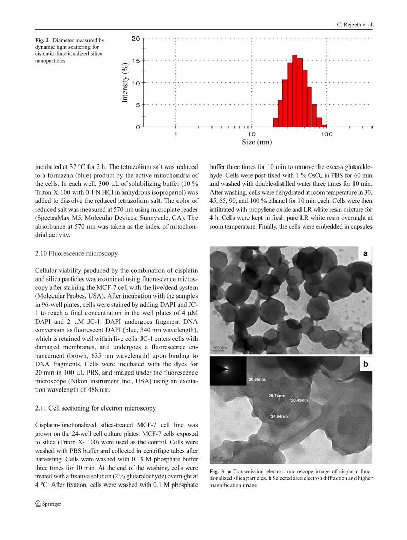
Fig. 3 a Transmission electron microscope image of cisplatin-func-tionalized silica particles. b Selected area electron diffraction and highermagnification image
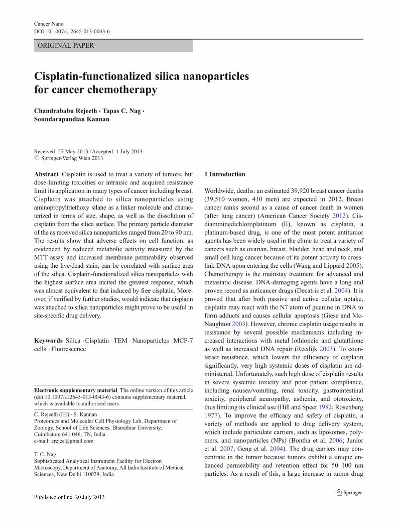
Fig. 2 Diameter measured bydynamic light scattering forcisplatin-functionalized silicananoparticles
C. Rejeeth et al.

(ElectronMicroscopy Sciences, USA) with freshly prepared LRwhite resin. Polymerization was carried out in an oven at 60 °Cfor 24 h. Sections from the embedded cells were cut on an Ultracut microtome (Reichert, Wien, Austria) and collected on 100-mesh copper TEM grids (Ted Pella Inc.,). Ultrathin sectionsmounted on copper grids were stained with 1 % aqueous uranylacetate (Ted Pella Inc.,) for 15 min and lead citrate (ElectronMicroscopy Sciences, USA) for 1.5 min.
2.12 Statistical analysis
Appropriate statistical procedures (Student t test for means,Stat graphics plus 3.3 software) were applied in the statisticalanalysis of the experimental data.
3 Results and discussion
3.1 Synthesis and characterization of cisplatin-functionalizedsilica particles
In this study, schematic diagrams shows (Fig. 1) the surface ofsilica particles was modified by the attachment of cisplatinmolecules, making use of the silanol groups on the silicasurface. Silanol groups are thought to be predominantly iso-lated from each other, with the surface concentration reportedto vary between two and four silanol groups per squarenanometer (Fripiat 1982; Iler 1979; Katz and Milewski1987; Mathias and Wannemacher 1988). In a study of silica,the silanol surface concentration was found to vary onlyslightly with surface area: 2.1 groups/nm2 for Triton X-100,which is made at high flame temperatures to facilitate largerprimary particle diameters, versus 2.4–2.5 groups/nm2 report-ed for Aerosil samples (Bhowmick et al. 2010). The observedreductions in the specific surface areas of the cisplatin-functionalized particles are explained by the attachment ofcisplatin molecules on the particle surface. The mean equiva-lent hydrodynamic diameters of cisplatin-functionalized silicasamples, as determined by DLS, are given in Fig. 2. Smallerparticles, with respect to hydrodynamic diameter, have agreater surface area and are more easily dispersed in aqueoussolutions, because of the presence of higher concentration, ona per gram of silica basis, of silanol groups on the surface ofthe higher specific surface area.
Silica particle morphology was observed using TEM. TheTEM image of the cisplatin-functionalized silica sample inFig. 3a shows particles that are round in shape, ranging from20 to 90 nm in diameter. The electron diffraction pattern,given in Fig. 3b (inset), shows the amorphous nature of thesample. In Fig. 3b, spherically shaped dark spots, believed to
0
0.05
0.1
0.15
0.2
0.25
0.3
0.35
0.4
0.45
50 150 250 350 450
ICP
-MS
Sign
al (
coun
ts)
Cis
plat
in/g
Silica surface area, m2 /g
Fig. 5 Estimation of maximumand minimum cisplatinattachment to the silica surfaceand inductively coupled plasmamass spectrometry result. Errorbars represent standarddeviations of triplicatemeasurements
Fig. 4 FT-IR spectra, before and after the reaction with cisplatin-functionalized silica nanoparticles. a Silica nanoparticles, b cisplatin,c cisplatin-functionalized silica nanoparticles
Cisplatin-functionalized silica nanoparticles for cancer

be platinum, are uniformly distributed on the surface of theparticles. Mapping of the platinum also suggests a relativelyuniform distribution of platinum on the silica particles. Ox-ygen and silicon result from the silica particles, and carbonfrom the TEM grid support and/or the APTES linker mole-cule on the particles.
3.2 FT-IR characterization of cisplatin-functionalized silicananoparticles
Figure 4a shows the FT-IR spectra of silica nanoparticles, thecharacteristic property of silica nanoparticles was showed bythe N–H and O–H stretching vibration at 3,607 cm−1 and the
SiH3OH bending vibration at 1,622 cm−1. The broad bandspectrum at 2,977 and 1,639 cm−1 shows the presence ofcisplatin (Fig. 4b). The PEGs bending vibration at 1,639 cm−1
demonstrating the characteristic property of cross-linked silicananoparticles functionalized with cisplatin. The characteristicpeak at 1,565 and 1,386 cm−1 clearly reveals bending vibrationof SiH3OH and stretching vibration of PEGs, respectively. Thecharacterized small peak at 1,565 and 1,490 cm−1 shows theHCL removed stretching of cisplatin-functionalized silicananoparticles (Fig. 4c). This result clearly shows the cross-linking of cisplatin-functionalized silica nanoparticles(supporting data S3).
3.3 Amount of cisplatin attached to the silica surface
ICP-MS results for the amount of cisplatin attached to thesilica surface are presented in Fig. 5, along with estimates ofthe minimum and maximum expected cisplatin loading. Themaximum amount of cisplatin attachment to the silica parti-cles was estimated assuming that silica particles contain2.5 silanol groups/nm2 on the surface (Katz and Milewski1987), and assuming a 1:1 binding ratio between the silanol
0
10
20
30
40
50
60
70
80
Triton -X 100 Cisplatin 5µg/ml Nanoparticles
% A
bsor
banc
e at
570
nm r
elat
ive
to
cont
rol
24 hrs
36 hrs
48 hrs
a
0
20
40
60
80
100
120
140
Cisplatin(0.1µg/ml)
Cisplatin(0.5µg/ml)
Cisplatin(2.5µg/ml)
Cisplatin(5µg/ml)
Cisplatin(10µg/ml)
% A
bsor
banc
e at
570
nm
rela
tive
to
cont
rol
b 24 hrs
36 hrs
48 hrs
Fig. 7 MTT assay of cisplatin-functionalized silica particles on MCF7. aAfter 24, 36, and 48 h and b dose-dependent study of free cisplatin. Resultsare presented as mean values compared to the control, and error barsrepresent standard deviation of four experiments. Asterisks denote statisti-cally significant (p>0.05) differences between experiment and control
a
b
0
0.2
0.4
0.6
0.8
1
1.2
1.4
1.6
1.8
After 24 hrs After 36 hrs After 48 hrs
% o
f cis
plat
in d
isso
lved
Silica NPs
0
0.5
1
1.5
2
2.5
3
3.5
4
4.5
0 24 36 48
ICP
-MS
Sign
al (c
ount
s )
Time, hrsFig. 6 a Percentage cisplatin dissolved as a function of time. b Disso-lution rate of cisplatin from the surface of silica particles to PBS bufferestimated by inductively coupled plasma-mass spectrometry. Errorbars represent standard deviations of triplicate measurements
C. Rejeeth et al.

groups, APTES, and cisplatin. A minimum amount of cis-platin attachment was estimated assuming that 50 % reactionhad occurred between the silanol groups and APTES, andbetween APTES and cisplatin. As shown in Fig. 5, thecisplatin loading is projected to be roughly proportional tothe surface area of the silica nanoparticles. This was ob-served experimentally, with cisplatin loading ranging frommaximum 0.4 g cisplatin/g silica for APTES to 0.039 gcisplatin/g silica for silanol groups. These results indicatethe significance of nanoparticle-based materials in terms oftheir high loading capacity.
3.4 Dissolution of cisplatin from silica particles
Cisplatin-functionalized silica particles were used to investi-gate the release of cisplatin in PBS buffer (pH 7.4) with time.The percentages of cisplatin released from functionalizedsilica samples are shown in (Fig. 6a) and the dissolution rateconstants of cisplatin from the samples are shown in Fig. 6b.The solubility of cisplatin in water is 2,350 mg/L (Kim et al.2009). As shown in Fig. 6a, the percentages of cisplatinreleased are low but non-zero. In Fig. 6b, it can be seen thatthe dissolution rate constant of cisplatin from the cisplatin-
Fig. 8 Live/dead images ofMCF7 cells viewed under thefluorescence microscope afterinteraction with a control, bsilica nanoparticles, c freecisplatin, d cisplatin-functionalized silicananoparticles
Cisplatin-functionalized silica nanoparticles for cancer
functionalized silica particles is ten times greater than that ofthe lower specific surface area of surfactant (supporting dataS1) 50 samples at 24 h. It can also be observed that thecisplatin dissolution rate decreases with time for all of thecisplatin-functionalized samples. Results also indicate that thedissolution of cisplatin in PBS buffer from the particle sur-faces does not reach equilibrium within 48 h of time. Thishigher dissolution rate constant of cisplatin from the cisplatin-functionalized silica nanoparticles is attributed to the higherspecific surface areas of the particles compared to the othersamples (supporting data S2).
3.5 In vitro effects on metabolic function
We evaluated the effects of cisplatin-functionalized silicananoparticles and cisplatin alone on cellular metabolic activ-ity using the MTTassay after 24, 36, and 48 h, and the resultsare presented in Fig. 7a. MTT results for different doses offree cisplatin after 48 h are represented in Fig. 7b. Significantdifferences (p≤0.05) between control and sample, as denot-ed by asterisks in Fig. 7a, were observed for the surface areasamples, Triton X-100-cisplatin, at the earlier time points.Significant differences between control and sample wereobserved for silica–cisplatin samples by 48 h. The MCF-7cells exhibits the smallest decrease in metabolic activity inthe case of cisplatin-functionalized silica sample, even after48 h of incubation. Student t test indicates that the MTT
results for each of the silica samples and the 5 μg/mL cis-platin sample are significantly different (p≤0.05). Resultsalso show that reduction in metabolic activity after treatmentwith the Triton–cisplatin construct (45 % compared to con-trol) is very close to the response from the cells treated withcisplatin (38 % compared to control). An almost equal re-duction in metabolic activity can be observed after 48 h whenthe cells are incubated with free cisplatin at different doses(0.1–10 μg/mL) as shown in Fig. 7b. With the silica–cisplat-in system, it is important to note, however, that no apparentsynergistic effects were observed in terms of adverse effectson cellular function, unlike in prior reports of thebenzo(a)pyrene/iron oxide micron-sized particle system(Garcon et al. 2001; Garry et al. 2004). Nor were any miti-gative effects on cellular function observed, as in the case ofsilica synthesized with chitosan, compared to silica synthe-sized in the absence of chitosan (Chang et al. 2007). Expo-sure to the combination of cisplatin with silica that had thehighest specific surface area resulted a similar effect onmetabolic activity as compared to exposure to the equivalentamount of free cisplatin.
The MCF-7 cells were treated with cisplatin-functionalizedsilica particles of specific surface area for 24 h, and viability wasobserved by staining live and dead cells with DAPI and JC-1,respectively. Fluorescence stained breast cancer cells after treat-ment with the control (nontreated), Triton X-100 silica particles,cisplatin-functionalized particles, and free cisplatin are shown in
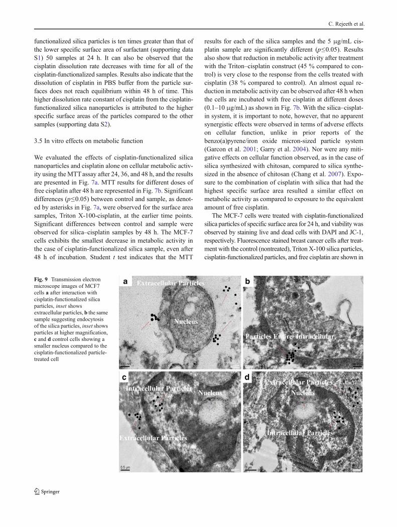
Fig. 9 Transmission electronmicroscope images of MCF7cells a after interaction withcisplatin-functionalized silicaparticles, inset showsextracellular particles, b the samesample suggesting endocytosisof the silica particles, inset showsparticles at higher magnification,c and d control cells showing asmaller nucleus compared to thecisplatin-functionalized particle-treated cell
C. Rejeeth et al.

Fig. 8a, b, c, and d respectively. Remarkable morphologicalchanges were observed under the fluorescence microscopewhen cells were treated with cisplatin-functionalized particles.In Fig. 8d, the number of cells remaining after treatment withthe particles is less than the others. The MTT results also haveshown that the greatest reduction in metabolic activity wasproduced by the treated cells, which is in agreement with thefluorescence microscopy images. Results showed that after 24 hof incubation, cisplatin-functionalized silica particles and freecisplatin have the strongest adverse effect on viability comparedto the control. In the fluorescence images, most of the cellsappeared dead after treatment with cisplatin-functionalized silicaparticles and free cisplatin, Fig. 8c, d. In contrast, treatment withcontrol (nontreated) led to slate significant adverse effects in thecells; most of the cells are alive as shown in Fig. 8a. This studyindicates that nanoparticle-mediated morphological alterationmay occur in the cells and to study these morphological changesat a higher level of resolution, we further observed them in thetransmission electron microscope.
3.6 Cellular response to cisplatin-functionalized silicaparticles
Cisplatin-functionalized silica particle breast cancer cell in-teractions were investigated by TEM analysis of thin sec-tioned cell samples. Representative micrographs are shownin Fig. 9, with images chosen after analyzing 50–100 cellsfrom each sample. The morphology of MCF-7 cells aftertreatment with cisplatin-functionalized silica particles isshown in Fig. 9a, b whereas MCF-7 cells after treatmentwith control silica particles are shown in Fig. 9c, d. TEMimages clearly show the internalization of the both controland cisplatin-functionalized silica particles inside the cells(Fig. 9c). In both the cases, extracellular particles are alsovisible in the images. Images also show that intracellularparticles are confined inmembrane-bound vacuoles (Fig. 9a).Internalized particles are found in aggregated form and mayhave entered the cells by the endocytosis process as sug-gested by the image in Fig. 9b. Membrane-bound aggregatedparticles are observed to be very close to the nucleus of thecell in some cases. The morphological changes in MCF-7cells after 48 h of growth are also shown in Fig. 9. Theimages shown in Fig. 9 are the representatives of entire cellpopulation after analyzing 50–100 cells in each sample.Images of cells after treatment with control silica particles(Fig. 9c, d) revealed that the cell membranes are moreintegrated compared to the cells treated with cisplatin-functionalized silica particles (Fig. 9a, b). It also can beobserved that nucleus of the cells treated with cisplatin-functionalized silica (Fig. 9a) is much larger compared tothe cells treated with control silica particles (Fig. 9c) (Meijeret al. 2001).
TEM analysis of the MCF-7 cells can shed light in under-standing the possible interaction mechanisms of cisplatin-functionalized silica particles with the cells. Cisplatin mustbe able to react with DNA to affect cell function, so it needs toreach the cell nucleus. In the TEM images, both extracellularand intracellular particles are visible suggesting two possibil-ities. The extracellular particles may be releasing cisplatin intosolution, which is then taken up by the cells. Free cisplatin isbelieved to enter cells via both passive and active transportroutes (Ohmori et al. 1993). Cisplatin-functionalized silicananoparticles can enhance both possible interaction mecha-nisms by bringing high loadings of cisplatin near the cells forpassive transport of free cisplatin to take place. The intracel-lular particles, on the contrary, may carry bound cisplatin intothe MCF-7 cells, which is then released into the cytoplasmwith higher surface area corresponding to greater rates ofactive transport.
4 Conclusions
In conclusion, successful attachment of a controlled amountof toxic cisplatin molecules on the surface of the nano-sizedsilica particles was demonstrated and the particles werecharacterized in terms of particle size and surface area. Inthis study, we have demonstrated that the dissolution rate ofcisplatin from the surface of the particles to the solutionphase depends on the surface areas of the particles. Biolog-ical studies have shown that, the impaired cellular function inthe MCF-7 induced by cisplatin-functionalized silica parti-cles, as indicated by the reduced metabolic activity and cellviability. These effects appeared to be correlated with thecisplatin functionalized surface areas of the particles. Appar-ent uptake of the particles by the cells and morphologicaldifferences after active interaction with the cisplatin-functionalized silica particles were observed by TEM. Theimportance of our study lies in showing the combinatorialeffect of relatively high surface area nanoparticles and toxicmolecules at the cellular level. In the wake of increasing useof nanoparticles, which may be either surface modified priorto end use in drug delivery constructs or consumer products,or upon release into the environment, this study issues acautionary note that the properties of the nanoparticles, es-pecially specific surface area, may affect biological response.
Acknowledgments This work was financially supported by DST-NANOMISSION (DST No. SR/NM/NS-60/2010) Ministry of Scienceand technology and UGC-NON-SAP (G2/6966/UGC NON-SAP(Zoology)/2010) New Delhi, Govt. of India. The authors greatly ac-knowledged DRDO Centre for Life Sciences for nanoparticles charac-terization studies, Electron Microscopy Centre, AIIMS, New-Delhi andSankara Nethralaya for their kind assistance with fluorescence micros-copy studies. The authors are very much thankful to all faculty members
Cisplatin-functionalized silica nanoparticles for cancer

of the Department of Zoology, Bharathiar University for their constantsupport and encouragement throughout this study.
References
American Cancer Society (2012) Cancer facts and figures 2012. Amer-ican Cancer Society, Atlanta
Bhowmick TK,YoonD, PatelM, Fisher J, Ehrman S (2010) In vitro effectsof cisplatin-functionalized silica nanoparticles on chondrocytes. JNanoparticle Res 12:2757–2770
Bontha S, Kabanov AV, Bronich TK (2006) Polymer micelles withcross-linked ionic cores for delivery of anticancer drugs. J ControlRelease 114:163–174
Chang JS, Chang KLB, Hwang DF, Kong ZL (2007) In vitrocytotoxicitiy of silica nanoparticles at high concentrations stronglydepends on the metabolic activity type of the cell line. Environ SciTechnol 41:2064–2068
Decatris MP, Sundar S, O'Byrne KJ (2004) Platinum-based chemotherapyin metastatic breast cancer: current status. Cancer Treat Rev 30:53–81
Fripiat JJ (1982) Silanol groups and properties of silica surfaces. ACSSymp Serc 194:165–184
Gan S, Yang P, Yang W (2009) Photo activation of alkyl C-H andsilanization: a simple and general route to prepare high-densityprimary amines on inert polymer surfaces for protein immobiliza-tion. Bio macromol 10:1238–1243
Garcon G, Gosset P, Garry S, Marez T, Hannothiaux MH, Shirali P(2001) Pulmonary induction of proinflammatory mediators fol-lowing the rat exposure to benzo(a)pyrene-coated onto Fe2O3
particles. Toxicol Lett 121:107–117Garry S, Nesslany F, Aliouat E, Haguenoer JM, Marzin D (2004)
Hematite (Fe2O3) acts by oxydative stress and potentiatesbenzo[a]pyrene genotoxicity. Mutat Res 563:117–129
Geng L, Osusky K, Konjeti S, Fu A, Hallahan D (2004) Radiation-guided drug delivery to tumor blood vessels results in improvedtumor growth delay. J Control Release 99:369–381
Giese B, McNaughton D (2003) Interaction of anticancer drug cisplatinwith guanine: density functional theory and surface enhancedRaman spectroscopy study. Biopolymers 72:472–489
Hill JM, Speer RJ (1982) Organo-platinum complexes as antitumoragents. Anticancer Res 2:173–186
Iler RK (1979) The chemistry of silica: solubility, polymerization,colloid and surface properties, and biochemistry. Wiley, NewYork, xxiv, 866
Jang LS, Liu HJ (2009) Fabrication of protein chips based on 3-aminopropyltriethoxysilane as a monolayer. Biomed Microdevices11:331–338
Junior AD, Mota LG, Nunan EA, Wainstein AJ, Wainstein AP, Leal AS,Cardoso VN, De Oliveira MC (2007) Tissue distribution evaluation
of stealth pH-sensitive liposomal cisplatin versus free cisplatin inEhrlich tumor-bearing mice. Life Sci 80:659–664
Katz HS, Milewski JV (1987) Handbook of fillers for plastics. VanNostrand Reinhold Co, New York
Kim JK, Anderson J, Jun HW, Repka MA, Jo S (2009) Self-assemblingpeptide amphiphile-based nanofiber gel for bioresponsive cisplatindelivery. Mol Pharm 6:978–985
Libertino S, Giannazzo F, Aiello V, Scandurra A, Sinatra F, Renis M, FicheraM (2008) XPS and AFM characterization of the enzyme glucoseoxidase immobilized on SiO(2) surfaces. Langmuir 24:1965–1972
Liu T, Wang S, Chen G (2009) Immobilization of trypsin on silica-coated fiberglass core in microchip for highly efficient proteolysis.Talanta 77:1767–1773
Maeda H, Wu J, Sawa T, Matsumura Y, Hori K (2000) Tumor vascularpermeability and the EPR effect in macromolecular therapeutics.A review. J Control Release 65:271–284
Mathias J, Wannemacher G (1988) Basic characteristics and applica-tions of Aerosil.30. The chemistry and physics of the Aerosilsurface. J Colloid Interface Sci 125:61–68
Meijer C, van Luyn MJA, Nienhuis EF, Blom N, Mulder NH, de VriesEGE (2001) Ultrastructural morphology and localisation ofcisplatin-induced platinum–DNA adducts in a cisplatin-sensitiveand -resistant human small cell lung cancer cell line using electronmicroscopy. Biochem Pharmacol 61:573–578
Mosmann T (1983) Rapid colorimetric assay for cellular growth andsurvival: application to proliferation and cytotoxicity assays. JImmunol Methods 65:55–63
Nguyen TH, Kim YU, Kim KJ, Choi SS (2009) Investigation ofstructural transition of dsDNA on various substrates studied byatomic force microscopy. J Nanosci Nanotechnol 9:2162–2168
Ohmori T, Morikage T, Sugimoto Y, Fujiwara Y, Kasahara K, Nishio K,Ohta S, Sasaki Y, Takahashi T, Saijo N (1993) The mechanism ofthe difference in cellular uptake of platinum derivatives in non-small cell lung cancer cell line (PC-14) and its cisplatin-resistantsubline (PC-14/ CDDP) Jpn. J Cancer Res 84:83–92
Reedijk J (2003) New clues for platinum antitumor chemistry: kineti-cally controlled metal binding to DNA. Proc Natl Acad Sci U S A100:3611–3616
Rejeeth C, Kannan S, Muthuchelian K (2012a) Development of in vitrogene delivery system using ORMOSIL nanoparticle: analysis ofp53 gene expression in cultured breast cancer cell (MCF-7). Can-cer Nano 3:55–63
Rejeeth C, Kannan S, Salem A (2012b) Novel luminescent silicananoparticles (LSN): p53 gene delivery system in breast cancerin vitro and in vivo. Journal of Pharmacy and Pharmacology doi:10.1111/j.2042-7158.2012.01547.x (In press)
Rosenberg B (1977) Noble metal complexes in cancer chemotherapy.Adv Exp Med Biol 91:129–150
Wang D, Lippard SJ (2005) Cellular processing of platinum anticancerdrugs. Nat Rev Drug Discov 4:307–320
Wurster DE, Taylor PW (1965) Dissolution rates. J Pharm Sci 54:169–175
C. Rejeeth et al.