chronic lymphocytic leukemia -...
TRANSCRIPT
Hematology 2002 193
Chronic Lymphocytic Leukemia
Neil E. Kay, Terry J. Hamblin, Diane F. Jelinek, Gordon W. Dewald, John C. Byrd,Sherif Farag, Margaret Lucas, and Thomas Lin
This update of early stage B-cell chronic lympho-cytic leukemia (B-CLL) embraces current informa-tion on the diagnosis, biology, and interventionrequired to more fully develop algorithms formanagement of this disease. Emphasis on earlystage is based on the rapid advancement in ourunderstanding of the disease parameters and ourincreasing ability to predict for a given early stagepatient whether there is a need for more aggressivemanagement.
In Section I, Dr. Terry Hamblin addresses thenature of the disease, accurate diagnostic proce-dures, evidence for an early “preclinical” phase, theuse of newer prognostic features to distinguishwho will be likely to progress or not, and whether itis best to watch or treat early stage disease.
In Section II, Dr. Neil Kay and colleaguesaddress the biologic aspects of the disease andhow they may relate to disease progression.Review of the newer insights into gene expression,
recurring genetic defects, role of cytokines/autocrine pathways, and the interaction of the CLLB cell with the microenvironment are emphasized.The relationship of these events to both triggerdisease progression and as opportunities for futuretherapeutic intervention even in early stage diseaseis also considered.
In Section III, Dr. John Byrd and colleaguesreview the historical and now current approachesto management of the previously untreated pro-gressive B-CLL patient. They discuss what decisiontree could be used in the initial decision to treat agiven patient. The use of single agents versusnewer combination approaches such aschemoimmunotherapy are discussed here. Inaddition, the place of marrow transplant and someof the newer antibodies available for treatment of B-CLL are considered. Finally, a challenge to utilizeour growing knowledge of the biology of B-CLL inthe early stage B-CLL is proffered.
I. THE NATURE AND NATURAL HISTORY OF
EARLY-STAGE B-CLL
Terry J. Hamblin, MD*
Some people think that we are getting better at treatingB-cell chronic lymphocytic leukemia (B-CLL) and pro-duce survival curves that seem to show an improved sur-vival for more recently diagnosed cases. At least part ofthat improvement is artificial, caused by earlier diagno-sis or even the recognition as B-CLL of conditions thatwould not previously have crossed the threshold of di-agnosis. As we diagnose B-CLL on lower and lower lym-phocyte counts, we need to be doubly sure that the diag-nosis is correct. It may be that a similar distinction needsto be made between B-CLL and “monoclonal lympho-cytosis of undetermined origin” as that between myeloma
and monoclonal gammopathies of undetermined signifi-cance (MGUS). As the proportion of cases diagnosed inearly stage increases it becomes vital for the practicingphysician to know who is likely to require therapy. Itmay be that the “watchful waiting” paradigm for earlystage B-CLL needs to be challenged.
Diagnosis of True B-CLLAs time has passed we have lowered the threshold fordiagnosing B-CLL. At one time a lymphocyte count of10,000/mm3 was required1 but today we are happy with5000/mm3.2 This reflects a greater certainty that we candistinguish B-CLL from other types of lymphocytosis, acertainty provided by the availability of immunologicmarkers. One consequence has been to include within thediagnosis more patients whose B-CLL is utterly benign.
This lowered guard demands a sterner conformityto the correct profile of lymphocyte markers in order todefine B-CLL and exclude other types of exfoliating lym-phoma in which a mild lymphocytosis is common (Table1). The characteristic B-CLL pattern (CD5 and CD23
* Royal Bournemouth Hospital, Castle Lane East,Bournemouth BH7 7DW, Dorset, United Kingdom
194 American Society of Hematology
positive, surface immunoglobulin weak, CD79b weakor absent, FMC7 negative) has been codified by the RoyalMarsden group.3 A score of 4 or 5 is found in almost alltrue B-CLLs. Very occasionally a true B-CLL will haveonly a weakly positive CD5 but the use of this scoringsystem will almost always exclude other “spillover lym-phomas.” Apart from mantle cell lymphomas these willall be CD5 negative, and mantle cell lymphoma is easilydistinguished even when the cellular morphology is simi-lar; it is CD23 negative and has strong surface immuno-globulin staining. Confirmation of mantle cell lymphomais by detection of the t(11;14) translocation and nuclearcyclin D1 staining. These “spillover lymphomas” willbe excluded from further discussion.
Monoclonal Lymphocytosis ofUndetermined Significance
Every journey begins with a single step. Every tumorbegins with a single cell. The NCI guidelines set a mini-mal threshold for the diagnosis of CLL in the certainknowledge that the tumor will have existed before it isdiagnosed. Exactly how early might CLL be diagnosed?And does it emerge from a profusion of proliferationsthat are monoclonal but not malignant? And can we tellthe difference? Rawstron et al4 have designed a simpleflow cytometric assay to detect minimal residual dis-ease in patients with B-CLL who have been treated withintensive chemotherapy. This assay, based on the stain-ing pattern of CD5, CD19, CD20, and CD79b, can de-tect small numbers of B-CLL cells with a sensitivity of1 in 105. When the researchers applied this assay to rou-tine blood counts coming through their laboratory inLeeds 3% of individuals over 40 years of age had a de-tectable population of cells with an identical marker pro-file to that of B-CLL. These cells were monotypic with
respect to immunoglobulin light chains, and in the fewpatients where they had enough material to sequencethe immunoglobulin heavy chain genes, they were clearlymonoclonal. However, we should emphasize that “mono-clonal” does not necessarily mean “malignant.” A B-CLL-like population could be detected twice as com-monly in individuals over the age of 60 as in those be-tween 40 and 59. As few as 3 cells per mm3 could bereliably detected, and in most the B-CLL-like cells rep-resented a minority of the circulating B cells. Becausethe samples were anonymized, it was not possible to re-test on a sequential basis. It is conceivable that thesewere only transient events.
These findings have yet to be reproduced in otherlaboratories, but if they are confirmed they will suggestthat there is a monoclonal lymphocytosis of undeter-mined significance (MLUS) that bears a relationship toB-CLL similar to that MGUS bears to myeloma and thatis present in a sizable proportion of elderly individuals.As with MGUS, and indeed as with myelodysplastic syn-drome and carcinoma of the prostate, it is certain thatmany cases of MLUS that could be diagnosed will re-main latent throughout an individual’s lifetime. Undoubt-edly the Leeds study will prompt others to verify this,but it is a project for a young hematologist.
This is not a new concept. Han et al5 described abenign monoclonal B cell lymphocytosis without dis-ease progression over 6-24 years, but all of these had alymphocyte count > 10,000/mm3. The term B-MLUS hasbeen used extensively by Mellstedt and his colleagues6-
10 to refer to patients with an isolated monoclonal B-celllymphocytosis in blood and bone marrow but no othersigns or symptoms of B-CLL. The European classifica-tion of Binet et al11 allows for lymphadenopathy in stageA disease whereas the Swedish MLUS patients do notseem to differ substantially from Rai Stage 0 B-CLL.12
To add to the confusion, during 2002 two papers haveapplied the term MLUS to proliferations of CD5-nega-tive lymphocytes, distinguishing them from early stageB-CLL.13,14 While semantically correct, this usage is un-helpful.
Montserrat et al described “smoldering” B-CLL,15
which was defined as Rai stage 0 with non-diffuse bonemarrow histology, hemoglobin ≥ 13 g/dL, blood lym-phocytes < 30,000/mm3, and a lymphocyte doubling timeof > 12 months. An analysis of the French CooperativeGroup’s trial of Stage A patients16 demonstrated that asubgroup they designated A′, with hemoglobin > 12 g/dL, lymphocyte count < 30,000/mm3, and fewer than80% lymphocytes on bone marrow aspiration, was lesslikely to progress that other stage A patients.
The French criteria require a bone marrow aspira-tion, and the Spanish a bone marrow trephine. In view
Table 1. Immunophenotype of B-cell chronic lymphocyticleukemia (B-CLL) and lymphomas that resemble it.
Mantle Cell FollicularAntigen B-CLL Lymphoma SLVL Lymphoma
sIg Weak ++ ++ ++
CD5 ++ ++ – –
CD19 ++ ++ ++ ++
CD20 + ++ ++ ++
CD22 Weak or – ++ ++ ++
CD23 ++ – – –
CD79b Weak or – ++ ++ ++
FMC7 – ++ ++ ++
CD10 – – – ++
Abbreviations: sIg , surface immunoglobulin; SLVL, spleniclymphoma with villous lymphocytes.

Hematology 2002 195
of the utility of blood tests, it may be appropriate to ques-tion the necessity of such invasive techniques in B-CLL.I would contend that bone marrow examination does nothelp in the diagnosis of B-CLL. Undoubtedly it givesprognostic information; future clinical trials must decidewhether it gives information unavailable from blood testsalone.
Two Types of B-CLL?The division of B-CLL into stable and progressive dis-ease was noted by David Galton 36 years ago.17 Whathas been unclear is whether there is a qualitative differ-ence between the B-CLL that pursues a progressivecourse and “smoldering” or stable B-CLL, or simply aquantitative one. Are they part of a spectrum? Does onemetamorphose into the other? Is it, like the transforma-tion of myelodysplastic syndrome to acute leukemia, amatter of the acquisition of further genetic damage?
Some answers to these questions have become avail-able from the recognition of two subsets of B-CLL basedon the IgV
H mutational status.18,19 These two subsets have
separate and distinct natural histories. Binet stage A pa-tients without somatic mutations have a median survivalof 8 years; those with somatic mutations have a mediansurvival of 25 years.18
Somatic mutations occur in B cells in the germinalcenter under the influence of mutator enzymes gener-ated in response to contact with antigen presented by
follicular dendritic cells in the presence of T cells.20
Stimulation of the B cell receptor in any other circum-stances, whether by T independent antigen or bysuperantigen, is thought not to induce somatic mutations(Figure 1).
It seems clear that B-CLL always derives from amature cell.21 To use the term “memory” cell is contro-versial; perhaps to call it an “experienced” cell is moreappropriate at present. Moreover, the two subsets arevery similar to each other, differing from other types oflymphoma by the expression of a wide range of differ-ent genes, but from each other by the differential ex-pression of very few.22,23 However, the mutational stateis fixed. There has been no case of B-CLL in which mu-tations have been lost or gained by the clone during thecourse of the disease. There has been much debate as towhether B-CLL is one or two diseases. The fact that themutated subset is equally common in males and femaleswhile the unmutated subset is three times as common inmales suggests two separate entities, but what is clear isthat the two subsets are distinct and do not metamor-phose one into the other.
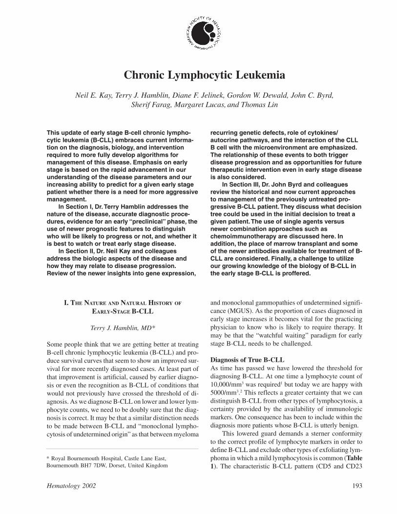
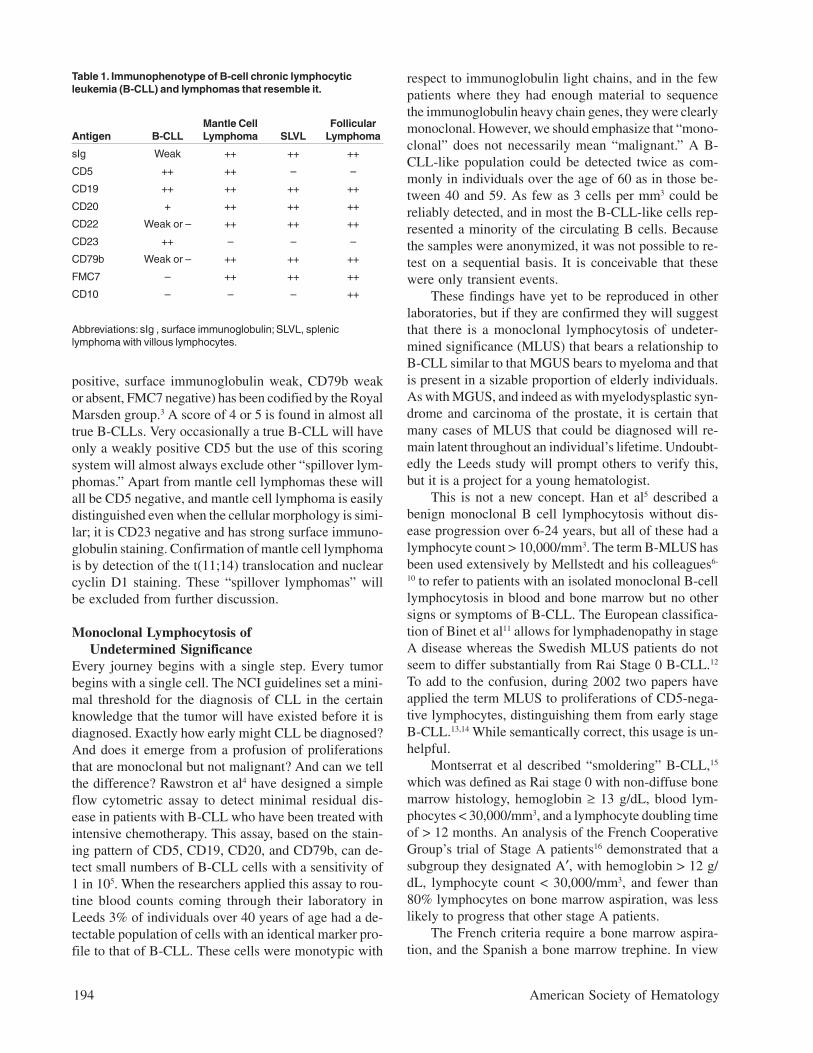
Natural History of B-CLL with Mutated V GenesIs it correct to assume that B-CLL with mutated V genesis the same as “smoldering” B-CLL? Clearly not, sincesome cases progress to advanced stage disease. How-ever, it is likely that all “smoldering” cases are containedwithin the mutated subset. It is interesting to note that,although the Leeds group had only limited material fromtheir study of routine blood counts, in three cases theywere able to sequence the immunoglobulin V genes. Allthree had somatic mutations.4 Survival curves on our ownBinet stage A patients that are censored for B-CLL-un-related deaths demonstrate that all patients withunmutated V genes are dead within 12 years of diagno-sis, whereas the first death in the group with mutated Vgenes does not occur until 27 years after diagnosis, andonly 2/70 have died from a B-CLL-related cause (Fig-ure 2). In our hands, when the mutated subset progressesit does so slowly, and the survival curve resembles thatof stable rather than progressive B-CLL (Figure 3). Nev-ertheless, the late complications of B-CLL, severe hypo-gammaglobulinemia and autoimmune hemolytic anemia,do occur in the mutated subset, and in our experiencehemolytic anemia may be the presenting feature of lowcount stage 0 disease that is thereafter never troublesome.
Consequences for ManagementA consensus has arisen that early stage B-CLL shouldnot be treated with chemotherapy; rather, watchful wait-ing should be employed. The best supporting evidencefor this approach is the meta-analysis of 2048 early stage
Figure 1. Diagrammatic representation of the two subsets ofchronic lymphocytic leukemia (CLL).
Conventionally, B cells meet antigen in the germinal center in thecontext of T cells and antigen presenting cells. This stimulatessomatic mutation of the immunoglobulin V genes, and a memory Bcell emerges from the germinal center. The unmutated subset mightbe explained by envisioning unconventional stimulation of the B cellreceptor outside the germinal center, either by T-independentantigens or superantigen. This does not induce somatic mutation,but does stimulate an “experienced” immunophenotype.
196 American Society of Hematology
patients in 7 trials randomized between immediate ordeferred treatment with chlorambucil (with or withoutprednisolone).24 This demonstrated no benefit for eitherarm. The 10-year survival was slightly worse (but notstatistically significantly so) for those treated early (44%versus 47%). There is, therefore, no need to treat pa-tients with stage A disease with chlorambucil unless thereis evidence of a falling hemoglobin or platelet count,progression to a later stage of disease, unsightly or un-comfortable lymphadenopathy, a lymphocyte doublingtime of < 12 months, or transformation of the disease.
However, it must be admitted that all advanced B-CLL has passed through an early stage. In the largeFrench study,25 51% of patients allocated to the deferredtreatment arm eventually required treatment, and 27%eventually died of a cause related to B-CLL. Further-more, chlorambucil is by no means the most effectivetreatment for B-CLL. The combination of fludarabine,cyclophosphamide, and rituximab has been reported toinduce complete responses of 60% in advanced disease,with half of those tested achieving a molecular remis-sion.26 In cancer, generally it is seldom prudent to waituntil the disease progressed before applying the mosteffective treatment. On the other hand, it would be fool-hardy to apply a potentially toxic treatment to a groupof individuals who will never be bothered by their bloodcondition. What is needed is a foolproof way of predict-ing who will progress and who will not.
Prognostic Factors for Stable and Progressive DiseaseWatchful waiting is clearly an appropriate managementfor at least half of all B-CLLs. From the foregoing it isclear that we can be more selective than applying thispolicy simply to all early stage disease. The French A′16
and Montserrat’s “smoldering”15 criteria are predictive,
but when observed over a 5-year period, 25% of A′ casesand 13% of “smoldering” cases progressed, and 46% ofA′′ and 43% of “active” cases failed to progress.27
A number of serum factors have been suggested aspotential prognostic indicators for early stage disease.Patients with early stage B-CLL who had serum thymi-dine kinase levels greater than 7.0 U/L had a signifi-cantly shorter progression-free interval than those witha lower level.28 Serum soluble CD23 segregates Binetstage B disease into more or less aggressive forms.29
Serum levels of β2-microglobulin greater than 4.0 mg/L
are adverse prognostic factors in a wide range of lym-phoid malignancies,30 including B-CLL.31
Chromosomal analysis is also predictive. Patientswith a normal karyotype or isolated 13q14 deletions havea benign prognosis.32 On the other hand, the 10-15% ofpatients with 17p13 or 11q23 deletions do very badlyindeed.33,34
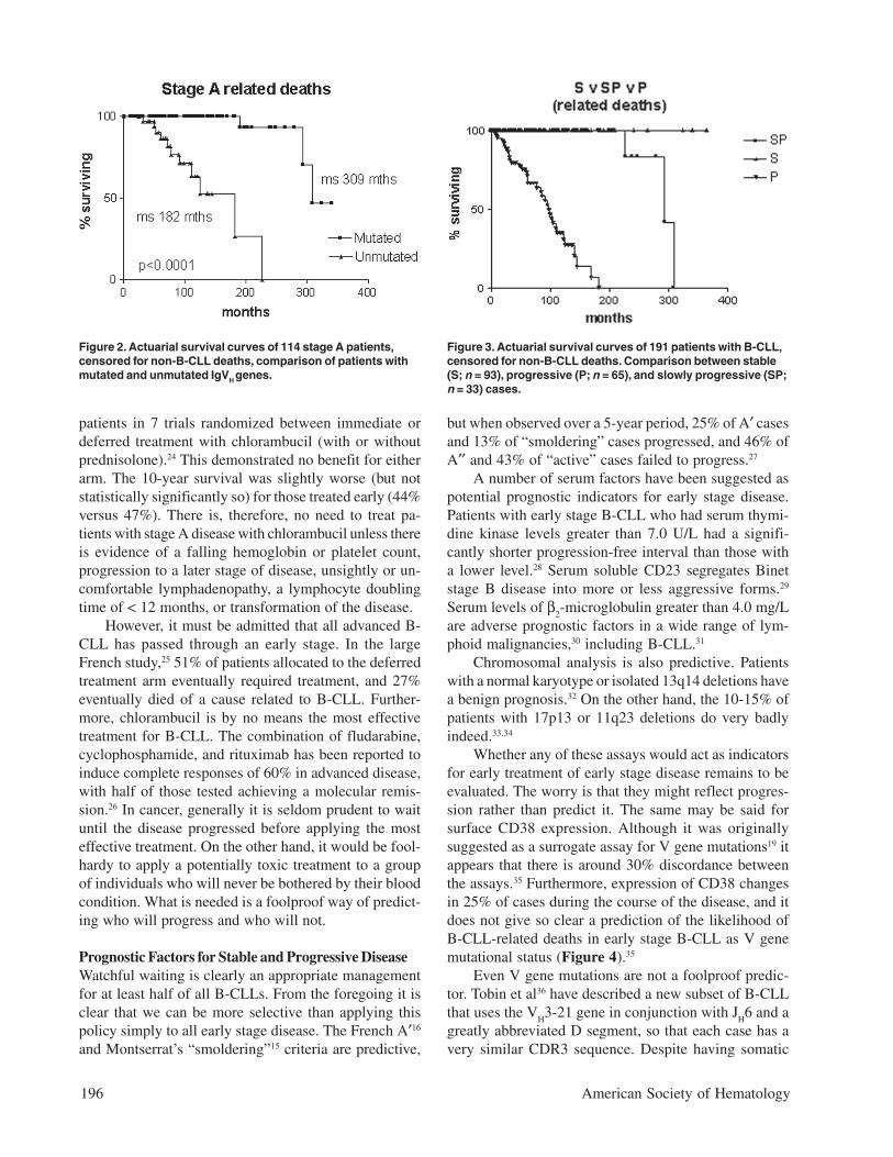
Whether any of these assays would act as indicatorsfor early treatment of early stage disease remains to beevaluated. The worry is that they might reflect progres-sion rather than predict it. The same may be said forsurface CD38 expression. Although it was originallysuggested as a surrogate assay for V gene mutations19 itappears that there is around 30% discordance betweenthe assays.35 Furthermore, expression of CD38 changesin 25% of cases during the course of the disease, and itdoes not give so clear a prediction of the likelihood ofB-CLL-related deaths in early stage B-CLL as V genemutational status (Figure 4).35
Even V gene mutations are not a foolproof predic-tor. Tobin et al36 have described a new subset of B-CLLthat uses the V
H3-21 gene in conjunction with J
H6 and a
greatly abbreviated D segment, so that each case has avery similar CDR3 sequence. Despite having somatic
Figure 2. Actuarial survival curves of 114 stage A patients,censored for non-B-CLL deaths, comparison of patients withmutated and unmutated IgVH genes.
Figure 3. Actuarial survival curves of 191 patients with B-CLL,censored for non-B-CLL deaths. Comparison between stable(S; n = 93), progressive (P; n = 65), and slowly progressive (SP;n = 33) cases.
Hematology 2002 197
mutations, this subset has a survival curve that resemblesthe unmutated subset. Time will reveal whether thereare any other exceptions.
For the moment watchful waiting is still the watch-word. At the same time, prospective clinical trials shouldevaluate whether any of the available prognostic factorscan be used to successfully guide early treatment of earlystage B-CLL that is destined to be fatal.
II. PROGRESSION EVENTS IN
B-CHRONIC LYMPHOCYTIC LEUKEMIA
Neil E. Kay, MD,1* Diane F. Jelinek, PhD,2*and Gordon W. Dewald, PhD3*
Advances in the understanding of biologic processes inmany malignant diseases have generated uniquerationale(s) for their evaluation and management. Thus,the knowledge that leukemic myeloid cells with at(15;17) translocation have an abnormal retinoic acidreceptor fusion structure has ultimately generated thesuccessful use of retinoic acid therapies. The carefuldissection of biologic events in colon cancer has uncov-ered several new areas for focus on both disease pro-gression and unique preventative approaches (use ofCox-2 inhibitors). Similarly, the study of the easily avail-able circulating blood B cells from patients with B-CLLshould be just as helpful in unraveling the mysteries of
this leukemic cell process. Even though the clinical stag-ing systems of Rai and Binet have been very useful inpredicting overall survival, they cannot predict anindividual’s risk of disease progression in the early stage.Current helpful biologic paradigms that are linked to theCLL B cell include their low level of cell cycle activity,resistance to apoptosis, expression levels of CD38, divi-sion into germline versus somatic mutation-type clones,common recurring genetic defects, and defective anti-gen presenting capacity. Other less well known but rel-evant biologic aspects include the capacity of B-CLLcells to produce multiple cytokines that impact apoptosis/drug resistance and their ability to be induced to undergocell signaling by these cytokines with subsequent alter-ation of gene expression. In addition, the presence ofadhesion molecules that relate to CLL circulatory pat-terns and acquisition of additional genetic defects areimportant biologic events relevant to clinical outcome.Finally, it is necessary to consider the impact of the mi-croenvironment on the CLL B cell. Currently, microenvi-ronmental candidates found to affect important biologicevents in the CLL B cell include circulating nurse cells,stromal cells in node and marrow, and the hypoxic atmo-sphere known to exist in CLL B cell tissue sites (e.g., mar-row). We propose that the detailed sequential analysis ofthese biologic events in early stage B-CLL disease wouldbe most instructive in predicting disease progression andin devising unique treatment strategies. This section willdiscuss some of these biologic events, in particular genetic,immunologic, angiogenesis, and apoptosis parameters, withan emphasis on early-stage B-CLL and discussion of earlyintervention points related to these parameters.
Genetic Events in B-CLLWhile the exact origin of the malignant B cell in thisdisease is unknown and not the subject of this review,there has been a considerable amount of effort dedicatedto more precise determinations of the genetic nature ofthe CLL B cell. Given the nature of the disease and ourcurrent understanding of the site of origin of CLL B cells,most of the cytogenetic studies have been aimed at mar-row and/or blood cells. These have involved the use ofconventional cytogenetics, fluorescence in situ hybrid-ization (FISH), and most recently gene profiling assaysand immunoglobulin (Ig) V
H mutational status. The ad-
vances in understanding the genetic events using theseapproaches have initially been begrudgingly obtainedbut are now accelerating.
CytogeneticsWith the introduction of B-cell mitogens, it has beenpossible to generate metaphase cells for analysis by con-ventional G-banding techniques. Importantly, this ap-
* Mayo Clinic, 1Division of Hematology, Department ofMedicine, 2Dept. of Immunology, 3Dept. of Laboratory Med.and Pathology, 200 First Street, Rochester, MN 55905
This work is in part supported by the National Cancer Institute(R01 CA95241-01, R21 CA87155-1, and R21 CA 91542-1).
Figure 4. Actuarial survival curves of 114 stage A patients,censored for non-B-CLL deaths, comparison of patients withCD38 positive and CD38 negative B-CLL.
198 American Society of Hematology
proach revealed heterogeneous, clonal genetic changesin a significant proportion of patients (~40%-50%).1,2
Contributions from this period of analysis also includedthe more common recurring defects, including trisomy12,1,2 11q–,3 deletion of 6,4,5 and abnormalities of chro-mosome 13.2,6 Interestingly, these findings have beencorrelated with clinical outcome and/or disease course.When single cytogenetic abnormalities are examined,trisomy 12 or chromosome 6 defects are associated withshortened survival.2 However, sole abnormalities of chro-mosome 13q14 are linked to a more favorable clinicaloutcome.2 Atypical morphology is also now linked tothe presence of trisomy 12.7,8 Clonal evolution in B-CLLwas first demonstrated using this approach with one studyshowing that karyotypic evolution can be detected inaround 40% of the patients over a 1-year duration.5 Inaddition, complex karyotypes could be found in B-CLLcells, and cohort studies showed poor clinical outcomefor those patients. While this technique has helped inlooking at critical genetic events, a significant percentageof CLL patients show no genetic defects likely due to theneed for more extensive in vitro CLL B cell division.
FISHConsiderable advances in definition of common recur-ring genetic defects have been uncovered using the FISHmethod, which does not require mitoses. It is now pos-sible to find these defects in higher percentages of CLLB cells, including those of patients with early-stage dis-ease. Indeed, the use of FISH has largely replaced con-ventional cytogenetics in the determination of geneticdefects in B-CLL. When FISH is used, the most com-mon gene defects are deletions involving chromosomeband 13q14, trisomy 12, deletions of 11q22.3-q23.1,deletions of 6q21-q23, and, finally, mutations or dele-tions of 17p13.9-11 Our studies have used a “CLL FISH”panel that detects common recurring genetic defects in
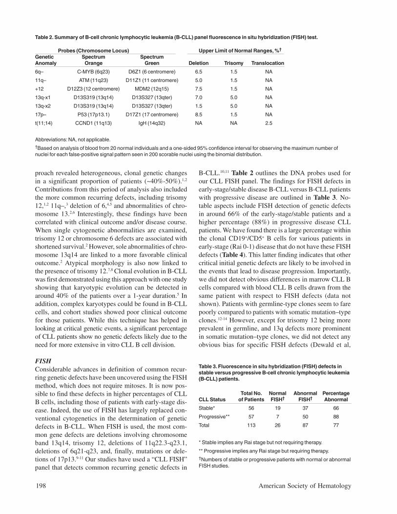
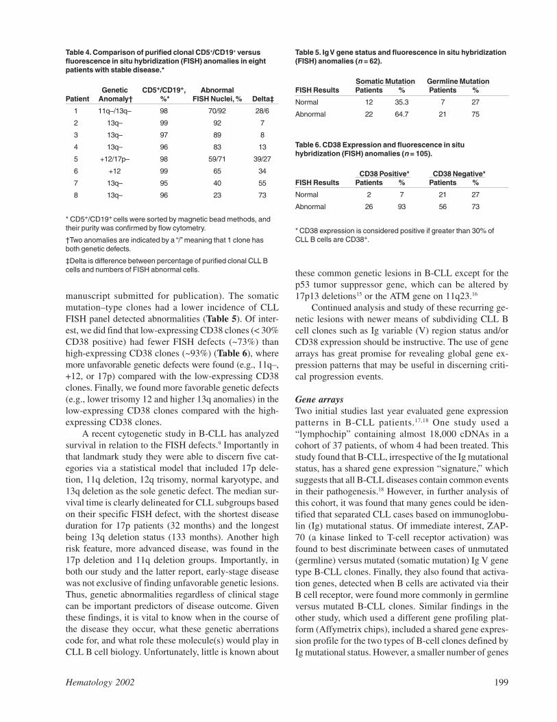
B-CLL.10,11 Table 2 outlines the DNA probes used forour CLL FISH panel. The findings for FISH defects inearly-stage/stable disease B-CLL versus B-CLL patientswith progressive disease are outlined in Table 3. No-table aspects include FISH detection of genetic defectsin around 66% of the early-stage/stable patients and ahigher percentage (88%) in progressive disease CLLpatients. We have found there is a large percentage withinthe clonal CD19+/CD5+ B cells for various patients inearly-stage (Rai 0-1) disease that do not have these FISHdefects (Table 4). This latter finding indicates that othercritical initial genetic defects are likely to be involved inthe events that lead to disease progression. Importantly,we did not detect obvious differences in marrow CLL Bcells compared with blood CLL B cells drawn from thesame patient with respect to FISH defects (data notshown). Patients with germline-type clones seem to farepoorly compared to patients with somatic mutation–typeclones.12-14 However, except for trisomy 12 being moreprevalent in germline, and 13q defects more prominentin somatic mutation–type clones, we did not detect anyobvious bias for specific FISH defects (Dewald et al,
Table 2. Summary of B-cell chronic lymphocytic leukemia (B-CLL) panel fluorescence in situ hybridization (FISH) test.
Table 3. Fluorescence in situ hybridization (FISH) defects instable versus progressive B-cell chronic lymphocytic leukemia(B-CLL) patients.
Total No. Normal Abnormal PercentageCLL Status of Patients FISH† FISH† Abnormal
Stable* 56 19 37 66
Progressive** 57 7 50 88
Total 113 26 87 77
* Stable implies any Rai stage but not requiring therapy.
** Progressive implies any Rai stage but requiring therapy.†Numbers of stable or progressive patients with normal or abnormalFISH studies.
Abbreviations: NA, not applicable.†Based on analysis of blood from 20 normal individuals and a one-sided 95% confidence interval for observing the maximum number ofnuclei for each false-positive signal pattern seen in 200 scorable nuclei using the binomial distribution.
Probes (Chromosome Locus) Upper Limit of Normal Ranges, %†
Genetic Spectrum SpectrumAnomaly Orange Green Deletion Trisomy Translocation
6q– C-MYB (6q23) D6Z1 (6 centromere) 6.5 1.5 NA
11q– ATM (11q23) D11Z1 (11 centromere) 5.0 1.5 NA
+12 D12Z3 (12 centromere) MDM2 (12q15) 7.5 1.5 NA
13q-x1 D13S319 (13q14) D13S327 (13qter) 7.0 5.0 NA
13q-x2 D13S319 (13q14) D13S327 (13qter) 1.5 5.0 NA
17p– P53 (17p13.1) D17Z1 (17 centromere) 8.5 1.5 NA
t(11;14) CCND1 (11q13) IgH (14q32) NA NA 2.5
Hematology 2002 199
manuscript submitted for publication). The somaticmutation–type clones had a lower incidence of CLLFISH panel detected abnormalities (Table 5). Of inter-est, we did find that low-expressing CD38 clones (< 30%CD38 positive) had fewer FISH defects (~73%) thanhigh-expressing CD38 clones (~93%) (Table 6), wheremore unfavorable genetic defects were found (e.g., 11q–,+12, or 17p) compared with the low-expressing CD38clones. Finally, we found more favorable genetic defects(e.g., lower trisomy 12 and higher 13q anomalies) in thelow-expressing CD38 clones compared with the high-expressing CD38 clones.
A recent cytogenetic study in B-CLL has analyzedsurvival in relation to the FISH defects.9 Importantly inthat landmark study they were able to discern five cat-egories via a statistical model that included 17p dele-tion, 11q deletion, 12q trisomy, normal karyotype, and13q deletion as the sole genetic defect. The median sur-vival time is clearly delineated for CLL subgroups basedon their specific FISH defect, with the shortest diseaseduration for 17p patients (32 months) and the longestbeing 13q deletion status (133 months). Another highrisk feature, more advanced disease, was found in the17p deletion and 11q deletion groups. Importantly, inboth our study and the latter report, early-stage diseasewas not exclusive of finding unfavorable genetic lesions.Thus, genetic abnormalities regardless of clinical stagecan be important predictors of disease outcome. Giventhese findings, it is vital to know when in the course ofthe disease they occur, what these genetic aberrationscode for, and what role these molecule(s) would play inCLL B cell biology. Unfortunately, little is known about
these common genetic lesions in B-CLL except for thep53 tumor suppressor gene, which can be altered by17p13 deletions15 or the ATM gene on 11q23.16
Continued analysis and study of these recurring ge-netic lesions with newer means of subdividing CLL Bcell clones such as Ig variable (V) region status and/orCD38 expression should be instructive. The use of genearrays has great promise for revealing global gene ex-pression patterns that may be useful in discerning criti-cal progression events.
Gene arraysTwo initial studies last year evaluated gene expressionpatterns in B-CLL patients.17,18 One study used a“lymphochip” containing almost 18,000 cDNAs in acohort of 37 patients, of whom 4 had been treated. Thisstudy found that B-CLL, irrespective of the Ig mutationalstatus, has a shared gene expression “signature,” whichsuggests that all B-CLL diseases contain common eventsin their pathogenesis.18 However, in further analysis ofthis cohort, it was found that many genes could be iden-tified that separated CLL cases based on immunoglobu-lin (Ig) mutational status. Of immediate interest, ZAP-70 (a kinase linked to T-cell receptor activation) wasfound to best discriminate between cases of unmutated(germline) versus mutated (somatic mutation) Ig V genetype B-CLL clones. Finally, they also found that activa-tion genes, detected when B cells are activated via theirB cell receptor, were found more commonly in germlineversus mutated B-CLL clones. Similar findings in theother study, which used a different gene profiling plat-form (Affymetrix chips), included a shared gene expres-sion profile for the two types of B-cell clones defined byIg mutational status. However, a smaller number of genes
Table 4. Comparison of purified clonal CD5+/CD19+ versusfluorescence in situ hybridization (FISH) anomalies in eightpatients with stable disease.*
Genetic CD5+/CD19+, AbnormalPatient Anomaly† %* FISH Nuclei, % Delta‡
1 11q–/13q– 98 70/92 28/6
2 13q– 99 92 7
3 13q– 97 89 8
4 13q– 96 83 13
5 +12/17p– 98 59/71 39/27
6 +12 99 65 34
7 13q– 95 40 55
8 13q– 96 23 73
* CD5+/CD19+ cells were sorted by magnetic bead methods, andtheir purity was confirmed by flow cytometry.
†Two anomalies are indicated by a “/” meaning that 1 clone hasboth genetic defects.
‡Delta is difference between percentage of purified clonal CLL Bcells and numbers of FISH abnormal cells.
Table 5. Ig V gene status and fluorescence in situ hybridization(FISH) anomalies (n = 62).
Somatic Mutation Germline MutationFISH Results Patients % Patients %
Normal 12 35.3 7 27
Abnormal 22 64.7 21 75
Table 6. CD38 Expression and fluorescence in situhybridization (FISH) anomalies (n = 105).
CD38 Positive* CD38 Negative*FISH Results Patients % Patients %
Normal 2 7 21 27
Abnormal 26 93 56 73
* CD38 expression is considered positive if greater than 30% ofCLL B cells are CD38+.
200 American Society of Hematology
(around 30) were identified with differential expressionthat could distinguish Ig somatic mutation–type versusgermline-type clones.17 CLL gene profile analysisshowed that CLL B cells were most closely related tomemory B cells. Specific genes found to be over-expressed included guanine nucleotide exchange factorswith implications for the Ras pathway in the pathogen-esis of this leukemia. They also found the enhanced ex-pression of mRNA encoding for cytokine (e.g., IL-4R)or chemokine receptors (e.g., CCR7) when compared tonormal memory B cells.
Our gene array approach was to compare CLL Bcells purified from 38 patients of either somatic muta-tion–type or germline-type clones to highly purifiedblood B cells.19 Once again, we found the CLL geneexpression signature pattern is distinct from that of nor-mal B cells, but we were not able to clearly distinguishbetween germline- and somatic mutation–type clones.However, with the use of a novel software program(genes@work, IBM, New York), it was possible to dis-cern gene expression patterns between the two Ig V
H
clones when factoring in if they were either CD38 high-or low-expression clones. This program also allowed usto further refine the differential gene expression patternbetween control B cells and CLL clones so only 81 geneswere discriminating. Some of these genes included thosecoding for LEF1, TGFBR3, and fibromodulin, 2 of whichcan be implicated in tumorigenesis. Of interest, it wasnot possible to discriminate between early- and late-stageB-CLL using this approach. Thus, gene array approachesare likely identifying genetic events that occur relativelyearly in the disease course. Collectively, this approachhas many implications for investigation in B-CLL. Dis-covery of unique markers for B-CLL will be valuable indiagnosis, identification of events that occur in CLL Bcells leading to disease progression, and eventually po-tential therapeutic targets. This would be particularlyuseful where we find clear discrimination of geneticevents between normal cells and CLL B cells.
Immune Defects in B-CLLWhile the primary event in B-CLL is the malignant trans-formation of the CLL B cell, it is likely that progressionis linked to an abnormal immune system in the host. Pro-gression of this disease could be related to dysfunctionalcomponents of the host immune cellular arm. Early workshowed that while absolute T-cell numbers are elevatedin untreated B-CLL, there are a large number of immunecell defects.20 These defects affect all cellular compo-nents of the immune system, including quantitative andqualitative aspects of the normal B-cell pool, T-cell sub-sets, natural killer (NK) cells, and dendritic cells. NKcells are deficit in function, as are dendritic cells.21,22 The
latter two aspects are likely to have important roles indisease progression, since NK cells are the first line ofdefense in dealing with malignancy, and dendritic cellsare crucial in the induction of an effective cytotoxic T-cell response to tumor cells. One additional factor in theoverall dendritic cell capacity for B-CLL patients is theobservation that the CLL B cell has a defective antigen-presenting capacity and can render the T cell anergic.23
Thus, blood and splenic CD4 T cells of patients withCLL failed to express surface CD154 after CD3 cross-linking. However, normal T cells exposed to increasinglevels of CLL B cells will lose CD154 expression.23
These same authors have shown that with gene transferof CD40L, the CLL B cell can become a better antigenpresenting cell.24 We have shown that T cells in B-CLLhave a polarized Th-2 type cytokine profile with in-creased spontaneous synthesis and secretion of IL-4.25
Since IL-4 is an anti-apoptosis cytokine for CLL B cells,IL-4 creates a favorable microenvironment for the leu-kemic CLL B cells. In addition, the polarization of Th-2type cells can result in lesser T-cell cytotoxic capacityand could favor tumor progression.
Does the progression of B-CLL relate to these im-mune abnormalities? To a certain extent, this questioncould be answered if we knew when these arrays of im-mune abnormalities occurred in the course of a specificpatient’s illness. Current evidence does suggest that theimmune defects accrue with increasing stage.20 The im-pact of chemotherapy certainly can add to the deficitarray of immune abnormalities seen in a given patient.Attention to correction of these immune defects, even inearly-stage CLL patients, may yield additional time with-out morbidity and/or without the need for therapy. Con-sideration of cytokines such as IL-2, interferon-α/γ, IL-7, and/or IL-12 as immune adjuvants designed to en-hance the host cellular resistance to clonal emergencein early-stage disease may be warranted.
Angiogenesis in B-CLLThe impact of neovascularization and angiogenic path-ways in B-CLL has only recently begun to be explored.The findings to date suggest that abnormal vasculariza-tion exists in the bone marrow and nodes of B-CLL pa-tients and that this is associated with progressive dis-ease.26,27 The etiology of the abnormal blood vessel for-mation in B-CLL tissue sites is likely related to the spon-taneous production by CLL B cells of the proangiogenicfactor VEGF. The B-CLL cells are also capable of gen-erating increased amounts of VEGF under hypoxic con-ditions.27,28 However, there are other proangiogenic fac-tors produced by B-CLL cells, including bFGF.26,28 Sincethe marrow site is normally under hypoxic conditions, itis likely the in vivo spontaneous VEGF production of
Hematology 2002 201
CLL cells is augmented by increasing tumor burden. Theincreased vascularization noted in marrow and nodes ofB-CLL patients is highly suggestive that, like solid tu-mor development, the B-CLL process may depend onabnormal blood flow for disease progression. Evidencefor this includes our observations that abnormal marrowangiogenesis is positively associated with increased Raistage.26 In addition, elevated serum VEGF levels corre-late with advanced disease stage.29 However when in-tracellular levels are measured the lower the VEGF valuethe poorer the clinical outcome.30 The full angiogenicpotential of CLL B cells is revealed by the leukemic CLLB cell ability to produce both pro- and anti-angiogenicfactors. This analysis has shown that CLL B cells pro-duce more anti-angiogenic factor (e.g., thrombospondin-1 [TSP-1]) and less VEGF for early-stage disease com-pared with late stage and for somatic mutation-type ver-sus germline-type clones.28 It would appear that CLL Bcells are capable of undergoing “angiogenic switching,”which has been shown in solid tumors to be linked todisease progression. The exact mechanism(s) for thisbiologic maneuver in B-CLL is not clear, but exposureto hypoxic conditions can induce switching in vitro. Inaddition, we and others have shown that matrixmetalloproteinases or gelatinases (MMP) 9 and 2 aresynthesized and secreted by CLL B cells.31 These mol-ecules are crucial in the proteolytic degradation of com-ponents of the extracellular matrix (e.g., fibronectin,collagen) leading to enhanced angiogenesis. Recent workhas shown that interferons can down-regulate the func-tion of MMP-9 secreted by CLL B cells.31 Further workdesigned to delineate mechanisms for angiogenic switch-ing in B-CLL cells will surely add to our ability to eithertreat the disease more effectively or keep the disease ina more stable phase. Finally, analysis of the angiogeniccapacity of the CLL B cell has revealed that CLL B cellsmay utilize the VEGF pathway as an autocrine pathwayto increase their resistance to apoptosis. CLL B cells notonly produce and secrete VEGF but also possess mem-brane receptors for VEGFR1 (Flt-1) and VEGFR2(KDR). We have also shown that at least one of thesereceptors, VEGFR1, is spontaneously phosphorylated inCLL B cells. Addition of VEGF in vitro significantlyincreases the ability of a CLL B cell to resist undergoingspontaneous or drug-induced apoptosis. Thus, interrup-tion of this pathway by various mediators (e.g., anti-VEGF or anti-VEGFR antibodies, receptor tyrosine ki-nase inhibitors) may prove useful in the management ofB-CLL. bFGF has also been shown to be associated withincreased CLL B cell apoptosis resistance, suggestingthat vascular factors play a key role in this importantfeature of the CLL B cell.32 While the VEGF pathway islikely contributing to the significant resistance to
apoptosis seen in CLL B cell clones, there are many re-dundant mechanisms that regulate this parameter.
ApoptosisThis biologic parameter is a central feature of the leuke-mic nonproliferating CD5+/CD19+ B cells of B-CLLpatients. A full understanding of the pathways related toapoptosis resistance in these cells is critical both in un-derstanding the disease process and in devising uniquestrategies. There is a growing and very significant bodyof literature defining the mechanisms of apoptosis inhealth and disease. Clearly, defects in normal apoptoticmachinery contribute in many ways to tumor progres-sion. Any consideration of the apoptosis status in B-CLLneeds to consider the existing leukemic cell genetic de-fects and the surrounding microenvironment (e.g., stro-mal cells, vascular supply), which modifies these ge-netic events, since this interaction could lead to diseaseprogression. The latter feature is underscored by the dra-matic induction of in vitro apoptosis for the long-livedCLL B cell when taken out of the host. This section willbe limited to consideration of CLL B cell apoptosis as itrelates to both autocrine pathways and cellular interac-tions believed to be linked to disease progression in B-CLL. In addition, how the knowledge of these eventscan be used as new therapeutic intervention points willbe considered.
Early-stage B-CLL often has expanded polyclonalT cells in blood;20 in specific, it is known that the T-helper (CD4+) cell is frequently found in infiltrated nodesand marrow.33,34 Since cytokines elaborated by T cells,such as IL-4 and interferon (α and γ), are able to in-crease CLL B cell apoptosis resistance, the intimacy ofthese 2 cells could work to the advantage of the CLL Bcell. CD4+ T cells may be able to regulate disease pro-gression by virtue of a CD40-CD40L interaction wheresurvivin is induced in CLL B cells. Survivin is an inhibi-tor of apoptosis that can also modulate cell prolifera-tion. Thus, this type of interaction can result in the in-crease of survivin-positive CLL B cells.35 These survivin-positive CLL B cells tend to have higher proliferativerates and increases in Bcl-2,35 and ultimately this inter-action may be related to disease progression.
Other cellular components that may be contributingto apoptosis and disease progression are both the den-dritic cell and the bone marrow stromal cell. Studies ofmarrow stromal cells in B-CLL show that the leukemicCLL B cell once bound to stromal cells is further pro-tected from cell death.36 Of interest, patients in the early-stage phase of the CLL disease process can have CLLcells found in close association with follicular dendriticcells, suggesting that this interaction could facilitate dis-ease progression.37 Definition of the surface membrane

202 American Society of Hematology
Table 7. Cytokines and their receptors on chronic lymphocyticleukemia (CLL) B cells.
Cytokine† Cytokine Receptors‡ Biologic Effect§
IL-2 Present Inhibits apoptosis
IL-4 Present Inhibits apoptosis
IL-8 Present Inhibits apoptosis
IL-10 Present Induces apoptosis
TNFα ? Inhibits apoptosis
TGFβ Present Induces apoptosis
Interferon α/γ Present Inhibits apoptosis
VEGF Present (both VEGFR1 Inhibits apoptosisand VEGFR2)
bFGF ? Induces drug resistance
CXCR4 Present Stimulates chemotaxis
CXCR3 Present Unknown
†Cytokines have been shown to be synthesized and secreted byCLL B cells.‡Receptors specific for the respective cytokine have been found onCLL B cell membranes.§Functional impact of each cytokine on the clonal CLL B cellpopulation.
Abbreviations: IL, interleukin; TNF, tumor necrosis factor; TGF,tumor growth factor; VEGF, vascular endothelial growth factor;bFGF, basic fibroblast growth factor.
Figure 5. The maintenance and expansion of the malignant B-cell chronic lymphocytic leukemia (B-CLL) clone is primarilyrelated to the genetic events induced by various inherited orenvironmental processes that lead to transformation. However,the overall process of leukemic B-cell clone survivorship isalso linked to a complex circuitry of cytokines and chemokinesthat act in both autocrine and paracrine pathways. Importantly,stromal cells, dendritic cells, endothelial cells, nurse cells, andvarious immune cells contribute to this cytokine/chemokinearray, which ultimately contributes to the behavior of the CLLB cell. Apparent physical association of the leukemic CLL Bcell with these various cell types suggests that there issignificant cellular crosstalk that assists in the induction of amicroenvironment favorable to the leukemic CLL B cell.
determinants that lead/facilitate these cellular interac-tions could be helpful in delaying disease progression.
More recently, a nurse like cell has been describedin the blood of B-CLL patients, and this cell is able toconfer significant increases in apoptosis resistance toCLL B cells.38,39 The exact mechanism of resistance ap-pears to be linked to elaboration of SDF-1, which bindsto its cognate receptor CXCR4. Given that these cellu-lar aspects exist in early-stage disease, interruption ofthe cellular interactions between CLL B cells and theserespective elements represents a candidate for therapeuticintervention. If this can be done with minimal toxicity,then it may be feasible to consider using these interven-tions in early-stage patients to prevent progression. Theredundancy of CLL B cell apoptosis resistance is fur-ther underscored by our recent finding of a unique path-way mediated by a family of molecules related to tumornecrosis factors designated as April//Blys.40
Finally, a list of cytokines and their respective re-ceptors found on CLL B cells are featured in Table 7.Clearly, the microenvironmental influence on a CLL Bcell clone will be strongly related to the cell types andthe nature of the cytokines secreted by those cell typesin the immediate area. Issues in the future will be to knowwhich of these cytokines predominate in the CLL B cell
involved tissue sites and which of these cytokines havethe most impact on CLL B cell viability. In addition, itwill be important to know if these CLL B cells acquireautocrine pathways sequentially and if these acquiredpathways relate to disease progression. Importantly, eachof these cytokine pathways provides an opportunity fortherapeutic interventions. Thus, we have shown that apseudomonas exotoxin-conjugated IL-4 molecule41 isable to kill a significant percentage of CLL B cells (datanot shown). Since the IL-4 receptor is not as well ex-pressed in other tissue sites, this approach has the ad-vantage of working in a selective fashion as well.
SummaryThis discussion has attempted to highlight the emergingareas of biology that will help us understand disease pro-gression in B-CLL. Clearly, this is not going to be onesimple equation but will likely involve the use of com-plex models for developing the best formulae to helppredict progression. Figure 5 outlines a schematic modelof the interplay between CLL cells and their microenvi-ronment. While genetic events in the transformation ofthis B cell are crucial to the disease process, this malig-nant cell has the potential to have multiple points of in-teraction with its neighbors and the secreted moleculesof the resident cells. Understanding the dynamics of thisleukemic cell alone and within the framework of the tis-sue milieu will ultimately lead us to both more accu-
Hematology 2002 203
rate predictions for progression and, we hope, bettertherapies.
III. TO TREAT OR NOT TO TREAT THE
PATIENT WITH CLL: THAT IS THE QUESTION
John C. Byrd, MD,* Sherif Farag, MD, PhD,‡
Margaret Lucas, PA-C,‡ and Thomas Lin, MD, PhD‡
Therapeutic Intervention for CLLCLL is one of the most common types of adult leuke-mia, with registry data suggesting that 7000 patients arediagnosed and 4500 die related to this disease each yearin the United States.1 As the most common leukemia,virtually all oncologists, hematologists, and internists willcare for patients with CLL. Important issues in CLL carecan be divided by four junctures met during the courseof each patient’s disease: (1) making the definitive diag-nosis of CLL and determining individual patient prog-nosis; (2) determining when to treat the CLL; (3) deter-mining how to initially treat the CLL; and (4) determin-ing a salvage regimen suitable for an individual patient’scase in relapse. Herein we focus entirely on the initialdiagnosis and the initial treatment of CLL. For furtherdiscussion of salvage therapy, the reader is referred to arecent review.2
Now That I Have CLL, What Does It Mean to Me?At initial presentation, the great majority (80-90%) ofpatients with CLL will be asymptomatic. In this setting,the first question is how will this disease and its associ-ated complications impact the patient’s well being? Amajor focus of research in CLL over the past 3 decadeshas been to examine clinical and laboratory features thatpredict which patients will develop symptomatic disease.(reviewed in ref. 3) As with other cancers, outcome of
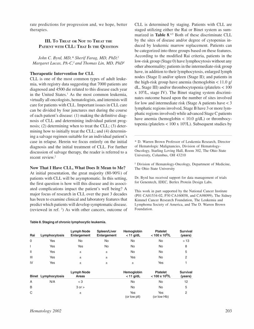
CLL is determined by staging. Patients with CLL arestaged utilizing either the Rai or Binet system as sum-marized in Table 8.4,5 Both of these discriminate CLLby the sites of disease and/or degree of cytopenias in-duced by leukemic marrow replacement. Patients canbe categorized into three groups based on these features.According to the modified Rai criteria, patients in thelow-risk group (Stage 0) have lymphocytosis without anyother abnormality; patients in the intermediate-risk grouphave, in addition to their lymphocytosis, enlarged lymphnodes (Stage I) and/or spleen (Stage II); and patients inthe high-risk group have anemia (hemoglobin < 11.0 g/dL, Stage III) and/or thrombocytopenia (platelets < 100x 109/L, stage IV). The Binet staging system discrimi-nates outcome based upon the number of sites involvedfor low and intermediate risk (Stage A patients have < 3lymphatic regions involved, Stage B have 3 or more lym-phatic regions involved) while advanced Stage C patientshave anemia (hemoglobin < 10.0 g/dL) or thrombocy-topenia (platelets < 100 x 109/L). Subsequent studies by
* D. Warren Brown Professor of Leukemia Research, Directorof Hematologic Malignancies, Division of Hematology-Oncology, Starling Loving Hall, Room 302, The Ohio StateUniversity, Columbus, OH 43210
‡ Division of Hematology-Oncology, Department of Medicine,The Ohio State University
Dr. Byrd has received support for data management of trialsfor Genentech, IDEC, Berlex Protein Design Labs.
This work in part supported by the National Cancer Institute(P01 CA81534-02, P30 CA160058, and CA98099), The SidneyKimmel Cancer Research Foundation, The Leukemia andLymphoma Society of America, and The D. Warren BrownFoundation.
Table 8. Staging of chronic lymphocytic leukemia.
Lymph Node Spleen/Liver Hemoglobin Platelet SurvivalRai Lymphocytosis Enlargement Enlargement < 11 g/dL < 100 x 109/L (years)
0 Yes No No No No > 13
I Yes Yes No No No 8
II Yes ± ± No No 5
III Yes ± ± Yes No 2
IV Yes ± ± ± Yes 1
Lymph Node Hemoglobin Platelet SurvivalBinet Lymphocytosis Areas < 11 g/dL < 100 x 109/L (years)
A N/A < 3 No No 12
B 3 or > No No 5
C ± Yes Yes 2(or low plt) (or low Hb)
204 American Society of Hematology
several groups have further dissected therapeutic out-come of early stage CLL and have identified a groupthat can be considered to have smoldering CLL. Whiledifferent criteria are utilized,6-8 these patients generallyhave Binet Stage A disease, non-diffuse bone marrowinvolvement, a lymphocyte count of < 30 × 109/L, he-moglobin > 13 g/dL, and lymphocyte doubling timegreater than 12 months. It is very unusual for patients tohave diffuse bone marrow involvement with the otherfeatures listed (less than 2%) resulting in many practi-tioners (including the authors) not evaluating bone mar-row morphology at diagnosis. In these patients, the riskof progression to symptomatic CLL is low (14-17%) at5 years, and clinical outcome in this group is the sameas the age-matched control population.6-8
Nonetheless, both staging systems fail to considerfactors such as age and recently identified genetic orother molecular markers as prognostic features. A bet-ter understanding of these recently identified featureswill allow improved decisions regarding the appropriateinitial intervention. These prognostic features are out-lined in Sections I and II. These newer prognostic fea-tures and our developing understanding of CLL as a dis-ease in evolution has re-opened the question of the roleof treatment in early stage disease for patients with poorprognostic features.
I’m 55 Years Old and Too Young to Have ThisDiagnosis, What Does It Mean to Me?
CLL is generally a disease of the elderly, with a medianage at diagnosis exceeding 60 years. Only 10% of pa-tients diagnosed with CLL are under the age of 50 and1-2% are younger than 40. Several series have exam-ined clinical features of younger (age < 50-55) CLLpatients and have consistently noted a higher male pre-dominance.9-12 In some series a higher frequency ofRichter’s transformation has been noted.9,10 The stagedistribution of patients at diagnosis is similar betweenyounger and elderly patients across all series reportedand the Rai and Binet staging systems are effective atpredicting clinical outcome. While younger patients havean improved survival when compared to older patientswith CLL, their median survival was noted to be 12.3years in one series whereas age-matched controls had alife expectancy of 31.2 years.12 In another series,9 CLLpatients under age 55 had an expected 10-year survivalof 45.3% versus the 96.2% survival in the control popu-lation. In this second series, only 40% of the youngerpatients remained without treatment whereas 49% of theelderly patients had required therapy. In each of the largereported series, only patients with smoldering CLL ap-pear relatively unaffected by their disease, with a 94%12-year survival in one series9 and an 85% 14-year pre-
dicted survival in another series.12 Thus, early treatmentshould not be considered for young patients with smol-dering CLL until some evidence of progression occurs(i.e., patient no longer qualifying as smoldering CLL),but consideration of early intervention trials with the goalof cure would appear to be of most relevance to youngerpatients where loss of absolute life expectancy is greatest.
When to Treat CLL?Clinicians caring for patients with CLL must focus uponthe most critical clinical juncture, that being when toinitiate treatment. In the absence of symptoms, observa-tion without therapy is the current standard of care. Thisis based on mature data from seven different studies dem-onstrating no improvement in overall survival for symp-tomatic CLL patients receiving early therapeutic inter-vention with chlorambucil therapy (reviewed in 13). Ameta-analysis13 that included data from six of these tri-als and outcomes of 2048 patients demonstrated a slightlyhigher death rate (42.6%) among those treated early ver-sus those randomized to deferred therapy (41.6%), al-though this difference was not statistically significant.Deaths attributable to secondary malignancies were simi-lar in both groups,13,14 despite one early report15 noting ahigher risk of epithelial malignancies in patients receiv-ing early intervention. It must be noted that in each ofthese studies, a large proportion of patients with smol-dering or early stage disease were included. For thispopulation of patients, therapeutic benefit would haveto improve overall survival in excess of that observed inthe age-matched control population without CLL. Fur-thermore, each of these studies employed chlorambucil,an agent that yields a low complete response rate. Withmore effective therapies and use of risk stratification,re-consideration of this approach in well-designed trialsseems quite prudent. This is further substantiated by thenow recognized genetic clonal evolution of CLL thatoccurs over time from initial diagnosis.16-19 This clonalevolution coincides with the CLL cells becoming moreresistant to apoptosis.20 Efforts within the German CLLStudy Group and several US groups are underway toexamine this question with newer CLL therapies.
Determining what constitutes sufficient symptoma-tology to initiate treatment in CLL is subjective and con-founds cross-interpretation of clinical trials. To assistclinicians and assure uniform study entrance criteria, anNCI sponsored Working Group on CLL21 establishedguidelines for initiation of treatment. These indicationsinclude the presence of non-autoimmune cytopenias (RaiStage III and IV), symptomatic lymphadenopathy orhepatosplenomegaly, disease-related B-symptoms orfatigue, extreme lymphocytosis (greater than 150-300 ×109/L), and autoimmune hemolytic anemia or thromb-
Hematology 2002 205
ocytopenia not controlled with steroids. Incorporationof the patient’s individuality and associated co-morbidproblems must also be considered. In this manner, it isimperative for the hematologist to critically question ifthe symptoms are from CLL or another unrelated diag-nosis. For instance, fatigue might be reflective of under-lying cardiovascular disease, sleep apnea, depression,hypothyroidism, or secondary malignancy. Careful as-sessment of the CLL patient as an internist first and thenas a hematologist will assure treatment is not initiatedfor a source unrelated to the underlying disease. Indeed,the decision of when to start therapy remains one of themost challenging issues physicians caring for patientswith CLL have to make.
How to Treat Symptomatic CLL?There has been considerable debate regarding what isthe best initial therapeutic approach for symptomaticCLL. CLL virtually always presents as a systemic dis-ease and is generally treated with chemotherapy. Untilrecently, the goal of therapy has been one of palliationonly, without hope of complete remission of the CLL.Introduction of new therapies outlined below and novelcombination approaches has begun shifting the thera-peutic goal of CLL to that of attaining a complete re-mission as defined by a recent consensus paper on re-sponse evaluation in CLL.21 A summary of differentclasses of therapies employed for the treatment of pre-viously untreated, but symptomatic CLL patients followsbelow.
Alkylator therapyThe initial treatment of patients with symptomatic CLLhas often involved therapy with chlorambucil. While thistherapy is effective at palliating the majority of CLLpatients, no randomized studies have established its ben-efit relative to overall survival as compared to observa-tion alone. Prednisone is often included with chloram-bucil despite three randomized studies not demonstrat-ing a survival advantage to its addition (reviewed in 13).Furthermore, a meta-analysis that included 424 patientsfrom three separate trials randomized to receive chloram-bucil versus chlorambucil and prednisone could not dem-onstrate a survival benefit.13 Prednisone and other corti-costeroids as a single agent have a minimal responserate in CLL, predispose to opportunistic infections, andcan accentuate hyperleukocytosis.22 Outside of support-ive therapy for autoimmune complications, there is littledata to support including corticosteroids in the initialtreatment of CLL. Since the introduction of chloram-bucil, a variety of studies have examined different sched-ules and dose intensities in previously untreated CLLwith varied results. The most promising regimen was a
high-dose regimen (approximately 5 times the usual dos-ing) reported by Jaksik and colleagues.23 A fixed 15 mgdaily dose of chlorambucil was administered until tox-icity or complete remission. A control arm received stan-dard dose chlorambucil. An alternative response assess-ment different than the NCI criteria was utilized that didnot require bone marrow evaluation. After attainmentof CR, patients received a twice-weekly dose of 15 mgof chlorambucil for 3 years. Even without the ability todirectly compare these results to other regimens that usedthe NCI response criteria,21 this regimen was impres-sive. The treatment arm showed significantly higher CRrate (70% versus 31%, P < 0.0001) and overall survival(P < 0.005) as compared to the control arm where pa-tients received 75 mg of chlorambucil monthly for 6cycles followed by maintenance as outlined above. He-matological toxicity was manageable with rapid dosereduction for cytopenias. A subsequent Phase III studyby this same group has demonstrated this high dosechlorambucil regimen to be superior to mini-CHOP (cy-clophosphamide, adriamycin, vincristine, and pred-nisone)24 and to have a similar toxicity and efficacy pro-file as fludarabine.25 The major limitation to buildingupon this regimen with other active agents is myelo-suppression.23-25 However, the lack of cellular and hu-moral immune suppression observed with chlorambucilas compared with the nucleoside analogs or Campath-1H make this regimen the most promising for investiga-tions incorporating immune based therapy (i.e., vac-cines). Specific details of chlorambucil dosing and usefor CLL patients have been reported.26
Combination chemotherapynot including purine analogsA variety of different combination regimens used in non-Hodgkin’s lymphoma such as mini-CHOP, CAP (cyclo-phosphamide, doxorubicin, and prednisone), COP (cy-clophosphamide, vincristine, and prednisone), andchlorambucil + epirubicin have been examined for effi-cacy against chlorambucil-based regimens in previouslyuntreated CLL (reviewed in 13). These studies have notdemonstrated the superiority of combination-based ap-proaches over chlorambucil regimens with respect to pro-longation of survival. Differentiating the frequency ofcomplete response rates in each of these studies is quitedifficult because the NCI 96 criteria21 were not employed.Additionally, most studies did not require confirmatorybone marrow biopsies at completion of therapy or didnot report how many CR patients had residual nodules(PR by modified NCI 96 criteria). A meta-analysis ofthese studies has been reported as well that corroboratesthe lack of benefit of these NHL therapies for CLL.13
206 American Society of Hematology
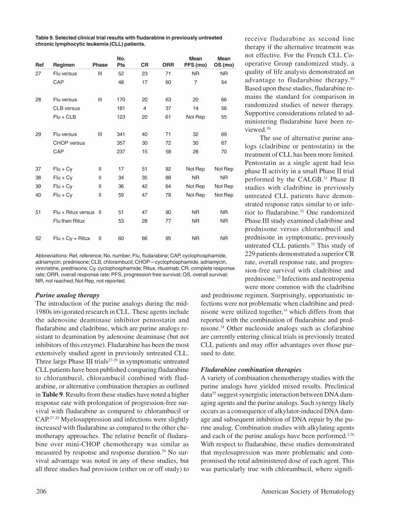
Purine analog therapyThe introduction of the purine analogs during the mid-1980s invigorated research in CLL. These agents includethe adenosine deaminase inhibitor pentostatin andfludarabine and cladribine, which are purine analogs re-sistant to deamination by adenosine deaminase (but notinhibitors of this enzyme). Fludarabine has been the mostextensively studied agent in previously untreated CLL.Three large Phase III trials27-29 in symptomatic untreatedCLL patients have been published comparing fludarabineto chlorambucil, chlorambucil combined with flud-arabine, or alternative combination therapies as outlinedin Table 9. Results from these studies have noted a higherresponse rate with prolongation of progression-free sur-vival with fludarabine as compared to chlorambucil orCAP.27-29 Myelosuppression and infections were slightlyincreased with fludarabine as compared to the other che-motherapy approaches. The relative benefit of fludara-bine over mini-CHOP chemotherapy was similar asmeasured by response and response duration.29 No sur-vival advantage was noted in any of these studies, butall three studies had provision (either on or off study) to
receive fludarabine as second linetherapy if the alternative treatment wasnot effective. For the French CLL Co-operative Group randomized study, aquality of life analysis demonstrated anadvantage to fludarabine therapy.30
Based upon these studies, fludarabine re-mains the standard for comparison inrandomized studies of newer therapy.Supportive considerations related to ad-ministering fludarabine have been re-viewed.26
The use of alternative purine ana-logs (cladribine or pentostatin) in thetreatment of CLL has been more limited.Pentostatin as a single agent had lessphase II activity in a small Phase II trialperformed by the CALGB.31 Phase IIstudies with cladribine in previouslyuntreated CLL patients have demon-strated response rates similar to or infe-rior to fludarabine.32 One randomizedPhase III study examined cladribine andprednisone versus chlorambucil andprednisone in symptomatic, previouslyuntreated CLL patients.33 This study of229 patients demonstrated a superior CRrate, overall response rate, and progres-sion-free survival with cladribine andprednisone.33 Infections and neutropeniawere more common with the cladribine
and prednisone regimen. Surprisingly, opportunistic in-fections were not problematic when cladribine and pred-nisone were utilized together,33 which differs from thatreported with the combination of fludarabine and pred-nisone.34 Other nucleoside analogs such as clofarabineare currently entering clinical trials in previously treatedCLL patients and may offer advantages over those pur-sued to date.
Fludarabine combination therapiesA variety of combination chemotherapy studies with thepurine analogs have yielded mixed results. Preclinicaldata35 suggest synergistic interaction between DNA dam-aging agents and the purine analogs. Such synergy likelyoccurs as a consequence of alkylator-induced DNA dam-age and subsequent inhibition of DNA repair by the pu-rine analog. Combination studies with alkylating agentsand each of the purine analogs have been performed.2,36
With respect to fludarabine, these studies demonstratedthat myelosupression was more problematic and com-promised the total administered dose of each agent. Thiswas particularly true with chlorambucil, where signifi-
Table 9. Selected clinical trial results with fludarabine in previously untreatedchronic lymphocytic leukemia (CLL) patients.
No. Mean MeanRef Regimen Phase Pts CR ORR PFS (mo) OS (mo)
27 Flu versus III 52 23 71 NR NR
CAP 48 17 60 7 54
28 Flu versus III 170 20 63 20 66
CLB versus 181 4 37 14 56
Flu + CLB 123 20 61 Not Rep 55
29 Flu versus III 341 40 71 32 69
CHOP versus 357 30 72 30 67
CAP 237 15 58 28 70
37 Flu + Cy II 17 51 92 Not Rep Not Rep
38 Flu + Cy II 34 35 88 NR NR
39 Flu + Cy II 36 42 64 Not Rep Not Rep
40 Flu + Cy II 59 47 78 Not Rep Not Rep
51 Flu + Ritux versus II 51 47 90 NR NR
Flu then Ritux 53 28 77 NR NR
52 Flu + Cy + Ritux II 60 66 95 NR NR
Abbreviations: Ref, reference; No, number; Flu, fludarabine; CAP, cyclophosphamide,adriamycin, prednisone; CLB, chlorambucil; CHOP—cyclophosphamide, adriamycin,vincristine, prednisone; Cy, cyclophosphamide; Ritux, rituximab; CR, complete responserate; ORR, overall response rate; PFS, progression free survival; OS, overall survival;NR, not reached; Not Rep, not reported.
Hematology 2002 207
cant myelosuppression was observed and increased tox-icity prompted early discontinuation of this therapeuticarm in the large US intergroup trial.28 Three recent phaseII reports37-39 have demonstrated the feasibility of com-bining fludarabine with cyclophosphamide with or with-out filgrastim support as summarized in Table 9. Thesetwo therapeutic regimens are now being compared inongoing parallel Phase III studies by the US and Ger-man CLL intergroup. A fourth study,40 also summarizedin Table 9, utilized oral fludarabine combined with cy-clophosphamide. Unfortunately, the oral form offludarabine is not available in many countries, includ-ing the United States, and will therefore have limiteduse in clinical trials outside of Europe. Determining thebenefit of cyclophosphamide as measured by improvedCR rate will be important in deciding how to add anti-body therapies to current CLL regimens. Combinationstudies incorporating cyclophosphamide or otheralkylator agents with cladribine have noted significantmyelosuppression and infectious morbidity. Combina-tion of the less myelosuppressive agent pentostatin withchlorambucil and prednisone in one Phase II study ofuntreated CLL patients noted an overall response rate of87%, including a 44% complete response rate.41 Oppor-tunistic infections were problematic in this study, likely asa consequence of the corticosteroids in the absence of an-timicrobial and antiviral prophylaxis.
Without new combination studies of cladribine,clofarabine, or pentostatin in previously untreated CLLpatients, it is likely that future studies performed by largecooperative groups will focus on adding monoclonalantibodies to fludarabine containing regimens.
CAMPATH-1HThis antibody is a humanized anti-CD52 antibody whoseantigen is expressed on greater than 95% of mature Band T lymphocytes. Studies with CAMPATH-1H in un-treated and heavily pre-treated CLL have yielded prom-ising results leading to its approval in May 2001 forfludarabine-refractory CLL (reviewed in 42). Across allof the studies performed in CLL it is clearly demon-strated that CAMPATH-1H preferentially eliminates CLLtumor cells from the blood, bone marrow, and spleen.Responses in nodal sites of disease occur but almost al-ways are partial responses and diminish in proportion tothe size of the lymph nodes. Common toxicities observedwith CAMPATH-1H include self-limited cytokine releasesyndrome, immunosuppression, and neutropenia. Thisimmunosuppression can be quite severe when CAMPATH-1H is administered at high doses or for a prolongedperiod, resulting in a high frequency of opportunisticinfections.
One large study of CAMPATH-1H monotherapy in pre-
viously untreated CLL patients has been reported.43 Thistrial administered CAMPATH-1H subcutaneously and noteda 19% CR rate and an 87% overall response rate witheffective tumor elimination in nodal sites of involvementas well. In this study subcutaneous administration ofCAMPATH-1H greatly diminished the infusion toxicityobserved with this drug. However, infectious complica-tions were still noted with 10% of patients reactivatingcytomegalovirus. Three other Phase II studies ongoingor recently completed by the M.D. Anderson CancerCenter, the Cancer and Leukemia Group B (CALGB),and the CLL Research Consortium have administeredCAMPATH-1H in an abbreviated schedule following in-duction therapy with fludarabine-based therapies toeliminate minimal residual disease. Only one of thesehas been reported and noted efficacy in terms of im-proving final response but significant infectious morbid-ity in patients receiving this regimen.44 Because of thepotential immunosuppression associated with CAMPATH-1H, it is likely that future studies with this antibody willfocus on elimination of minimal residual disease. APhase III trial by the German CLL study group is cur-rently examining the benefit of such therapy after in-duction therapy with fludarabine or fludarabine and cy-clophosphamide.
RituximabThis is an anti-CD20 chimeric monoclonal antibody.Rituximab administered using a NHL schedule (reviewedin 45) had little activity in previously treated CLL pa-tients. However, two Phase II studies have subsequentlydemonstrated that dose escalation with the weekly NHLschedule46 or more frequent (thrice weekly) administra-tion47 increases the clinical response significantly withminimal toxicity as compared to that observed with cy-totoxic chemotherapy. In contrast, two studies48,49 havedemonstrated that weekly administration of rituximabas administered to NHL patients has significant activityin previously untreated CLL patients but a low CR rateas observed with CAMPATH-1H.43 Unlike CAMPATH-1H,rituximab has less myelosuppression and potential forcellular immune suppression and is therefore a goodcandidate to combine concurrently with other activeagents in CLL. In addition, it has been demonstrated thatrituximab monotherapy down-regulates the anti-apoptotic proteins mcl-1 and XIAP expression in CLLcells in vivo,50 thus offering enhanced response tofludarabine-based therapy. Given the low complete re-sponse rates with rituximab monotherapy in both previ-ously untreated and treated CLL, this antibody will likelymake its greatest contribution in CLL with combinationtreatment strategies.
Two recent studies have combined rituximab with
208 American Society of Hematology
fludarabine-based therapies in previously untreated CLL,as summarized in Table 9.51,52 The first randomized PhaseII study was undertaken by the CALGB and has recentlybeen reported.51 Patients were randomized to receiveeither 6 monthly courses of fludarabine concurrent withrituximab followed 2 months later by 4 weekly doses ofrituximab for consolidation therapy or sequentialfludarabine monotherapy followed 2 months later byrituximab consolidation therapy. One hundred four pa-tients were randomized to the concurrent (n = 51) orsequential (n = 53) regimens. During the induction por-tion of treatment, patients receiving the concurrent regi-men experienced more grade 3 or 4 neutropenia (74%versus 41%) and grade 3 or 4 infusion-related toxicity(20% versus 0%) as compared to the sequential arm.The consolidation rituximab therapy was tolerated wellin both arms. All other toxicities including infectionswere similar between the two arms. The overall responserate with the concurrent regimen was 90% (47% com-plete response [CR], 43% partial response [PR]; 95%confidence interval [CI] .82, .98) compared to 77% (28%CR, 49% PR; 95% CI .66, .99) with the sequential regi-men. With a median follow-up time of 23 months, the
median response duration and survival have not beenreached for either regimen. No unexpected long-termtoxicities have been observed with this regimen.
A second study undertaken by the M.D. Andersongroup in previously untreated CLL patients has addedrituximab (375 mg/m2 with the first cycle and 500 mg/m2 for subsequent cycles) to a slightly attenuated doseof fludarabine (25 mg/m2 IV, days 1-3) and cyclophos-phamide (250 mg/m2 IV, days 1-3) for 6 cycles.52 In apreliminary report of 60 patients who completed treat-ment, a 66% CR rate was noted and 95% overall re-sponse was noted. Furthermore, 22 of 37 (59%) patientsin CR had no evidence of minimal residual disease byPCR detection. Toxicity was quite acceptable other thana higher frequency of neutropenia as compared to pre-vious studies by the same group with fludarabine andcyclophosphamide alone. The results of both the CALGBstudy and the M.D. Anderson Study are promising andemphasize the importance of both the US intergroupstudy (E2297) and the German study group Phase IIIstudies examining the addition of cyclophosphamide tofludarabine in previously untreated CLL patients. If thesestudies are positive, it is likely that future Phase III stud-ies incorporating rituximab will use fludarabine and cy-clophosphamide as the standard therapy. However, inthe absence of positive studies, omission of cyclophos-phamide will likely occur.
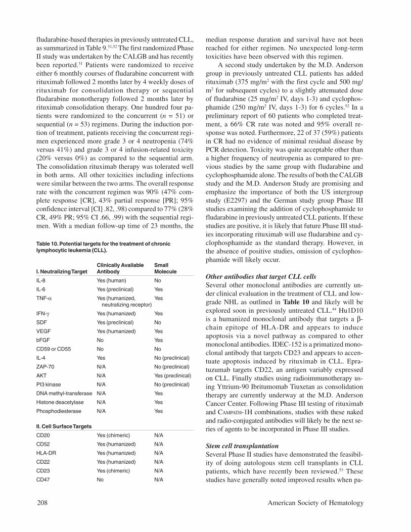
Other antibodies that target CLL cellsSeveral other monoclonal antibodies are currently un-der clinical evaluation in the treatment of CLL and low-grade NHL as outlined in Table 10 and likely will beexplored soon in previously untreated CLL.44 Hu1D10is a humanized monoclonal antibody that targets a β-chain epitope of HLA-DR and appears to induceapoptosis via a novel pathway as compared to othermonoclonal antibodies. IDEC-152 is a primatized mono-clonal antibody that targets CD23 and appears to accen-tuate apoptosis induced by rituximab in CLL. Epra-tuzumab targets CD22, an antigen variably expressedon CLL. Finally studies using radioimmunotherapy us-ing Yttrium-90 Ibritumomab Tiuxetan as consolidationtherapy are currently underway at the M.D. AndersonCancer Center. Following Phase III testing of rituximaband CAMPATH-1H combinations, studies with these nakedand radio-conjugated antibodies will likely be the next se-ries of agents to be incorporated in Phase III studies.
Stem cell transplantationSeveral Phase II studies have demonstrated the feasibil-ity of doing autologous stem cell transplants in CLLpatients, which have recently been reviewed.53 Thesestudies have generally noted improved results when pa-
Table 10. Potential targets for the treatment of chroniclymphocytic leukemia (CLL).
Clinically Available SmallI. Neutralizing Target Antibody Molecule
IL-8 Yes (human) No
IL-6 Yes (preclinical) Yes
TNF-α Yes (humanized, Yes neutralizing receptor)
IFN-γ Yes (humanized) Yes
SDF Yes (preclinical) No
VEGF Yes (humanized) Yes
bFGF No Yes
CD59 or CD55 No No
IL-4 Yes No (preclinical)
ZAP-70 N/A No (preclinical)
AKT N/A Yes (preclinical)
PI3 kinase N/A No (preclinical)
DNA methyl-transferase N/A Yes
Histone deacetylase N/A Yes
Phosphodiesterase N/A Yes
II. Cell Surface Targets
CD20 Yes (chimeric) N/A
CD52 Yes (humanized) N/A
HLA-DR Yes (humanized) N/A
CD22 Yes (humanized) N/A
CD23 Yes (chimeric) N/A
CD47 No N/A
Hematology 2002 209
tients were transplanted in complete remission or withminimal residual clinically detectable disease and couldbe converted to a PCR negative status.54 A randomizedPhase III trial in Europe is currently testing the efficacyof autologous bone marrow transplant following initialcytoreductive therapy. The role of standard allogeneicstem cell transplant in CLL is much less defined, exceptfor the very young patient with refractory disease, dueto the high treatment-related morbidity. Less toxic pre-parative regimens (i.e., mini transplants) that induce sig-nificant immunosuppression to allow engraftment, butdo not produce significant regimen related morbidity,have broadened the use of allogeneic stem cell trans-plant in CLL.55 Patients not attaining a complete remis-sion with initial therapy or having high risk genetic ab-normalities [i.e., del(11q22-q23), del(17p13), unmutatedsomatic V
H gene status, and p53 mutations] should be
considered good candidates for early application of thismodality on well designed clinical trials.
Other ApproachesThe recognition of important intracellular targets thatinhibit CLL cell apoptosis and other cytokines, chemo-kines, and angiogeneic peptides that provide a favorableenvironment for CLL cells to survive provides uniqueopportunities to eliminate residual disease followingadministration of initial CLL therapy. Furthermore, evenin the absence of detectable minimal residual disease,such an intervention might prevent re-growth of CLL.Major advances in using sophisticated computerizedmodeling to predict the chemical structure of small mol-ecules that will inhibit these intraceullar targets haveoccurred and these agents are now entering the clinic.In addition, significant advances in the technical aspectsof developing humanized monoclonal antibodies haveoccurred and there are now many agents (see Table 10)that target and neutralize cytokines, chemokines, orangiogeneic peptides and that are available for clinicaltrials. As the next decade of CLL studies is initiated, wemust now consider rational application of therapeuticapproaches that either treat minimal residual disease orprevent its emergence following effective cytoreductivetherapy outlined above. Such application in clinical tri-als applies not only to patients receiving therapy forsymptomatic CLL, but also to patients with early asymp-tomatic disease with poor prognostic features. Such atherapeutic approach to completely eliminate detectableCLL combined with continued laboratory investigationwill provide the best opportunity to alter the natural his-tory of this disease.
REFERENCES
I. The Nature and Natural History ofEarly-Stage B-CLL
1. Cheson BD, Bennett JM, Rai KR. Guidelines for clinicalprotocols for chronic lymphocytic leukemia: recommendationsof the National Cancer Institute-sponsored working group. AmJ Hematol. 1988;29:152-163.
2. Cheson BD, Bennett JM, Grever M, et al. National CancerInstitute-sponsored Working Group guidelines for chroniclymphocytic leukemia: revised guidelines for diagnosis andtreatment. Blood. 1996;87:4900-4907.
3. Moreau EJ, Matutes E, A’Hern RP, et al. Improvement of thechronic lymphocytic leukemia scoring system with themonoclonal antibody SN8 (CD79b). Am J Clin Pathol.1997;108:378-378.
4. Rawstron AC, Green MJ, Kuzmiccki A, Kennedy B, Jack AS,Hillmen P. Monoclonal B lymphocytes with the characteristicsof “indolent” chronic lymphocytic leukemia are present in 3.5%of adults with normal blood counts. Blood. 2002;100:635-639.
5. Han T, Ozer M, Gavigan M, et al. Benign monoclonal B celllymphocytosis – a benign variant of B-CLL: Clinical, immuno-logic, phenotypic and cytogenetic studies in 10 patients. Blood.1984;64:244-252.
6. Kimby E, Mellstedt H, Bjorkholm M, Holm G. Clonal cellsurface structures related to differentiation, activation andhoming in B-cell chronic lymphocytic leukemia and mono-clonal lymphocytosis of undetermined significance. Eur JHaematol. 1989;43:452-459.
7. Garcia C, Rosen A, Kimby E et al. Higher T-cell imbalance andgrowth factor receptor expression in B-cell chronic lymphocyticleukemia (B-B-CLL) as compared to monoclonal B-celllymphocytosis of undetermined significance (B-MLUS). LeukRes. 1989;13:31-37.
8. Aguilar-Santelises M, Amador JF, Mellstedt H, Jondal M. LowIL-1 beta production in leukemic cells from progressive B cellchronic leukemia (B-B-CLL). Leuk Res. 1989;13:937-942.
9. Aman P, Mellstedt H. The leukemic B-cell population ofpatients with monoclonal lymphocytosis of undeterminedsignificance (MLUS) are functionally distinct from the chroniclymphocytic leukemia (B-CLL) derived cell population. LeukRes. 1991;15:715-719.
10. Aguilar-Santelises M, Loftenius A, et al . Serum levels ofhelper factors (IL-1 alpha, IL-1 beta and IL-6), T-cell products(sCD4 and sCD8), sIL-2R and beta 2-microglobulin in patientswith B-B-CLL and benign B lymphocytosis. Leuk Res.1992;16:607-613.
11. Binet J-L, Leporier M, Dighiero G, et al. A clinical stagingsystem for chronic lymphocytic leukemia. Cancer.1977;40:855-864.
12. Rai KR, Sawitsky A, Cronkite ER, et al. Clinical staging ofchronic lymphocytic leukemia. Blood. 1977;46:219-234.
13. Wang C, Amato D, Fernandes B. CD5-negative phenotype ofmonoclonal B-lymphocytosis of undetermined significance(MLUS). Am J Hematol. 2002;69:147-149.
14. Wang C, Amato D, Rabah R, Zheng J, Fernandes B. Differentia-tion of monoclonal B lymphocytosis of undetermined signifi-cance (MLUS) and chronic lymphocytic leukemia (B-CLL)with weak CD5 expression from CD5- B-CLL. Leuk Res. 2002:In press.
15. Montserrat E, Vinolas N, Reverter JC, Rozman C. Naturalhistory of chronic lymphocytic leukemia: on the progressionand prognosis of early clinical stages. Nouv Rev Fr d’Hematol1988;30:359-361.
16. French Cooperative group on Chronic Lymphocytic Leukemia.

210 American Society of Hematology
Effects of chlorambucil and therapeutic decision in initial formsof chronic lymphocytic leukemia (stage A): results of arandomised trial on 612 patients. Blood. 1990;75:1414-1421.
17. Galton DAG. The pathogenesis of chronic lymphocyticleukaemia. Can Med Assoc J. 1966;94:1005-1010.
18. Hamblin TJ, Davis Z, Gardiner A, Oscier DG, Stevenson FK.Unmutated Ig V(H) genes are associated with a more aggres-sive form of chronic lymphocytic leukemia. Blood.1999;94:1848-1854.
19. Damle RN, Wasil T, Fais F, et al. Ig V gene mutation status andCD38 expression as novel prognostic indicators in chroniclymphocytic leukemia. Blood. 1999;94:1840-1847.
20. MacLennan IC. Germinal centers. Annu Rev Immunol.1994;12:117-139.
21. Damle RN, Ghiotto F, Valleto A, et al. B-cell chronic lympho-cytic leukaemia cells express a surface membrane phenotype ofactivated, antigen-experienced B lymphocytes. Blood.2002;99:4087-4093.
22. Rosenwald A, Alizadeh AA, Widhopf G, et al. Relation of geneexpression phenotype to immunoglobulin mutation genotype inB cell chronic lymphocytic leukemia. J Exp Med.2001;194:1639-1647.
23. Klein U, Tu Y, Stolovitzky GA, et al. Gene expression profilingof B cell chronic lymphocytic leukemia reveals a homogeneousphenotype related to memory B cells. J Exp Med.2001;194:1625-1638.
24. B-CLL Trialists Collaborative Group. Chemotherapeuticoptions in chronic lymphocytic leukemia: a meta-analysis ofthe randomized trials. J Natl Canc Inst. 1999;91:861-868.
25. Dighiero G, Maloum K, Desablens B, et al. Chlorambucil inindolent chronic lymphocytic leukemia. N Engl J Med.1998;338:1506-1514.
26. O’Brien S. Campath-1H and rituximab [abstract]. LeukLymphoma. 2001;42 (suppl 1):13-14.
27. Montserrat E, Vinolas N, Reverter J-C, Rozman C. Chroniclymphocytic leukemia in early stage: “smoldering” and “active”forms. In: Cheson BD (ed). Chronic Lymphocytic Leukemia:Scientific Advances and Clinical Developments. New York;Marcel Dekker: 1993:281-296.
28. Hallek M, Langenmayer I, Nerl C, et al. Elevated serumthymidine kinase levels identify a subgroup at high risk ofdisease progression in early, non-smoldering chronic lympho-cytic leukemia. Blood. 1999;93:1732-1737.
29. Molica S, Levato D, Dell’Olio M, et al. Cellular expression andserum circulating levels of CD23 in B-cell chronic lymphocyticleukemia. Implications for prognosis. Haematologica.1996;81:428-433.
30. Kantarjian HM, Smith T, Estey E, et al. Prognostic significanceof elevated serum beta-2-microglobulin levels in adult acutelymphocytic leukemia. Am J Med. 1994;93:599-604.
31. Ibrahim S, Keating M, Do KA, et al. CD38 expression as animportant prognostic factor in B-cell chronic lymphocyticleukemia. Blood. 2001;98:181-186.
32. Juliusson G, Oscier DG, Fitchett M, et al. Prognostic subgroupsin B-cell chronic lymphocytic leukemia defined by specificchromosomal abnormalities. N Engl J Med. 1990;323:720-724.
33. Oscier DG, Gardiner AC, Mould SJ, et al. Multivariate analysisof prognostic factors in CLL: Clinical stage VH gene, muta-tional status, and loss or mutation of the p53 gene are indepen-dent prognostic factors. Blood. 2002;100:1177-1184.
34. Kröber A, Seiler T, Benner A, et al. VH mutation status, CD38
expression level, genomic aberrations, and survival in chroniclymphocytic leukemia. Blood. 2002;100:1410-1416.
35. Hamblin TJ, Orchard JA, Ibbotson RE, et al. CD38 expressionand immunoglobulin variable region mutations are independent
prognostic variables in chronic lymphocytic leukemia, butCD38 expression may vary during the course of the disease.Blood. 2002;99:1023-1029.
36. Tobin G, Thunberg U, Johnson A, et al. Somatically mutatedIgVH3-21 genes characterize a new subset of chronic lympho-cytic leukemia. Blood. 2002;99:2262-2264.
II. Progression Events inB-Chronic Lymphocytic Leukemia
1. Han T, Ozer H, Sadamori N, et al. Prognostic importance ofcytogenetic abnormalities in patients with chronic lymphocyticleukemia. N Engl J Med. 1984;310:288-292.
2. Juliusson G, Oscier DG, Fitchett M, et al. Prognostic subgroupsin B-cell chronic lymphocytic leukemia defined by specificchromosomal abnormalities. N Engl J Med. 1990;323:720-724.
3. Neilson JR, Auer R, White D, et al. Deletions at 11q identify asubset of patients with typical CLL who show consistentdisease progression and reduced survival. Leukemia.1997;11:1929-1932.
4. Zhang Y, Weber-Matthiesen K, Siebert R, Matthiesen P,Schlegelberger B. Frequent deletions of 6q23-24 in B-cell non-Hodgkin’s lymphomas detected by fluorescence in situhybridization. Genes Chromosomes Cancer. 1997;18:310-313.
5. Finn WG, Kay NE, Kroft SH, Church S, Peterson LC. Second-ary abnormalities of chromosome 6q in B-cell chroniclymphocytic leukemia: a sequential study of karyotypicinstability in 51 patients. Am J Hematol. 1998;59:223-229.
6. Kroft SH, Finn WG, Kay NE, Peterson LC. Isolated 13q14abnormalities and normal karyotypes are associated withtypical lymphocyte morphology in B-cell chronic lymphocyticleukemia. Am J Clin Pathol. 1997;107:275-282.
7. Criel A, Wlodarska I, Meeus P, et al. Trisomy 12 is uncommonin typical chronic lymphocytic leukaemias. Br J Haematol.1994;87:523-528.
8. Finn WG, Thangavelu M, Yelavarthi KK, et al. Karyotypecorrelates with peripheral blood morphology andimmunophenotype in chronic lymphocytic leukemia. Am J ClinPathol. 1996;105:458-467.
9. Dohner H, Stilgenbauer S, Benner A, et al. Genomic aberrationsand survival in chronic lymphocytic leukemia. N Engl J Med.2000;343:1910-1916.
10. Wyatt WA, Meyer R, Flynn H, et al. Karyotype evolution andRai stage in B-cell chronic lymphocytic lymphoma (B-CLL)[abstract]. Blood. 2000;96:367a.
11. Meyer R, Brockman S, Paternoster S, et al. Fluorescence-labeled DNA probes to study indolent and aggressive B-CLL:comparison to Rai stage, level of clonal B-cells, IgVH mutationstatus and conventional cytogenetics [abstract]. Blood.2001;98:360a.
12. Damle R, Wasil T, Fais F, et al. IgV gene mutation status andCD38 expression as novel prognostic indicators in chroniclymphocytic leukemia. Blood. 1999;94:1840-1847.
13. Hamblin T, Davis Z, Gardiner A, Oscier D, Stevenson F.Unmutated Ig V-H genes are associated with a more aggressiveform of chronic lymphocytic leukemia. Blood. 1999;94:1848-1854.
14. Jelinek DF, Tschumper RC, Geyer SM, et al. Analysis of clonalB-cell CD38 and immunoglobulin variable region sequencestatus in relation to clinical outcome for B-chronic lymphocyticleukaemia. Br J Haematol. 2001;115:854-861.
15. Dohner H, Fischer K, Bentz M, et al. p53 gene deletion predictsfor poor survival and non-response to therapy with purineanalogs in chronic B-cell leukemias. Blood. 1995;85:1580-1589.
16. Stankovic T, Kidd AM, Sutcliffe A, et al. ATM mutations and

Hematology 2002 211
phenotypes in ataxia-telangiectasia families in the British Isles:expression of mutant ATM and the risk of leukemia, lymphoma,and breast cancer. Am J Hum Genet. 1998;62:334-345.
17. Klein U, Tu Y, Stolovitzky GA, et al. Gene expression profilingof B cell chronic lymphocytic leukemia reveals a homogeneousphenotype related to memory B cells. J Exp Med.2001;194:1625-1638.
18. Rosenwald A, Alizadeh AA, Widhopf G, et al. Relation of geneexpression phenotype to immunoglobulin mutation genotype inB cell chronic lymphocytic leukemia. J Exp Med.2001;194:1639-1647.
19. Tschumper RC, Kay NE, Iturria SJ, et al. Gene expressionprofiling of germline (GL)/CD38lo, GL/CD38hi, somaticmutation (SM)/CD38lo, and SM/CD38hi subsets of B-CLL[abstract]. Blood. 2001;98:471a.
20. Bartik MM, Welker D, Kay NE. Impairments in immune cellfunction in B cell chronic lymphocytic leukemia. Semin Oncol.1998;25:27-33.
21. Orsini E, Chiaretti S, Romana Mauro F, Guarini A, Foa R.Defective dendritic cell compartment in chronic lymphocyticleukemia patients [abstract]. Blood. 2001;98:731a.
22. Stadelmeyer E, Grube R, Chraust S, Linkesch W, Strunk D.Alteration of dendritic cell function in B-CLL [abstract]. Blood.2001;98:287b.
23. Cantwell M, Hua T, Pappas J, Kipps TJ. Acquired CD40-liganddeficiency in chronic lymphocytic leukemia. Nat Med.1997;3:984-989.
24. Kato K, Cantwell MJ, Sharma S, Kipps TJ. Gene transfer ofCD40-ligand induces autologous immune recognition ofchronic lymphocytic leukemia B cells. J Clin Invest.1998;101:1133-1144.
25. Kay NE, Han L, Bone N, Williams G. Interleukin 4 content inchronic lymphocytic leukaemia (CLL) B cells and blood CD8+T cells from B-CLL patients: impact on clonal B-cell apoptosis.Br J Haematol. 2001;112:760-767.
26. Kini A, Kay N, Peterson L. Increased bone marrow angiogen-esis in B-cell chronic lymphocytic leukemia. Leukemia.2000;14:1414-1418.
27. Chen H, Treweeke AT, West DC, et al. In vitro and in vivoproduction of vascular endothelial growth factor by chroniclymphocytic leukemia cells. Blood. 2000;96:3181-3187.
28. Kay NE, Bone ND, Tschumper RC, et al. B-CLL cells arecapable of synthesis and secretion of both pro- and anti-angiogenic molecules. Leukemia. 2002;16:911-919.
29. Molica S, Vitelli G, Levato D, Gandolfo GM, Liso V. Increasedserum levels of vascular endothelial growth factor predict riskof progression in early B-cell chronic lymphocytic leukaemia.Br J Haematol. 1999;107:605-610.
30. Aguayo A, O’Brien S, Keating M, et al. Clinical relevance ofintracellular vascular endothelial growth factor levels in B-cellchronic lymphocytic leukemia. Blood. 2000;96:768-780.
31. Bauvois B, Dumont J, Mathiot C, Kolb JP. Production of matrixmetalloproteinase-9 in early stage B-CLL: suppression byinterferons. Leukemia. 2002;16:791-798.
32. Menzel T, Rahman A, Calleja E, et al. Elevated intracellularlevel of basic fibroblast growth factor correlates with stage ofchronic lymphocytic leukemia and is associated with resistanceto Fludarabine. Blood. 1996;87:1056-1063.
33. Ghia P, Granziero L, Chilosi M, Caligaris-Cappio F. Chronic Bcell malignancies and bone marrow microenvironment. SeminCancer Biol. 2002;12:149-155.
34. Pizzolo G, Chilosi M, Ambrosetti A, Semenzato G, Fiore-Donati L, Perona G. Immunohistologic study of bone marrowinvolvement in B-chronic lymphocytic leukemia. Blood.1983;62:1289-1296.
35. Granziero L, Ghia P, Circosta P, et al. Survivin is expressed on
CD40 stimulation and interfaces proliferation and apoptosis inB-cell chronic lymphocytic leukemia. Blood. 2001;97:2777-2783.
36. Lagneaux L, Delforge A, De Bruyn C, Bernier M, Bron D.Adhesion to bone marrow stroma inhibits apoptosis of chroniclymphocytic leukemia cells. Leuk Lymphoma. 1999;35:445-453.
37. Chilosi M, Pizzolo G, Caligaris-Cappio F, et al. Immunohis-tochemical demonstration of follicular dendritic cells in bonemarrow involvement of B-cell chronic lymphocytic leukemia.Cancer. 1985;56:328-332.
38. Burger JA, Tsukada N, Burger M, Zvaifler NJ, Dell’Aquila M,Kipps TJ. Blood-derived nurse-like cells protect chroniclymphocytic leukemia B cells from spontaneous apoptosisthrough stromal cell-derived factor-1. Blood. 2000;96:2655-2663.
39. Burger JA, Burger M, Kipps TJ. Chronic lymphocytic leukemiaB cells express functional CXCR4 chemokine receptors thatmediate spontaneous migration beneath bone marrow stromalcells. Blood. 1999;94:3658-3667.
40. Novak AJ, Bram RJ, Kay NE, Jelinek DF. Aberrant expressionof B-lymphocyte stimulator by B chronic lymphocytic leukemiacells: a mechanism for survival Blood. 2002; In press.
41. Kawakami K, Kawakami M, Puri RK. Overexpressed cellsurface interleukin-4 receptor molecules can be successfullytargeted for antitumor cytotoxin therapy. Crit Rev Immunol.2001;21:299-310.
III. To Treat or Not to Treat The Patient with CLL:That Is the Question
1. Jemal A, Thomas A, Murray T, Thun M. Cancer statistics 2002.CA Cancer J Clin. 2002;52:23-47.
2. Byrd JC, Waselenko JK, Keating M, Rai K, Grever MR. Noveltherapies for chronic lymphocytic leukemia in the 21st century.Semin Oncol. 2000;27:587-597.
3. Zwiebel JA, Cheson BD. Chronic lymphocytic leukemia:staging and prognostic factors. Semin Oncol. 1998;25:42-59.
4. Rai KR, Sawitsky A, Cronkite EP, et al. Clinical staging ofchronic lymphocytic leukemia. Blood. 1975;46:219-234.
5. Binet JL, Auquier A, Dighiero G, et al. A new prognosticclassification of chronic lymphocytic leukemia derived from amultivariate survival analysis. Cancer. 1981;48:198-206.
6. Montserrat E, Vinolas N, Reverter JC, Rozman C. Naturalhistory of chronic lymphocytic leukemia: on the progressionand prognosis of early clinical stages. Nouv Rev Fr Hematol.1988;30:359-361.
7. Molica S. Progression and survival studies in early chroniclymphocytic leukemia. Blood. 1991;78:895-899.
8. French Cooperative Group on Chronic Lymphocytic Leu-kaemia. Natural history of stage A chronic lymphocyticleukemia untreated patients. Br J Haematol. 1990;76:45-57.
9. Mauro FR, Foa R, Giannarelli D, et al. Clinical characteristicsand outcome of young chronic lymphocytic leukemia patients:a single institution study of 204 cases. Blood. 1999;94:448-454.
10. de Lima M, O’Brien S, Lerner S, Keating MJ. Chroniclymphocytic leukemia in the young patient. Semin Oncol.1998;25:107-116.
11. Molica S, Brugiatelli M, Callea V, et al. Comparison of youngerversus older B-cell chronic lymphocytic leukemia patients forclinical presentation and prognosis: a retrospective study of 53cases. Eur J Haematol. 1994;52:216-221.
12. Montserrat E, Gomis F, Vallespi T, et al. Presenting features andprognosis of chronic lymphocytic leukemia in younger adults.Blood. 1991;78:1545-1551.
13. CLL Trialists’ Collaborative Group. Chemotherapeutic options
212 American Society of Hematology
in chronic lymphocytic leukemia: a meta-analysis of therandomized trials. J Natl Cancer Inst. 1999;91:861-868.
14. Dighiero G, Maloum K, Desablens B, et al. Chlorambucil inindolent chronic lymphocytic leukemia. French CooperativeGroup on Chronic Lymphocytic Leukemia. N Engl J Med.1998;338:1506-1514.
15. French Cooperative Group on Chronic Lymphocytic Leukemia.Effects of chlorambucil and therapeutic decision in initial formsof chronic lymphocytic leukemia (stage A): results of arandomized clinical trial on 612 patients. Blood. 1990;75:1414-1421.
16. Dohner H, Stilgenbauer S, Benner A, et al. Genomic aberrationsand survival in chronic lymphocytic leukemia. N Engl J Med.2000;343:1910-1916.
17. Cuneo A, Bigoni R, Rigolin GM, et al. Late appearance of the11q22.3-23.1 deletion involving the ATM locus in B-cellchronic lymphocytic leukemia and related disorders. Clinico-biological significance. Haematologica. 2002;87:44-51.
18. Hamblin TJ, Orchard JA, Ibbotson RE, et al. CD38 expressionand immunoglobulin variable region mutations are independentprognostic variables in chronic lymphocytic leukemia, butCD38 expression may vary during the course of the disease.Blood. 2002;99:1023-1029.
19. Cordone I, Masi S, Mauro FR, et al. p53 expression in b-cellchronic lymphocytic leukemia: a marker for disease progres-sion and poor prognosis. Blood. 1998;91:4132-4349.
20. Oliveira GB, Pereira FG, Metze K, Lorand-Metze I. Spontane-ous apoptosis in chronic lymphocytic leukemia and itsrelationship to clinical and cell kinetic parameters. Cytometry.2001;1546:329-335.
21. Cheson BD, Bennett JM, Grever M, et al. National CancerInstitute-sponsored Working Group guidelines for chroniclymphocytic leukemia: revised guidelines for diagnosis andtreatment. Blood. 1996;87:4990-4997.
22. Kyle RA, McParland CE, Dameshek W. Large doses ofprednisone and prednisolone in the treatment of malignantproliferative disorders. Ann Intern Med. 1962;57:713-761.
23. Jaksic B, Brugiatelli M. High dose continuous chlorambucil vsintermittent chlorambucil plus prednisone for treatment of B-CLL—IGCI CLL-01 trial. Nouv Rev Fr Hematol. 1988;30:437-442.
24. Jaksic B, Brugiatelli M, Krc I, et al. High dose chlorambucilversus Binet’s modified cyclophosphamide, doxorubicin,vincristine, and prednisone regimen in the treatment of patientswith advanced B-cell chronic lymphocytic leukemia: results ofan international multicenter randomized trial. InternationalSociety for Chemo-Immunotherapy, Vienna. Cancer.1997;79:2107-2114.
25. Jaksic B, Brugiatelli M, Suciu S, et al. Fludarabine vs. highdose chlorambucil in advanced B-cell chronic lymphocyticleukaemia (B-CLL): results of EORTC phase-II randomisedtrial after 4 years of median follow-up [abstract]. Blood.2000;96:758a.
26. Byrd JC, Rai KR, Sausville ER, Grever MR. Old and newtherapies in chronic lymphocytic leukemia: now is the time fora reassessment of therapeutic goals. Semin Oncol. 1998,25:65-74.
27. Johnson S, Smith AG, Loffler H, et al. Multicentre prospectiverandomized trial of fludarabine versus cyclophosphamide,doxorubicin, and prednisone (CAP) for treatment of advanced-stage chronic lymphocytic leukemia. Lancet. 1996;347:1432-1438.
28. Rai KR, Peterson BL, Appelbaum FR, et al. Fludarabinecompared with chlorambucil as primary therapy for chroniclymphocytic leukemia. N Engl J Med. 2000;343:1750-1757.
29. Leporrier M, Chevret S, Cazin B, et al. Randomized comparisonof fludarabine, CAP, and ChOP in 938 previously untreatedstage B and C chronic lymphocytic leukemia patients. FrenchCooperative Group on Chronic Lymphocytic Leukemia. Blood.2001;98:2319-2325.
30. Levy V, Porcher R, Delabarre F, Leporrier M, Cazin B, ChevretS. Evaluating treatment strategies in chronic lymphocyticleukemia: use of quality-adjusted survival analysis. FrenchCooperative CLL Group. J Clin Epidemiol. 2001;54:747-754.
31. Dillman RO, Mick R, McIntyre OR. Pentostatin in chroniclymphocytic leukemia: a phase II trial of Cancer and Leukemiagroup B. J Clin Oncol. 1989;7:433-438.
32. Robak T. Cladribine in the treatment of chronic lymphocyticleukemia. Leuk Lymphoma. 2001;40:551-564.
33. Robak T, Blonski JZ, Kasznicki M, et al. Cladribine withprednisone versus chlorambucil with prednisone as first-linetherapy in chronic lymphocytic leukemia: report of a prospec-tive, randomized, multicenter trial. Blood. 2000;96:2723-2729.
34. O’Brien S, Kantarjian H, Beran M, et al. Results of fludarabineand prednisone therapy in 264 patients with chronic lympho-cytic leukemia with multivariate analysis-derived prognosticmodel for response to treatment. Blood. 1993;82:1695-1700.
35. Yamauchi T, Nowak BJ, Keating MJ, Plunkett W. DNA repairinitiated in chronic lymphocytic leukemia lymphocytes by 4-hydroperoxycyclophosphamide is inhibited by fludarabine andclofarabine. Clin Cancer Res. 2001;11:3580-3589.
36. Robak T, Blonski JZ, Kasznicki M, et al. Cladribine combinedwith cyclophosphamide and mitoxantrone as front-line therapyin chronic lymphocytic leukemia. Leukemia. 2001;15:1510-1516.
37. Flinn IW, Byrd JC, Morrison C, et al. Fludarabine andcyclophosphamide with filgrastim support in patients withpreviously untreated indolent lymphoid malignancies. Blood.2000;96:71-75.
38. O’Brien SM, Kantarjian HM, Cortes J, et al. Results of thefludarabine and cyclophosphamide combination regimen inchronic lymphocytic leukemia. J Clin Oncol. 2001;19:1414-1420.
39. Flinn IW, Jemiai Y, Bennett JM, et al. Fludarabine andcyclophosphamide achieves high complete response rate inpatients with previously untreated chronic lymphocyticleukemia: ECOG 1997 [abstract]. Blood. 2001;98:633a.
40. Cazin B, Maloum K, Divine M, et al. Oral fludarabine andcyclophosphamide in previously untreated CLL: preliminarydata on 59 patients [abstract]. Blood. 2001;98:772a.
41. Oken MM, Lee S, Cassileth PA, Krigel RL. Pentostatin,chlorambucil, and prednisone for the treatment of chroniclymphocytic leukemia: Eastern Cooperative Oncology GroupProtocol E1488 [abstract]. Proc Am Soc Clin Oncol.1998;17:6a.
42. Flynn JM, Byrd JC. Campath-1H monoclonal antibody therapy.Curr Opin Oncol. 2000;12:574-581.
43. Lundin J, Kimby E, Bjorkholm M, et al. Phase II trial ofsubcutaneous anti-CD52 monoclonal antibody alemtuzumab(Campath-1H) as first-line treatment for patients with B-cellchronic lymphocytic leukemia (B-CLL). Blood. In press.
44. Keating MJ, O’Brien SJ. Eradication of minimal residualdisease in patients with chronic lymphocytic leukemia usingalemtuzumab (Campath-1H). CLL: The Cutting Edge. Baylor-Charles A. Sammons Cancer Center, Dallas, TX. 2001;6(4):5-7.
45. Cataland SR, Lucas M, Byrd JC. Antibody therapy of acute andchronic leukemias. Curr Pharm Biotechnol. 2001;4:357-367.
46. O’Brien SM, Kantarjian H, Thomas DA, et al. Rituximab dose-escalation trial in chronic lymphocytic leukemia. J Clin Oncol.2001;19:2165-2170.
Hematology 2002 213
47. Byrd JC, Murphy T, Howard RS, et al. Rituximab using a thriceweekly dosing schedule in B-cell chronic lymphocytic leukemiaand small lymphocytic lymphoma demonstrates clinical activityand acceptable toxicity. J Clin Oncol. 2001;19:2153-2164.
48. Hainsworth JD, Litchy S, Burris HA, Greco FA. Rituximab asfirst-line and maintenance therapy for patients with smalllymphocytic lymphoma (SLL) and chronic lymphocyticleukemia (CLL) [abstract]. Blood. 2001;98:363a.
49. Thomas DA, O’Brien S, Giles FJ, et al. Single agent rituxan inearly stage chronic lymphocytic leukemia (CLL) [abstract].Blood. 2001;98:364a.
50. Byrd JC, Kitada S, Flinn IW, et al. The mechanism of tumor cellclearance by rituximab in vivo in patients with B-cell chroniclymphocytic leukemia: evidence of caspase activation andapoptosis induction. Blood. 2002;99:1038-1043.
51. Byrd JC, Peterson BL, Morrison VA, et al. Randomized phase IIstudy of concurrent versus sequential treatment with rituximabwith fludarabine in symptomatic, untreated patients with B-cellchronic lymphocytic leukemia: superior complete responserates with concurrent therapy. Blood. In press.
52. Wierda W, O’Brien S, Albitar M, et al. Combined fludarabine,cyclophosphamide, and rituximab achieves a high completeremission rate as initial treatment for chronic lymphocyticleukemia [abstract]. Blood. 2001;98:3210a.
53. Dreger P, Montserrat E. Autologous and allogeneic stem celltransplantation for chronic lymphocytic leukemia. Leukemia.2002;16:985-992.
54. Schultze JL, Donovan JW, Gribben JG. Minimal residualdisease detection after myeloablative chemotherapy in chroniclymphatic leukemia. J Mol Med. 1999;77:259-265.
55. Khouri IF, Keating M, Korbling M, et al. Transplant-lite:induction of graft-versus-malignancy using fludarabine-basednonablative chemotherapy and allogeneic blood progenitor-celltransplantation as treatment for lymphoid malignancies. J ClinOncol. 1998;16:2817-2824.