chemotaxis in bacteria - ilya nemenman: theoretical...
TRANSCRIPT

Copyright 1975. A II riyhts reserved
CHEMOTAXIS IN BACTERIA ~886
Julius Adler
Departments of Biochemistry and Genetics, University of Wisconsin,Madison, Wisconsin 53706
CONTENTSOVERVIEW ................................................................. 342DEMONSTRATION AND MEASUREMENT OF CHEMOTAXIS IN BACTERIA ........ 342THE MOVEMENT OF INDIVIDUAL BACTERIA IN A CHEMICAL GRADIENT ....... 343THE DETECTION OF CHEMICALS BY BACTERIA: CHEMOSENSORS .............. 344What is Detected? ................................................................... 344The Number of Different Chemosensors ................................................. 345Nature of the Chemosensors ........................................................... 346COMMUNICATION OF SENSORY INFORMATION FROM CHEMOSENSORS TO
THE FLAGELLA ........................................................ 350THE FUNCTIONING OF FLAGELLA TO PRODUCE BACTERIAl. MOTION ........ 351THE RESPONSE OF FLAGELLA TO SENSORY INFORMATION ................... 352INTEGRATION OF MULTIPLE SENSORY DATA BY BACTERIA ................... 353ROLE OF THE CYTOPLASMIC MEMBRANE ................................... 353UNANSWERED QUESTIONS ................................................... 354RELATION OF BACTERIAL CHEMOTAXIS TO BEHAVIORAL BIOLOGY AND
NEUROBIOLOGY ................................................. 354
"I am not entirely happy about my diet of flies and bugs, but it’s the way I’m made.A spider has to pick up a living somehow or other, and 1 happen to be a trapper.I just naturally build a web and trap flies and other insects. My mother was a trapperbefore me. Her mother was a trapper before her. All our family have been trappers.Way back for thousands and thousands of years we spiders have been laying for fliesand bugs."
"It’s a miserable inheritance," said Wilbur, gloomily. He was sad because his newfriend was so bloodthirsty.
"Yes, it is," agreed Charlotte. "But I can’t help it. I don’t know how the first spider in¯ the early days of the world happened to think up this fancy idea of spinning a web,but she did, and it was clever of her, too. And since then, all of us spiders have hadto work the same trick. It’s not a bad pitch, on the whole."
Ann
u. R
ev. B
ioch
em. 1
975.
44:3
41-3
56. D
ownl
oade
d fr
om a
rjou
rnal
s.an
nual
revi
ews.
org
by E
MO
RY
UN
IVE
RSI
TY
on
08/2
6/10
. For
per
sona
l use
onl
y.

342 ADLER
recently. For reviews of the literature up to about 1960, see Berg (4), Weibull (5), Ziegler (6). This review will restrict itself to the recent work on chemotaxis Escherichia coli and Salmonella typhimurium. Some of this work is also covered inBerg’s review (4), and a review by Parkinson (7) should be consulted for a complete treatment of the genetic aspects.
OVERVIEW
Motile bacteria are attracted by certain chemicals and repelled by others; this ispositive and negative chemotaxis. Chemotaxis can be dissected by means of thefollowing questions :1. How do individual bacteria move in a gradient of attractant or repellent ?2. How do bacteria detect the chemicals ?3. How is the sensory information communicated to the flagella?4. How do bacterial flagella produce motion ?5. How do flagella respond to the sensory information in order to bring about the
appropriate change in direction ?6. In the case of multiple or conflicting sensory data, how is the information
integrated ?
DEMONSTRATION AND MEASUREMENT OFCHEMOTAXIS IN BACTERIA
Work before 1965, although valuable (4-6), was carried out in complex media andwas largely of a subjective nature. It was therefore necessary to develop conditionsfor obtaining motility and chemotaxis in defined media (8-11) and to find objective,quantitative methods for demonstrating chemotaxis.
(a) Plate method: For positive chemotaxis, a petri dish containing metabolizableattractant, salts needed for growth, and soft agar (a low enough concentration sothat the bacteria can swim) is inoculated in the center with the bacteria. As thebacteria grow, they consume the local supply of attractant, thus creating a gradient,which they follow to form a ring of bacteria surrounding the inoculum (8). Fornegative chemotaxis, a plug of hard agar containing repellent is planted in a petridish containing soft agar and bacteria concentrated enough to be visibly turbid ; thebacteria soon vacate the area around the plug (12). By searching in the area of theplate traversed by wild-type bacteria, one can isolate mutants in positive or negativechemotaxis (for example, 10, 12-15).
(b) Capillary method: In the 1880s Pfeffer observed bacterial chemotaxis inserting a capillary containing a solution of test chemical into a bacterial suspensionand then looking microscopically for accumulation of bacteria at the mouth of andinside the capillary (positive chemotaxis) or movement of bacteria away from thecapillary (negative chemotaxis) (2, 3). For positive chemotaxis this procedure been converted into an objective, quantitative assay by measuring the number ofbacteria accumulating inside a capillary containing attractant solution (10, 11). Fornegative chemotaxis, repellent in the capillary decreases the number of cells that will
Ann
u. R
ev. B
ioch
em. 1
975.
44:3
41-3
56. D
ownl
oade
d fr
om a
rjou
rnal
s.an
nual
revi
ews.
org
by E
MO
RY
UN
IVE
RSI
TY
on
08/2
6/10
. For
per
sona
l use
onl
y.

CHEMOTAXIS IN BACTERIA 343
enter (12). Alternatively, repellent is placed with the bacteria but not in th~ capillary;the number of bacteria fleeing into the capillary for refuge is then measured (12).Unlike in the plate method, where bacteria make the gradient of attractant bymetabolizing the chemical, here the experimenter provides the gradient; hencenonmetabolizable chemicals can be studied.
(c) Defined gradients : Quantitative analysis of bacterial migration has been achievedby making defined gradients of attractant (16) or repellent (17), and then determiningthe distribution of bacteria in the gradient by measuring scattering of a laser beamby the bacteria. The method allows the experimenter to vary the shape of the gradient.
(d) A change in the bacterium’s tumbling frequency in response to a chemicalgradient, described next, is also to be regarded as a demonstration and a measure-ment of chemotaxis.
THE MOVEMENT OF INDIVIDUAL BACTERIA IN ACHEMICAL GRADIENT
The motion of bacteria can of course be observed microscopically by eye, recordedby microcinematography, or followed as tracks that form on photographic film aftertime exposure (18, 19). Owing to the very rapid movement of bacteria, however,significant progress was not made until the invention of an automatic trackingmicroscope, which allowed objective, quantitative, and much faster observations (20).A slower, manual tracking microscope has also been used (21). A combination these methods has led to the following conclusions.
In the absence of a stimulus (i.e. no attractant or repellent present, or else constant, uniform concentration--no gradient) a bacterium such as E. coil or S.typhimurium swims in a smooth, straight line for a number of seconds--a "run," thenit thrashes around for a fraction of a second--a "tumble" (or abruptly changes itsdirection--a "twiddle"); and then it again swims in a straight line, but in a new,randomly chosen direction (22). (A tumble is probably a series of very brief runsand twiddles.)
Compared to this unstimulated state, cells tumble less frequently (i.e. they swim inlonger runs) when they encounter increasing concentrations of attractant (22, 23) they tumble more frequently when the concentration decreases (23). For repellents,the opposite is true: bacteria encountering an increasing concentration tumble moreoften, while a decreasing concentration suppresses tumbling (17). (See Figure [-Much smaller concentration changes are needed to bring about suppression oftumbling than stimulation of tumbling (22, 24).]
All this applies not only to spatial gradients (for example, a higher concentrationof chemical to the right than to the left) but also to temporal gradients (a higherconcentration of chemical now than earlier). The important discovery that bacteriacan be stimulated by temporal gradients of chemicals was made by mixing bacteriaquickly with increasing or decreasing concentrations of attractant (23) or repellent(17) and then immediately observing the alteration of tumbling frequency. After short while (depending on the extent and the direction of the concentration change),the tumbling frequency returns to the unstimulated state (17, 23). A different way
Ann
u. R
ev. B
ioch
em. 1
975.
44:3
41-3
56. D
ownl
oade
d fr
om a
rjou
rnal
s.an
nual
revi
ews.
org
by E
MO
RY
UN
IVE
RSI
TY
on
08/2
6/10
. For
per
sona
l use
onl
y.

344 ADLER
provide temporal gradients is to destroy or synthesize an attractant enzymatically ;as the concentration of attractant changes, the tumbling frequency is measured (25).[For a history of the use of temporal stimulation in the study of bacterial behavior,see the introduction to (25).] The fact that bacteria can "remember" that there is different concentration now than before has led to the proposal that bacteria have akind of "memory" (23, 24).
The possibility that a bacterium in a spatial gradient compares the concentrationat each end of its cell has not been ruled out, but it is not necessary to invoke it now,and in addition, the concentration differencc at the two ends would be too small tobe effective for instantaneous comparison (23, 24).
These crucial studies (17, 22, 23, 25) point to the regulation of tumbling frequencyas a central feature of chemotaxis. The results are summarized in Figure 1.
By varying the tumbling frequency in this manner, the bacteria migrate in a"biased random walk" (24) toward attractants and away from repellents : motion a favorable direction is prolonged, and motion in an unfavorable direction isterminated.
[Bacteria that have one or more flagella located at the pole ("polar flagellation,"as in Spirillum or Pseudomonas) back up instead of tumbling (26, 27). Even bacteriathat have flagella distributed all over ("peritrichous flagellation," as in E. coli orS. typhimurium) will go back and forth instead of tumbling if the medium issufficiently viscous (28).]
THE DETECTION OF CHEMICALS BY BACTERIA :CHEMOSENSORS
What is Detected?
Until 1969 it was not known if bacteria detected the attractants themselves orinstead measured some product of metabolism of the attractants, for example ATP.The latter idea was eliminated and the former established by the following results(10). (a) Some extensively metabolized chemicals are not attractants. This includeschemicals that are the first products in the metabolism of chemicals that do attract.(b) Some essentially nonmetabolizable chemicals attract bacteria : nonmetabolizableanalogs of metabolizable attractants attract bacteria, and mutants blocked in themetabolism of an attractant are still attracted to it. (c) Chemicals attractbacteria even in the presence of a metabolizable, nonattracting chemical.(d) Attractants that are closely related in structure compete with each other but not
INCREASING CONCENTRATION OF ATTRACTANT~’~I~,DECREASED FREQUENCY OF TUMBLING
DECREASING CONCENTRATION OF REPELLENT ~
DECREASING CONCENTRATION OF ATTRACTANT ~INCREASING CONCENTRATION OF REPELLENT ..~INCREASED FREQUENCY OV TUMBLING
Figure 1 Effect of change of chemical concentration on tumbling frequency.
Ann
u. R
ev. B
ioch
em. 1
975.
44:3
41-3
56. D
ownl
oade
d fr
om a
rjou
rnal
s.an
nual
revi
ews.
org
by E
MO
RY
UN
IVE
RSI
TY
on
08/2
6/10
. For
per
sona
l use
onl
y.

CHEMOTAXlS IN BACTERIA 345
with structurally nnrelated attractants. (e) Mutants lacking the detection mechanismbut normal in metabolism can be isolated.(f) Transport of a chemical into the ceilsis neither sufficient nor necessary for it to attract.
Thus, bacteria can sense attractants per se: these cells are equipped withsensory devices, "chemoreceptors," that measure changes in concentration of certainchemicals and report the changes to the flagella (10). It is a characteristic feature this and many other sensory functions that when the stimulus intensity changes,there is a response for a brief period only, i.e. the response is transient (17, 23). contrast, all other responses of bacteria to changes in concentration of a chemicalpersist as long as the new concentration is maintained. For example, when theconcentration of lactose is increased (over a certain range), there is a persistingincrease in the rate of lactose transport, the rate of lactose metabolism, or the rateof fl-galactosidase synthesis. To emphasize this unique feature, the sensory devicesfor chemotaxis will now be called "chemosensors."
"Chemoreceptor" will now be used for that part of the chemosensor tfiat "receives"the chemicals--a component that recognizes or binds the chemicals detected. A¢hemosensor must in addition have a component--the "signaller" that signals tothc flagella the change in the fraction of chemoreceptor occupied by the chemical.Further, a chemosensor may contain transport components for the sensed chemical,needed either directly or to place the chemoreceptor in a proper conformation.Bacteria have "receptors" for other chemicals--phage receptors, bacteriocin recep-tors, enzymes, repressors, etc, but these do not serve a sensory function in themanner just defined.
Since metabolism of the attractants is not involved in sensing them (10), themechanism of positive chemotaxis does not rely upon the attractant’s valuc to thecell. Similarly, negative chemotaxis is not mediated by the harmful effects of arepellent (12): (a) repellents are detected at concentrations too low to be harmful;(b) not all harmful chemicals are repellents; and (c) not all repellents are harmful.Nevertheless, the survival value of chemotaxis must lie in bringing the bacteria into anutritious environment (the attractants might signal the presence of other undetectednutrients) and away from a noxious one.
The chemosensors serve to alert the bacterium to changes in its environment.
The Number of Different Chemosensors
For both positive and negative chemotaxis the following criteria have been used todivide the chemicals into chemosensor classes. (a) For a number of chemosensors,mutants lacking the corresponding taxis, "specifically nonchemotactic mutants,"have been isolated (10, 12~15, 29-31). (b) Competition experiments: chemical
¯present in high enough concentration to saturate its chemoreceptor, will completelyblock the response to B if the two are detected by the same chemoreceptor bnt notif they are detected by different chemoreceptors (10, 12, 17, 3003). (c) Many of chemosensors are inducible, each being separately induced by a chemical it candetect (10, 15).
Table 1 Fists chemosensors identified so far for positive chemotaxis in E. coil, andTable 2 for negative chemotaxis in E. coli. Altogether evidence exists for about
Ann
u. R
ev. B
ioch
em. 1
975.
44:3
41-3
56. D
ownl
oade
d fr
om a
rjou
rnal
s.an
nual
revi
ews.
org
by E
MO
RY
UN
IVE
RSI
TY
on
08/2
6/10
. For
per
sona
l use
onl
y.

346 ADLER
Table 1 Partial list of chemosensors for positive chemotaxis in Escherichia coli
ThresholdAttractant Molaritya
N-acetyl-glucosamine sensor
N-Acetyl-D-glucosamine 1 X 10 sFructose sensor
D-Fructose 1 x 10⁻⁵
Galactose sensorD-Galactose 1 X 10.6
D-Glucose 1 × l0-6
D-Fucose 2 x 10 5Glucose sensor
D-Glucose 3 x 10 6Mannose sensor
D-Glucose 3 x 10 6D-Mannose 3 x 10⁻⁶
Maltose sensorMaltose 3 x 10 6
Mannitol sensorD-Mannitol 7 x 10⁻⁶
Ribose sensorD-Ribose 7 X 10⁻⁶
Sorbitol sensorD-Sorbitol 1 × 10⁻⁵
Trehalose sensorTrehalose 6 × 10⁻⁶
Aspartate sensorL-Aspartate 6 × 10-s
L-Glutamate 5 × 10 6
Serine sensorL-Serine 3 x 10⁻⁷
L-Cysteine 4 × 10⁻⁶
L-Alanine 7 x 10-s
Glycine 3 x 10⁻⁵
Data from (15, 29, 33); see more complete listing of specificities there. O2 is alsoattractive to E. coli (8, 34), as are certain inorganic ions (unpublished data).
a The threshold values are lower in mutants unable to take up or metabolize a chemical.
For example, the threshold for D-galactose is 100 times lower in a mutant unable to take upand metabolize this sugar (15).
20 different chemosensors in E. coli, but the evidence for each of them is not equallystrong. Oxygen taxis (8, 34) has not yet been studied from the point of view of chemosensor. S. typhimurium, insofar as its repertoire has been investigated, showssome of the same responses as E. coli (16, 17, 23, 30, 35).
Nature of the Chemosensors
Protein components of some of the chemosensors have been identified by acombination of biochemical and genetic techniques. Each chemosensor, it is believed,
Ann
u. R
ev. B
ioch
em. 1
975.
44:3
41-3
56. D
ownl
oade
d fr
om a
rjou
rnal
s.an
nual
revi
ews.
org
by E
MO
RY
UN
IVE
RSI
TY
on
08/2
6/10
. For
per
sona
l use
onl
y.

CHEMOTAXIS IN BACTERIA 347
Table 2 Partial list of chemosensors for negative chemotaxis in Escherichia colia
ThresholdRepellent Molarity
Fatty acid sensor
Acetate (C2) 3 x -4
Propionate (C3) 2 x 10 4n-Butyrate, isobutyrate (C4) 1 x 10.4
n-Valerate, isovalerate (C5) 1 x 10.4
n-Caproate (C6) 1 x 10⁻⁴
n-Heptanoate (C7) 6 x 10⁻³
n-Caprylate (C8) 3 x 10 zAlcohol sensor
Methanol (C1) 1 x 10⁻¹Ethanol (C2) 1 × 10.3
n-Propanol (C3) 4 x 10⁻³iso-Propanol (C3) 6 x 10⁻⁴
iso-Butanol (C4) 1 x 10⁻³
iso-Amylalcohol (C5) 7 x 10⁻³
Hydrophobic amino acid sensor
L-Leucine 1 × 1 -4L-Isoleucine 1.5 × 10⁻⁴
L-Valine 2.5 × 10⁻⁴
L-Tryptophan 1 x 10⁻³
L-Phenylalanine 3 x 10 3L-Glutamine 3 x 10.3
L-Histidine 5 x 10.3
Indole sensor
Indole 1 × 10.6
Skatole 1 × 10⁻⁶
Aromatic sensors
Benzoate 1 x 10⁻⁴
Salicylate 1 x 10⁻⁴
H÷ sensor
Low pH pH 6.5
OH- sensor
High pH pH 7.5Sulfide sensor
Na:S 3 x 10- 3
2-Propanethiol 3 x 10⁻³Metallic cation sensor
CoSO~ 2 x 10 4NiSO. 2 x I0-5
a Data from (12). See more complete listing of specificities there.
Ann
u. R
ev. B
ioch
em. 1
975.
44:3
41-3
56. D
ownl
oade
d fr
om a
rjou
rnal
s.an
nual
revi
ews.
org
by E
MO
RY
UN
IVE
RSI
TY
on
08/2
6/10
. For
per
sona
l use
onl
y.
348 ADLER
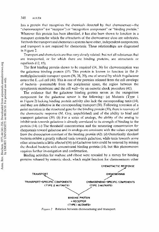
has a protein that recognizes the chemicals detected by that chemosensor--the"chemoreceptor" (or "receptor") or "recognition component" or "binding protein."Wherever lhis protein has been identified, it has also been shown to fimction in atransport system for which the attractants of the chemosensor class are substrates.Yet both the transport and chemotaxis systems have other, independent components,and transport is not required for chemotaxis. These relationships are diagramedin Figure 2.
Transport and chemotaxis are thus very closely related ; but not all substances thatare transported, or for which there are binding proteins, are attractants orrepellents (12, 15).
The first binding protein shown to be required (14, 36) for chemoreception wasthe galactose binding protein (37). This protein is known to function in the fl-methylgalactoside transport system (36, 38, 39), one of several by which D-galactoseenters the E. coli cell (40). This is one of the proteins released from the cell envelopeof bacteria--presumably from the periplasmic space, the region between thecytoplasmic membrane and the cell wall--by an osmotic shock procedure (41).
The evidence that the galactose binding protein serves as the recognitioncomponent for the galactose sensor is the following: (a) Mutants (Type in Figure 2) lacking binding protein activity also lack the corresponding taxis (14),and they are defective in the corresponding transport (38). Following reversion of point mutation in the structural gene for the binding protein (39), there is recovery the chemotactic response (M. Goy, unpublished) and of the ability to bind andtransport galactose (39). (b) For a series of analogs, the ability of the analog inhibit taxis towards galactose is directly correlated to its strength of binding to theprotein (14). (c) The threshold concentration and the saturating concentration chemotaxis toward galactose and its analogs are consistent with the values expectedfrom the dissociation constant of the binding protein (42). (d) Osmotically shockedbacteria exhibit a greatly reduced taxis towards galactose, while taxis towards someother attractants is little affecled (14). (e) Galactose taxis could be restored by mixingthe shocked bacteria with concentrated binding protein (14), but this phenomenonrequires further investigation and confirmation.
Binding activities for maltose and ribose were revealed by a survey for bindingproteins released by osmotic shock, which might function for chemosensors other
CHEMOTACTIE RESPONSE
TRANSPORT CHEMOSENSING
TRANSPORT-SPECIFIC COMPONENTS CHEMOSENSING-SPECIFIC COMPONENTS(TYPE ~. MUTANTS) (TYPE 3 MUTANTS)
BINDING PROTEIN= RECEPTOR
(TYPE I MUTANTS)
Fi~/ure 2 Relation between chemosensing and transport.
Ann
u. R
ev. B
ioch
em. 1
975.
44:3
41-3
56. D
ownl
oade
d fr
om a
rjou
rnal
s.an
nual
revi
ews.
org
by E
MO
RY
UN
IVE
RSI
TY
on
08/2
6/10
. For
per
sona
l use
onl
y.

CHEMOTAXIS IN BACTERIA 349
than galactose (14). The binding protein for maltose has now been purified (43), mutants lacking it fail to carry out maltose taxis (31), as well as being defective in thetransport of maltose (31, 43). That for ribose has been purified from S. typhimuriumand serves as ribose chemoreceptor by criteria a to c above (30, 35).
Mutants of Type 2 (Figure 2) are defective in transport but not necessarily chemotaxis, even though the two share a common binding protein. Thus, certaincomponents of the transport system, and the process of transport itself, are notrequired for chemotaxis (at least for certain chemosensors). This has been studiedextensively in the case ofgalactose, where transport is clearly not required (l 0, 14, 44).Two genes of Type 2 were found for the/~-methylgalactoside transport system forgalactose (44). Some of the mutations in these genes abolished transport withoutaffecting chemotaxis; other mutations in these genes affected chemotaxis as well (44).Such chemotactic defects may reflect interactions, direct or indirect, that thesecomponents normally have with the chemosensing machinery, or some kind ofunusual interaction of the mutated component with the binding protein that wouldhinder its normal function in chemosensing. Two genes whose products areinvolved in the transport system for maltose (45) can be mutated without affectingtaxis toward that sugar (15, 31). Type 2 gene products are most likely located in thecytoplasmic membrane, since they function in transport.
Mutants of Type 3 are defective in chemosensing but not in transport. Pre-sumably they have defects in gene products "signallers" (44)--which signal infor-mation from the binding protein to the rest of the chemotaxis machinery withouthaving a role in the transport mechanism. Such mutants, defective only for galactosetaxis or jointly for galactose and ribose taxis, are known (44). The chemistry andlocation of Type 3 gene products are as yet undescribed.
A mutant in the binding protein gene (by the criterion of complementation) known that binds and transports galactose normally but fails to carry out galactosetaxis, presumably because this binding protein is altered at a site for interactionwith thc Type 3 gene product (44). Conversely, some mutations mapping in thegene for the galactose (44) or maltose (31) binding proteins affect transport but binding or chemotaxis. The binding protein thus appears to have three sites--onefor binding the ligand, one for interacting with the next transport components, andone for interacting with the next chemotaxis components.
Whereas the binding proteins mentioned above can be removed from the cellenvelope by osmotic shock, other binding proteins exist that are tightly bound to thecytoplasmic membrane. Examples of such are the enzymes II of the phosphotransferasesystem, a phosphoenolpyruvate-dependent mechanism for the transport of certainsugars (46, 47). A number of sugar sensors utilize enzymes II as recognitioncomponents: for example, the glucose and mannose sensors are serviced byglucose enzyme II and mannose enzyme II, respectively (29). In these cases,enzyme I and HPr (a phosphate-carrier protein) of the phosphotransferase system(46) are also required for optimum chemotaxis (29). This could mean that phosphoryla-tion and transport of the sugars are required for chemotaxis in these cases; thatenzyme I and HPr must be present for interaction of enzyme II with subsequentchemosensing components; or, as seems most likely, that the enzyme II binds
Ann
u. R
ev. B
ioch
em. 1
975.
44:3
41-3
56. D
ownl
oade
d fr
om a
rjou
rnal
s.an
nual
revi
ews.
org
by E
MO
RY
UN
IVE
RSI
TY
on
08/2
6/10
. For
per
sona
l use
onl
y.

350
sugars more effectively afte~ it has been phosphorylated by phosphoenolpyruvateunder the influence of enzyme I and HPr. The products of the phosphotransferasesystem, the phosphorylated sugars, are not attractants, even when they can betransported by a hexose phosph, ate transport system (15, 29). This rules out the ideathat the phosphotransferase system is required to transport and’phosphorylate thesugars so that they will be available to an internal chemoreceptor, and indicatesinstead that interaction of the sugar specifically with the phosphotransferase systemsomehow leads .to chemotaxis (29). Certainly it is not the metabolism of thephosphorylated sugars that brings about chemotaxis: several cases of non-metabolizable phosphorylated sugars are known, yet the corresponding free sugarsare attractants (29).
Bacteria detect changes over time in the concentration of attractant or repellent(17, 23, 25), and experiments with whole cells indicate that it is the time rate of changeof the binding protein fraction occupied by ligand that the chemotactic machineryappears to detect (25, 42). How this is achieved remains unknown. A conformationalchange occurs when ligand (galactose) interacts with its purified binding protein (48,49), and possibly this change is sensed by the next component in the system, butnothing is known about this linkage.
COMMUNICATION OF SENSORY INFORMATION FROMCHEMOSENSORS TO THE FLAGELLA
Somehow the chemosensors must signal to the flagella that a change in chemicalconcentration has been encountered. The nature of this system of transmittinginformation to the flagella is entirely unknown, but several mechanisms have beensuggested (10, 23, 50).
(a) The membrane potential alters, either increasing or decreasing for attractants,with the opposite effect for repellents. The change propagates along the cellmembrane to the base of the flagellum. The cause of the change in membranepotential is a change in the rate of influx or etttux of some ion(s) when the concentra-tion of attractant or repellent is changed.
(b) The level of a low molecular weight transmitter changes, increasing decreasing with attractant or repellent. The transmitter diffuses to the base of theflagella. Calculations (4) indicate that diffusion of a substance of low molecularweight is much too slow to aceount for the practically synchronous reversal offlagella at the two ends of Spirillum volutans, which occurs in response tochemotactic stimuli (26, 27). Thus for this organism, at least, a change in membranepotential appears to be the more likely of the two mechanisms.
Although the binding protein of chemosensors is probably distributed all aroundthe cell because it is shared with transport, possibly only those protein molecules atthe base of the flagellum serve for chemoreception. In that case, communicationbetween the chemosensors and flagella could be less elaborate, taking place bymeans of direct protein-protein interaction.
Several tools are available for exploring the transmission system. One is the studyof mutants that may be defective in this system; these are the "generally non-
Ann
u. R
ev. B
ioch
em. 1
975.
44:3
41-3
56. D
ownl
oade
d fr
om a
rjou
rnal
s.an
nual
revi
ews.
org
by E
MO
RY
UN
IVE
RSI
TY
on
08/2
6/10
. For
per
sona
l use
onl
y.
CHEMOTAXIS IN BACTERIA 351
chemotactic mutants," strains unable (fully or partly) to respond to any attractant repellent (10, 12, 51). Some of these mutants swim smoothly, never tumbling (51-53),while others~"tumbling mutants"~tumble most of the time (28, 53-55). Geneticstudies (56-58) have revealed that the generally nonchemotactic mutants map four genes (53, 58). One of these gene products must be located in the flagellum,presumably at the base, since some mutations lead to motile, nonchemotactic cellswhile other mutations in the same gene lead to absence of flagella (59). The locationof the other three gene products is unknown. The function of the four gene productsis also unknown, but it has been, suggested (7, 53) that they play a role in thegeneration and control of tumbling at the level of a "twiddle generator" (22).
A second tool comes from the discovery that methionine is required for chemotaxis(60), perhaps at the level of the transmission system. Without methionine,chemotactically wild-type bacteria do not carry out chemotaxis (11, 55, 60, 61) tumble (55, 60, 62). This is not the case for tumbling mutants (55), unless they first "aged" in the absence of methionine (62), presumably to remove a store methionine or a product formed from it. There is evidence that methionine functionsvia S-adenosylmethionine (55, 61-64), but the mechanism of action of methionine chemotaxis remains to be discovered.
THE FUNCTIONING OF FLAGELLA TO PRODUCEBACTERIAL MOTION
For reviews of bacterial flagella and how they function, see (5, 65-70).For many years it was considered that bacterial flagella work either by means of a
wave that propagates down the flagellum, as is known to be the case for eucaryoticflagella, or by rotating as rigid or semirigid helices [-for a review of the history, see(71)]. Recently it was argued from existing evidence that the latter view is correct (71),and this was firmly established by the following experiment (72). E. coli cells with onlyone flagellum (obtained by growth on o-glucose, a catabolite repressor of flagellasynthesis) (9) were tethered to a glass slide by means of antibody to the filaments.(The antibody of course reacts with the filament and just happens to stick to glass.)Now that the filament is no longer free to rotate, the cell instead rotates, usuallycounterclockwise, sometimes cl.oekwise (72). By using such tethered cells, thedynamics of the flagellar motor were then characterized (73).
Energy for this rotation comes from the intermediate of oxidative phosphorylation(the proton gradient in the Mitchell hypothesis), not from ATP directly (64, 74), unlikethe case of eucaryotic flagella or muscle; this is true for both counterclockwise andclockwise rotation (64).
In S. typhimurium, light having the action spectrum of flavins brings abouttumbling, and this might in some way be caused by interruption of the energy flowfrom electron transport (75).
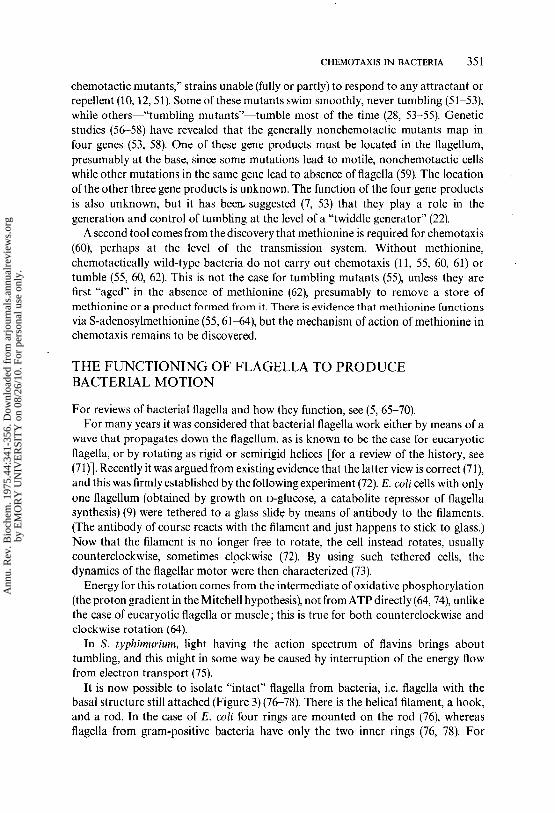
It is now possible to isolate "intact" flagella from bacteria, i.e. flagella with thebasal structure still attached (Figure 3) (76-78). There is the helical filament, a hook,and a rod. In the case of E. coli four rings are mounted on the rod (76), whereasflagella from gram-positive bacteria have only the two inner rings (76, 78). For
Ann
u. R
ev. B
ioch
em. 1
975.
44:3
41-3
56. D
ownl
oade
d fr
om a
rjou
rnal
s.an
nual
revi
ews.
org
by E
MO
RY
UN
IVE
RSI
TY
on
08/2
6/10
. For
per
sona
l use
onl
y.

352 ADLER
MEMBRANE
PEPTIDOGL¥C, ANLAYER
CYTOPLASMICMEMBRANE
BA,.~AL
Fi.qure 3 Model of the flagellar base of E. co/i (76, 77). Dimensions are in nanometers.
E. coli it has been established that the outer ring is attached to the outer membraneand the inner ring to the cytoplasmic membrane (Figure 3) (77). The basal body (a) anchors the flagellum into the cell envelope; (b) provides contact with cytoplasmic membrane, the place where the energy originates; and (c) very likelyconstitutes the motor (or a part of it) that drives the rotation.
The genetics of synthesis of bacterial flagella is being vigorously pursued inE. coli and S. typhimurium (28, 79, 80). It is consistent with such a complex structurethat at least 20 genes are required for the assembly and function of an E. coli flagellum(79) and many of these are homologous to those described in Sahnonella (28, 80).
THE RESPONSE OF FLAGELLA TOSENSORY INFORMATION
Addition of attractants to E. coli cells, tethered to glass by means of antibody toflagella, causes counterclockwise rotation to the cells as viewed from above (52).(Were the flagellum free to rotate, this would correspond to clockwise rotation of theflagellum and swimming toward the observer, as viewed frown above. But since aconvention of physics demands that the direction of rotation be defined as the objectis viewed moving away from the observer, the defined direction of the flagellarrotation is counterclockwise.) On the other hand, addition of repellents causes clock-wise rotation of the cells (52). These responses last for a short time, depending on thestrength of the stimulus ; then the rotation returns to the unstimulated state, mostlycounterclockwise (52).
Mutants of E. coli that swim smoothly and never tumble always rotate counter-clockwise, while mutants that almost always tumble rotate mostly clockwise (52).
From these results and from the prior knowledge that increase of attractantconcentration causes smooth swimming (i.e. suppressed tumbling) (22, 23, 25) while
Ann
u. R
ev. B
ioch
em. 1
975.
44:3
41-3
56. D
ownl
oade
d fr
om a
rjou
rnal
s.an
nual
revi
ews.
org
by E
MO
RY
UN
IVE
RSI
TY
on
08/2
6/10
. For
per
sona
l use
onl
y.

CHEMOTAXIS IN BACTERIA 353
addition of repellents causes tumbling (17), it was concluded that smooth swimmingresults from counterclockwise rotation of flagella and tumbling from clockwiserotation (52).
When there are several flagella originating from various places around the cell, as inE. coli or S. typhimurium, the flagella function together as a bundle propelling thebacterium from behind (75, 81, 82). Apparently the bundle of flagella survivescounterclockwise rotation of the individual flagella to bring about smooth swimming(no tumbling), but comes apart as a result of clockwise rotation of individualflagella to produce tumbling. That tumbling occurs concomitantly with theflagellar bundle flying apart has actually been observed by use of such high intensitylight that individual flagella could be seen (75).
Presumably less than a second of clockwise rotation can bring about a tumble, andthe long periods of clockwise rotation reported (52) result from the use of unnaturallylarge repellent stimuli. (The corresponding statement can be made for the largeattractant stimuli used.) Some kind of a recovery process is required for return to theunstimulated tumbling frequency. The mechanism of recovery is as yet unknown,but it appears that methionine is somehow involved (55, 62).
The information developed so far is summarized in Figure 4.The reversal frequency of the flagellum of a Pseudomonad is also altered by
gradients of attractant or repellent, and reversal of flagellar rotation can explain thebacking up of polarly flagellated bacteria (27).
INTEGRATION OF MULTIPLE SENSORY DATABY BACTERIA
Bacteria are capable of integrating multiple sensory inputs, apparently byalgebraically adding the stimuli (17). For example, the response to a decrease repellent concentration could be overcome by superimposing a decrease in con-centration of attractant (17). Whether bacteria will "decide" on attraction repulsion in a "conflict" situation (a capillary containing both attractant andrepellent) depends on the relative effective concentration of the two chemicals, i.e.how far each is present above its threshold concentration (3, 83). The mechanismfor summing the opposing signals is unknown.
ROLE OF THE CYTOPLASMIC MEMBRANE
There is increasing evidence that the cytoplasmic membrane plays a crucial role inchemotaxis. (a) Some of the binding proteins that serve in chemoreception--theenzymes II of the phosphotransferase system (29)~are firmly bound to the
SUPPRESSION OFCOUNTERCLOCKWISE TUMBLING
~RECOVER~ROTATION OF FLAGELLA~(SMOOTH SWIMMING)
CHEMICAL CHANGE IN/~ CHEMOSENSOR ~
GRADIENT SIGNAL
~CLOCKWlSE ROTATION -~ TUMBLING ~RECOVERY
OF FLAGELLA
Figure 4 Summary scheme ofch~motaxis.
Ann
u. R
ev. B
ioch
em. 1
975.
44:3
41-3
56. D
ownl
oade
d fr
om a
rjou
rnal
s.an
nual
revi
ews.
org
by E
MO
RY
UN
IVE
RSI
TY
on
08/2
6/10
. For
per
sona
l use
onl
y.

354 ADLER
cytoplasmic membrane (46, 47). Binding proteins that can be released by osmoticslaock are located in the periplasmic space (41), perhaps loosely in contact with thecytoplasmic membrane. (b) The base of the flagellum has a ring embedded in thecytoplasmic membrane (77). (c) The energy source for motility comes from oxidativephosphorylation (64, 74), a process that along with electron transport is membrane-associated (84). (d) Chemotaxis, but not motility, is unusually highly dependent temperature, which suggested a requirement for fluidity in the membrane lipids (11).This requirement for a fluid membrane was actually established by meas3~ring thetemperature dependence of chemotaxis in an unsaturated fatty acid auxotroph thathad various fatty acids incorporated (85). (e) A number of reagents (for example,ether or chloroform) that affect membrane properties inhibit chemotaxis atconcentrations that do not inhibit motility (26, 86-88).
This involvement of the cytoplasmic membrane in chemotaxis, especially thelocation there of the chemoreceptors and flagella, makes the membrane potentialhypothesis for transmission of information from chemoreceptors to flagellaplausible, but of course by no means proves it.
UNANSWERED QUESTIONS
While the broad outlines of bacterial chemotaxis have perhaps been sketched, thebiochemical mechanisms involved remain to be elucidated: How do chemosensorswork? By what means do they communicate with the flagella? What is themechanism that drives the motor for rotating the flagella ? What is the mechanism ofthe gear that shifts the direction of flagellar rotation ? How does the cell recover fromthe stimulus ? How are multiple sensory data processed ? What are the functions ofthe cytoplasmic membrane in chemotaxis ?
RELATION OF BACTERIAL CHEMOTAXIS TOBEHAVIORAL BIOLOGY AND NEUROBIOLOGY
The inheritance of behavior (see opening quotation) and its underlying biochemicalmechanisms are nowhere more amenable to genetic and biochemical investigationthan in the bacteria. From the earliest studies of bacterial behavior (2, 3, 89-91) the present (8, 10, 24, 42, 50, 92, 93) people have hoped that this relatively simplesystem could tell us something about the mechanisms of behavior of animals andman. Certainly, striking similarities exist between sensory reception in bacteria and inhigher organisms (16, 24, 42, 92, 93).
Already in 1889 Alfred Binet wrote in The Psychic Life of Micro-organisms (89)
If the existence of psychological phenomena in lower organisms is denied, it will benecessary to assume that these phenomena can be superadded in the course ofevolution, in proportion as an organism grows more perfect and complex. Nothingcould be more inconsistent with the teachings of general physiology, which shows usthat all vital phenomena are previously present in non-differentiated cells.
Ann
u. R
ev. B
ioch
em. 1
975.
44:3
41-3
56. D
ownl
oade
d fr
om a
rjou
rnal
s.an
nual
revi
ews.
org
by E
MO
RY
UN
IVE
RSI
TY
on
08/2
6/10
. For
per
sona
l use
onl
y.
CHEMOTAXIS IN BACTERIA 355
Literature Cited
1. Engelmann, T. W. 1881. Pfliiger’s Arch.Gesamte Physiol. Menschen Tiere 25:285-92
2. Pfeffer, W. 1884. Untersuch. Bot. Inst.Tiibingen 1 : 363-482
3. Pfeffer, W. 1888. Untersuch. Bot. Inst.Tiibingen 2 : 58~661
4. Berg, H. C. 1975. Ann. Rev. Biophys.Bioeng. 4:119-36
5. Weibull, C. 1960. Bacteria 1 : 153-2056. Ziegler, H. 1962. Encyclopedia of Plant
Physiology, ed. W. Ruhland, 17-II:484-532. Berlin: Springer. (In German)
7. Parkinson, J. S. 1975. Cell. In press8. Adler, J. 1966. Science 153:708-169. Adler, J., Templeton, B. 1967. J. Gen.
Microbiol. 46:175-8410. Adler, J. 1969. Science 166:1588-9711. Adler, J. 1973. J. Gen. Microbiol. 74:
77-9112. Tso, W.-W., Adler, J. 1974. J. Bacteriol.
118 : 560-7613. Hazelbauer, G. L., Mesibov, R. E., Adler,
J. 1969. Proc. Nat. Acad. Sci. USA64:1300-7
14. Hazelbauer, G. L., Adler, J. 1971. NatureNew Biol. 230(12): 101-4
15. Adler, J., Hazelbauer, G. U, Dahl, M. M.1973. J..Bacteriol. 115 : 824-47
16. Dahlquist, F. W., Lovely, P., Koshland,D. E. Jr. 1972. Nature New Biol. 236:120-23
17. Tsang, N., Macnab, R, Koshland, D. E.Jr. 1973. Science 181 : 60-63
18. Dryl, S. 1958. Bull. Acad. Pol. Sci. 6:429-32
19. Vaituzis, Z., Doetsch, R. N. 1969.Appl. Microbiol. 17:584-88
20. Berg, H. C. 1971. Rev. Sci. Instrum.42 : 868-71
21. Lovely, P., Macnab, R., Dahlquist, F. W.,Koshland, D. E. Jr. 1974. Rev. Sci.lnstrum. 45 : 683-86
22. Berg, H. C., Brown, D. A. 1972. Nature239 : 500-4
23. Maenab, R. M., Koshland, D. E. Jr.1972. Proe. Nat. Acad. Sci. USA 69:2509-12
24. Koshland, D. E. Jr. 1974. FEBS Lett.40 (Suppl.): $3-$9
25. Brown, D. A., Berg, H. C. 1974. Proc.Nat. Acad. Sci. USA 71 : 1388-92
26. Caraway, B. H., Krieg, N. R. 1972.Can. J. Microbiol. 18:1749-59
27. Taylor, B. L., Koshland, D. E. Jr. 1974.J. Bacteriol. 119:640-42
28. Vary, P. S., Stoeker, B. A. D. 1973.Genetics 73 : 229-45
29. Adler, J., Epstein, W. 1974. Proc. Nat.
Acad. Sci. USA. 71:2895-99
30. Aksamit, R. R., Koshland, D. E. Jr. 1974.Biochemistry 13 : 4473-78
31. Hazelbauer, G. L. 1975. J. Bdcteriol. Inpress
32. Rothert, W. 1901. Flora 88:371-42133. Mesibov, R., Adler, J. 1972. J. Bacteriol.
112:315-2634. Baracchini, O., Sherris, J. C. 1959. J.
Pathol. Bacteriol. 77 : 565-7435. Aksamit, R., Koshland, D. E. Jr. 1972.
Biochem. Biophys. Res. Commun. 48:1348-53
36. Kalckar, H. M. 1971. Science 174: 557-65
37. Anraku, Y. 1968. J. Biol. Chem. 243:3116-22
38. Boos, W. 1969.Eur.J. Biochem. 10:66-7339. Boos, W. 1972. J. Biol. Chem. 247:
5414-2440. Rotman, B., Ganesan, A. K., Guzman,
R. 1968. J. Mol. Biol. 36:247-6041. Heppel, L. A. 1967. Science 156: 1451-
5542. Mesibov, R., Ordal, G. W., Adler, J. 1973.
J. Gen. Physiol. 62 : 203-2343. Kellerman, O., Szmelman, S. 1974. Eur.
J. Biochem. 47:139-4944. Ordal, G. W., Adler, J. 1974. J. Bacteriol.
117 : 517-2645. Hofnung, M., Hatfield, D., Schwartz,
M. 1974. J. Bacter~ol. 117:40-4746. Roseman, S. 1972. Metab. Pathways
6:41-8947. Kundig, Wo., Roseman, S. 1971. J. Biol.
Chem. 246 : 1407-1848. Boos, W., Gordon, A. S., Hall, R. E.,
Price, H. D. 1972. J. Biol. Chem. 247:917-24
49. Rotman, B., Ellis, J. H. Jr. 1972. J.Baeteriol. 111 : 791-96
50. Doetsch, R. N. 1972. J. Theor. Biol. 35:55-66
51. Armstrong, J. B., Adler, J., Dahl, M. M.1967. J. Bacteriol. 93 : 390-98
52. Larsen, S. H., Reader, R. W., Kort, E. N.,Tso, W.-W., Adler, J. 1974. Nature249 : 74-77
53. Parkinson, J. S. 1975. Nature 252:317-19
54. Armstrong, J. B. 1968. Chemotaxis inEscherichia coll. PhD thesis, Univ. ofWisconsin, Madison, 43-45 ; 85-86
55. Aswad, D., Koshland, D. E. Jr. 1974.J. Bacteriol. 118 : 640-45
56. Armstrong, J. B., Adler, J. 1969. Genetics61 : 61-66
57. Armstrong, J. B., Adler, J. 1969. J.Bacteriol. 97 : 156-61
Ann
u. R
ev. B
ioch
em. 1
975.
44:3
41-3
56. D
ownl
oade
d fr
om a
rjou
rnal
s.an
nual
revi
ews.
org
by E
MO
RY
UN
IVE
RSI
TY
on
08/2
6/10
. For
per
sona
l use
onl
y.

356 ADLER
58. Parkinson, J. S. 19751 J. Bacteriol. Inpress
59. Silverman, M., Simon, M. 1973. J.Bacteriol. 116 : 114~2
60. Adler, J., Dahl, M. M. 1967. J. Gen.Microbiol. 46:161-73
61. Armstrong, J. B. 1972. Can. J. Microbiol.18 : 591-96
62. Springer, M. S., Kort. E. N., Larsen,S. H., Ordal, G. W., Readcr, R. W.,Tso, W.-W., Adler, J. 1975. J. Bacteriol.In press
63.Armstrong, J. B. 1972. Can. J. Microbiol.18 : 1695-1701
64. Larsen, S. H., Adler, J., Gargus, J. J.,Hogg, R. W. 1974. Proc. Nat. Acad.Sci. USA 71 : 1239~43
65. Newton, B. A., Kerridge, D. 1965. Syrup.Soc. Gen. Microbiol. 15:220-49
66. Doetsch, R. N., Hageage, G. J. 1968. Biol.Rev. 43 : 317~52
67. Iino, T. 1969. Bacteriol. Rev. 33:454-7568. Asakura, S. 1970. Advan. Biophys. 1 : 99-
15569. Smith, R. W., Koffter, H. 1971. Advan.
Microb. Physiol. 6 : 219-33970. Doetsch, R.N. 1971. Crit. Rev. Microbiol.
1 : 73-10371. Berg, H. C., Anderson, R. A. 1973.
Nature 245 : 380-8272.Silverman, M., Simon, M. 1974. Nature
249 : 73-7473. Berg, H. C. 1974. Nature 249:77-7974. Thipayathasana, P., Valentine, R. C.
1974. Biochim. Biophys. Acta 347:464-68
75. Macnab, R., Koshland, D. E. Jr. 1974.J. Mol. Biol. 84 : 399-406
76. DePamphilis, M. L., Adler, J. 1971.J. Baaeriol. 105 : 384-95
77. DePamphilis, M. L., Adler, J. 1971.J. Baaeriol. 105:396-407
78.Dimmitt, K., Simon, M. 1971. J.BacLeriol. 105 : 369-75
79. Hilemn, M., Silverman, M., Simon, M.1975. J. Supramol. Struct. 2:In press
80. Yamaguchi, S., lino, T., Horiguchi, T.,Ohta, K. 1972. d. Gen. Microbiol. 70:59-75
81. Pijper, A. 1957. Er(Jeb. Mikrobiol.Immunitaetsforsch. Exp. Ther. 30:37-91
82. Pijper, A.; Nunn, A. J. 1949. J. Roy.Microsc. Soc. 69:138-42
83. Adler, J., Tso, W.-W. 1974. Science 184:1292-94
84. Harold, F. M. 1972. Bacteriol. Rev. 36:172-230
85. Lofgren, K. W., Fox, C. F. 1974.J. Bacteriol. 118:1181 82
86. Rothert, W. 1904. Jahrb. Wiss. Bot.39 : 1-70
87. Chet, I., Fogel, S., Mitchell, R. 1971.J. Bacteriol. 106 : 863-67
88. Faust, M. A., Doetsch, R. N. 1971.Can. J. Microbiol. 17:191-96
89. Binet, A. 1889. The Psychic Life ofMicro-organisms, iv v. Chicago : OpenCourt.
90. Verworn, M. 1889. Psycho-Physio-logische Protisten-Studien. Jena : Fischer
91. Jennings, H. S. 1906. Behavior of theLower Organisms. Republished byIndiana Univ. Press, Bloomington, 1962
92. Clayton, R. K. 1953. Arch. Mikrobiol.19:141-65
93. Clayton, R. K. 1959. See Ref. 6, 17-I:371 87
Ann
u. R
ev. B
ioch
em. 1
975.
44:3
41-3
56. D
ownl
oade
d fr
om a
rjou
rnal
s.an
nual
revi
ews.
org
by E
MO
RY
UN
IVE
RSI
TY
on
08/2
6/10
. For
per
sona
l use
onl
y.