chemotaxis by natural populations of coral reef bacteria · 2017-05-23 · original article...
TRANSCRIPT
ORIGINAL ARTICLE
Chemotaxis by natural populations of coral reefbacteria
Jessica Tout1, Thomas C Jeffries1, Katherina Petrou1, Gene W Tyson2, Nicole S Webster3,Melissa Garren4, Roman Stocker4, Peter J Ralph1 and Justin R Seymour1
1Plant Functional Biology and Climate Change Cluster, University of Technology Sydney, Sydney, New SouthWales, Australia; 2Australian Centre for Ecogenomics, School of Chemistry and Molecular Biosciences,University of Queensland, St Lucia, Queensland, Australia; 3Australian Institute of Marine Science,Townsville, Queensland, Australia and 4Department of Civil and Environmental Engineering,Ralph M. Parsons Laboratory, Massachusetts Institute of Technology, Cambridge, MA, USA
Corals experience intimate associations with distinct populations of marine microorganisms, butthe microbial behaviours underpinning these relationships are poorly understood. There is evidencethat chemotaxis is pivotal to the infection process of corals by pathogenic bacteria, but thisevidence is limited to experiments using cultured isolates under laboratory conditions. Wemeasured the chemotactic capabilities of natural populations of coral-associated bacteria towardschemicals released by corals and their symbionts, including amino acids, carbohydrates,ammonium and dimethylsulfoniopropionate (DMSP). Laboratory experiments, using a modifiedcapillary assay, and in situ measurements, using a novel microfabricated in situ chemotaxis assay,were employed to quantify the chemotactic responses of natural microbial assemblages on theGreat Barrier Reef. Both approaches showed that bacteria associated with the surface of the coralspecies Pocillopora damicornis and Acropora aspera exhibited significant levels of chemotaxis,particularly towards DMSP and amino acids, and that these levels of chemotaxis were significantlyhigher than that of bacteria inhabiting nearby, non-coral-associated waters. This pattern wassupported by a significantly higher abundance of chemotaxis and motility genes in metagenomeswithin coral-associated water types. The phylogenetic composition of the coral-associatedchemotactic microorganisms, determined using 16S rRNA amplicon pyrosequencing, differed fromthe community in the seawater surrounding the coral and comprised known coral associates,including potentially pathogenic Vibrio species. These findings indicate that motility andchemotaxis are prevalent phenotypes among coral-associated bacteria, and we propose thatchemotaxis has an important role in the establishment and maintenance of specific coral–microbeassociations, which may ultimately influence the health and stability of the coral holobiont.The ISME Journal (2015) 9, 1764–1777; doi:10.1038/ismej.2014.261; published online 23 January 2015
Introduction
Corals host bacterial communities that are phylo-genetically distinct, more active and more abundantthan the bacterial communities in the surroundingseawater (Ducklow and Mitchell, 1979a; Paul et al.,1986; Rohwer et al., 2001, 2002; Frias-Lopez et al.,2002; Kellogg, 2004; Rosenberg et al., 2007; Sweetet al., 2011). Although the recent application ofmolecular techniques has begun to unravel thecomplex nature of coral–bacteria interactions(Rohwer et al., 2002; Rosenberg et al., 2007;Ceh et al., 2011, 2012), we only have a rudimentaryunderstanding of the ecological mechanisms and
bacterial behaviours underpinning these ecologicalassociations. Recent work focussed on coral patho-gens has revealed that chemotaxis may be apotentially important phenotype among coral-asso-ciated bacteria (Banin et al., 2001; Rosenberg et al.,2007; Meron et al., 2009; Vega Thurber et al., 2009;Garren et al., 2014).
Chemotaxis may be a particularly beneficialphenotype within reefs because the coral surfacemicroenvironment is characterised by strong gra-dients of chemical cues and organic material (Kuhlet al., 1995; Mass et al., 2010). Coral mucus and theexudates of the symbiotic dinoflagellate Symbiodi-nium sp. are highly enriched in microbial growthsubstrates including amino acids, carbohydrates andthe organic sulphur compound dimethylsulfonio-propionate (DMSP) (Von Holt and Von Holt, 1968;Ducklow and Mitchell, 1979b; Meikle et al., 1988;Wild et al., 2005, 2010; Raina et al., 2009, 2010,2013; Garren et al., 2014), and gradients of these
Correspondence: J Tout, Plant Functional Biology and ClimateChange Cluster, University of Technology Sydney, PO Box 123Broadway, Sydney, New South Wales 2007, Australia.E-mail: [email protected] 22 July 2014; revised 1 December 2014; accepted5 December 2014; published online 23 January 2015
The ISME Journal (2015) 9, 1764–1777& 2015 International Society for Microbial Ecology All rights reserved 1751-7362/15
www.nature.com/ismej
materials extend from the coral surface into theimmediately surrounding seawater (Garren et al.,2014). The capacity to employ chemotaxis to exploitthe resource-rich or infochemical-rich coral surfacemicroenvironment may thus provide considerableadvantages for reef microorganisms by enablingaccess to limiting substrates or potential animalhosts.
Marine bacteria exhibit high-performancemotility (Mitchell et al., 1995, 1996; Grossart et al.,2001) and chemotaxis (Stocker et al., 2008; Seymouret al., 2009; Stocker and Seymour, 2012), and thereis evidence that bacterial chemotaxis may be anecologically important phenotype within coralreefs (Banin et al., 2001; Rosenberg et al., 2007;Meron et al., 2009; Vega Thurber et al., 2009; Garrenet al., 2014). Some of the earliest work on marinebacterial chemotaxis demonstrated that coral andSymbiodinium exudates are potent chemoattractants(Chet and Mitchell, 1976; Bartlett and Matsumura,1986), and chemotaxis and motility are importantphenotypes for the coral pathogens Vibrio shiloiand V. coralliilyticus to locate, invade and colonisetheir coral hosts (Banin et al., 2001; Korenand Rosenberg, 2006; Rosenberg et al., 2007;Meron et al., 2009; Kimes et al., 2011). Recently, ithas been demonstrated that V. coralliilyticusexhibits extremely strong chemotactic responsestowards DMSP to locate heat-stressed coloniesof its coral host, Pocillopora damicornis (Garrenet al., 2014).
Although increasing evidence suggests that che-motaxis is an important phenotype among marinebacterial populations (Blackburn et al., 1998;Stocker et al., 2008; Stocker and Seymour, 2012),most research in this area has relied on the use ofcultured isolates and experiments performed underlaboratory conditions. Yet, the majority of marinebacteria are not amenable to cultivation, excludingpotentially important representatives of naturalbacterial communities from laboratory experiments(Jannasch and Jones, 1959; Hoppe, 1976; Bianchiand Giuliano, 1996). In addition, it is important toestablish the extent to which swimming andchemosensory capabilities of laboratory isolatesreflect their natural state, and how isolates respondto chemoattractants in the presence of naturalpopulations. Here, we aim to expand our under-standing of coral–microbe interactions by examiningchemotaxis among natural populations of coral-associated bacteria using a combination of labora-tory-based and in situ experiments.
Materials and methods
This study was conducted on Heron Island in theCapricorn Bunker Group on the southern GreatBarrier Reef, Australia (231260S, 1511540E) duringtwo consecutive winter sampling seasons in July2010 and July 2011.
Laboratory chemotaxis experimentsTo quantify the level of chemotaxis demonstrated bynatural communities of coral reef bacteria, weperformed a set of laboratory-based studies usingseawater samples collected from coral-associatedand nearby non-coral-associated environments. Sea-water (1 l) was collected from two environments:(i) by placing the mouth of a sterile 1-l Schott bottleimmediately adjacent (o1 cm distance) to the sur-face of colonies of the coral species Pocilloporadamicornis, in 1.5 m depth within the reef ofHeron Island (2312602800S, 15115501100E) (‘coralassociated’) (Supplementary Figure S1) and (ii) atthe surface of deeper (10 m depth) open water,outside of the Heron Island reef overlayinga large patch of sandy substrate, where no coralswere present within a radius of 10 m (2312600400S,15115502000E) (‘non-coral associated’) (SupplementaryFigure S1). Water samples were returned to theHeron Island Research Station laboratory andused immediately (within 10 min) for chemotaxisexperiments.
In these laboratory experiments, we examinedchemotactic responses using a modified version ofthe capillary assay (Pfeffer, 1884; Adler, 1973),whereby sterile 1-ml syringes were filled with150 ml of putative chemoattractant. Syringes wereinserted into 100-ml vials containing seawatercollected from the environment (Dennis et al.,2013). Each of three replicates involved a singlesyringe being inserted into one vial of seawater.Once placed into the vial of seawater chemo-tattractants diffused out of the syringe into theseawater suspension, and chemotactic bacteriawithin the seawater migrated into the syringe.Putative chemoattractants were selected accordingto their relevance as components of coral mucus(Von Holt and Von Holt, 1968; Muscatine andCernichiari, 1969; Ducklow and Mitchell, 1979b;Meikle et al., 1988; Hill et al., 1995; Fitzgerald andSzmant, 1997; Broadbent et al., 2002; Broadbentand Jones, 2004; Wild et al., 2005, 2010; Rainaet al., 2009, 2010) and included a suite of aminoacids, carbohydrates, dimethylsulfonipropionateand filtered seawater (FSW) (SupplementaryInformation).
In situ chemotaxis assay (ISCA) experimentsAlthough the laboratory experiments were designedto provide a first glimpse into the chemotacticcapacity of natural communities of coral-associatedbacteria, laboratory-based measurements may beinfluenced by bottle effects, changes in communitycomposition or the change in physical conditionsfrom ocean to the laboratory. To examine coral–microbe chemotaxis within the natural coral reefenvironment, we complemented the laboratoryexperiments with in situ chemotaxis measurements,using a newly developed microfluidic-based plat-form, the ISCA. The ISCA was engineered using soft
Chemotaxis by coral reef bacteriaJ Tout et al
1765
The ISME Journal
lithography techniques (Whitesides et al., 2001;Seymour et al., 2008) to create a high-throughputmethod for chemotaxis quantification in situ,allowing for the simultaneous and replicatedtesting of multiple chemoattractants underidentical conditions, as well as on-chip controls.Each ISCA consists of a matrix of 24 cylindricalwells embedded within a B30-cm2 slab of thesoft, inert polymer polydimethylsiloxane. Eachwell has a diameter of 10 mm and a height of1 mm, resulting in a volume of B80 ml. Each wellhas two 1-mm diameter, 5-mm-high ports, whichare openings connecting the interior of the chamberto the external environment. Individual wellsare filled with 80 ml of putative chemoattractantusing a pipette. Over the course of a 30-mindeployment, the chemoattractant gradually leaksinto the external environment through thetwo inlet ports via molecular diffusion, creatinga gradient in the surrounding seawater thattriggers the migration of chemotactic bacteria intothe wells.
ISCA deployments were conducted 1 year sub-sequent to the laboratory experiments. The shortbranches of P. damicornis (used in the laboratoryexperiments) prevented the placement of ISCAs inbetween the coral branches, meaning that non-intrusive ISCA experiments close to the surface ofthis coral species were not possible. Consequently,we focussed this component of the study on theabundant coral species Acropora aspera andA. palifera, which have a more deeply branchingstructure that allowed for ISCAs to be placed inbetween coral branches (Supplementary Figure S2).ISCAs were deployed on the surface of A. aspera,located on Heron Island reef crest, and onthe surface of A. palifera within lagoon(Supplementary Figure S1). These coral speciesrepresent the dominant species within these tworegions of Heron Island reef (Wild et al., 2004). Foreach coral deployment, ISCAs were carefullyinserted into the coral branches (SupplementaryFigure S2), so that the wells were facing away fromthe centre of the coral. ISCAs were also deployed on thesandy substrate within Heron Island lagoon and withinopen water away from any coral (SupplementaryInformation; Supplementary Figure S1). We nowdescribe the bacterial communities inhabitingeach of these environments as ‘coral-associated’and ‘non-coral-associated’ bacteria, respectively.For each water-type tested, two ISCAs weredeployed in this fashion, so that samples could becollected for both 16S rRNA amplicon sequencingand flow cytometric analysis, respectively. For eachISCA deployment, four chemoattractants weretested simultaneously and each was replicated fourtimes across the ISCA. These included: (1) anequimolar mix (100 mM each) of the amino acidsarginine, histidine, isoleucine, leucine, lysine,methionine, phenylalanine, threonine, tryptophanand valine (Sigma-Aldrich, Sydney, NSW, Australia);
(2) an equimolar mix (100mM each) of the carbohy-drates arabinose, fucose, galactose, glucose andmannose (Sigma-Aldrich); (3) 100mM of ammoniumchloride (Sigma-Aldrich); (4) 100mM DMSP (TokyoChemical Industry, Tokyo, Japan). To avoid thegeneration of secondary chemical gradients, allchemoattractants for both syringe assays and ISCAdeployments were diluted in 0.2mm FSW, usingseawater collected from the relevant sampling envir-onment. This FSW was also used as a control in eachexperiment.
Chemotaxis sample preparation and analysisFor both the laboratory and in situ experiments,the intensity of chemotaxis was determinedusing flow cytometry to quantify the numberof cells that migrated into syringes (laboratoryexperiments) and ISCA wells (in situ experiments).Upon completion of the assays, samples wereimmediately (within 10 min) fixed with glutaralde-hyde (1% final concentration) for 20 min and frozenin liquid nitrogen before being stored at � 80 1C.Samples were stained with SYBR Green I (1:10 000)(Invitrogen, Molecular Probes, Eugene, OR, USA) andanalysed using a Becton Dickinson LSR II flowcytometer (BD Biosciences, San Jose, CA, USA).Bacterial populations were discriminated accordingto SYBR Green fluorescence and side-scatter (Marieet al., 1997; Seymour et al., 2007).
DNA extraction, 16S rRNA gene sequencing andanalysisDNA samples were quick-frozen in liquid nitrogenand stored at � 80 1C. Prior to extraction, DNAsamples were quick-thawed in a bath of hot waterand a 5-ml aliquot from each sample was transferredinto individual microfuge tubes. Genomic DNA wasextracted from the samples using MicroLYSIS reagent(Microzone, Haywards Heath, UK) in a 1:5 dilution.The lysis protocol involved one cycle of the following7-step thermocycling conditions: 65 1C for 15 min,96 1C for 2 min, 65 1C for 4 min, 96 1C for 1 min, 65 1Cfor 1 min, 96 1C for 30 s and hold at 20 1C.
The four replicate samples of each chemo-attractant across the ISCA designated for DNAcollection were pooled to account for biologicalvariability. The composition of the microbialcommunities responding to each chemoattractantwas determined using 16S rRNA amplicon pyrose-quencing. Extracted DNA was amplified usinguniversal 16S primers 803F (50-ATTAGATACCCTGGTAGTC-30) and 1392R (50-ACGGGCGGTGTGTRC-30)(Supplementary Information). Amplicons weresequenced using the 454 GS-FLX pyrosequencingplatform (Roche, Brandford, CT, USA). Homopolymererrors were removed using Acacia (Bragg et al., 2012),and 16S rRNA gene sequences were analysed usingthe QIIME pipeline (Caporaso et al., 2010; Kuczynskiet al., 2011) (Supplementary Information).
Chemotaxis by coral reef bacteriaJ Tout et al
1766
The ISME Journal
Metagenomic analysis of bacterial communitiesFor each of the four water types where ISCAs weredeployed, metagenomes from bulk seawater werealso sequenced. For each water type, 10 l of seawaterwas collected in sterile 10-l Schott bottles(Supplementary Information). Shotgun meta-genomic libraries were generated for each of thefour samples using the 454 pyrosequencing platform(454 GS-FLX, Roche). Further details of meta-genomic sequencing, data quality control and analysisare provided in Tout et al. (2014). For this study,post quality control sequence analysis focussed ongenes associated with bacterial chemotaxis andmotility, which were identified by comparingsequences to the KEGG database at the hierarchalfunctional level one (Supplementary Information).
Chemotactic index and statistical analysis of dataFor both laboratory and in situ experiments, theaccumulation of bacteria in response to thechemoattractants was expressed in terms of achemotactic index, Ic (mean±s.d.). The Ic valuewas calculated by normalising the concentration ofcells responding to a given chemoattractant to thenumber of cells responding to the correspondingFSW control. Positive chemotaxis was identifiedwhen Ic was significantly (Po0.05) greater than 1.Ic data were tested for normality using theKolmogorov–Smirnov test, and Levene’s test wasused to test for homogeneity of variance. In caseswhere these assumptions were not met, log10
transformations were performed. To comparechemotaxis levels in the laboratory experiments,three-way analysis of variance was used (watertype� chemoattractant� concentration). For the ISCAexperiments, chemotactic responses within watertypes were compared using a paired T-test (chemoat-tractant), whereas a two-way analysis of variance wasused to determine differences between the water types(water type� chemoattractant).
A network analysis approach was employed toexamine chemotactic preferences at the levelof the OTUs (Operational Taxonomic Units). Thisprovided information on whether chemotactic OTUsdemonstrated a specialist response, whereby theywere only chemotactic to a specific chemoattractant,or a generalist response, whereby they exhibitedchemotaxis towards multiple attractants. OTUsresponding to the tested chemoattractants wereplotted using Cytoscape 2.8.3 (www.cytoscape.org)(Shannon et al., 2003; Smoot et al., 2011; Fuhrmanand Steele, 2008; Fan et al., 2012; Needham et al.,2013) according to the QIIME pipeline using theforce-directed Cytoscape layout: edge-weightedspring embedded according to e-weights (Kamadaand Kawai, 1989; Caporaso et al., 2010; Kuczynskiet al., 2011) (Supplementary Information). Knowncoral-associated OTUs where identified according toRaina et al. (2010) and were colour coded to highlighttheir distribution among the chemoattractants.
Differences in the relative abundance of motilityand chemotaxis genes between the four metage-nomes were identified using Fisher’s exact test with aBenjamini false discovery rate multiple test correc-tion, within the statistical analysis of metagenomicprofiles package (Parks and Beiko, 2010). All quotedq-values represent corrected values, with onlyvalues of o0.05 reported (Parks and Beiko, 2010).Confidence intervals (95%) were determined usingthe Newcombe–Wilson method. Multivariate statis-tical software (PRIMER v6) was used to measure thedegree of similarity between metagenomes (Clarkeand Gorley, 2006). Data were square-root trans-formed and the Bray–Curtis similarity was calcu-lated between samples. SIMPER analysis (Clarke,1993) was used to identify the metabolic categoriescontributing most to the dissimilarity between themetagenomes.
Results
Laboratory chemotaxis experimentsDMSP invoked a strong chemotactic response bybacteria from coral-associated water, as evidencedby cell concentrations in DMSP-containing syringesreaching 2–15 times higher values than the control(that is, Ic¼ 2–15; Figure 1a). However, no signifi-cant difference was measured in the total number ofcells responding to different concentrations ofDMSP (Figure 1a; Supplementary Table S1). Coral-associated bacteria exhibited significantly (Po0.05)higher levels of chemotaxis towards all concentra-tions of DMSP (Ic¼ 2–15) than the non-coral-asso-ciated bacteria, which did not display chemotaxistowards any of the tested DMSP concentrations(Ic¼ 0.11–0.18) (Figures 1a and b; SupplementaryTable S1).
Among the coral-associated bacteria collectedfrom seawater adjacent to P. damicornis, significant(Po0.001) positive chemotaxis was also observedtowards several of the tested amino acids. Thestrongest chemotactic response in the coral-associated bacteria was to 1 mM tryptophan(Ic¼ 28.1±4.9 s.d.) (Figure 1c), where Ic increasedwith higher concentrations of the attractant andchemotaxis to all concentrations of tryptophan wassignificantly greater than to the FSW control.Significant (Po0.001) chemotaxis towards all con-centrations of aspartic acid (Ic¼ 5.2±3.4 s.d.) andcasamino acid (Ic¼15.0±13.6 s.d.) was also mea-sured (Figure 1c; Supplementary Table S2), wherethe 10-mM concentration invoked the highestresponse in both cases. Responses varied signifi-cantly according to concentration, but acrossall of the tested amino acids there was no generaltrend of increasing or decreasing Ic with increasingamino-acid concentration. Bacteria from the coral-associated seawater exhibited significantly(Po0.001) higher levels of chemotaxis towardsaspartic acid, tryptophan and casamino acids than
Chemotaxis by coral reef bacteriaJ Tout et al
1767
The ISME Journal
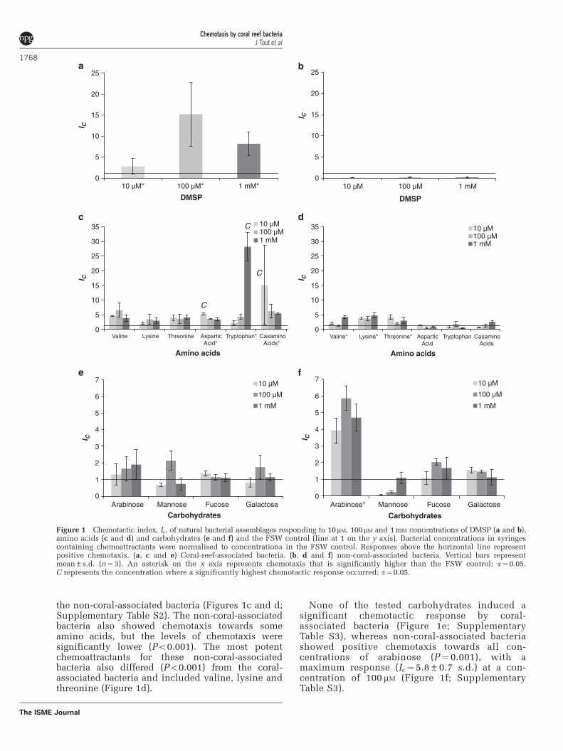
the non-coral-associated bacteria (Figures 1c and d;Supplementary Table S2). The non-coral-associatedbacteria also showed chemotaxis towards someamino acids, but the levels of chemotaxis weresignificantly lower (Po0.001). The most potentchemoattractants for these non-coral-associatedbacteria also differed (Po0.001) from the coral-associated bacteria and included valine, lysine andthreonine (Figure 1d).
None of the tested carbohydrates induced asignificant chemotactic response by coral-associated bacteria (Figure 1e; SupplementaryTable S3), whereas non-coral-associated bacteriashowed positive chemotaxis towards all con-centrations of arabinose (P¼ 0.001), with amaximum response (Ic¼ 5.8±0.7 s.d.) at a con-centration of 100 mM (Figure 1f; SupplementaryTable S3).
0
5
10
15
20
25
10 µM 100 µM 1 mM
DMSP
0
5
10
15
20
25
10 µM* 100 µM* 1 mM*
I CI C
I C
I CI C
I C
DMSP
0
1
2
3
4
5
6
7
Arabinose Mannose Fucose Galactose
Carbohydrates
10 µM
100 µM
1 mM
0
1
2
3
4
5
6
7
Arabinose* Mannose Fucose Galactose
Carbohydrates
10 µM
100 µM
1 mM
0
5
10
15
20
25
30
35
Valine Lysine Threonine AsparticAcid*
Tryptophan* CasaminoAcids*
Amino acids
10 µM100 µM1 mM
0
5
10
15
20
25
30
35
Valine* Lysine* Threonine* AsparticAcid
Tryptophan CasaminoAcids
Amino acids
10 µM100 µM1 mM
C
C
C
Figure 1 Chemotactic index, Ic, of natural bacterial assemblages responding to 10mM, 100mM and 1 mM concentrations of DMSP (a and b),amino acids (c and d) and carbohydrates (e and f) and the FSW control (line at 1 on the y axis). Bacterial concentrations in syringescontaining chemoattractants were normalised to concentrations in the FSW control. Responses above the horizontal line representpositive chemotaxis. (a, c and e) Coral-reef-associated bacteria. (b, d and f) non-coral-associated bacteria. Vertical bars representmean±s.d. (n¼ 3). An asterisk on the x axis represents chemotaxis that is significantly higher than the FSW control; a¼0.05.C represents the concentration where a significantly highest chemotactic response occurred; a¼ 0.05.
Chemotaxis by coral reef bacteriaJ Tout et al
1768
The ISME Journal
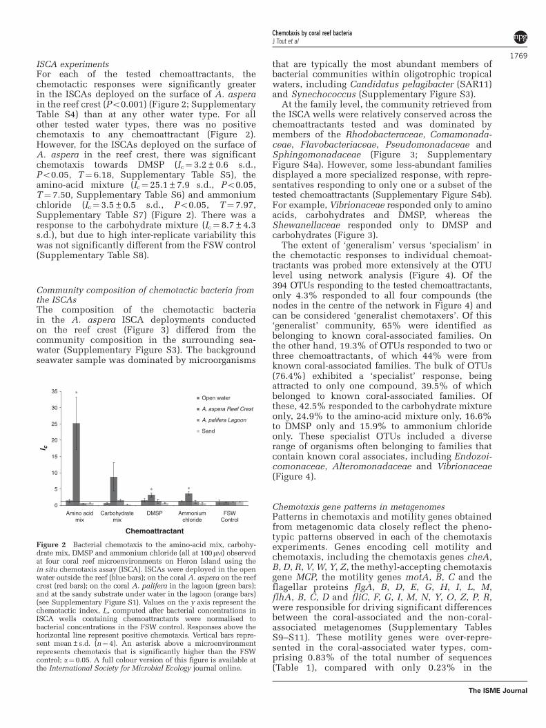
ISCA experimentsFor each of the tested chemoattractants, thechemotactic responses were significantly greaterin the ISCAs deployed on the surface of A. asperain the reef crest (Po0.001) (Figure 2; SupplementaryTable S4) than at any other water type. For allother tested water types, there was no positivechemotaxis to any chemoattractant (Figure 2).However, for the ISCAs deployed on the surface ofA. aspera in the reef crest, there was significantchemotaxis towards DMSP (Ic¼ 3.2±0.6 s.d.,Po0.05, T¼ 6.18, Supplementary Table S5), theamino-acid mixture (Ic¼ 25.1±7.9 s.d., Po0.05,T¼ 7.50, Supplementary Table S6) and ammoniumchloride (Ic¼ 3.5±0.5 s.d., Po0.05, T¼ 7.97,Supplementary Table S7) (Figure 2). There was aresponse to the carbohydrate mixture (Ic¼ 8.7±4.3s.d.), but due to high inter-replicate variability thiswas not significantly different from the FSW control(Supplementary Table S8).
Community composition of chemotactic bacteria fromthe ISCAsThe composition of the chemotactic bacteriain the A. aspera ISCA deployments conductedon the reef crest (Figure 3) differed from thecommunity composition in the surrounding sea-water (Supplementary Figure S3). The backgroundseawater sample was dominated by microorganisms
that are typically the most abundant members ofbacterial communities within oligotrophic tropicalwaters, including Candidatus pelagibacter (SAR11)and Synechococcus (Supplementary Figure S3).
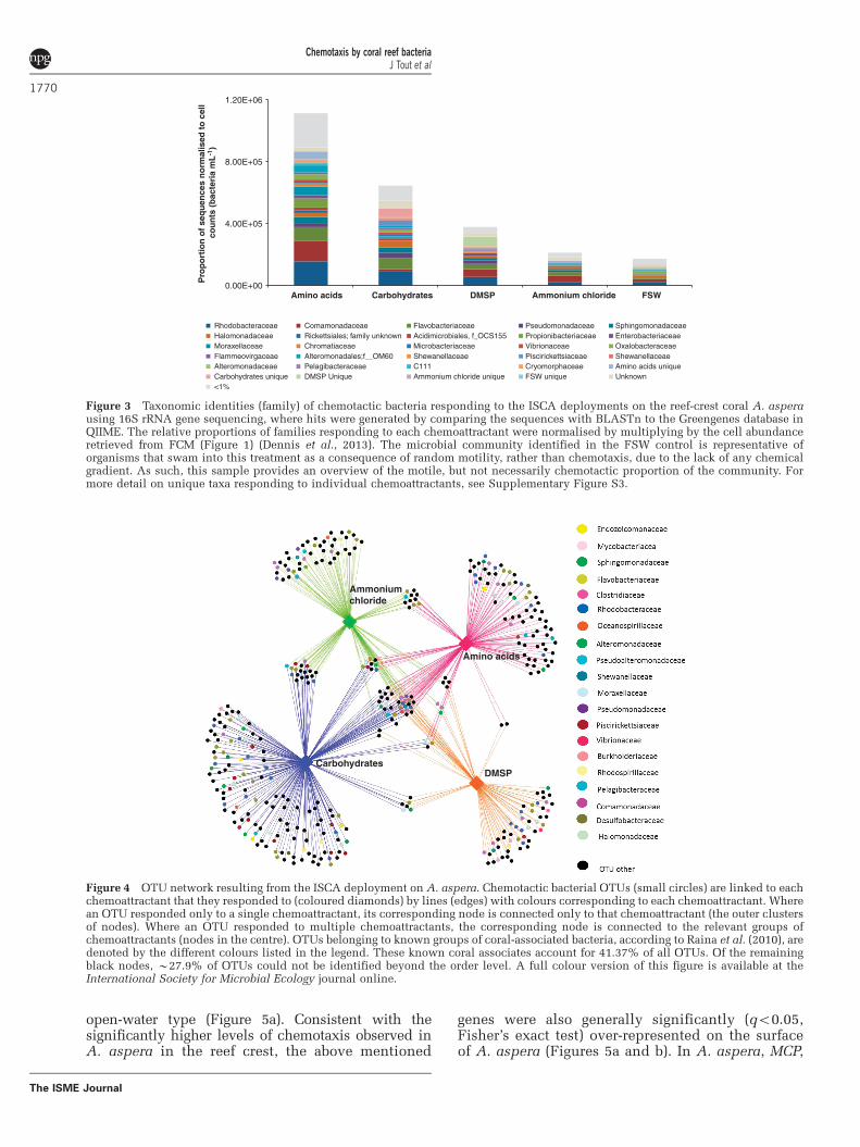
At the family level, the community retrieved fromthe ISCA wells were relatively conserved across thechemoattractants tested and was dominated bymembers of the Rhodobacteraceae, Comamonada-ceae, Flavobacteriaceae, Pseudomonadaceae andSphingomonadaceae (Figure 3; SupplementaryFigure S4a). However, some less-abundant familiesdisplayed a more specialized response, with repre-sentatives responding to only one or a subset of thetested chemoattractants (Supplementary Figure S4b).For example, Vibrionaceae responded only to aminoacids, carbohydrates and DMSP, whereas theShewanellaceae responded only to DMSP andcarbohydrates (Figure 3).
The extent of ‘generalism’ versus ‘specialism’ inthe chemotactic responses to individual chemoat-tractants was probed more extensively at the OTUlevel using network analysis (Figure 4). Of the394 OTUs responding to the tested chemoattractants,only 4.3% responded to all four compounds (thenodes in the centre of the network in Figure 4) andcan be considered ‘generalist chemotaxers’. Of this‘generalist’ community, 65% were identified asbelonging to known coral-associated families. Onthe other hand, 19.3% of OTUs responded to two orthree chemoattractants, of which 44% were fromknown coral-associated families. The bulk of OTUs(76.4%) exhibited a ‘specialist’ response, beingattracted to only one compound, 39.5% of whichbelonged to known coral-associated families. Ofthese, 42.5% responded to the carbohydrate mixtureonly, 24.9% to the amino-acid mixture only, 16.6%to DMSP only and 15.9% to ammonium chlorideonly. These specialist OTUs included a diverserange of organisms often belonging to families thatcontain known coral associates, including Endozoi-comonaceae, Alteromonadaceae and Vibrionaceae(Figure 4).
Chemotaxis gene patterns in metagenomesPatterns in chemotaxis and motility genes obtainedfrom metagenomic data closely reflect the pheno-typic patterns observed in each of the chemotaxisexperiments. Genes encoding cell motility andchemotaxis, including the chemotaxis genes cheA,B, D, R, V, W, Y, Z, the methyl-accepting chemotaxisgene MCP, the motility genes motA, B, C and theflagellar proteins flgA, B, D, E, G, H, I, L, M,flhA, B, C, D and fliC, F, G, I, M, N, Y, O, Z, P, R,were responsible for driving significant differencesbetween the coral-associated and the non-coral-associated metagenomes (Supplementary TablesS9–S11). These motility genes were over-repre-sented in the coral-associated water types, com-prising 0.83% of the total number of sequences(Table 1), compared with only 0.23% in the
0
5
10
15
20
25
30
35
Amino acidmix
Carbohydratemix
DMSP Ammoniumchloride
FSWControl
I c
Chemoattractant
Open water
A. aspera Reef Crest
A. palifera Lagoon
Sand
∗
∗ ∗
Figure 2 Bacterial chemotaxis to the amino-acid mix, carbohy-drate mix, DMSP and ammonium chloride (all at 100mM) observedat four coral reef microenvironments on Heron Island using thein situ chemotaxis assay (ISCA). ISCAs were deployed in the openwater outside the reef (blue bars); on the coral A. aspera on the reefcrest (red bars); on the coral A. palifera in the lagoon (green bars);and at the sandy substrate under water in the lagoon (orange bars)(see Supplementary Figure S1). Values on the y axis represent thechemotactic index, Ic, computed after bacterial concentrations inISCA wells containing chemoattractants were normalised tobacterial concentrations in the FSW control. Responses above thehorizontal line represent positive chemotaxis. Vertical bars repre-sent mean±s.d. (n¼4). An asterisk above a microenvironmentrepresents chemotaxis that is significantly higher than the FSWcontrol; a¼0.05. A full colour version of this figure is available atthe International Society for Microbial Ecology journal online.
Chemotaxis by coral reef bacteriaJ Tout et al
1769
The ISME Journal
open-water type (Figure 5a). Consistent with thesignificantly higher levels of chemotaxis observed inA. aspera in the reef crest, the above mentioned
genes were also generally significantly (qo0.05,Fisher’s exact test) over-represented on the surfaceof A. aspera (Figures 5a and b). In A. aspera, MCP,
0.00E+00
4.00E+05
8.00E+05
1.20E+06
Amino acids Carbohydrates DMSP Ammonium chloride FSW
Pro
po
rtio
n o
f se
qu
ence
s n
orm
alis
ed t
o c
ell
cou
nts
(b
acte
ria
mL
-1)
Rhodobacteraceae Comamonadaceae Flavobacteriaceae Pseudomonadaceae SphingomonadaceaeHalomonadaceae Rickettsiales; family unknown Acidimicrobiales, f_OCS155 Propionibacteriaceae EnterobacteriaceaeMoraxellaceae Chromatiaceae Microbacteriaceae Vibrionaceae OxalobacteraceaeFlammeovirgaceae Alteromonadales;f__OM60 Shewanellaceae Piscirickettsiaceae ShewanellaceaeAlteromonadaceae Pelagibacteraceae C111 Cryomorphaceae Amino acids uniqueCarbohydrates unique DMSP Unique Ammonium chloride unique FSW unique Unknown<1%
Figure 3 Taxonomic identities (family) of chemotactic bacteria responding to the ISCA deployments on the reef-crest coral A. asperausing 16S rRNA gene sequencing, where hits were generated by comparing the sequences with BLASTn to the Greengenes database inQIIME. The relative proportions of families responding to each chemoattractant were normalised by multiplying by the cell abundanceretrieved from FCM (Figure 1) (Dennis et al., 2013). The microbial community identified in the FSW control is representative oforganisms that swam into this treatment as a consequence of random motility, rather than chemotaxis, due to the lack of any chemicalgradient. As such, this sample provides an overview of the motile, but not necessarily chemotactic proportion of the community. Formore detail on unique taxa responding to individual chemoattractants, see Supplementary Figure S3.
Ammonium chloride
Amino acids
DMSPCarbohydrates
Figure 4 OTU network resulting from the ISCA deployment on A. aspera. Chemotactic bacterial OTUs (small circles) are linked to eachchemoattractant that they responded to (coloured diamonds) by lines (edges) with colours corresponding to each chemoattractant. Wherean OTU responded only to a single chemoattractant, its corresponding node is connected only to that chemoattractant (the outer clustersof nodes). Where an OTU responded to multiple chemoattractants, the corresponding node is connected to the relevant groups ofchemoattractants (nodes in the centre). OTUs belonging to known groups of coral-associated bacteria, according to Raina et al. (2010), aredenoted by the different colours listed in the legend. These known coral associates account for 41.37% of all OTUs. Of the remainingblack nodes, B27.9% of OTUs could not be identified beyond the order level. A full colour version of this figure is available at theInternational Society for Microbial Ecology journal online.
Chemotaxis by coral reef bacteriaJ Tout et al
1770
The ISME Journal
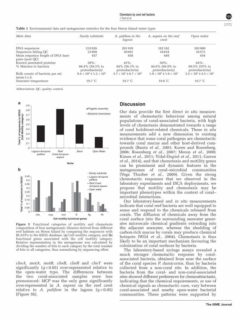
cheA, motA, motB, cheB, cheR and cheY weresignificantly (qo0.05) over-represented relative tothe open-water type. The differences betweenthe two coral-associated samples were lesspronounced: MCP was the only gene significantlyover-represented in A. aspera on the reef crestrelative to A. palifera in the lagoon (qo0.05)(Figure 5b).
Discussion
Our data provide the first direct in situ measure-ments of chemotactic behaviour among naturalpopulations of coral-associated bacteria, with highlevels of chemotaxis demonstrated towards a rangeof coral holobiont-related chemicals. These in situmeasurements add a new dimension to existingevidence that some coral pathogens are chemotactictowards coral mucus and other host-derived com-pounds (Banin et al., 2001; Koren and Rosenberg,2006; Rosenberg et al., 2007; Meron et al., 2009;Kimes et al., 2011; Vidal-Dupiol et al., 2011; Garrenet al., 2014), and that chemotaxis and motility genescan be prominent and dynamic features in themetagenomes of coral–microbial communities(Vega Thurber et al., 2009). Given the strongchemotactic responses that we observed in thelaboratory experiments and ISCA deployments, wepropose that motility and chemotaxis may beimportant phenotypes within the context of coral–microbial interactions.
Our laboratory-based and in situ measurementsindicate that coral reef bacteria are well equipped tosense and respond to the chemicals released fromcorals. The diffusion of chemicals away from thecoral surface into the surrounding seawater gener-ates microscale chemical gradients extending intothe adjacent seawater, whereas the shedding ofcarbon-rich mucus by corals may produce chemicalhotspots (Wild et al., 2004). Chemotaxis is thuslikely to be an important mechanism favouring thecolonisation of coral surfaces by bacteria.
The laboratory-based syringe assays revealed amuch stronger chemotactic response by coral-associated bacteria, obtained from near the surfaceof the coral species P. damicornis, than by bacteriacollected from a non-coral site. In addition, thebacteria from the coral- and non-coral-associatedsites showed different preferences for chemoattractants,indicating that the chemical requirements, or use ofchemical signals as chemotactic cues, vary betweencoral-associated and nearby open-water bacterialcommunities. These patterns were supported by
Table 1 Environmental data and metagenome statistics for the four Heron Island water types
Meta data Sandy substrate A. palifera in thelagoon
A. aspera on the reefcrest
Open water
DNA sequences 233 026 201 910 182 182 103 900Sequences failing QC 23 899 20 691 18 818 10 573Mean sequence length of DNA basepairs (post QC)
457 450 449 454
Known annotated proteins 58%; 45%; 50%; 64%;% Matches to bacteria 88.4% (58.3% to
proteobacteria)84% (56.3% toproteobacteria)
84.6% (60.9% toproteobacteria)
89.5% (55% toproteobacteria)
Bulk counts of bacteria per ml,mean±s.d.
9.4� 104±1.2� 104 3.7� 105±6.7� 104 1.0� 106±1.6� 105 3.5�106±4.5�105
Seawater temperature 19.7 1C 19.7 1C 19.6 1C 19.7 1C
Abbreviation: QC, quality control.
0
0.1
0.2
0.3
0.4
0.5
0.6
0.7
0.8
0.9
Lagoon-Acroporapalifera
ReefCrest-Acropora
aspera
Sand Open Water
Tota
l % o
f se
qu
ence
s to
KE
GG
(C
ell M
oti
lity
cate
go
ry)
Niche
Flagellar assembly
Bacterial chemotaxis
0
0.05
0.1
0.15
0.2
0.25
0.3
0.35
0.4
0.45
che mcp mot fli flg flh
Tota
l % o
f se
qu
ence
s to
KE
GG
Cell motility functional genes
Sandy substrate
Lagoon-Acroporapalifera
Reef Crest-Acropora asperaOpen water
Figure 5 Functional categories of motility and chemotaxiscomposition of four metagenomic libraries derived from differentreef habitats on Heron Island by comparing the sequences withBLASTn to the KEGG database. (a) Cell motility category, and (b)functional genes associated with the cell motility category.Relative representation in the metagenome was calculated bydividing the number of hits to each category by the total numberof hits to all categories, thus normalizing by sequencing effort.
Chemotaxis by coral reef bacteriaJ Tout et al
1771
The ISME Journal
the results of the in situ experiments, wherebybacteria associated with the surface of the coralA. aspera exhibited significantly higher levels ofchemotactic capability than bacteria from any otherwater type around the reef or from the open water.
The short-branching morphology of P. damicorniswas not amenable to ISCA deployment, so ISCAexperiments were performed on the more deeplybranching A. aspera in the reef crest and A. paliferain the lagoon. Although this prevents direct compa-rison of our laboratory and in situ results, thegeneral pattern of higher levels of chemotaxis oncoral surfaces was conserved between approaches.
Elevated levels of chemotaxis observed among themicrobial communities associated with the reef crestcoral A. aspera in the ISCA experiments weresupported by metagenomic analysis, where genesassociated with motility and chemotaxis weresignificantly more abundant in the seawater asso-ciated with A. aspera than in any other sample. Ofnote, motility and chemotaxis genes in the A. asperasample were significantly more abundant than inthe sample obtained from the other coral speciesA. palifera located in the lagoon, which is directly inline with the differences in chemotactic responsesobserved between the bacterial communities asso-ciated with these two coral species in the ISCAexperiments. Taken together, our results demon-strate that chemotaxis is heterogeneous across acoral reef and between communities associated withdifferent corals. The different chemtoaxis patternsobserved between the bacterial communities asso-ciated with the two Acropora species may be due tointer-coral variability in microbial community com-position and function (Rohwer et al., 2001, 2002;Tout et al., 2014), or slight differences in themorphology of the two coral species may havealtered the biophysical environment inhabited bythe resident microbes (Wallace, 1999). In addition,the location of the corals within the Heron Islandreef system may be responsible for some of thesedifferences. A. aspera was chosen for sampling inthe reef crest environment, whereas A. palifera waschosen within the lagoon, because these speciesdominate the coral communities within these twohabitats, respectively (Wild et al., 2004). The reefcrest, where the A. aspera deployment occurred isan environment characterised by a wall of Acroporacorals, where 69% of the benthos is covered by hardcorals (Salmond et al., 2013). In contrast, in thelagoon where the ISCAs were deployed onA. palifera, hard corals contribute to only 6% ofthe total benthic cover (the lowest hard coral coveron Heron Island) (Salmond et al., 2013) and the bulkof the substrate is sand. As a consequence, the localenvironment surrounding the different coral colo-nies varied substantially, with differences in thebulk microbial community within these two habitatspotentially contributing to the differences observedhere. Finally, Vega Thurber et al. (2009) showedsignificant shifts in chemotaxis genes among the
microbial communities associated with stressedcorals relative to healthy individuals, and althoughno notable signs of disease or stress were apparent ineither coral tested here, there remains the possibilitythat differences in the health status of the coral hostsmay contribute to differences in the level ofchemotaxis observed here.
Natural communities of coral-associated bacteriashowed strong chemotactic responses towardsDMSP in both the laboratory assays and the ISCAdeployments. This is consistent with DMSP beingan important source of carbon and reduced sulphurfor marine bacteria (Howard et al., 2006) and achemical cue for some coral pathogens (Garren et al.,2014). DMSP is abundant on coral reefs (Broadbentet al., 2002; Broadbent and Jones, 2004; Raina et al.,2010), with coral mucus concentrations (up to 62mM)the highest reported in the marine environment(Broadbent et al., 2002; Broadbent and Jones, 2004).There is now evidence that both the coral symbiontsSymbiodinium spp. (Keller et al., 1989; Broadbentet al., 2002) and the coral animal (Raina et al., 2013)have the capacity to produce significant quantitiesof DMSP. Our results confirm that, similar to othermarine microorganisms (Miller et al., 2004;Seymour et al., 2010; Sharp et al., 2012), coral-reef-associated bacteria use chemotaxis to enhancetheir access to DMSP or to follow DMSP gradients asa cue to locate the host (Garren et al., 2014). Thedominant bacterial taxa responding to DMSPincluded known coral-associated species that havethe capacity to degrade DMSP (Flavobacteriaceae)and DMS (Comamonadaceae) or both (Rhodobacter-acea, Pseudomonaceae and Halomonaceae) (Rainaet al., 2010). It is notable that we also observed someVibrio sequences in the DMSP sample, becauserecent evidence suggests that DMSP can be a potentchemoattractant for coral pathogens belonging tothis genus (Garren et al., 2014).
Strong chemotactic responses to several aminoacids by coral-associated bacteria were alsoobserved using both the syringe assays and theISCA deployments. The amino acids used in thechemotaxis assays have previously been found inthe mucus of several coral species (Ducklow andMitchell, 1979b; Meikle et al., 1988; Fitzgerald andSzmant, 1997). Chemotaxis towards amino acids iswell recognised among enteric bacteria (Adler, 1966;Mesibov and Adler, 1972; Kim et al., 2001; Baineret al., 2003; Frank et al., 2011), has been demon-strated in some marine bacteria (Barbara andMitchell, 2003a,b) and our results indicate that itmay also be involved in coral–microbe interactionsin coral reef environments.
Marine bacteria have been shown to exhibitchemotaxis towards carbohydrates present in theexudates of phytoplankton (Hellebust, 1965; Belland Mitchell, 1972) and in the mucus of theHawaiian squid Euprymna scolopes light organ(DeLoney-Marino et al., 2003). Despite the prominenceof carbohydrates in coral mucus (Ducklow and
Chemotaxis by coral reef bacteriaJ Tout et al
1772
The ISME Journal
Mitchell, 1979b; Meikle et al., 1988; Wild et al., 2005),the chemotactic response of coral-associatedbacteria towards the tested carbohydrates in boththe syringe assays and the ISCAs were neversignificantly different from the FSW control. Takentogether, these findings indicate that carbohydratespreviously shown to occur in coral mucus do notappear to be an important chemical cue for coral-associated bacteria.
Our results demonstrate that natural populationsof coral-associated bacteria exhibit chemotaxistowards ammonium. Aquatic heterotrophic bacteriahave previously been shown to use chemotaxis toexploit patches of inorganic substrates includingammonium (Stocker and Seymour, 2012; Denniset al., 2013). Within coral reefs, nitrogen is often alimiting nutrient in the water column (Thomas andOwen, 1971; Crossland et al., 1984; Moore et al.,2013), as nitrate and ammonium concentrations areoften o2 mM (Crossland et al., 1984; Bythell, 1990).However, ammonium and other inorganic nutrientsare likely to be generated as metabolic by-productsin the coral holobiont (Kawaguti, 1953; Muscatineand D’Elia, 1978; Siboni et al., 2008), and ammo-nium levels can reach up to 50 mM within coralmucus (Wild et al., 2005). Therefore the ability touse chemotaxis to exploit elevated levels of inor-ganic nutrients near the surface of corals mayprovide a competitive advantage for some coral reefbacteria.
The microbial community identified in the FSWcontrol is representative of organisms that swaminto this treatment as a consequence of randommotility, rather than chemotaxis, due to the lack ofany chemical gradient. As such, this sample pro-vides an overview of the motile, but not necessarilychemotactic proportion of the community. Thechoice of chemoattractant concentrations in ourexperiments was guided by concentrations pre-viously shown to occur within coral microenviron-ments, such as coral mucus (Broadbent and Jones,2004; Wild et al., 2004, 2005, 2010). In most cases,no significant difference was observed in thechemotactic response between different concentra-tions. However, the coral-associated bacteria’sresponse to tryptophan increased with increasingconcentrations. On the other hand, in the case ofaspartic and casamino acids, the strongest chemo-taxis occurred in response to the lowest testedconcentration of 10 mM, which is perhaps indicativeof an inhibitory response associated with saturationof chemoreceptors at higher concentrations(Mesibov et al., 1973). These patterns indicate thatthe chemotactic sensitivities and thresholds forcoral-associated bacteria vary between differenttypes of compounds, which may reflect the relativeavailability and concentrations of these substrates inthe environment.
Recent measurements of the chemotacticbehaviour of a natural bacterial population from alake revealed a strong phylogenetic partitioning
in the response towards inorganic compounds(Dennis et al., 2013). Our results have expandedupon this work by identifying members of thechemotactic bacterial communities associated withthe coral species A. aspera, and demonstrating thatthese differed substantially from the community inthe surrounding seawater. This indicates thatchemotaxis may act as a behavioural filter, favouringcertain species over others in associating with coralsand thus determining the composition of microbialcommunities within specific coral microniches.
At the family level of taxonomic resolution, wefound that the same bacterial groups were typicallythe dominant responders to all chemoattractants,indicating that microorganisms belonging to arestricted range of bacterial families perform che-motaxis in the coral reef environment. These groupsincluded families with members that have metabolicrequirements for the tested chemoattractants (forexample, Flavobacteriaceae for DMSP; Raina et al.,2010) or are known coral associates (Rhodobacter-aceae, Comamonadaceae, Flavobacteriaceae andPseudomonadaceae (Raina et al., 2010; Morrowet al., 2012)). However, at a finer taxonomicresolution it became clear that specialist chemotac-tic responses, whereby organisms only exhibitedchemotaxis to certain chemicals, were common.There were some families where representativesonly responded to one or a subset of the chemoat-tractants tested. These more-specialised groups ofchemotaxers included members of the Shewanella-ceae and Vibrionaceae families. The Shewanella-ceae only exhibited chemotaxis towards DMSP andcarbohydrates. This family includes known coral-associated bacteria (Shnit-Orland and Kushmaro,2009; Raina et al., 2010; Ceh et al., 2012) and speciesthat degrade DMSP (Raina et al., 2010). Chemotaxisby members of the marine Shewanellaceae towardsamino acids and algae has previously been observed(Barbara and Mitchell, 2003a,b), and our resultsdemonstrate that members of this known group ofcoral associates may use chemotaxis towards DMSPwithin the coral holobiont.
Clear differentiation of the chemotactic commu-nity into groups of ‘specialist’ and ‘generalist’chemotaxers became particularly evident whenresponses were assessed at the OTU level. Approxi-mately 4.3% of all OTUs responded to all fourcompounds tested, whereas 19.3% responded to2 and 3 chemoattractants. This suggests that a subsetof the chemotactic community is made of ‘generalistchemotaxers’ that have the capacity to sense anddirect movement in response to diverse chemicalscompounds. Currently, little is known about thechemoreceptors used by marine bacteria (Stockerand Seymour, 2012), but the generalist chemotaxersidentified here may either have multiple sets ofchemoreceptors, which allow them to respond to avariety of compounds (Wadhams and Armitage,2004; Parales et al., 2013), or single chemoreceptorsthat allow for binding of multiple compounds
Chemotaxis by coral reef bacteriaJ Tout et al
1773
The ISME Journal
(Adler, 1969; Glekas et al., 2012). However, thebulk (76.4%) of chemotactic OTUs identified herecan be classified as ‘specialist chemotaxers’, as theyresponded to only one compound. Many of thesespecialist chemotaxers were also known coralassociates including members of Endozoicomona-ceae, Rhodobacteraceae, Vibrionaceae and Myco-bacteriacea. In the environment, the heterogeneityin chemotactic responses observed here could leadto strong niche partitioning among the bacterialcommunity, suggesting that this behaviour couldunderpin some of the heterogeneity in microbialcommunity composition previously observedbetween different coral hosts (Rohwer et al., 2002).
The partitioning of coral–microbe communitiesobserved here may be encouraged by differentialrelease of chemoattractants from the coral holobiontunder varying environmental conditions. There isevidence that the production and release of keychemoattractants, such as DMSP, can vary markedlyunder different environmental conditions and coralhealth states (Raina et al., 2013; Garren et al., 2014).From a bacterium’s perspective, chemotaxis serves avariety of potential ecological functions, including(i) providing cells with greater access to importantgrowth substrates and nutrients in otherwise oligo-trophic habitats (Stocker et al., 2008; Seymour et al.,2009), (ii) enhancing exposure to terminal electronacceptors/donors (Schweinitzer and Josenhans, 2010)or (iii) use of infochemicals that may providepathogenic microorganisms with information aboutthe location, health and potential susceptibility ofcoral hosts to infection (Garren et al., 2014).Heterogeneity in the strength of chemotaxis towardsdifferent chemoattractants observed across thedifferent water types is perhaps indicative of varyingecological strategies among microbial communitieson coral reefs.
Conclusions
We have provided the first in situ quantification ofbacterial chemotaxis on a coral reef. Previousstudies have shown that different habitats andmicroenviroments on coral reefs host phylogeneti-cally distinct communities (Rohwer et al., 2002;Rohwer and Kelly, 2004), but our results show thatdifferent reef features (for example, coral species andwater types) also host distinct microbial phenotypiccapacities, specifically the ability to performchemotaxis. In addition, we have provided a directmechanism for how chemical gradients associatedwith coral surfaces may be involved in the establish-ment of specific coral–bacterial relationships, andhow microbial chemotaxis might shape the compo-sition of coral reef bacterial communities. As such,we suggest that within the chemically and physicallycomplex coral microenvironment, bacterial beha-viours, including motility and chemotaxis, may befundamental drivers of patterns in microbial
diversity and metabolism, coral infection dynamicsand chemical cycling processes.
Conflict of Interest
The authors declare no conflict of interest.
Acknowledgements
This research was funded by the Australian ResearchCouncil Grant DP110103091 to JRS, GWT and RS and theHuman Frontiers in Science Program award no. RGY0089to RS and JRS. This research was funded in part by theGordon and Betty Moore Foundation through Grant #3801to JRS, RS and GWT and an Investigator Award (grantGBMF3783) to RS; a post-graduate award to JT from theDepartment of Environmental Science and ClimateChange Cluster at the University of Technology Sydney.JRS and NSW were funded through Australian ResearchCouncil Future Fellowships FT130100218 andFT120100480, respectively. We are grateful to the GreatBarrier Reef Marine Park Authority for coral collectionpermits G09/31733.1 (PJR, University of Technology,Sydney). Amplicons were sequenced using the 454 GS-FLX pyrosequencing platform (Roche) at the AustralianCentre for Ecogenomics (University of Queensland, Aus-tralia). Metagenomic sequencing was carried out at theRamaciotti Centre for Gene Function Analysis at theUniversity of New South Wales. The metagenomes can beaccessed through MG-Rast (http://metagenomics.anl.gov)under the project numbers 4483104.3 (A. palifera in thelagoon), 4483105.3 (A. aspera on the reef crest), 4483106.3(sandy substrate) and 4483107.3 (open water).
References
Adler J. (1966). Chemotaxis in bacteria. Science 153:708–716.
Adler J. (1969). Chemoreceptors in bacteria. Science 166:1588–1597.
Adler J. (1973). A method for measuring chemotaxis anduse of the method to determine optimum conditionsfor chemotaxis by Escherichia coli. J Gen Microbiol 74:77–91.
Bainer R, Park H, Cluzel P. (2003). A high-throughputcapillary assay for bacterial chemotaxis. J MicrobiolMethods 55: 315–319.
Banin E, Israely T, Fine M, Loya Y, Rosenberg E. (2001).Role of endosymbiotic zooxanthellae and coralmucus in the adhesion of the coral-bleaching pathogenVibrio shiloi to its host. FEMS Microbiol Lett 199:33–37.
Barbara GM, Mitchell JG. (2003a). Marine bacterialorganisation around point-like sources of amino acids.FEMS Microbiol Ecol 43: 99–109.
Barbara GM, Mitchell JG. (2003b). Bacterial tracking ofmotile algae. FEMS Microbiol Ecol 44: 79–87.
Bartlett DG, Matsumura P. (1986). Behavioural responses tochemical cues by bacteria. J Chem Ecol 12: 1071–1089.
Bell W, Mitchell R. (1972). Chemotactic and growthresponses of marine bacteria to algal extracellularproducts. The Biol Bull 143: 265–277.
Chemotaxis by coral reef bacteriaJ Tout et al
1774
The ISME Journal
Bianchi A, Giuliano L. (1996). Enumeration of viablebacteria in the marine pelagic environment. ApplMicrobiol 62: 174–177.
Blackburn N, Fenchel T, Mitchell J. (1998). Microscalenutrient patches in planktonic habitats shown bychemotactic bacteria. Science 282: 2254–2256.
Bragg L, Stone G, Imelfort M, Hugenholtz P, Tyson GW.(2012). Fast, accurate error-correction of ampliconpyrosequences using Acacia. Nat Methods 9: 425–426.
Broadbent AD, Jones GB, Jones RJ. (2002). DMSP in coralsand benthic algae from the Great Barrier Reef. EstuarCoast Shelf Sci 55: 547–555.
Broadbent AD, Jones GB. (2004). DMS and DMSP inmucus ropes, coral mucus, surface films and sedimentpore waters from coral reefs in the Great Barrier Reef.Mar Freshwater Res 55: 849–855.
Bythell JC. (1990). Nutrient uptake in the reef-buildingcoral Acropora palmate at natural environmentalconcentrations. Mar Ecol Prog Ser 68: 65–69.
Caporaso JG, Kuczynski J, Stombaugh J, Bittinger K,Bushman FD, Costello EK et al. (2010). QIIME allowsanalysis of high-throughput community sequencingdata. Nat Methods 7: 335–336.
Ceh J, van Keulen M, Bourne DG. (2011). Coral-associatedbacterial communities on Ningaloo Reef, WesternAustralia. FEMS Microbiol Ecol 75: 134–144.
Ceh J, Raina J-B, Soo RM, van Keulen M, Bourne DG.(2012). Coral-bacterial communities before and after acoral mass spawning event on Ningaloo Reef. PLoSOne 7: e36920.
Chet I, Mitchell R. (1976). Ecological aspects of microbialchemotactic behaviour. Annu Rev Microbiol 30: 221–239.
Clarke KR, Gorley RN. (2006). PRIMER v6: User Manual/Tutorial. PRIMER-E: Plymouth, UK.
Clarke KR. (1993). Non-parametric multivariate analysesof changes in community structure. Aust J Ecol 18:117–143.
Crossland CJ, Hatcher BG, Atkinson MJ, Smith SV. (1984).Dissolved nutrients of a high-latitude coral reef,Houtman Abrolhos Islands, Western Australia. MarEcol Prog Ser 14: 159–163.
DeLoney-Marino CR, Wolfe AJ, Visick KL. (2003).Chemoattraction of Vibrio fischeri to Serine, nucleo-sides and N-Acetylneuraminic Acid, a component ofsquid light-organ Mucus. Appl Environ Microbiol 69:7527–7530.
Dennis PG, Seymour J, Kumbun K, Tyson GW. (2013).Diverse populations of lake water bacteria exhibitchemotaxis towards inorganic nutrients. ISME J 7:1661–1664.
Ducklow HW, Mitchell R. (1979a). Bacterial populationsand adaptations in the mucus layers of living corals.Limnol Oceanogr 24: 715–725.
Ducklow HW, Mitchell R. (1979b). Composition of mucusreleased by coral reef coelenterates. Limnol Oceanogr24: 706–714.
Fan L, Reynolds D, Liu M, Stark S, Kjelleberg S,Webster NS, Thomas T. (2012). Functional equiva-lence and evolutionary convergence in complexcommunities of microbial sponge symbionts. ProcNatl Acad Sci USA 109: E1878–E1887.
Fitzgerald LM, Szmant AM. (1997). Biosynthesis of‘essential’ amino acids by scleractinian corals.Biochem J 322: 213–221.
Frank V, Koler M, Furst S, Vaknin A. (2011). The physicaland functional thermal sensitivity of bacterial chemor-eceptors. J Mol Biol 411: 554–566.
Frias-Lopez J, Zerkl AL, Bonheyo GT, Fouke BW. (2002).Partitioning of bacterial communities between sea-water and healthy, black band diseased and dead coralsurfaces. Appl Environ Microbiol 68: 2214–2228.
Fuhrman J, Steele JA. (2008). Community structure ofmarine bacterioplankton: patterns, networks, and rela-tionships to function. Aquat Microb Ecol 53: 69–81.
Garren M, Son K, Raina JB, Rusconi R, Menolascina F,Shapiro OH et al. (2014). A bacterial pathogen usesdimethylsulfoniopropionate as a cue to target heat-stressed corals. ISME J 8: 999–1007.
Glekas GD, Mulhern BJ, Kroc A, Duelfer KA, Lei V, Rao CV,Ordal GW. (2012). The Bacillus subtilis chemoreceptorMcpC senses multiple ligands using two discretemechanisms. J Biol Chem 287: 39412–39418.
Grossart HP, Riemann L, Azam F. (2001). Bacterial motilityin the seas and its ecological implications. AquatMicrob Ecol 25: 247–258.
Hellebust JA. (1965). Excretion of some organic com-pounds by marine phytoplankton. Limnol Oceanogr10: 192–206.
Hill RW, Dacey WH, Krupp DA. (1995). Dimethylsulfo-nioprpionate in reef corals. Bull Mar Sci 57: 489–494.
Hoppe HG. (1976). Determination and properties ofactively metabolising heterotrophic bacteria in thesea, investigated by means of micro-autoradiography.Mar Biol 36: 291–302.
Howard EC, Henriksen JR, Buchan A, Reisch CR, Burg-mann H, Welsh R et al. (2006). Bacterial taxa that limitsulfur flux from the ocean. Science 314: 649–652.
Jannasch HW, Jones GE. (1959). Bacterial populations inseawater as determined by different methods ofenumeration. Limnol Oceanogr 4: 128–139.
Kamada T, Kawai S. (1989). An algorithm for drawinggeneral undirected graphs. Inform Process Lett 31:7–15.
Kawaguti S. (1953). Ammonium metabolism of reef corals.Biol J Okayama Univ 1: 171–176.
Keller MD, Bellows WK, Guillard RL. (1989). Dimethyl-sulfide production in marine phytoplankton.In Saltzman ES, Cooper JC (eds) Biogenic Sulfur inthe Environment. American Chemical Society:Washington, DC, pp 167–182.
Kellogg C. (2004). Tropical Archaea: diversity associatedwith the surface microlayers of corals. Mar Ecol ProgSer 273: 81–88.
Kim C, Jackson M, Lux R, Khan S. (2001). Determinants ofchemotactic signal amplification in Escherichia coli.J Mol Biol 307: 119–135.
Kimes NE, Grim CJ, Johnson WR, Hasan NA, Tall BD,Kothary MH et al. (2011). Temperature regulationof virulence factors in the pathogen Vibrio coralliily-ticus. ISME J 6: 835–846.
Koren O, Rosenberg E. (2006). Bacteria associated withmucus and tissues of the coral Oculina patagonica insummer and winter. Appl Environ Microbiol 728:5254–5259.
Kuczynski J, Stombaugh J, Walters WA, Gonzalex A,Caporaso JG, Knight R. (2011). Using QIIME toanalyse 16S Rrna gene sequences from microbialcommunities. Curr Protoc Bioinformatics Chapter 10:Unit 10.7.
Kuhl M, Cohen Y, Dalsgaard T, Jørgensen BB, Revsbech NP.(1995). Microenvironment and photosynthesis ofzooxanthellae in sceractinian corals studied withmicrosensors for O2, pH, and light. Mar Ecol ProgSer 117: 159–172.
Chemotaxis by coral reef bacteriaJ Tout et al
1775
The ISME Journal

Marie D, Partensky F, Jacquet S. (1997). Enumeration andcell cycle analysis of natural populations of marinepicoplankton by flow cytometry using the nucleic acidstain SYBR Green I. Appl Environ Microbiol 63:186–193.
Mass T, Genin A, Shavit U, Grinstein M, Tchernov D.(2010). Flow enhances photosynthesis in marinebenthic autotrophs by increasing the efflux of oxygenfrom the organism to the water. Proc Natl Acad SciUSA 107: 2527–2531.
Meikle P, Richards GN, Yellowlees D. (1988). Structuralinvestigations on the mucus from six species of coral.Mar Biol 99: 187–193.
Meron D, Efrony R, Johnson WR, Schaefer AL,Morris PJ, Rosenberg E et al. (2009). Role offlagella in virulence of the coral pathogen Vibriocoralliilyticus. Appl Environ Microbiol 75:5704–5707.
Mesibov R, Adler J. (1972). Chemotaxis toward aminoacids in Escherichia coli. J Bacteriol 112: 315–326.
Mesibov R, Ordal GW, Adler J. (1973). The range ofattractant concentrations for bacterial chemotaxis andthe threshold and size of response over the range.J Gen Physiol 62: 203–223.
Miller TR, Hnilicka K, Dziedzic A, Desplats P, Belas R.(2004). Chemotaxis of Silicibacter sp. strain TM1040toward dinoflagellate products. Appl Environ Micro-biol 70: 4692–4701.
Mitchell JG, Pearson L, Bonazinga A, Dillon S, Khoury H,Paxinos R. (1995). Long lag times and high velocitiesin the motility of natural assemblages of marinebacteria. Appl Environ Microbiol 61: 877–882.
Mitchell JG, Pearson L, Dillon S. (1996). Clustering ofmarine bacteria in seawater enrichments. ApplEnviron Microbiol 62: 3716–3721.
Moore CM, Mills MM, Arrigo KR, Berman-Frank I,Bopp L, Boyd PW et al. (2013). Processes andpatterns of oceanic nutrient limitation. Nat Geosci 6:701–710.
Morrow KM, Moss AG, Chadwick NE, Liles MR. (2012).Bacterial associates of two Caribbean coralspecies reveal species-specific distribution andgeographic variability. Appl and Environ Microbiol78: 6438–6449.
Muscatine L, Cernichiari E. (1969). Assimilation ofphotosynthetic products of Zooxanthellae by a reefcoral. Biol Bull 137: 506–523.
Muscatine L, D’Elia CFD. (1978). The uptake, retentionand release of ammonium by reef corals. LimnolOceanogr 23: 725–734.
Needham DM, Chow CET, Cram JA, Sachdeva R, Parada A,Fuhrman JA. (2013). Short-term observation of marinebacterial and viral communities: patterns, connectionsand resilience. ISME J 7: 1274–1285.
Parales RE, Luu RA, Chen GY, Liu X, Wu V, Lin P et al.(2013). Pseudomonas putida F1 has multiple chemor-eceptors with overlapping specificity for organicacids. Microbiology 159: 1086–1096.
Parks DH, Beiko RG. (2010). Identifying biologicallyrelevant differences between metagenomic commu-nities. Bioinformatics 26: 715–721.
Paul JH, DeFlaun MF, Jeffrey WH. (1986). Elevated levelsof microbial activity on the coral surface microlayer.Mar Ecol Prog Ser 33: 29–40.
Pfeffer W. (1884). Lokomotorische Richtungsbewgungendurchchemische Reize. Untersuchungen aus demBotanischen Institut Tubingen 1: 363–482.
Raina JB, Tapiolas D, Willis BL, Bourne DG. (2009).Coral associated bacteria and their role in thebiogeochemical cycling of sulfur. Appl EnvironMicrobiol 75: 3429–3501.
Raina JB, Dinsdale EA, Willis BL, Bourne DG. (2010). Dothe organic sulphur compounds DMSP and DMS drivecoral microbial associations? Trends Microbiol 18:101–108.
Raina JB, Tapiolas DM, Foret S, Lutz A, Abrego D, Ceh Jet al. (2013). DMSP biosynthesis by an animal and itsrole in coral thermal stress response. Nature 502:677–682.
Rohwer F, Breitbart M, Jara J, Azam F, Knowlton N. (2001).Diversity of bacteria associated with the Caribbeancoral Montastraea franksi. Coral Reefs 20: 85–91.
Rohwer F, Seguritan V, Azam F, Knowlton N. (2002).Diversity and distribution of coral-associated bacteria.Mar Ecol Prog Ser 243: 1–10.
Rohwer F, Kelly S. (2004). Culture independent analysesof coral-associated microbes. In Rosenberg E, Loya Y(eds) Coral Health and Disease. Springer: New York,NY, USA, pp 265–277.
Rosenberg E, Koren O, Reshef L, Efrony R,Zilber-Rosenberg I. (2007). The role of microorganismsin coral health, disease and evolution. Nat Rev 5:355–362.
Salmond J, Loder J, Passenger J, Phinn S, Roelfsema C,Rempel C. (2013). Reef check Australia 2013 HeronIsland Reef Healthy Report. Reef Check FoundationLtd: Brisbane, Australia.
Schweinitzer T, Josenhans C. (2010). Bacterial energy taxis:a global strategy? Arch Microbiolo 192: 507–520.
Seymour JR, Seuront L, Mitchell JG. (2007). Microscalegradients of planktonic microbial communities abovethe sediment surface in a mangrove estuary. EstuarCoast Shelf Sci 73: 651–666.
Seymour JR, Ahmed T, Marcos, Stocker R. (2008).A microfluidic chemotaxis assay to study microbialbehaviour in diffusing nutrient patches. LimnolOceanogr Methods 6: 477–488.
Seymour JR, Marcos, Stocker R. (2009). Resource patchformation and exploitation throughout the marinemicrobial food web. Am Nat 173: e15–e29.
Seymour JR, Ahmed T, Simo R, Stocker R. (2010).Chemoattraction to DMSP in the marine microbialfoodweb. Science 329: 342–34.
Sharp KH, Distal D, Paul VJ. (2012). Diversity andDynamics of bacterial communities in early life stagesof the Caribbean coral Porites astreoides. ISME J 6:790–801.
Shannon P, Markiel A, Ozier O, Baliga NS, Wang JT,Ramage D et al. (2003). Cytoscape: a softwareenvironment for integrated models of biomolecularinteraction networks. Genome Res 13: 2498–2504.
Shnit-Orland M, Kushmaro A. (2009). Coral mucus-associated bacteria: a possible first line of defence.FEMS Microbiol Ecol 67: 371–380.
Siboni N, Ben-Dov E, Sivan A, Kushmaro A. (2008). Globaldistribution and diversity of coral-associated Archaeaand their possible role in the coral holobion nitrogencycle. Environ Microbiol 10: 2979–2990.
Smoot M, Ono K, Ruscheinski J, Wna PL, Ideker T. (2011).Cytoscape 2.8: new features for data integrationand network visualisation. Bioinformatics 27:431–432.
Stocker R, Seymour JR, Samadani A, Hunt DE, Polz MF.(2008). Rapid chemotactic response enables marine
Chemotaxis by coral reef bacteriaJ Tout et al
1776
The ISME Journal
bacteria to exploit ephemeral microscale nutrientpatches. Proc Natl Acad Sci USA 105: 4209–4214.
Stocker R, Seymour JR. (2012). Ecology and physics ofbacterial chemotaxis in the ocean. Microbiol Mol BiolRev 76: 792–812.
Sweet MJ, Croquer A, Bythell JC. (2011). Bacterialassemblages differ between compartments within thecoral holobiont. Coral Reefs 30: 39–52.
Thomas WH, Owen RW Jr. (1971). Estimating phytoplanktonproduction from ammonium and chlorophyll concen-trations in nutrient-poor water of the eastern tropicalpacific ocean. Fish Bull 69: 87–92.
Tout J, Jeffries TC, Webster NS, Stocker R, Ralph PJ,Seymour JR. (2014). Variability in microbial commu-nity composition and function between differentniches within a coral reef. Microb Ecol 67: 540–552.
Vega Thurber R, Willner-Hall D, Rodriguez-Mueller B,Desnues C, Edwards RA, Angly F et al. (2009).Metagenomic analysis of stressed coral holobionts.Environ Microbiol 11: 2148–2163.
Vidal-Dupiol J, Ladriere O, Meistertzheim A-L, Foure L,Adjeroud M, Mitta G. (2011). Physiological responsesof the scleractinina coral Pocillopora damicornis to
bacterial stress from Vibrio coralliilyticus. J Exp Biol214: 1533–1543.
Von Holt C, Von Holt M. (1968). The secretion of organiccompounds by zooxanthellae isolated from varioustypes of Zoanthus. Comp Biochem Physiol 24: 83–92.
Wadhams GH, Armitage JP. (2004). Making sense of it all:bacterial chemotaxis. Nat Rev 5: 1024–1037.
Wallace C. (1999). Staghorn Corals of the World: aRevision of the Genus Acropora. CSIRO: Collingwood,Australia.
Whitesides GM, Ostuni E, Takayama S, Jiang X, Ingber DE.(2001). Soft lithography in biology and biochemistry.Ann Rev Biomed Eng 3: 335–373.
Wild C, Huettel M, Klueter A, Kremb SG, Rasheed MYM,Jorgensen B. (2004). Coral mucus functions as anenergy carrier and particle trap in the reef ecosystem.Nature 428: 66–70.
Wild C, Holger W, Huettel M. (2005). Influence of coralmucus on nutrient fluxes in carbonate sands. Mar EcolProg Ser 287: 87–98.
Wild C, Naumann M, Niggl W, Haas A. (2010). Carbohy-drate composition of mucus released by sclearactinianwarm- and cold-water reef corals. Aquat Biol 10: 41–45.
Supplementary Information accompanies this paper on The ISME Journal website (http://www.nature.com/ismej)
Chemotaxis by coral reef bacteriaJ Tout et al
1777
The ISME Journal