chapter # 12 central dogma of life · chapter # 12 central dogma of life the ‗central dogma‘ is...
TRANSCRIPT
Chapter # 12 Central Dogma of Life
The ‗Central Dogma‘ is the process by which the instructions in DNA are converted into a
functional product. It was first proposed in 1958 by Francis Crick, discoverer of the structure
of DNA.
The central dogma of molecular biology explains the flow of genetic information, from DNA
to RNA, to make a functional product, a protein?
The central dogma suggests that DNA contains the information needed to make all of our
proteins, and that RNA is a messenger that carries this information to the ribosomes?.
The ribosomes serve as factories in the cell where the information is ‗translated‘ from a code
into the functional product.
The process by which the DNA instructions are converted into the functional product is
called gene expression?.
Gene expression has two key stages - transcription? and translation
?.
In transcription, the information in the DNA of every cell is converted into small, portable
RNA messages.
During translation, these messages travel from where the DNA is in the cell nucleus to the
ribosomes where they are ‗read‘ to make specific proteins.
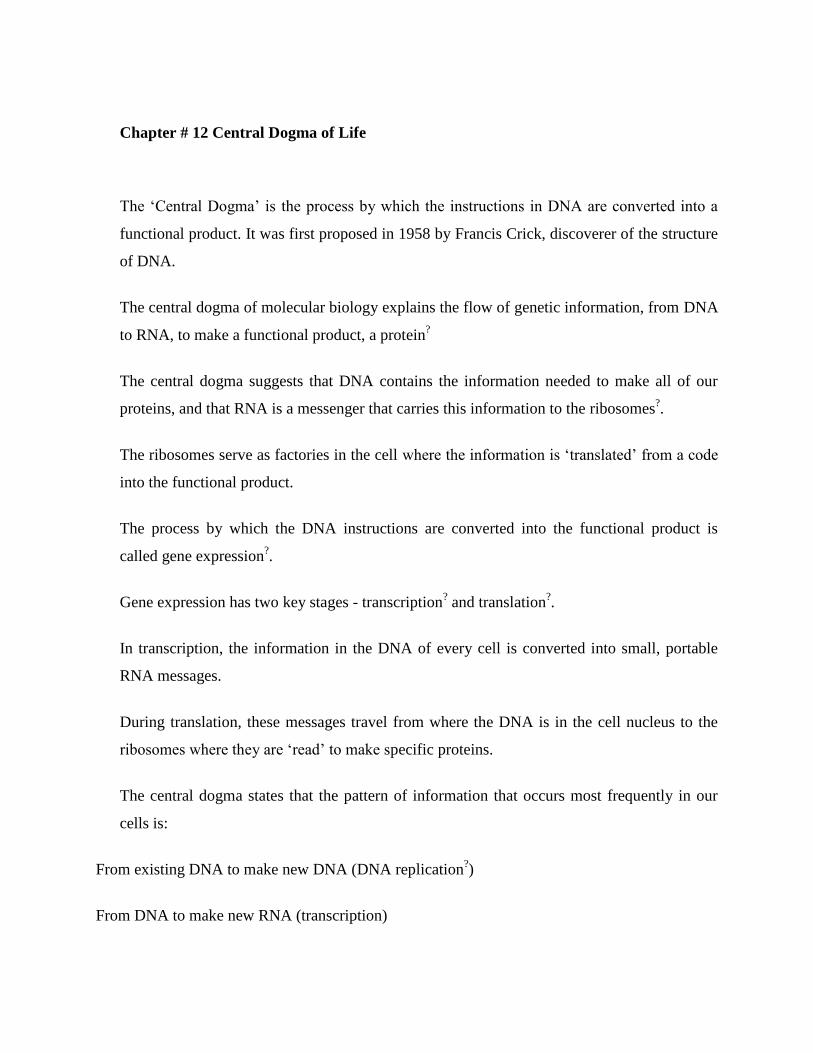
The central dogma states that the pattern of information that occurs most frequently in our
cells is:
From existing DNA to make new DNA (DNA replication?)
From DNA to make new RNA (transcription)
From RNA to make new proteins (translation).
Reverse transcription is the transfer of information from RNA to make new DNA, this occurs
in the case of retroviruses, such as HIV?. It is the process by which the genetic information
from RNA is assembled into new DNA.
The central dogma has also been described as "DNA makes RNA and RNA makes
protein,"[3]
a positive statement which was originally termed the sequence hypothesis by
Crick. However, this simplification does not make it clear that the central dogma as stated by
Crick does not preclude the reverse flow of information from RNA to DNA, only ruling out
the flow from protein to RNA or DNA. Crick's use of the word dogma was unconventional,
and has been controversial.
The dogma is a framework for understanding the transfer of sequence information between
information-carrying biopolymers, in the most common or general case, in living organisms.
There are 3 major classes of such biopolymers: DNA and RNA (both nucleic acids), and
protein. There are 3×3 = 9 conceivable direct transfers of information that can occur between
these. The dogma classes these into 3 groups of 3: 3 general transfers (believed to occur
normally in most cells), 3 special transfers (known to occur, but only under specific
conditions in case of some viruses or in a laboratory), and 3 unknown transfers (believed
never to occur). The general transfers describe the normal flow of biological information:
DNA can be copied to DNA (DNA replication), DNA information can be copied into mRNA
(transcription), and proteins can be synthesized using the information in mRNA as a template
(translation).
DNA replications
In the sense that DNA replication must occur if genetic material is to be provided for the
progeny of any cell, whether somatic or reproductive, the copying from DNA to DNA
arguably is the fundamental step in the central dogma. A complex group of proteins called
the replisome performs the replication of the information from the parent strand to the
complementary daughter strand.
The replisome comprises:
a helicase that unwinds the superhelix as well as the double-stranded DNA helix to create a
replication fork SSB protein that binds open the double-stranded DNA to prevent it from
reassociating RNA primase that adds a complementary RNA primer to each template strand
as a starting point for replication DNA polymerase III that reads the existing template chain
from its 3' end to its 5' end and adds new complementary nucleotides from the 5' end to the 3'
end of the daughter chain DNA polymerase I that removes the RNA primers and replaces
them with DNA.
DNA ligase that joins the two Okazaki fragments with phosphodiester bonds to produce a
continuous chain. This process typically takes place during S phase of the cell cycle.
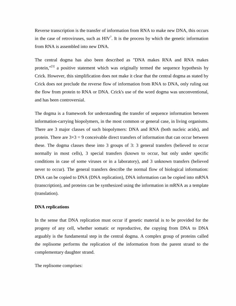
Transcription
Transcription is the process by which the information contained in a section of DNA is
replicated in the form of a newly assembled piece of messenger RNA (mRNA). Enzymes
facilitating the process include RNA polymerase and transcription factors. In eukaryotic cells
the primary transcript is (pre-mRNA). Pre-mRNA must be processed for translation to
proceed. Processing includes the addition of a 5' cap and a poly-A tail to the pre-mRNA
chain, followed by splicing. Alternative splicing occurs when appropriate, increasing the
diversity of the proteins that any single mRNA can produce. The product of the entire
transcription process that began with the production of the pre-mRNA chain, is a mature
mRNA chain.
Translation
The mature mRNA finds its way to a ribosome, where it gets translated. In prokaryotic cells,
which have no nuclear compartment, the processes of transcription and translation may be
linked together without clear separation. In eukaryotic cells, the site of transcription (the cell
nucleus) is usually separated from the site of translation (the cytoplasm), so the mRNA must
be transported out of the nucleus into the cytoplasm, where it can be bound by ribosomes.
The ribosome reads the mRNA triplet codons, usually beginning with an AUG
(adenine−uracil−guanine), or initiator methionine codon downstream of the ribosome
binding site. Complexes of initiation factors and elongation factors bring aminoacylated
transfer RNAs (tRNAs) into the ribosome-mRNA complex, matching the codon in the
mRNA to the anti-codon on the tRNA. Each tRNA bears the appropriate amino acid residue
to add to the polypeptide chain being synthesised. As the amino acids get linked into the
growing peptide chain, the chain begins folding into the correct conformation. Translation
ends with a stop codon which may be a UAA, UGA, or UAG triplet.
The mRNA does not contain all the information for specifying the nature of the mature
protein. The nascent polypeptide chain released from the ribosome commonly requires
additional processing before the final product emerges. For one thing, the correct folding
process is complex and vitally important. For most proteins it requires other chaperone
proteins to control the form of the product. Some proteins then excise internal segments from
their own peptide chains, splicing the free ends that border the gap; in such processes the
inside "discarded" sections are called inteins. Other proteins must be split into multiple
sections without splicing. Some polypeptide chains need to be cross-linked, and others must
be attached to cofactors such as haem (heme) before they become functional.
Special transfers of biological sequential information
Reverse transcription
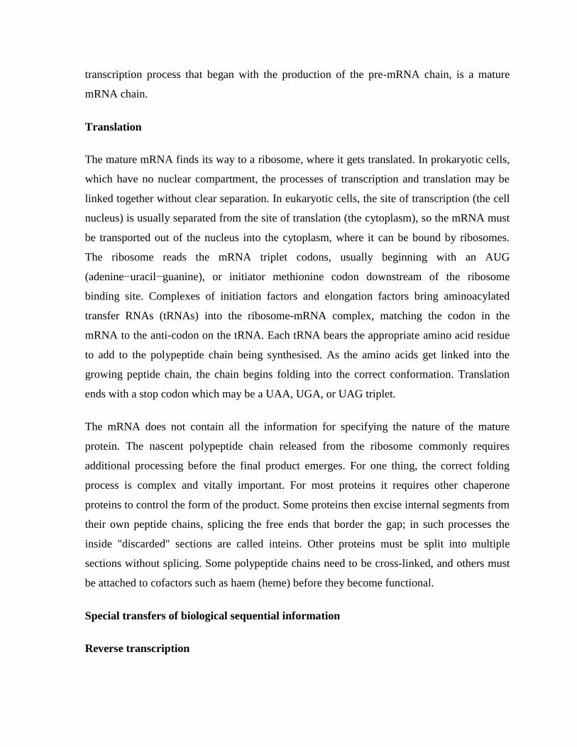
Unusual flow of information highlighted in green
Reverse transcription is the transfer of information from RNA to DNA (the reverse of normal
transcription). This is known to occur in the case of retroviruses, such as HIV, as well as in
eukaryotes, in the case of retrotransposons and telomere synthesis. It is the process by which
genetic information from RNA gets transcribed into new DNA.
RNA replication
RNA replication is the copying of one RNA to another. Many viruses replicate this way. The
enzymes that copy RNA to new RNA, called RNA-dependent RNA polymerases, are also
found in many eukaryotes where they are involved in RNA silencing.
RNA editing, in which an RNA sequence is altered by a complex of proteins and a "guide
RNA", could also be seen as an RNA-to-RNA transfer.
Direct translation from DNA to protein
Direct translation from DNA to protein has been demonstrated in a cell-free system (i.e. in a
test tube), using extracts from E. coli that contained ribosomes, but not intact cells. These cell
fragments could synthesize proteins from single-stranded DNA templates isolated from other
organisms (e,g., mouse or toad), and neomycin was found to enhance this effect. However, it
was unclear whether this mechanism of translation corresponded specifically to the genetic
code.
tRNA and genetic code:
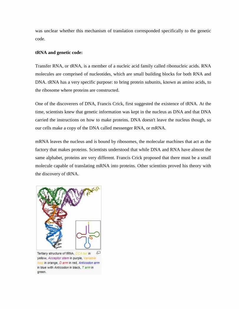
Transfer RNA, or tRNA, is a member of a nucleic acid family called ribonucleic acids. RNA
molecules are comprised of nucleotides, which are small building blocks for both RNA and
DNA. tRNA has a very specific purpose: to bring protein subunits, known as amino acids, to
the ribosome where proteins are constructed.
One of the discoverers of DNA, Francis Crick, first suggested the existence of tRNA. At the
time, scientists knew that genetic information was kept in the nucleus as DNA and that DNA
carried the instructions on how to make proteins. DNA doesn't leave the nucleus though, so
our cells make a copy of the DNA called messenger RNA, or mRNA.
mRNA leaves the nucleus and is bound by ribosomes, the molecular machines that act as the
factory that makes proteins. Scientists understood that while DNA and RNA have almost the
same alphabet, proteins are very different. Francis Crick proposed that there must be a small
molecule capable of translating mRNA into proteins. Other scientists proved his theory with
the discovery of tRNA.
The structure of tRNA
Function of tRNA
The job of tRNA is to read the message of nucleic acids, or nucleotides, and translate it into
proteins, or amino acids. The process of making a protein from an mRNA template is called
translation.
How does tRNA read the mRNA? It reads the mRNA in three-letter nucleotide sequences
called codons. Each individual codon corresponds to an amino acid. There are four
nucleotides in mRNA. There is one tRNA molecule for each and every codon.
Interestingly, there are only 21 amino acids. This brings up the idea that our genetic code is
redundant. That is, we have 64 codons but only 21 amino acids. How do we resolve this?
More than one codon can specify for an amino acid.
This table (Figure 2) shows all the combinations of nucleic acids, or codons, as well as which
amino acid is specified by which codon. As you can see, not every amino acid has four
codons. In fact, methionine only has one.
Notice, however, that each codon has only one corresponding amino acid. Thus we say that
the genetic code is redundant, but not ambiguous. For example, the codons GUU, GUC,
GUA, and GUG all code for Valine (redundancy), and none of them specify any other amino
acid (no ambiguity).
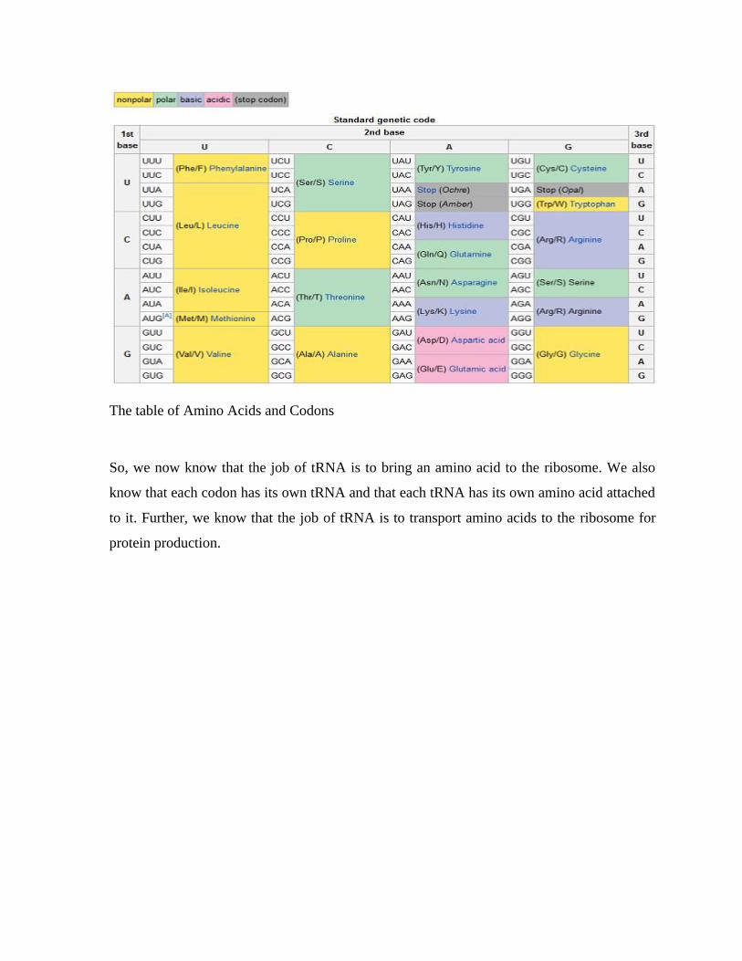
The table of Amino Acids and Codons
So, we now know that the job of tRNA is to bring an amino acid to the ribosome. We also
know that each codon has its own tRNA and that each tRNA has its own amino acid attached
to it. Further, we know that the job of tRNA is to transport amino acids to the ribosome for
protein production.
Chapter # 13 TRANSCRIPTION
RNA Polymerases:
RNA polymerase is an enzyme that is responsible for copying a DNA sequence into an RNA
sequence, duyring the process of transcription. As complex molecule composed of protein
subunits, RNA polymerase controls the process of transcription, during which the
information stored in a molecule of DNA is copied into a new molecule of messenger RNA.
RNA polymerases have been found in all species, but the number and composition of these
proteins vary across taxa. For instance, bacteria contain a single type of RNA polymerase,
while eukaryotes (multicellular organisms and yeasts) contain three distinct types. In spite of
these differences, there are striking similarities among transcriptional mechanisms. For
example, all species require a mechanism by which transcription can be regulated in order to
achieve spatial and temporal changes in gene expression.
BACTERIAL TRANSCRIPTION
Bacterial transcription is the process in which messenger RNA transcripts of genetic material
in bacteria are produced, to be translated for the production of proteins. Bacterial
transcription occurs in the cytoplasm alongside translation. Unlike in eukaryotes, bacterial
transcription and translation can occur simultaneously. This is impossible in eukaryotes,
where transcription occurs in a membrane-bound nucleus while translation occurs outside the
nucleus in the cytoplasm. In bacteria genetic material is not enclosed in a membrane-
enclosed nucleus and has access to ribosomes in the cytoplasm.
Transcription is known to be controlled by a variety of regulators in bacteria. Many of these
transcription factors are homodimers containing helix-turn-helix DNA-binding motifs
Initiation
The following steps occur, in order, for transcription initiation:
RNA polymerase (RNAP) binds to one of several specificity factors, ζ, to form a
holoenzyme. In this form, it can recognize and bind to specific promoter regions in the DNA.
The -35 region and the -10 ("Pribnow box") region comprise the core prokaryotic promoter,
and |T| stands for the terminator. The DNA on the template strand between the +1 site and
the terminator is transcribed into RNA, which is then translated into protein. At this stage, the
DNA is double-stranded ("closed"). This holoenzyme/wound-DNA structure is referred to as
the closed complex.
The DNA is unwound and becomes single-stranded ("open") in the vicinity of the initiation
site (defined as +1). This holoenzyme/unwound-DNA structure is called the open complex.
The RNA polymerase transcribes the DNA (the beta subunit initiates the synthesis), but
produces about 10 abortive (short, non-productive) transcripts which are unable to leave the
RNA polymerase because the exit channel is blocked by the ζ-factor.
The ζ-factor eventually dissociates from the core enzyme, and elongation proceeds.
Elongation
Promoters can differ in "strength"; that is, how actively they promote transcription of their
adjacent DNA sequence. Promoter strength is in many (but not all) cases, a matter of how
tightly RNA polymerase and its associated accessory proteins bind to their respective DNA
sequences. The more similar the sequences are to a consensus sequence, the stronger the
binding is. Additional transcription regulation comes from transcription factors that can
affect the stability of the holoenzyme structure at initiation.
Most transcripts originate using adenosine-5'-triphosphate (ATP) and, to a lesser extent,
guanosine-5'-triphosphate (GTP) (purine nucleoside triphosphates) at the +1 site. Uridine-5'-
triphosphate (UTP) and cytidine-5'-triphosphate (CTP) (pyrimidine nucleoside triphosphates)
are disfavoured at the initiation site.
Termination
Two termination mechanisms are well known:
Intrinsic termination (also called Rho-independent transcription termination) involves
terminator sequences within the RNA that signal the RNA polymerase to stop. The
terminator sequence is usually a palindromic sequence that forms a stem-loop hairpin
structure that leads to the dissociation of the RNAP from the DNA template.
Rho-dependent termination uses a termination factor called ρ factor (rho factor) which is a
protein to stop RNA synthesis at specific sites. This protein binds at a rho utilisation site on
the nascent RNA strand and runs along the mRNA towards the RNAP. A stem loop structure
upstream of the terminator region pauses the RNAP, when ρ-factor reaches the RNAP, it
causes RNAP to dissociate from the DNA, terminating transcription
TRANSCRIPTION IN EUKARYOTES
Eukaryotic transcription is the elaborate process that eukaryotic cells use to copy genetic
information stored in DNA into units of RNA replica. Gene transcription occurs in both
eukaryotic and prokaryotic cells. Unlike prokaryotic RNA polymerase that initiates the
transcription of all different types of RNA, RNA polymerase in eukaryotes (including
humans) comes in three variations, each encoding a different type of gene. A eukaryotic cell
has a nucleus that separates the processes of transcription and translation. Eukaryotic
transcription occurs within the nucleus where DNA is packaged into nucleosomes and higher
order chromatin structures. The complexity of the eukaryotic genome necessitates a great
variety and complexity of gene expression control.
Overview
Transcription is the process of copying genetic information stored in a DNA strand into a
transportable complementary strand of RNA. Eukaryotic transcription takes place in the
nucleus of the cell and proceeds in three sequential stages: initiation, elongation, and
termination. The transcriptional machinery that catalyzes this complex reaction has at its core
three multi-subunit RNA polymerases. RNA polymerase I is responsible for transcribing
RNA that codes for genes that become structural components of the ribosome.
Protein coding genes are transcribed into messenger RNAs (mRNAs) that carry the
information from DNA to the site of protein synthesis. Although mRNAs possess great
diversity, they are not the most abundant RNA species made in the cell. The so called non-
coding RNAs account for the large majority of the transcriptional output of a cell. These non-
coding RNAs perform a variety of important cellular functions.
RNA Polymerase
Eukaryotes have three nuclear RNA polymerases, each with distinct roles and properties
Name Location Product
RNA
Polymerase I
(Pol I, Pol A)
nucleolus
larger ribosomal RNA (rRNA) (28S, 18S,
5.8S)
RNA
Polymerase II
(Pol II, Pol B)
nucleus
messenger RNA (mRNA), most small
nuclear RNAs (snRNAs), small interfering
RNA (siRNAs) and micro RNA (miRNA).
RNA
Polymerase III
(Pol III, Pol C)
nucleus (and possibly
the nucleolus-
nucleoplasm
interface)
transfer RNA (tRNA), other small RNAs
(including the small 5S ribosomal RNA (5s
rRNA), snRNA U6, signal recognition
particle RNA (SRP RNA) and other stable
short RNAs
RNA polymerase I (Pol I) catalyses the transcription of all rRNA genes except 5S. These
rRNA genes are organised into a single transcriptional unit and are transcribed into a
continuous transcript. This precursor is then processed into three rRNAs: 18S, 5.8S, and 28S.
The transcription of rRNA genes takes place in a specialised structure of the nucleus called
the nucleolus, where the transcribed rRNAs are combined with proteins to form ribosomes.
RNA polymerase II (Pol II) is responsible for the transcription of all mRNAs, some snRNAs,
siRNAs, and all miRNAs. Many Pol II transcripts exist transiently as single strand precursor
RNAs (pre-RNAs) that are further processed to generate mature RNAs. For example,
precursor mRNAs (pre-mRNAs)are extensively processed before exiting into the cytoplasm
through the nuclear pore for protein translation.
RNA polymerase III (Pol III) transcribes small non-coding RNAs, including tRNAs, 5S
rRNA, U6 snRNA, SRP RNA, and other stable short RNAs such as ribonuclease P RNA.
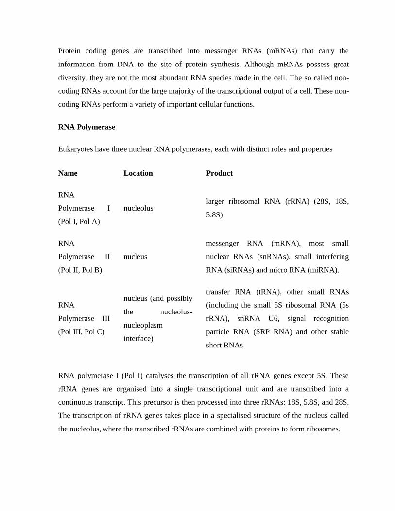
Structure of eukaryotic RNA polymerase II (light blue) in complex with α-amanitin (red), a
strong poison found in death cap mushrooms that targets this vital enzyme
RNA Polymerases I, II, and III contain 14, 12, and 17 subunits, respectively. All three
eukaryotic polymerases have five core subunits that exhibit homology with the β, β‘, αI, α
II,
and ω subunits of E. coli RNA polymerase. An identical ω-like subunit (RBP6) is used by all
three eukaryotic polymerases, while the same α-like subunits are used by Pol I and III. The
three eukaryotic polymerases share four other common subunits among themselves. The
remaining subunits are unique to each RNA polymerase. The additional subunits found in Pol
I and Pol III relative to Pol II, are homologous to Pol II transcription factors.
Crystal structures of RNA polymerases I and II provide an opportunity to understand the
interactions among the subunits and the molecular mechanism of eukaryotic transcription in
atomic detail.
The carboxyl terminal domain (CTD) of RPB1, the largest subunit of RNA polymerase II,
plays an important role in bringing together the machinery necessary for the synthesis and
processing of Pol II transcripts. Long and structurally disordered, the CTD contains multiple
repeats of heptapeptide sequence YSPTSPS that are subject to phosphorylation and other
posttranslational modifications during the transcription cycle. These modifications and their
regulation constitute the operational code for the CTD to control transcription initiation,
elongation and termination and to couple transcription and RNA processing.
Initiation
The initiation of gene transcription in eukaryotes occurs in specific steps. First, an RNA
polymerase along with general transcription factors binds to the promoter region of the gene
to form a closed complex called the preinitiation complex. The subsequent transition of the
complex from the closed state to the open state results in the melting or separation of the two
DNA strands and the positioning of the template strand to the active site of the RNA
polymerase. Without the need of a primer, RNA polymerase can initiate the synthesis of a
new RNA chain using the template DNA strand to guide ribonucleotide selection and
polymerization chemistry.
However, many of the initiated syntheses are aborted before the transcripts reach a
significant length (~10 nucleotides). During these abortive cycles, the polymerase keeps
making and releasing short transcripts until it is able to produce a transcript that surpasses ten
nucleotides in length. Once this threshold is attained, RNA polymerase escapes the promoter
and transcription proceeds to the elongation phase.
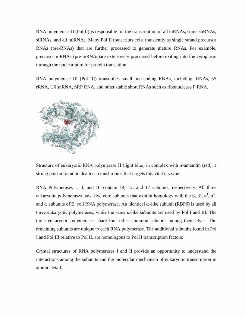
Here is a diagram of the attachment of RNA polymerase II to the de-helicized DNA.
Eukaryotic promoters and general transcription factors
Pol II-transcribed genes contain a region in the immediate vicinity of the transcription start
site (TSS) that binds and positions the preinitiation complex. This region is called the core
promoter because of its essential role in transcription initiation. Different classes of sequence
elements are found in the promoters. For example, the TATA box is the highly conserved
DNA recognition sequence for the TATA box binding protein, TBP, whose binding initiates
transcription complex assembly at many genes.
Eukaryotic genes also contain regulatory sequences beyond the core promoter. These cis-
acting control elements bind transcriptional activators or repressors to increase or decrease
transcription from the core promoter. Well-characterized regulatory elements include
enhancers, silencers, and insulators. These regulatory sequences can be spread over a large
genomic distance, sometimes located hundreds of kilobases from the core promoters.
General transcription factors are a group of proteins involved in transcription initiation and
regulation. These factors typically have DNA-binding domains that bind specific sequence
elements of the core promoter and help recruit RNA polymerase to the transcriptional start
site. General transcription factors for RNA polymerase II include TFIID, TFIIA, TFIIB,
TFIIF, TFIIE, and TFIIH.
Assembly of preinitiation complex
To prepare for transcription, a complete set of general transcription factors and RNA
polymerase need to be assembled at the core promoter to form the ~2 million dalton
preinitiation complex. For example, for promoters that contain a TATA box near the TSS,
the recognition of TATA box by the TBP subunit of TFIID initiates the assembly of a
transcription complex. The next proteins to enter are TFIIA and TFIIB, which stabilize the
DNA-TFIID complex and recruit Pol II in association with TFIIF and additional transcription
factors. TFIIF serves as the bridge between the TATA-bound TBP and polymerase. One of
the last transcription factors to be recruited to the preinitiation complex is TFIIH, which
plays an important role in promoter melting and escape.
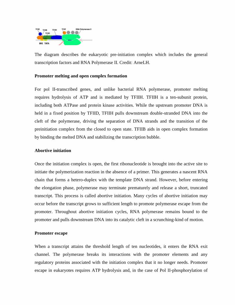
The diagram describes the eukaryotic pre-initiation complex which includes the general
transcription factors and RNA Polymerase II. Credit: ArneLH.
Promoter melting and open complex formation
For pol II-transcribed genes, and unlike bacterial RNA polymerase, promoter melting
requires hydrolysis of ATP and is mediated by TFIIH. TFIIH is a ten-subunit protein,
including both ATPase and protein kinase activities. While the upstream promoter DNA is
held in a fixed position by TFIID, TFIIH pulls downstream double-stranded DNA into the
cleft of the polymerase, driving the separation of DNA strands and the transition of the
preinitiation complex from the closed to open state. TFIIB aids in open complex formation
by binding the melted DNA and stabilizing the transcription bubble.
Abortive initiation
Once the initiation complex is open, the first ribonucleotide is brought into the active site to
initiate the polymerization reaction in the absence of a primer. This generates a nascent RNA
chain that forms a hetero-duplex with the template DNA strand. However, before entering
the elongation phase, polymerase may terminate prematurely and release a short, truncated
transcript. This process is called abortive initiation. Many cycles of abortive initiation may
occur before the transcript grows to sufficient length to promote polymerase escape from the
promoter. Throughout abortive initiation cycles, RNA polymerase remains bound to the
promoter and pulls downstream DNA into its catalytic cleft in a scrunching-kind of motion.
Promoter escape
When a transcript attains the threshold length of ten nucleotides, it enters the RNA exit
channel. The polymerase breaks its interactions with the promoter elements and any
regulatory proteins associated with the initiation complex that it no longer needs. Promoter
escape in eukaryotes requires ATP hydrolysis and, in the case of Pol II-phosphorylation of
the CTD. Meanwhile, the transcription bubble collapses down to 12-14 nucleotides,
providing kinetic energy required for the escape.
Elongation
After escaping the promoter and shedding most of the transcription factors for initiation, the
polymerase acquires new factors for the next phase of transcription: elongation.[21][22]
Transcription elongation is a processive process. Double stranded DNA that enters from the
front of the enzyme is unzipped to avail the template strand for RNA synthesis. For every
DNA base pair separated by the advancing polymerase, one hybrid RNA:DNA base pair is
immediately formed. DNA strands and nascent RNA chain exit from separate channels; the
two DNA strands reunite at the trailing end of the transcription bubble while the single strand
RNA emerges alone.
Elongation factors
Among the proteins recruited to polymerase are elongation factors, thus called because they
stimulate transcription elongation. There are different classes of elongation factors. Some
factors can increase the overall rate of transcribing, some can help the polymerase through
transient pausing sites, and some can assist the polymerase to transcribe through chromatin.
One of the elongation factors, P-TEFb, is particularly important. P-TEFb phosphorylates the
second residue (Ser-2) of the CTD repeats (YSPTSPS) of the bound Pol II. P-TEFb also
phosphorylates and activates SPT5 and TAT-SF1. SPT5 is a universal transcription factor
that helps recruit 5'-capping enzyme to Pol II with a CTD phosphorylated at Ser-5. TAF-SF1
recruits components of the RNA splicing machinery to the Ser-2 phosphorylated CTD. P-
TEFb also helps suppress transient pausing of polymerase when it encounters certain
sequences immediately following initiation.
Transcription fidelity
Transcription fidelity is achieved through multiple mechanisms. RNA polymerases select
correct nucleoside triphosphate (NTP) substrate to prevent transcription errors. Only the NTP
which correctly base pairs with the coding base in the DNA is admitted to the active center.
RNA polymerase performs two known proof reading functions to detect and remove
misincorporated nucleotides: pyrophosphorylytic editing and hydrolytic editing. The former
removes the incorrectly inserted ribonucleotide by a simple reversal of the polymerization
reaction, while the latter involves backtracking of the polymerase and cleaving of a segment
of error-containing RNA product. Elongation factor TFIIS stimulates an inherent
ribonuclease activity in the polymerase, allowing the removal of misincorporated bases
through limited local RNA degradation. Note that all reactions (phosphodiester bond
synthesis, pyrophosphorolysis, phosphodiester bond hydrolysis) are performed by RNA
polymerase by using a single active center.
Pausing, poising, and backtracking
Transcription elongation is not a smooth ride along the DNA railway. For proofreading, the
polymerase is made to back-up, erase some of the RNA it has already made and have another
go at transcription. In general, RNA polymerase does not transcribe through a gene at a
constant pace. Rather it pauses periodically at certain sequences, sometimes for long periods
of time before resuming transcription. In extreme cases, for example, when the polymerase
encounters a damaged nucleotide, it comes to a complete halt. More often, an elongating
polymerase is stalled near the promoter. Promoter-proximal pausing during early elongation
is a commonly used mechanism for regulating genes poised to be expressed rapidly or in a
coordinated fashion. Pausing is mediated by a complex called NELF (negative elongation
factor) in collaboration with DSIF (DRB-sensitivity-inducing factor containing SPT4/SPT5).
The blockage is released once the polymerase receives an activation signal, such as the
phosphorylation of Ser-2 of CTD tail by P-TEFb. Other elongation factors such as ELL and
TFIIS stimulate the rate of elongation by limiting the length of time that polymerase pauses.
RNA processing
Elongating polymerase is associated with a set of protein factors required for various types of
RNA processing. mRNA is capped as soon as it emerges from the RNA-exit channel of the
polymerase. After capping, dephosphorylation of Ser-5 within the CTD repeats may be
responsible for dissociation of the capping machinery. Further phosphorylation of Ser-2
causes recruitment of the RNA splicing machinery that catalyzes the removal of non-coding
introns to generate mature mRNA. Alternative splicing expands the protein complements in
eukaryotes. Just as with 5‘-capping and splicing, the CTD tail is involved in recruiting
enzymes responsible for 3‘-polyadenylation, the final RNA processing event that is coupled
with the termination of transcription.
Termination
The last stage of transcription is termination, which leads to the dissociation of the complete
transcript and the release of RNA polymerase from the template DNA.The process differs for
each of the three RNA polymerases. The mechanism of termination is the least understood of
the three transcription stages.
Factor-dependent termination
The termination of transcription of pre-rRNA genes by polymerase Pol I is performed by a
system that needs a specific transcription termination factor. The mechanism used bears
some resemblance to the rho-dependent termination in prokaryotes. Eukaryotic cells contain
hundreds of ribosomal DNA repeats, sometimes distributed over multiple chromosomes.
Termination of transcription occurs in the ribosomal intergenic spacer region that contains
several transcription termination sites upstream of a Pol I pausing site. Through a yet
unknown mechanism, the 3‘-end of the transcript is cleaved, generating a large primary
rRNA molecule that is further processed into the mature 18S, 5.8S and 28S rRNAs.
As Pol II reaches the end of a gene, two protein complexes carried by the CTD, CPSF
(cleavage and polyadenylation specificity factor) and CSTF (cleavage stimulation factor),
recognize the poly-A signal in the transcribed RNA. Poly-A-bound CPSF and CSTF recruit
other proteins to carry out RNA cleavage and then polyadenylation. Poly-A polymerase adds
approximately 200 adenines to the cleaved 3‘ end of the RNA without a template. The long
poly-A tail is unique to transcripts made by Pol II.
In the process of terminating transcription by Pol I and Pol II, the elongation complex does
not dissolve immediately after the RNA is cleaved. The polymerase continues to move along
the template, generating a second RNA molecule associated with the elongation complex.
Two models have been proposed to explain how termination is achieved at last. The
allosteric model states that when transcription proceeds through the termination sequence, it
causes disassembly of elongation factors and/or an assembly of termination factors that cause
conformational changes of the elongation complex. The torpedo model suggests that a 5' to 3'
exonuclease degrades the second RNA as it emerges from the elongation complex.
Polymerase is released as the highly processive exonuclease overtakes it. It is proposed that
an emerging view will express a merge of these two models.
Factor-independent termination
RNA polymerase III can terminate transcription efficiently without involvement of additional
factors. The Pol III termination signal consists of a stretch of thymines (on the nontemplate
strand) located within 40bp downstream from the 3' end of mature RNAs. The poly-T
termination signal pauses Pol III and causes it to back track to the nearest RNA hairpin to
become a ―dead-end‖ complex. Consistent with the allosteric mechanism of termination, the
RNA hairpin allosterically opens Pol III and causes the elongation complex to disintegrate.
The extensive structure embedded in the Pol III-transcript thus is responsible for the factor-
independent release of Pol III at the end of a gene. RNA-duplex-dependent termination is an
ancient mechanism that dates back to the last universal common ancestor.
Eukaryotic transcriptional control
The regulation of gene expression in eukaryotes is achieved through the interaction of several
levels of control that acts both locally to turn on or off individual genes in response to a
specific cellular need and globally to maintain a chromatin-wide gene expression pattern that
shapes cell identity. Because eukaryotic genome is wrapped around histones to form
nucelosomes and higher-order chromatin structures, the substrates for transcriptional
machinery are in general partially concealed. Without regulatory proteins, many genes are
expressed at low level or not expressed at all. Transcription requires displacement of the
positioned nucleosomes to enable the transcriptional machinery to gain access of the DNA.
All steps in the transcription are subject to some degree of regulation. Transcription initiation
in particular is the primary level at which gene expression is regulated. Targeting the rate-
limiting initial step is the most efficient in terms of energy costs for the cell. Transcription
initiation is regulated by cis-acting elements (enhancers, silencers, isolators) within the
regulatory regions of the DNA, and sequence-specific trans-acting factors that act as
activators or repressors. Gene transcription can also be regulated post-initiation by targeting
the movement of the elongating polymerase.
Global control and epigenetic regulation
The eukaryotic genome is organized into a compact chromatin structure that allows only
regulated access to DNA. The chromatin structure can be globally ―open‖ and more
transcriptionally permissive or globally ―condensed‖ and transcriptionally inactive. The
former (euchromatin) is lightly packed and rich in genes under active transcription. The latter
(heterochromatin) includes gene-poor regions such as telomeres and centromeres but also
regions with normal gene density but transcriptionally silenced. Transcription can be silenced
by histone modification (deaceltylation and methylation), RNA interference, and/or DNA
methylation.
The gene expression patterns that define cell identity have to be inherited through cell
division. This process is called epigenetic regulation. DNA methylation is reliably inherited
through the action of maintenance methylases that modify the nascent DNA strand generated
by replication. In mammalian cells, DNA methylation is the primary marker of
transcriptionally silenced regions. Specialized proteins can recognize the marker and recruit
histone deacetylases and methylases to re-establish the silencing. Nucleosome histone
modifications could also be inherited during cell division, however, it is not clear whether it
can work independently without the direction by DNA methylation.
Gene-specific activation
The two main tasks of transcription initiation are to provide RNA polymerase with an access
to the promoter and to assemble general transcription factors with polymerase into a
transcription initiation complex. Diverse mechanisms of initiating transcription by overriding
inhibitory signals at the gene promoter have been identified. Eukaryotic genes have acquired
extensive regulatory sequences that encompass a large number of regulator-binding sites and
spread overall kilobases (sometimes hundreds of kilobases) from the promoter–-both
upstream and downstream. The regulator binding sites are often clustered together into units
called enhancers. Enhancers can facilitate highly cooperative action of several transcription
factors (which constitute enhanceosomes). Remote enhancers allow transcription regulation
at a distance. Insulators situated between enhancers and promoters help define the genes that
an enhancer can or cannot influence.
Eukaryotic transcriptional activators have separate DNA-binding and activating functions.
Upon binding to its cis-element, an activator can recruit polymerase directly or recruit other
factors needed by the transcriptional machinery. An activator can also recruit nucleosome
modifiers that alter chromatin in the vicinity of the promoter and thereby help initiation.
Multiple activators can work together, either by recruiting a common or two mutually
dependent components of the transcriptional machinery, or by helping each other bind to
their DNA sites. These interactions can synergize multiple signaling inputs and produce
intricate transcriptional responses to address cellular needs.
Gene-specific repression
Eukaryotic transcription repressors share some of the mechanisms used by their prokaryotic
counterparts. For example, by binding to a site on DNA that overlaps with the binding site of
an activator, a repressor can inhibit binding of the activator. But more frequently, eukaryotic
repressors inhibit the function of an activator by masking its activating domain, preventing its
nuclear localization, promoting its degradation, or inactivating it through chemical
modifications. Repressors can directly inhibit transcription initiation by binding to a site
upstream of a promoter and interacting with the transcriptional machinery. Repressors can
indirectly repress transcription by recruiting histone modifiers (deacetylases and methylases)
or nucelosome remodeling enzymes that affect the accessibility of the DNA. Repressing
histone and DNA modifications are also the basis of transcriptional silencing that can spread
along the chromatin and switch off multiple genes.
Elongation and termination control
The elongation phase starts once assembly of the elongation complex has been completed,
and progresses until a termination sequence is encountered. The post-initiation movement of
RNA polymerase is the target of another class of important regulatory mechanisms. For
example, the transcriptional activator Tat affects elongation rather than initiation during its
regulation of HIV transcription. In fact, many eukaryotic genes are regulated by releasing a
block to transcription elongation called promoter-proximal pausing. Pausing can influence
chromatin structure at promoters to facilitate gene activity and lead to rapid or synchronous
transcriptional responses when cells are exposed to an activation signal. Pausing is associated
with the binding of two negative elongation factors, DSIF (SPT4/SPT5) and NELF, to the
elongation complex. Other factors can also influence the stability and duration of the paused
polymerase.[44]
Pause release is triggered by the recruitment of the P-TEFb kinase.
Transcription termination has also emerged as an important area of transcriptional regulation.
Termination is coupled with the efficient recycling of polymerase. The factors associated
with transcription termination can also mediate gene looping and thereby determine the
efficiency of re-initiation.
Transcription-coupled DNA repair
When transcription is arrested by the presence of a lesion in the transcribed strand of a gene,
DNA repair proteins are recruited to the stalled RNA polymerase to initiate a process called
transcription-coupled repair. Central to this process is the general transcription factor TFIIH
that has ATPase activity. TFIIH causes a conformational change in the polymerase, to expose
the transcription bubble trapped inside, in order for the DNA repair enzymes to gain access to
the lesion. Thus, RNA polymerase serves as damage-sensing protein in the cell to target
repair enzymes to genes that are being actively transcribed.
Comparisons between prokaryotic and eukaryotic transcription
Eukaryotic transcription is more complex than prokaryotic transcription. For instance, in
eukaryotes the genetic material (DNA), and therefore transcription, is primarily localized to
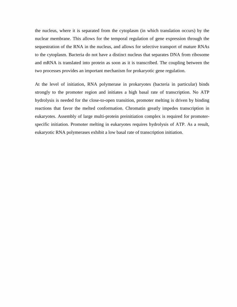
the nucleus, where it is separated from the cytoplasm (in which translation occurs) by the
nuclear membrane. This allows for the temporal regulation of gene expression through the
sequestration of the RNA in the nucleus, and allows for selective transport of mature RNAs
to the cytoplasm. Bacteria do not have a distinct nucleus that separates DNA from ribosome
and mRNA is translated into protein as soon as it is transcribed. The coupling between the
two processes provides an important mechanism for prokaryotic gene regulation.
At the level of initiation, RNA polymerase in prokaryotes (bacteria in particular) binds
strongly to the promoter region and initiates a high basal rate of transcription. No ATP
hydrolysis is needed for the close-to-open transition, promoter melting is driven by binding
reactions that favor the melted conformation. Chromatin greatly impedes transcription in
eukaryotes. Assembly of large multi-protein preinitiation complex is required for promoter-
specific initiation. Promoter melting in eukaryotes requires hydrolysis of ATP. As a result,
eukaryotic RNA polymerases exhibit a low basal rate of transcription initiation.
CHAPTER # 14 RNA SPLICING
Pre-mRNA Splicing
Because eukaryotic pre-mRNAs are transcribed from intron containing genes, the sequences
encoded by the intronic DNA must be removed from the primary transcript prior to the
RNA's becoming biologically active. The process of intron removal is called RNA splicing,
or pre-mRNA splicing. The intron-exon junctions (splice-sites) in the precursor mRNA (pre-
mRNA) of eukaryotes are recognized by trans-acting factors (prokaryotes RNAs are mostly
polycistronic). In pre-mRNA splicing the intronic sequences are excised and the exons are
ligated to generate the spliced mRNA.
Group I introns occur in nuclear, mitochondrial and chloroplast rRNA genes, group II introns
in mitochondrial and chloroplast mRNA genes.
Many of the group I and group II introns are self-splicing in that no additional protein factors
are necessary for the intron to be efficiently and accurately excised and the strands
reattached. "The nucleotide sequence of group II self-splicing introns is highly conserved,
and hence these introns fold into an evolutionarily conserved three-dimensional structure,
which can undergo a self-splicing reaction in the absence of any trans-acting factors.
In contrast, the nucleotide sequences and length of nuclear pre-mRNA introns is highly
variable, except for the short conserved sequences at the 5´ and 3´ splice sites and the branch
points. Therefore nuclear pre-mRNA splicing requires trans-acting factors, which interact
with these short conserved sequences, and from which the catalytically active spliceosome is
assembled.
The conserved sequences are: 5' splice site = AGguragu; 3' splice site = yyyyyyy nagG (y=
pyrimidine); branch site = ynyuray (r = purine, n = nucleotide)
Expressed differently, the highly conserved, consensus sequence for the 5' donor splice site
is (for RNA): (A or C)AG/GUAAGU. That is, most exons end with AG and introns begin
with GU (GT for DNA). The highly conserved, consensus sequence for the 3' acceptor splice
site is (for RNA): (C/U)less than 10N(C/T)AG/G, where most introns end in AG after a long
stretch of pyrimidines. The branch site within introns (area of lariat formation close to the
acceptor site during splicing) has the consensus sequence UAUAAC. In most cases, U can be
replaced by C and A can be replaced by G. However, the penultimate (bold) A residue is
fully conserved (invariant).
Group I introns require an external guanosine nucleotide as a cofactor. The 3'-OH of the
guanosine nucleotide acts as a nucleophile to attack the 5'-phosphate of the intron's 5'
nucleotide. The 3' end of the 5' exon is termed the splice donor site. The 3'-OH at the 3' splice
donor end of the 5' exon next attacks the splice acceptor site at the 5' nucleotide of the 3'
exon, releasing the intron and covalently attaching the two exons together.
Pre-mRNA processing takes place in the nucleus of eukaryotes, whereas lack of a nuclear
membrane in prokaryotes permits initiation of translation while transcription is not yet
complete.
Pre-mRNA processing events include capping of the 5‘ end on the pre-mRNA, pre-mRNA
splicing to remove intronic sequences, and polyadenylation of the 3‘ end of the pre-mRNA.
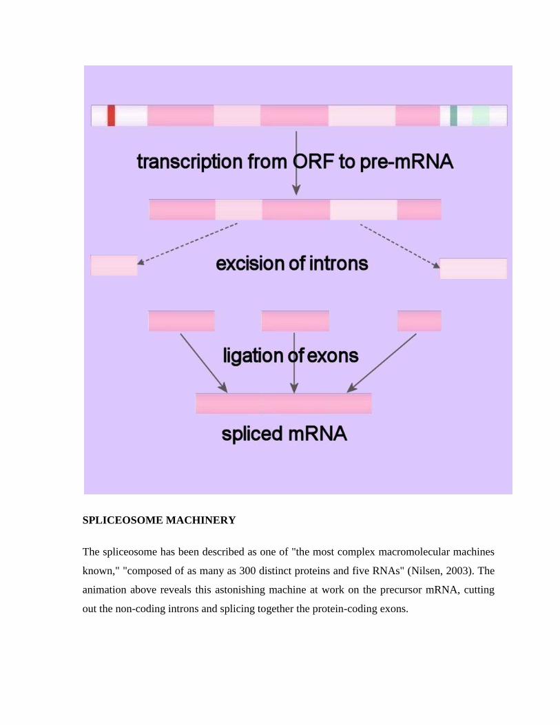
SPLICEOSOME MACHINERY
The spliceosome has been described as one of "the most complex macromolecular machines
known," "composed of as many as 300 distinct proteins and five RNAs" (Nilsen, 2003). The
animation above reveals this astonishing machine at work on the precursor mRNA, cutting
out the non-coding introns and splicing together the protein-coding exons.
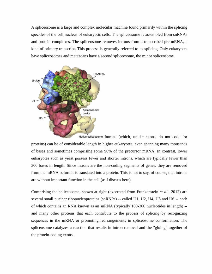
A spliceosome is a large and complex molecular machine found primarily within the splicing
speckles of the cell nucleus of eukaryotic cells. The spliceosome is assembled from snRNAs
and protein complexes. The spliceosome removes introns from a transcribed pre-mRNA, a
kind of primary transcript. This process is generally referred to as splicing. Only eukaryotes
have spliceosomes and metazoans have a second spliceosome, the minor spliceosome.
Introns (which, unlike exons, do not code for
proteins) can be of considerable length in higher eukaryotes, even spanning many thousands
of bases and sometimes comprising some 90% of the precursor mRNA. In contrast, lower
eukaryotes such as yeast possess fewer and shorter introns, which are typically fewer than
300 bases in length. Since introns are the non-coding segments of genes, they are removed
from the mRNA before it is translated into a protein. This is not to say, of course, that introns
are without important function in the cell (as I discuss here).
Comprising the spliceosome, shown at right (excerpted from Frankenstein et al., 2012) are
several small nuclear ribonucleoproteins (snRNPs) -- called U1, U2, U4, U5 and U6 -- each
of which contains an RNA known as an snRNA (typically 100-300 nucleotides in length) --
and many other proteins that each contribute to the process of splicing by recognizing
sequences in the mRNA or promoting rearrangements in spliceosome conformation. The
spliceosome catalyzes a reaction that results in intron removal and the "gluing" together of
the protein-coding exons.
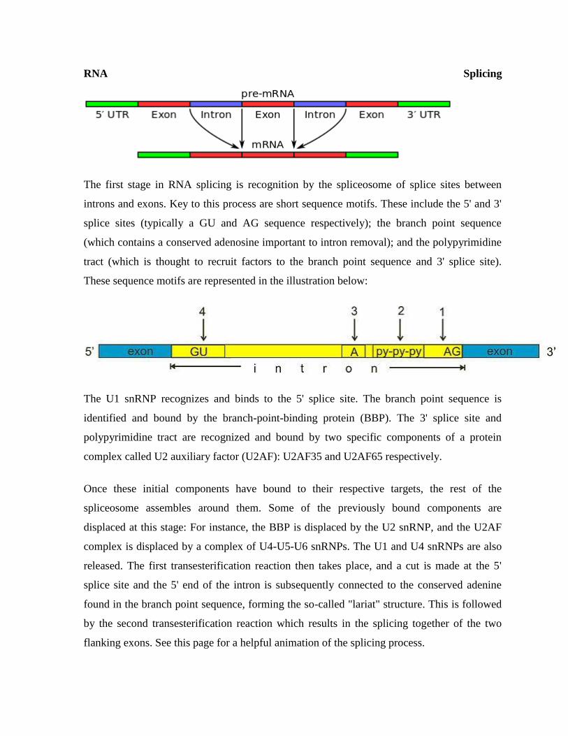
RNA Splicing
The first stage in RNA splicing is recognition by the spliceosome of splice sites between
introns and exons. Key to this process are short sequence motifs. These include the 5' and 3'
splice sites (typically a GU and AG sequence respectively); the branch point sequence
(which contains a conserved adenosine important to intron removal); and the polypyrimidine
tract (which is thought to recruit factors to the branch point sequence and 3' splice site).
These sequence motifs are represented in the illustration below:
The U1 snRNP recognizes and binds to the 5' splice site. The branch point sequence is
identified and bound by the branch-point-binding protein (BBP). The 3' splice site and
polypyrimidine tract are recognized and bound by two specific components of a protein
complex called U2 auxiliary factor (U2AF): U2AF35 and U2AF65 respectively.
Once these initial components have bound to their respective targets, the rest of the
spliceosome assembles around them. Some of the previously bound components are
displaced at this stage: For instance, the BBP is displaced by the U2 snRNP, and the U2AF
complex is displaced by a complex of U4-U5-U6 snRNPs. The U1 and U4 snRNPs are also
released. The first transesterification reaction then takes place, and a cut is made at the 5'
splice site and the 5' end of the intron is subsequently connected to the conserved adenine
found in the branch point sequence, forming the so-called "lariat" structure. This is followed
by the second transesterification reaction which results in the splicing together of the two
flanking exons. See this page for a helpful animation of the splicing process.
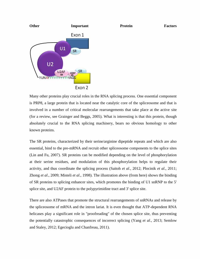
Other Important Protein Factors
Many other proteins play crucial roles in the RNA splicing process. One essential component
is PRP8, a large protein that is located near the catalytic core of the spliceosome and that is
involved in a number of critical molecular rearrangements that take place at the active site
(for a review, see Grainger and Beggs, 2005). What is interesting is that this protein, though
absolutely crucial to the RNA splicing machinery, bears no obvious homology to other
known proteins.
The SR proteins, characterized by their serine/arginine dipeptide repeats and which are also
essential, bind to the pre-mRNA and recruit other spliceosome components to the splice sites
(Lin and Fu, 2007). SR proteins can be modified depending on the level of phosphorylation
at their serine residues, and modulation of this phosphorylation helps to regulate their
activity, and thus coordinate the splicing process (Saitoh et al., 2012; Plocinik et al., 2011;
Zhong et al., 2009; Misteli et al., 1998). The illustration above (from here) shows the binding
of SR proteins to splicing enhancer sites, which promotes the binding of U1 snRNP to the 5'
splice site, and U2AF protein to the polypyrimidine tract and 3' splice site.
There are also ATPases that promote the structural rearrangements of snRNAs and release by
the spliceosome of mRNA and the intron lariat. It is even thought that ATP-dependent RNA
helicases play a significant role in "proofreading" of the chosen splice site, thus preventing
the potentially catastrophic consequences of incorrect splicing (Yang et al., 2013; Semlow
and Staley, 2012; Egecioglu and Chanfreau, 2011).
Composition
Each spliceosome is composed of five small nuclear RNAs (snRNA), and a range of
associated protein factors. When these small RNA are combined with the protein factors,
they make an RNA-protein complex called snRNP.
The snRNAs that make up the major spliceosome are named U1, U2, U4, U5, and U6, and
participate in several RNA-RNA and RNA-protein interactions. The RNA component of the
small nuclear ribonucleic protein or snRNP (pronounced "snurp") is rich in uridine (the
nucleoside analog of the uracil nucleotide).
The canonical assembly of the spliceosome occurs anew on each hnRNA (pre-mRNA). The
hnRNA contains specific sequence elements that are recognized and utilized during
spliceosome assembly. These include the 5' end splice, the branch point sequence, the
polypyrimidine tract, and the 3' end splice site. The spliceosome catalyzes the removal of
introns, and the ligation of the flanking exons.
Introns typically have a GU nucleotide sequence at the 5' end splice site, and an AG at the 3'
end splice site. The 3' splice site can be further defined by a variable length of
polypyrimidines, called the polypyrimidine tract (PPT), which serves the dual function of
recruiting factors to the 3' splice site and possibly recruiting factors to the branch point
sequence (BPS). The BPS contains the conserved Adenosine required for the first step of
splicing.
A group of less abundant snRNAs, U11, U12, U4atac, and U6atac, together with U5, are
subunits of the so-called minor spliceosome that splices a rare class of pre-mRNA introns,
denoted U12-type. The minor spliceosome is located in the nucleus like its major
counterpart, though there are exceptions in some specialised cells including anucleate
platelets and the dendroplasm of neuronal cells.
New evidence derived from the first crystal structure of a group II intron suggests that the
spliceosome is actually a ribozyme, and that it uses a two–metal ion mechanism for catalysis.
In addition, many proteins exhibit a zinc-binding motif, which underscores the importance of
zinc metal in the splicing mechanism.
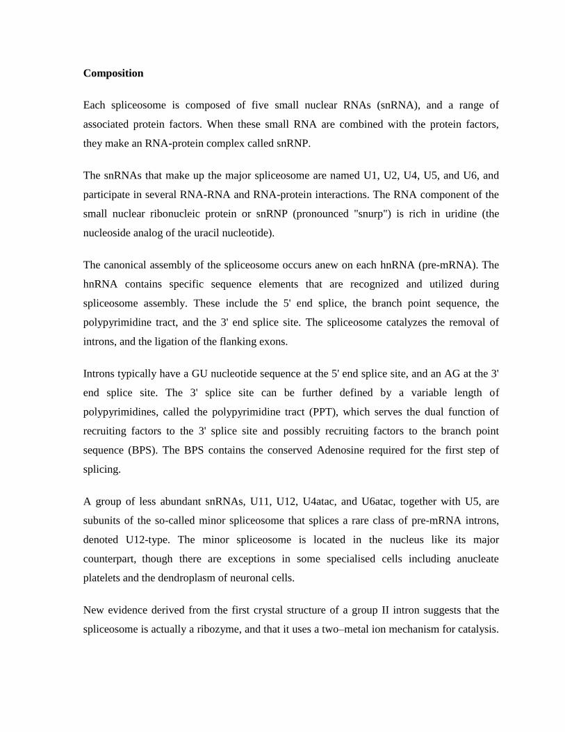
Above are electron microscopy fields of negatively stained yeast (Saccharomyces cerevisiae)
tri-snRNPs. Below left is a schematic illustration of the interaction of tri-snRNP proteins
with the U4/U6 snRNA duplex. Below right is a cartoon model of the yeast tri-snRNP with
shaded areas corresponding to U5 (gray), U4/U6 (orange) and the linker region (yellow).
Alternative splicing
Alternative splicing (the re-combination of different exons) is a major source of genetic
diversity in eukaryotes. Splice variants have been used to account for the relatively small
number of genes in the human genome. For years the estimate widely varied, with top
estimates reaching 100,000 genes, but now, due to the Human Genome Project, the figure is
believed to be closer to 20,000 genes. One particular Drosophila gene (Dscam, the
Drosophila homolog of the human Down syndrome cell adhesion molecule DSCAM) can be
alternatively spliced into 38,000 different mRNA
The Exon Junction Complex
The exon junction complex (EJC) is a protein complex comprised of several protein
components (RNPS1, Y14, SRm160, Aly/REF and Magoh) left behind near splice junctions
by the splicing process (Hir and Andersen, 2008). Their function is to mark the transcript as
processed, and thus ready for export from the nucleus to the cytoplasm, and translation at the
ribosome. The EJC is typically found 20 to 24 nucleotides upstream of the splice junction.
The EJC also plays an important role in nonsense mediated decay, a surveillance system used
in eukaryotes to destroy transcripts containing premature stop codons (Trinkle-Mulcahy et
al., 2009; Chang et al., 2007; Gehring et al., 2005). Upon encountering an EJC during
translation, the ribosome displaces the complex from the mRNA. The ribosome then
continues until it reaches a stop codon. If, however, the mRNA contains a stop codon before
the EJC, the nonsense mediated decay pathway is triggered. The EJC and its position thus
contribute to transcript quality control.
The Evolution of the Spliceosome
A popular hypothesis regarding the origins of the spliceosome is that its predecessor was
self-splicing RNA introns (e.g. Valadkhan, 2007). Such a hypothesis makes sense of several
observations. For example, a simpler way to achieve splicing presumably would be to bring
the splice sites together in one step to directly cleave and rejoin them. The proposed scenario,
however, would explain the use of a lariat intermediate, since a lariat is generated by group II
RNA intron sequences (Lambowitz1 and Zimmerly, 2011; Vogel and Borner, 2002).
The hypothesis also helps to clarify why RNA molecules play such an important part in the
splicing process. Examples of self-splicing RNA introns still exist today (e.g., in the nuclear
rRNA genes of the ciliate Tetrahymena) (Hagen and Cech, 1999; Price et al., 1995; Price and
Cech, 1988; Kruger et al., 1982).
These observations may be taken as evidence as to the spliceosome's evolutionary
predecessor, but they are hardly helpful in elucidating a plausible scenario for transitioning
from one to the other. The spliceosome machinery is far more complex and sophisticated
than autocatalytic ribozymes, involving not just five RNAs but hundreds of proteins.
CHAPTER # 15 RNA EDITING
RNA editing is a molecular process through which some cells can make discrete changes to
specific nucleotide sequences within a RNA molecule after it has been generated by RNA
polymerase. RNA editing is relatively rare, and common forms of RNA processing (e.g.
splicing, 5'-capping and 3'-polyadenylation) are not usually included as editing. Editing
events may include the insertion, deletion, and base substitution of nucleotides within the
edited RNA molecule.
RNA editing has been observed in some tRNA, rRNA, mRNA or miRNA molecules of
eukaryotes and their viruses, archaea and prokaryotes. RNA editing occurs in the cell nucleus
and cytosol, as well as within mitochondria and plastids. In vertebrates, editing is rare and
usually consists of a small number of changes to the sequence of affected molecules. In other
organisms, extensive editing (pan-editing) can occur; in some cases the majority of
nucleotides in a mRNA sequence may result from editing.
RNA-editing processes show great molecular diversity, and some appear to be evolutionarily
recent acquisitions that arose independently. The diversity of RNA editing phenomena
includes nucleobase modifications such as cytidine (C) to uridine (U) and adenosine (A) to
inosine (I) deaminations, as well as non-templated nucleotide additions and insertions. RNA
editing in mRNAs effectively alters the amino acid sequence of the encoded protein so that it
differs from that predicted by the genomic DNA sequence.
Editing by insertion or deletion
RNA editing through the addition and deletion of uracil has been found in kinetoplasts from
the mitochondria of Trypanosoma brucei[3]
Because this may involve a large fraction of the
sites in a gene, it is sometimes called "pan-editing" to distinguish it from topical editing of
one or a few sites.
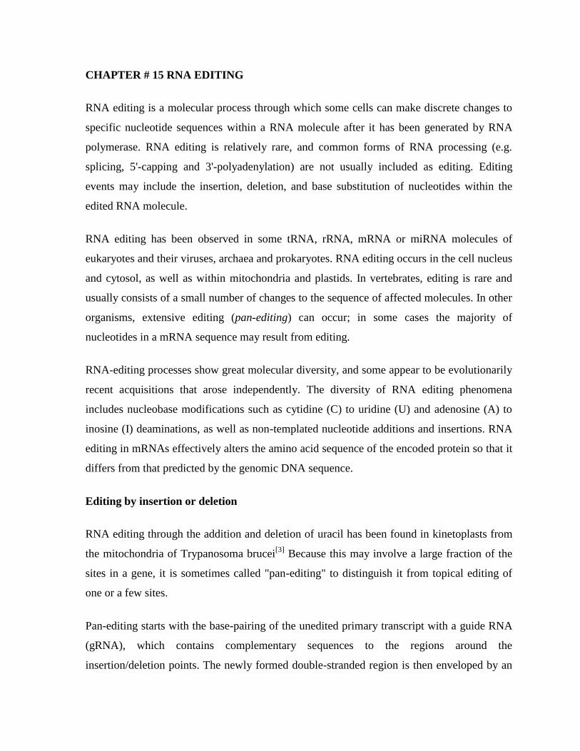
Pan-editing starts with the base-pairing of the unedited primary transcript with a guide RNA
(gRNA), which contains complementary sequences to the regions around the
insertion/deletion points. The newly formed double-stranded region is then enveloped by an
editosome, a large multi-protein complex that catalyzes the editing. The editosome opens the
transcript at the first mismatched nucleotide and starts inserting uridines. The inserted
uridines will base-pair with the guide RNA, and insertion will continue as long as A or G is
present in the guide RNA and will stop when a C or U is encountered. The inserted
nucleotides cause a frameshift and result in a translated protein that differs from its gene.
The Effect of Uracil Insertion in pre-mRNA transcripts
The mechanism of the editosome involves an endonucleolytic cut at the mismatch point
between the guide RNA and the unedited transcript. The next step is catalyzed by one of the
enzymes in the complex, a terminal U-transferase, which adds Us from UTP at the 3‘ end of
the mRNA. The opened ends are held in place by other proteins in the complex. Another
enzyme, a U-specific exoribonuclease, removes the unpaired Us. After editing has made
mRNA complementary to gRNA, an RNA ligase rejoins the ends of the edited mRNA
transcript. As a consequence, the editosome can edit only in a 3‘ to 5‘ direction along the
primary RNA transcript. The complex can act on only a single guide RNA at a time.
Therefore, a RNA transcript requiring extensive editing will need more than one guide RNA
and editosome complex.
Editing by deamination
C-to-U editing
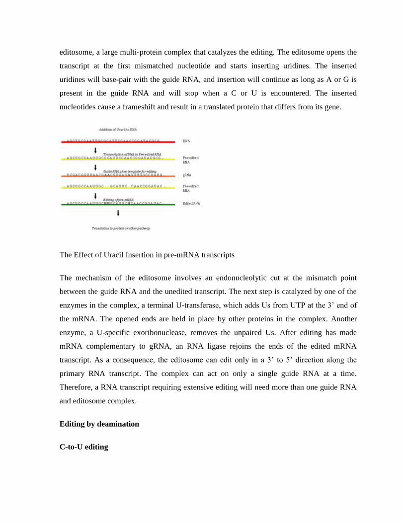
The Effect of C-to-U RNA Editing on the Human ApoB gene
The editing involves cytidine deaminase that deaminates a cytidine base into a uridine base.
An example of C-to-U editing is with the apolipoprotein B gene in humans. Apo B100 is
expressed in the liver and apo B48 is expressed in the intestines. In the intestines, the mRNA
has a CAA sequence edited to be UAA, a stop codon, thus producing the shorter B48 form.
C-to-U editing often occurs in the mitochondrial RNA of flowering plants. Different plants
have different degrees of C-to-U editing; eight editing events occur in mitochondria of the
moss Funaria hygrometrica , where as over 1700 editing events occur in the lycophytes
Isoetes engelmanii. C-to-U editing is performed by members of the pentatricopeptide repeat
(PPR) protein family. Angiosperms have large PPR families, acting as trans -factors for cis -
elements lacking a consensus sequence; Arabidopsis has around 450 members in its PPR
family. There have been a number of discoveries of PPR proteins in both plastids and
mitochondria.
A-to-I editing
A-to-I editing is the main form of RNA editing in mammals and occurs in regions of double-
stranded RNA (dsRNA). Adenosine deaminases acting on RNA (ADARs) are the RNA-
editing enzymes involved in the hydrolytic deamination of Adenosine to Inosine (A-to-I
editing). A-to-I editing can be specific (a single adenosine is edited within the stretch of
dsRNA) or promiscuous (up to 50% of the adenosines are edited). Specific editing occurs
within short duplexes (e.g., those formed in an mRNA where intronic sequence base pairs
with a complementary exonic sequence), while promiscuous editing occurs within longer
regions of duplex (e.g., pre- or pri-miRNAs, duplexes arising from transgene or viral
expression, duplexes arising from paired repetitive elements). There are many effects of A-
to-I editing, arising from the fact that I behaves as if it is G both in translation and when
forming secondary structures. These effects include alteration of coding capacity, altered
miRNA or siRNA target populations, heterochromatin formation, nuclear sequestration,
cytoplasmic sequestration, endonucleolytic cleavage by Tudor-SN, inhibition of miRNA and
siRNA processing, and altered splicing.
Alternative mRNA editing
Alternative U-to-C mRNA editing was first reported in WT1 (Wilms Tumor-1) transcripts,
and non-classic G-A mRNA changes were first observed in HNRNPK (heterogeneous
nuclear ribonucleoprotein K) transcripts in both malignant and normal colorectal samples.
The latter changes were also later seen alongside non-classic U-to-C alterations in brain cell
TPH2 (tryptophan hydroxylase 2) transcripts. Although the reverse amination might be the
simplest explanation for U-to-C changes, transamination and transglycosylation mechanisms
have been proposed for plant U-to-C editing events in mitochondrial transcripts. A recent
study reported novel G-to-A mRNA changes in WT1 transcripts at two hotspots, proposing
the APOBEC3A (apolipoprotein B mRNA editing enzyme, catalytic polypeptide 3A) as the
enzyme implicated in this class of alternative mRNA editing. It was also shown that
alternative mRNA changes were associated with canonical WT1 splicing variants, indicating
their functional significance.
RNA editing in plant mitochondria and plastids
It has been shown in previous studies that the only types of RNA editing seen in the plants‘
mitochondria and plastids are conversion of C-to-U and U-to-C (very rare). RNA-editing
sites are found mainly in the coding regions of mRNA, introns, and other non-translated
regions. In fact, RNA editing can restore the functionality of tRNA molecules. The editing
sites are found primarily upstream of mitochondrial or plastid RNAs. While the specific
positions for C to U RNA editing events have been fairly well studied in both the
mitochondrion and plastid, the identity and organization of all proteins comprising the
editosome have yet to be established. Members of the expansive PPR protein family have
been shown to function as trans-acting factors for RNA sequence recognition. Specific
members of the MORF (Multiple Organellar RNA editing Factor) family are also required
for proper editing at several sites. As some of these MORF proteins have been shown to
interact with members of the PPR family, it is possible MORF proteins are components of
the editosome complex. An enzyme responsible for the trans- or deamination of the RNA
transcript remains elusive, though it has been proposed that the PPR proteins may serve this
function as well.
RNA editing is essential for the normal functioning of the plant‘s translation and respiration
activity. Editing can restore the essential base-pairing sequences of tRNAs, restoring
functionality. It has also been linked to the production of RNA-edited proteins that are
incorporated into the polypeptide complexes of the respiration pathway. Therefore, it is
highly probable that polypeptides synthesized from unedited RNAs would not function
properly and hinder the activity of both mitochondria and plastids.
C-to-U RNA editing can create start and stop codons, but it cannot destroy existing start and
stop codons. A cryptic start codon is created when the codon ACG is edited to AUG.
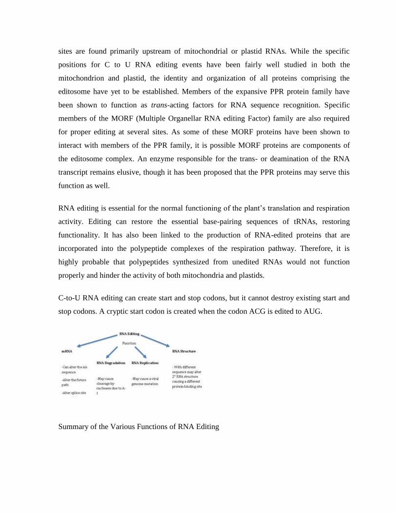
Summary of the Various Functions of RNA Editing
RNA editing in viruses
RNA editing in viruses (i.e., measles, mumps, or parainfluenza) are used for stability and
generation of protein variants. Viral RNAs are transcribed by a virus-encoded RNA-
dependent RNA polymerase, which is prone to pausing and ―stuttering‖ at certain nucleotide
combinations. In addition, up to several hundred non-templated A's are added by the
polymerase at the 3‘ end of nascent mRNA. These As help stabilize the mRNA. Furthermore,
the pausing and stuttering of the RNA polymerase allows the incorporation of one or two Gs
or As upstream of the translational codon. The addition of the non-templated nucleotides
shifts the reading frame, which generates a different protein.
Origin and evolution of RNA editing
The RNA-editing system seen in the animal may have evolved from mononucleotide
deaminases, which have led to larger gene families that include the apobec-1 and adar genes.
These genes share close identity with the bacterial deaminases involved in nucleotide
metabolism. The adenosine deaminase of E. coli cannot deaminate a nucleoside in the RNA;
the enzyme‘s reaction pocket is too small to for the RNA strand to bind to. However, this
active site is widened by amino acid changes in the corresponding human analog genes,
APOBEC1 and ADAR, allowing deamination. The gRNA-mediated pan-editing in
trypanosome mitochondria, involving templated insertion of U residues, is an entirely
different biochemical reaction. The enzymes involved have been shown in other studies to be
recruited and adapted from different sources. But, the specificity of nucleotide insertion via
the interaction between the gRNA and mRNA are similar to the tRNA editing processes in
the animal and Acanthamoeba mithochondria. Eukaryotic ribose methylation of rRNAs by
guide RNA molecules is a similar form of modification.
Thus, RNA editing evolved more than once. Several adaptive rationales for editing have been
suggested.[45]
Editing is often described as a mechanism of correction or repair to compensate
for defects in gene sequences. However, in the case of gRNA-mediated editing, this
explanation does not seem possible because if a defect happens first, there is no way to
generate an error-free gRNA-encoding region, which presumably arises by duplication of the
original gene region. This thinking leads to an evolutionary proposal called "constructive
neutral evolution" in which the order of steps is reversed, with the gratuitous capacity for
editing preceding the "defect".
RNA editing may be involved in RNA degradation
A study looked at the involvement of RNA editing in RNA degradation. The researchers
specifically looked at the interaction between ADAR and UPF1, an enzyme involved in the
nonsense-mediated mRNA decay pathway (NMD). They found that ADAR and UPF1 are
found within the suprasliceosome and they form a complex that leads to the down-regulation
of specific genes. The exact mechanism or the exact pathways that these two are involved in
are unknown at this time. The only fact that this research has shown is that they form a
complex and down-regulate specific genes.
CHAPTER # 16 TRANSLATION
In molecular biology and genetics, translation is the process in which cellular ribosomes
create proteins.
In translation, messenger RNA (mRNA)—produced by transcription from DNA—is decoded
by a ribosome to produce a specific amino acid chain, or polypeptide. The polypeptide later
folds into an active protein and performs its functions in the cell. The ribosome facilitates
decoding by inducing the binding of complementary tRNA anticodon sequences to mRNA
codons. The tRNAs carry specific amino acids that are chained together into a polypeptide as
the mRNA passes through and is "read" by the ribosome. The entire process is a part of gene
expression.
In brief, translation proceeds in four phases:
Initiation: The ribosome assembles around the target mRNA. The first tRNA is attached at
the start codon.
Elongation: The tRNA transfers an amino acid to the tRNA corresponding to the next codon.
Translocation: The ribosome then moves (translocates) to the next mRNA codon to continue
the process, creating an amino acid chain.
Termination: When a stop codon is reached, the ribosome releases the polypeptide.
In bacteria, translation occurs in the cell's cytoplasm, where the large and small subunits of
the ribosome bind to the mRNA. In eukaryotes, translation occurs in the cytosol or across the
membrane of the endoplasmic reticulum in a process called vectorial synthesis. In many
instances, the entire ribosome/mRNA complex binds to the outer membrane of the rough
endoplasmic reticulum (ER); the newly created polypeptide is stored inside the ER for later
vesicle transport and secretion outside of the cell.
Many of transcribed RNA, such as transfer RNA, ribosomal RNA, and small nuclear RNA,
do not undergo translation into proteins.
A number of antibiotics act by inhibiting translation. These include anisomycin,
cycloheximide, chloramphenicol, tetracycline, streptomycin, erythromycin, and puromycin.
Prokaryotic ribosomes have a different structure from that of eukaryotic ribosomes, and thus
antibiotics can specifically target bacterial infections without any harm to a eukaryotic host's
cells.
The basic process of protein production is addition of one amino acid at a time to the end of a
protein. This operation is performed by a ribosome. The choice of amino acid type to add is
determined by an mRNA molecule. Each amino acid added is matched to a three nucleotide
subsequence of the mRNA. For each such triplet possible, the corresponding amino acid is
accepted. The successive amino acids added to the chain are matched to successive
nucleotide triplets in the mRNA. In this way the sequence of nucleotides in the template
mRNA chain determines the sequence of amino acids in the generated amino acid chain.
Addition of an amino acid occurs at the C-terminus of the peptide and thus translation is said
to be amino-to-carboxyl directed.
The mRNA carries genetic information encoded as a ribonucleotide sequence from the
chromosomes to the ribosomes. The ribonucleotides are "read" by translational machinery in
a sequence of nucleotide triplets called codons. Each of those triplets codes for a specific
amino acid.
The ribosome molecules translate this code to a specific sequence of amino acids. The
ribosome is a multisubunit structure containing rRNA and proteins. It is the "factory" where
amino acids are assembled into proteins. tRNAs are small noncoding RNA chains (74-93
nucleotides) that transport amino acids to the ribosome. tRNAs have a site for amino acid
attachment, and a site called an anticodon. The anticodon is an RNA triplet complementary
to the mRNA triplet that codes for their cargo amino acid.
Aminoacyl tRNA synthetases (enzymes) catalyze the bonding between specific tRNAs and
the amino acids that their anticodon sequences call for. The product of this reaction is an
aminoacyl-tRNA. This aminoacyl-tRNA is carried to the ribosome by EF-Tu, where mRNA
codons are matched through complementary base pairing to specific tRNA anticodons.
Aminoacyl-tRNA synthetases that mispair tRNAs with the wrong amino acids can produce
mischarged aminoacyl-tRNAs, which can result in inappropriate amino acids at the
respective position in protein. This "mistranslation" of the genetic code naturally occurs at
low levels in most organisms, but certain cellular environments cause an increase in
permissive mRNA decoding, sometimes to the benefit of the cell.
The ribosome has three sites for tRNA to bind. They are the aminoacyl site (abbreviated A),
the peptidyl site (abbreviated P) and the exit site (abbreviated E). With respect to the mRNA,
the three sites are oriented 5‘ to 3‘ E-P-A, because ribosomes move toward the 3' end of
mRNA. The A site binds the incoming tRNA with the complementary codon on the mRNA.
The P site holds the tRNA with the growing polypeptide chain. The E site holds the tRNA
without its amino acid. When an aminoacyl-tRNA initially binds to its corresponding codon
on the mRNA, it is in the A site. Then, a peptide bond forms between the amino acid of the
tRNA in the A site and the amino acid of the charged tRNA in the P site. The growing
polypeptide chain is transferred to the tRNA in the A site. Translocation occurs, moving the
tRNA in the P site, now without an amino acid, to the E site; the tRNA that was in the A site,
now charged with the polypeptide chain, is moved to the P site. The tRNA in the E site
leaves and another aminoacyl-tRNA enters the A site to repeat the process.
After the new amino acid is added to the chain, and after the mRNA is released out of the
nucleus and into the ribosome's core, the energy provided by the hydrolysis of a GTP bound
to the translocase EF-G (in prokaryotes) and eEF-2 (in eukaryotes) moves the ribosome
down one codon towards the 3' end. The energy required for translation of proteins is
significant. For a protein containing n amino acids, the number of high-energy phosphate
bonds required to translate it is 4n-1. The rate of translation varies; it is significantly higher
in prokaryotic cells (up to 17-21 amino acid residues per second) than in eukaryotic cells (up
to 6-9 amino acid residues per second).
In activation, the correct amino acid is covalently bonded to the correct transfer RNA
(tRNA). The amino acid is joined by its carboxyl group to the 3' OH of the tRNA by an ester
bond. When the tRNA has an amino acid linked to it, it is termed "charged". Initiation
involves the small subunit of the ribosome binding to the 5' end of mRNA with the help of
initiation factors (IF). Termination of the polypeptide happens when the A site of the
ribosome faces a stop codon (UAA, UAG, or UGA). No tRNA can recognize or bind to this
codon. Instead, the stop codon induces the binding of a release factor protein that prompts the
disassembly of the entire ribosome/mRNA complex.
The process of translation is highly regulated in both eukaryotic and prokaryotic organisms.
Regulation of translation can impact the global rate of protein synthesis which is closely
coupled to the metabolic and proliferative state of a cell. In addition, recent work has
revealed that genetic differences and their subsequent expression as mRNAs can also impact
translation rate in an RNA-specific manner.
CHAPTER # 17 POST TRANSLATIONAL MODIFICATIONS
Protein post-translational modification (PTM) increases the functional diversity of the
proteome by the covalent addition of functional groups or proteins, proteolytic cleavage of
regulatory subunits or degradation of entire proteins. These modifications include
phosphorylation, glycosylation, ubiquitination, nitrosylation, methylation, acetylation,
lipidation and proteolysis and influence almost all aspects of normal cell biology and
pathogenesis. Therefore, identifying and understanding PTMs is critical in the study of cell
biology and disease treatment and prevention.
Introduction
Within the last few decades, scientists have discovered that the human proteome is vastly
more complex than the human genome. While it is estimated that the human genome
comprises between 20,000 and 25,000 genes, the total number of proteins in the human
proteome is estimated at over 1 million. These estimations demonstrate that single genes
encode multiple proteins. Genomic recombination, transcription initiation at alternative
promoters, differential transcription termination, and alternative splicing of the transcript are
mechanisms that generate different mRNA transcripts from a single gene .
The increase in complexity from the level of the genome to the proteome is further facilitated
by protein post-translational modifications (PTMs). PTMs are chemical modifications that
play a key role in functional proteomics, because they regulate activity, localization and
interaction with other cellular molecules such as proteins, nucleic acids, lipids, and cofactors.
Post-translational modifications are key mechanisms to increase proteomic diversity. While
the genome comprises 20-25,000 genes, the proteome is estimated to encompass over 1
million proteins. Changes at the transcriptional and mRNA levels increase the size of the
transcriptome relative to the genome, and the myriad of different post-translational
modifications exponentially increases the complexity of the proteome relative to both the
transcriptome and genome.
Additionally, the human proteome is dynamic and changes in response to a legion of stimuli,
and post-translational modifications are commonly employed to regulate cellular activity.
PTMs occur at distinct amino acid side chains or peptide linkages and are most often
mediated by enzymatic activity. Indeed, it is estimated that 5% of the proteome comprises
enzymes that perform more than 200 types of post-translational modifications (4). These
enzymes include kinases, phosphatases, transferases and ligases, which add or remove
functional groups, proteins, lipids or sugars to or from amino acid side chains, and proteases,
which cleave peptide bonds to remove specific sequences or regulatory subunits. Many
proteins can also modify themselves using autocatalytic domains, such as autokinase and
autoprotolytic domains.
Post-translational modification can occur at any step in the "life cycle" of a protein. For
example, many proteins are modified shortly after translation is completed to mediate proper
protein folding or stability or to direct the nascent protein to distinct cellular compartments
(e.g., nucleus, membrane). Other modifications occur after folding and localization are
completed to activate or inactivate catalytic activity or to otherwise influence the biological
activity of the protein. Proteins are also covalently linked to tags that target a protein for
degradation. Besides single modifications, proteins are often modified through a combination
of post-translational cleavage and the addition of functional groups through a step-wise
mechanism of protein maturation or activation.
Protein PTMs can also be reversible depending on the nature of the modification. For
example, kinases phosphorylate proteins at specific amino acid side chains, which is a
common method of catalytic activation or inactivation. Conversely, phosphatases hydrolyze
the phosphate group to remove it from the protein and reverse the biological activity.
Proteolytic cleavage of peptide bonds is a thermodynamically favorable reaction and
therefore permanently removes peptide sequences or regulatory domains.
Consequently, the analysis of proteins and their post-translational modifications is
particularly important for the study of heart disease, cancer, neurodegenerative diseases and
diabetes. The characterization of PTMs, although challenging, provides invaluable insight
into the cellular functions underlying etiological processes. Technically, the main challenges
in studying post-translationally modified proteins are the development of specific detection
and purification methods. Fortunately, these technical obstacles are being overcome with a
variety of new and refined proteomics technologies.
Post-Translational Modifications
As noted above, the large number of different PTMs precludes a thorough review of all
possible protein modifications. Therefore, this overview only touches on a small number of
the most common types of PTMs studied in protein research today. Furthermore, greater
focus is placed on phosphorylation, glycosylation and ubiquitination, and therefore these
PTMs are described in greater detail on pages dedicated to the respective PTM.
Phosphorylation
Reversible protein phosphorylation, principally on serine, threonine or tyrosine residues, is
one of the most important and well-studied post-translational modifications. Phosphorylation
plays critical roles in the regulation of many cellular processes including cell cycle, growth,
apoptosis and signal transduction pathways.
Glycosylation
Protein glycosylation is acknowledged as one of the major post-translational modifications,
with significant effects on protein folding, conformation, distribution, stability and activity.
Glycosylation encompasses a diverse selection of sugar-moiety additions to proteins that
ranges from simple monosaccharide modifications of nuclear transcription factors to highly
complex branched polysaccharide changes of cell surface receptors. Carbohydrates in the
form of aspargine-linked (N-linked) or serine/threonine-linked (O-linked) oligosaccharides
are major structural components of many cell surface and secreted proteins.
Ubiquitination
Ubiquitin is an 8-kDa polypeptide consisting of 76 amino acids that is appended to the ε-
NH2 of lysine in target proteins via the C-terminal glycine of ubiquitin. Following an initial
monoubiquitination event, the formation of a ubiquitin polymer may occur, and
polyubiquitinated proteins are then recognized by the 26S proteasome that catalyzes the
degradation of the ubiquitinated protein and the recycling of ubiquitin.
S-Nitrosylation
Nitric oxide (NO) is produced by three isoforms of nitric oxide synthase (NOS) and is a
chemical messenger that reacts with free cysteine residues to form S-nitrothiols (SNOs). S-
nitrosylation is a critical PTM used by cells to stabilize proteins, regulate gene expression
and provide NO donors, and the generation, localization, activation and catabolism of SNOs
are tightly regulated.
S-nitrosylation is a reversible reaction, and SNOs have a short half life in the cytoplasm
because of the host of reducing enzymes, including glutathione (GSH) and thioredoxin, that
denitrosylate proteins. Therefore, SNOs are often stored in membranes, vesicles, the
interstitial space and lipophilic protein folds to protect them from denitrosylation. For
example, caspases, which mediate apoptosis, are stored in the mitochondrial intermembrane
space as SNOs. In response to extra- or intracellular cues, the caspases are released into the
cytoplasm, and the highly reducing environment rapidly denitrosylates the proteins, resulting
in caspase activation and the induction of apoptosis.
S-nitrosylation is not a random event, and only specific cysteine residues are S-nitrosylated.
Because proteins may contain multiple cysteines and due to the labile nature of SNOs, S-
nitrosylated cysteines can be difficult to detect and distinguish from non-S-nitrosylated
amino acids. The biotin switch assay, developed by Jaffrey et al., is a common method of
detecting SNOs, and the steps of the assay are listed below:
All free cysteines are blocked.
All remaining cysteines (presumably only those that are denitrosylated) are denitrosylated.
The now-free thiol groups are then biotinylated.
Biotinylated proteins are detected by SDS-PAGE and Western blot analysis or mass
spectrometry.
Methylation
The transfer of one-carbon methyl groups to nitrogen or oxygen (N- and O-methylation,
respectively) to amino acid side chains increases the hydrophobicity of the protein and can
neutralize a negative amino acid charge when bound to carboxylic acids. Methylation is
mediated by methyltransferases, and S-adenosyl methionine (SAM) is the primary methyl
group donor.
Methylation occurs so often that SAM has been suggested to be the most-used substrate in
enzymatic reactions after ATP. Additionally, while N-methylation is irreversible, O-
methylation is potentially reversible. Methylation is a well-known mechanism of epigenetic
regulation, as histone methylation and demethylation influences the availability of DNA for
transcription. Amino acid residues can be conjugated to a single methyl group or multiple
methyl groups to increase the effects of modification.
N-Acetylation
N-acetylation, or the transfer of an acetyl group to nitrogen, occurs in almost all eukaryotic
proteins through both irreversible and reversible mechanisms. N-terminal acetylation requires
the cleavage of the N-terminal methionine by methionine aminopeptidase (MAP) before
replacing the amino acid with an acetyl group from acetyl-CoA by N-acetyltransferase
(NAT) enzymes. This type of acetylation is co-translational, in that N-terminus is acetylated
on growing polypeptide chains that are still attached to the ribosome. While 80-90% of
eukaryotic proteins are acetylated in this manner, the exact biological significance is still
unclear.
Acetylation at the ε-NH2 of lysine (termed lysine acetylation) on histone N-termini is a
common method of regulating gene transcription. Histone acetylation is a reversible event
that reduces chromosomal condensation to promote transcription, and the acetylation of these
lysine residues is regulated by transcription factors that contain histone acetyletransferase
(HAT) activity. While transcription factors with HAT activity act as transcription co-
activators, histone deacetylase (HDAC) enzymes are co-repressors that reverse the effects of
acetylation by reducing the level of lysine acetylation and increasing chromosomal
condensation.
Sirtuins (silent information regulator) are a group of NAD-dependent deacetylases that target
histones. As their name implies, they maintain gene silencing by hypoacetylating histones
and have been reported to aid in maintaining genomic stability.
While acetylation was first detected in histones, cytoplasmic proteins have been reported to
also be acetylated, and therefore acetylation seems to play a greater role in cell biology than
simply transcriptional regulation. Furthermore, crosstalk between acetylation and other post-
translational modifications, including phosphorylation, ubiquitination and methylation, can
modify the biological function of the acetylated protein.
Protein acetylation can be detected by chromosome immunoprecipitation (ChIP) using
acetyllysine-specific antibodies or by mass spectrometry, where an increase in histone by 42
mass units represents a single acetylation.
Lipidation
Lipidation is a method to target proteins to membranes in organelles (endoplasmic reticulum
[ER], Golgi apparatus, mitochondria), vesicles (endosomes, lysosomes) and the plasma
membrane. The four types of lipidation are:
C-terminal glycosyl phosphatidylinositol (GPI) anchor
N-terminal myristoylation
S-myristoylation
S-prenylation
Each type of modification gives proteins distinct membrane affinities, although all types of
lipidation increase the hydrophobicity of a protein and thus its affinity for membranes. The
different types of lipidation are also not mutually exclusive, in that two or more lipids can be
attached to a given protein.
GPI anchors tether cell surface proteins to the plasma membrane. These hydrophobic
moieties are prepared in the ER, where they are then added to the nascent protein en bloc.
GPI-anchored proteins are often localized to cholesterol- and sphingolipid-rich lipid rafts,
which act as signaling platforms on the plasma membrane. This type of modification is
reversible, as the GPI anchor can be released from the protein by phosphoinositol-specific
phospholipase C. Indeed, this lipase is used in the detection of GPI-anchored proteins to
release GPI-anchored proteins from membranes for gel separation and analysis by mass
spectrometry.
N-myristoylation is a method to give proteins a hydrophobic handle for membrane
localization. The myristoyl group is a 14-carbon saturated fatty acid (C14), which gives the
protein sufficient hydrophobicity and affinity for membranes, but not enough to permanently
anchor the protein in the membrane. N-myristoylation can therefore act as a conformational
localization switch, in which protein conformational changes influence the availability of the
handle for membrane attachment. Because of this conditional localization, signal proteins
that selectively localize to membrane, such as Src-family kinases, are N-myristoylated.
N-myristoylation is facilitated specifically by N-myristoyltransferase (NMT) and uses
myristoyl-CoA as the substrate to attach the myristoyl group to the N-terminal glycine.
Because methionine is the N-terminal amino acid of all eukaryotic proteins, this PTM
requires methionine cleavage by the above-mentioned MAP prior to addition of the myristoyl
group; this represents one example of multiple PTMs on a single protein.
S-palmitoylation adds a C16 palmitoyl group from palmitoyl-CoA to the thiolate side chain
of cysteine residues via palmitoyl acyl transferases (PATs). Because of the longer
hydrophobic group, this anchor can permanently anchor the protein to the membrane. This
localization can be reversed, though, by thioesterases that break the link between the protein
and the anchor; thus, S-palmitoylation is used as an on/off switch to regulate membrane
localization. S-palmitoylation is often used to strengthen other types of lipidation, such as
myristoylation or farnesylation (see below). S-palmitoylated proteins also selectively
concentrate at lipid rafts.
S-prenylation covalently adds a farnesyl (C15) or geranylgeranyl (C20) group to specific
cysteine residues within 5 amino acids from the C-terminus via farnesyl transferase (FT) or
geranylgeranyl transferases (GGT I and II). Unlike S-palmitoylation, S-prenylation is
hydrolytically stable. Approximately 2% of all proteins are prenylated, including all
members of the Ras superfamily. This group of molecular switches is farnesylated,
geranylgeranylated or a combination of both. Additionally, these proteins have specific 4-
amino acid motifs at the C-terminus that determine the type of prenylation at single or dual
cysteines. Prenylation occurs in the ER and is often part of a stepwise process of PTMs that
is followed by proteolytic cleavage by Rce1 and methylation by isoprenyl cysteine
methyltransferase (ICMT).
Proteolysis
Peptide bonds are indefinitely stable under physiological conditions, and therefore cells
require some mechanism to break these bonds. Proteases comprise a family of enzymes that
cleave the peptide bonds of proteins and are critical in antigen processing, apoptosis, surface
protein shedding and cell signaling.
The family of over 11,000 proteases varies in substrate specificity, mechanism of peptide
cleavage, location in the cell and the length of activity. While this variation suggests a wide
array of functionalities, proteases can generally be separated into groups based on the type of
proteolysis. Degradative proteolysis is critical to remove unassembled protein subunits and
misfolded proteins and to maintain protein concentrations at homeostatic concentrations by
reducing a given protein to the level of small peptides and single amino acids. Proteases also
play a biosynthetic role in cell biology that includes cleaving signal peptides from nascent
proteins and activating zymogens, which are inactive enzyme precursors that require
cleavage at specific sites for enzyme function. In this respect, proteases act as molecular
switches to regulate enzyme activity.
Proteolysis is a thermodynamically favorable and irreversible reaction. Therefore, protease
activity is tightly regulated to avoid uncontrolled proteolysis through temporal and/or spatial
control mechanisms including regulation by cleavage in cis or trans and
compartmentalization (e.g., proteasomes, lysosomes).
The diverse family of proteases can be classified by the site of action, such as
aminopeptidases and carboxypeptidase, which cleave at the amino or carboxy terminus of a
protein, respectively. Another type of classification is based on the active site groups of a
given protease that are involved in proteolysis. Based on this classification strategy, greater
than 90% of known proteases fall into one of four categories as follows:
Serine proteases
Cysteine proteases
Aspartic acid proteases
Zinc metalloproteases
CHAPTER # 18 REGULATORY ELEMENTS
RNA molecules that act as regulators were known in bacteria for years before the first
microRNAs (miRNAs) and short interfering RNAs (siRNAs) were discovered in eukaryotes.
In 1981, the ~108 nucleotide RNA I was found to block ColE1 plasmid replication by base
pairing with the RNA that is cleaved to produce the replication primer. This work was
followed by the 1983 discovery of a ~70 nucleotide RNA which is transcribed from the
pOUT promoter of the Tn10 transposon and represses transposition by preventing translation
of the transposase mRNA. The first chromosomally-encoded small RNA regulator, reported
in 1984, was the 174 nucleotide Escherichia coli MicF RNA, which inhibits translation of the
mRNA encoding the major outer membrane porin OmpF. These first small RNA regulators,
and a handful of others, were identified by gel analysis due to their abundance, by multicopy
phenotypes, or by serendipity.
While a few bacterial RNA regulators were identified early on, their prevalence and their
contributions to numerous physiological responses were not initially appreciated. In 2001–
2002, four groups reported the identification of many new small RNAs through systematic
computational searches for conservation and orphan promoter and terminator sequences in
the intergenic regions of E. coli. Additional RNAs were discovered by direct detection using
cloning-based techniques or microarrays with probes in intergenic regions. Variations of
these approaches, aided by the availability of many new bacterial genome sequences, have
led to the identification of regulatory RNAs in an ever-increasing number of bacteria.
Enabled by recent technical advances, including multilayered computational searches deep
sequencing and tiled microarrays with full genome coverage, hundreds of candidate
regulatory RNA genes in various bacteria have now been predicted. In E. coli alone, ~80
small transcripts have been verified, increasing the total number of genes identified for this
organism by 2%.
In this review, we will focus our discussion on bacterial small RNAs that act as regulators. A
limited number of small RNAs carry out specific housekeeping functions, namely the 4.5S
RNA component of the signal recognition particle, the RNase P RNA responsible for
processing of tRNAs and other RNAs, and tmRNA, which acts as both a tRNA and mRNA
to tag incompletely translated proteins for degradation and to release stalled ribosomes. We
will not discuss these RNAs further, although their actions, as well as those of some tRNAs,
can have regulatory consequences.
In addition, a few defining features are worthy of mention at the outset. Riboswitches are part
of the mRNA they regulate, usually found within the 5‘ untranslated region (5‘-UTR), and
hence act in cis. Most of the regulatory RNAs that act in trans by base pairing with other
RNAs are synthesized as discrete transcripts with dedicated promoter and terminator
sequences. Given that the longest of these RNAs, RNAIII of Staphylococcus aureus, is still
only 514 nucleotides, the RNAs are commonly referred to as small RNAs (sRNAs). We
prefer this term to ―noncoding RNA‖, the term frequently used in eukaryotes, since a number
of the sRNAs, including RNAIII, also encode proteins. In contrast to the base pairing sRNAs,
some sRNAs that modulate protein activity as well as the CRISPR RNAs are processed out
of longer transcripts.
Regulatory Functions of Bacterial RNAs
Regulatory RNAs can modulate transcription, translation, mRNA stability, and DNA
maintenance or silencing. They achieve these diverse outcomes through a variety of
mechanisms, including changes in RNA conformation, protein binding, base pairing with
other RNAs, and interactions with DNA.
Riboswitches
Perhaps the simplest bacterial RNA regulatory elements are sequences at the 5‘ end of
mRNAs, or less frequently at the 3‘ end, that can adopt different conformations in response
to environmental signals, including stalled ribosomes, uncharged tRNAs, elevated
temperatures, or small molecule ligands. These elements were first described decades ago in
elegant studies characterizing transcription attenuation. In this process, stalled ribosomes lead
to changes in mRNA structure, affecting transcription elongation through the formation of
terminator or antiterminator structures in the mRNA. Later studies showed that sequences
found in transcripts encoding tRNA synthetases, termed ―Tboxes‖, bind the corresponding
uncharged tRNAs, and that other leader sequences, known as ―RNA thermometers‖, fold in a
manner that is sensitive to temperature. In both of these cases, the alternate structures lead to
changes in the expression of the downstream gene.
More recently, it was found that leader sequences could bind small molecules and adopt
different conformations in the presence or absence of metabolites. These metabolite sensors,
denoted ―riboswitches‖, directly regulate the genes involved in the uptake and use of the
metabolite. In fact, in some cases, the presence of a riboswitch upstream of an
uncharacterized or mis-annotated gene has helped to clarify the physiological role of the gene
product. An ever-increasing number and variety of riboswitches are being identified in
bacteria, as well as in some eukaryotes. For example, as many as 2% of all Bacillus subtilis
genes are regulated by riboswitches which bind metabolites ranging from flavin
mononucleotide (FMN) and thiamin pyrophosphate to S-adenosylmethionine, lysine and
guanine.
Riboswitches generally consist of two parts: the aptamer region, which binds the ligand, and
the so-called expression platform, which regulates gene expression through alternative RNA
structures that affect transcription or translation. Upon binding of the ligand, the riboswitch
changes conformation. These changes usually involve alternative hairpin structures which
form or disrupt transcriptional terminators or antiterminators, or which occlude or expose
ribosome binding sites. In general, most riboswitches repress transcription or translation in
the presence of the metabolite ligand; only a few riboswitches that activate gene expression
have been characterized.
Gene Arrangement and Regulatory Functions of Ligand- and Protein-binding
Regulatory RNAs
Due to the modular nature of riboswitches, the same aptamer domain can mediate different
regulatory outcomes or operate through distinct mechanisms in different contexts. For
example, the cobalamin riboswitch, which binds the coenzyme form of vitamin B12, operates
by transcription termination for the btuB genes in Gram-positive bacteria but modulates
translation initiation for the cob operons of Gram-negative bacteria. Some transcripts carry
tandem riboswitches, which can integrate distinct physiological signals, and one notable
riboswitch, the glmS leader sequence, even acts as a ribozyme to catalyze self-cleavage.
Upon binding of its cofactor glucosamine-6-phosphate, the glmS riboswitch cleaves itself and
inactivates the mRNA encoding the enzyme that generates glucosamine-6-phosphate, thus
effecting a negative feedback loop for metabolite levels. In principle, riboswitches could be
used in conjunction with any reaction associated with RNA, not just transcription, translation
and RNA processing, but also RNA modification, localization or splicing.
Generally, the riboswitches in Gram-positive bacteria affect transcriptional attenuation, while
the riboswitches in Gram-negative bacteria more frequently inhibit translation. Possibly the
preferential use of transcriptional termination in Gram-positive organisms is linked to the fact
that genes are clustered together in larger biosynthetic operons where more resources would
be wasted if the full-length transcript is synthesized. Gram-positive organisms also appear to
rely more on cis-acting riboswitches than Gram-negative organisms, for which more trans-
acting sRNA regulators are known. Research directions pursued in studies of the different
organisms, however, may bias these generalizations.
sRNAs That Modulate Protein Activity
Three protein-binding sRNAs have intrinsic activity (RNase P) or contribute essential
functions to a ribonucleoprotein particle (4.5S and tmRNA). In contrast, three other protein-
binding sRNAs (CsrB, 6S, and GlmY) act in a regulatory fashion to antagonize the activities
of their cognate proteins by mimicking the structures of other nucleic acids.
The CsrB and CsrC RNAs of E. coli modulate the activity of CsrA, an RNA-binding protein
that regulates carbon usage and bacterial motility upon entry into stationary phase and other
nutrient-poor conditions. CsrA dimers bind to GGA motifs in the 5‘ UTR of target mRNAs,
thereby affecting the stability and/or translation of the mRNA. The CsrB and CsrC RNAs
each contain multiple GGA binding sites, 22 and 13 respectively, for CsrA. Thus, when CsrB
and CsrC levels increase, the sRNAs effectively sequester the CsrA protein away from
mRNA leaders. Transcription of the csrB and csrC genes is induced by the BarA-UvrB two-
component regulators when cells encounter nutrient poor growth conditions, though the
signal for this induction is not known. The CsrB and CsrC RNAs also are regulated at the
level of stability through the CsrD protein, a cyclic di-GMP binding protein, which recruits
RNase E to degrade the sRNAs. CsrB and CsrC homologs (such as RsmY and RsmZ) have
been found to antagonize the activities of CsrA homologs in a range of bacteria including
Salmonella, Erwinia, Pseudomonas, and Vibrio where they impact secondary metabolism,
quorum sensing and epithelial cell invasion.
The E. coli 6S RNA mimics an open promoter to bind to and sequester the ζ70
-containing
RNA polymerase. When 6S is abundant, especially in stationary phase, it is able to complex
with much of the ζ70
-bound, housekeeping form of RNA polymerase, but is not associated
with the ζS-bound, stationary phase form of RNA polymerase. The interaction between 6S
and ζ70
-holoenzyme inhibits transcription from certain ζ70
promoters and increases
transcription from some ζS regulated promoters, in part by altering the competition between
ζ70
-and ζS-holoenzyme binding to promoters. Interestingly, the 6S RNA can serve as a
template for the transcription of 14–20 nucleotide product RNAs (pRNAs) by RNA
polymerase, especially during outgrowth from stationary phase. In fact, it is thought that
transcription from 6S when NTP concentrations increase may be a way to release ζ70
-RNA
polymerase. It is not known whether the pRNAs themselves have a function. The 6S RNA is
processed out of a longer transcript and accumulates during stationary phase, but the details
of this regulation have not been elucidated. There are multiple 6S homologs in a number of
organisms, including two in B. subtilis. The roles of these homologs again are not known, but
it is tempting to speculate that they inhibit the activities of alternative ζ factor forms of RNA
polymerase.
One additional sRNA, GlmY, has recently been proposed to have a protein-binding mode of
action and is thought to function by titrating an RNA processing factor away from a
homologous sRNA, GlmZ. Both GlmZ and GlmY promote accumulation of the GlmS
glucosamine-6-phosphate synthase, however they do so by distinct mechanisms. The full-
length GlmZ RNA base pairs with and activates translation of the glmS mRNA. Although the
GlmY RNA is highly homologous to GlmZ in sequence and predicted secondary structure,
GlmY lacks the region that is complementary to the glmS mRNA target and does not directly
activate glmS translation. Instead, GlmY expression inhibits a GlmZ processing event that
renders GlmZ unable to activate glmS translation. Although not yet conclusively shown,
GlmY most likely stabilizes the full-length GlmZ by competing with GlmZ for binding to the
YhbJ protein that targets GlmZ for processing. The GlmY RNA is also processed and its
levels are negatively regulated by poly-adenylation.
CsrB RNA simulates an mRNA element, 6S imitates a DNA structure, and GlmY mimics
another sRNA, raising the question as to what other molecules, nucleic acid or otherwise,
might yet uncharacterized sRNAs mimic?
Cis-encoded Base Pairing sRNAs
In contrast to the few known protein-binding sRNAs, most characterized sRNAs regulate
gene expression by base pairing with mRNAs and fall into two broad classes: those having
extensive potential for base pairing with their target RNA and those with more limited
complementarity. We will first focus on sRNAs that are encoded in cis on the DNA strand
opposite the target RNA and share extended regions of complete complementarity with their
target, often 75 nucleotides or more. While the two transcripts are encoded in the same
region of DNA, they are transcribed from opposite strands as discrete RNA species and
function in trans as diffusible molecules. For the few cases where it has been examined, the
initial interaction between the sRNA and target RNA involves only limited pairing, though
the duplex can subsequently be extended. The most well-studied examples of cis-encoded
antisense sRNAs reside on plasmids or other mobile genetic elements, however chromosomal
versions of these sRNAs increasingly are being found.
Gene Arrangement and Regulatory Functions of Base Pairing Regulatory RNAs
Most of the cis-encoded antisense sRNAs expressed from bacteriophage, plasmids and
transposons function to maintain the appropriate copy number of the mobile element. They
achieve this through a variety of mechanisms, including inhibition of replication primer
formation and transposase translation, as mentioned for plasmid ColE1 RNA I and Tn10
pOUT RNA, respectively. Another common group act as antitoxins to repress the translation
of toxic proteins that kill cells from which the mobile element has been lost.
In general, the physiological roles of the cis-encoded antisense sRNAs expressed from
bacterial chromosomes are less well understood. A subset promote degradation and/or
repress translation of mRNAs encoding proteins that are toxic at high levels. In E. coli, there
are also two sRNAs, IstR and OhsC, that are encoded directly adjacent to genes encoding
potentially toxic proteins. Although these sRNAs are not true antisense RNAs, they do
contain extended regions of perfect complementarity (19 and 23 nucleotides) with the toxin
mRNAs. Interestingly, most of these sRNAs appear to be expressed constitutively. Some of
the chromosomal antitoxin sRNAs are homologous to plasmid antitoxin sRNAs (for
example, the Hok/Sok loci present in the E. coli chromosome) or are located in regions
acquired from mobile elements (for example, the RatA RNA of B. subtilis found in a remnant
of a cryptic prophage). These observations indicate that the antitoxin sRNA and
corresponding toxin genes might have been acquired by horizontal transfer. The
chromosomal versions may simply be non-functional remnants. However, some cis-encoded
antisense antitoxin sRNAs do not have known homologs on mobile elements. In addition,
given that bacteria have multiple copies of several loci, all of which are expressed in the
cases examined, it is tempting to speculate that the antitoxin sRNAs-toxin proteins encoded
on the chromosome provide beneficial functions. Although high levels of the toxins kill cells,
more moderate levels produced from single-copy loci under inducing conditions may only
slow growth. Thus one model proposes that chromosomal toxin-antitoxin modules induce
slow growth or stasis under conditions of stress to allow cells time to repair damage or
otherwise adjust to their environment. Another possibility is that certain modules may be
retained in bacterial chromosomes as a defense against plasmids bearing homologous
modules, assuming that the chromosomal antisense sRNA can repress the expression of the
plasmid-encoded toxin.
Another group of cis-encoded antisense sRNAs modulates the expression of genes in an
operon. Some of these sRNAs are encoded in regions complementary to intervening
sequence between ORFs. For example, in E. coli, base pairing between the stationary phase-
induced GadY antisense sRNA and the gadXW mRNA leads to cleavage of the duplex
between the gadX and gadW genes and increased levels of a gadX transcript. For the
virulence plasmid pJM1 of Vibrio anguillarum, the interaction between the RNAβ antisense
sRNA and the fatDCBAangRT mRNA leads to transcription termination after the fatA gene,
thus reducing expression of the downstream angRT genes. In Synechocystis, the iron-stress
repressed IsrR antisense sRNA base pairs with sequences within isiA coding region of the
isiAB transcript and leads to decreased levels of an isiA transcript. In this case, it is not
known whether isiB expression is also affected.
The list of cis-encoded antisense sRNAs is far from complete, especially for chromosomal
versions, and other mechanisms of action are sure to be found.
Trans-encoded Base Pairing sRNAs
Another class of base pairing sRNAs is the trans-encoded sRNAs, which, in contrast to the
cis-encoded antisense sRNAs, share only limited complementarity with their target mRNAs.
These sRNAs regulate the translation and/or stability of target mRNAs and are, in many
respects, functionally analogous to eukaryotic miRNAs.
The majority of the regulation by the known trans-encoded sRNAs is negative. Base pairing
between the sRNA and its target mRNA usually leads to repression of protein levels through
translational inhibition, mRNA degradation, or both. The bacterial sRNAs characterized to
date primarily bind to the 5‘ UTR of mRNAs and most often occlude the ribosome binding
site, though some sRNAs such as GcvB and RyhB inhibit translation through base pairing far
upstream of the AUG of the repressed gene. The sRNA-mRNA duplex is then frequently
subject to degradation by RNase E. For the few characterized sRNA-mRNA interactions, the
inhibition of ribosome binding is the main contributor to reduced protein levels, while the
subsequent degradation of the sRNA-mRNA duplex is thought to increase the robustness of
the repression and make the regulation irreversible. However, sRNAs can also activate
expression of their target mRNAs through an anti-antisense mechanism whereby base pairing
of the sRNA disrupts an inhibitory secondary structure which sequesters the ribosome
binding site. Theoretically, base pairing between a trans-encoded sRNA and its target could
promote transcription termination or antitermination, as has been found for some cis-encoded
sRNAs, or alter mRNA stability through changes in poly-adenylation.
For trans-encoded sRNAs, there is little correlation between the chromosomal location of the
sRNA gene and the target mRNA gene. In fact, each trans-encoded sRNA typically base
pairs with multiple mRNAs. The capacity for multiple base pairing interactions results from
the fact that trans-encoded sRNAs make more limited contacts with their target mRNAs in
discontinuous patches, rather than extended stretches of perfect complementarity, as for cis-
encoded antisense sRNAs. The region of potential base pairing between trans-encoded
sRNAs and target mRNAs typically encompasses ~10–25 nucleotides, but in all cases where
it has been examined only a core of the nucleotides seem to be critical for regulation. For
example, although the SgrS sRNA has the potential to form 23 base pairs with the ptsG
mRNA across a stretch of 32 nucleotides, only four single mutations in SgrS significantly
affected downregulation of ptsG.
In many cases, the RNA chaperone Hfq is required for trans-encoded sRNA-mediated
regulation, presumably to facilitate RNA-RNA interactions due to limited complementarity
between the sRNA and target mRNA. The hexameric Hfq ring, which is homologous to Sm
and Sm-like proteins involved in splicing and mRNA decay in eukaryotes, may actively
remodel the RNAs to melt inhibitory secondary structures. Hfq also may serve passively as a
platform to allow sRNAs and mRNAs to sample potential complementarity, effectively
increasing the local concentrations of sRNAs and mRNAs. It should be noted that when the
E. coli SgrS RNA is pre-annealed with the ptsG mRNA in vitro, the Hfq protein is no longer
required. However, in vivo in E. coli, sRNAs no longer regulate their target mRNAs in hfq
mutant strains, and all trans-encoded base pairing sRNAs examined to date co-
immunoprecipitate with Hfq. In fact, enrichment of sRNAs by co-immunoprecipitation with
Hfq proved to be a fruitful approach to identify and validate novel sRNAs in E. coli and has
been extended to other bacteria, such as S. typhimurium.
Beyond facilitating base pairing, Hfq contributes to sRNA regulation through modulating
sRNA levels. Somewhat counterintuitively, most E. coli sRNAs are less stable in the absence
of Hfq, presumably because Hfq protects sRNAs from degradation in the absence of base
pairing with mRNAs. Once base paired with target mRNAs, many of the known sRNA-
mRNA pairs are subject to degradation by RNase E, and Hfq may also serve to recruit RNA
degradation machinery through its interactions with RNase E and other components of the
degradosome. In addition, competition between sRNAs for binding to Hfq may be a factor
controlling sRNA activity in vivo.
Although all characterized E. coli trans-encoded sRNAs require Hfq for regulation of their
targets, the need for an RNA chaperone may not be universal. For example, VrrA RNA
repression of OmpA protein expression in V. cholerae is not eliminated in hfq mutant cells,
though the extent of repression is higher in cells expressing Hfq. In general, longer stretches
of base pairing, as is the case for the cis-encoded antisense sRNAs that usually do not require
Hfq for function, and/or high concentrations of the sRNA may obviate a chaperone
requirement.
In contrast to cis-encoded sRNAs, several of which are expressed constitutively, most of the
trans-encoded sRNAs are synthesized under very specific growth conditions. In E. coli for
example, these regulatory RNAs are induced by low iron (Fur-repressed RyhB), oxidative
stress (OxyR-activated OxyS), outer membrane stress (ζE-induced MicA and RybB),
elevated glycine (GcvA-induced GcvB), changes in glucose concentration (CRP-repressed
Spot42 and CRP-activated CyaR), and elevated glucose-phosphate levels (SgrR-activated
SgrS). In fact, it is possible that every major transcription factor in E. coli may control the
expression of one or more sRNA regulators. It is also noteworthy that a number of the
sRNAs are encoded adjacent to the gene encoding their transcription regulator, including E.
coli OxyR-OxyS, GcvA-GcvB, and SgrR-SgrS.
The fact that a given base pairing sRNA often regulates multiple targets means that a single
sRNA can globally modulate a particular physiological response, in much the same manner
as a transcription factor, but at the post-transcriptional level. Well-characterized regulatory
effects of these sRNAs include the down regulation of iron-sulfur cluster containing enzymes
under conditions of low iron (E. coli RyhB), repression of outer membrane porin proteins
under conditions of membrane stress (E. coli MicA and RybB), and repression of quorum
sensing at low cell density (Vibrio Qrr). The fact that direct or indirect negative feedback
regulation is observed for a number of sRNAs emphasizes that sRNAs are integrated into
regulatory circuits. In E. coli for example, ryhB is repressed when iron is released after RyhB
down-regulates iron-sulfur enzymes, and micA and rybB are repressed when membrane stress
is relieved upon their down-regulation of outer membrane porins. As another example, the
Qrr sRNAs in Vibrio base pair with and inhibit expression of the mRNAs encoding the
transcription factors responsible for the activation of the qrr genes.
CRISPR RNAs
A unique class of recently discovered regulatory RNAs is the CRISPR RNAs, which provide
resistance to bacteriophage and prevent plasmid conjugation. CRISPR systems share certain
similarities with eukaryotic siRNA-driven gene silencing, although they exhibit distinct
features as well, and present an exciting new arena of RNA research. The CRISPR sequences
have been found in ~40% of bacteria and ~90% of archaea sequenced to date, emphasizing
their wide-ranging importance.
CRISPR sequences (Clustered Regularly Interspaced Short Palindromic Repeats) are highly
variable DNA regions which consist of a ~550 bp leader sequence followed by a series of
repeat-spacer units. The repeated DNA can vary from 24 to 47 base pairs, but the same repeat
sequence usually appears in each unit in a given CRISPR array, and is repeated two to 249
times. The repeat sequences diverge significantly between bacteria, but can be grouped into
12 major types and often contain a short 5–7 base pair palindrome. Unlike other repeated
sequences in bacterial chromosomes, the CRISPR repeats are regularly interspersed with
unique spacers of 26 to 72 base pairs; these spacers are not typically repeated in a given
CRISPR array. Although the repeats can be similar between species, the spacers between the
repeats are not conserved at all, often varying even between strains, and are most often found
to be homologous to DNA from phages and plasmids, an observation that was initially
perplexing.
Gene Arrangement and Regulatory Functions of CRISPR RNAs
Adjacent to the CRISPR DNA array are several CRISPR-associated (CAS) genes. Two to six
core CAS genes seem to be associated with most CRISPR systems, but different CRISPR
subtypes also have specific CAS genes encoded in the flanking region. Other CAS genes,
that are never present in strains lacking the repeats, may be found in genomic locations
distant from the CRISPR region(s). The molecular functions of the CAS proteins are still
mostly obscure, but they often contain RNA- or DNA-binding domains, helicase motifs, and
endo- or exonuclease domains.
After the initial report of CRISPR sequences in 1989, several different hypotheses were
advanced as to possible functions of these repeats. The proposal that CRISPRs confer
resistance to phages came in 2005 with findings that the spacers often contain homology to
phage or plasmids. Another major advance was the discovery that the CRISPR DNA arrays
are transcribed in bacteria and archaea. The full-length CRISPR RNA initially extends the
length of the entire array, but is subsequently processed into shorter fragments the size of a
single repeat-spacer unit. Recently, it was shown that the E. coli K12 CasA-E proteins
associate to form a complex termed Cascade, for CRISPR-Associated Complex for Antiviral
Defense. The CasE protein within the Cascade complex is responsible for processing of the
full-length CRISPR RNA transcript.
Importantly, it was demonstrated that new spacers corresponding to phage sequences are
integrated into existing CRISPR arrays during phage infection and that these new spacers
confer resistance to subsequent infections with the cognate phage, or other phage bearing the
same sequence. The new spacers are inserted at the beginning of the array, such that the 5‘
end of the CRISPR region is hypervariable between strains and conveys information about
the most recent phage infections, while the 3‘ end spacers are consequences of more ancient
infections. Single nucleotide point mutations in the bacterial spacers or the phage genome
abolish phage resistance and, further, introduction of novel phage sequences as spacers in
engineered CRISPR arrays provides de novo immunity to bacteria that have never
encountered this phage. Similar observations were recently made for spacers found to
correspond to sequences present on conjugative plasmids.
These findings, together with the observation that some CAS genes encode proteins with
functions potentially analagous to eukaryotic RNAi enzymes, have led to a model for
CRISPR RNA function. The CRISPR DNA array is transcribed into a long RNA, which is
processed by the Cascade complex of CAS proteins into a single repeat-spacer unit known as
a crRNA. The crRNAs, which are single-stranded unlike double-stranded siRNAs, are
retained in the Cascade complex. By analogy with eukaryotic RNAi systems, Cascade or
other CAS effector proteins may then direct base pairing of the crRNA spacer sequence with
phage or plasmid nucleic acid targets. Until recently, it was not known whether the crRNAs
would target DNA or RNA, but CRISPR spacers generated from both strands of phage genes
can effectively confer phage resistance. In addition, the insertion of an intron into the target
gene DNA in a conjugative plasmid abolishes interference by crRNAs, even though the
uninterrupted target sequence is regenerated in the spliced mRNA. These results all point to
DNA as the direct target, but how the crRNAs interact with the DNA and what occurs
subsequently are still unknown. Further studies addressing the details of the molecular
mechanism behind CRISPR RNA-mediated ―silencing‖ of foreign DNA and how new
spacers are selected and then acquired are eagerly anticipated and will provide further insight
into the similarities and differences with the eukaryotic RNAi machinery.
The CRISPR system has broad evolutionary implications. The extreme variability of
CRISPR arrays between organisms and even strains of the same species provides useful tools
for researchers to genotype strains and to study horizontal gene transfer and micro-evolution.
The CRISPR loci record the history of recent phage infection and allow differentiation
between strains of the same species. This property can be used to identify pathogenic
bacterial strains and track disease progression world-wide, as well as to monitor the
population dynamics of non-pathogenic bacteria. Additionally, the presence of phage
sequences within the CRISPR arrays that confer resistance against infection provide a strong
selective pressure for the mutation of phage genomes and may partially underlie the rapid
phage mutation rate.
Dual Function RNAs
The distinctions between some of the categories of RNA regulators discussed above as well
as between the RNA regulators and other RNAs can be blurry. For example, a few of the
trans-encoded base pairing sRNAs encode proteins in addition to base pairing with target
mRNAs. The S. aureus RNAIII has been shown to base pair with mRNAs encoding
virulence factors and a transcription factor, but also encodes a 26 amino acid δ-hemolysin
peptide. Similarly, the E. coli SgrS RNA, which blocks translation of the ptsG mRNA
encoding a sugar-phosphate transporter, is translated to produce the 43 amino acid SgrT
protein. In this case, the SgrT protein is thought to reinforce the regulation exerted by SgrS
by independently down-regulating glucose uptake through direct or indirect inhibition of the
PtsG protein. We predict that other regulatory sRNAs will be found to encode small proteins
and that conversely some mRNAs encoding small proteins will be found to have additional
roles as sRNA regulators. It also deserves mention that some of the cis-encoded antisense
sRNAs, in addition to regulating their cognate sense mRNA, may base pair with other
mRNAs via limited complementarity or, in independent roles, bind proteins to affect other
functions. Similarly, while riboswitches are synthesized as part of an mRNA, the small
transcripts that are generated by transcription attenuation or autocleavage potentially could
go on to perform other functions as their own entities.
Factors Influencing Regulation by RNAs
While there has been a great explosion in the discovery and characterization of RNA
regulators in the past ten years, a number of critical questions about their regulatory
mechanisms remain to be answered.
RNA Structures, Levels, and Localization
What are the structures of the RNAs and how do they impact ligand, protein and mRNA
binding? Three-dimensional structures for several riboswitches, both in the presence and
absence of their respective ligands, have been solved in recent years. These studies have
shown that some riboswitches have a single, localized ligand-binding pocket. In these cases,
the conformational changes induced by ligand binding are confined to a small region. In
other riboswitches, the ligand-binding site is comprised of at least two distinct sites, such that
ligand binding results in more substantial changes in the global tertiary structure. In contrast,
no three-dimensional structures have been solved for bacterial sRNAs. In fact, the secondary
structures for only a limited number of sRNAs have been probed experimentally. Another
generally unknown quantity, which has important implications for how an RNA interacts
with other molecules, is the concentration of the RNA. After induction, the OxyS RNA has
been estimated to be present at 4,500 molecules per cell, but it is not known whether this is
typical for other sRNAs and whether all of the sRNA molecules are active. Do nucleotide
modifications or metabolite binding alter the abundance or activities of any of the sRNAs? It
is also intriguing to ask whether any of the regulatory RNAs show specific subcellular
localization or are even secreted. In eukaryotes, localization of regulatory RNAs to specific
subcellular structures, such as P bodies and Cajal bodies, is connected to their functions. It is
plausible that subcellular localization similarly impacts regulatory RNA function in bacteria.
In support of this idea, RNase E has been found to bind membranes in vitro, and membrane
targeting of the ptsG mRNA-encoded protein is required for efficient SgrS sRNA repression
of this transcript. Another attractive, but untested, hypothesis is that bacterial RNAs might be
secreted into a host cell where they could modulate eukaryotic cell functions.
Proteins Involved
What proteins are associated with regulatory RNAs and how do the proteins impact the
actions of the RNAs? So far much of the attention has been focused on the RNA chaperone
Hfq. Even so, the details of how this protein binds to sRNAs and impacts their functions are
murky. For example, structural and mutational studies indicate that both faces of the donut-
like Hfq hexamer can make contacts with RNA, but it is not clear whether the sRNA and
mRNA bind both faces simultaneously, whether the sRNA and mRNA bind particular faces,
and whether base pairing is facilitated by changes in RNA structure or proximity between the
two RNAs or both. The Hfq protein has been shown to copurify with the ribosomal protein
S1, components of the RNase E degradosome, and polynucleotide phosphorylase, among
others, but these are all abundant RNA-binding proteins and the in vivo relevance of these
interactions is poorly understood. In addition, only half of all sequenced Gram-negative and
Gram-positive species and one archaeon have Hfq homologs. Do other proteins substitute for
Hfq in the organisms that do not have homologs, or does base pairing between sRNAs and
their target mRNAs not require an RNA chaperone in these cases?
It is likely still other proteins that act on or in conjuction with the regulatory RNAs remain to
be discovered. The RNase E and RNase III endonucleases are known to cleave base pairing
sRNAs and their targets, but these may not be the only ribonucleases to degrade the RNAs.
Pull-down experiments with tagged sRNAs indicate that other proteins, such as RNA
polymerase, also bind the RNA regulators, but again the physiological relevance of this
interaction is not known. In addition, genetic studies hint at the involvement of proteins such
as YhbJ, which antagonizes GlmY and GlmZ activity, though the activity of this protein is
still mysterious.
Requirements for Productive Base Pairing
What are the rules for productive base pairing? Trans-encoded sRNAs bind to their target
mRNAs using discontiguous and imperfect base pairing, of which often only a core set of
interactions is essential, stimulating questions as to how specificity between sRNAs and
mRNAs is imparted and how such limited pairing can cause translation inhibition or RNA
degradation. Several algorithms for the predictions of base pairing targets for trans-encoded
sRNAs have been developed and reviewed in. However, the accuracy of these predictions
has been varied. For some sRNAs, such as RyhB and GcvB, there are distinct conserved
single-stranded regions, which appear to be required for base pairing with most targets and
are associated with more accurate predictions. For other sRNAs such as OmrA and OmrB,
few known targets were predicted in initial searches. Mutational studies to define the base
pairing interactions with known OmrA and OmrB targets highlight possible impediments to
computational predictions. These can include the lack of knowledge about the sRNA
domains required for base pairing, limited base pairing interactions, and base pairing to
mRNA regions outside the immediate vicinity of the ribosome binding site. Recent
systematic analysis indicates sRNAs can block translation by pairing with sequences in the
coding region, as far downstream as the fifth codon. Other factors such as the position of Hfq
binding and the secondary structures of both the mRNA and sRNA are also likely to impact
base pairing in ways that have not been formalized. In vitro studies exploring the role of Hfq
in facilitating the pairing between the RprA and DsrA RNAs and the rpoS mRNA show that
binding between Hfq, the mRNA and the sRNAs is clearly influenced by what portion of the
rpoS 5‘ leader is assayed. With an increasing number of validated targets that can serve as
training sets, the ability to accurately predict targets should significantly improve.
As with eukaryotic miRNAs and siRNAs, there may be mechanistic differences between the
trans- and cis-encoded base pairing sRNAs based on their different properties. Trans-
encoded sRNAs, which have imperfect base pairing with their targets like miRNAs, often
interact with Hfq. In contrast, cis-encoded sRNAs, which have complete complementarity
with targets like siRNAs, do not appear to require Hfq, but tend to be more structured and
may use other factors to aid in base pairing. These differences may have broader implications
for the types of targets regulated, the nature of the proteins required, as well as the
mechanistic details of base pairing.
New Mechanisms of Action
What novel mechanisms of action remain to be uncovered? Most sRNAs characterized to
date base pair in the 5‘ UTR of target mRNAs near the ribosome binding site, however other
locations for base pairing and consequent mechanisms of regulation are possible. Only a few
bacterial ribozymes have been described. Will other sRNAs or riboswitches be found to have
enzymatic activity? As already alluded to, the mechanism of crRNA action in targeting and
interfering with DNA is not understood. Completely novel mechanisms may be revealed by
further studies of the CRISPR sequences. Finally, nearly a third of the E. coli sRNAs
identified to date, and the vast majority of those in other organisms, have yet to be
characterized in significant detail. These too may have unanticipated roles and modes of
action.
Physiological Roles of Regulatory RNAs
In addition to further exploring the mechanisms by which riboswitches, sRNAs and crRNAs
act, it is worth reflecting on what is known, as well as what is not understood, about the
physiological roles of these regulators.
Association with Specific Responses
A number of themes are emerging with respect to the physiological roles of riboswitches and
sRNAs. In general terms, riboswitches, protein binding sRNAs, trans-encoded base pairing
sRNAs and some cis-encoding base pairing sRNAs mediate responses to changing
environmental conditions by modulating metabolic pathways or stress responses.
Riboswitches and T-boxes tend to regulate biosynthetic genes, as these elements directly
sense the concentrations of various metabolites, while some RNA thermometers, such as the
5‘-UTR of the mRNA encoding the heat shock sigma factor, control transcriptional
regulators. The CsrB and 6S families of sRNAs also control the expression of large numbers
of genes in response to decreases in nutrient availability by repressing the activities of global
regulators. The trans-encoded base pairing sRNAs mostly contribute to the ability to survive
various environmental insults by modulating the translation of regulators or repressing the
synthesis of unneeded proteins. In particular, it is intriguing that a disproportionate number
of trans-encoded sRNAs regulate outer membrane proteins (MicA, MicC, MicF, RybB,
CyaR, OmrA and OmrB) or transporters (SgrS, RydC, GcvB). Other pervasive themes
include RNA-mediated regulation of iron metabolism, not only in bacteria but also in
eukaryotes, as well as RNA regulators of quorum sensing.
Pathogenesis presents a set of behaviors one might expect to be regulated by sRNAs since
bacterial infections involve multiple rounds of rapid and coordinated responses to changing
conditions. The central role of sRNAs in modulating the levels of outer membrane proteins,
which are key targets for the immune system, as well as other responses important for
survival under conditions found in host cells, such as altered iron levels, also implicates these
RNA regulators in bacterial survival in host cells. Indeed, although these studies are still at
the early stages, several sRNAs have been shown to alter infection. These include members
of the CsrB family of sRNAs in Salmonella, Erwinia, Yersinia, Vibrio and Pseudomonads
which bind to and antagonize CsrA family proteins that are global regulators of virulence
genes; RyhB of Shigella which represses a transcriptional activator of virulence genes;
RNAIII of Staphylococcus which both base pairs with mRNAs encoding virulence factors
and encodes the δ-hemolysin peptide; and the Qrr sRNAs of Vibrio which regulate quorum
and hfq mutants of a wide range of bacteria also show reduced virulence. Some sRNAs, such
as a number of sRNAs encoded in Salmonella and Staphylococcus pathogenicity islands,
show differential expression under pathogenic conditions. Other sRNAs, such as five in
Listeria monocytogenes, are specific to pathogenic strains. Finally, thermosensors and
riboswitches can have roles in as regulators of pathogenesis, upregulating virulence genes
upon increased temperature encountered in host cells or upon binding signals such as the
―second messenger‖ cyclic di-GMP. Further studies of these and other pathogenesis-
associated regulatory RNAs could lead to opportunities for interfering with disease.
A subset of the cis-encoded antisense sRNAs expressed from bacterial chromosomes act as
antitoxins but their physiological roles are not clear. They may also be involved in altering
cell metabolism in response to various stresses enabling survival. Alternatively, they may
play a role in protecting against foreign DNA. This is clearly the function of CRISPR RNAs,
which have been demonstrated to repress bacteriophage and plasmid entry into the cell, and
in principle could be used to silence genes from other mobile elements.
Physiological Roles of Multiple Copies
Some sRNAs including OmrA/OmrB, Prr1/Prr2, Qrr1–5, 6S homologs, CsrB homologs,
GlmY/GlmZ, and several toxin-antitoxin modules are present in multiple copies in a given
bacterium. Although the physiological advantages of the repeated sRNA genes are only
understood in a subset of cases, multiple copies can have several different roles.
Possible Roles of Duplicated RNA Genes
Firstly, homologous RNAs can act redundantly, serving as back ups in critical pathways or to
increase the sensitivity of a response. In V. cholerae, any single Qrr RNA is sufficient to
repress quorum sensing by down regulating the HapR transcription factor, and the deletion of
all four qrr genes is required to constitutively activate the quorum sensing behaviors. Since
the effectiveness of sRNA regulation is directly related to their abundance relative to mRNA
targets, this redundancy has been proposed to permit an ultrasensitive, switch-like response
for quorum sensing in V. cholerae and may help amplify a small input signal to achieve a
large output.
Secondly, repeated RNAs can act additively, as in the case of the V. harveyi Qrr sRNAs. In
this case, the five qrr genes have divergent promoter regions and are differentially expressed,
suggesting each Qrr sRNA may respond to different metabolic indicators to integrate various
environmental signals. Deletion of individual Qrr genes affects the extent of quorum sensing
behaviors, indicating they do not act redundantly. Rather, the total amount of Qrr sRNAs in
V. harveyi produces distinct levels of regulated genes, such that altering the abundance of any
given Qrr sRNA changes the extent of the response. This additive regulation is thought to
allow fine-tuning of luxR levels across a gradient of expression, leading to precise, tailored
amounts of gene expression. It is surprising that within the same quorum sensing system in
two related species of Vibrio, the multiple Qrr sRNAs operate according to two distinct
mechanisms. While the reason for this is not clear, the difference illustrates the evolvability
of RNA regulators and the regulatory nuances that can be provided by having multiple
copies.
A third possibility is that the duplicated RNAs can act independently of each other. This
could occur in several ways. For base pairing sRNAs, each sRNA could regulate a different
set of genes, most likely in a somewhat overlapping manner. For protein-binding sRNAs,
different homologs could interact with distinct proteins, giving rise to variations in the core
complexes. As mentioned above, various B. subtilis 6S isoforms could repress RNA
polymerase bound to different ζ factors. Homologous RNA species also can employ very
different mechanisms of action, as observed for the E. coli GlmY and GlmZ RNAs. GlmZ
functions by base pairing, while GlmY likely acts as a mimic to titrate away YhbJ and other
factors that inactive GlmZ.
In some cases it is still perplexing why multiple copies are maintained. One example is the
toxin-antitoxin modules, which are not only encoded by multiple genes in E. coli
chromosomes, but which can vary in gene number even within the same species. Redundant
RNAs may simply indicate a recent evolutionary event, which has not yet undergone
variation to select new functions. Alternatively, additional genes may be selected by the
pressure to maintain at least one copy across a population. Complete answers to the question
of why various regulatory RNA genes are duplicated await more characterization of each set
of RNAs.
Advantages of Regulatory RNAs
RNA regulators have several advantages over protein regulators. They are less costly to the
cell and can be faster to produce, since they are shorter than most mRNAs (~100–200
nucleotides compared to 1,000 nucleotides for the average ~350 amino acid E. coli protein)
and do not require the extra step of translation.
The effects of the RNA regulators themselves also can be very fast. For cis-acting
riboswitches, the coupling of a sensor directly to an mRNA allows a cell to respond to the
signal in an extremely rapid and sensitive manner. Similarly, since sRNAs are faster to
produce than proteins and act post-transcriptionally, it was anticipated that, in the short term,
they could shut off or turn on expression more rapidly than protein-based transcription
factors. Indeed this expectation is supported by some dynamic simulations. Other unique
aspects of sRNA regulation revealed by recent modeling studies are related to the threshold
linear response provided by sRNAs, in contrast to the straight linear response provided by
transcription factors. Most sRNAs characterized thus far act stoichiometrically through the
noncatalytic mechanisms of mRNA degradation or competitive inhibition of translation,
reactions in which the relative concentrations of the sRNA and mRNA are critical. Thus for
negatively-acting sRNAs, when [sRNA] ≫ [mRNA], gene expression is tightly shut off, but
when [mRNA] ≫ [sRNA], the sRNA has little effect on expression. This threshold property
of sRNA repression suggests that sRNAs are not generally as effective as proteins at
transducing small or transient input signals. In contrast, when input signals are large and
persistent, sRNAs are hypothesized to be better than transcription factors at strongly and
reliably repressing proteins levels, as well as at filtering noise. Moreover, sRNA-based
regulation is thought to be ultra-sensitive to changes in sRNA and mRNA levels around the
critical threshold, especially in the case of multiple, redundant sRNAs as in the V. cholerae
Qrr quorum sensing system, which is proposed to lead to switch-like ―all or nothing‖
behavior.
Additional features of different subsets of the RNA regulators provide other advantages.
Some riboswitches lead to transcription termination or self-cleavage and some base pairing
sRNAs direct the cleavage of their targets, rendering their regulatory effects irreversible. For
the cis-encoded antisense sRNAs and the CRISPR RNAs, the extensive complementarity
with the target nucleic acids imparts extremely high specificity. In contrast, the ability of
trans-encoded sRNAs to regulate many different genes allows these sRNAs to control entire
physiological networks with varying degrees of stringency and outcomes. The extent and
quality of base pairing with sRNAs can prioritize target mRNAs for differential regulation
and could be used by cells to integrate different states into gene expression programs. In
addition, when multiple target mRNAs of a given sRNA are expressed in a cell, their relative
abundance and binding affinities can strongly influence expression of each other through
cross-talk. Conversely, competition between different sRNAs for Hfq or a specific mRNA is
likely to alter dynamics within a regulatory network. Finally, base pairing flexibility
presumably also allows rapid evolution of sRNAs and mRNA targets.
Moreover, while not an advantage per se, RNA regulators usually act at a level
complementary to protein regulators, most often functioning at the post-transcriptional level
as opposed to transcription factors that act before sRNAs or enzymes such as kinases or
proteases that act after sRNAs. Different combinations of these protein and RNA regulators
can provide a variety of regulatory outcomes, such as extremely tight repression, an
expansion in the genes regulated in response to a single signal or conversely an increase in
the number of signals sensed by a given gene (Shimoni et al., 2007).
Evolution of Regulatory RNAs
We do not yet know whether all bacteria contain regulatory RNAs or whether we are coming
close to having identified all sRNAs and riboswitches in well-studied bacteria. Given the
redundancy in the sRNAs being found, the searches for certain classes of sRNAs, in
particular sRNAs encoded in intergenic regions and expressed under typical laboratory
conditions, appear near saturation in E. coli. However, other types of sRNAs, such as cis-
encoded antisense sRNAs and sRNAs whose expression is tightly regulated, may still be
missing from the lists of identified RNA regulators.
Are RNA regulators remnants of the RNA world or are the genes recent additions to bacterial
genomes? We propose that the answer to this question is both. Some of the regulators such as
riboswitches and CRISPR systems, which are very broadly conserved, are likely to have
ancient evolutionary origins. In contrast, while regulation by base pairing may long have
been in existence, individual antisense regulators, both cis- and trans-encoded sRNAs may
be recently acquired and rapidly evolving. This is exemplified by the poor conservation of
sRNA sequences across bacteria. For example, the Prr RNAs of Pseudomonas bear almost no
resemblance to the equivalent RyhB sRNA of E. coli although both are repressed by Fur and
act on similar targets (Wilderman et al., 2004). One might imagine that the expression of a
spurious transcript, either antisense or with limited complementarity to a bona fide mRNA,
which provides some selective advantage could easily be fixed in a population.
It is intriguing to note that distinct RNA regulators have been used to solve specific
regulatory problems, emphasizing the pervasiveness and adaptability of RNA-mediated
regulation. For example, in B. subtilis, the glmS mRNA is inactivated by the self-cleavage of
the glucosamine-6-phosphate-responsive cis-acting riboswitch (Collins et al., 2007), whereas
in E. coli, the glmS mRNA is positively regulated by the two trans-acting sRNAs GlmY and
GlmZ. As another example, RyhB-like trans-encoded sRNAs repress the expression of iron-
containing enzymes during iron starvation in various bacteria, while the cis-encoded IsiR
sRNA of Synechocystis represses expression of the IsiA protein, a light harvesting antenna,
under iron replete conditions .
Applications of Regulatory RNAs
The central roles played by RNA regulators in cellular physiology make them attractive for
use as tools to serve as biosensors or to control bacterial growth either positively or
negatively. Endogenous RNAs could serve as signals of the environmental status of the cell.
For example, the levels of the RyhB and OxyS sRNAs, respectively, are powerful indicators
of the iron status and hydrogen peroxide concentration in a cell. CRISPR sequences provide
insights into the history of the extracellular DNA encountered by the bacteria and have been
used to genotype strains during infectious disease outbreaks. Regarding the control of
bacterial cell growth, one can imagine how riboswitches might be exploited as drug targets
given their potential to bind a wide variety of compounds. Similarly, since interference with
the functions of some of the sRNAs is detrimental to growth and several sRNAs contribute to
virulence, these regulators and their interacting proteins also could be targeted by
antibacterial therapies. Alternatively, ectopic expression of specific regulatory RNAs might
be used to increase stress resistance and facilitate bacterial survival in various industrial or
ecological settings.
RNA also presents a powerful system for rational design as it is modular, easily synthesized
and manipulated, and can attain an enormous diversity of sequence, structure, and function.
Although less developed than in eukaryotes, the application of synthetic RNAs is being
explored in bacteria. For example, riboswitch elements have been engineered to use novel
ligands, and sRNAs have been designed to base pair with novel transcripts. Engineered
CRISPR repeats present an obvious mechanism by which to repress uptake of specific DNA
sequences. Limitations to these approaches include incomplete repression observed for the
synthetic riboswitches and base pairing sRNAs thus far, off target effects, as well as
problems in delivering the RNA regulators into cells where they might be of greatest utility.
Nevertheless, synthetic RNAs have potential to provide a variety of useful tools and
therapeutics in the future.
CHAPTER # 19 REGULATION OF GENE EXPRESSION
Regulation of gene expression includes a wide range of mechanisms that are used by cells to
increase or decrease the production of specific gene products (protein or RNA), and is informally
termed gene regulation. Sophisticated programs of gene expression are widely observed in
biology, for example to trigger developmental pathways, respond to environmental stimuli, or
adapt to new food sources. Virtually any step of gene expression can be modulated, from
transcriptional initiation, to RNA processing, and to the post-translational modification of a
protein.
Gene regulation is essential for viruses, prokaryotes and eukaryotes as it increases the versatility
and adaptability of an organism by allowing the cell to express protein when needed. Although
as early as 1951, Barbara McClintock showed interaction between two genetic loci, Activator
(Ac) and Dissociator (Ds), in the color formation of maize seeds, the first discovery of a gene
regulation system is widely considered to be the identification in 1961 of the lac operon,
discovered by Jacques Monod, in which some enzymes involved in lactose metabolism are
expressed by E. coli only in the presence of lactose and absence of glucose.
Furthermore, in multicellular organisms, gene regulation drives the processes of cellular
differentiation and morphogenesis, leading to the creation of different cell types that possess
different gene expression profiles, and hence produce different proteins/have different
ultrastructures that suit them to their functions (though they all possess the genotype, which
follows the same genome sequence).
The initiating event leading to a change in gene expression include activation or deactivation of
receptors. Also, there is evidence that changes in a cell's choice of catabolism leads to altered
gene expressions
Regulated stages of gene expression
Any step of gene expression may be modulated, from the DNA-RNA transcription step to post-
translational modification of a protein. The following is a list of stages where gene expression is
regulated, the most extensively utilised point is Transcription Initiation:
Chromatin domains
Transcription
Post-transcriptional modification
RNA transport
Translation
mRNA degradation
Modification of DNA
In eukaryotes, the accessibility of large regions of DNA can depend on its chromatin structure,
which can be altered as a result of histone modifications directed by DNA methylation, ncRNA,
or DNA-binding protein. Hence these modifications may up or down regulate the expression of a
gene. Some of these modifications that regulate gene expression are inheritable and are referred
to as epigenetic regulation.
Structural
Transcription of DNA is dictated by its structure. In general, the density of its packing is
indicative of the frequency of transcription. Octameric protein complexes called nucleosomes are
responsible for the amount of supercoiling of DNA, and these complexes can be temporarily
modified by processes such as phosphorylation or more permanently modified by processes such
as methylation. Such modifications are considered to be responsible for more or less permanent
changes in gene expression levels.
Chemical
Methylation of DNA is a common method of gene silencing. DNA is typically methylated by
methyltransferase enzymes on cytosine nucleotides in a CpG dinucleotide sequence (also called
"CpG islands" when densely clustered). Analysis of the pattern of methylation in a given region
of DNA (which can be a promoter) can be achieved through a method called bisulfite mapping.
Methylated cytosine residues are unchanged by the treatment, whereas unmethylated ones are
changed to uracil. The differences are analyzed by DNA sequencing or by methods developed to
quantify SNPs, such as Pyrosequencing (Biotage) or MassArray (Sequenom), measuring the
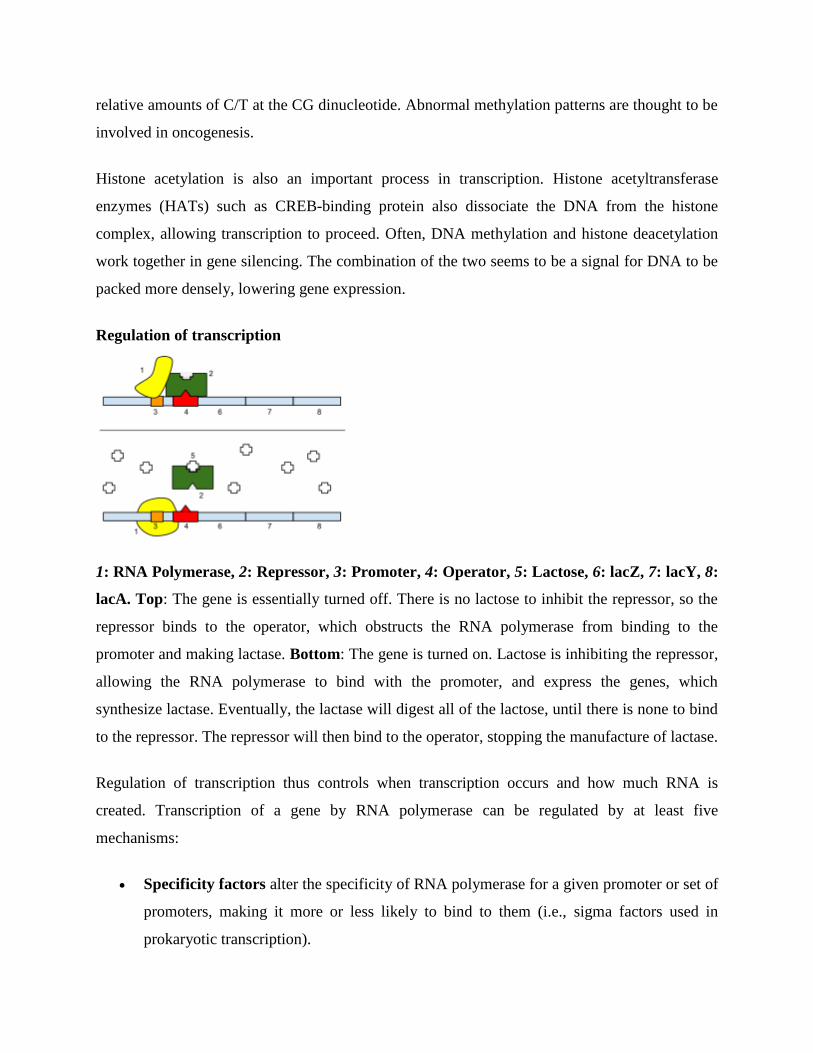
relative amounts of C/T at the CG dinucleotide. Abnormal methylation patterns are thought to be
involved in oncogenesis.
Histone acetylation is also an important process in transcription. Histone acetyltransferase
enzymes (HATs) such as CREB-binding protein also dissociate the DNA from the histone
complex, allowing transcription to proceed. Often, DNA methylation and histone deacetylation
work together in gene silencing. The combination of the two seems to be a signal for DNA to be
packed more densely, lowering gene expression.
Regulation of transcription
1: RNA Polymerase, 2: Repressor, 3: Promoter, 4: Operator, 5: Lactose, 6: lacZ, 7: lacY, 8:
lacA. Top: The gene is essentially turned off. There is no lactose to inhibit the repressor, so the
repressor binds to the operator, which obstructs the RNA polymerase from binding to the
promoter and making lactase. Bottom: The gene is turned on. Lactose is inhibiting the repressor,
allowing the RNA polymerase to bind with the promoter, and express the genes, which
synthesize lactase. Eventually, the lactase will digest all of the lactose, until there is none to bind
to the repressor. The repressor will then bind to the operator, stopping the manufacture of lactase.
Regulation of transcription thus controls when transcription occurs and how much RNA is
created. Transcription of a gene by RNA polymerase can be regulated by at least five
mechanisms:
Specificity factors alter the specificity of RNA polymerase for a given promoter or set of
promoters, making it more or less likely to bind to them (i.e., sigma factors used in
prokaryotic transcription).
Repressors bind to the Operator, coding sequences on the DNA strand that are close to
or overlapping the promoter region, impeding RNA polymerase's progress along the
strand, thus impeding the expression of the gene.The image to the right demonstrates
regulation by a repressor in the lac operon.
General transcription factors position RNA polymerase at the start of a protein-coding
sequence and then release the polymerase to transcribe the mRNA.
Activators enhance the interaction between RNA polymerase and a particular promoter,
encouraging the expression of the gene. Activators do this by increasing the attraction of
RNA polymerase for the promoter, through interactions with subunits of the RNA
polymerase or indirectly by changing the structure of the DNA.
Enhancers are sites on the DNA helix that are bound by activators in order to loop the
DNA bringing a specific promoter to the initiation complex. Enhancers are much more
common in eukaryotes than prokaryotes, where only a few examples exist (to date).
Silencers are regions of DNA sequences that, when bound by particular transcription
factors, can silence expression of the gene.
Post-transcriptional regulation
After the DNA is transcribed and mRNA is formed, there must be some sort of regulation on
how much the mRNA is translated into proteins. Cells do this by modulating the capping,
splicing, addition of a Poly(A) Tail, the sequence-specific nuclear export rates, and, in several
contexts, sequestration of the RNA transcript. These processes occur in eukaryotes but not in
prokaryotes. This modulation is a result of a protein or transcript that, in turn, is regulated and
may have an affinity for certain sequences.
Three prime untranslated regions and microRNAs
Three prime untranslated regions (3'-UTRs) of messenger RNAs (mRNAs) often contain
regulatory sequences that post-transcriptionally influence gene expression. Such 3'-UTRs often
contain both binding sites for microRNAs (miRNAs) as well as for regulatory proteins. By
binding to specific sites within the 3'-UTR, miRNAs can decrease gene expression of various
mRNAs by either inhibiting translation or directly causing degradation of the transcript. The 3'-
UTR also may have silencer regions that bind repressor proteins that inhibit the expression of a
mRNA.
The 3'-UTR often contains miRNA response elements (MREs). MREs are sequences to which
miRNAs bind. These are prevalent motifs within 3'-UTRs. Among all regulatory motifs within
the 3'-UTRs (e.g. including silencer regions), MREs make up about half of the motifs.
As of 2014, the miRBase web site, an archive of miRNA sequences and annotations, listed
28,645 entries in 233 biologic species. Of these, 1,881 miRNAs were in annotated human
miRNA loci. miRNAs were predicted to have an average of about four hundred target mRNAs
(affecting expression of several hundred genes). Freidman et al. estimate that >45,000 miRNA
target sites within human mRNA 3'-UTRs are conserved above background levels, and >60% of
human protein-coding genes have been under selective pressure to maintain pairing to miRNAs.
Direct experiments show that a single miRNA can reduce the stability of hundreds of unique
mRNAs.[7]
Other experiments show that a single miRNA may repress the production of hundreds
of proteins, but that this repression often is relatively mild (less than 2-fold).
The effects of miRNA dysregulation of gene expression seem to be important in cancer. For
instance, in gastrointestinal cancers, a 2015 paper identified nine miRNAs as epigenetically
altered and effective in down-regulating DNA repair enzymes.
The effects of miRNA dysregulation of gene expression also seem to be important in
neuropsychiatric disorders, such as schizophrenia, bipolar disorder, major depressive disorder,
Parkinson's disease, Alzheimer's disease and autism spectrum disorders.[12][13][14]
Regulation of translation
The translation of mRNA can also be controlled by a number of mechanisms, mostly at the level
of initiation. Recruitment of the small ribosomal subunit can indeed be modulated by mRNA
secondary structure, antisense RNA binding, or protein binding. In both prokaryotes and
eukaryotes, a large number of RNA binding proteins exist, which often are directed to their
target sequence by the secondary structure of the transcript, which may change depending on
certain conditions, such as temperature or presence of a ligand (aptamer). Some transcripts act as
ribozymes and self-regulate their expression.
Examples of gene regulation
Enzyme induction is a process in which a molecule (e.g., a drug) induces (i.e., initiates or
enhances) the expression of an enzyme.
The induction of heat shock proteins in the fruit fly Drosophila melanogaster.
The Lac operon is an interesting example of how gene expression can be regulated.
Viruses, despite having only a few genes, possess mechanisms to regulate their gene
expression, typically into an early and late phase, using collinear systems regulated by
anti-terminators (lambda phage) or splicing modulators (HIV).
GAL4 is a transcriptional activator that controls the expression of GAL1, GAL7, and
GAL10 (all of which code for the metabolic of galactose in yeast). The GAL4/UAS
system has been used in a variety of organisms across various phyla to study gene
expression.
Developmental biology
Main article: morphogen
A large number of studied regulatory systems come from developmental biology. Examples
include:
The colinearity of the Hox gene cluster with their nested antero-posterior patterning
It has been speculated that pattern generation of the hand (digits - interdigits) The
gradient of Sonic hedgehog (secreted inducing factor) from the zone of polarizing activity
in the limb, which creates a gradient of active Gli3, which activates Gremlin, which
inhibits BMPs also secreted in the limb, resulting in the formation of an alternating
pattern of activity as a result of this reaction-diffusion system.
Somitogenesis is the creation of segments (somites) from a uniform tissue (Pre-somitic
Mesoderm, PSM). They are formed sequentially from anterior to posterior. This is
achieved in amniotes possibly by means of two opposing gradients, Retinoic acid in the
anterior (wavefront) and Wnt and Fgf in the posterior, coupled to an oscillating pattern
(segmentation clock) composed of FGF + Notch and Wnt in antiphase.
Sex determination in the soma of a Drosophila requires the sensing of the ratio of
autosomal genes to sex chromosome-encoded genes, which results in the production of
sexless splicing factor in females, resulting in the female isoform of doublesex.
Circuitry
Up-regulation and down-regulation
Up-regulation is a process that occurs within a cell triggered by a signal (originating internal or
external to the cell), which results in increased expression of one or more genes and as a result
the protein(s) encoded by those genes. On the converse, down-regulation is a process resulting
in decreased gene and corresponding protein expression.
Up-regulation occurs, for example, when a cell is deficient in some kind of receptor. In
this case, more receptor protein is synthesized and transported to the membrane of the
cell and, thus, the sensitivity of the cell is brought back to normal, reestablishing
homeostasis.
Down-regulation occurs, for example, when a cell is overstimulated by a
neurotransmitter, hormone, or drug for a prolonged period of time, and the expression of
the receptor protein is decreased in order to protect the cell (see also tachyphylaxis).
Inducible vs. repressible systems
Gene Regulation can be summarized by the response of the respective system:
Inducible systems - An inducible system is off unless there is the presence of some
molecule (called an inducer) that allows for gene expression. The molecule is said to
"induce expression". The manner by which this happens is dependent on the control
mechanisms as well as differences between prokaryotic and eukaryotic cells.
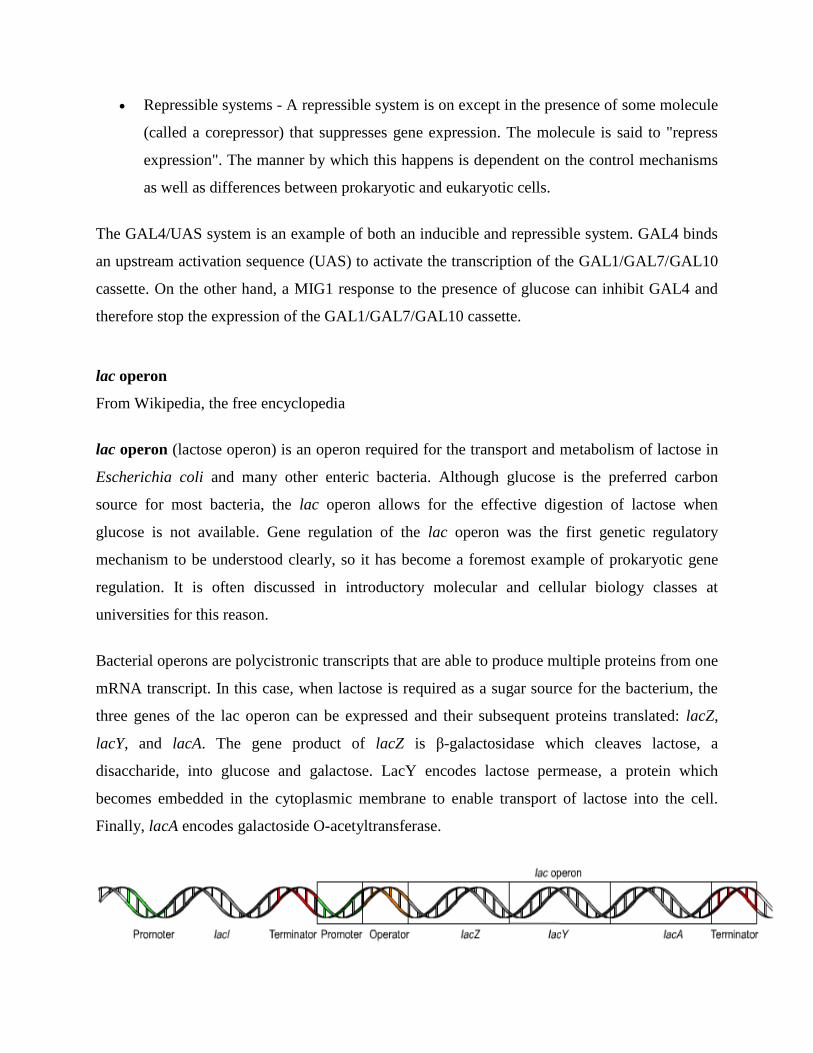
Repressible systems - A repressible system is on except in the presence of some molecule
(called a corepressor) that suppresses gene expression. The molecule is said to "repress
expression". The manner by which this happens is dependent on the control mechanisms
as well as differences between prokaryotic and eukaryotic cells.
The GAL4/UAS system is an example of both an inducible and repressible system. GAL4 binds
an upstream activation sequence (UAS) to activate the transcription of the GAL1/GAL7/GAL10
cassette. On the other hand, a MIG1 response to the presence of glucose can inhibit GAL4 and
therefore stop the expression of the GAL1/GAL7/GAL10 cassette.
lac operon
From Wikipedia, the free encyclopedia
lac operon (lactose operon) is an operon required for the transport and metabolism of lactose in
Escherichia coli and many other enteric bacteria. Although glucose is the preferred carbon
source for most bacteria, the lac operon allows for the effective digestion of lactose when
glucose is not available. Gene regulation of the lac operon was the first genetic regulatory
mechanism to be understood clearly, so it has become a foremost example of prokaryotic gene
regulation. It is often discussed in introductory molecular and cellular biology classes at
universities for this reason.
Bacterial operons are polycistronic transcripts that are able to produce multiple proteins from one
mRNA transcript. In this case, when lactose is required as a sugar source for the bacterium, the
three genes of the lac operon can be expressed and their subsequent proteins translated: lacZ,
lacY, and lacA. The gene product of lacZ is β-galactosidase which cleaves lactose, a
disaccharide, into glucose and galactose. LacY encodes lactose permease, a protein which
becomes embedded in the cytoplasmic membrane to enable transport of lactose into the cell.
Finally, lacA encodes galactoside O-acetyltransferase.
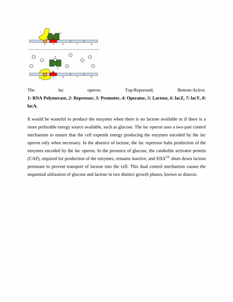
The lac operon. Top:Repressed, Bottom:Active.
1: RNA Polymerase, 2: Repressor, 3: Promoter, 4: Operator, 5: Lactose, 6: lacZ, 7: lacY, 8:
lacA.
It would be wasteful to produce the enzymes when there is no lactose available or if there is a
more preferable energy source available, such as glucose. The lac operon uses a two-part control
mechanism to ensure that the cell expends energy producing the enzymes encoded by the lac
operon only when necessary. In the absence of lactose, the lac repressor halts production of the
enzymes encoded by the lac operon. In the presence of glucose, the catabolite activator protein
(CAP), required for production of the enzymes, remains inactive, and EIIAGlc
shuts down lactose
permease to prevent transport of lactose into the cell. This dual control mechanism causes the
sequential utilization of glucose and lactose in two distinct growth phases, known as diauxie.
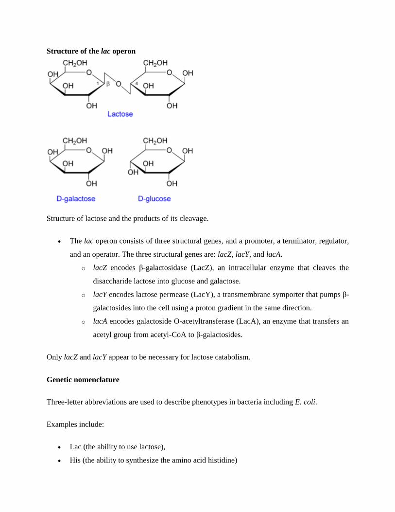
Structure of the lac operon
Structure of lactose and the products of its cleavage.
The lac operon consists of three structural genes, and a promoter, a terminator, regulator,
and an operator. The three structural genes are: lacZ, lacY, and lacA.
o lacZ encodes β-galactosidase (LacZ), an intracellular enzyme that cleaves the
disaccharide lactose into glucose and galactose.
o lacY encodes lactose permease (LacY), a transmembrane symporter that pumps β-
galactosides into the cell using a proton gradient in the same direction.
o lacA encodes galactoside O-acetyltransferase (LacA), an enzyme that transfers an
acetyl group from acetyl-CoA to β-galactosides.
Only lacZ and lacY appear to be necessary for lactose catabolism.
Genetic nomenclature
Three-letter abbreviations are used to describe phenotypes in bacteria including E. coli.
Examples include:
Lac (the ability to use lactose),
His (the ability to synthesize the amino acid histidine)
Mot (swimming motility)
SmR (resistance to the antibiotic streptomycin)
In the case of Lac, wild type cells are Lac+ and are able to use lactose as a carbon and energy
source, while Lac− mutant derivatives cannot use lactose. The same three letters are typically
used (lower-case, italicized) to label the genes involved in a particular phenotype, where each
different gene is additionally distinguished by an extra letter. The lac genes encoding enzymes
are lacZ, lacY, and lacA. The fourth lac gene is lacI, encoding the lactose repressor—"I" stands
for inducibility.
One may distinguish between structural genes encoding enzymes, and regulatory genes encoding
proteins that affect gene expression. Current usage expands the phenotypic nomenclature to
apply to proteins: thus, LacZ is the protein product of the lacZ gene, β-galactosidase. Various
short sequences that are not genes also affect gene expression, including the lac promoter, lac p,
and the lac operator, lac o. Although it is not strictly standard usage, mutations affecting lac o
are referred to as lac oc, for historical reasons.
Regulation
Specific control of the lac genes depends on the availability of the substrate lactose to the
bacterium. The proteins are not produced by the bacterium when lactose is unavailable as a
carbon source. The lac genes are organized into an operon; that is, they are oriented in the same
direction immediately adjacent on the chromosome and are co-transcribed into a single
polycistronic mRNA molecule. Transcription of all genes starts with the binding of the enzyme
RNA polymerase (RNAP), a DNA-binding protein, which binds to a specific DNA binding site,
the promoter, immediately upstream of the genes. Binding of RNA polymerase to the promoter is
aided by the cAMP-bound catabolite activator protein (CAP, also known as the cAMP receptor
protein). However, the lacI gene (regulatory gene for lac operon) produces a protein that blocks
RNAP from binding to the promoter of the operon. This protein can only be removed when
allolactose binds to it, and inactivates it. The protein that is formed by the lacI gene is known as
the lac repressor. The type of regulation that the lac operon undergoes is referred to as negative
inducible, meaning that the gene is turned off by the regulatory factor (lac repressor) unless some
molecule (lactose) is added. Because of the presence of the lac repressor protein, genetic
engineers who replace the lacZ gene with another gene will have to grow the experimental
bacteria on agar with lactose available on it. If they do not, the gene they are trying to express
will not be expressed as the repressor protein is still blocking RNAP from binding to the
promoter and transcribing the gene. Once the repressor is removed, RNAP then proceeds to
transcribe all three genes (lacZYA) into mRNA. Each of the three genes on the mRNA strand has
its own Shine-Dalgarno sequence, so the genes are independently translated. The DNA sequence
of the E. coli lac operon, the lacZYA mRNA, and the lacI genes are available from GenBank.
The first control mechanism is the regulatory response to lactose, which uses an intracellular
regulatory protein called the lactose repressor to hinder production of β-galactosidase in the
absence of lactose. The lacI gene coding for the repressor lies nearby the lac operon and is
always expressed (constitutive). If lactose is missing from the growth medium, the repressor
binds very tightly to a short DNA sequence just downstream of the promoter near the beginning
of lacZ called the lac operator. The repressor binding to the operator interferes with binding of
RNAP to the promoter, and therefore mRNA encoding LacZ and LacY is only made at very low
levels. When cells are grown in the presence of lactose, however, a lactose metabolite called
allolactose, made from lactose by the product of the lacZ gene, binds to the repressor, causing an
allosteric shift. Thus altered, the repressor is unable to bind to the operator, allowing RNAP to
transcribe the lac genes and thereby leading to higher levels of the encoded proteins.
The second control mechanism is a response to glucose, which uses the catabolite activator
protein (CAP) homodimer to greatly increase production of β-galactosidase in the absence of
glucose. Cyclic adenosine monophosphate (cAMP) is a signal molecule whose prevalence is
inversely proportional to that of glucose. It binds to the CAP, which in turn allows the CAP to
bind to the CAP binding site (a 16 bp DNA sequence upstream of the promoter on the left in the
diagram below, about 60 bp upstream of the transcription start site), which assists the RNAP in
binding to the DNA. In the absence of glucose, the cAMP concentration is high and binding of
CAP-cAMP to the DNA significantly increases the production of β-galactosidase, enabling the
cell to hydrolyse lactose and release galactose and glucose.
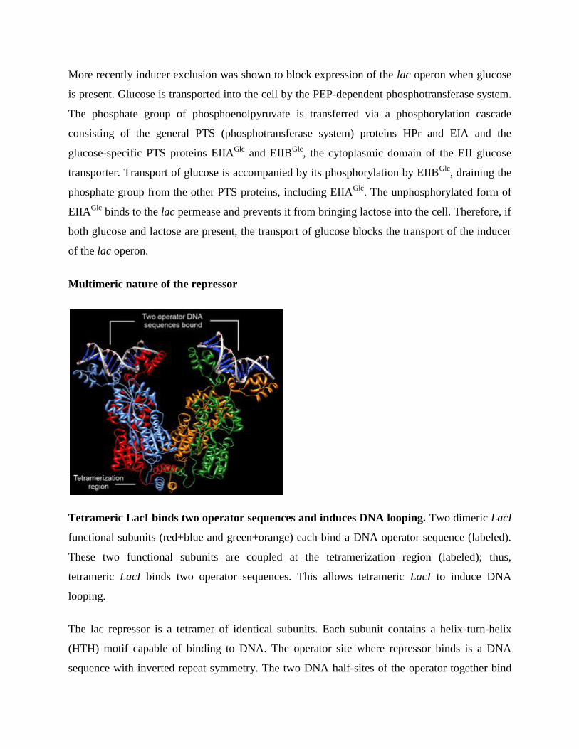
More recently inducer exclusion was shown to block expression of the lac operon when glucose
is present. Glucose is transported into the cell by the PEP-dependent phosphotransferase system.
The phosphate group of phosphoenolpyruvate is transferred via a phosphorylation cascade
consisting of the general PTS (phosphotransferase system) proteins HPr and EIA and the
glucose-specific PTS proteins EIIAGlc
and EIIBGlc
, the cytoplasmic domain of the EII glucose
transporter. Transport of glucose is accompanied by its phosphorylation by EIIBGlc
, draining the
phosphate group from the other PTS proteins, including EIIAGlc
. The unphosphorylated form of
EIIAGlc
binds to the lac permease and prevents it from bringing lactose into the cell. Therefore, if
both glucose and lactose are present, the transport of glucose blocks the transport of the inducer
of the lac operon.
Multimeric nature of the repressor
Tetrameric LacI binds two operator sequences and induces DNA looping. Two dimeric LacI
functional subunits (red+blue and green+orange) each bind a DNA operator sequence (labeled).
These two functional subunits are coupled at the tetramerization region (labeled); thus,
tetrameric LacI binds two operator sequences. This allows tetrameric LacI to induce DNA
looping.
The lac repressor is a tetramer of identical subunits. Each subunit contains a helix-turn-helix
(HTH) motif capable of binding to DNA. The operator site where repressor binds is a DNA
sequence with inverted repeat symmetry. The two DNA half-sites of the operator together bind
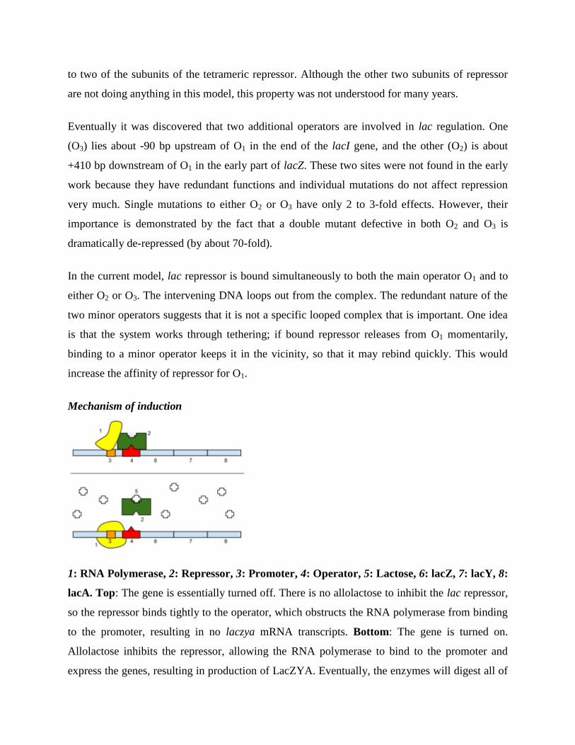
to two of the subunits of the tetrameric repressor. Although the other two subunits of repressor
are not doing anything in this model, this property was not understood for many years.
Eventually it was discovered that two additional operators are involved in lac regulation. One
(O3) lies about -90 bp upstream of O1 in the end of the lacI gene, and the other (O2) is about
+410 bp downstream of O1 in the early part of lacZ. These two sites were not found in the early
work because they have redundant functions and individual mutations do not affect repression
very much. Single mutations to either O2 or O3 have only 2 to 3-fold effects. However, their
importance is demonstrated by the fact that a double mutant defective in both O2 and O3 is
dramatically de-repressed (by about 70-fold).
In the current model, lac repressor is bound simultaneously to both the main operator O1 and to
either O2 or O3. The intervening DNA loops out from the complex. The redundant nature of the
two minor operators suggests that it is not a specific looped complex that is important. One idea
is that the system works through tethering; if bound repressor releases from O1 momentarily,
binding to a minor operator keeps it in the vicinity, so that it may rebind quickly. This would
increase the affinity of repressor for O1.
Mechanism of induction
1: RNA Polymerase, 2: Repressor, 3: Promoter, 4: Operator, 5: Lactose, 6: lacZ, 7: lacY, 8:
lacA. Top: The gene is essentially turned off. There is no allolactose to inhibit the lac repressor,
so the repressor binds tightly to the operator, which obstructs the RNA polymerase from binding
to the promoter, resulting in no laczya mRNA transcripts. Bottom: The gene is turned on.
Allolactose inhibits the repressor, allowing the RNA polymerase to bind to the promoter and
express the genes, resulting in production of LacZYA. Eventually, the enzymes will digest all of
the lactose, until there is no allolactose that can bind to the repressor. The repressor will then
bind to the operator, stopping the transcription of the LacZYA genes.
The repressor is an allosteric protein, i.e. it can assume either one of two slightly different
shapes, which are in equilibrium with each other. In one form the repressor will bind to the
operator DNA with high specificity, and in the other form it has lost its specificity. According to
the classical model of induction, binding of the inducer, either allolactose or IPTG, to the
repressor affects the distribution of repressor between the two shapes. Thus, repressor with
inducer bound is stabilized in the non-DNA-binding conformation. However, this simple model
cannot be the whole story, because repressor is bound quite stably to DNA, yet it is released
rapidly by addition of inducer. Therefore it seems clear that inducer can also bind to the
repressor when the repressor is already bound to DNA. It is still not entirely known what the
exact mechanism of binding is.
Role of non-specific binding
Non-specific binding of the repressor to DNA plays a crucial role in the repression and induction
of the Lac-operon. The specific binding site for the Lac-repressor protein is the operator. The
non-specific interaction is mediated mainly by charge-charge interactions while binding to the
operator is reinforced by hydrophobic interactions. Additionally, there is an abundance of non-
specific DNA sequences to which the repressor can bind. Essentially, any sequence that is not
the operator, is considered non-specific. Studies have shown, that without the presence of non-
specific binding, induction (or unrepression) of the Lac-operon could not occur even with
saturated levels of inducer. It had been demonstrated that, without non-specific binding, the basal
level of induction is ten thousand times smaller than observed normally. This is because the non-
specific DNA acts as sort of a "sink" for the repressor proteins, distracting them from the
operator. The non-specific sequences decrease the amount of available repressor in the cell. This
in turn reduces the amount of inducer required to unrepress the system.
Lactose analogs
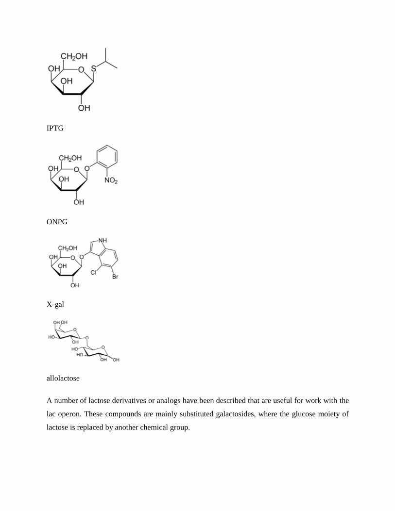
IPTG
ONPG
X-gal
allolactose
A number of lactose derivatives or analogs have been described that are useful for work with the
lac operon. These compounds are mainly substituted galactosides, where the glucose moiety of
lactose is replaced by another chemical group.
Isopropyl-β-D-thiogalactoside (IPTG) is frequently used as an inducer of the lac operon
for physiological work.[1]
IPTG binds to repressor and inactivates it, but is not a substrate
for β-galactosidase. One advantage of IPTG for in vivo studies is that since it cannot be
metabolized by E. coli its concentration remains constant and the rate of expression of lac
p/o-controlled genes, is not a variable in the experiment. IPTG intake is dependent on the
action of lactose permease in P. fluorescens, but not in E. coli.
Phenyl-β-D-galactose (phenyl-Gal) is a substrate for β-galactosidase, but does not
inactivate repressor and so is not an inducer. Since wild type cells produce very little β-
galactosidase, they cannot grow on phenyl-Gal as a carbon and energy source. Mutants
lacking repressor are able to grow on phenyl-Gal. Thus, minimal medium containing only
phenyl-Gal as a source of carbon and energy is selective for repressor mutants and
operator mutants. If 108 cells of a wild type strain are plated on agar plates containing
phenyl-Gal, the rare colonies which grow are mainly spontaneous mutants affecting the
repressor. The relative distribution of repressor and operator mutants is affected by the
target size. Since the lacI gene encoding repressor is about 50 times larger than the
operator, repressor mutants predominate in the selection.
Other compounds serve as colorful indicators of β-galactosidase activity.
o ONPG is cleaved to produce the intensely yellow compound, orthonitrophenol,
and is commonly used as a substrate for assay of β-galactosidase in vitro.
o Colonies that produce β-galactosidase are turned blue by X-gal (5-bromo-4-
chloro-3-indolyl-β-D-galactoside).
Allolactose is an isomer of lactose and is the inducer of the lac operon. Lactose is
galactose-(β1->4)-glucose, whereas allolactose is galactose-(β1->6)-glucose. Lactose is
converted to allolactose by β-galactosidase in an alternative reaction to the hydrolytic
one. A physiological experiment which demonstrates the role of LacZ in production of
the "true" inducer in E. coli cells is the observation that a null mutant of lacZ can still
produce LacY permease when grown with IPTG but not when grown with lactose. The
explanation is that processing of lactose to allolactose (catalyzed by β-galactosidase) is
needed to produce the inducer inside the cell.
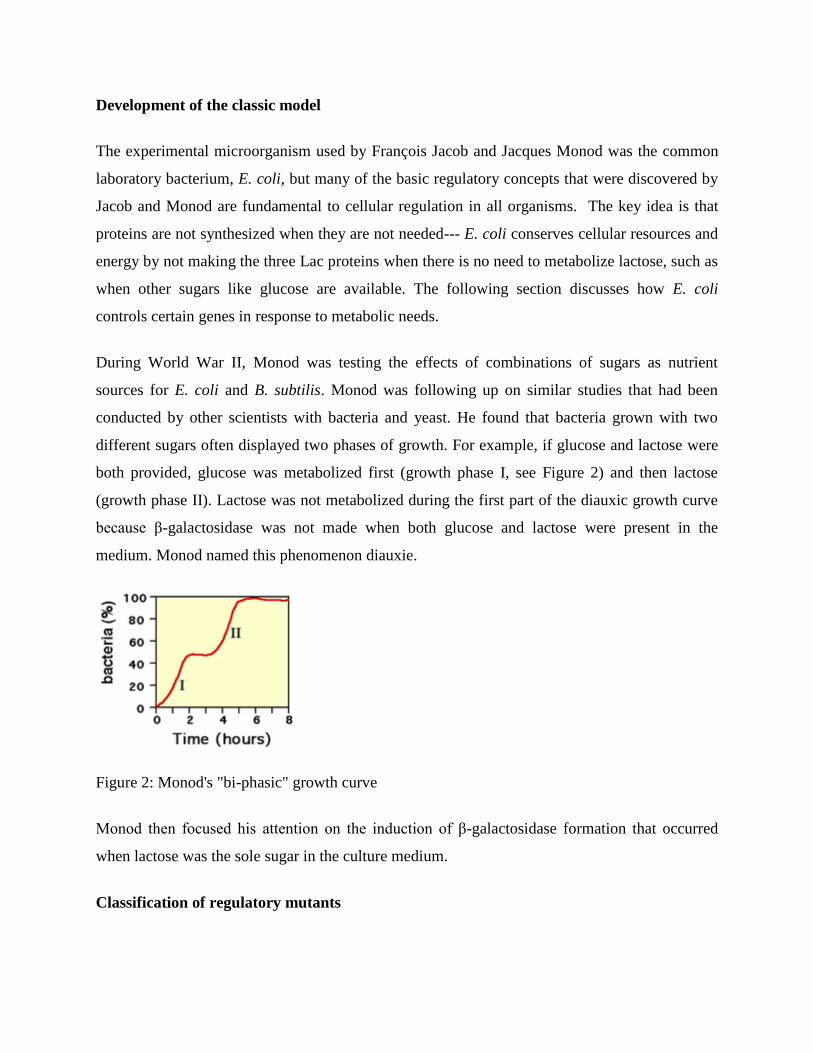
Development of the classic model
The experimental microorganism used by François Jacob and Jacques Monod was the common
laboratory bacterium, E. coli, but many of the basic regulatory concepts that were discovered by
Jacob and Monod are fundamental to cellular regulation in all organisms. The key idea is that
proteins are not synthesized when they are not needed--- E. coli conserves cellular resources and
energy by not making the three Lac proteins when there is no need to metabolize lactose, such as
when other sugars like glucose are available. The following section discusses how E. coli
controls certain genes in response to metabolic needs.
During World War II, Monod was testing the effects of combinations of sugars as nutrient
sources for E. coli and B. subtilis. Monod was following up on similar studies that had been
conducted by other scientists with bacteria and yeast. He found that bacteria grown with two
different sugars often displayed two phases of growth. For example, if glucose and lactose were
both provided, glucose was metabolized first (growth phase I, see Figure 2) and then lactose
(growth phase II). Lactose was not metabolized during the first part of the diauxic growth curve
because β-galactosidase was not made when both glucose and lactose were present in the
medium. Monod named this phenomenon diauxie.
Figure 2: Monod's "bi-phasic" growth curve
Monod then focused his attention on the induction of β-galactosidase formation that occurred
when lactose was the sole sugar in the culture medium.
Classification of regulatory mutants
A conceptual breakthrough of Jacob and Monod was to recognize the distinction between
regulatory substances and sites where they act to change gene expression. A former soldier,
Jacob used the analogy of a bomber that would release its lethal cargo upon receipt of a special
radio transmission or signal. A working system requires both a ground transmitter and a receiver
in the airplane. Now, suppose that the usual transmitter is broken. This system can be made to
work by introduction of a second, functional transmitter. In contrast, he said, consider a bomber
with a defective receiver. The behavior of this bomber cannot be changed by introduction of a
second, functional aeroplane.
To analyze regulatory mutants of the lac operon, Jacob developed a system by which a second
copy of the lac genes (lacI with its promoter, and lacZYA with promoter and operator) could be
introduced into a single cell. A culture of such bacteria, which are diploid for the lac genes but
otherwise normal, is then tested for the regulatory phenotype. In particular, it is determined
whether LacZ and LacY are made even in the absence of IPTG (due to the lactose repressor
produced by the mutant gene being non-functional). This experiment, in which genes or gene
clusters are tested pairwise, is called a complementation test.
This test is illustrated in the figure (lacA is omitted for simplicity). First, certain haploid states
are shown (i.e. the cell carries only a single copy of the lac genes). Panel (a) shows repression,
(b) shows induction by IPTG, and (c) and (d) show the effect of a mutation to the lacI gene or to
the operator, respectively. In panel (e) the complementation test for repressor is shown. If one
copy of the lac genes carries a mutation in lacI, but the second copy is wild type for lacI, the
resulting phenotype is normal---but lacZ is expressed when exposed to inducer IPTG. Mutations
affecting repressor are said to be recessive to wild type (and that wild type is dominant), and this
is explained by the fact that repressor is a small protein which can diffuse in the cell. The copy of
the lac operon adjacent to the defective lacI gene is effectively shut off by protein produced from
the second copy of lacI.
If the same experiment is carried out using an operator mutation, a different result is obtained
(panel (f)). The phenotype of a cell carrying one mutant and one wild type operator site is that
LacZ and LacY are produced even in the absence of the inducer IPTG; because the damaged
operator site, does not permit binding of the repressor to inhibit transcription of the structural
genes. The operator mutation is dominant. When the operator site where repressor must bind is
damaged by mutation, the presence of a second functional site in the same cell makes no
difference to expression of genes controlled by the mutant site.
A more sophisticated version of this experiment uses marked operons to distinguish between the
two copies of the lac genes and show that the unregulated structural gene(s) is(are) the one(s)
next to the mutant operator (panel (g). For example, suppose that one copy is marked by a
mutation inactivating lacZ so that it can only produce the LacY protein, while the second copy
carries a mutation affecting lacY and can only produce LacZ. In this version, only the copy of the
lac operon that is adjacent to the mutant operator is expressed without IPTG. We say that the
operator mutation is cis-dominant, it is dominant to wild type but affects only the copy of the
operon which is immediately adjacent to it.
This explanation is misleading in an important sense, because it proceeds from a description of
the experiment and then explains the results in terms of a model. But in fact, it is often true that
the model comes first, and an experiment is fashioned specifically to test the model. Jacob and
Monod first imagined that there must be a site in DNA with the properties of the operator, and
then designed their complementation tests to show this.
The dominance of operator mutants also suggests a procedure to select them specifically. If
regulatory mutants are selected from a culture of wild type using phenyl-Gal, as described above,
operator mutations are rare compared to repressor mutants because the target-size is so small.
But if instead we start with a strain which carries two copies of the whole lac region (that is
diploid for lac), the repressor mutations (which still occur) are not recovered because
complementation by the second, wild type lacI gene confers a wild type phenotype. In contrast,
mutation of one copy of the operator confers a mutant phenotype because it is dominant to the
second, wild type copy.
Regulation by cyclic AMP
Explanation of diauxie depended on the characterization of additional mutations affecting the lac
genes other than those explained by the classical model. Two other genes, cya and crp,
subsequently were identified that mapped far from lac, and that, when mutated, result in a
decreased level of expression in the presence of IPTG and even in strains of the bacterium
lacking the repressor or operator. The discovery of cAMP in E. coli led to the demonstration that
mutants defective the cya gene but not the crp gene could be restored to full activity by the
addition of cAMP to the medium.
The cya gene encodes adenylate cyclase, which produces cAMP. In a cya mutant, the absence of
cAMP makes the expression of the lacZYA genes more than ten times lower than normal.
Addition of cAMP corrects the low Lac expression characteristic of cya mutants. The second
gene, crp, encodes a protein called catabolite activator protein (CAP) or cAMP receptor protein
(CRP).
However the lactose metabolism enzymes are made in small quantities in the presence of both
glucose and lactose (sometimes called leaky expression) due to the fact that the LacI repressor
rapidly associates/dissociates from the DNA rather than tightly binding to it, which can allow
time for RNAP to bind and transcribe mRNAs of lacZYA. Leaky expression is necessary in order
to allow for metabolism of some lactose after the glucose source is expended, but before lac
expression is fully activated.
In summary:
When lactose is absent then there is very little Lac enzyme production (the operator has
Lac repressor bound to it).
When lactose is present but a preferred carbon source (like glucose) is also present then a
small amount of enzyme is produced (Lac repressor is not bound to the operator).
When glucose is absent, CAP-cAMP binds to a specific DNA site upstream of the
promoter and makes a direct protein-protein interaction with RNAP that facilitates the
binding of RNAP to the promoter.
The delay between growth phases reflects the time needed to produce sufficient quantities of
lactose-metabolizing enzymes. First, the CAP regulatory protein has to assemble on the lac
promoter, resulting in an increase in the production of lac mRNA. More available copies of the
lac mRNA results in the production (see translation) of significantly more copies of LacZ (β-
galactosidase, for lactose metabolism) and LacY (lactose permease to transport lactose into the
cell). After a delay needed to increase the level of the lactose metabolizing enzymes, the bacteria
enter into a new rapid phase of cell growth.
The diagram below summarizes these statements.
lac operon in detail
Two puzzles of catabolite repression relate to how cAMP levels are coupled to the presence of
glucose, and secondly, why the cells should even bother. After lactose is cleaved it actually
forms glucose and galactose (easily converted to glucose). In metabolic terms, lactose is just as
good a carbon and energy source as glucose. The cAMP level is related not to intracellular
glucose concentration but to the rate of glucose transport, which influences the activity of
adenylate cyclase. (In addition, glucose transport also leads to direct inhibition of the lactose
permease.) As to why E. coli works this way, one can only speculate. All enteric bacteria ferment
glucose, which suggests they encounter it frequently. It is possible that a small difference in
efficiency of transport or metabolism of glucose v. lactose makes it advantageous for cells to
regulate the lac operon in this way.
Use in molecular biology
The lac gene and its derivatives are amenable to use as a reporter gene in a number of bacterial-
based selection techniques such as two hybrid analysis, in which the successful binding of a
transcriptional activator to a specific promoter sequence must be determined. In LB plates
containing X-gal, the colour change from white colonies to a shade of blue corresponds to about
20-100 β-galactosidase units, while tetrazolium lactose and MacConkey lactose media have a
range of 100-1000 units, being most sensitive in the high and low parts of this range respectively.
Since MacConkey lactose and tetrazolium lactose media both rely on the products of lactose
breakdown, they require the presence of both lacZ and lacY genes. The many lac fusion
techniques which include only the lacZ gene are thus suited to the X-gal plates or ONPG liquid
broths
Catabolite repression
Carbon catabolite repression, or simply catabolite repression, is an important part of global
control system of various bacteria and other micro-organisms. Catabolite repression allows
micro-organisms to adapt quickly to a preferred (rapidly metabolisable) carbon and energy
source first. This is usually achieved through inhibition of synthesis of enzymes involved in
catabolism of carbon sources other than the preferred one. The catabolite repression was first
shown to be initiated by glucose and therefore sometimes referred to as the glucose effect.
However, the term "glucose effect" is actually a misnomer since other carbon sources are known
to induce catabolite repression.
Escherichia coli
Catabolite repression was extensively studied in Escherichia coli. E. coli grows faster on glucose
than on any other carbon source. For example, if E. coli is placed on an agar plate containing
only glucose and lactose, the bacteria will use glucose first and lactose second. When glucose is
available in the environment, the synthesis of β-galactosidase is under repression due to the
effect of catabolite repression caused by glucose. The catabolite repression in this case is
achieved through the utilization of phosphotransferase system.
An important enzyme from the phosphotranferase system called Enzyme II A (EIIA) plays a
central role in this mechanism. There are different catabolite-specific EIIA in a single cell, even
though different bacterial groups have specificities to different sets of catabolites. In enteric
bacteria one of the EIIA enzymes in their set is specific for glucose transport only. When
glucose levels are high inside the bacteria, EIIA mostly exists in its unphosphorylated form. This
leads to inhibition of adenylyl cyclase and lactose permease, therefore cAMP levels are low and
lactose can not be transported inside the bacteria. After some time, the glucose is all used up and
the second preferred carbon source (i.e. lactose) has to be used by bacteria. Absence of glucose
will "turn off" catabolite repression.
Furthermore, when glucose levels are low the phosphorylated form of EIIA accumulates and
consequently activates the enzyme adenylyl cyclase, which will produce high levels of cAMP.
cAMP binds to catabolite activator protein (CAP) and together they will bind to a promoter
sequence on the lac operon. However, this is not enough for the lactose genes to be transcribed.
Lactose must be present inside the cell to remove the lactose repressor from the operator
sequence (transcriptional regulation). When these two conditions are satisfied, it means for the
bacteria that glucose is absent and lactose is available. Next, bacteria start to transcribe lactose
gene and produce β-galactosidase enzymes for lactose metabolism. The example above is a
simplification of a complex process. Catabolite repression is considered to be a part of global
control system and therefore it affects more genes rather than just lactose gene transcription.
Bacillus subtilis
Gram positive bacteria such as Bacillus subtilis have a cAMP-independent catabolite repression
mechanism controlled by catabolite control protein A (CcpA). In this alternative pathway CcpA
negatively represses other sugar operons so they are off in the presence of glucose. It works by
the fact that Hpr is phosphorylated by a specific mechanism, when glucose enters through the
cell membrane protein EIIC, and when Hpr is phosphoralated it can then allow CcpA to block
transcription of the alternative sugar pathway operons at their respective cre sequence binding
sites. Note that E. coli has a similar cAMP-independent catabolite repression mechanism that
utilizes a protein called catabolite repressor activator (Cra).
gal operon
The gal operon is a prokaryotic operon, which encodes enzymes necessary for galactose
metabolism. The operon contains two operators, OE (for external) and OI. The former is just
before the promoter, and the latter is just after the galE gene (the first gene in the operon).
Repression of gene expression works via binding of repressor molecules to the two operators.
These repressors dimerize, creating a loop in the DNA. The loop as well as hindrance from the
external operator prevent RNA polymerase from binding to the promoter, and thus prevent
transcription.
The gal operon of E. coli consists of 3 structural genes: galE (epimerase), galT (galactose
transferase), and galK (galactokinase), which are transcribed from two overlapping promoters
PG1 and PG2 upstream from galE. Regulation of the operon is complex since the GalE product,
an epimerase that converts UDP-glucose into UDP-galactose, is required for the formation of
UDP-galactose for cell wall biosynthesis, in particular the cell wall component
lipopolysaccharide, even when cells are not using galactose as a carbon/energy source.
The gal operon is controlled by CRP-cAMP as for the lac operon. CRP-cAMP binds to -35
region promoting transcription from PG1 but inhibiting transcription from PG2. When cells are
grown in glucose, basal level transcription occurs from PG2. The unlinked galR gene encodes
the repressor for this system. A tetrameric GalR repressor binds to 2 operators, one located at
+55 and one located at -60 relative to the PG1 start site. Looping of the DNA blocks the access
of RNA polymerase to promoters and/or inhibits formation of the open complex. When GalR
binds as a dimer to the -60 site only, promoter PG2 is activated, not repressed, allowing basal
levels of GalE to be produced. In this state promoter PG1 is inactivated through interactions with
the alpha subunit of RNA polymerase
trp operon
From Wikipedia, the free encyclopedia
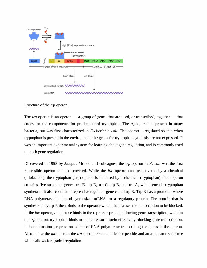
Structure of the trp operon.
The trp operon is an operon — a group of genes that are used, or transcribed, together — that
codes for the components for production of tryptophan. The trp operon is present in many
bacteria, but was first characterized in Escherichia coli. The operon is regulated so that when
tryptophan is present in the environment, the genes for tryptophan synthesis are not expressed. It
was an important experimental system for learning about gene regulation, and is commonly used
to teach gene regulation.
Discovered in 1953 by Jacques Monod and colleagues, the trp operon in E. coli was the first
repressible operon to be discovered. While the lac operon can be activated by a chemical
(allolactose), the tryptophan (Trp) operon is inhibited by a chemical (tryptophan). This operon
contains five structural genes: trp E, trp D, trp C, trp B, and trp A, which encode tryptophan
synthetase. It also contains a repressive regulator gene called trp R. Trp R has a promoter where
RNA polymerase binds and synthesizes mRNA for a regulatory protein. The protein that is
synthesized by trp R then binds to the operator which then causes the transcription to be blocked.
In the lac operon, allolactose binds to the repressor protein, allowing gene transcription, while in
the trp operon, tryptophan binds to the repressor protein effectively blocking gene transcription.
In both situations, repression is that of RNA polymerase transcribing the genes in the operon.
Also unlike the lac operon, the trp operon contains a leader peptide and an attenuator sequence
which allows for graded regulation.
It is an example of repressible negative regulation of gene expression. Within the operon's
regulatory sequence, the operator is blocked by the repressor protein in the presence of
tryptophan (thereby preventing transcription) and is liberated in tryptophan's absence (thereby
allowing transcription). The process of attenuation (explained below) complements this
regulatory action.
Repression
The operon operates by a negative repressible feedback mechanism. The repressor for the trp
operon is produced upstream by the trpR gene, which is constitutively expressed at a low level.
Synthesized TrpR monomers associate into tetramers. These tetramers are inactive and are
dissolved in the nucleoplasm. When tryptophan is present, these tryptophan repressor tetramers
bind to tryptophan, causing a change in the repressor conformation, allowing the repressor to
bind to the operator. This prevents RNA polymerase from binding to and transcribing the operon,
so tryptophan is not produced from its precursor. When tryptophan is not present, the repressor is
in its inactive conformation and cannot bind the operator region, so transcription is not inhibited
by the repressor.
Attenuation
Mechanism of transcriptional attenuation of the trp operon.
Attenuation is a second mechanism of negative feedback in the trp operon. The repression
system targets the intracellular trp concentration whereas the attenuation responds to the
concentration of charged tRNAtrp
. Thus, the trpR repressor decreases gene expression by altering
the initiation of transcription, while attenuation does so by altering the process of transcription
that's already in progress. While the TrpR repressor decreases transcription by a factor of 70,
attenuation can further decrease it by a factor of 10, thus allowing accumulated repression of
about 700-fold. Attenuation is made possible by the fact that in prokaryotes (which have no
nucleus), the ribosomes begin translating the mRNA while RNA polymerase is still transcribing
the DNA sequence. This allows the process of translation to affect transcription of the operon
directly.
At the beginning of the transcribed genes of the trp operon is a sequence of at least 130
nucleotides termed the leader transcript (trpL). Lee and Yanofsky (1977) found that the
attenuation efficiency is correlated with the stability of a secondary structure embedded in trpL,
and the 2 constituent hairpins of the terminator structure were later elucidated by Oxender et al.
(1979). This transcript includes four short sequences designated 1-4, each of which is partially
complementary to the next one. Thus, three distinct secondary structures (hairpins) can form: 1-
2, 2-3 or 3-4. The hybridization of sequences 1 and 2 to form the 1-2 structure is rare because the
RNA Polymerase waits for a ribosome to attach before continuing transcription past sequence 1,
however if the 1-2 hairpin were to form it would prevent the formation of the 2-3 structure (but
not 3-4). The formation of a hairpin loop between sequences 2-3 prevents the formation of
hairpin loops between both 1-2 and 3-4. The 3-4 structure is a transcription termination sequence
(abundant in G/C and immediately followed by several uracil residues), once it forms RNA
polymerase will disassociate from the DNA and transcription of the structural genes of the
operon can not occur (see below for a more detailed explanation). The functional importance of
the 2nd hairpin for the transcriptional termination is illustrated by the reduced transcription
termination frequency observed in experiments destabilizing the central G+C pairing of this
hairpin.
Part of the leader transcript codes for a short polypeptide of 14 amino acids, termed the leader
peptide. This peptide contains two adjacent tryptophan residues, which is unusual, since
tryptophan is a fairly uncommon amino acid (about one in a hundred residues in a typical E. coli
protein is tryptophan). The strand 1 in trpL encompasses the region encoding the trailing residues
of the leader peptide: Trp, Trp, Arg, Thr, Ser; conservation is observed in these 5 codons
whereas mutating the upstream codons do not alter the operon expression. If the ribosome
attempts to translate this peptide while tryptophan levels in the cell are low, it will stall at either
of the two trp codons. While it is stalled, the ribosome physically shields sequence 1 of the
transcript, preventing the formation of the 1-2 secondary structure. Sequence 2 is then free to
hybridize with sequence 3 to form the 2-3 structure, which then prevents the formation of the 3-4
termination hairpin, which is why the 2-3 structure is called an anti-termination hairpin. In the
presence of the 2-3 structure, RNA polymerase is free to continue transcribing the operon.
Mutational analysis and studies involving complementary oligonucleotides demonstrate that the
stability of the 2-3 structure corresponds to the operon expression level. If tryptophan levels in
the cell are high, the ribosome will translate the entire leader peptide without interruption and
will only stall during translation termination at the stop codon. At this point the ribosome
physically shields both sequences 1 and 2. Sequences 3 and 4 are thus free to form the 3-4
structure which terminates transcription. This terminator structure forms when no ribosome stalls
in the vicinity of the Trp tandem (i.e. Trp or Arg codon): either the leader peptide is not
translated or the translation proceeds smoothly along the strand 1 with abundant charged
tRNAtrp. More over, the ribosome is proposed to only block about 10 nts downstream, thus
ribosome stalling in either the upstream Gly or further downstream Thr do not seem to affect the
formation of the termination hairpin. The end result is that the operon will be transcribed only
when tryptophan is unavailable for the ribosome, while the trpL transcript is constitutively
expressed.
This attenuation mechanism is experimentally supported. First, the translation of the leader
peptide and ribosomal stalling are directly evidenced to be necessary for inhibiting the
transcription termination. Moreover, mutational analysis destabilizing or disrupting the base-
pairing of the antiterminator hairpin results in increased termination of several folds; consistent
with the attenuation model, this mutation fails to relieve attenuation even with starved Trp. In
contrast, complementary oligonucleotides targeting strand 1 increases the operon expression by
promoting the antiterminator formation. Furthermore, in histidine operon, compensatory
mutation shows that the pairing ability of strands 2-3 matters more than their primary sequence
in inhibiting attenuation.
In attenuation, where the translating ribosome is stalled determines whether the termination
hairpin will be formed. In order for the transcribing polymerase to concomitantly capture the
alternative structure, the time scale of the structural modulation must be comparable to that of the
transcription. To ensure that the ribosome binds and begins translation of the leader transcript
immediately following its synthesis, a pause site exists in the trpL sequence. Upon reaching this
site, RNA polymerase pauses transcription and apparently waits for translation to begin. This
mechanism allows for synchronization of transcription and translation, a key element in
attenuation.
A similar attenuation mechanism regulates the synthesis of histidine, phenylalanine and
threonine.
Chapter # 20 Control of Gene Expression in Prokaryotes
The quintessential example which still stands as the paradigm of transcriptional control is the
Lac Operon, first developed by Jacob and Monod and verified each year faithfully by second
year science students.
Background:
E. coli (we are back with these pesky little coliforms) have the ability to grow in media which
contain lactose as their sole carbon source. Lactose is a sugar found in milk (a disaccharide).
To metabolise this sugar the bugs must produce two enzymes, beta galactosidase and lac
permease. The lac permease allows the lactose to enter the cell. The beta galactosidase cleaves
the bond joining the two monosaccharides (known as a beta galactoside bond, a type of
glycosidic bond). Once the sugar has been cleaved the two monosaccharides can be utilized by
the cell‘s glycolytic ―house keeping‖ enzymes. If the E. coli are grown up on media with other
carbon sources there is very little activity of these two enzymes.
Is this modulation of enzyme activity a transcriptional event or simply an activation/deactivation
of pre-existing enzyme activity? The answer is it is a transcriptional event. To test for this a
protein synthesis inhibitor is included in the incubation. The induction does not occur.
The term induction means that the activity of an enzyme (beta gal in our example) increases after
the addition of a compound, in this case lactose. How does the presence of lactose, as the sole
carbon source, control the level of transcription of the enzymes that catalyse its utilization?
The experiments by Jacob and Monod, working with bacteria containing an extra copy of the
genes(extra chromosomal) for the lac operon, found
that the control of this gene expression had two elements; a cis acting factor and a trans acting
factor. They isolated many mutants of E. coli where the lesion was either on the genomic DNA
or on the extra chromosomal copy. From the analysis of these mutants the lac operon model was
developed.
Mutants of E. coliwhich have lost the ability to control beta gal gene expression tend to fall into
two categories: constitutive high producers and no producers. The high producers have high
levels of beta galactosidase activity, whether lactose is present or not. The no producers have no
activity whether lactose is present or not.
Cis acting factors could only affect gene expression on the same piece of DNA, while trans
acting factors could influence gene expression on other copies of the gene located on separate
pieces of DNA (extra chromosomal) in the cell.
The model proposed contains lac I, a promoter region, an operator region, the transcription unit
which contains lac Z, lac Y and lac A. The gene product of lac I is a protein, known as the lac
repressor. This protein is a tetramer with a high affinity for the operator region of the lac operon.
There are only a few copies in the cell.
The promoter is the region where RNA polymerase binds (at the -10 and -35 regions). The
repressor binds at the operator (-10 to 0).
Lac Z is the structural gene coding for beta galactosidase, lac Y for lac permease and lac A for a
transacetylase of unknown function. These three genes are all transcribed in one long mRNA,
known as a polycistronic mRNA.
The lac operon is under two forms of control; positive and negative control.
A protein is said to exert negative control when its binding prevents an event. The repressor is an
example of negative control. When the repressor is bound to the operator the RNA polymerase
cannot bind to the promoter. No RNA polymerase, no transcription no enzyme activity. Once
lactose enters the cell a small amount of it is converted to allolactose by the few copies of beta
gal present in the induced cell. The allolactose binds to the repressor and results in its
dissociation from the operator.
In the lab we use a compound IPTG which is an analogue of the allolactose and is thus described
as an inducer. A protein exerts positive control when its binding results in an event. The
catabolite activator protein (CAP) or as it is sometimes known, the cAMP receptor protein (CRP)
is an example of positive control. This protein binds to an activation site within the promoter
region only when it is complexed to cAMP. cAMP is a derivative of ATP, synthesized by
adenylyl cyclase and is a second messenger in both eukaryotic and prokaryotic cells. In this case
the significance of cAMP is that its concentration is low when intracellular [glucose] is high and
vice versa. The other important fact is that cAMP reversibly binds to CAP in a concentration
dependent manner.
In summary: When intracellular glucose levels are high cAMP is
low CAP is not associated with cAMP and is not bound to the DNA.
When intracellular [glucose] is low cAMP is high cAMP-CAP is complexed and associated with
the DNA at the activation site. This complex will increase the frequency of initiation of
transcription by RNA polymerase at the lac promoter. This protein complex acts on a number of
operons around the genome and is described as global regulation.
The best way to understand the lac operon is to take a couple of scenarios.
1.Lactose (+) and glucose (-)
2.Lactose (+) and glucose (+)
3.Lactose (-) and glucose (+)
4.Lactose (-) and glucose (-)
In scenario 1 some of the Lactose entering the cell via the few lac permease transporters
available has been converted to allolactose and has resulted in the removal of the repressor from
the operator. The
promoter is now unmasked and RNA polymerase can now bind and initiate transcription.
However it won‘t do this very frequently without the help of the cAMP-CAP bound to the
activation site. This protein complex binding puts a 90o kink in the DNA and interacts with the
alpha subunit of RNA polymerase. Without the cAMP CAP the lac promoter is a weak promoter
varying significantly from the consensus sequence at -10 and -35. The combination of the two
controls means beta gal and lac
permease are transcribed at high levels.
In scenario 2the repressor is off the operator but the CAP (without cAMP) is not bound to the
DNA so initiation only occurs at a low rate
little transcription.
In scenario 3the repressor is bound to the operator and the CAP (without cAMP) is not bound to
the DNA. Very little transcription of the lac operon genes is happening now.
In scenario 4the cell is starving! The repressor is on the operator but the cAMP CAP is on the
DNA. If the repressor is bound there is no transcription RNA polymerase has no access. The
regulation of the lac operon by the repressor is described as specific regulation of gene
expression; the control is only exerted over the three structural genes following the operator. The
CAP-cAMP complex is an example of global regulation; it exerts its influence over a number of
genes scattered throughout the genome. Genes which encode other catabolic enzymes involved
in carbohydrate metabolism e.g. the arabinose operon (arabinose is another alternative sugar to
glucose which E. coli can survive on if they have nothing else) also come under the control of
the CAP-cAMP complex. Again, if glucose is present at a sufficient concentration then it is in
the organism‘s interest to down-regulate the synthesis of genes which catalyse the catabolism of
arabinose. The idea is save the energy and use glucose. The enzymes which catalyse glucose
catabolism are not inducible i.e glycolysis is too fundamental to survival to be switched on and
off.
Cis acting factors: the operator sequence, the promoter sequence, the activation sequence.
Trans acting factors: the repressor, the sigma factor
The trp operon.
The trp operon is another example of specific control of gene expression operating in our friend
E. coli. One of the aspects of this example that makes it different is the genes here are
biosynthetic rather than catabolic.
Background.
E. coli have the ability to synthesise tryptophan (an amino acid) from the compound chorismate.
It requires 5 enzymes to do this; the gene products of genes trpE to trpA.
The trp operon also has a leader sequence which will attenuate trpE – trpA expression at
intermediate [trp]. The bacteria do not want to make the enzymes for tryptophan biosynthesis
when there is plenty of tryptophan around. To control this an operon is in place with a repressor
that binds to the trp operator when trp is bound (the opposite of the lac operon).
The association of the trp with the repressor protein is non-covalent and concentration
dependent. Once the intracellular [trp] concentration falls the trp comes off the repressor and the
repressor dissociates itself from the DNA leaving the promoter region free to bind RNA
polymerase.
The repressor in this case works in the opposite way to the lac repressor. If the trp is present, the
repressor binds, whereas in the lac operon, if the lactose is present the repressor dissociates. This
behaviour is explained by the purpose of both enzymes. Beta galactosidase is a catalytic enzyme.
It is needed when the lactose is present to metabolise the lactose. If glucose is around the bugs
can use that instead and there is no need to make beta gal. In the case of the trp operon the gene
products are biosynthetic enzymes. They do not need to be synthesized if there is enough of the
product they are synthesizing. The trp repressor and the lac repressor are great examples of
proteins that bind to DNA in a base sequence specific manner. Their affinity for DNA is altered
several thousand fold by the binding of a metabolite; lactose or tryptophan. The difference is that
the trp repressor has greater affinity for the operator when the trp is bound, while the lac
repressor has greater affinity when the allolactose is NOT bound.
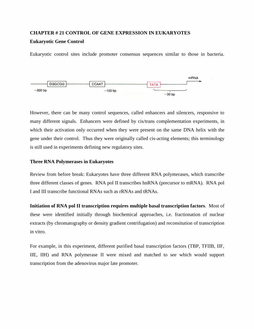
CHAPTER # 21 CONTROL OF GENE EXPRESSION IN EUKARYOTES
Eukaryotic Gene Control
Eukaryotic control sites include promoter consensus sequences similar to those in bacteria.
However, there can be many control sequences, called enhancers and silencers, responsive to
many different signals. Enhancers were defined by cis/trans complementation experiments, in
which their activation only occurred when they were present on the same DNA helix with the
gene under their control. Thus they were originally called cis-acting elements; this terminology
is still used in experiments defining new regulatory sites.
Three RNA Polymerases in Eukaryotes
Review from before break: Eukaryotes have three different RNA polymerases, which transcribe
three different classes of genes. RNA pol II transcribes hnRNA (precursor to mRNA). RNA pol
I and III transcribe functional RNAs such as rRNAs and tRNAs.
Initiation of RNA pol II transcription requires multiple basal transcription factors. Most of
these were identified initially through biochemical approaches, i.e. fractionation of nuclear
extracts (by chromatography or density gradient centrifugation) and reconsitution of transcription
in vitro.
For example, in this experiment, different purified basal transcription factors (TBP, TFIIB, IIF,
IIE, IIH) and RNA polymerase II were mixed and matched to see which would support
transcription from the adenovirus major late promoter.
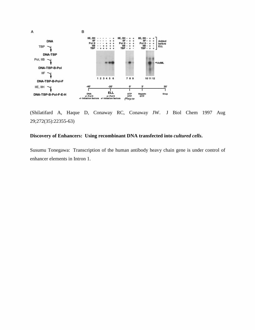
(Shilatifard A, Haque D, Conaway RC, Conaway JW. J Biol Chem 1997 Aug
29;272(35):22355-63)
Discovery of Enhancers: Using recombinant DNA transfected into cultured cells.
Susumu Tonegawa: Transcription of the human antibody heavy chain gene is under control of
enhancer elements in Intron 1.
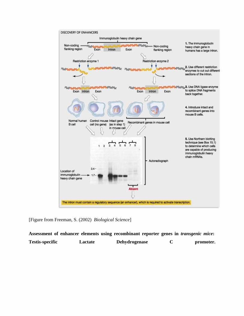
[Figure from Freeman, S. (2002) Biological Science]
Assessment of enhancer elements using recombinant reporter genes in transgenic mice:
Testis-specific Lactate Dehydrogenase C promoter.
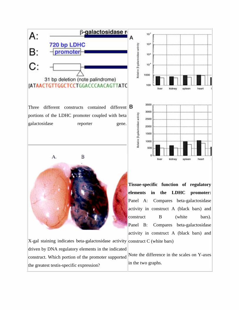
Three different constructs contained different
portions of the LDHC promoter coupled with beta
galactosidase reporter gene.
X-gal staining indicates beta-galactosidase activity
driven by DNA regulatory elements in the indicated
construct. Which portion of the promoter supported
the greatest testis-specific expression?
Tissue-specific function of regulatory
elements in the LDHC promoter:
Panel A: Compares beta-galactosidase
activity in construct A (black bars) and
construct B (white bars).
Panel B: Compares beta-galactosidase
activity in construct A (black bars) and
construct C (white bars)
Note the difference in the scales on Y-axes
in the two graphs.
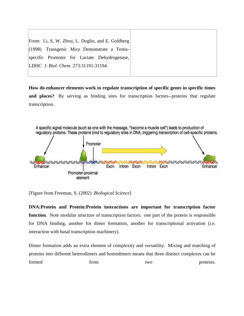
From: Li, S, W. Zhou, L. Doglio, and E. Goldberg
(1998) Transgenic Mice Demonstrate a Testis-
specific Promoter for Lactate Dehydrogenase,
LDHC J. Biol. Chem. 273:31191-31194.
How do enhancer elements work to regulate transcription of specific genes in specific times
and places? By serving as binding sites for transcription factors--proteins that regulate
transcription.
[Figure from Freeman, S. (2002) Biological Science]
DNA:Protein and Protein:Protein interactions are important for transcription factor
function. Note modular structure of transcription factors: one part of the protein is responsible
for DNA binding, another for dimer formation, another for transcriptional activation (i.e.
interaction with basal transcription machinery).
Dimer formation adds an extra element of complexity and versatility. Mixing and matching of
proteins into different heterodimers and homodimers means that three distinct complexes can be
formed from two proteins.
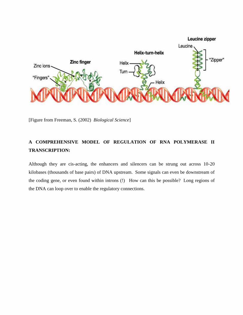
[Figure from Freeman, S. (2002) Biological Science]
A COMPREHENSIVE MODEL OF REGULATION OF RNA POLYMERASE II
TRANSCRIPTION:
Although they are cis-acting, the enhancers and silencers can be strung out across 10-20
kilobases (thousands of base pairs) of DNA upstream. Some signals can even be downstream of
the coding gene, or even found within introns (!) How can this be possible? Long regions of
the DNA can loop over to enable the regulatory connections.
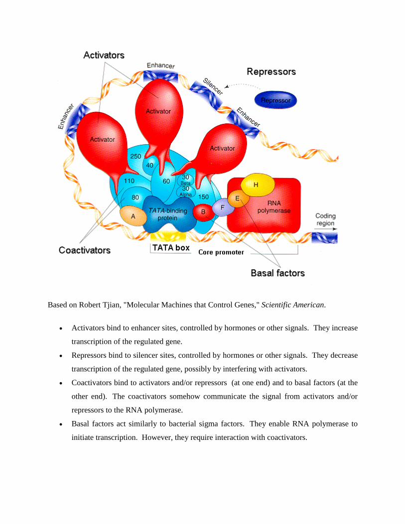
Based on Robert Tjian, "Molecular Machines that Control Genes," Scientific American.
Activators bind to enhancer sites, controlled by hormones or other signals. They increase
transcription of the regulated gene.
Repressors bind to silencer sites, controlled by hormones or other signals. They decrease
transcription of the regulated gene, possibly by interfering with activators.
Coactivators bind to activators and/or repressors (at one end) and to basal factors (at the
other end). The coactivators somehow communicate the signal from activators and/or
repressors to the RNA polymerase.
Basal factors act similarly to bacterial sigma factors. They enable RNA polymerase to
initiate transcription. However, they require interaction with coactivators.
How were all these control proteins figured out? Robert Tjian explains some experiments:
The researchers tested human cell extracts for a sigma-like protein: one that (1) bound to
DNA and (2) stimulated RNA transcription in the test tube. They tested many, many
proteins, and found one: SP1.
SP1 only increased transcription when the DNA contained "GC box" sequence (an
enhancer). Without GC box, only basal (low-level) transcription occurred.
The "zinc finger" domain of SP1 was essential for binding to GC box. The "glutamine-
rich domain" was not needed for DNA binding, but was needed to increase
transcription. The researchers guessed that the glutamine-rich domain bound to basal
factors needed for low-level transcription, and converted it to high-level transcription.
Basal "Factor D" (known to bind TATA box) was suspected to be the target of SP1. To
Tjian's surprise, however, when Factor D was better purified, SP1 failed to increase
transcription. Therefore he guessed that Factor D included the TATA Binding Protein
plus some other factor. The other factor(s) turned out to be eight coactivators.
Splicing of hnRNA to make mRNA
The first transcript of RNA from a eukaryotic gene is not yet ready for transcription. It is called
hnRNA, for high-molecular-weight nuclear RNA. In order for the RNA to exit the nucleus, and
for proteins to be translated by ribosomes in the cytoplasm, the following processing steps must
first occur:
Capping of the 5' sequence with 5' methyl-7-guanidine (the "m-7-G cap")
Addition of a run of adenine nucleotides to the 3' OH end (the "poly-A tail")
Splicing out of the intron sequences
Interestingly, retroviruses such as HIV which use an RNA genome have a "cap" and "tail,"
enabling them to mimic harmless messenger RNA.
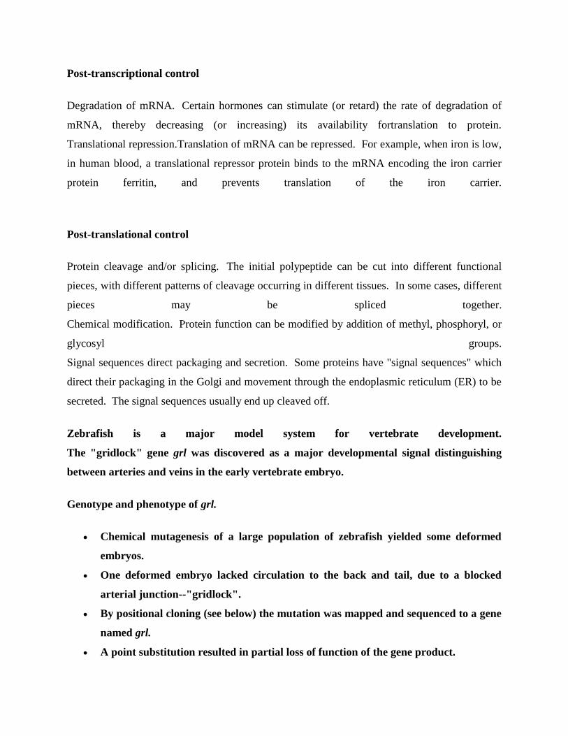
Post-transcriptional control
Degradation of mRNA. Certain hormones can stimulate (or retard) the rate of degradation of
mRNA, thereby decreasing (or increasing) its availability fortranslation to protein.
Translational repression.Translation of mRNA can be repressed. For example, when iron is low,
in human blood, a translational repressor protein binds to the mRNA encoding the iron carrier
protein ferritin, and prevents translation of the iron carrier.
Post-translational control
Protein cleavage and/or splicing. The initial polypeptide can be cut into different functional
pieces, with different patterns of cleavage occurring in different tissues. In some cases, different
pieces may be spliced together.
Chemical modification. Protein function can be modified by addition of methyl, phosphoryl, or
glycosyl groups.
Signal sequences direct packaging and secretion. Some proteins have "signal sequences" which
direct their packaging in the Golgi and movement through the endoplasmic reticulum (ER) to be
secreted. The signal sequences usually end up cleaved off.
Zebrafish is a major model system for vertebrate development.
The "gridlock" gene grl was discovered as a major developmental signal distinguishing
between arteries and veins in the early vertebrate embryo.
Genotype and phenotype of grl.
Chemical mutagenesis of a large population of zebrafish yielded some deformed
embryos.
One deformed embryo lacked circulation to the back and tail, due to a blocked
arterial junction--"gridlock".
By positional cloning (see below) the mutation was mapped and sequenced to a gene
named grl.
A point substitution resulted in partial loss of function of the gene product.
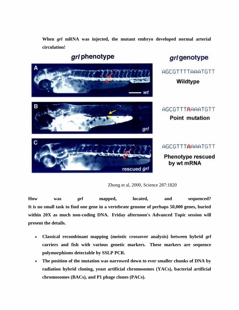
When grl mRNA was injected, the mutant embryo developed normal arterial
circulation!
Zhong et al, 2000, Science 287:1820
How was grl mapped, located, and sequenced?
It is no small task to find one gene in a vertebrate genome of perhaps 50,000 genes, buried
within 20X as much non-coding DNA. Friday afternoon's Advanced Topic session will
present the details.
Classical recombinant mapping (meiotic crossover analysis) between hybrid grl
carriers and fish with various genetic markers. These markers are sequence
polymorphisms detectable by SSLP PCR.
The position of the mutation was narrowed down to ever smaller chunks of DNA by
radiation hybrid cloning, yeast artificial chromosomes (YACs), bacterial artificial
chromosomes (BACs), and P1 phage clones (PACs).
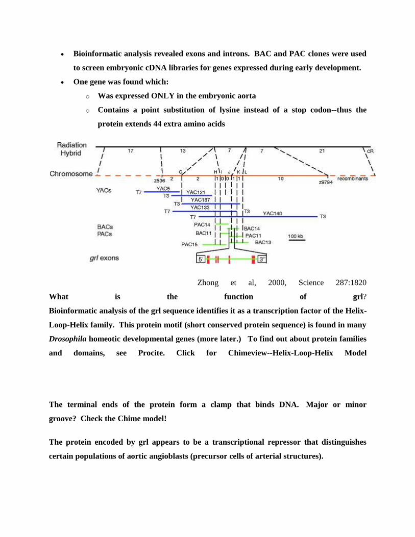
Bioinformatic analysis revealed exons and introns. BAC and PAC clones were used
to screen embryonic cDNA libraries for genes expressed during early development.
One gene was found which:
o Was expressed ONLY in the embryonic aorta
o Contains a point substitution of lysine instead of a stop codon--thus the
protein extends 44 extra amino acids
Zhong et al, 2000, Science 287:1820
What is the function of grl?
Bioinformatic analysis of the grl sequence identifies it as a transcription factor of the Helix-
Loop-Helix family. This protein motif (short conserved protein sequence) is found in many
Drosophila homeotic developmental genes (more later.) To find out about protein families
and domains, see Procite. Click for Chimeview--Helix-Loop-Helix Model
The terminal ends of the protein form a clamp that binds DNA. Major or minor
groove? Check the Chime model!
The protein encoded by grl appears to be a transcriptional repressor that distinguishes
certain populations of aortic angioblasts (precursor cells of arterial structures).
CHAPTER # 22 INTRODUCTION TO PLASMIDS
Plasmids are commonly used to multiply (make many copies of) or express particular genes.
Types of Plasmids
Plasmids used in genetic engineering are called vectors . Plasmids serve as important tools in
genetics and biotechnology labs, where they are commonly used to multiply (make many copies
of) or express particular genes. Many plasmids are commercially available for such uses. The
gene to be replicated is inserted into copies of a plasmid containing genes that make cells
resistant to particular antibiotics. The gene is also inserted into a multiple cloning site (MCS, or
polylinker), which is a short region containing several commonly used restriction sites allowing
the easy insertion of DNA fragments.
A Plasmid Map of pUC19
pUC19 is one of a series of plasmid cloning vectors created by Messing and co-workers in the
University of California. The p in its name stands for plasmid and UC represents the University
in which it was created. It is a circular double stranded DNA and has 2686 base pairs. pUC19 is
one of the most widely used vector molecules as the recombinants, or the cells into which
foreign DNA has been introduced, can be easily distinguished from the non-recombinants based
on color differences of colonies on growth media. pUC18 is similar to pUC19, but the multiple
cloning site region is reversed.
Next, the plasmids are inserted into bacteria by a process called transformation. Then, the bacteria are
exposed to the particular antibiotics. Only bacteria that take up copies of the plasmid survive, since the
plasmid makes them resistant. In particular, the protecting genes are expressed (used to make a protein)
and the expressed protein breaks down the antibiotics. In this way, the antibiotics act as a filter, selecting
only the modified bacteria. Finally, these bacteria can be grown in large amounts, harvested, and lysed
(often using the alkaline lysis method) to isolate the plasmid of interest.
Another major use of plasmids is to make large amounts of proteins. In this case, researchers
grow bacteria containing a plasmid harboring the gene of interest. Just as the bacterium produces
proteins to confer its antibiotic resistance, it can also be induced to produce large amounts of
proteins from the inserted gene. This is a cheap and easy way of mass-producing a gene or the
protein it then codes for; for example, insulin or even antibiotics.
One way of grouping plasmids is by their ability to transfer to other bacteria. Conjugative
plasmids contain tra genes, which perform the complex process of conjugation, the transfer of
plasmids to another bacterium. Non-conjugative plasmids are incapable of initiating conjugation,
hence they can be transferred only with the assistance of conjugative plasmids. An intermediate
class of plasmids are mobilizable, and carry only a subset of the genes required for transfer. They
can parasitize a conjugative plasmid, transferring at high frequency only in its presence.
Plasmids are now being used to manipulate DNA, and may possibly be a tool for curing many
diseases.
It is possible for plasmids of different types to coexist in a single cell. Several different plasmids
have been found in E. coli. However, related plasmids are often incompatible, in the sense that
only one of them survives in the cell line, due to the regulation of vital plasmid functions. Thus,
plasmids can be assigned into incompatibility groups.
Another way to classify plasmids is by function. There are five main classes:
Fertility F-plasmids, which contain tra genes. They are capable of conjugation and result in the
expression of sex pilli.
Resistance plasmids, which contain genes that provide resistance against antibiotics or poisons.
They were historically known as R-factors, before the nature of plasmids was understood.
Col plasmids, which contain genes that code for bacteriocins, proteins that can kill other bacteria.
Degradative plasmids, which enable the digestion of unusual substances, e.g. toluene and salicylic
acid.
Virulence plasmids, which turn the bacterium into a pathogen.
CHAPTER # 23 INTRODUCTION TO VECTORS
Vector (molecular biology)
In molecular cloning, a vector is a DNA molecule used as a vehicle to artificially carry foreign
genetic material into another cell, where it can be replicated and/or expressed. A vector
containing foreign DNA is termed recombinant DNA. The four major types of vectors are
plasmids, viral vectors, cosmids, and artificial chromosomes. Of these, the most commonly used
vectors are plasmids. Common to all engineered vectors are an origin of replication, a
multicloning site, and a selectable marker.
The vector itself is generally a DNA sequence that consists of an insert (transgene) and a larger
sequence that serves as the "backbone" of the vector. The purpose of a vector which transfers
genetic information to another cell is typically to isolate, multiply, or express the insert in the
target cell. Vectors called expression vectors (expression constructs) specifically are for the
expression of the transgene in the target cell, and generally have a promoter sequence that drives
expression of the transgene. Simpler vectors called transcription vectors are only capable of
being transcribed but not translated: they can be replicated in a target cell but not expressed,
unlike expression vectors. Transcription vectors are used to amplify their insert.
Insertion of a vector into the target cell is usually called transformation for bacterial cells,
transfection for eukaryotic cells, although insertion of a viral vector is often called transduction.
Characteristics
Plasmids
Plasmids are double-stranded and generally circular DNA sequences that are capable of
automatically replicating in a host cell. Plasmid vectors minimalistically consist of an origin of
replication that allows for semi-independent replication of the plasmid in the host. Plasmids are
found widely in many bacteria, for example in Escherichia coli, but may also be found in a few
eukaryotes, for example in yeast such as Saccharomyces cerevisiae.[1]
Bacterial plasmids may be
conjugative/transmissible and non-conjugative:
conjugative: mediate DNA transfer through conjugation and therefore spread rapidly
among the bacterial cells of a population; e.g., F plasmid, many R and some col plasmids.
nonconjugative- do not mediate DNA through conjugation, e.g., many R and col
plasmids.
The pBR322 plasmid is one of the first plasmids widely used as a cloning vector.
Plasmids with specially-constructed features are commonly used in laboratory for cloning
purposes. These plasmid are generally non-conjugative but may have many more features,
notably a "multiple cloning site" where multiple restriction enzyme cleavage sites allow for the
insertion of a transgene insert. The bacteria containing the plasmids can generate millions of
copies of the vector within the bacteria in hours, and the amplified vectors can be extracted from
the bacteria for further manipulation. Plasmids may be used specifically as transcription vectors
and such plasmids may lack crucial sequences for protein expression. Plasmids used for protein
expression, called expression vectors, would include elements for translation of protein, such as a
ribosome binding site, start and stop codons.
Viral vectors
Viral vectors are generally genetically engineered viruses carrying modified viral DNA or RNA
that has been rendered noninfectious, but still contain viral promoters and also the transgene,
thus allowing for translation of the transgene through a viral promoter. However, because viral
vectors frequently are lacking infectious sequences, they require helper viruses or packaging
lines for large-scale transfection. Viral vectors are often designed for permanent incorporation of
the insert into the host genome, and thus leave distinct genetic markers in the host genome after
incorporating the transgene. For example, retroviruses leave a characteristic retroviral integration
pattern after insertion that is detectable and indicates that the viral vector has incorporated into
the host genome.
Transcription
Transcription is a necessary component in all vectors: the premise of a vector is to multiply the
insert (although expression vectors later also drive the translation of the multiplied insert). Thus,
even stable expression is determined by stable transcription, which generally depends on
promoters in the vector. However, expression vectors have a variety of expression patterns:
constitutive (consistent expression) or inducible (expression only under certain conditions or
chemicals). This expression is based on different promoter activities, not post-transcriptional
activities. Thus, these two different types of expression vectors depend on different types of
promoters.
Viral promoters are often used for constitutive expression in plasmids and in viral vectors
because they normally force constant transcription in many cell lines and types reliably.
Inducible expression depends on promoters that respond to the induction conditions: for
example, the murine mammary tumor virus promoter only initiates transcription after
dexamethasone application and the Drosophilia heat shock promoter only initiates after high
temperatures.
Expression
Expression vectors produce proteins through the transcription of the vector's insert followed by
translation of the mRNA produced, they therefore require more components than the simpler
transcription-only vectors. Expression in different host organism would require different
elements, although they share similar requirements, for example a promoter for initiation of
transcription, a ribosomal binding site for translation initiation, and termination signals.
Prokaryotes expression vector
Promoter - commonly used inducible promoters are promoters derived from lac operon
and the T7 promoter. Other strong promoters used include Trp promoter and Tac
Promoter, which a hybrid of both the Trp and Lac Operon promoters.
Ribosome binding site (RBS) Follows the promoter, and promotes efficient translation of
the protein of interest.
Translation initiation site - Shine-Dalgarno sequence enclosed in the RBS, 8 base-pairs
upstream of the AUG start codon.
Eukaryotes expression vector
Eukaryote expression vectors require sequences that encode for:
Polyadenylation tail: Creates a polyadenylation tail at the end of the transcribed pre-
mRNA that protects the mRNA from exonucleases and ensures transcriptional and
translational termination: stabilizes mRNA production.
Minimal UTR length: UTRs contain specific characteristics that may impede
transcription or translation, and thus the shortest UTRs or none at all are encoded for in
optimal expression vectors.
Kozak sequence: Vectors should encode for a Kozak sequence in the mRNA, which
assembles the ribosome for translation of the mRNA.
Features
Modern artificially-constructed vectors contain essential components as well as other additional
features:
Origin of replication: Necessary for the replication and maintenance of the vector in the
host cell.
Promoter: Promoters are used to drive the transcription of the vector's transgene as well
as the other genes in the vector such as the antibiotic resistance gene. Some cloning
vectors need not have a promoter for the cloned insert but it is an essential component of
expression vectors so that the cloned product may be expressed.
Cloning site: This may be a multiple cloning site or other features that allow for the
insertion of foreign DNA into the vector through ligation.
Genetic markers: Genetic markers for viral vectors allow for confirmation that the vector
has integrated with the host genomic DNA.
Antibiotic resistance: Vectors with antibiotic-resistance open reading frames allow for
survival of cells that have taken up the vector in growth media containing antibiotics
through antibiotic selection.
Epitope: Vector contains a sequence for a specific epitope that is incorporated into the
expressed protein. Allows for antibody identification of cells expressing the target
protein.
Reporter genes: Some vectors may contain a reporter gene that allows for identification
of plasmid that contains inserted DNA sequence. An example is lacZ-α which codes for
the N-terminus fragment of β-galactosidase, an enzyme that digests galactose. A multiple
cloning site is located within lacZ-α, and an insert successfully ligated into the vector will
disrupt the gene sequence, resulting in an inactive β-galactosidase. Cells containing
vector with an insert may be identified using blue/white selection by growing cells in
media containing an analogue of galactose (X-gal). Cells expressing β-galactosidase
(therefore doesn't contain an insert) appear as blue colonies. White colonies would be
selected as those that may contain an insert. Other commonly used reporters include
green fluorescent protein and luciferase.
Targeting sequence: Expression vectors may include encoding for a targeting sequence in
the finished protein that directs the expressed protein to a specific organelle in the cell or
specific location such as the periplasmic space of bacteria.
Protein purification tags: Some expression vectors include proteins or peptide sequences
that allows for easier purification of the expressed protein. Examples include
polyhistidine-tag, glutathione-S-transferase, and maltose binding protein. Some of these
tags may also allow for increased solubility of the target protein. The target protein is
fused to the protein tag, but a protease cleavage site positioned in the polypeptide linker
region between the protein and the tag allows the tag to be removed later.