ch. 6 hematology and immunology blood and the lymphatic system
TRANSCRIPT

Ch. 6 Hematology and Immunology
Blood and the Lymphatic System

Hematology is the medical specialty that studies the anatomy and physiology of the blood and uses diagnostic tests, medical and surgical procedures, and drugs to treat blood diseases.
Immunology is the medical specialty that studies the anatomy and physiology of the lymphatic system and uses diagnostic tests, medical and surgical procedures, and drugs to treat lymphatic and immune response diseases.
Two million red blood cells die every second.
There are approximately 100,000 miles of blood vessels in the human body.
Seven percent of a humans body weight is made up of blood.
Each day 400 gallons of recycled blood are pumped through the kidneys.
By donating just one pint of blood, four lives can be saved.
Half your body’s red blood cells are replaced every seven days.
Every two seconds, someone needs blood.
There are approximately 1 billion red blood cells in two to three drops of blood.
Your body usually replaces the volume of the blood you donate within 24 hours.

Anatomy and Physiology
Blood-Contains blood cells, blood cell fragments, water, proteins, and clotting factors.
Function: transports oxygen, carbon dioxide, nutrients, and waste products.
Lymphatic System:-Consists of the lymphatic vessels, lymph fluid, lymph nodes, lymphoid tissues, and lymphoid organs.
-Forms a pathway throughout the body that is separate from that of the circulatory system.
Function: Defends the body against microorganisms and cancerous cells.

Anatomy of the Blood
1. Plasma-Yellow-ish clear liquid (about 90% water) that makes up 55% of the blood.
-Contains parts of the blood: erythrocytes, leukocytes, and platelets; albumin, bilirubin, hormones, complement proteins, and clotting factors; creatinine and urea.

Figure 6-2 Plasma

Erythrocytes (Red Blood Cells)-Most numerous of the parts within the plasma.-Red blood cells are a round, somewhat flattened, red disk. -Do not have a nucleus. Live about 120 days.-Contains hemoglobin (iron-containing molecule) that bonds with oxygen and carbon dioxide.-Hematopoiesis, the process of forming blood cells, occurs in the red marrow of long or flat bones.

Figure 6-3 Hematopoiesis.

Life Cycle of an Erythrocytes:
Begin as stem cells, erythroblasts, normoblasts, reticulocytes, and finally erythrocytes.
When your body experiences blood loss, the kidneys secrete erythropoietin hormone that stimulates the production and maturity of erythrocytes.
When blood cells die, their parts are recycled and stored in the body other uses.

Leukocytes (White Blood Cells)
Granulocytes:neutrophilseosinophilsbasophils
Agranulocytes:lymphocytesmonocytes
-Can be identified by the presence or absence of granules in their cytoplasm and the shape of their nucleus.

Neutrophils-Most common leukocyte, making up 40 to 60% of leukocytes in blood. Short lived: hours to days.-Nucleus has many segments or lobes, so they are also known as polymorphonucleated leukocytes (PMNs), polys, segs, or segmenters.-Develop from the red bone marrow.
Function: engulf and destroy bacteria.

Eosinophils-Make up just 1 to 4% of leukocytes-Nucleus has two lobes-Develop in the red marrow
Function: engulf and destroy foreign cells (pollen, animal dander, etc.); release chemicals that kill parasites

Basophils-Least common leukocyte, making up 0.5 to 1% of leukocytes.-Nucleus has more than one lobe.-Develop in the red marrow
Function: release histamine at the site of tissue injury; release heparin, an anticoagulant.

Lymphocytes-Make up 20 to 40% of leukocytes.-Smallest of the white blood cells.-Nucleus is round and nearly fills the cell.-Some lymphocytes live for just a few days, while others live for many years.-Begin development in red marrow; some become B cells or natural killer cells; others migrate to the thymus to become T cells.
Function: destroy viruses and produce antibodies.

Monocytes-Make up 2 to 4% of leukocytes.-Largest of the white blood cells. -Have a large amount of cytoplasm, and nucleus is large and kidney bean shaped.-Develop in the red marrow.Function: are phagocytes that engulf and destroy microorganisms, cancerous cells, dead leukocytes, and cellular debris. Are also macrophages.
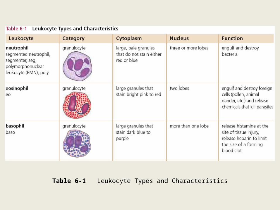
Table 6-1 Leukocyte Types and Characteristics

Table 6-1 (continued) Leukocyte Types and Characteristics
Thrombocytes-Different from other blood cells because they are only cell fragments; also called Platelets.-Active in the blood-clotting process.-Begin in the red marrow as stem cells that then become megakaryoblasts, and then mature into megakaryocytes, a very large cell. -Cytoplasm of the megakaryocyte breaks away at the edges to form cell fragments (thrombocytes) that are released into the blood.
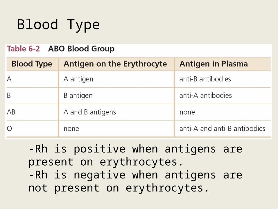
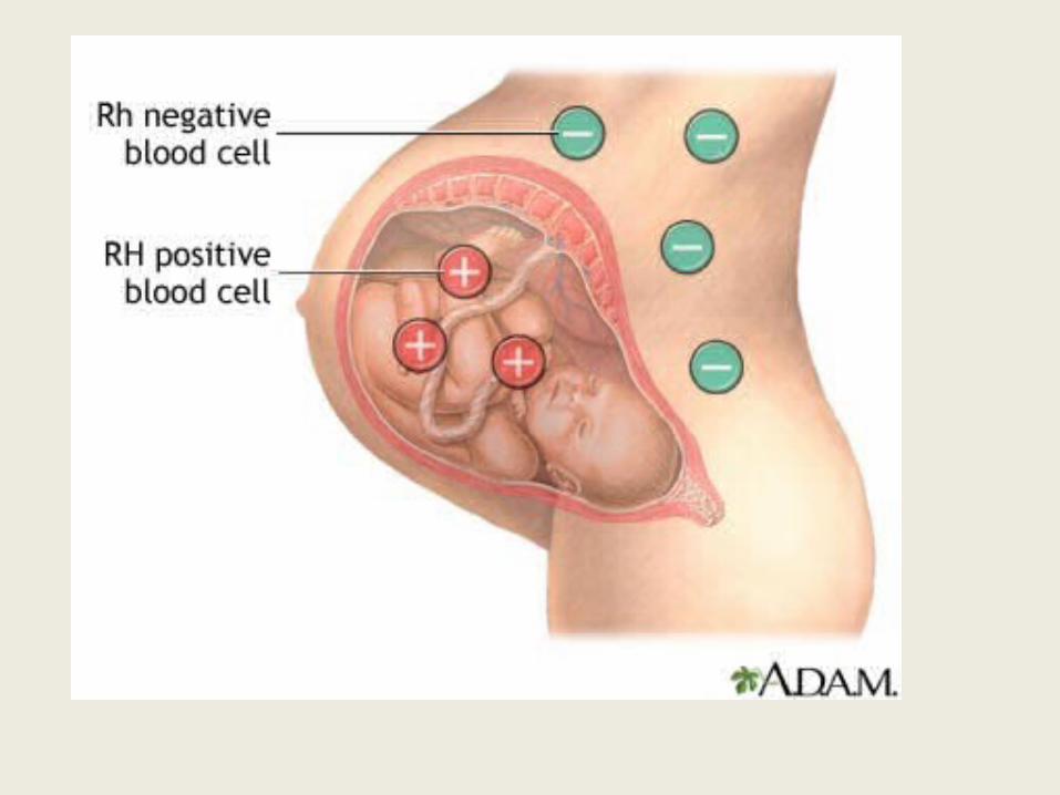
Blood Type
-Rh is positive when antigens are present on erythrocytes.-Rh is negative when antigens are not present on erythrocytes.

Physiology of Blood Clotting
1. Platelet Aggregation- Thrombocytes form clumps to decrease blood loss.
2. Coagulation- Blood clot forms. (fibrin)
3. Hemostasis- Cessation of bleeding.
After clotting factors are activated, the remaining fluid portion of plasma is called serum.

Figure 6-11 Blood clotSusumu Nishinaga/Photo Researchers, Inc.
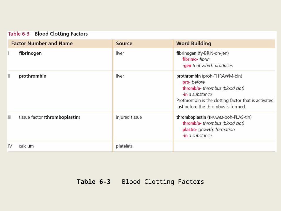
Table 6-3 Blood Clotting Factors

Table 6-3 (continued) Blood Clotting Factors
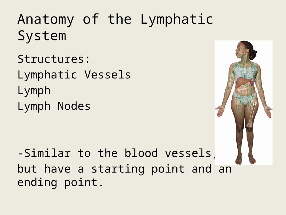
Anatomy of the Lymphatic System
Structures:Lymphatic VesselsLymphLymph Nodes
-Similar to the blood vessels, but have a starting point and an ending point.
Beginning Point: fluid enters a lymphatic capillary and becomes lymph.
End Point: ducts empty into large veins in the neck.

Characteristics:-Lymphatic capillaries have large openings in their walls that allow microorganisms and cancerous cells to enter.
-Valves within the system keep the lymph flowing in one direction.
-Lymph Nodes- function to filter the lymph, contain lymphocytes and macrophages that destroy microorganisms.
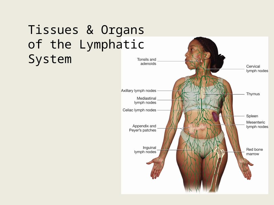
Tissues & Organs of the Lymphatic System
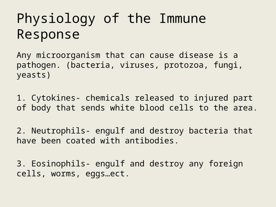
Physiology of the Immune Response
Any microorganism that can cause disease is a pathogen. (bacteria, viruses, protozoa, fungi, yeasts)
1. Cytokines- chemicals released to injured part of body that sends white blood cells to the area.
2. Neutrophils- engulf and destroy bacteria that have been coated with antibodies.
3. Eosinophils- engulf and destroy any foreign cells, worms, eggs…ect.

4. Basophils- release histamine in response to microorganisms.
5. Monocytes- engulf and destroy pathogens that have been coated with antibodies. Also known as a macrophage.Produce: interferon, interleukin, & tumor necrosis factor.
6. LymphocytesNK (natural killer) Cells- recognize and destroy pathogens.B Cells- from the red bone marrow; work with macrophages, produce antibodies; activate T Cells.T Cells- from the thymus gland; Helper T-Cells, Memory T-Cells, Cytotoxic T-Cells, & Suppressor T-Cells
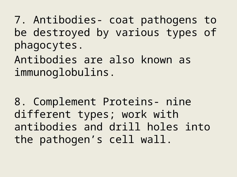
7. Antibodies- coat pathogens to be destroyed by various types of phagocytes. Antibodies are also known as immunoglobulins.
8. Complement Proteins- nine different types; work with antibodies and drill holes into the pathogen’s cell wall.
Diseases and Conditions
Blood:-Blood Dyscrasia
-Hemorrhage
-Pancytopenia
-Septicemia
Erythrocytes:-Abnormal Red Blood Cell Morphology
-AnemiaAplastic anemiaFolic Acid Deficiency anemiaIron Deficiency anemiaPernicious anemiaSickle Cell anemia
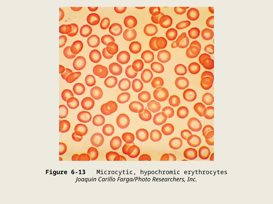
Figure 6-13 Microcytic, hypochromic erythrocytesJoaquin Carillo Farga/Photo Researchers, Inc.
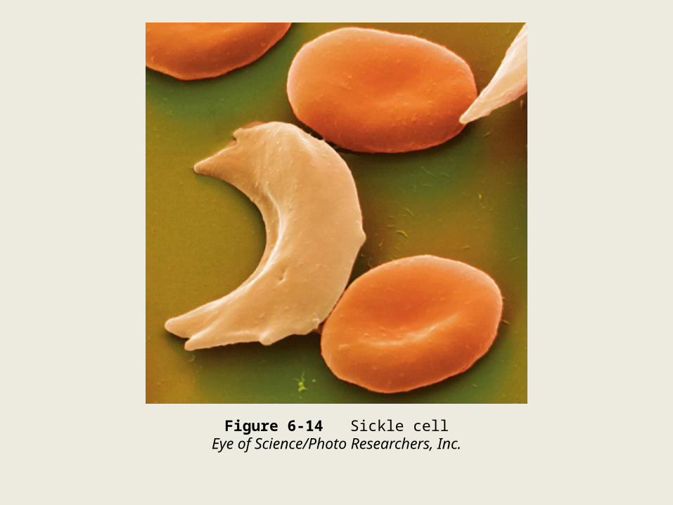
Figure 6-14 Sickle cellEye of Science/Photo Researchers, Inc.
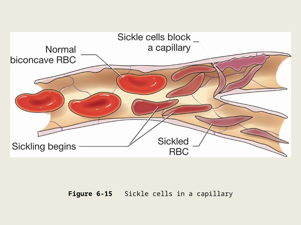
Figure 6-15 Sickle cells in a capillary

Erythrocytes: -Anisocytosis
-Poikilocytosis
-Polycythemia Vera
-Thalassemia
-Transfusion Reaction

Leukocytes:-Acquired Immunodeficiency Syndrome (AIDS)
-Leukemia
-Mononucleosis
-Multiple Myeloma

Figure 6-16 Human immunodeficiency virusChris Bjornberg/Photo Researchers, Inc.
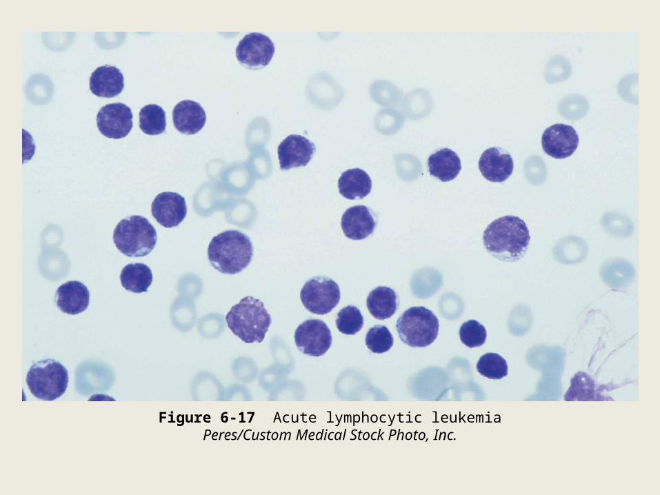
Figure 6-17 Acute lymphocytic leukemiaPeres/Custom Medical Stock Photo, Inc.
Thrombocytes:-Coagulopathy
-Deep Venous Thrombosis (DVT)
-Disseminated Intravascular Coagulation (DIC)
-Hemophilia
-Thrombocytopenia

Figure 6-20 Deep venous thrombosis

Lymphatic System:-Graft-Versus-Host Disease (GVHD)
-Lymphadenopathy
-Lymphedema
-LymphomaHodgkin’s LymphomaNon-Hodgkin’s Lymphoma
-Splenomegaly
-Thymoma
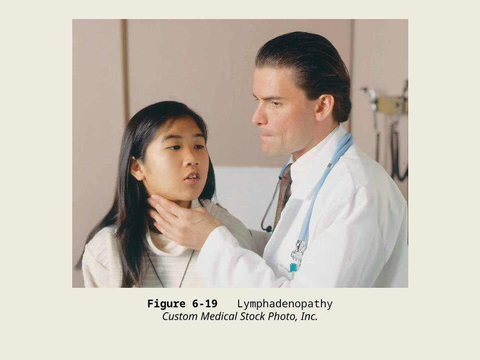
Figure 6-19 LymphadenopathyCustom Medical Stock Photo, Inc.

Autoimmune Disorders:-Diabetes Mellitus, Type 1
-Graves’ Disease
-Hashimoto’s Thyroiditis
-Inflammatory Bowel Disease
-Multiple Sclerosis

Autoimmune Disorders: -Myasthenia Gravis
-Psoriasis
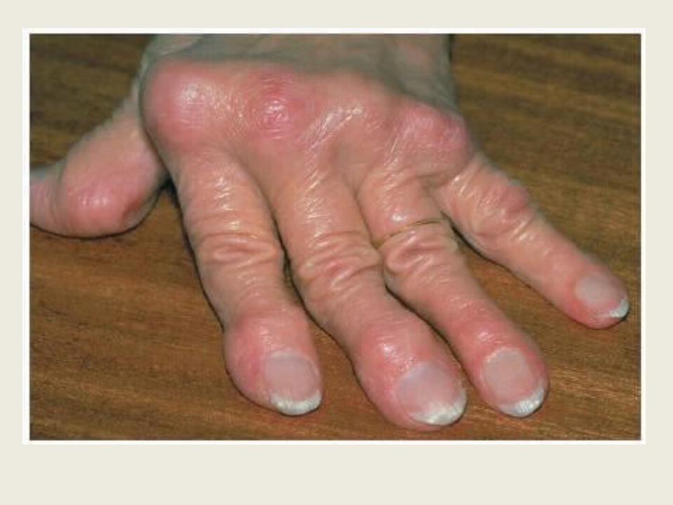
-Rheumatoid Arthritis
-Scleroderma
-Systemic Lupus Erythematosus


Laboratory and Diagnostic Procedures
Blood Cell Tests:-Blood Type
-Complete Blood Count (CBC) with Differential
-Peripheral Blood Smear

Table 6-4 Complete Blood Count (CBC) with Differential

Table 6-4 Complete Blood Count (CBC) with Differential

Coagulation Tests:-Activated Clotting Time (ACT)
-Partial Thromboplastin Time (PTT)
-Prothrombin Time (PT)

Other Blood Tests:-Blood Chemistries
-Ferritin

Other Blood Tests:-Total iron-binding capacity (TIBC)
-Human Immunodeficiency Virus (HIV) TestsELISA―First screening test done for HIV.Western Blot―Used to confirm a positive ELISA and make a diagnosis of HIV infection.Viral RNA Load Test―Measures tiny amounts of HIV RNA and monitors progression of the disease and response to antiretroviral drugs.p24 Antigen Test―Detects the protein p24 in HIVCD4 count―Used to monitor the progression of the disease and response to antiretroviral drugs.
Saliva Test:-OraSure
Quick screening test that is done in the doctor’s office or clinic. It uses the same technology as the ELISA blood test.
Serum Tests:-Electrophoresis-Monospot
Urine Tests:-Bence Jones Protein -Schilling Test
Radiologic Procedures:-Color Flow Duplex Ultrasonography
-Lymphangiography
Medical Procedures:-Bone Marrow Aspiration -Phlebotomy
-Vaccination
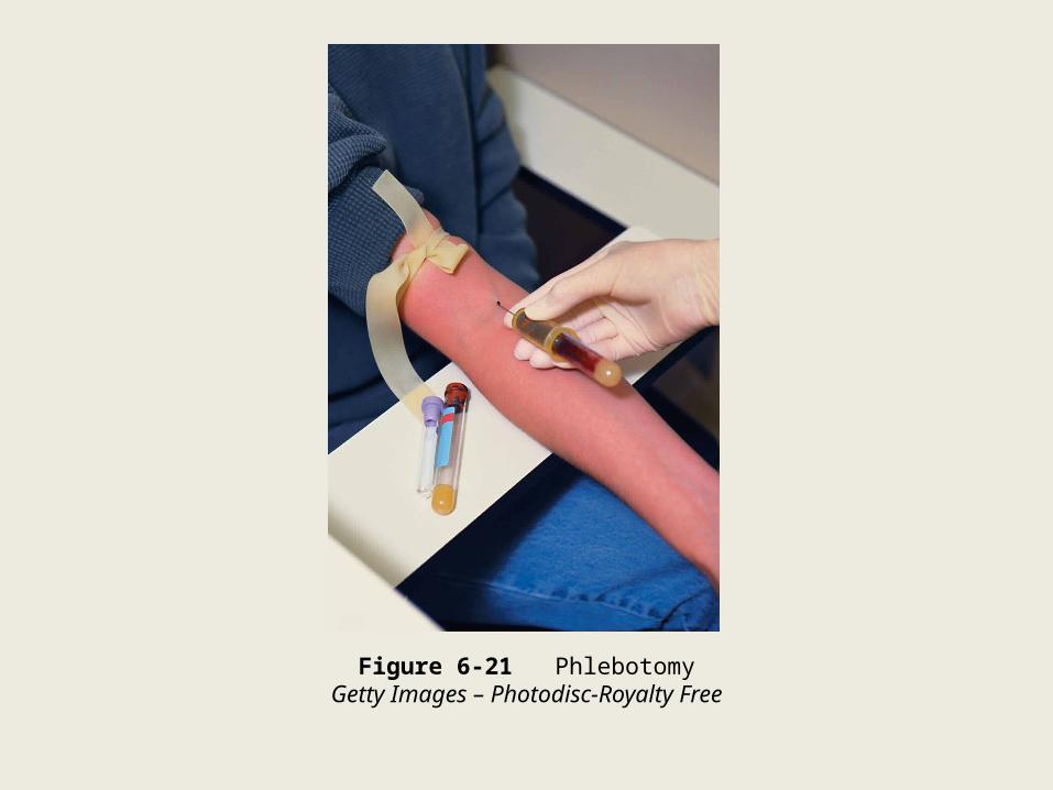
Figure 6-21 PhlebotomyGetty Images – Photodisc-Royalty Free
Blood Donation and Transfusion Procedures:-Blood donation
-Blood Transfusion
-Bone Marrow Transplantation (BMT)
-Plasmapheresis
-Stem cell transplantation

Surgical Procedures:-Lymph node biopsy
-Lymph node dissection
-Splenectomy
-Thymectomy