cellular/molecular proneurotrophin ... · cellular/molecular...
TRANSCRIPT
Cellular/Molecular
Proneurotrophin-3 Is a Neuronal Apoptotic Ligand:Evidence for Retrograde-Directed Cell Killing
Hiroko Yano,1 Risa Torkin,2,3* Laura Andres Martin,2* Moses V. Chao,1 and Kenneth K. Teng2
1Molecular Neurobiology Program, Skirball Institute of Biomolecular Medicine, New York University School of Medicine, New York, New York 10016,2Department of Medicine, Weill Cornell Medical College, New York, New York 10065, and 3Cell Biology Program, The Hospital for Sick Children, Toronto,Ontario M5G 1X8, Canada
Although mature neurotrophins are well described trophic factors that elicit retrograde survival signaling, the precursor forms ofneurotrophins (i.e., proneurotrophins) can function as high-affinity apoptotic ligands for selected neural populations. An outstandingquestion is whether target-derived proneurotrophins might affect neuronal survival/death decisions through a retrograde transportmechanism. Since neurotrophin-3 (NT-3) is highly expressed in non-neural tissues that receive peripheral innervation, we investigatedthe localized actions of its precursor (proNT-3) on sympathetic neurons in the present study. Pharmacological inhibition of intracellularfurin proteinase activity in 293T cells resulted in proNT-3 release instead of mature NT-3, whereas membrane depolarization in cerebellargranule neurons stimulated endogenous proNT-3 secretion, suggesting that proNT-3 is an inducible bona fide ligand in the nervoussystem. Our data also indicate that recombinant proNT-3 induced sympathetic neuron death that is p75 NTR- and sortilin-dependent, withhallmark features of apoptosis including JNK (c-Jun N-terminal kinase) activation and nuclear fragmentation. Using compartmentalizedculture systems that segregate neuronal cell bodies from axons, proNT-3, acting within the distal axon compartment, elicited sympatheticneuron death and overrode the survival-promoting actions of NGF. Together, these results raise the intriguing possibility that dysregu-lation of proneurotrophin processing/release by innervated targets can be deleterious to the neurons projecting to these sites.
IntroductionProneurotrophins are the precursors of a small family of peptidegrowth factors that include nerve growth factor (NGF), brain-derived neurotrophic factor (BDNF), neurotrophin-3 (NT-3),and neurotrophin-4 (NT-4). Until recently, fully processed ma-ture neurotrophins were believed to be the sole ligands responsi-ble for their diverse actions in and outside of the nervous system(Lewin and Barde, 1996; Segal, 2003; Gentry et al., 2004; Teng andHempstead, 2004). However, both NGF and BDNF precursorscan be released as soluble ligands with distinct biological activities(Lee et al., 2001; Nykjaer et al., 2004; Pang et al., 2004; Teng et al.,2005; Woo et al., 2005; Nagappan et al., 2009; Yang et al., 2009a).This supports a paradigm in which proneurotrophins and theirmature counterparts interact with distinct coreceptor complexes,thereby inducing diametrically opposite cellular responses. Thus,mature neurotrophins activate the Trk receptor tyrosine kinases
to elicit well defined signal transduction cascades, whereas anunrelated p75 NTR receptor serves to restrict the fidelity of ligandbinding to cognate Trk receptor (Segal, 2003; Gentry et al., 2004;Teng and Hempstead, 2004). In contrast, proNGF and proBDNFengage p75 NTR and the vps10p domain-containing receptor sor-tilin (Nykjaer et al., 2004; Teng et al., 2005; Willnow et al., 2008),with both ligands exerting proapoptotic actions on peripheraland central neurons (Lee et al., 2001; Nykjaer et al., 2004; Teng etal., 2005; Volosin et al., 2006; Jansen et al., 2007).
NT-3 was identified as the third neurotrophin family mem-ber, based on sequence conservation with NGF and BDNF(Maisonpierre et al., 1990a). Early in vitro work demonstratedthat NT-3 promotes the survival of neuronal subpopulations(Maisonpierre et al., 1990a; Lamballe et al., 1991) and that it isretrogradely transported by both peripheral and central neurons(DiStefano et al., 1992), consistent with its role as a target-derivedneurotrophic factor. Compared with other neurotrophins, NT-3exhibits the most widespread distribution in non-neuronal tis-sues, including many targets of sympathetic and sensory inner-vations (Schecterson and Bothwell, 1992; Katoh-Semba et al.,1996, 1998). In vivo studies of gene targeted animals deficient inNT-3 or it receptor TrkC also support important functions forthis ligand in peripheral and CNS development (Minichiello andKlein, 1996; Bates et al., 1999; Kahn et al., 1999; Ma et al., 2002;von Bohlen und Halbach et al., 2003). Although it is well knownthat p75 NTR modulates the specificity of NT-3 binding toTrkA–C (Bibel et al., 1999; Mischel et al., 2001; Kuruvilla et al.,2004), activation of p75 NTR alone by NT-3 has also been shown toinduce cell death (Friedman, 2000; Wang et al., 2000). Together,
Received May 1, 2009; revised Sept. 1, 2009; accepted Oct. 13, 2009.This work was supported in part by National Institutes of Health Grants NS21072 and HD23315 (M.V.C.) and
NS057627 (K.K.T.) and The James Birrell Neuroblastoma Fund at the Hospital for Sick Children (R.T.) (R.T. was at WeillCornell Medical College at the time of the study). We are grateful to Dr. Barbara Hempstead for her advice andgenerous support. We thank Dr. Francis Lee for helpful discussion, Dr. Robert Campenot for sharing his expertise oncompartmentalized culture system, and Drs. Anders Nykjaer and Claus Munck Petersen for communicating theirunpublished data. We also thank Taeho Kim for his advice on proneurotrophin immunoprecipitation.
*R.T. and L.A.M. contributed equally to this work.Correspondence should be addressed to Kenneth K. Teng, Weill Cornell Medical College, Room D602, 1300 York
Avenue, New York, NY 10065. E-mail: [email protected]. Yano’s present address: Brigham and Women’s Hospital, Harvard Medical School, Boston, MA 02115.R. Torkin’s present address: Acorda Therapeutics, Hawthorne, NY 10532.DOI:10.1523/JNEUROSCI.2059-09.2009
Copyright © 2009 Society for Neuroscience 0270-6474/09/2914790-13$15.00/0
14790 • The Journal of Neuroscience, November 25, 2009 • 29(47):14790 –14802
these findings are consistent with the hypothesis that NT-3 selec-tively uses different receptor complexes to achieve distinct bio-logical endpoints.
Similar to other neurotrophins, NT-3 is synthesized as a high-molecular-weight precursor (proNT-3) that undergoes furin/proconvertase-mediated cleavage for its release as a mature dimer(Seidah et al., 1996). Interestingly, perturbation of processingresults in proNT-3 secretion instead of mature NT-3 (Seidah etal., 1996; Farhadi et al., 2000). Given the study by Ginty andcolleagues (Kuruvilla et al., 2004) that NT-3 acts as an interme-diate target-derived neuritogenic factor for innervating sympa-thetic fibers, we explored the possibility that locally releasedproNT-3 might elicit alternative action on sympathetic neurondevelopment and provide evidence for target-derived proNT-3 asa retrograde apoptotic ligand.
Materials and MethodsCell lines. HEK 293T cells were maintained in DMEM supplemented with10% fetal bovine serum, 1% penicillin/streptomycin, and 1% pyruvate.Parental PC12 cells and PC12 nnr5 (Green et al., 1986) were maintained inDMEM, 10% calf serum, 5% horse serum, 1% penicillin/streptomycin,and 1% pyruvate.
Generation of expression vectors and recombinant proNT-3 protein. Hu-man full-length preproNT-3 cDNA was amplified by PCR using primersto introduce a 5� SacI site with an optimized Kozak consensus for trans-lational initiation, and a heptahistidine (His7) tag, stop codon, andBamHI site at the 3� terminus. In parallel, point mutation of KR to AA(amino acid 137, 138; according to GenBank accession numberNP_002518) was performed using PCR-based mutagenesis to generatecleavage-resistant proNT-3 cDNA. Constructs encoding native orcleavage-resistant His7-tagged proNT-3 cDNAs, subcloned in pBlue-script II SK (pBS NT-3-His7 or pBS proNT-3-His7, respectively), werebidirectionally sequenced. Next, recombinant baculoviral expressionvectors encoding native or cleavage-resistant His7-tagged proNT-3cDNA were generated using the Bac-to-Bac Baculovirus Expression Sys-tem by subcloning a PstI–EcoRI insert from pBS NT-3-His7 or pBSproNT-3-His7 into pFastBac I vector. Baculoviral stocks were amplifiedand propagated using Spodoptera frugiperda (Sf9) cells cultured in Sf-900II SFM for 72 h, whereas High Five cells cultured in Express Five SFMwere used for protein purification. All baculovirus expression systemrelated reagents and cells were purchased from Invitrogen.
For mammalian expression studies, native and cleavage-resistantpreproNT-3 cDNAs were PCR-subcloned into pCMV-Tag 4a vector(Stratagene) using the above described 5�-primer and a separate 3�-primer that removes the NT-3 stop codon to generate C-terminal FLAG-tagged version of both molecules. The resulting constructs werebidirectionally sequenced.
Purification of His7-tagged recombinant proNT-3 and mature NT-3.Cellular lysates from baculovirus-infected High Five cells (5 multiplici-ties of infection; 60 h after infection) were prepared in a detergent-freelysis buffer consisting of 50 mM Na-phosphate, pH 7.8, 300 mM NaCl, 20mM imidazole plus proteinase inhibitors and were used as sources. His7-tagged proNT-3 or mature NT-3 was purified by Ni � ion chromatogra-phy (ProBond resins) according to the manufacturer’s instruction(Invitrogen). Briefly, after washing in 50 mM Na-phosphate, pH 7.8, 300mM NaCl, 20 mM imidazole, proNT-3-His7 or mature NT-3-His7 waseluted with 50 mM Na-phosphate, pH 7.8, 300 mM NaCl, and 500 mM
imidazole, and were collected in 1 ml fractions. Purification was monitoredby Western blot analysis using an anti-NT-3 antiserum that recognizes bothproNT-3 and mature NT-3 (sc-547; Santa Cruz Biotechnology) and bysilver staining. Fractions containing purified recombinant proteins werepooled and dialyzed against three changes of HBSS (Invitrogen) andstored in aliquots at �80°C until use. Final concentrations of purifiedmature NT-3 and proNT-3 were determined by quantitative Westernblot using an anti-NT3 antibody (sc-547) against serial dilutions of re-combinant NT-3 (Promega) as standards. All experiments were con-ducted with His7-tagged proNT-3 and His7-tagged mature NT-3 that
were purified and processed at the same time to ensure internalconsistence.
TrkC-dependent signaling and neuritogenesis assay. To compare TrkCactivation by NT-3 versus proNT-3, PC12 nnr5 cells (Green et al., 1986)were stably transfected with an expression plasmid encoding full-lengthTrkC receptor followed by clonal selection in 500 �g/ml G418. Anti-Trkimmunoprecipitation of TrkC-expressing PC12 nnr5 clones were per-formed with an anti-Trk antiserum (sc-11; Santa Cruz Biotechnology)followed by anti-phosphotyrosine (sc-7020; Santa Cruz Biotechnology)Western blotting. Total cellular lysates were also Western blotted with ananti-pErk antiserum (Ab 9101; Cell Signaling Technology) to corrobo-rate activation of downstream TrkC-dependent signaling or with com-bined anti-Erk1/2 antisera (sc-93 and sc-153; Santa Cruz Biotechnology)to verify equality of sample loading.
For neuritogenic assay, PC12 cells were transiently transfected withgreen fluorescent protein (GFP) with or without full-length TrkC cDNAby Lipofectamine 2000 (Invitrogen). Twenty-four hours later, replicacultures were treated with NGF, mature NT-3, or proNT-3 in DMEMcontaining 0.1% FBS. Paraformaldehyde-fixed, GFP-positive PC12 cellswith neurites more than two cell body diameters in length were scored aspositive 48 h later. At least 100 cells were counted from randomly chosenfields per culture conditions by an observer blinded to the treatments.
Primary sympathetic neuron cultures and assessment of apoptosis. Dis-sociated superior cervical ganglion (SCG) neurons were isolated frompostnatal day 0 (P0) to P2 rats. Alternatively, cultured SCG neurons wereprepared from p75 NTR-null mice (Lee et al., 1992) and wild-type controlanimals. Unless otherwise stated, neurons were plated on laminin-coatedPermanox slides and maintained for 7 d in NGF as described previously(Nykjaer et al., 2004; Teng et al., 2005). On the day of the experiment,replicate cultures were rinsed five times with NGF-free medium (MEM,10% FBS, 0.45% glucose, 2 mM glutamine, 1% pyruvate, 1% penicillin/streptomycin) and treated with either NGF, mature NT-3, proNT-3, orequivalent quantities of diluent (50 mM Na-phosphate, pH 7.8, 300 mM
NaCl, 500 mM imidazole dialyzed with three changes of HBSS). Whereapplicable, parallel cultures were concomitantly treated with various an-tagonists or inhibitors as indicated at the time of ligand addition. After36 – 48 h, SCG cultures were processed for terminal deoxynucleotidyltransferase-mediated biotinylated UTP nick end labeling (TUNEL)analysis (Roche Molecular Biochemicals) and counterstained with anti-neuronal specific �-tubulin (TuJ1) (Covance) and 4�,6�-diamidino-2-phenylindole (DAPI) to visualize nuclei. Apoptotic neurons, identifiedby TUNEL positivity and/or fragmented nuclei, were scored blinded as totreatment conditions by the observer, and at least 200 cells were countedfor each culture condition. Where applicable, statistical analyses (Stu-dent’s t test) were performed on the indicated paired samples with sig-nificance ( p � 0.05) indicated by an asterisk (*). For some experiments,SCG cultures were fixed and stained with phospho-c-Jun N-terminalkinase (JNK) or phospho-c-Jun antibodies (both from Cell SignalingTechnology) along with TuJ1 and DAPI. Indirect immunofluorescenceanalysis was performed using the appropriate secondary antibodies toidentify apoptotic neurons.
Compartmentalized cultures. Dissociated SCG neurons were plated oncollagen-coated 35 mm tissue culture plates previously seated with aTeflon divider Camp10 (Tyler Research Instruments) and cultured asdescribed previously (MacInnis and Campenot, 2002; Ye et al., 2003)with the following modifications. Freshly plated SCG neuron cultureswere treated with 5-fluorodeoxy-uridine for the first 7 d to eliminatecontaminating non-neuronal cells in the center (cell body) compart-ment. NGF was used at 20 ng/ml (in MEM containing 0.3% methylcel-lulose, 2.5% FBS, and 0.4% glucose) in the center compartment and at100 ng/ml (in MEM containing 0.3% methylcellulose, and 0.4% glucose)in the distal axon compartment to induce axonal outgrowth. Seven to10 d later and on visual inspection of the cultures to ensure axonal growthinto the distal compartment, NGF was removed from the center com-partment and cultures were treated with 100 ng/ml NGF exclusively inthe distal compartment to enrich for neurons that bear processes in thedistal compartment. Another 3 d later, NGF within the distal compart-ment was reduced to 10 ng/ml. Twenty-four hours later, 40 nm orangefluorescent microspheres (Invitrogen) were added (at 1:1000 dilution in
Yano et al. • ProNT-3 Induces Neuronal Apoptosis J. Neurosci., November 25, 2009 • 29(47):14790 –14802 • 14791
MEM plus 10 ng/ml NGF) to the distal axonchamber. Neurons that were viable with activeretrograde transport were thus identified bythe uptake of fluorescent microspheres intotheir cell bodies (Ye et al., 2003). Only replicacultures that were retrogradely labeled wereused for additional analysis. Where applicable,statistical analyses (Student’s t test) wereperformed on the indicated paired sampleswith significance ( p � 0.05) indicated by anasterisk (*).
Generation of anti-proNT-3 and sortilin an-tisera. A glutathione S-transferase (GST) fu-sion protein containing the prodomain ofhuman proNT-3 (amino acids 21–136) wasgenerated by PCR subcloning the correspond-ing DNA fragment into the EcoRI–XhoI sitesof pGEX6p-1 vector, followed by IPTG(isopropyl-�-D-thiogalactopyranoside) induc-tion in bacteria transformant and purificationvia glutathione-Sepharose chromatography.Rabbit antisera (Ab 19573 and 19574) wereproduced by repeated immunization with theGST-NT-3 prodomain fusion protein (PoconoRabbit Farm and Laboratory). High-titer anti-sera (as well as preimmune control sera fromthe respective animals) were sequentially puri-fied by negative adsorption to immobilizedGST and IgG enriched by protein A-Sepharosechromatography. Additional affinity purifica-tion of the 19574 antiserum was performed us-ing conjugated GST-NT-3 prodomain fusionprotein. The resulting antisera (referred to asanti-proNT-3) were dialyzed in PBS and storedin aliquots at �80°C before additionalcharacterization.
Full-length human sortilin cDNA was usedas a template to generate His6-tagged truncatedsortilin lacking the transmembrane and intra-cellular domain (amino acids 34 –757) andsubcloned into the pFastBac I baculoviral vec-tor. Baculoviral stocks were amplified andpropagated using S. frugiperda (Sf9) cells, whereas High Five cells cul-tured in Express Five SFM were used for protein purification by Ni-ionchromatography. Rabbit anti-sortilin antiserum was obtained by re-peated immunization with purified His6-tagged truncated sortilin (Po-cono Rabbit Farm and Laboratory) and was further IgG enriched byprotein A-Sepharose chromatography. Specificity of the anti-sortilin anti-serum was verified by Western blotting against lysates of 293 cells trans-fected with human sortilin cDNA and compared with a commercialmonoclonal antibody against sortilin (anti-NTR3; BD Biosciences) (sup-plemental Fig. 1, available at www.jneurosci.org as supplementalmaterial).
Tissue preparation for proNT-3/NT-3 Western blot. Initially, we surveyedseveral different published protocols for neurotrophin extraction fromtissues (Katoh-Semba et al., 1996; Zhang et al., 2001; Yang et al., 2009b)but have found great variability in how well proNT-3 and mature NT-3can be extracted. The following procedure was eventually adopted be-cause it affords the most efficient and reproducible means for proNT-3/NT-3 recovery from both neural and non-neural sources: Freshlydissected tissues from rats were immediately processed in ice-cold tissuelysis buffer [20 mM Tris, pH 7.4, 150 mM NaCl, 1% NP-40, 1% TritonX-100, 0.05% SDS, 10% glycerol, 1 mM PMSF, and proteinase inhibitormixture (Sigma-Aldrich)] (Yang et al., 2009b) using a hand-held tissuehomogenizer (Omni International), except that hippocampal lysate wasobtained by pooling six to eight dissected hippocampi (kept on dry iceuntil the end of the dissection) before the homogenization step. Tissuelysates were incubated at 4°C with constant shaking for 15 min and were thencleared by centrifugation at 13,000 � g for 15 min at 4°C before
Bradford-based quantitation (Bio-Rad Laboratories). Aliquoted samples(in 1� SDS-PAGE buffer) were boiled and immediately stored at �80°Cbefore additional analysis.
Western blotting analysis of proNT-3 and mature NT-3 expression invarious tissues was performed as described previously (Randolph et al.,2007) using PVDF (polyvinylidene difluoride) membrane in 8% nonfatdry milk with a rabbit anti-NT-3 antibody (sc-547; Santa Cruz Biotech-nology; 1:1000 dilution). To demonstrate specificity, the anti-NT-3 an-tiserum was pretreated with 100-fold molar excess of recombinant NT-3(Promega) for 1.5 h at 25°C in PBS before using it for Western blotting(Randolph et al., 2007). As a control, anti-NT-3 antiserum that did notreceive recombinant NT-3 was processed in parallel before use.
Endogenous proNT-3 immunoprecipitation. Dissociated P6 cerebellargranule neurons were cultured on poly-L-lysine-coated 150 mm tissueculture plates and maintained in MEM supplemented with 10% fetalbovine serum, 25 mM KCl, 2 mM GlutaMax, 0.45% glucose, 1% penicil-lin/streptomycin, and 1% pyruvate. Cultures were also treated with5-fluorodeoxy-uridine for the first 3 d to eliminate contaminating non-neuronal cells. On day in vitro (DIV) 6, cultured granule neurons wererinsed once with Neurobasal medium (containing 1� B27 supplement,0.1 mg/ml bovine serum albumin, and 0.5 �M GlutaMax) for 1 h beforetreatment with 25 mM KCl to stimulate proNT-3 release. Where indi-cated, the cell-permeable furin inhibitor I (Dec-RVKR-CMK; Calbio-chem) was used at 30 �M. To capture secreted proNT-3, a goat anti-NT-3antiserum (sc-13380; Santa Cruz Biotechnology) was added to the cul-ture medium (1:100 dilution) at the time of treatment. In addition, toprevent proteolytic degradation of proNT-3, a cell-impermeant �2 anti-
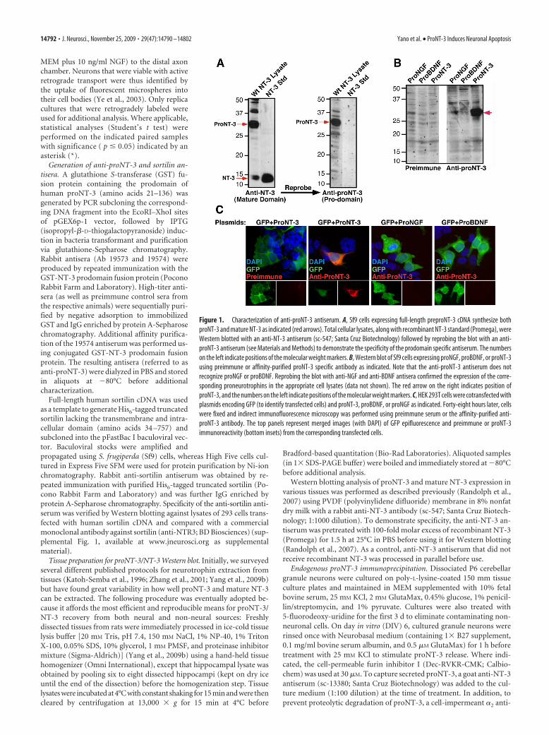
Figure 1. Characterization of anti-proNT-3 antiserum. A, Sf9 cells expressing full-length preproNT-3 cDNA synthesize bothproNT-3 and mature NT-3 as indicated (red arrows). Total cellular lysates, along with recombinant NT-3 standard (Promega), wereWestern blotted with an anti-NT-3 antiserum (sc-547; Santa Cruz Biotechnology) followed by reprobing the blot with an anti-proNT-3 antiserum (see Materials and Methods) to demonstrate the specificity of the prodomain specific antiserum. The numberson the left indicate positions of the molecular weight markers. B, Western blot of Sf9 cells expressing proNGF, proBDNF, or proNT-3using preimmune or affinity-purified proNT-3 specific antibody as indicated. Note that the anti-proNT-3 antiserum does notrecognize proNGF or proBDNF. Reprobing the blot with anti-NGF and anti-BDNF antisera confirmed the expression of the corre-sponding proneurotrophins in the appropriate cell lysates (data not shown). The red arrow on the right indicates position ofproNT-3, and the numbers on the left indicate positions of the molecular weight markers. C, HEK 293T cells were cotransfected withplasmids encoding GFP (to identify transfected cells) and proNT-3, proBDNF, or proNGF as indicated. Forty-eight hours later, cellswere fixed and indirect immunofluorescence microscopy was performed using preimmune serum or the affinity-purified anti-proNT-3 antibody. The top panels represent merged images (with DAPI) of GFP epifluorescence and preimmune or proNT-3immunoreactivity (bottom insets) from the corresponding transfected cells.
14792 • J. Neurosci., November 25, 2009 • 29(47):14790 –14802 Yano et al. • ProNT-3 Induces Neuronal Apoptosis
plasmin inhibitor (Calbiochem) was added to all the cultures (Teng et al.,2005). Twenty-four hours later, neuronal conditioned media were col-lected, cleared of cellular debris by centrifugation, and supplementedwith the proteinase inhibitors PMSF, leupeptin, and aprotinin. ProteinA/G-agarose beads (Pierce) were then added to the media for 16 h toimmunoprecipitate proNT-3, followed by three washes in Tris lysisbuffer (TLB) containing 20 mM Tris, pH 7.4, 150 mM NaCl, 2 mM CaCl2,and 10% glycerol. To elute captured proNT-3, washed agarose beadswere incubated with 0.1 M glycine, pH 2.5, for 10 min at 4°C. Eluates wereimmediately neutralized with 1.5 M Tris, pH 8.8, before addition of 5�SDS-PAGE sample buffer, followed by Western blotting with anti-NT-3or anti-proNT-3 antisera.
ProNT-3 and coreceptors immunoprecipitation. HEK 293T cells weretransfected with mammalian expression vectors encoding p75 NTR,and/or Myc-tagged sortilin in the presence or absence of FLAG-taggedcleavage-resistant proNT-3. Forty-eight hours later, cells were harvestedin lysis buffer containing 20 mM Tris, pH 7.4, 150 mM NaCl, 1% NP-40supplemented with proteinase inhibitors. To detected intracellularproNT-3 interaction with sortilin, anti-FLAG immunoprecipitates wereWestern blotted with a rabbit anti-Myc antibody (Abcam) according tostandard protocol.
To detect p75 NTR and sortilin binding to exogenously added proNT-3,293T cells expressing both, or either, receptor were treated for 1 h at 37°Cwith conditioned medium (MEM containing 2 mM GlutaMax, 0.45%glucose, 1% penicillin/streptomycin, and 1% pyruvate) from 293T
cells transfected with FLAG-tagged proNT-3cDNA. Receptor-expressing cells were thenharvested in TLB before overnight immuno-precipitation with a biotinylated goat anti-p75 NTR antibody (BAF367; R&D Systems)using streptavidin beads (Pierce). After fivewashes in TLB, the receptor-bound proNT-3was eluted with 0.1 M glycine, pH 2.5, and pro-cessed as described above for Western blottinganalysis.
ResultsPrevious work demonstrated that the pre-cursors of NGF and BDNF (i.e., proNGFand proBDNF, respectively) are secretedligands with opposing biological actionsto their mature neurotrophin counter-parts. During development, NT-3 exhib-its the highest level of expression amongthe neurotrophins examined in a varietyof target tissues (Maisonpierre et al.,1990b; Katoh-Semba et al., 1996, 1998). Yetit remains unclear whether NT-3 is releasedas a higher molecular weight form (i.e.,proNT-3), and if so, how proNT-3 affectsneuronal functions. Therefore, we firstsought evidence of endogenous proNT-3secretion in the present study.
Neuronal release ofendogenous proNT-3Rabbit antisera, raised against the prodo-main of human prepro-NT-3 (referred toas anti-proNT-3), were assessed for reac-tivity toward proNT-3 but not matureNT-3 or other proneurotrophins. Asshown in Figure 1A, Sf9 cells infected witha full-length preproNT-3 containing viralvector express both mature NT-3 (�14.5kDa) and the high-molecular-weightproNT-3 (�32 kDa), based on Westernblot analysis using a commercial anti-
NT-3 antiserum that recognizes the C-terminal portion of bothNT-3 and proNT-3. Reprobing the membrane with our anti-proNT-3 antiserum revealed only the 32 kDa proNT-3 species.The specificity of the anti-proNT-3 antiserum was confirmed byWestern blot analysis of lysates from insect cells expressing eitherproNGF, proBDNF, or proNT-3. These proteins were generatedfrom expression constructs in which the furin consensus sites ofthe neurotrophins were mutated to block cleavage (Negro et al.,1994; Seidah et al., 1996; Lee et al., 2001; Teng et al., 2005) (seeFig. 3A). As shown in Figure 1B, immunopositive signal was onlydetectable from cells that expressed proNT-3. Reprobing the blotwith anti-NGF and anti-BDNF antisera confirmed the presenceof proNGF and proBDNF, respectively, in the corresponding celllysates (data not shown). Consistent with the results from Figure1B, proNT-3 antiserum specifically recognizes HEK 293 cells thatwere transfected with proNT-3, but not proNGF or proBDNF,expression plasmid by immunofluorescence microscopy (Fig.1C). These results together indicate that our antiserum specifi-cally recognizes the prodomain of proNT-3 with no detectablecross-reactivity to mature NT-3 or other proneurotrophins.
Multiple tissues from newborn rat contain proNT-3 and ma-ture NT-3 at varying abundance and ratios as assessed by anti-
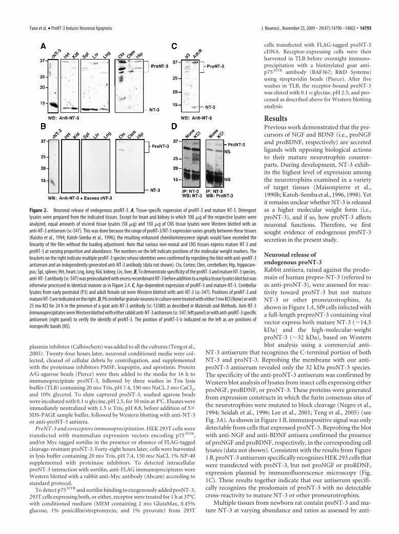
Figure 2. Neuronal release of endogenous proNT-3. A, Tissue-specific expression of proNT-3 and mature NT-3. Detergentlysates were prepared from the indicated tissues. Except for heart and kidney in which 100 �g of the respective lysates wereanalyzed, equal amounts of visceral tissue lysates (50 �g) and 150 �g of CNS tissue lysates were Western blotted with ananti-NT-3 antiserum (sc-547). This was done because the range of proNT-3/NT-3 expression varies greatly between these tissues(Kaisho et al., 1994; Katoh-Semba et al., 1996); the resulting enhanced chemiluminescence signals would have exceeded thelinearity of the film without the loading adjustment. Note that various non-neural and CNS tissues express mature NT-3 andproNT-3 at varying proportion and abundance. The numbers on the left indicate positions of the molecular weight markers. Thebrackets on the right indicate multiple proNT-3 species whose identities were confirmed by reprobing the blot with anti-proNT-3antiserum and an independently generated anti-NT-3 antibody (data not shown). Ctx, Cortex; Cbm, cerebellum; Hip, hippocam-pus; Spl, spleen; Hrt, heart; Lng, lung; Kid, kidney; Liv, liver. B, To demonstrate specificity of the proNT-3 and mature NT-3 species,anti-NT-3 antibody (sc-547) was preincubated with excess recombinant NT-3 before addition to a replica tissue lysates blot but wasotherwise processed in identical manner as in Figure 2 A. C, Age-dependent expression of proNT-3 and mature NT-3. Cerebellarlysates from early postnatal (P3) and adult female rat were Western blotted with anti-NT-3 (sc-547). Positions of proNT-3 andmature NT-3 are indicated on the right. D, P6 cerebellar granule neurons in culture were treated with either 5 mM KCl (None) or with25 mM KCl for 24 h in the presence of a goat anti-NT-3 antibody (sc-13380) as described in Materials and Methods. Anti-NT-3immunoprecipitates were Western blotted with either rabbit anti-NT-3 antiserum (sc-547; left panel) or with anti-proNT-3 specificantiserum (right panel) to verify the identify of proNT-3. The position of proNT-3 is indicated on the left as are positions ofnonspecific bands (NS).
Yano et al. • ProNT-3 Induces Neuronal Apoptosis J. Neurosci., November 25, 2009 • 29(47):14790 –14802 • 14793
NT-3 Western blot analysis (Fig. 2A). The specificity of proNT-3and mature NT-3 immunoreactivity was confirmed by probingreplica blot with the same anti-NT-3 antiserum in the presence ofexcess recombinant NT-3 (Fig. 2B). Although we cannot rule outthe possibility that lower molecular weight proNT-3 isoformsthat were present in some of the tissues arose from partial pro-teolytic degradation during sample preparation, it is noteworthythat proNT-3 of various sizes (�28 –37 kDa) have been predictedto exist because of alternative 5� exon usage as well as multiplein-frame ATG start sites within the NT-3 transcripts (Leingartnerand Lindholm, 1994; Kendall et al., 2000).
Interestingly, representative CNS tissues of P1 rat appear toexpress predominantly the high-molecular-weight proNT-3. Al-though prolonged exposure to film did reveal trace amount ofmature NT-3 in these CNS tissues (data not shown), it is apparentthat the ratio of proNT-3 to mature NT-3 is significantly greaterin the developing CNS, compared with visceral organs of thesame age. In contrast, mature NT-3 is readily detectable in theadult brain. As shown in Figure 2C, both proNT-3 and matureNT-3 are present in the cerebellum of older animals, consistentwith a recent finding that the relative expression of proBDNF andmature BDNF is developmentally regulated (Yang et al., 2009b).
Past studies have documented a high level of NT-3 expressionin the cerebellum (Lindholm et al., 1993; Katoh-Semba et al.,1998) and activity-dependent release of NT-3 from cultured cer-ebellar granule neurons (Sadakata et al., 2004, 2007). TheseELISA-based assays, however, did not distinguish whetherproNT-3 or mature NT-3 is secreted. Since cerebellum expressesmostly proNT-3 at early postnatal age (Fig. 2A), we performedanti-NT-3 immunoprecipitation analysis from conditioned me-dia of KCl-depolarized cultured cerebellar granule neurons (Fig.2D) to directly examine whether proNT-3 is released in an activity-dependent manner. Our data indicate that, under basal culture con-dition, there was negligible proNT-3 release from P6 granuleneurons. However, on membrane depolarization, a significantamount of proNT-3 can be detected in the media of these cultures.
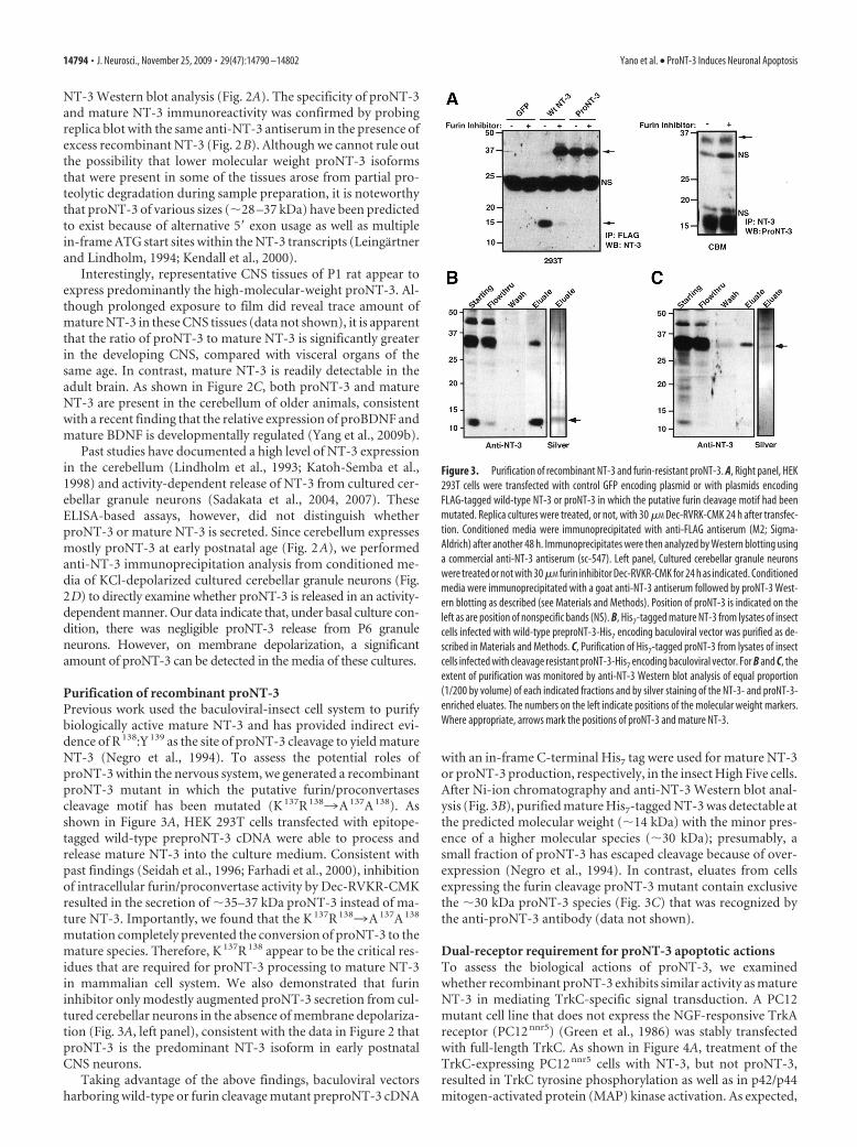
Purification of recombinant proNT-3Previous work used the baculoviral-insect cell system to purifybiologically active mature NT-3 and has provided indirect evi-dence of R 138:Y 139 as the site of proNT-3 cleavage to yield matureNT-3 (Negro et al., 1994). To assess the potential roles ofproNT-3 within the nervous system, we generated a recombinantproNT-3 mutant in which the putative furin/proconvertasescleavage motif has been mutated (K 137R 1383A 137A 138). Asshown in Figure 3A, HEK 293T cells transfected with epitope-tagged wild-type preproNT-3 cDNA were able to process andrelease mature NT-3 into the culture medium. Consistent withpast findings (Seidah et al., 1996; Farhadi et al., 2000), inhibitionof intracellular furin/proconvertase activity by Dec-RVKR-CMKresulted in the secretion of �35–37 kDa proNT-3 instead of ma-ture NT-3. Importantly, we found that the K 137R 1383A 137A 138
mutation completely prevented the conversion of proNT-3 to themature species. Therefore, K 137R 138 appear to be the critical res-idues that are required for proNT-3 processing to mature NT-3in mammalian cell system. We also demonstrated that furininhibitor only modestly augmented proNT-3 secretion from cul-tured cerebellar neurons in the absence of membrane depolariza-tion (Fig. 3A, left panel), consistent with the data in Figure 2 thatproNT-3 is the predominant NT-3 isoform in early postnatalCNS neurons.
Taking advantage of the above findings, baculoviral vectorsharboring wild-type or furin cleavage mutant preproNT-3 cDNA
with an in-frame C-terminal His7 tag were used for mature NT-3or proNT-3 production, respectively, in the insect High Five cells.After Ni-ion chromatography and anti-NT-3 Western blot anal-ysis (Fig. 3B), purified mature His7-tagged NT-3 was detectable atthe predicted molecular weight (�14 kDa) with the minor pres-ence of a higher molecular species (�30 kDa); presumably, asmall fraction of proNT-3 has escaped cleavage because of over-expression (Negro et al., 1994). In contrast, eluates from cellsexpressing the furin cleavage proNT-3 mutant contain exclusivethe �30 kDa proNT-3 species (Fig. 3C) that was recognized bythe anti-proNT-3 antibody (data not shown).
Dual-receptor requirement for proNT-3 apoptotic actionsTo assess the biological actions of proNT-3, we examinedwhether recombinant proNT-3 exhibits similar activity as matureNT-3 in mediating TrkC-specific signal transduction. A PC12mutant cell line that does not express the NGF-responsive TrkAreceptor (PC12 nnr5) (Green et al., 1986) was stably transfectedwith full-length TrkC. As shown in Figure 4A, treatment of theTrkC-expressing PC12 nnr5 cells with NT-3, but not proNT-3,resulted in TrkC tyrosine phosphorylation as well as in p42/p44mitogen-activated protein (MAP) kinase activation. As expected,
Figure 3. Purification of recombinant NT-3 and furin-resistant proNT-3. A, Right panel, HEK293T cells were transfected with control GFP encoding plasmid or with plasmids encodingFLAG-tagged wild-type NT-3 or proNT-3 in which the putative furin cleavage motif had beenmutated. Replica cultures were treated, or not, with 30 �M Dec-RVRK-CMK 24 h after transfec-tion. Conditioned media were immunoprecipitated with anti-FLAG antiserum (M2; Sigma-Aldrich) after another 48 h. Immunoprecipitates were then analyzed by Western blotting usinga commercial anti-NT-3 antiserum (sc-547). Left panel, Cultured cerebellar granule neuronswere treated or not with 30 �M furin inhibitor Dec-RVKR-CMK for 24 h as indicated. Conditionedmedia were immunoprecipitated with a goat anti-NT-3 antiserum followed by proNT-3 West-ern blotting as described (see Materials and Methods). Position of proNT-3 is indicated on theleft as are position of nonspecific bands (NS). B, His7-tagged mature NT-3 from lysates of insectcells infected with wild-type preproNT-3-His7 encoding baculoviral vector was purified as de-scribed in Materials and Methods. C, Purification of His7-tagged proNT-3 from lysates of insectcells infected with cleavage resistant proNT-3-His7 encoding baculoviral vector. For B and C, theextent of purification was monitored by anti-NT-3 Western blot analysis of equal proportion(1/200 by volume) of each indicated fractions and by silver staining of the NT-3- and proNT-3-enriched eluates. The numbers on the left indicate positions of the molecular weight markers.Where appropriate, arrows mark the positions of proNT-3 and mature NT-3.
14794 • J. Neurosci., November 25, 2009 • 29(47):14790 –14802 Yano et al. • ProNT-3 Induces Neuronal Apoptosis
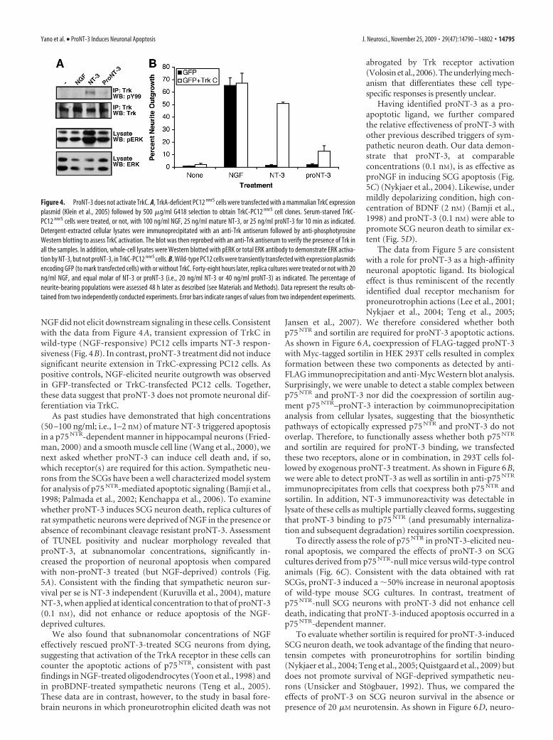
NGF did not elicit downstream signaling in these cells. Consistentwith the data from Figure 4A, transient expression of TrkC inwild-type (NGF-responsive) PC12 cells imparts NT-3 respon-siveness (Fig. 4B). In contrast, proNT-3 treatment did not inducesignificant neurite extension in TrkC-expressing PC12 cells. Aspositive controls, NGF-elicited neurite outgrowth was observedin GFP-transfected or TrkC-transfected PC12 cells. Together,these data suggest that proNT-3 does not promote neuronal dif-ferentiation via TrkC.
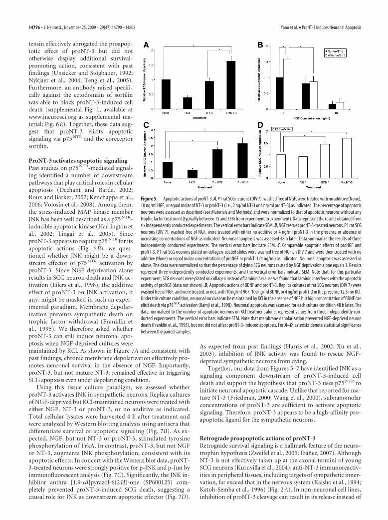
As past studies have demonstrated that high concentrations(50 –100 ng/ml; i.e., 1–2 nM) of mature NT-3 triggered apoptosisin a p75 NTR-dependent manner in hippocampal neurons (Fried-man, 2000) and a smooth muscle cell line (Wang et al., 2000), wenext asked whether proNT-3 can induce cell death and, if so,which receptor(s) are required for this action. Sympathetic neu-rons from the SCGs have been a well characterized model systemfor analysis of p75 NTR-mediated apoptotic signaling (Bamji et al.,1998; Palmada et al., 2002; Kenchappa et al., 2006). To examinewhether proNT-3 induces SCG neuron death, replica cultures ofrat sympathetic neurons were deprived of NGF in the presence orabsence of recombinant cleavage resistant proNT-3. Assessmentof TUNEL positivity and nuclear morphology revealed thatproNT-3, at subnanomolar concentrations, significantly in-creased the proportion of neuronal apoptosis when comparedwith non-proNT-3 treated (but NGF-deprived) controls (Fig.5A). Consistent with the finding that sympathetic neuron sur-vival per se is NT-3 independent (Kuruvilla et al., 2004), matureNT-3, when applied at identical concentration to that of proNT-3(0.1 nM), did not enhance or reduce apoptosis of the NGF-deprived cultures.
We also found that subnanomolar concentrations of NGFeffectively rescued proNT-3-treated SCG neurons from dying,suggesting that activation of the TrkA receptor in these cells cancounter the apoptotic actions of p75 NTR, consistent with pastfindings in NGF-treated oligodendrocytes (Yoon et al., 1998) andin proBDNF-treated sympathetic neurons (Teng et al., 2005).These data are in contrast, however, to the study in basal fore-brain neurons in which proneurotrophin elicited death was not
abrogated by Trk receptor activation(Volosin et al., 2006). The underlying mech-anism that differentiates these cell type-specific responses is presently unclear.
Having identified proNT-3 as a pro-apoptotic ligand, we further comparedthe relative effectiveness of proNT-3 withother previous described triggers of sym-pathetic neuron death. Our data demon-strate that proNT-3, at comparableconcentrations (0.1 nM), is as effective asproNGF in inducing SCG apoptosis (Fig.5C) (Nykjaer et al., 2004). Likewise, undermildly depolarizing condition, high con-centration of BDNF (2 nM) (Bamji et al.,1998) and proNT-3 (0.1 nM) were able topromote SCG neuron death to similar ex-tent (Fig. 5D).
The data from Figure 5 are consistentwith a role for proNT-3 as a high-affinityneuronal apoptotic ligand. Its biologicaleffect is thus reminiscent of the recentlyidentified dual receptor mechanism forproneurotrophin actions (Lee et al., 2001;Nykjaer et al., 2004; Teng et al., 2005;
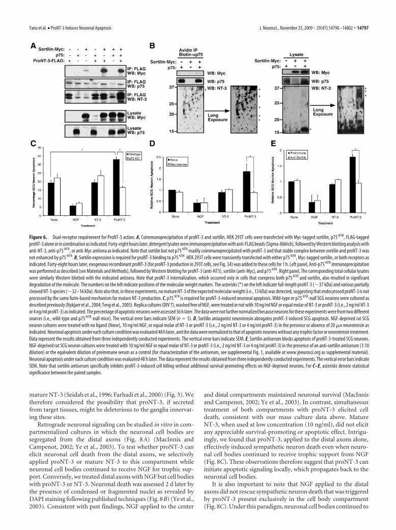
Jansen et al., 2007). We therefore considered whether bothp75 NTR and sortilin are required for proNT-3 apoptotic actions.As shown in Figure 6A, coexpression of FLAG-tagged proNT-3with Myc-tagged sortilin in HEK 293T cells resulted in complexformation between these two components as detected by anti-FLAG immunoprecipitation and anti-Myc Western blot analysis.Surprisingly, we were unable to detect a stable complex betweenp75 NTR and proNT-3 nor did the coexpression of sortilin aug-ment p75 NTR–proNT-3 interaction by coimmunoprecipitationanalysis from cellular lysates, suggesting that the biosyntheticpathways of ectopically expressed p75 NTR and proNT-3 do notoverlap. Therefore, to functionally assess whether both p75 NTR
and sortilin are required for proNT-3 binding, we transfectedthese two receptors, alone or in combination, in 293T cells fol-lowed by exogenous proNT-3 treatment. As shown in Figure 6B,we were able to detect proNT-3 as well as sortilin in anti-p75 NTR
immunoprecipitates from cells that coexpress both p75 NTR andsortilin. In addition, NT-3 immunoreactivity was detectable inlysate of these cells as multiple partially cleaved forms, suggestingthat proNT-3 binding to p75 NTR (and presumably internaliza-tion and subsequent degradation) requires sortilin coexpression.
To directly assess the role of p75 NTR in proNT-3-elicited neu-ronal apoptosis, we compared the effects of proNT-3 on SCGcultures derived from p75 NTR-null mice versus wild-type controlanimals (Fig. 6C). Consistent with the data obtained with ratSCGs, proNT-3 induced a �50% increase in neuronal apoptosisof wild-type mouse SCG cultures. In contrast, treatment ofp75 NTR-null SCG neurons with proNT-3 did not enhance celldeath, indicating that proNT-3-induced apoptosis occurred in ap75 NTR-dependent manner.
To evaluate whether sortilin is required for proNT-3-inducedSCG neuron death, we took advantage of the finding that neuro-tensin competes with proneurotrophins for sortilin binding(Nykjaer et al., 2004; Teng et al., 2005; Quistgaard et al., 2009) butdoes not promote survival of NGF-deprived sympathetic neu-rons (Unsicker and Stogbauer, 1992). Thus, we compared theeffects of proNT-3 on SCG neuron survival in the absence orpresence of 20 �M neurotensin. As shown in Figure 6D, neuro-
Figure 4. ProNT-3 does not activate TrkC. A, TrkA-deficient PC12 nnr5 cells were transfected with a mammalian TrkC expressionplasmid (Klein et al., 2005) followed by 500 �g/ml G418 selection to obtain TrkC-PC12 nnr5 cell clones. Serum-starved TrkC-PC12 nnr5 cells were treated, or not, with 100 ng/ml NGF, 25 ng/ml mature NT-3, or 25 ng/ml proNT-3 for 10 min as indicated.Detergent-extracted cellular lysates were immunoprecipitated with an anti-Trk antiserum followed by anti-phosphotyrosineWestern blotting to assess TrkC activation. The blot was then reprobed with an anti-Trk antiserum to verify the presence of Trk inall the samples. In addition, whole-cell lysates were Western blotted with pERK or total ERK antibody to demonstrate ERK activa-tion by NT-3, but not proNT-3, in TrkC-PC12 nnr5 cells. B, Wild-type PC12 cells were transiently transfected with expression plasmidsencoding GFP (to mark transfected cells) with or without TrkC. Forty-eight hours later, replica cultures were treated or not with 20ng/ml NGF, and equal molar of NT-3 or proNT-3 (i.e., 20 ng/ml NT-3 or 40 ng/ml proNT-3) as indicated. The percentage ofneurite-bearing populations were assessed 48 h later as described (see Materials and Methods). Data represent the results ob-tained from two independently conducted experiments. Error bars indicate ranges of values from two independent experiments.
Yano et al. • ProNT-3 Induces Neuronal Apoptosis J. Neurosci., November 25, 2009 • 29(47):14790 –14802 • 14795
tensin effectively abrogated the proapop-totic effect of proNT-3 but did nototherwise display additional survival-promoting action, consistent with pastfindings (Unsicker and Stogbauer, 1992;Nykjaer et al., 2004; Teng et al., 2005).Furthermore, an antibody raised specifi-cally against the ectodomain of sortilinwas able to block proNT-3-induced celldeath (supplemental Fig. 1, available atwww.jneurosci.org as supplemental ma-terial; Fig. 6E). Together, these data sug-gest that proNT-3 elicits apoptoticsignaling via p75 NTR and the coreceptorsortilin.
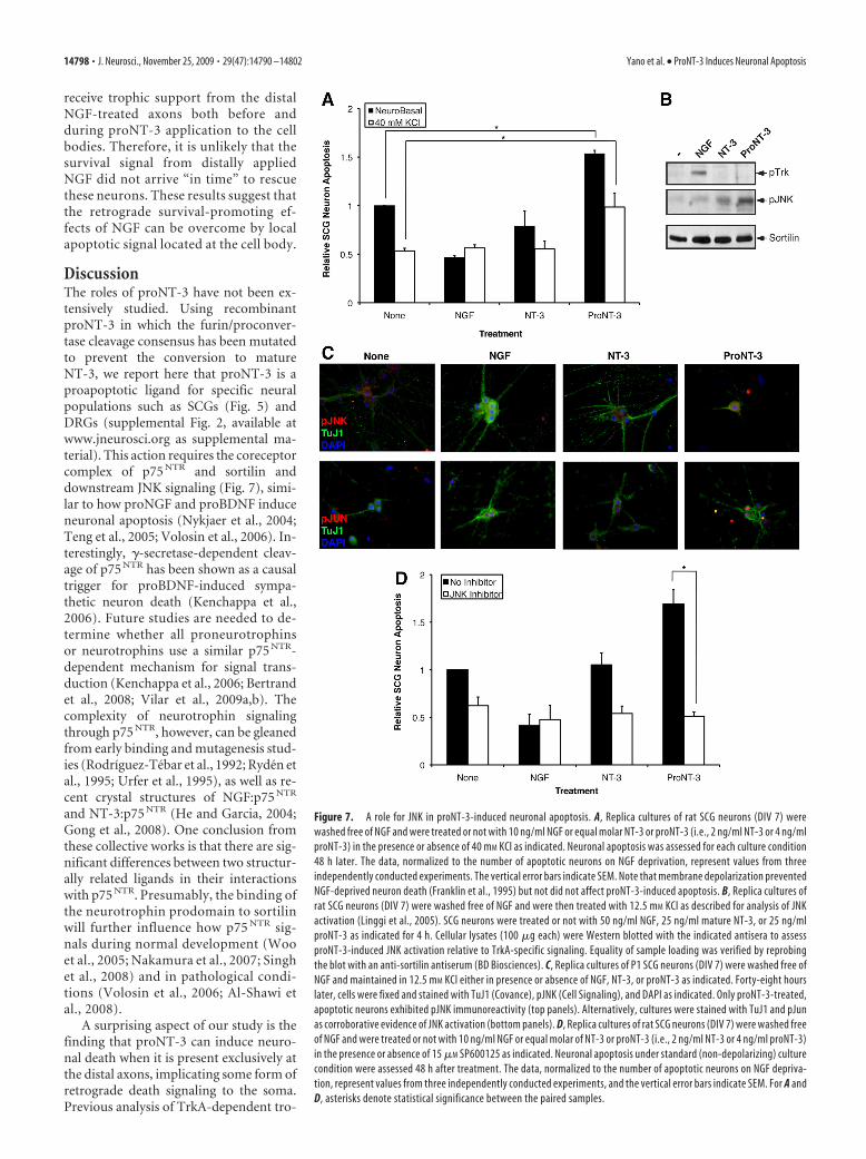
ProNT-3 activates apoptotic signalingPast studies on p75 NTR-mediated signal-ing identified a number of downstreampathways that play critical roles in cellularapoptosis (Dechant and Barde, 2002;Roux and Barker, 2002; Kenchappa et al.,2006; Volosin et al., 2008). Among them,the stress-induced MAP kinase memberJNK has been well described as a p75 NTR-inducible apoptotic kinase (Harrington etal., 2002; Linggi et al., 2005). SinceproNT-3 appears to require p75 NTR for itsapoptotic actions (Fig. 6B), we ques-tioned whether JNK might be a down-stream effector of p75 NTR activation byproNT-3. Since NGF deprivation aloneresults in SCG neuron death and JNK ac-tivation (Eilers et al., 1998), the additiveeffect of proNT-3 on JNK activation, ifany, might be masked in such an exper-imental paradigm. Membrane depolar-ization prevents sympathetic death ontrophic factor withdrawal (Franklin etal., 1995). We therefore asked whetherproNT-3 can still induce neuronal apo-ptosis when NGF-deprived cultures weremaintained by KCl. As shown in Figure 7A and consistent withpast findings, chronic membrane depolarization effectively pro-motes neuronal survival in the absence of NGF. Importantly,proNT-3, but not mature NT-3, remained effective in triggeringSCG apoptosis even under depolarizing condition.
Using this tissue culture paradigm, we assessed whetherproNT-3 activates JNK in sympathetic neurons. Replica culturesof NGF-deprived but KCl-maintained neurons were treated witheither NGF, NT-3 or proNT-3, or no additive as indicated.Total cellular lysates were harvested 4 h after treatment andwere analyzed by Western blotting analysis using antisera thatdifferentiate survival or apoptotic signaling (Fig. 7B). As ex-pected, NGF, but not NT-3 or proNT-3, stimulated tyrosinephosphorylation of TrkA. In contrast, proNT-3, but not NGFor NT-3, augments JNK phosphorylation, consistent with itsapoptotic effects. In concert with the Western blot data, proNT-3-treated neurons were strongly positive for p-JNK and p-Jun byimmunofluorescent analysis (Fig. 7C). Significantly, the JNK in-hibitor anthra [1,9-cd]pyrazol-6(2H)-one (SP600125) com-pletely prevented proNT-3-induced SCG death, suggesting acausal role for JNK as downstream apoptotic effector (Fig. 7D).
As expected from past findings (Harris et al., 2002; Xu et al.,2003), inhibition of JNK activity was found to rescue NGF-deprived sympathetic neurons from dying.
Together, our data from Figures 5–7 have identified JNK as asignaling component downstream of proNT-3-induced celldeath and support the hypothesis that proNT-3 uses p75 NTR toinitiate neuronal apoptotic cascade. Unlike that reported for ma-ture NT-3 (Friedman, 2000; Wang et al., 2000), subnanomolarconcentrations of proNT-3 are sufficient to activate apoptoticsignaling. Therefore, proNT-3 appears to be a high-affinity pro-apoptotic ligand for the sympathetic neurons.
Retrograde proapoptotic actions of proNT-3Retrograde survival signaling is a hallmark feature of the neuro-trophin hypothesis (Zweifel et al., 2005; Ibanez, 2007). AlthoughNT-3 is not effectively taken up at the axonal termini of youngSCG neurons (Kuruvilla et al., 2004), anti-NT-3 immunoreactiv-ities in peripheral tissues, including targets of sympathetic inner-vation, far exceed that in the nervous system (Kaisho et al., 1994;Katoh-Semba et al., 1996) (Fig. 2A). In non-neuronal cell lines,inhibition of proNT-3 cleavage can result in its release instead of
Figure 5. Apoptotic actions of proNT-3. A, P1 rat SCG neurons (DIV 7), washed free of NGF, were treated with no additive (None),10 ng/ml NGF, or equal molar of NT-3 or proNT-3 (i.e., 2 ng/ml NT-3 or 4 ng/ml proNT-3) as indicated. The percentage of apoptoticneurons were assessed as described (see Materials and Methods) and were normalized to that of apoptotic neurons without anytrophic factor treatment (typically between 15 and 25% from experiment to experiment). Data represent the results obtained fromsix independently conducted experiments. The vertical error bars indicate SEM. B, NGF rescues proNT-3-treated neurons. P1 rat SCGneurons (DIV 7), washed free of NGF, were treated with either no additive or 4 ng/ml proNT-3 in the presence or absence ofincreasing concentrations of NGF as indicated. Neuronal apoptosis was assessed 48 h later. Data summarize the results of threeindependently conducted experiments. The vertical error bars indicate SEM. C, Comparable apoptotic effects of proNGF andproNT-3. P1 rat SCG neurons plated on collagen-coated slides were washed free of NGF on DIV 7 and were then treated with noadditive (None) or equal molar concentrations of proNGF or proNT-3 (4 ng/ml) as indicated. Neuronal apoptosis was assessed asabove. The data were normalized so that the percentage of dying SCG neurons caused by NGF deprivation alone equals 1. Resultsrepresent three independently conducted experiments, and the vertical error bars indicate SEM. Note that, for this particularexperiment, SCG neurons were plated on collagen instead of laminin because we found that laminin interferes with the apoptoticactivity of proNGF (data not shown). D, Apoptotic actions of BDNF and proNT-3. Replica cultures of rat SCG neurons (DIV 7) werewashed free of NGF, and were treated, or not, with 10 ng/ml NGF, 100 ng/ml BDNF, or 4 ng/ml proNT-3 in the presence 12.5 mM KCl.Under this culture condition, neuronal survival can be maintained by KCl in the absence of NGF but high concentration of BDNF canelicit death via p75 NTR activation (Bamji et al., 1998). Neuronal apoptosis was assessed for each culture condition 48 h later. Thedata, normalized to the number of apoptotic neurons on KCl treatment alone, represent values from three independently con-ducted experiments. The vertical error bars indicate SEM. Note that membrane depolarization prevented NGF-deprived neurondeath (Franklin et al., 1995), but not did not affect proNT-3-induced apoptosis. For A–D, asterisks denote statistical significancebetween the paired samples.
14796 • J. Neurosci., November 25, 2009 • 29(47):14790 –14802 Yano et al. • ProNT-3 Induces Neuronal Apoptosis
mature NT-3 (Seidah et al., 1996; Farhadi et al., 2000) (Fig. 3). Wetherefore considered the possibility that proNT-3, if secretedfrom target tissues, might be deleterious to the ganglia innervat-ing these sites.
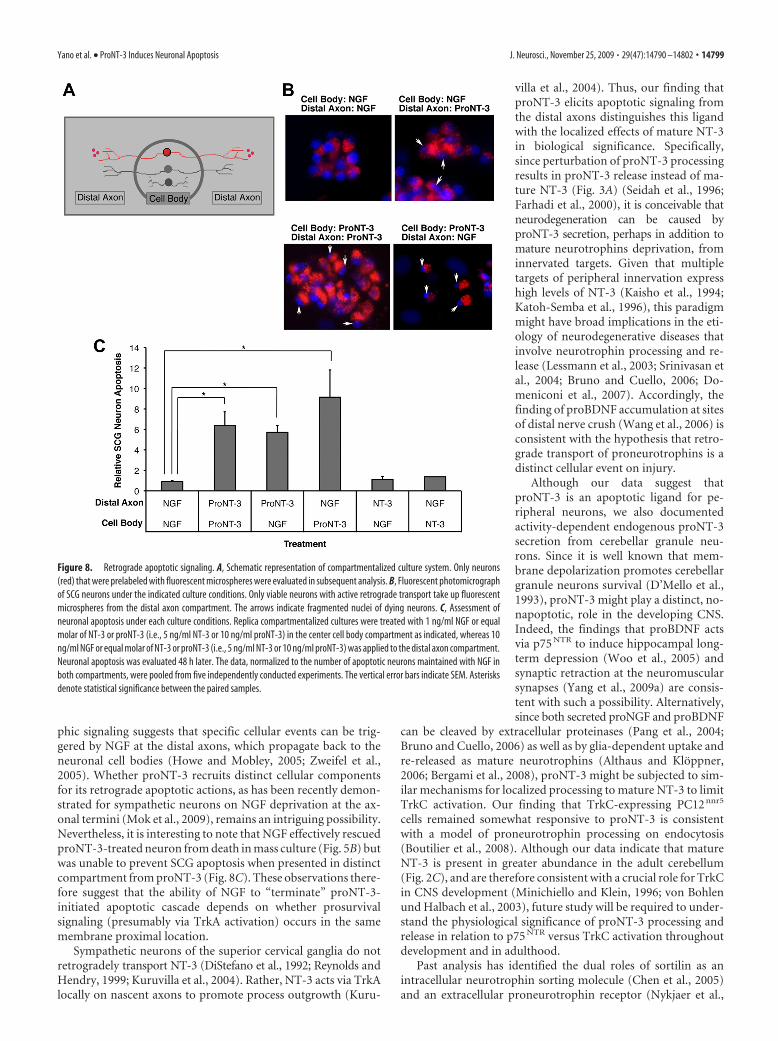
Retrograde neuronal signaling can be studied in vitro in com-partmentalized cultures in which the neuronal cell bodies aresegregated from the distal axons (Fig. 8A) (MacInnis andCampenot, 2002; Ye et al., 2003). To test whether proNT-3 canelicit neuronal cell death from the distal axons, we selectivelyapplied proNT-3 or mature NT-3 to this compartment whileneuronal cell bodies continued to receive NGF for trophic sup-port. Conversely, we treated distal axons with NGF but cell bodieswith proNT-3 or NT-3. Neuronal death was assessed 2 d later bythe presence of condensed or fragmented nuclei as revealed byDAPI staining following published techniques (Fig. 8B) (Ye et al.,2003). Consistent with past findings, NGF applied to the center
and distal compartments maintained neuronal survival (MacInnisand Campenot, 2002; Ye et al., 2003). In contrast, simultaneoustreatment of both compartments with proNT-3 elicited celldeath, consistent with our mass culture data above. MatureNT-3, when used at low concentration (10 ng/ml), did not elicitany appreciable survival-promoting or apoptotic effect. Intrigu-ingly, we found that proNT-3, applied to the distal axons alone,effectively induced sympathetic neuron death even when neuro-nal cell bodies continued to receive trophic support from NGF(Fig. 8C). These observations therefore suggest that proNT-3 caninitiate apoptotic signaling locally, which propagates back to theneuronal cell bodies.
It is also important to note that NGF applied to the distalaxons did not rescue sympathetic neuron death that was triggeredby proNT-3 present exclusively in the cell body compartment(Fig. 8C). Under this paradigm, neuronal cell bodies continued to
Figure 6. Dual-receptor requirement for ProNT-3 action. A, Coimmunoprecipitation of proNT-3 and sortilin. HEK 293T cells were transfected with Myc-tagged sortilin, p75 NTR, FLAG-taggedproNT-3 alone or in combination as indicated. Forty-eight hours later, detergent lysates were immunoprecipitation with anti-FLAG beads (Sigma-Aldrich), followed by Western blotting analysis withanti-NT-3, anti-p75 NTR, or anti-Myc antisera as indicated. Note that sortilin but not p75 NTR readily coimmunoprecipitated with proNT-3 and that stable complex between sortilin and proNT-3 wasnot enhanced by p75 NTR. B, Sortilin expression is required for proNT-3 binding to p75 NTR. HEK 293T cells were transiently transfected with either p75 NTR, Myc-tagged sortilin, or both receptors asindicated. Forty-eight hours later, exogenous recombinant proNT-3 (for proNT-3 production in 293T cells, see Fig. 3A) was added to these cells for 1 h. Left panel, Anti-p75 NTR immunoprecipitationwas performed as described (see Materials and Methods), followed by Western blotting for proNT-3 (anti-NT3), sortilin (anti-Myc), and p75 NTR. Right panel, The corresponding total cellular lysateswere similarly Western blotted with the indicated antisera. Note that proNT-3 internalization, which occurred only in cells that coexpress both p75 NTR and sortilin, also resulted in significantdegradation of the molecule. The numbers on the left indicate positions of the molecular weight markers. The asterisks (*) on the left indicate full-length proNT-3 (�37 kDa) and various partiallycleaved NT-3 species (�32–16 kDa). Note also that, in these experiments, no mature NT-3 of the expected molecular weight (i.e., 13 kDa) was detected, suggesting that endocytosed proNT-3 is notprocessed by the same furin-based mechanism for mature NT-3 production. C, p75 NTR is required for proNT-3-induced neuronal apoptosis. Wild-type or p75 NTR-null SCG neurons were cultured asdescribed previously (Nykjaer et al., 2004; Teng et al., 2005). Replica cultures (DIV 7), washed free of NGF, were treated or not with 10 ng/ml NGF or equal molar of NT-3 or proNT-3 (i.e., 2 ng/ml NT-3or 4 ng/ml proNT-3) as indicated. The percentage of apoptotic neurons were assessed 36 h later. The data were not further normalized because neurons for these experiments were from two differentsources (i.e., wild-type and p75 NTR-null mice). The vertical error bars indicate SEM (n � 5). D, Sortilin antagonist neurotensin abrogates proNT-3 induced SCG apoptosis. NGF-deprived rat SCGneuron cultures were treated with no ligand (None), 10 ng/ml NGF, or equal molar of NT-3 or proNT-3 (i.e., 2 ng/ml NT-3 or 4 ng/ml proNT-3) in the presence or absence of 20 �M neurotensin asindicated. Neuronal apoptosis under each culture condition was evaluated 48 h later, and the data were normalized to that of apoptotic neurons without any trophic factor or neurotensin treatment.Data represent the results obtained from three independently conducted experiments. The vertical error bars indicate SEM. E, Sortilin antiserum blocks apoptosis of proNT-3-treated SCG neurons.NGF-deprived rat SCG neuron cultures were treated with 10 ng/ml NGF or equal molar of NT-3 or proNT-3 (i.e., 2 ng/ml NT-3 or 4 ng/ml proNT-3) in the presence of an anti-sortilin antiserum (1:10dilution) or the equivalent dilution of preimmune serum as a control (for characterization of the antiserum, see supplemental Fig. 1, available at www.jneurosci.org as supplemental material).Neuronal apoptosis under each culture condition was evaluated 48 h later. The data represent the results obtained from three independently conducted experiments. The vertical error bars indicateSEM. Note that sortilin antiserum specifically inhibits proNT-3-induced cell killing without additional survival-promoting effects on NGF-deprived neurons. For C–E, asterisks denote statisticalsignificance between the paired samples.
Yano et al. • ProNT-3 Induces Neuronal Apoptosis J. Neurosci., November 25, 2009 • 29(47):14790 –14802 • 14797
receive trophic support from the distalNGF-treated axons both before andduring proNT-3 application to the cellbodies. Therefore, it is unlikely that thesurvival signal from distally appliedNGF did not arrive “in time” to rescuethese neurons. These results suggest thatthe retrograde survival-promoting ef-fects of NGF can be overcome by localapoptotic signal located at the cell body.
DiscussionThe roles of proNT-3 have not been ex-tensively studied. Using recombinantproNT-3 in which the furin/proconver-tase cleavage consensus has been mutatedto prevent the conversion to matureNT-3, we report here that proNT-3 is aproapoptotic ligand for specific neuralpopulations such as SCGs (Fig. 5) andDRGs (supplemental Fig. 2, available atwww.jneurosci.org as supplemental ma-terial). This action requires the coreceptorcomplex of p75 NTR and sortilin anddownstream JNK signaling (Fig. 7), simi-lar to how proNGF and proBDNF induceneuronal apoptosis (Nykjaer et al., 2004;Teng et al., 2005; Volosin et al., 2006). In-terestingly, �-secretase-dependent cleav-age of p75 NTR has been shown as a causaltrigger for proBDNF-induced sympa-thetic neuron death (Kenchappa et al.,2006). Future studies are needed to de-termine whether all proneurotrophinsor neurotrophins use a similar p75 NTR-dependent mechanism for signal trans-duction (Kenchappa et al., 2006; Bertrandet al., 2008; Vilar et al., 2009a,b). Thecomplexity of neurotrophin signalingthrough p75 NTR, however, can be gleanedfrom early binding and mutagenesis stud-ies (Rodríguez-Tebar et al., 1992; Ryden etal., 1995; Urfer et al., 1995), as well as re-cent crystal structures of NGF:p75 NTR
and NT-3:p75 NTR (He and Garcia, 2004;Gong et al., 2008). One conclusion fromthese collective works is that there are sig-nificant differences between two structur-ally related ligands in their interactionswith p75 NTR. Presumably, the binding ofthe neurotrophin prodomain to sortilinwill further influence how p75 NTR sig-nals during normal development (Wooet al., 2005; Nakamura et al., 2007; Singhet al., 2008) and in pathological condi-tions (Volosin et al., 2006; Al-Shawi etal., 2008).
A surprising aspect of our study is thefinding that proNT-3 can induce neuro-nal death when it is present exclusively atthe distal axons, implicating some form ofretrograde death signaling to the soma.Previous analysis of TrkA-dependent tro-
Figure 7. A role for JNK in proNT-3-induced neuronal apoptosis. A, Replica cultures of rat SCG neurons (DIV 7) werewashed free of NGF and were treated or not with 10 ng/ml NGF or equal molar NT-3 or proNT-3 (i.e., 2 ng/ml NT-3 or 4 ng/mlproNT-3) in the presence or absence of 40 mM KCl as indicated. Neuronal apoptosis was assessed for each culture condition48 h later. The data, normalized to the number of apoptotic neurons on NGF deprivation, represent values from threeindependently conducted experiments. The vertical error bars indicate SEM. Note that membrane depolarization preventedNGF-deprived neuron death (Franklin et al., 1995) but not did not affect proNT-3-induced apoptosis. B, Replica cultures ofrat SCG neurons (DIV 7) were washed free of NGF and were then treated with 12.5 mM KCl as described for analysis of JNKactivation (Linggi et al., 2005). SCG neurons were treated or not with 50 ng/ml NGF, 25 ng/ml mature NT-3, or 25 ng/mlproNT-3 as indicated for 4 h. Cellular lysates (100 �g each) were Western blotted with the indicated antisera to assessproNT-3-induced JNK activation relative to TrkA-specific signaling. Equality of sample loading was verified by reprobingthe blot with an anti-sortilin antiserum (BD Biosciences). C, Replica cultures of P1 SCG neurons (DIV 7) were washed free ofNGF and maintained in 12.5 mM KCl either in presence or absence of NGF, NT-3, or proNT-3 as indicated. Forty-eight hourslater, cells were fixed and stained with TuJ1 (Covance), pJNK (Cell Signaling), and DAPI as indicated. Only proNT-3-treated,apoptotic neurons exhibited pJNK immunoreactivity (top panels). Alternatively, cultures were stained with TuJ1 and pJunas corroborative evidence of JNK activation (bottom panels). D, Replica cultures of rat SCG neurons (DIV 7) were washed freeof NGF and were treated or not with 10 ng/ml NGF or equal molar of NT-3 or proNT-3 (i.e., 2 ng/ml NT-3 or 4 ng/ml proNT-3)in the presence or absence of 15 �M SP600125 as indicated. Neuronal apoptosis under standard (non-depolarizing) culturecondition were assessed 48 h after treatment. The data, normalized to the number of apoptotic neurons on NGF depriva-tion, represent values from three independently conducted experiments, and the vertical error bars indicate SEM. For A andD, asterisks denote statistical significance between the paired samples.
14798 • J. Neurosci., November 25, 2009 • 29(47):14790 –14802 Yano et al. • ProNT-3 Induces Neuronal Apoptosis
phic signaling suggests that specific cellular events can be trig-gered by NGF at the distal axons, which propagate back to theneuronal cell bodies (Howe and Mobley, 2005; Zweifel et al.,2005). Whether proNT-3 recruits distinct cellular componentsfor its retrograde apoptotic actions, as has been recently demon-strated for sympathetic neurons on NGF deprivation at the ax-onal termini (Mok et al., 2009), remains an intriguing possibility.Nevertheless, it is interesting to note that NGF effectively rescuedproNT-3-treated neuron from death in mass culture (Fig. 5B) butwas unable to prevent SCG apoptosis when presented in distinctcompartment from proNT-3 (Fig. 8C). These observations there-fore suggest that the ability of NGF to “terminate” proNT-3-initiated apoptotic cascade depends on whether prosurvivalsignaling (presumably via TrkA activation) occurs in the samemembrane proximal location.
Sympathetic neurons of the superior cervical ganglia do notretrogradely transport NT-3 (DiStefano et al., 1992; Reynolds andHendry, 1999; Kuruvilla et al., 2004). Rather, NT-3 acts via TrkAlocally on nascent axons to promote process outgrowth (Kuru-
villa et al., 2004). Thus, our finding thatproNT-3 elicits apoptotic signaling fromthe distal axons distinguishes this ligandwith the localized effects of mature NT-3in biological significance. Specifically,since perturbation of proNT-3 processingresults in proNT-3 release instead of ma-ture NT-3 (Fig. 3A) (Seidah et al., 1996;Farhadi et al., 2000), it is conceivable thatneurodegeneration can be caused byproNT-3 secretion, perhaps in addition tomature neurotrophins deprivation, frominnervated targets. Given that multipletargets of peripheral innervation expresshigh levels of NT-3 (Kaisho et al., 1994;Katoh-Semba et al., 1996), this paradigmmight have broad implications in the eti-ology of neurodegenerative diseases thatinvolve neurotrophin processing and re-lease (Lessmann et al., 2003; Srinivasan etal., 2004; Bruno and Cuello, 2006; Do-meniconi et al., 2007). Accordingly, thefinding of proBDNF accumulation at sitesof distal nerve crush (Wang et al., 2006) isconsistent with the hypothesis that retro-grade transport of proneurotrophins is adistinct cellular event on injury.
Although our data suggest thatproNT-3 is an apoptotic ligand for pe-ripheral neurons, we also documentedactivity-dependent endogenous proNT-3secretion from cerebellar granule neu-rons. Since it is well known that mem-brane depolarization promotes cerebellargranule neurons survival (D’Mello et al.,1993), proNT-3 might play a distinct, no-napoptotic, role in the developing CNS.Indeed, the findings that proBDNF actsvia p75 NTR to induce hippocampal long-term depression (Woo et al., 2005) andsynaptic retraction at the neuromuscularsynapses (Yang et al., 2009a) are consis-tent with such a possibility. Alternatively,since both secreted proNGF and proBDNF
can be cleaved by extracellular proteinases (Pang et al., 2004;Bruno and Cuello, 2006) as well as by glia-dependent uptake andre-released as mature neurotrophins (Althaus and Kloppner,2006; Bergami et al., 2008), proNT-3 might be subjected to sim-ilar mechanisms for localized processing to mature NT-3 to limitTrkC activation. Our finding that TrkC-expressing PC12 nnr5
cells remained somewhat responsive to proNT-3 is consistentwith a model of proneurotrophin processing on endocytosis(Boutilier et al., 2008). Although our data indicate that matureNT-3 is present in greater abundance in the adult cerebellum(Fig. 2C), and are therefore consistent with a crucial role for TrkCin CNS development (Minichiello and Klein, 1996; von Bohlenund Halbach et al., 2003), future study will be required to under-stand the physiological significance of proNT-3 processing andrelease in relation to p75 NTR versus TrkC activation throughoutdevelopment and in adulthood.
Past analysis has identified the dual roles of sortilin as anintracellular neurotrophin sorting molecule (Chen et al., 2005)and an extracellular proneurotrophin receptor (Nykjaer et al.,
Figure 8. Retrograde apoptotic signaling. A, Schematic representation of compartmentalized culture system. Only neurons(red) that were prelabeled with fluorescent microspheres were evaluated in subsequent analysis. B, Fluorescent photomicrographof SCG neurons under the indicated culture conditions. Only viable neurons with active retrograde transport take up fluorescentmicrospheres from the distal axon compartment. The arrows indicate fragmented nuclei of dying neurons. C, Assessment ofneuronal apoptosis under each culture conditions. Replica compartmentalized cultures were treated with 1 ng/ml NGF or equalmolar of NT-3 or proNT-3 (i.e., 5 ng/ml NT-3 or 10 ng/ml proNT-3) in the center cell body compartment as indicated, whereas 10ng/ml NGF or equal molar of NT-3 or proNT-3 (i.e., 5 ng/ml NT-3 or 10 ng/ml proNT-3) was applied to the distal axon compartment.Neuronal apoptosis was evaluated 48 h later. The data, normalized to the number of apoptotic neurons maintained with NGF inboth compartments, were pooled from five independently conducted experiments. The vertical error bars indicate SEM. Asterisksdenote statistical significance between the paired samples.
Yano et al. • ProNT-3 Induces Neuronal Apoptosis J. Neurosci., November 25, 2009 • 29(47):14790 –14802 • 14799
2004; Teng et al., 2005). Our findings are consistent with thelatter work and implicate sortilin as a p75 NTR coreceptor forproNT-3. Given the observation that proNT-3 prodomain cleav-age can be uncoupled from the secretory pathway (Figs. 2, 3)(Seidah et al., 1996; Farhadi et al., 2000), elucidating the molec-ular interactions between proNT-3 and sortilin will be crucial tounderstand whether sortilin also modulates proNT-3 processingand release. Although sortilin is but one of several related Vps-10p domain-containing molecules (Hermans-Borgmeyer et al.,1998; Mazella, 2001; Hermey et al., 2004), our data using a sorti-lin antagonist (Fig. 6D) and an anti-sortilin blocking antiserum(Fig. 6E) strongly suggest that the apoptotic action of proNT-3requires sortilin. Consistent with these findings, proNT-3 uptakein a surrogate cell line can only be reconstituted in the presence ofsortilin and p75 NTR (Fig. 6B). However, we cannot rule out thepossibility that in vivo where both p75 NTR and multiple sortilinfamily members are expressed at varying abundance and in dif-ferent cell types, the specificity of proneurotrophin actions mightbe mediated by additional sortilin family members.
In summary, our study raises the intriguing possibility ofproNT-3 as a target-derived apoptotic factor. Along with the re-ported role of truncated TrkC in mediating mature NT-3 actions(Esteban et al., 2006), NT-3 and its precursor proNT-3 are capa-ble of eliciting a plethora of biological events that differ in termsof coreceptor use and mechanisms of downstream signaling, con-sistent with their widespread expressions in both neural and non-neural tissues.
ReferencesAl-Shawi R, Hafner A, Olsen J, Olson J, Chun S, Raza S, Thrasivoulou C,
Lovestone S, Killick R, Simons P, Cowen T (2008) Neurotoxic and neu-rotrophic roles of proNGF and the receptor sortilin in the adult andageing nervous system. Eur J Neurosci 27:2103–2114.
Althaus HH, Kloppner S (2006) Mature pig oligodendrocytes rapidly pro-cess human recombinant pro-nerve growth factor and do not undergocell death. J Neurochem 98:506 –517.
Bamji SX, Majdan M, Pozniak CD, Belliveau DJ, Aloyz R, Kohn J, CausingCG, Miller FD (1998) The p75 neurotrophin receptor mediates neuro-nal apoptosis and is essential for naturally occurring sympathetic neurondeath. J Cell Biol 140:911–923.
Bates B, Rios M, Trumpp A, Chen C, Fan G, Bishop JM, Jaenisch R (1999)Neurotrophin-3 is required for proper cerebellar development. Nat Neurosci2:115–117.
Bergami M, Santi S, Formaggio E, Cagnoli C, Verderio C, Blum R, BerningerB, Matteoli M, Canossa M (2008) Uptake and recycling of pro-BDNFfor transmitter-induced secretion by cortical astrocytes. J Cell Biol183:213–221.
Bertrand MJ, Kenchappa RS, Andrieu D, Leclercq-Smekens M, Nguyen HN,Carter BD, Muscatelli F, Barker PA, De Backer O (2008) NRAGE, ap75NTR adaptor protein, is required for developmental apoptosis in vivo.Cell Death Differ 15:1921–1929.
Bibel M, Hoppe E, Barde YA (1999) Biochemical and functional interac-tions between the neurotrophin receptors trk and p75NTR. EMBO J18:616 – 622.
Boutilier J, Ceni C, Pagdala PC, Forgie A, Neet KE, Barker PA (2008) Pro-neurotrophins require endocytosis and intracellular proteolysis to induceTrkA activation. J Biol Chem 283:12709 –12716.
Bruno MA, Cuello AC (2006) Activity-dependent release of precursor nervegrowth factor, conversion to mature nerve growth factor, and its degra-dation by a protease cascade. Proc Natl Acad Sci U S A 103:6735– 6740.
Chen ZY, Ieraci A, Teng H, Dall H, Meng CX, Herrera DG, Nykjaer A,Hempstead BL, Lee FS (2005) Sortilin controls intracellular sortingof brain-derived neurotrophic factor to the regulated secretory path-way. J Neurosci 25:6156 – 6166.
Dechant G, Barde YA (2002) The neurotrophin receptor p75(NTR): novelfunctions and implications for diseases of the nervous system. Nat Neu-rosci 5:1131–1136.
DiStefano PS, Friedman B, Radziejewski C, Alexander C, Boland P, Schick
CM, Lindsay RM, Wiegand SJ (1992) The neurotrophins BDNF, NT-3,and NGF display distinct patterns of retrograde axonal-transport in pe-ripheral and central neurons. Neuron 8:983–993.
D’Mello SR, Galli C, Ciotti T, Calissano P (1993) Induction of apoptosis incerebellar granule neurons by low potassium: inhibition of death byinsulin-like growth factor I and cAMP. Proc Natl Acad Sci U S A90:10989 –10993.
Domeniconi M, Hempstead BL, Chao MV (2007) Pro-NGF secreted by as-trocytes promotes motor neuron cell death. Mol Cell Neurosci34:271–279.
Eilers A, Whitfield J, Babij C, Rubin LL, Ham J (1998) Role of the Jun kinasepathway in the regulation of c-Jun expression and apoptosis in sympa-thetic neurons. J Neurosci 18:1713–1724.
Esteban PF, Yoon HY, Becker J, Dorsey SG, Caprari P, Palko ME, Coppola V,Saragovi HU, Randazzo PA, Tessarollo L (2006) A kinase-deficientTrkC receptor isoform activates Arf6-Rac1 signaling through the scaffoldprotein tamalin. J Cell Biol 173:291–299.
Farhadi HF, Mowla SJ, Petrecca K, Morris SJ, Seidah NG, Murphy RA (2000)Neurotrophin-3 sorts to the constitutive secretory pathway of hip-pocampal neurons and is diverted to the regulated secretory pathwayby coexpression with brain-derived neurotrophic factor. J Neurosci20:4059 – 4068.
Franklin JL, Sanz-Rodriguez C, Juhasz A, Deckwerth TL, Johnson EM Jr(1995) Chronic depolarization prevents programmed death of sympa-thetic neurons in vitro but does not support growth: requirement forCa 2� influx but not Trk activation. J Neurosci 15:643– 664.
Friedman WJ (2000) Neurotrophins induce death of hippocampal neuronsvia the p75 receptor. J Neurosci 20:6340 – 6346.
Gentry JJ, Barker PA, Carter BD (2004) The p75 neurotrophin receptor:multiple interactors and numerous functions. Prog Brain Res 146:25–39.
Gong Y, Cao P, Yu HJ, Jiang T (2008) Crystal structure of the neurotrophin-3and p75NTR symmetrical complex. Nature 454:789–793.
Green SH, Rydel RE, Connolly JL, Greene LA (1986) PC12 cell mutantsthat possess low- but not high-affinity nerve growth factor receptorsneither respond to nor internalize nerve growth factor. J Cell Biol102:830 – 843.
Harrington AW, Kim JY, Yoon SO (2002) Activation of Rac GTPase by p75is necessary for c-Jun N-terminal kinase-mediated apoptosis. J Neurosci22:156 –166.
Harris CA, Deshmukh M, Tsui-Pierchala B, Maroney AC, Johnson EM Jr(2002) Inhibition of the c-Jun N-terminal kinase signaling pathway bythe mixed lineage kinase inhibitor CEP-1347 (KT7515) preserves metab-olism and growth of trophic factor-deprived neurons. J Neurosci22:103–113.
He XL, Garcia KC (2004) Structure of nerve growth factor complexed withthe shared neurotrophin receptor p75. Science 304:870 – 875.
Hermans-Borgmeyer I, Hampe W, Schinke B, Methner A, Nykjaer A, SusensU, Fenger U, Herbarth B, Schaller HC (1998) Unique expression patternof a novel mosaic receptor in the developing cerebral cortex. Mech Dev70:65–76.
Hermey G, Plath N, Hubner CA, Kuhl D, Schaller HC, Hermans-BorgmeyerI (2004) The three sorCS genes are differentially expressed and regulatedby synaptic activity. J Neurochem 88:1470 –1476.
Howe CL, Mobley WC (2005) Long-distance retrograde neurotrophic sig-naling. Curr Opin Neurobiol 15:40 – 48.
Ibanez CF (2007) Message in a bottle: long-range retrograde signaling in thenervous system. Trends Cell Biol 17:519 –528.
Jansen P, Giehl K, Nyengaard JR, Teng K, Lioubinski O, Sjoegaard SS,Breiderhoff T, Gotthardt M, Lin F, Eilers A, Petersen CM, Lewin GR,Hempstead BL, Willnow TE, Nykjaer A (2007) Roles for the pro-neurotrophin receptor sortilin in neuronal development, aging and braininjury. Nat Neurosci 10:1449 –1457.
Kahn MA, Kumar S, Liebl D, Chang R, Parada LF, De Vellis J (1999) Micelacking NT-3, and its receptor TrkC, exhibit profound deficiencies in CNSglial cells. Glia 26:153–165.
Kaisho Y, Shintani A, Nishida M, Fukumoto H, Igarashi K (1994) Develop-mental changes of neurotrophin-3 level in the mouse brain detected by ahighly sensitive enzyme immunoassay. Brain Res 666:143–146.
Katoh-Semba R, Kaisho Y, Shintani A, Nagahama M, Kato K (1996) Tissuedistribution and immunocytochemical localization of neurotrophin-3 inthe brain and peripheral tissues of rats. J Neurochem 66:330 –337.
Katoh-Semba R, Semba R, Takeuchi IK, Kato K (1998) Age-related changes
14800 • J. Neurosci., November 25, 2009 • 29(47):14790 –14802 Yano et al. • ProNT-3 Induces Neuronal Apoptosis
in levels of brain-derived neurotrophic factor in selected brain regions ofrats, normal mice and senescence-accelerated mice: a comparison tothose of nerve growth factor and neurotrophin-3. Neurosci Res31:227–234.
Kenchappa RS, Zampieri N, Chao MV, Barker PA, Teng HK, Hempstead BL,Carter BD (2006) Ligand-dependent cleavage of the P75 neurotrophinreceptor is necessary for NRIF nuclear translocation and apoptosis insympathetic neurons. Neuron 50:219 –232.
Kendall S, Yeo M, Henttu P, Tomlinson DR (2000) Alternative splicing ofthe neurotrophin-3 gene gives rise to different transcripts in a number ofhuman and rat tissues. J Neurochem 75:41– 47.
Klein M, Hempstead BL, Teng KK (2005) Activation of STAT5-dependenttranscription by the neurotrophin receptor Trk. J Neurobiol 63:159 –171.
Kuruvilla R, Zweifel LS, Glebova NO, Lonze BE, Valdez G, Ye H, Ginty DD(2004) A neurotrophin signaling cascade coordinates sympathetic neu-ron development through differential control of TrkA trafficking andretrograde signaling. Cell 118:243–255.
Lamballe F, Klein R, Barbacid M (1991) TrkC, a new member of the trkfamily of tyrosine protein kinases, is a receptor for neurotrophin-3. Cell66:967–979.
Lee KF, Li E, Huber LJ, Landis SC, Sharpe AH, Chao MV, Jaenisch R (1992)Targeted mutation of the gene encoding the low affinity NGF receptorp75 leads to deficits in the peripheral sensory nervous system. Cell69:737–749.
Lee R, Kermani P, Teng KK, Hempstead BL (2001) Regulation of cell sur-vival by secreted proneurotrophins. Science 294:1945–1948.
Leingartner A, Lindholm D (1994) Two promoters direct transcription ofthe mouse NT-3 gene. Eur J Neurosci 6:1149 –1159.
Lessmann V, Gottmann K, Malcangio M (2003) Neurotrophin secretion:current facts and future prospects. Prog Neurobiol 69:341–374.
Lewin GR, Barde YA (1996) Physiology of the neurotrophins. Annu RevNeurosci 19:289 –317.
Lindholm D, Castren E, Tsoulfas P, Kolbeck R, Berzaghi Mda P, LeingartnerA, Heisenberg CP, Tessarollo L, Parada LF, Thoenen H, Tesarollo L(1993) Neurotrophin-3 induced by tri-iodothyronine in cerebellar gran-ule cells promotes Purkinje cell differentiation. J Cell Biol 122:443– 450.
Linggi MS, Burke TL, Williams BB, Harrington A, Kraemer R, Hempstead BL,Yoon SO, Carter BD (2005) Neurotrophin receptor interacting factor(NRIF) is an essential mediator of apoptotic signaling by the p75 neuro-trophin receptor. J Biol Chem 280:13801–13808.
Ma L, Harada T, Harada C, Romero M, Hebert JM, McConnell SK, Parada LF(2002) Neurotrophin-3 is required for appropriate establishment ofthalamocortical connections. Neuron 36:623– 634.
MacInnis BL, Campenot RB (2002) Retrograde support of neuronal sur-vival without retrograde transport of nerve growth factor. Science295:1536 –1539.
Maisonpierre PC, Belluscio L, Squinto S, Ip NY, Furth ME, Lindsay RM,Yancopoulos GD (1990a) Neurotrophin-3: a neurotrophic factor re-lated to NGF and BDNF. Science 247:1446 –1451.
Maisonpierre PC, Belluscio L, Friedman B, Alderson RF, Wiegand SJ, FurthME, Lindsay RM, Yancopoulos GD (1990b) NT-3, BDNF, and NGF inthe developing rat nervous system: parallel as well as reciprocal patterns ofexpression. Neuron 5:501–509.
Mazella J (2001) Sortilin/neurotensin receptor-3: a new tool to investigateneurotensin signaling and cellular trafficking? Cell Signal 13:1– 6.
Minichiello L, Klein R (1996) TrkB and TrkC neurotrophin receptors coop-erate in promoting survival of hippocampal and cerebellar granule neu-rons. Genes Dev 10:2849 –2858.
Mischel PS, Smith SG, Vining ER, Valletta JS, Mobley WC, Reichardt LF(2001) The extracellular domain of p75NTR is necessary to inhibitneurotrophin-3 signaling through TrkA. J Biol Chem 276:11294 –11301.
Mok SA, Lund K, Campenot RB (2009) A retrograde apoptotic signal orig-inating in NGF-deprived distal axons of rat sympathetic neurons in com-partmented cultures. Cell Res 19:546 –560.
Nagappan G, Zaitsev E, Senatorov VV Jr, Yang J, Hempstead BL, Lu B (2009)Control of extracellular cleavage of ProBDNF by high frequency neuronalactivity. Proc Natl Acad Sci U S A 106:1267–1272.
Nakamura K, Namekata K, Harada C, Harada T (2007) Intracellular sortilinexpression pattern regulates proNGF-induced naturally occurring celldeath during development. Cell Death Differ 14:1552–1554.
Negro A, Tavella A, Grandi C, Skaper SD (1994) Production and character-
ization of recombinant rat brain-derived neurotrophic factor andneurotrophin-3 from insect cells. J Neurochem 62:471– 478.
Nykjaer A, Lee R, Teng KK, Jansen P, Madsen P, Nielsen MS, Jacobsen C,Kliemannel M, Schwarz E, Willnow TE, Hempstead BL, Petersen CM(2004) Sortilin is essential for proNGF-induced neuronal cell death. Na-ture 427:843– 848.
Palmada M, Kanwal S, Rutkoski NJ, Gustafson-Brown C, Johnson RS,Wisdom R, Carter BD (2002) c-jun is essential for sympathetic neuronaldeath induced by NGF withdrawal but not by p75 activation. J Cell Biol158:453– 461.
Pang PT, Teng HK, Zaitsev E, Woo NT, Sakata K, Zhen S, Teng KK, YungWH, Hempstead BL, Lu B (2004) Cleavage of proBDNF by tPA/plasminis essential for long-term hippocampal plasticity. Science 306:487– 491.
Quistgaard EM, Madsen P, Grøftehauge MK, Nissen P, Petersen CM, ThirupSS (2009) Ligands bind to Sortilin in the tunnel of a ten-bladed beta-propeller domain. Nat Struct Mol Biol 16:96 –98.
Randolph CL, Bierl MA, Isaacson LG (2007) Regulation of NGF and NT-3protein expression in peripheral targets by sympathetic input. Brain Res1144:59 – 69.
Reynolds AJ, Hendry IA (1999) A technique for 125I-labelling of neurotro-phins and the use of retrograde axonal transport as a bioassay. Brain ResBrain Res Protoc 3:308 –312.
Rodríguez-Tebar A, Dechant G, Gotz R, Barde YA (1992) Binding ofneurotrophin-3 to its neuronal receptors and interactions with nervegrowth factor and brain-derived neurotrophic factor. EMBO J 11:917–922.
Roux PP, Barker PA (2002) Neurotrophin signaling through the p75 neu-rotrophin receptor. Prog Neurobiol 67:203–233.
Ryden M, Murray-Rust J, Glass D, Ilag LL, Trupp M, Yancopoulos GD,McDonald NQ, Ibanez CF (1995) Functional analysis of mutant neuro-trophins deficient in low-affinity binding reveals a role for p75LNGFR inNT-4 signalling. EMBO J 14:1979 –1990.
Sadakata T, Mizoguchi A, Sato Y, Katoh-Semba R, Fukuda M, Mikoshiba K,Furuichi T (2004) The secretory granule-associated protein CAPS2 reg-ulates neurotrophin release and cell survival. J Neurosci 24:43–52.
Sadakata T, Kakegawa W, Mizoguchi A, Washida M, Katoh-Semba R, ShutohF, Okamoto T, Nakashima H, Kimura K, Tanaka M, Sekine Y, Itohara S,Yuzaki M, Nagao S, Furuichi T (2007) Impaired cerebellar developmentand function in mice lacking CAPS2, a protein involved in neurotrophinrelease. J Neurosci 27:2472–2482.
Schecterson LC, Bothwell M (1992) Novel roles for neurotrophins are sug-gested by BDNF and NT-3 mRNA expression in developing neurons.Neuron 9:449 – 463.
Segal RA (2003) Selectivity in neurotrophin signaling: theme and variations.Annu Rev Neurosci 26:299 –330.
Seidah NG, Benjannet S, Pareek S, Chretien M, Murphy RA (1996) Cellularprocessing of the neurotrophin precursors of NT3 and BDNF by themammalian proprotein convertases. FEBS Lett 379:247–250.
Singh KK, Park KJ, Hong EJ, Kramer BM, Greenberg ME, Kaplan DR, MillerFD (2008) Developmental axon pruning mediated by BDNF-p75NTR-dependent axon degeneration. Nat Neurosci 11:649 – 658.
Srinivasan B, Roque CH, Hempstead BL, Al-Ubaidi MR, Roque RS (2004)Microglia-derived pronerve growth factor promotes photoreceptor celldeath via p75 neurotrophin receptor. J Biol Chem 279:41839 – 41845.
Teng HK, Teng KK, Lee R, Wright S, Tevar S, Almeida RD, Kermani P, TorkinR, Chen ZY, Lee FS, Kraemer RT, Nykjaer A, Hempstead BL (2005)ProBDNF induces neuronal apoptosis via activation of a receptor com-plex of p75NTR and sortilin. J Neurosci 25:5455–5463.
Teng KK, Hempstead BL (2004) Neurotrophins and their receptors: signal-ing trios in complex biological systems. Cell Mol Life Sci 61:35– 48.
Unsicker K, Stogbauer F (1992) Screening of adrenal medullary neuropep-tides for putative neurotrophic effects. Int J Dev Neurosci 10:171–179.
Urfer R, Tsoulfas P, O’Connell L, Shelton DL, Parada LF, Presta LG (1995)An immunoglobulin-like domain determines the specificity of neurotro-phin receptors. EMBO J 14:2795–2805.
Vilar M, Charalampopoulos I, Kenchappa RS, Simi A, Karaca E, Reversi A,Choi S, Bothwell M, Mingarro I, Friedman WJ, Schiavo G, Bastiaens PI,Verveer PJ, Carter BD, Ibanez CF (2009a) Activation of the p75 neuro-trophin receptor through conformational rearrangement of disulphide-linked receptor dimers. Neuron 62:72– 83.
Vilar M, Charalampopoulos I, Kenchappa RS, Reversi A, Klos-Applequist JM,Karaca E, Simi A, Spuch C, Choi S, Friedman WJ, Ericson J, Schiavo G,
Yano et al. • ProNT-3 Induces Neuronal Apoptosis J. Neurosci., November 25, 2009 • 29(47):14790 –14802 • 14801
Carter BD, Ibanez CF (2009b) Ligand-independent signaling bydisulfide-crosslinked dimers of the p75 neurotrophin receptor. J Cell Sci122:3351–3357.
Volosin M, Song W, Almeida RD, Kaplan DR, Hempstead BL, Friedman WJ(2006) Interaction of survival and death signaling in basal forebrain neu-rons: roles of neurotrophins and proneurotrophins. J Neurosci 26:7756 –7766.
Volosin M, Trotter C, Cragnolini A, Kenchappa RS, Light M, Hempstead BL,Carter BD, Friedman WJ (2008) Induction of proneurotrophins andactivation of p75NTR-mediated apoptosis via neurotrophin receptor-interacting factor in hippocampal neurons after seizures. J Neurosci28:9870 –9879.
von Bohlen und Halbach O, Minichiello L, Unsicker K (2003) Haploinsuf-ficiency in trkB and/or trkC neurotrophin receptors causes structuralalterations in the aged hippocampus and amygdala. Eur J Neurosci18:2319 –2325.
Wang H, Wu LL, Song XY, Luo XG, Zhong JH, Rush RA, Zhou XF (2006)Axonal transport of BDNF precursor in primary sensory neurons. EurJ Neurosci 24:2444 –2452.
Wang S, Bray P, McCaffrey T, March K, Hempstead BL, Kraemer R (2000)p75(NTR) mediates neurotrophin-induced apoptosis of vascular smoothmuscle cells. Am J Pathol 157:1247–1258.
Willnow TE, Petersen CM, Nykjaer A (2008) VPS10P-domain receptors—
regulators of neuronal viability and function. Nat Rev Neurosci 9:899 –909.
Woo NH, Teng HK, Siao CJ, Chiaruttini C, Pang PT, Milner TA, HempsteadBL, Lu B (2005) Activation of p75NTR by proBDNF facilitates hip-pocampal long-term depression. Nat Neurosci 8:1069 –1077.
Xu Z, Kukekov NV, Greene LA (2003) POSH acts as a scaffold for a multi-protein complex that mediates JNK activation in apoptosis. EMBO J22:252–261.
Yang F, Je HS, Ji Y, Nagappan G, Hempstead B, Lu B (2009a) Pro-BDNF-induced synaptic depression and retraction at developing neuromuscularsynapses. J Cell Biol 185:727–741.
Yang J, Siao CJ, Nagappan G, Marinic T, Jing D, McGrath K, Chen ZY, MarkW, Tessarollo L, Lee FS, Lu B, Hempstead BL (2009b) Neuronal releaseof proBDNF. Nat Neurosci 12:113–115.
Ye H, Kuruvilla R, Zweifel LS, Ginty DD (2003) Evidence in support ofsignaling endosome-based retrograde survival of sympathetic neurons.Neuron 39:57– 68.
Yoon SO, Casaccia-Bonnefil P, Carter B, Chao MV (1998) Competitive sig-naling between TrkA and p75 nerve growth factor receptors determinescell survival. J Neurosci 18:3273–3281.
Zhang SH, Zhou XF, Rush RA (2001) Extraction and quantification of theneurotrophins. Methods Mol Biol 169:31– 41.
Zweifel LS, Kuruvilla R, Ginty DD (2005) Functions and mechanisms ofretrograde neurotrophin signalling. Nat Rev Neurosci 6:615– 625.
14802 • J. Neurosci., November 25, 2009 • 29(47):14790 –14802 Yano et al. • ProNT-3 Induces Neuronal Apoptosis