cellular toxicity of nanoparticles by: amber cardell
TRANSCRIPT

Cellular Toxicity of Nanoparticles
By: Amber Cardell

Nanoparticles
10-9m – very small 1-100nm in size Characteristics different
from bulk material to nanoscale
Changes chemical and/or physical properties
http://www.sas.muohio.edu/pse/ppsfaculty/kerr.html

Nanoparticles- High Surface Area to Volume Ratio
High surface tension Tendency to adhere and clump Increases solubility Drives diffusion Reduces melting temp Increases catalytic activity
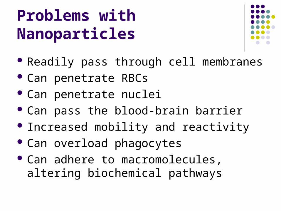
Problems with Nanoparticles
Readily pass through cell membranes Can penetrate RBCs Can penetrate nuclei Can pass the blood-brain barrier Increased mobility and reactivity Can overload phagocytes Can adhere to macromolecules, altering
biochemical pathways

Regulation of Nanoparticles
Not regulated by the EPA or FDA MSDS does not differentiate nanosize material Causes pollution as byproducts Commonly used in commercial products
Cosmetics Protective coatings Toothpaste Stain resistant clothing Suntan lotion

Materials and Methods Cell culture
Chinese Hamster Ovary Cells (CHO-K1) Transformed immortal cell
line Sensitive to toxic agents
Centrifuge cells Aseptic technique
70% ethanol- to sanitize all supplies
UV light under hood to eliminate microbial contaminants in the work area

Materials and Methods
Controlled environment Culture plates and flasks 5% CO2, 95% air 37°C Liquid Media
90% Kaighn’s Modification Essential nutrients for
CHO cells 10% fetal bovine serum Penicillin-Streptomycin
solution

Materials and Methods
Trypsin Detaches cells
Hanks Balanced Salt Solution Rinses cells pH indicator Solvent for nanoparticles
PBS Rinses cells

Particles Used Latex Beads
800 nm- fine particulate matter
Dyed blue 10 μl
Talc particles 15 nm diameter Concentrations
0.1 mg/ml 1.0 mg/ml 10 mg/ml (protein content)
Silica 7 nm diameter Concentrations
0.1 mg/ml 1.0 mg/ml 10 mg/ml (protein content)

Toxicity Assays
Phase Contrast Microscope
Trypan Blue Dead cells uptake dye Stored in nucleus
Total Protein Content Bradford assay Spectrophotometer
Standard curve Lower shows toxicity

CHO Cells- Control
NormalHealthyCell
Cells treated with Media24 hr exposure
100x
400x
1,000x1,000x

ExperimentalResults
Cells treated with Latex Beads10μl24 hr exposure
Beads inside cell
Beads outside cell
100x
400x400x 1,000x
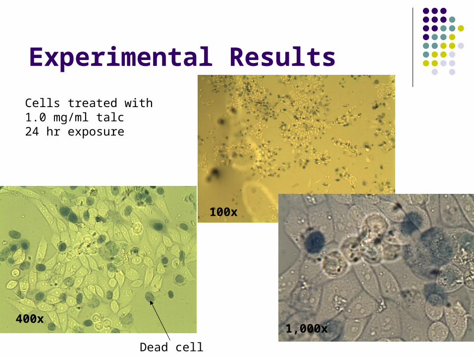
Experimental Results
Cells treated with1.0 mg/ml talc24 hr exposure
Dead cell
100x
400x1,000x

Experimental ResultsCells treated with 1.0 mg/ml Silica24 hr exposure
Dead Cell
100x
400x
1,000x

Cellular Toxicity Cell Mortality
0%
10%
20%
30%
40%
50%
60%
70%
Treatment
% M
ort
ality

Total Cell Protein ContentSpectrophotometer Readings at 595 nm
Standard CurveR2 = 0.9284
0
0.2
0.4
0.6
0.8
1
1.2
0 0.5 1 1.5 2 2.5
Concentration of Protein (mg/ml)
abso
rban
ce

Total Protein ContentTotal Protein Content
00.20.40.6
0.81
1.21.4
Media2 ml
Latexbeads10 μl
Talc0.1
mg/ml
Talc1.0
mg/ml
Talc 10mg/ml
Silica0.1
mg/ml
Silica1.0
mg/ml
Silica10
mg/ml
Treatments
Pro
tein
Co
nte
nt
(mg
/ml)

Conclusions
Nanoparticles were uptaken by the cells Cell mortality increased with exposure to
nanoparticles Silica seemed to be the most toxic agent Further toxicity research is needed before
continuing the use of nanoparticles in commercial products

Acknowledgements
Dr. Jacqueline Jordan- CSU Rishit Patel and Samantha Stuckey- CSU Derek Truyen Pham- Emory