cell structure and genetic control
TRANSCRIPT
Cell Structure and Genetic Control
Human physiology

Cell•Basic unit of structure and function of the
body.▫Highly organized molecular factory.
•Great diversity of function.▫Organ physiology derived from complex
functions of the cell.•3 principal parts:
▫Plasma membrane.▫Cytoplasm and organelles.▫Nucleus.

Plasma Membrane
• Is selectively permeable.• Composition:
▫ Double layer of phospholipids due to hydrophobic/hydrophilic parts. Restrict passage of H20 and H20 soluble ions.
▫ Proteins span or partially span the membrane. Provide structural support, transport molecules, serve as
receptors.▫Negatively charged carbohydrates attach to
the outer surface. Involved with regulatory molecules.

Plasma Membrane (continued)

Cytoplasm, Organelles, Nucleoli
• Cytoplasm:▫ Aqueous content of the cell.
• Organelles:▫ Sub-cellular structures within the cytoplasm.
• Nucleus:▫ Is a large spheroid body.▫ Largest of the organelles.▫ Contains the genetic material (DNA).▫Nucleoli:
Centers for production of ribosomes.

Cytoplasm, Organelles, Nucleoli (continued)
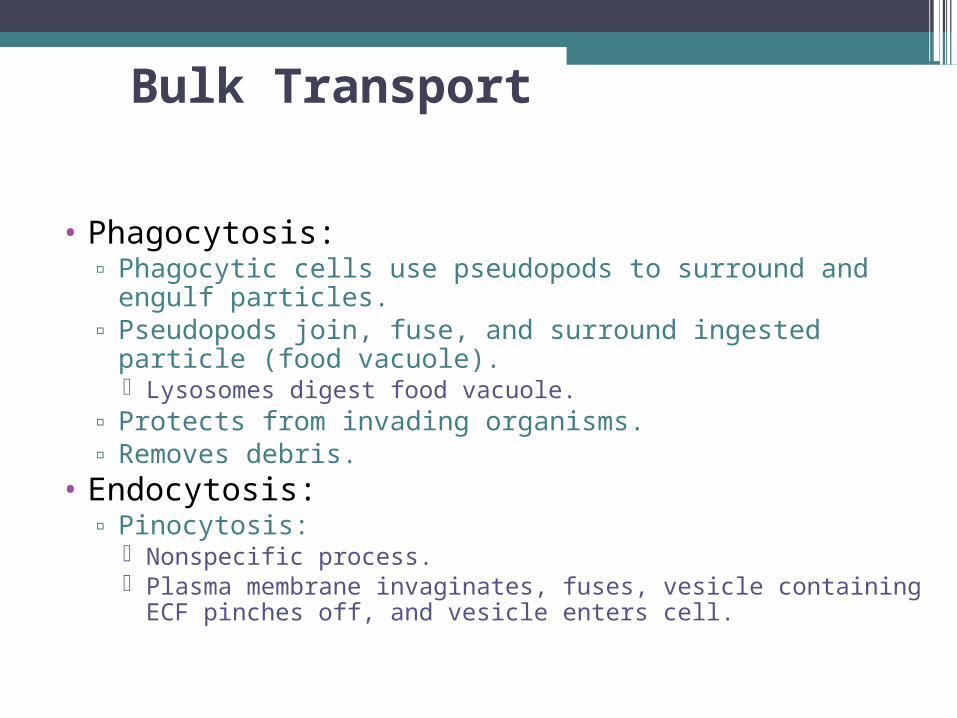
Bulk Transport
• Phagocytosis:▫ Phagocytic cells use pseudopods to surround and engulf
particles.▫ Pseudopods join, fuse, and surround ingested particle (food
vacuole). Lysosomes digest food vacuole.
▫ Protects from invading organisms.▫ Removes debris.
• Endocytosis:▫ Pinocytosis:
Nonspecific process. Plasma membrane invaginates, fuses, vesicle containing ECF
pinches off, and vesicle enters cell.

Bulk Transport (continued)
• Receptor-mediated endocytosis:▫ Interaction of molecules in ECF with specific membrane
receptor proteins.▫ Membrane invaginates, fuses, pinches off and forms
vesicle.▫ Vesicle enters cell.
• Exocytosis:▫ Process by which cellular products are secreted into
extracellular environment.▫ Proteins and other molecules to be secreted are packaged
in vesicles by Golgi complex.▫ Vesicles fuse with plasma membrane and release contents
into extracellular environment.

Cilia, Flagella, Microvilli
•Cilia:▫Tiny hair-like structures that project from
the surface of the cell. Stroke in unison.
Respiratory tract, uterine tube.
•Flagella:▫Simple whip-like structure that propels
sperm through its environment.•Microvilli:
▫Numerous folds (finger-like projections) increase surface area. Aid absorption.

Cytoplasm and Cytoskeleton
• Cytoplasm:▫ Jelly-like matrix within
the cell.▫ Includes organelles
and cytosol.▫ Highly organized
structure with microtubules and microfilaments that function as cytoskeleton.
• Cytoskeleton:▫ Actin and myosin
(microfilaments).▫ Spindle apparatus
(microtubules).

Lysosomes
▫Primary: Contain only digestive enzymes.
▫Secondary: Primary lysosome fuses with food vacuole or organelle.
Contain partially digested remnants of other organelles and organic material.
▫Residual body: Contain undigested wastes.
▫Autophagy: Process that destroys worn-out organelles, so that they
can be continuously replaced.▫Apoptosis (programmed cell death):
Lysosomes release digestive enzymes into the cell.

Peroxisomes
•Membrane-enclosed organelles.▫Contain specific enzymes that promote
oxidative reactions.▫Oxidize molecules and form H202.
•Catalase: converts H202 H20 + 02.•Oxidation of toxic molecules by
peroxisomes is an important function of liver and kidney cells.

Mitochondria
• Sites for energy production of all cells; but mature RBCs.
• Contain own DNA, can reproduce themselves.
• Structure:▫ Outer membrane:
smooth.▫ Inner membrane: cristae.▫ Cristae and matrix
compartmentalize mitochondrion space. Have different roles in
energy generation.

Ribosomes
•Protein factories:▫Proteins produced according to genetic
information contained in mRNA.▫Located in cytoplasm and on the surface of
endoplasmic reticulum.•rRNA molecules serve as enzymes
(ribozymes) required for protein synthesis.▫Contains 2 subunits composed of rRNA and
proteins.

Endoplasmic Reticulum (ER)
• Granular (rough) ER:▫ Bears ribosomes on
surface, in cells active in protein synthesis. Proteins enter cisternae
are modified for secretion.
• Agranular (smooth) ER:▫ Provides site for enzyme
reactions in steroid hormone production and inactivation.
▫ Storage of Ca2+ in striated muscle cells.

Golgi Complex
• Stacks of hollow, flattened sacks with cisternae.▫ One side of sack faces site
for entry of vesicles from ER that contain cellular products.
▫ Other site faces towards plasma membrane and releases vesicles of chemically modified products.
• Modifies proteins, separates according to destination, and packages into vesicles.

Cell Nucleus
Most cells have single nucleus.Enclosed by inner and outer membrane
(nuclear envelope).◦ Outer membrane is continuous with ER.
Nuclear pore complexes fuse inner and outer membranes together.◦ Selective active transport of proteins and RNA.
Regulation of gene expression. Transport of mRNA out of nucleus to ribosomes.
Nucleoli:◦ DNA contains the genes that code for the
production of mRNA.

Chromatin
• DNA within nucleus combines with protein (histones) to form chromatin.▫ Thread-like material that makes up the chromosomes. ▫ Histone proteins are positively charged and form spools
around which the negatively charged DNA strands wrap.• Euchromatin:
▫ Active in genetic transcription.• Heterochromatin:
▫ Contains genes that are permanently inactivated.

Chromatin (continued)

RNA Synthesis
•One gene codes for one polypeptide chain.▫Each gene is several thousand nucleotide
pairs long (DNA).•Each gene contains the code for the
production of a particular type of mRNA.▫For the genetic code to be translated into
synthesis of a particular protein, the DNA code is copied onto a strand of RNA (genetic transcription).

Genetic Transcription
• RNA-polymerase breaks weak hydrogen bonds between paired bases of DNA.▫ Regulatory molecules act as
transcription factors by binding to promoter region of gene, activating the gene.
• Double stranded DNA separates at region to be translated.▫ One freed strand of DNA
serves as guide. Freed bases pair with
complementary RNA nucleotide bases.
• RNA detaches.

Types of RNA
4 types of RNA produced within nucleus by transcription.◦Precursor mRNA pre-mRNA):
Altered in nucleus to form mRNA.◦Messenger RNA (mRNA):
Contains the code for synthesis of specific proteins.
◦Transfer RNA (tRNA): Decodes genetic message contained in
mRNA.◦Ribosomal RNA (rRNA):
Forms part of the ribosome structure.

Pre-mRNA
Contains excess bases within the pre-mRNA.
Introns: ◦ Regions of non-coding
DNA within a gene.Exons:
◦ Coding regions.Introns are removed
and the ends of exons spliced by snRNPs to produce mRNA.

Protein Synthesis
•Each mRNA passes through ribosomes forming a polyribosome.
•Association of mRNA with ribosomes is needed for genetic translation.
•Translation: ▫Production of specific protein according to
code contained in mRNA base sequence.

Protein Synthesis (continued)
Protein Synthesis (continued)
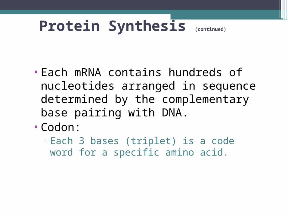
•Each mRNA contains hundreds of nucleotides arranged in sequence determined by the complementary base pairing with DNA.
•Codon:▫Each 3 bases (triplet) is a code word for a
specific amino acid.

Protein Synthesis (continued)

Transfer RNA
• Translation of the codons accomplished by tRNA and enzymes.▫ tRNA bends on itself,
making an anticodon (3 nucleotides that are complementary to codon of mRNA).
• Synthetase enzymes join specific amino acids to the ends of tRNA within a given codon.

Formation of a Polypeptide
•Anticodons of tRNA binds to mRNA codons.
•Each tRNA carries a specific amino acid.▫tRNA bring amino acids close together.▫Amino acid detaches from tRNA.
Enzymatically this amino acid is transferred to the amino acid on the next tRNA.
▫Polypeptide chain grows.•Interactions between amino acids cause
chain to twist and fold forming secondary and tertiary structure.

Translation of mRNA
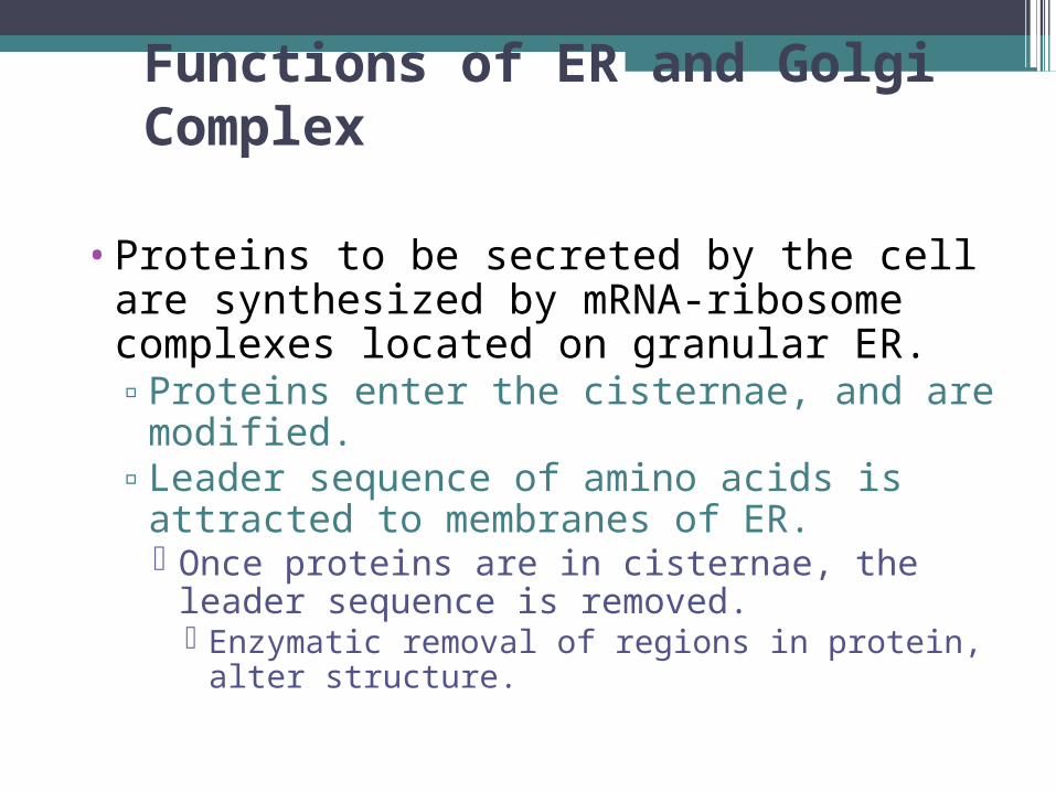
Functions of ER and Golgi Complex
•Proteins to be secreted by the cell are synthesized by mRNA-ribosome complexes located on granular ER.▫Proteins enter the cisternae, and are
modified.▫Leader sequence of amino acids is
attracted to membranes of ER. Once proteins are in cisternae, the leader
sequence is removed. Enzymatic removal of regions in protein, alter
structure.

Functions of ER and Golgi Complex (continued)
▫Secretory proteins are transported to Golgi complex. Further modified, packaged in vesicles, and secreted.

DNA Replication
• DNA is the only molecule in the body capable of replication.
• DNA helicases break weak hydrogen bonds to produce 2 free strands of DNA.
• Bases of each of the freed DNA strands can bind to complementary bases.
• Each copy is composed of one new strand and one strand from the original DNA molecule.
• Preserves the sequence of bases in DNA.

DNA Replication (continued)
DNA
•Law of Complementary Base Pairings:•# of purine bases = # pyrimadine
bases.▫Adenine only pairs with thymine.▫Guanine only pairs with cytosine.▫DNA polymerases join the nucleotides
together to form a second polynucleotide chain.

Cell Cycle•Interphase (non-dividing cell phases):
▫G1: Produces mRNA and proteins.
▫S: If cell is going to divide, DNA replicated.
▫ G2: Chromosome consists of 2 chromatids joined by
centromere. Each chromatid contains a complete double-helix DNA
molecule. Each chromatid will become a separate chromosome once mitotic division completed.
Completes interphase.

CyclinsCyclins promote different phases of the cell cycle.◦ During G1 phase an increase in cyclin D proteins activates
enzymes to move the cell quickly through the G1 phase. Overactivity of a gene that codes for cyclin D might cause
uncontrolled cell division (cancer).Oncogenes:
◦ Mutated forms of normal genes that contribute to cancer.Tumor suppressor genes:
◦ Inhibit cancer development.◦ Suppressor gene p53 indirectly blocks the ability of
cyclins to stimulate cell division. Induces the expression of gene p21, which inactivates the
cyclin-dependent kinases. Promotes cell differentiation.

Mitosis (M Phase)▫Prophase: Chromosomes become visible distinct structures.
▫Metaphase: Chromosomes line up single file along equator.
Action of spindle fibers attached to kinetochore▫Anaphase:
Centromeres split apart. Spindle fibers shorten, pulling the 2 chromatids in
each chromosome to opposite poles.▫Telophase:
Division of cytoplasm, producing 2 daughter cells.

Mitosis (continued)

Mitosis (continued)

Role of Centrosome• All animal cells have centrosome, located near
nucleus in non-dividing cell.▫ At center are 2 centrioles.
Each centriole composed of 9 bundles of microtubules. Microtubules grow out of pericentriolar material.
▫ Centrosome replicates itself during interphase (if cell is going to divide).
▫ Identical centrosomes move away from each other during prophase.
▫ Take up opposite poles by metaphase. Microtubules from both centrosomes form spindle fibers.
Spindle fibers pull chromosomes to opposite poles during anaphase.

Telomeres and Cell Division• Decreased ability of cells to divide is an indicator
of senescence (aging).▫ May be related to the loss of DNA sequences at the ends
of chromosomes (regions called telomeres). Telomeres serve as caps on the ends of DNA.
Prevent enzymes from mistaking the normal ends for broken DNA. DNA polymerase does not fully copy the DNA at end-regions.
Each time a chromosome replicates it loses 50-100 base pairs in its telomeres.
▫ Germinal cells can divide indefinitely due to an enzyme telomerase. Duplicates telomere DNA.

Meiosis (Reduction Division)•Cell division occurring in ovaries and testes to produce gametes (ova and sperm cells).
•Has 2 divisional sequences:▫First division:
Homologous chromosomes line up side by side along equator of cell.
Spindle fibers pull 1 member of the homologous pair to each pole. Each of the daughter cells contains 23
different chromosomes, consisting of 2 chromatids.

Meiosis (Reduction Division) (continued)
▫Second division: Each daughter cell divides, with duplicate
chromatids going to each new daughter cell. Testes: produce 4 sperm cells. Ovaries: produce one mature egg, polar bodies
die.

Cell Death• Pathologically:
▫ Cells deprived of blood supply swell, the membrane ruptures, and the cell bursts (necrosis).
• Apoptosis:▫ Cells shrink, membranes become bubbled, nuclei
condense.• Capsases (“executioner enzymes”):
▫ Mitochondria membranes become permeable to proteins and other products.
• Programmed cell death:▫ Physiological process responsible for remodeling of
tissues during embryonic development and tissue turnover in the adult.