cell metabolism article - carl s. thummel...
TRANSCRIPT

Cell Metabolism
Article
The LYR Factors SDHAF1 and SDHAF3Mediate Maturation of the Iron-Sulfur Subunitof Succinate DehydrogenaseUn Na,1,2,5 Wendou Yu,3,5,6 James Cox,2 Daniel K. Bricker,3 Knut Brockmann,4 Jared Rutter,2 Carl S. Thummel,3
and Dennis R. Winge1,2,*1Department of Medicine, University of Utah Health Sciences Center 5C426 School of Medicine, 30 North 1900 East, Salt Lake City,
UT 84132-2408, USA2Department of Biochemistry, University of Utah, 15 North Medical Drive East, Salt Lake City, UT 84112-5650, USA3Department of Human Genetics, University of Utah, 15 North 2030 East, Salt Lake City, UT 84112-5330, USA4Departments of Pediatrics and Pediatric Neurology, Faculty of Medicine, University of Gottingen, Robert Koch Strasse 40, 37075 Gottingen,
Germany5Co-first authors6Present address: Interdisciplinary Stem Cell Institute, Department of Pediatrics, Leonard M. Miller School of Medicine, University of Miami,
Miami, FL 33101, USA
*Correspondence: [email protected]://dx.doi.org/10.1016/j.cmet.2014.05.014
SUMMARY
Disorders arising from impaired assembly of succi-nate dehydrogenase (SDH) result in a myriad of pa-thologies, consistent with its unique role in linkingthe citric acid cycle and electron transport chain.In spite of this critical function, however, only a fewfactors are known to be required for SDH assemblyand function. We show here that two factors, Sdh6(SDHAF1) and Sdh7 (SDHAF3), mediate maturationof the FeS cluster SDH subunit (Sdh2/SDHB). Yeastand Drosophila lacking SDHAF3 are impaired inSDH activity with reduced levels of Sdh2. Drosophilalacking the Sdh7 ortholog SDHAF3 are hypersen-sitive to oxidative stress and exhibit muscular andneuronal dysfunction. Yeast studies revealed thatSdh6 and Sdh7 act together to promote Sdh2 matu-ration by binding to a Sdh1/Sdh2 intermediate, pro-tecting it from the deleterious effects of oxidants.These studies in yeast and Drosophila raise the pos-sibility that SDHAF3 mutations may be associatedwith idiopathic SDH-associated diseases.
INTRODUCTION
Succinate dehydrogenase (SDH) is an integral component of
both the mitochondrial respiratory chain and the tricarboxylic
acid (TCA) cycle. It catalyzes the two-electron oxidation of
succinate to fumarate with the reduction of ubiquinone to ubiq-
uinol (succinate:ubiquinone oxidoreductase). SDH is embedded
within the inner membrane (IM) of mitochondria and consists of
four nuclear-encoded subunits, designated Sdh1 through Sdh4
in yeast and SDHA through SDHD in mammalian cells. SDH
deficiency in humans results in infant encephalomyopathy,
myopathy, or tumorigenesis in the adult (Finsterer, 2008; Rustin
Cel
and Rotig, 2002). Loss-of-function mutations in human genes
for SDHA, SDHB, SDHC, and SDHD are strongly linked with
susceptibility to familial paraganglioma, pheochromocytoma,
gastrointestinal stromal tumors, and renal cell carcinoma (Bar-
della et al., 2011; Baysal et al., 2000; Feichtinger et al., 2010;
Janeway et al., 2011). Tumorigenesis arising from SDH defi-
ciency is purportedly related to the deleterious effects of supra-
physiological levels of succinate, which is a known inhibitor of a
myriad of a-ketoglutarate (aKG)-dependent enzymes, including
prolyl hydroxylases, histone, and DNA demethylases (Selak
et al., 2005; Xiao et al., 2012).
This tetrameric enzyme contains five redox cofactors,
including a covalently bound FAD and three iron-sulfur (FeS)
clusters in a hydrophilic segment consisting of two subunits
(Sdh1 and Sdh2) and a heme-containing membrane anchor
domain consisting of Sdh3 and Sdh4 subunits (Robinson and
Lemire, 1996). The FeS clusters facilitate electron transfer to
the ubiquinone-binding site formed between Sdh2 and themem-
brane subunits (Sun et al., 2005).
Assembly factors are often used to facilitate cofactor insertion
in mitochondrial respiratory complexes and mitigate unwanted
reactions during biogenesis. Recently, two SDH assembly fac-
tors associated with human pathogenesis were identified. Succi-
nate dehydrogenase assembly factor 1 (SDHAF1) was found in a
study of infantile mitochondrial diseases in which two families
hadmultiple children afflicted with leukoencephalopathy (Ghezzi
et al., 2009). Biochemical analyses revealed a SDH deficiency in
muscle samples and fibroblasts from these patients along with
missense mutations in SDHAF1. Deletion of the yeast ortholog
ofSDHAF1 (SDH6) resulted in a respiratory deficiency and a spe-
cific reduction in SDH activity (Ghezzi et al., 2009). SDH defi-
ciency has subsequently been reported in other patients that
carry SDHAF1 mutations (Ohlenbusch et al., 2012). Succinate
dehydrogenase assembly factor 2 (SDHAF2 or yeast Sdh5)
was shown to be required for the covalent attachment of FAD
to the catalytic SDHA (Sdh1) subunit (Hao et al., 2009). Yeast
lacking Sdh5 were respiratory deficient due to an absence of
SDH activity. Germline loss-of-function mutations in SDHAF2
l Metabolism 20, 253–266, August 5, 2014 ª2014 Elsevier Inc. 253
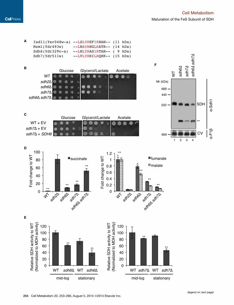
A
B
C
D
E
F
(legend on next page)
Cell Metabolism
Maturation of the FeS Subunit of SDH
254 Cell Metabolism 20, 253–266, August 5, 2014 ª2014 Elsevier Inc.

Cell Metabolism
Maturation of the FeS Subunit of SDH
were identified in SDH-deficient neuroendocrine paraganglioma
tumors (Hao et al., 2009). A number of SDH-deficient pathol-
ogies, including Leigh syndrome, gastrointestinal stromal tu-
mors, and neuroblastomas, have also been reported that lack
mutations in known SDH assembly factors or SDH structural
subunits (Feichtinger et al., 2010; Janeway et al., 2011).
Thus, additional SDH assembly factors may remain to be discov-
ered, potentially providing insights into the causes of idiopathic
SDH-associated diseases.
Sdh6 is amember of the LYR protein family that consists of ten
proteins in the human proteome and four in the yeast proteome
(Figure 1A). Within yeast, the founding member is the mitochon-
drial Isd11 protein that functions in the matrix FeS biogenesis
pathway as an effector of the Nfs1 cysteine desulfurase (Adam
et al., 2006; Wiedemann et al., 2006). We demonstrated that a
second LYR protein Mzm1 is a chaperone for the Rieske FeS
subunit of complex III (Atkinson et al., 2011; Cui et al., 2012).
The remaining yeast LYR proteins are Sdh6 and Acn9 (human
ortholog ACN9). Although Sdh6 is required for proper SDH activ-
ity, its molecular mechanism remains unknown. Moreover, Acn9
(designated Sdh7 in yeast and SDHAF3 in humans and flies) has
no known function. Here we show that these two factors are
required for SDH biogenesis in eukaryotes. Both Sdh6 and
Sdh7 protect Sdh2 maturation from the deleterious effects of
endogenous reactive oxygen species. We also report that loss
of SDHAF3 in Drosophila cells leads to a marked SDH deficiency
analogous to that in yeast, with defects inmuscular and neuronal
function in mutant flies. This study identifies functions of two
SDH assembly factors, providing a more complete understand-
ing of its critical role in cellular energy production and a potential
molecular framework for defining currently idiopathic SDH-asso-
ciated diseases.
RESULTS
Cells Lacking Sdh6 and Sdh7 Exhibit SDH DeficiencyAs a first step toward characterizing Sdh6 and Sdh7 function, we
examined the growth phenotypes of sdh6D or sdh7Dmutants on
nonfermentable carbon sources using S. cerevisiae. Compared
to wild-type cells, cells lacking Sdh6 or Sdh7 exhibited a partial
growth defect on glycerol/lactate medium and a severe growth
defect on acetate medium (Figure 1B), which is consistent
with previous studies (Ghezzi et al., 2009; McCammon, 1996).
We confirmed that the respiratory growth defects of sdh6D
and sdh7D mutants were attributed to deletions of SDH6 and
Figure 1. Succinate Dehydrogenase Deficiency in Cells Lacking Two L
(A) LX(L/A)YRXX(L/I)(R/K) motif conserved in four LYR motif family proteins in
biogenesis pathway (Adam et al., 2006); Mzm1, a protein facilitating the Rieske F
(B) Serial dilutions (10-fold) of cells starting from optical density 600 (OD600) = 0.
sources, as indicated, and incubated at 30�C.(C) Serial dilutions (10-fold) of cells were spotted on SC media lacking uracil and
(D) Metabolites extracted from cells cultured in synthetic minimal media cont
harvested at OD600 = 2. Relative levels of metabolites to WT are represented as
(E) Relative SDH activity in isolated mitochondria compared toWT. Mitochondria
24 hr (mid-log) and 48 hr (stationary). Data are shown as mean ± SD (n = 3; **p <
(F) Blue native (BN)-PAGE analysis to visualize protein complexes. Mitochondria
digitonin. After clarification, soluble fractions were separated on BN-PAGE and
subunit of SDH; F1b, a subunit of ATP synthase (complex V, CV) in oxidative phos
band is visualized by antisera to Sdh1, but not Sdh2. See also Figures S1 and S
Cel
SDH7, as respiratory growth of sdh6D and sdh7D cells
was restored with epitope-tagged Sdh6 and Sdh7, respectively
(Figure S1A, available online). Since both Sdh6 and Sdh7
belong to the LYR motif protein family (Figure 1A), we looked
for genetic interactions between their encoded proteins. The
sdh6D sdh7D double-deletion strain exhibited a marked syn-
thetic growth defect on glycerol/lactate medium (Figure 1B). In
addition, the overexpression of SDH6 partially suppressed the
respiratory growth defect of sdh7D cells (Figure 1C); however,
overexpression of SDH7 failed to restore respiratory function of
sdh6D cells.
Metabolomic profiling was used to identify the biological
process impaired in sdh6D and sdh7D mutants, assaying the
levels of approximately 100 polar metabolites. We cultured cells
in synthetic minimal medium with 2% raffinose and 0.2%
glucose to stationary phase. Metabolites extracted from cells
were analyzed using gas chromatography-mass spectrometry
(GC-MS) (Figure 1D). Cells lacking either Sdh6 or Sdh7 exhibited
elevated succinate levels and attenuated fumarate and malate
levels, consistent with impaired conversion of succinate to
fumarate by SDH in the citric acid cycle. These observations in
sdh6D cells are consistent with the previous study in fibroblasts
harboring a mutated SDHAF1 gene (Ghezzi et al., 2009). More-
over, the sdh6D sdh7D double mutant showed an enhanced
accumulation in succinate, consistent with the synergistic respi-
ratory growth defects in these cells.
To confirm that the increased succinate/fumarate ratio in
sdh7Dmutants was due to impaired SDH function, we quantified
SDH activity in mitochondria purified from wild-type (WT) and
mutant cells lacking Sdh6 or Sdh7. Mitochondria isolated from
sdh6D and sdh7D cells harvested at mid-log phase exhibited
modest diminutions of SDH activity, but the deficit was magni-
fied in stationary-phase cells (Figure 1E). The sdh6D sdh7D dou-
ble mutant showed markedly decreased SDH activity in mid-log
cultures (34% of WT; Figure S1B) relative to the single mutants,
in accordance with the synergistic respiratory growth defect
and TCA cycle intermediates in sdh6D sdh7D double mutants.
Enzymatic activities of pyruvate dehydrogenase, a-ketoglutarate
dehydrogenase, aconitase, malate dehydrogenase, and bc1complex III were unaffected in cells lacking Sdh6 or Sdh7 (Fig-
ures S1B and S1C).
Amoderate diminution of the assembled tetrameric SDH com-
plex was seen in mitochondria isolated from late-log cultures of
sdh6D or sdh7Dmutants as visualized by blue native (BN)-PAGE
(Figure 1F). The abundance of the SDH complex was further
YR Motif Family Proteins, Sdh6 and Sdh7
yeast. Isd11, a chaperone required for cysteine desulfurase activity in FeS
eS protein insertion into bc1 (Cui et al., 2012); Sdh6 (SDHAF1); and Sdh7.
5 were spotted on synthetic complete (SC) media containing different carbon
incubated at 30�C. EV, empty vector.
aining 2% raffinose/0.2% glucose were analyzed using GC-MS. Cells were
mean ± SEM (n R 4 biological replicates; *p < 0.05; **p < 0.005).
were isolated from cells grown in SCmedia plus 2% raffinose/0.2% glucose for
0.05).
isolated from the strains harvested at late-log phase were solubilized with 1%
then transferred to membranes for immunoblotting. Sdh1, a FAD-containing
phorylation. The band highlighted by ** is the Sdh1 assembly intermediate. This
2.
l Metabolism 20, 253–266, August 5, 2014 ª2014 Elsevier Inc. 255

A
C
E
B
D
F
G H
Figure 2. Sdh6 or Sdh7 Functions Are Linked to the Fe/S Sdh2 Subunit
(A) FAD-containing Sdh1 was visualized by UV excitation. Other proteins visualized by immunoblotting include Mdh1 (malate dehydrogenase) and Por1 (porin)
(loading control).
(B) 35S-methionine-labeled proteins were incubated with isolated mid-log versus stationary phase mitochondria for 30 min (pulse), followed by blocking protein
import with valinomycin for 30 and 60 min (chase), respectively. Radiolabeled proteins were resolved on SDS-PAGE and detected by autoradiography.
(legend continued on next page)
Cell Metabolism
Maturation of the FeS Subunit of SDH
256 Cell Metabolism 20, 253–266, August 5, 2014 ª2014 Elsevier Inc.

Cell Metabolism
Maturation of the FeS Subunit of SDH
attenuated in sdh6D sdh7D double-mutant cells. Thus, Sdh6 and
Sdh7 are required to maintain normal SDH levels and activity
in yeast. In addition to reduced levels of tetrameric SDH in the
mutant cells, a Sdh1 subcomplex is evident, and the same sub-
complex is seen in cells lacking Sdh2 or Sdh4 (** in Figures 1F
and S2). Therefore, Sdh6 and Sdh7 appear to function in SDH
assembly rather than in the regulation of SDH activity.
Maturation of Sdh2 Is Impaired in the Absence of Sdh6or Sdh7To characterize the roles of Sdh6 and Sdh7, the steady-state
levels of SDH subunits were quantified in mitochondria isolated
from mutant cells. The levels of Sdh2 were attenuated in both
sdh6D and sdh7D mutants and further diminished in the sdh6D
sdh7D double mutant (Figure 2A). In contrast, Sdh1 levels were
unchanged in the mutant cells, and covalent flavinylation of
Sdh1 was not significantly altered (Figure 2A). Cells lacking
Sdh5 are compromised in Sdh1 flavinylation, leading to a
reduced stability of Sdh1 (Hao et al., 2009; Kim et al., 2012).
A mitochondrial in vitro protein import assay was used to
address a role for Sdh6 and Sdh7 in Sdh2 maturation. The
import assay consisted of the in vitro import of 35S-methio-
nine-labeled Sdh2 into purified mitochondria isolated from
sdh2D cells. Sdh2-deficient mitochondria were used to ensure
that a pool of unassociated Sdh1 would be available for a stabi-
lizing interaction with imported Sdh2. Furthermore, the use of
sdh2D cells negates any changes in the mitochondrial mem-
brane potential (which drives protein import) by the loss of
Sdh6 or Sdh7. The stability of 35S-Sdh2 as monitored during
the chase phase of the reaction was compromised in cells lack-
ing Sdh6 or Sdh7, especially in mitochondria isolated from sta-
tionary-phase cultures (Figure 2B). This result is consistent with
the exacerbated defect in SDH activity seen in mutant cells at
late stages of growth. Unlike Sdh2, radioisotope-labeled Rip1,
a target of the LYR protein Mzm1, remained unaffected by
loss of either Sdh6 or Sdh7.
Sdh1 and Sdh2 accumulate in cells lacking the membrane
anchor subunits Sdh3 and/or Sdh4 (Figure S3A) (Kim et al.,
2012). We observed that cells depleted of the membrane an-
chor contain increased steady-state levels of both Sdh6 and
Sdh7 (Figure 2C). These results suggest that Sdh6 and Sdh7
may form stalled preassembly intermediates with Sdh1 and/or
Sdh2. We performed coimmunoprecipitation on epitope-tagged
Sdh6 and Sdh7 in mitochondria from WT cells and cells stalled
in SDH assembly. A fraction of Sdh1 and Sdh2 was copurified
with either Sdh6 or Sdh7 from cells lacking the membrane an-
(C) Sdh6-His6-3HA or Sdh7-His6-3HA under their own endogenous promoters w
endogenous Sdh6 or Sdh7 depleted, respectively. Steady-state levels are show
(D) Coimmunoprecipitation of Sdh6-His6-2Myc after crosslinking. Mitochondria
midylpropionate). The crosslinking reaction was stopped with Tris buffer (pH
magnetic beads. Bound substances to Myc beads were resolved on SDS-PA
aconitase.
(E) Standard coimmunoprecipitation of Sdh7-His6-2Myc was performed with iso
(F) Steady-state levels of Sdh2 in sdh1D mutants with overexpression of protein
genes.
(G) SDH activity in sdh6Dmutants with SDH2 overexpression was detected as de
not significant).
(H) Succinate levels in sdh7D mutants with SDH2 overexpression were measure
significant). See also Figure S3.
Cel
chor domain, but not in cells devoid of Sdh2 (Figures 2D, 2E,
and S3B). These interactions are seen in the presence and
absence of crosslinking prior to resin adsorption. These results
suggest that Sdh6 and Sdh7 interact with Sdh2 within a Sdh1/
Sdh2 subcomplex, which is known to accumulate in cells lack-
ing the membrane anchor domain (Kim et al., 2012). Moreover,
these interactions appear to occur in the mitochondrial matrix,
consistent with the known matrix location of Sdh1, Sdh2, and
Sdh6 (Ghezzi et al., 2009). Sdh7 is also a matrix protein, as
revealed by proteinase K treatment of purified mitochondria,
which degrades Sdh7 only in the presence of detergents and
not upon hypotonic disruption of only the outer membrane.
(Figure S3C).
The physical interactions of Sdh6 and Sdh7 with Sdh2 promp-
ted the hypothesis that Sdh6 and Sdh7 may be chaperones for
Sdh2. To test this, we first examined the ability of overexpressed
Sdh6 or Sdh7 to stabilize the highly labile Sdh2 present in sdh1D
cells (Kim et al., 2012). Elevated levels of Sdh6, but not Sdh7, led
to increased steady-state Sdh2 levels under these conditions
(Figure 2F). This result raised a possibility that Sdh6 may func-
tion as a chaperone for Sdh2 prior to its interaction with Sdh1.
We tested whether SDH2 overexpression in sdh6D mutants
or sdh7D mutants would restore SDH activity and suppress the
respiratory growth defects. However, SDH2 overexpression
neither restored SDH activity nor reversed succinate accumula-
tion or rescued respiratory growth of sdh6D mutants and sdh7D
mutants (Figures 2G, 2H, S4A, and S4B).
Once a holo-Sdh2/Sdh1 complex forms, the final step in SDH
biogenesis is the addition of the Sdh3/Sdh4 membrane anchor.
To address whether Sdh6 and Sdh7 are involved in the recruit-
ment of the membrane anchor, we tested whether co-overex-
pression of SDH3 and SDH4 would suppress the respiratory
defect in the sdh6D and sdh7D mutant cells. No restoration of
growth on acetate medium was observed with elevated cellular
levels of Sdh3 and Sdh4 (Figures S4C and S4D). Thus, Sdh6
and Sdh7 do not likely facilitate the recruitment of the Sdh3/
Sdh4 membrane anchor.
We conducted a series of studies to assess whether Sdh6 or
Sdh7 has an active role in FeS cluster insertion. These studies
failed to reveal a direct role of either factor in FeS cluster inser-
tion. First, affinity purification of Sdh2 in yeast leads to copurifi-
cation of Nfu1, Isu1, and Isa2, three key matrix proteins involved
in FeS biogenesis (data not shown). However, affinity purification
of Sdh6-His6-2Myc or Sdh7-His6-2Myc failed to adsorb Isu1 or
Isa2, whereas Sdh2 was associated. Second, whereas overex-
pression of ISA1 or ISA2 restores respiratory growth in grx5D
as expressed from plasmids in cells lacking either Sdh2 or Sdh4, along with
n by immunoblotting.
were solubilized with 1% digitonin in the presence of 1 mM dithiobis(succini-
7.4), and the supernatants were absorbed to anti-Myc antibody-conjugated
GE and detected by immunoblotting. Input, 4% of total lysates; Aco1, FeS
lated mitochondria without crosslinking. Input, 2% of total lysates.
s indicated. Yap1, transcription factor upregulating oxidative stress response
scribed in Figure 1E. Data are represented as mean ± SD (n = 3; **p < 0.05; ns,
d as described in Figure 1D. Mean ± SEM is shown (n = 6; **p < 0.05; ns, not
l Metabolism 20, 253–266, August 5, 2014 ª2014 Elsevier Inc. 257

A
B
C
Figure 3. Exogenous Antioxidants Rescue
the Growth Defect of sdh6D and sdh7D
Mutants
(A) Cells harboring either empty vector (EV) or
high-copy YAP1 plasmid were spotted on SC
media lacking leucine by 10-fold serial dilutions
and incubated at 30�C.(B) Serial dilutions (10-fold) of cells were spotted on
SCmediumwith the indicated carbon sources with
or without 5 mM N-acetylcysteine or 2 mM gluta-
thione and incubated at 30�C.(C) Succinate levels in cells overexpressing YAP1
were measured as described in Figure 1D. Cells
were harvested at OD600 = 1. Mean ± SEM is
shown (n = 6; *p < 0.005).
Cell Metabolism
Maturation of the FeS Subunit of SDH
mutant cells (Kim et al., 2010; Rodrıguez-Manzaneque et al.,
2002), overexpression of ISA1, ISA2, NFU1, or ISU1 failed to
suppress the respiratory defect of sdh6D or sdh7D cells (data
not shown). Third, 55Fe incorporation into Sdh2-His6-2Myc was
quantified in cells either containing or lacking Sdh6 or Sdh7. Im-
munocapture of Sdh2 failed to show any clear diminution in 55Fe
in the mutant cells (Figure S4E). However, interpretation of the55Fe study is complicated, since Sdh2 has three distinct FeS
centers, and Sdh6 or Sdh7 may have a restricted role with one
cluster. In addition, ascorbate is used during 55Fe labeling and,
as a reductant, may mimic the exogenous reductants in sup-
pressing the defects in the mutant cells.
258 Cell Metabolism 20, 253–266, August 5, 2014 ª2014 Elsevier Inc.
Antioxidants Ameliorate Defects insdh6D Mutants and sdh7D MutantsFurther clues to the function of Sdh6
emerged from a genetic suppressor
study in which extragenic suppressors
of the acetate growth defect of sdh6D
mutants were recovered. We generated
a high-copy plasmid library with partially
digested genomic DNA from sdh6D
mutants. This library was transformed to
sdh6D mutants, and the transformants
that exhibited enhanced growth on ace-
tatemediumwere collected. Interestingly,
multiple independent suppressors were
recovered that encoded Yap1, which is
a transcriptional activator that induces
the expression of a battery of antioxidant
genes, including thioredoxin, thioredoxin
reductase, and glutathione reductase,
in response tooxidative stress (Fernandes
et al., 1997). The overexpression of YAP1
in subcloned vectors robustly suppressed
the acetate growth defect of cells lacking
Sdh6 (Figure 3A). Consistent with the anti-
oxidant role of Yap1 expression, supple-
mental glutathione or N-acetylcysteine
also restored limited respiratory growth
in sdh6D mutants (Figure 3B).
Sinceweobserved agenetic interaction
between SDH6 and SDH7 (Figure 1), we
tested whether overexpression of YAP1
could also restore respiratory growth in sdh7D mutants. Indeed,
YAP1 overexpression suppressed the respiratory growth defect
of sdh7D mutants, although the suppression was less pro-
nounced compared to sdh6D mutants (Figure 3A). Moreover,
the addition of exogenous reductants restored limited respiratory
growth of sdh7Dmutants (Figure 3B). To confirm that YAP1 over-
expression restored SDH activity in sdh6D and sdh7D single-
mutant cells, we assessed SDH activity by measuring succinate
levels using metabolomics. Indeed, YAP1 overexpression
decreased succinate levels in both sdh6D and sdh7D mutants
significantly, while YAP1 overexpression did not affect succinate
levels in sdh2D mutants as a negative control (Figure 3C).
Cell Metabolism
Maturation of the FeS Subunit of SDH
Therefore, we conclude that YAP1 overexpression contributes to
the restoration of SDH activity in sdh6D and sdh7D mutants.
Elevated levels of Yap1, however, have no effect on the respira-
tory growth defects in sdh6D sdh7D double mutants (Figure 3A).
Sdh2 Is Stabilized by Sdh6 and Sdh7 under OxidativeStress ConditionsWe hypothesized that Sdh6 and Sdh7 may be important for SDH
maturation under oxidative stress conditions. Initially, we tested
whether cells lacking Sdh6 or Sdh7 were hypersensitive to the
superoxide anion generator paraquat. The respiratory growth
defect of sdh6D and sdh7D single mutants was dramatically
exacerbated in the presence of paraquat (Figure 4A). Likewise,
steady-state levels of Sdh2 were markedly attenuated in para-
quat-treated sdh6D and sdh7D mutants compared to WT cells
(Figure 4B). In contrast, steady-state levels of other mitochon-
drial FeS containing proteins were not significantly altered by
paraquat in the mutant cells (Figure 4B).
The paraquat sensitivity of sdh6D and sdh7D mutants may
arise from either enhanced ROS damage or the accumulation
of a pro-oxidant in the mutants. We observed that ROS levels
were not changed in untreated sdh6D and sdh7D mutants using
two different assays. First, we quantified aconitase (Aco1)
activity. The 4Fe-4S cluster in aconitase is susceptible to ROS
damage (Gardner, 2002); thus, aconitase activity is an indicator
of ROS stress in vivo. No diminution of aconitase activity was
seen in sdh6D and sdh7D mutants compared to WT (Figure 4C).
Second, we tested the mutant cells for hydrogen peroxide
sensitivity on rich glucose medium. As cells accumulate ROS,
they become sensitized to exogenous ROS and subsequently
lose viability (Khalimonchuk et al., 2007). After 2 hour treatment
of cells with 6 mM H2O2, the viability of sdh2D, sdh3D, and
sdh4Dmutant strainswas significantly compromised (Figure 4D).
However, sdh6D and sdh7D mutant cells exhibited no
growth defects. The lack of impairment in aconitase activity
and hydrogen peroxidase sensitivity suggest that ROS levels
are not elevated in sdh6D and sdh7D mutants as compared to
WT cells. It is notable that sdh1D and sdh5D single mutants
and the sdh4D sdh5D double mutant did not show hydrogen
peroxide sensitivity compared to sdh2D, sdh3D, and sdh4D
strains, which suggests that an assembly intermediate contain-
ing FAD-Sdh1 may be the source for electron leakage for the
generation of ROS in sdh2D, sdh3D, and sdh4D single mutants.
Theobservedhypersensitivity of sdh6D and sdh7Dmutant cells
to paraquat may imply that Sdh6 and Sdh7 are shielding the FeS
clusters in Sdh2 prior to full assembly. Superoxide inactivation of
the 4Fe-4S center in aconitase leads to a dissociation of one iron
ion forming an inactive 3Fe-4S center that can be reactivated by
supplemental iron salts (Gardner and Fridovich, 1992). We tested
whether supplemental iron salts would restore respiratory func-
tion to sdh6D and sdh7D mutant cells. Supplemental FeCl2, but
not ZnCl2, restored limited glycerol growth to both mutant cells
(Figure 4E). These data are consistent with a candidate role of
Sdh6 and Sdh7 in FeS cluster protection in SDH maturation.
Drosophila Sdhaf3 Mutants Are Sensitive to OxidativeStressThe Drosophila genome encodes a close ortholog of Sdh7 (Fig-
ure S5A) but has only a weak candidate homolog of Sdh6.
Cel
Accordingly, we examined the functions of Sdh7 in Drosophila
to determine if its roles in SDH assembly and activity have
been conserved through evolution and to define its possible
physiological functions. Gene targeting was used to generate a
null mutation in the Drosophila sdh7 ortholog (CG14898), which
we refer to here as dSdhaf3 (Figures S5B and S5C). These mu-
tants were outcrossed for six generations to w1118, which was
used as a control for most studies. dSdhaf3 mutants progress
normally through development and have a normal lifespan
when maintained on standard growth media. These animals
are, however, sensitive to ethanol (Figure 5A) and oxidative
stress, resulting from either paraquat treatment (Figure 5B) or
hyperoxia (Figure 5C). The response to hyperoxia is most pro-
nounced, with a 50% reduction in lifespan relative to controls,
althoughSdhB12081 hypomorphic mutants display amore severe
effect (Walker et al., 2006). Interestingly, most dSdhaf3 mutants
exposed to 100%oxygen for 4 days held their wings erect, a hall-
mark of mitochondrial dysfunction and muscle degeneration
(DeSimone et al., 1996; Greene et al., 2003). A similar abnormal
wing posture was observed in SdhB12081 mutants maintained
under normal conditions (10%–15%) or exposed to hyperoxia
(80%–100%).
dSdhaf3 Mutants Display Reduced SdhB Protein Levelsand SDH ActivityIf the function of dSdhaf3 has been conserved through evolution,
then dSdhaf3 mutants should display a specific defect in SDH
function. Consistent with this possibility, metabolomic profiling
of dSdhaf3 mutants revealed elevated succinate and reduced
levels of fumarate and malate (Figure 5D). Biochemical analysis
of mitochondrial extracts from dSdhaf3 mutants demonstrated
that dSdhaf3 mutants have normal levels of SdhA, but signifi-
cantly reduced levels of SdhB (Figure 5E), resulting in an approx-
imate 50% reduction in SDH enzymatic activity (Figures 5F and
5G), similar to the phenotypes of sdh7D yeast. SdhA is also
flavinylated normally in dSdhaf3 mutants, as expected (Fig-
ure S5E). Combining the hypomorphic SdhB12081 allele with
the dSdhaf3mutation resulted in a dramatic decrease in viability,
demonstrating a strong genetic interaction, consistent with the
reduced levels of SdhB in dSdhaf3 mutants and confirming the
functional interaction between dSdhaf3 and SDH (Figure S6A).
Interestingly, although dSdhaf3 mutants are fully viable and
fertile, they display a clear age-dependent reduction in move-
ment (Figure 5H). While mutants at 1 week of adult life show no
difference in motility relative to controls, mutants display an
approximately 50% reduction in movement by 2 weeks of age
and a more severe motility defect at later stages (Figure 5H).
Mutants are also significantly more sensitive to paralysis by
2 weeks of age, relative to controls (Figure S5F). Thus, like its
counterpart in yeast, dSdhaf3 is required to maintain normal
SDH levels and activity and proper wing muscle function and
motility in Drosophila.
Antioxidant and Genetic Rescue of dSdhaf3 MutantsConsistent with the ability of antioxidants to suppress the
growth defects in sdh7D yeast mutants, either a dietary (N-ace-
tylcysteine) or genetic (Sod2 expression) reduction in oxidative
stress rescued the hyperoxia sensitivity of dSdhaf3 mutants
(Figures 6A and 6B). Unlike the yeast studies, however,
l Metabolism 20, 253–266, August 5, 2014 ª2014 Elsevier Inc. 259

A
B
C
D
E
Figure 4. sdh6D and sdh7D Mutants Are Sensitive to Oxidative Stress
(A) Serial dilutions (10-fold) of cells were spotted on SC media with or without 2 mM paraquat with indicated carbon sources and incubated at 30�C.(B) Steady-state levels of proteins in mitochondria isolated from strains cultured in the presence of 2 mM paraquat. Yah1, ferredoxin of the mitochondrial matrix.
(C) Aconitase activity specific to cis-aconitate conversion in isolated mitochondria. Data are shown as mean ± SD (n = 3).
(legend continued on next page)
Cell Metabolism
Maturation of the FeS Subunit of SDH
260 Cell Metabolism 20, 253–266, August 5, 2014 ª2014 Elsevier Inc.

Cell Metabolism
Maturation of the FeS Subunit of SDH
overexpression of the putative Sdh6 homolog (encoded by
CG34229), using Act5C-GAL4 to drive a UAS-CG34229 trans-
gene in dSdhaf3mutants, had no effect on their sensitivity to hy-
peroxia (Figure S6B). This result, however, is difficult to interpret
because there is only limited sequence homology between
CG34229 and Sdh6. We conclude that this functional interac-
tion between Sdh6 and Sdh7 may not be conserved through
evolution or, alternatively, that CG34229 is not a functional ho-
molog of Sdh6.
The wing posture and motility defects in dSdhaf3 mutants
suggested that these animals suffer from muscular and
neuronal dysfunction. Consistent with this model, widespread
expression of WT dSdhaf3 (Act > dSdhaf3) in dSdhaf3 mutants
fully rescues their sensitivity to hyperoxia (Figure 6C), while
muscle-specific (C57 > dSdhaf3) or neuronal-specific (elav >
dSdhaf3) expression provides partial rescue (Figure 6D). Similar
effects are seen on the climbing defects in dSdhaf3 mutants
(Figures 6E and 6F). Genetic rescue in the fat body (Cg-GAL4)
or intestine (Mex-GAL4), however, provided no significant
rescue (data not shown). Widespread overexpression of
dSdhaf3 in wild-type flies has no significant effect on their
resistance to hyperoxia (data not shown). Taken together, we
conclude that dSdhaf3 function is conserved through evolution
and that proper SDH levels and activity are required for
resistance to oxidative stress as well as muscular and neuronal
function, consistent with their dependence on mitochondrial
oxidative phosphorylation.
SDHB Is Specifically Impaired in Human Cells Deficientin Wild-Type SDHAF1The human Sdh6 ortholog, SDHAF1, was shown previously to be
important for SDH activity and abundance in fibroblasts (Ghezzi
et al., 2009); however, its mechanism of action remained
undefined. We addressed whether SDHAF1 protects SDHB
(the human Sdh2 ortholog) from ROS damage similar to yeast
Sdh6. First, we determined steady-state levels of SDH structural
subunits in SDHAF1-depleted human embryonic kidney 293
(HEK293) cells by siRNA knockdown. SDHB and SDHC levels
were significantly reduced upon SDHAF1 depletion (Figure 7A).
SDHA levels, however, remained unaffected. Next, we tested
the effects of paraquat on SDHAF1-depleted HEK293 cells. In
accordance with the yeast and fly data, SDHB levels were further
attenuated in SDHAF1-deficient cells (Figure 7B). The sensitivity
to paraquat also suggests that SDHAF1 resembles Sdh6 in pro-
tecting holo-SDHB from ROS damage.
We also examined the steady-state SDHB levels in patient
fibroblasts with a known SDHAF1 mutation (Ohlenbusch et al.,
2012). Both SDHB and SDHC levels were diminished in mito-
chondria isolated from the patient fibroblasts, whereas SDHA
levels remained unaffected compared to controls (Figure 7C).
SDHB protein levels were <50% of wild-type controls, and
SDH enzyme activity was 52% and 40% in patients 1 and 2 rela-
tive to control values. Thus, the limited SDHB levels in the patient
cells likely contribute to reduced SDH function.
(D) Precultures grown up to late-log phase in YPDmedia were diluted 2-fold, follow
with sterile water, and 10-fold serial dilutions were spotted on YPD plate, followe
(E) Enhanced respiratory growths of sdh6D mutants and sdh7D mutants with iron
with or without FeCl2 or ZnCl2 with the indicated concentrations and then incuba
Cel
DISCUSSION
The present work demonstrates that maturation of the FeS sub-
unit of Sdh2 (SDHB) requires the participation of two assembly
factors, Sdh6 (SDHAF1) and Sdh7 (SDHAF3). These factors are
shown to guide Sdh2 maturation within the mitochondrial matrix
in the midst of endogenous oxidants. Yeast, flies, and mamma-
lian cells lacking one of these factors are impaired in SDH activity
and assembly, with Sdh2 exhibiting a heightened susceptibility
to oxidants. Normal oxidative metabolism in the mitochondria
leads to the formation of superoxide anions from the one-elec-
tron reduction of O2. Superoxide anions can readily dissociate
FeS clusters, rendering the assembly of FeS cluster centers
in mitochondrial enzymes susceptible to oxidative damage.
The present studies in yeast, flies, and mammalian cells suggest
that Sdh6 and Sdh7 shield one or more of the three FeS clusters
in Sdh2 from oxidants during assembly.
Yeast studies reveal that Sdh6 and Sdh7 act in concert in the
maturation of Sdh2 with a limited redundancy in function. Yeast
lacking either factor show a marked SDH deficiency in late-log
cultures that rely on oxidative metabolism. Under these condi-
tions, the mutant cells contain a reduced level of the assembled
tetrameric enzyme. These cells exhibit a hypersensitivity to the
superoxide generator paraquat. The respiratory defect of these
mutants is readily suppressed by overexpression of the Yap1
transcriptional activator of oxidative stress genes or exogenous
reductants. These studies highlight the role of Sdh6 and Sdh7 in
shielding Sdh2 maturation from deleterious effects of oxidants.
Flies lacking the Sdh7 ortholog SDHAF3, likewise, are hyper-
sensitive to paraquat and hyperoxia. The mutant flies show
diminished levels of active SDH and, as a result, accumulate suc-
cinate. The dSdhaf3 mutants are viable and fertile yet display
impaired movement that intensifies with age. The erect wing
phenotype exhibited under hyperoxic conditions and the motility
defects evident in agedmutant flies are consistent with muscular
and neuronal dysfunction. Moreover, neuronal or muscle-spe-
cific expression of wild-type dSdhaf3 is sufficient to partially
rescue the hyperoxia sensitivity of the mutants, demonstrating
the importance of SDH function in these tissues that rely heavily
on oxidative phosphorylation (OXPHOS). The hyperoxia sensi-
tivity of dSdhaf3 mutants is also partially suppressed by dietary
N-acetylcysteine or overexpression of the matrix mangano-
superoxide dismutase Sod2. These effects of antioxidantsmimic
the rescue of yeast sdh7D mutant oxidative growth and demon-
strate the apparent close conservation of Sdh7/SDHAF3 func-
tion through evolution.
Conservation in Sdh6/SDHAF1 function between yeast and
humans also exists. Attenuation of SDHAF1 in HEK293 culture
cells leads to a hypersensitivity in the stability of the FeS SDHB
subunit to paraquat. SDHB instability is also seen in two SDHAF1
patient fibroblast lines. One implication of the observed antioxi-
dant rescue of the defect of sdh6D yeast cells and dSdhaf3
mutant flies is the potential use of antioxidant therapeutics
for patients afflicted with SDHAF1 (and perhaps SDHAF3)
ed by addition of 6mMH2O2 and incubated for 2 hr at 30�C. Cells were washed
d by incubation at 30�C.supplementation. Serial dilutions (10-fold) of cells were spotted on SC media
ted at 30�C.
l Metabolism 20, 253–266, August 5, 2014 ª2014 Elsevier Inc. 261

A B C
D
E F G
H
Figure 5. dSdhaf3 Mutants Are Sensitive to Oxidative Stress and Display Reduced Levels of SdhB, Reduced SDH Activity, and Motility
Defects
(A–C) w1118 control (solid line) and dSdhaf3 mutant (dotted line) males (5 days old) were transferred to vials with (A) 5% ethanol, 1% agar in PBS, (B) 30 mM
paraquat in semidefined medium, or (C) 100% O2 with standard medium, and living animals were scored daily. Homozygous SdhB12081 mutants (dashed line)
were included in the hyperoxia experiment. Each graph was compiled from 3–5 experiments, using a total of 15–21 vials with 20 animals per vial. Error bars
represent ±SEM. dSdhaf3 mutants are significantly more sensitive than controls under each condition; p < 0.001.
(D) GC-MS was used to compare the relative levels of small metabolites in wild-type controls (gray boxes) and dSdhaf3 mutants (white boxes). n = 12 samples
from two independent experiments with 20 flies/sample (5 days old). ***p < 0.001.
(E) Proteins were extracted frommitochondria isolated fromw1118 controls, dSdhaf3mutants, orUAS-dSdhaf3/+ transformants and analyzed by immunoblotting
to detect SdhA, SdhB, and ATPa (subunit of complex V).
(legend continued on next page)
Cell Metabolism
Maturation of the FeS Subunit of SDH
262 Cell Metabolism 20, 253–266, August 5, 2014 ª2014 Elsevier Inc.

% s
urvi
val
N-acetyl cysteine rescue SOD2 rescue
% s
urvi
val
w1118dSdhaf3w1118 + NACdSdhaf3 + NAC
w1118dSdhaf3UAS-Sod2Act>Sod2, dSdhaf3
ubiquitous rescue muscle & neuronal rescuew1118
dSdhaf3UAS-dSdhaf3Act-GAL4Act>dSdhaf3
w1118dSdhaf3UAS-dSdhaf3C57>dSdhaf3elav>dSdhaf3 dS
dhaf
3
dSdh
af3
% s
urvi
val
% s
urvi
val
A B
C D
E
days days
days days
w11
18dS
dhaf3
Act-GAL4
Act>dS
dhaf3
dSdhaf3 dSdhaf3
F
UAS-dS
dhaf
3
UAS-dSdh
af3
w11
18dS
dhaf
3
C57>d
Sdha
f3ela
v>dS
dhaf
3ubiq. climbing rescue muscle & neuronal climbing rescue
******
******
Figure 6. dSdhaf3 Function Is Required in
the Muscles and Nervous System
w1118 control (blue solid line) and dSdhaf3 mutant
(red dotted line) males (5 days old) were trans-
ferred to vials with 100% O2 with standard me-
dium, and living animals were scored daily.
(A) 0.1% N-acetylcysteine was added to the cul-
ture medium for a quarter of the vials.
(B) Expression ofSod2 using theubiquitousAct5C-
GAL4 driver (Act>Sod2; purple line) partially res-
cues the hyperoxia sensitivity of dSdhaf3mutants.
Both NAC treatment and Sod2 expression signifi-
cantly rescue the hyperoxia sensitivity of dSdhaf3
mutants; p < 0.001.
(C) Expression of wild-type dSdhaf3 using
the ubiquitous Act5C-GAL4 driver (Act>dSdhaf3;
purple line) rescues the hyperoxia sensitivity of
dSdhaf3 mutants (p < 0.001).
(D) The muscle-specificC57-GAL4 driver provides
minor, but significant (p < 0.01), rescue of the
hyperoxia sensitivity of dSdhaf3 mutants (purple
line) relative to the control that carries the UAS-
dSdhaf3 transgene alone (green line), while the
CNS-specific elav-GAL4 driver provides more
efficient rescue (orange line) (p < 0.001).
(E and F) Expression of wild-type dSdhaf3 by using
either (E) the ubiquitousAct5C-GAL4driver (purple),
(F) the muscle-specific C57-GAL4 driver (purple),
or (F) the CNS-specific elav-GAL4 driver ( orange)
rescues the climbing defect in dSdhaf3mutants.
The Act>dSdhaf3 rescue in (C) and (E) was per-
formed in females, and other rescue studies were
performed in males (A, B, D, and F). The apparent
partial rescue of dSdhaf3mutants by a single copy
of theUAS-dSdhaf3 transgene (CandD, green line)
appears to be due to genetic background since
UAS-dSdhaf3 transformants have normal levels of
SdhB and SDH activity (E and F). Each graph was
compiled from two experiments with a total of 10
vials with 20 animals per vial. ***p < 0.001.
Cell Metabolism
Maturation of the FeS Subunit of SDH
mutations. Patients with SDHAF1 mutations have presented
with SDH-deficient leukoencephalopathy (Ghezzi et al., 2009;
Ohlenbusch et al., 2012) and have a survival window that may
be amenable to antioxidant therapy. Two case studies were
reported that attempted to alleviate clinical symptoms in SDH-
deficient patients harboring SDHAF1 mutations by supplement-
ing riboflavin and CoQ10. The clinical outcomes, however, were
not significantly improved (Jain-Ghai et al., 2013). More recently,
a candidate therapeutic, EPI-743, is being tested for treatment
of Leigh syndrome patients (Martinelli et al., 2012). EPI-743 is
a vitamin E quinone that is orally bioavailable and crosses
the blood-brain barrier (Shrader et al., 2011). Future studies
will focus on the efficacy of antioxidants with SDHAF1 patient
fibroblasts. In addition, it is important to note that every gene
encoding an SDH subunit or known assembly factor is causally
(F) A continuous colorimetric assay was used to measure SDH enzyme activity in
UAS-dSdhaf3/+ transformants. ***p < 0.001.
(G) Proteins from purified mitochondria were extracted from w1118 controls and d
and complex IV activity.
(H) Control w1118 flies and dSdhaf3 mutants were tested for motility in three inde
4 weeks of age. Climbing ability is reported as the number of flies that climbed ab
bottom. *p < 0.05; ***p < 0.001.
Cel
associated with human disease. We thus anticipate that
SDHAF3 mutations will be associated with one or more previ-
ously idiopathic SDH-associated diseases and propose that
SDHAF1 and SDHAF3 are candidate susceptibility factors for
undefined SDH-deficient tumors.
The present work provides insights into the physiological func-
tion of Sdh6 and Sdh7 in Sdh2 maturation. Sdh6 and Sdh7 are
shown to bind to the Sdh1/Sdh2 assembly intermediate that
accumulates in mutants lacking the SDH membrane anchor. In
addition, both Sdh6 and Sdh7 accumulate in the membrane an-
chormutant cells. Sdh6was found to impart stabilization to Sdh2
in cells lacking the FAD subunit Sdh1, suggesting that at least
Sdh6 has a specific interface for Sdh2. These assembly factors
do not appear to be apo-Sdh2 chaperones, since elevated levels
of Sdh2 do not suppress the respiratory defects of sdh6D or
extracts of purified mitochondria from w1118 controls, dSdhaf3 mutants, and
Sdhaf3 mutants, fractionated by nondenaturing PAGE, and analyzed for SDH
pendent experiments using a total of 18 vials with 20 adults/vial at 1, 2, 3, or
ove a line drawn 4 cm above the bottom of the vial 5 s after being tapped to the
l Metabolism 20, 253–266, August 5, 2014 ª2014 Elsevier Inc. 263

Figure 7. SDHB Is Destabilized in Human Cells with Reduced Levels of SDHAF1
(A) Relative SDHAF1 mRNA levels in HEK293 cells 72 hr after SDHAF1 knockdown using siRNA (left panel) and steady-state levels of proteins from total cell
lysates (right panel).
(B) HEK293 cells were treated with either control siRNA or SDHAF1 siRNA. Paraquat was added to cultures 24 hr after siRNA transfection. Total cell lysates were
obtained 48 hr after paraquat treatment.
(legend continued on next page)
Cell Metabolism
Maturation of the FeS Subunit of SDH
264 Cell Metabolism 20, 253–266, August 5, 2014 ª2014 Elsevier Inc.
Cell Metabolism
Maturation of the FeS Subunit of SDH
sdh7D mutants. Sdh6 and Sdh7 appear to be key chaperones
in holo-Sdh2 maturation during oxidative growth. However, a
recent report implicated SDHAF1 in an active role in FeS cluster
insertion (Maio et al., 2014).
Theobservation that the twoLYRproteinsSdh6andSdh7 func-
tionwith the FeS cluster Sdh2 subunit has significant implications
for the uncharacterized LYR proteins present in the human prote-
ome.Our studies raise thepossibility that future functional studies
of LYRM1, LYRM2, LYRM5, and LYRM9will reveal roles in matu-
ration of mammalian-specific FeS cluster enzymes.
EXPERIMENTAL PROCEDURES
Yeast Strains and Plasmids
All S. cerevisiae strains and plasmids used are listed in Tables S1 and S2,
respectively. Culture media and conditions are described in detail in the Sup-
plemental Experimental Procedures.
Mitochondrial Enzymatic Activity Assay
Succinate dehydrogenase (SDH) and aconitase activity assays were per-
formed as described previously (Atkinson et al., 2011). For SDH activity,
quinone-mediated reduction of dichlorophenolindophenol (DCPIP) upon suc-
cinate oxidation was measured with isolated mitochondria spectrophotomet-
rically at 600 nm. Aconitase activity wasmeasuredwith 100 mM cis-aconitate in
50 mM Tris (pH 7.4) at 240 nm in soluble fractions of mitochondria disrupted
by repetitive freeze-thaw. For malate dehydrogenase (MDH) activity, soluble
mitochondrial fractions were obtained using sonication. Oxidation of 0.2 mM
NADH was monitored in the presence of 2 mM oxaloacetate in 100 mM Tris
(pH 7.4) at 340 nm (Hayes et al., 1991).
Mitochondrial Protein Import Assay
Mitochondrial protein import assay was performed as described previously
(Wagener et al., 2011). Briefly, SDH2 and RIP1 open reading frames were
subcloned in pGEM-4Z for in vitro transcription and translation, respectively.
Radiolabeled precursor proteins were obtained using reticulocyte lysate
(Promega) in the presence of 35S-Met. Precursors were imported into 75 mg
of isolated mitochondria in 50 mM HEPES-KOH (pH 7.2) buffer containing
0.6 M sorbitol, 0.5 mg/ml BSA, 2 mM potassium phosphate, 75 mM KCl,
10 mM magnesium acetate, 2 mM EDTA, 2.5 mM MnCl2, 2 mM ATP, 2 mM
NADH, 10 mM creatine phosphate, 0.1 mg/ml creatine kinase, 2.5 mMmalate,
and 2.5 mM succinate for 30 min at 25�C for pulse. Import was stopped by
adding 5 mM valinomycin and then chased for the periods of time indicated.
Nonimported precursors were degraded by proteinase K on ice. Samples
were separated on SDS-PAGE and detected by autoradiography.
Coimmunoprecipitation
Mitochondria were solubilized in 10 mM sodium phosphate (pH 7.4), 500 mM
NaCl, 1mMEDTA, 1% digitonin, and 13 protease inhibitor cocktail (Roche) for
30 min on ice. Crosslinking was performed with solubilization by adding 1 mM
of dithiobis(succinimidylpropionate) (Pierce) for 30 min at room temperature
(RT). After centrifugation at 14,000 3 g, supernatants were incubated with
magnetic anti-Myc beads (Cell Signaling Technology) for 4 hr at 4�C. Beadswere washed with 10 mM sodium phosphate (pH 7.4), 500 mM NaCl, 1 mM
EDTA, 0.1% digitonin, and 1 mM PMSF. After washing three times, bound
substances were recovered by boiling with 23 SDS-PAGE sample buffer,
which was subjected to immunoblotting.
Drosophila Strains
Flies were maintained on standard Bloomington Stock Center medium with
malt at 25�C. The following stocks were obtained from the Bloomington Stock
(C) Steady-state levels of proteins in mitochondria isolated from control fibroblas
2012). The indicated percentages are relative levels of SDHB normalized to ATP
(D) Model of the role of Sdh6(SDHAF1) and Sdh7(SDHAF3) in maturation of Sdh2(
Sdh1 maturation requires covalent flavinylation by Sdh5, followed by formation o
paper Van Vranken et al., 2014).
Cel
Center: SdhAHP21216/CyO (Bloomington #22087), SdhB12081/CyO (Walker
et al., 2006), da-Gal4 (Wodarz et al., 1995), and Act5C-Gal4/CyO (Bloomington
#25374). The UAS-dSdhaf3 transformants were generated as described in the
Supplemental Experimental Procedures.
Statistics
Yeast data were analyzed using Microsoft Excel 2011. Data are presented as
mean ± SD or mean ± SEM, as indicated. Statistical significance was evaluated
usingStudent’s t test. p<0.05wasconsideredsignificant.Statistical analysisand
graphical presentation for Drosophila studies were performed using PRISM
software. Student’s t test was used for pairwise comparisons, and one-way
ANOVAwasused formultiplecomparisons.Flymetabolomicdataaregraphically
represented as box plots, with the box representing the lower and upper quar-
tiles, the horizontal line representing the median, and the bars representing the
minimumandmaximumdatapoints.All otherdataareshownas themean±SEM.
SUPPLEMENTAL INFORMATION
Supplemental Information includes Supplemental Experimental Procedures,
six figures, and two tables and can be found with this article online at http://
dx.doi.org/10.1016/j.cmet.2014.05.014.
AUTHOR CONTRIBUTIONS
U.N. designed the experiments, performed the cellular and biochemical ana-
lyses with yeast and mammalian cells, and wrote the paper. W.Y. designed
the experiments and performed the genetic experiments with Drosophila,
J.C. performed the metabolomics analyses, and D.K.B. initiated the
Drosophila study. K.B. provided the SDHAF1 patient fibroblast cells, J.R. pro-
vided the tissue culture facility, and C.S.T. conceived the Drosophila studies,
interpreted results, and contributed to the writing and financial support.
D.R.W. conceived the yeast and mammalian study and design, contributed
to writing the paper, interpreted results, provided financial support, and final-
ized the manuscript. U.N. and W.Y. contributed equally to this work.
ACKNOWLEDGMENTS
We thank the Bloomington Stock Center for providing fly stocks and FlyBase
for information used for this study. We acknowledge support of funds in
conjunction with grant P30 CA042014 awarded to Huntsman Cancer Institute.
D.K.B. was supported by the NIH Genetics Predoctoral Training Grant T32
GM007464. This research was supported by NIH RO1 ES03817 (D.R.W.)
and 1R01 GM094232 (C.S.T.).
Received: January 6, 2014
Revised: April 8, 2014
Accepted: May 16, 2014
Published: June 19, 2014
REFERENCES
Adam, A.C., Bornhovd, C., Prokisch, H., Neupert, W., and Hell, K. (2006). The
Nfs1 interacting protein Isd11 has an essential role in Fe/S cluster biogenesis
in mitochondria. EMBO J. 25, 174–183.
Atkinson, A., Smith, P., Fox, J.L., Cui, T.Z., Khalimonchuk, O., andWinge, D.R.
(2011). The LYR protein Mzm1 functions in the insertion of the Rieske Fe/S
protein in yeast mitochondria. Mol. Cell. Biol. 31, 3988–3996.
Bardella, C., Pollard, P.J., and Tomlinson, I. (2011). SDH mutations in cancer.
Biochim. Biophys. Acta 1807, 1432–1443.
Baysal, B.E., Ferrell, R.E., Willett-Brozick, J.E., Lawrence, E.C., Myssiorek, D.,
Bosch, A., van derMey, A., Taschner, P.E., Rubinstein,W.S., Myers, E.N., et al.
ts and patient fibroblasts harboring mutations on SDHAF1 (Ohlenbusch et al.,
5A levels by densitometry.
SDHB). Sdh6 and Sdh7 associate with Sdh2 within a Sdh1/Sdh2 intermediate.
f the Sdh1/Sdh2 subcomplex that is chaperoned by Sdh8 (see accompanying
l Metabolism 20, 253–266, August 5, 2014 ª2014 Elsevier Inc. 265

Cell Metabolism
Maturation of the FeS Subunit of SDH
(2000). Mutations in SDHD, a mitochondrial complex II gene, in hereditary
paraganglioma. Science 287, 848–851.
Cui, T.Z., Smith, P.M., Fox, J.L., Khalimonchuk, O., and Winge, D.R. (2012).
Late-stage maturation of the Rieske Fe/S protein: Mzm1 stabilizes Rip1 but
does not facilitate its translocation by the AAA ATPase Bcs1. Mol. Cell. Biol.
32, 4400–4409.
DeSimone, S., Coelho, C., Roy, S., VijayRaghavan, K., and White, K. (1996).
ERECT WING, the Drosophila member of a family of DNA binding proteins is
required in imaginal myoblasts for flight muscle development. Development
122, 31–39.
Feichtinger, R.G., Zimmermann, F., Mayr, J.A., Neureiter, D., Hauser-
Kronberger, C., Schilling, F.H., Jones, N., Sperl, W., and Kofler, B. (2010).
Low aerobic mitochondrial energy metabolism in poorly- or undifferentiated
neuroblastoma. BMC Cancer 10, 149.
Fernandes, L., Rodrigues-Pousada, C., and Struhl, K. (1997). Yap, a novel fam-
ily of eight bZIP proteins in Saccharomyces cerevisiae with distinct biological
functions. Mol. Cell. Biol. 17, 6982–6993.
Finsterer, J. (2008). Leigh and Leigh-like syndrome in children and adults.
Pediatr. Neurol. 39, 223–235.
Gardner, P.R. (2002). Aconitase: sensitive target and measure of superoxide.
Methods Enzymol. 349, 9–23.
Gardner, P.R., and Fridovich, I. (1992). Inactivation-reactivation of aconitase in
Escherichia coli. A sensitivemeasure of superoxide radical. J. Biol. Chem. 267,
8757–8763.
Ghezzi, D., Goffrini, P., Uziel, G., Horvath, R., Klopstock, T., Lochmuller, H.,
D’Adamo, P., Gasparini, P., Strom, T.M., Prokisch, H., et al. (2009).
SDHAF1, encoding a LYR complex-II specific assembly factor, is mutated in
SDH-defective infantile leukoencephalopathy. Nat. Genet. 41, 654–656.
Greene, J.C., Whitworth, A.J., Kuo, I., Andrews, L.A., Feany, M.B., and
Pallanck, L.J. (2003). Mitochondrial pathology and apoptotic muscle degener-
ation in Drosophila parkin mutants. Proc. Natl. Acad. Sci. USA 100, 4078–
4083.
Hao, H.X., Khalimonchuk, O., Schraders, M., Dephoure, N., Bayley, J.P.,
Kunst, H., Devilee, P., Cremers, C.W., Schiffman, J.D., Bentz, B.G., et al.
(2009). SDH5, a gene required for flavination of succinate dehydrogenase, is
mutated in paraganglioma. Science 325, 1139–1142.
Hayes, M.K., Luethy, M.H., and Elthon, T.E. (1991). Mitochondrial malate
dehydrogenase from corn : purification of multiple forms. Plant Physiol. 97,
1381–1387.
Jain-Ghai, S., Cameron, J.M., Al Maawali, A., Blaser, S., MacKay, N.,
Robinson, B., and Raiman, J. (2013). Complex II deficiency—a case report
and review of the literature. Am. J. Med. Genet. A. 161A, 285–294.
Janeway, K.A., Kim, S.Y., Lodish, M., Nose, V., Rustin, P., Gaal, J., Dahia, P.L.,
Liegl, B., Ball, E.R., Raygada, M., et al.; NIH Pediatric and Wild-Type GIST
Clinic (2011). Defects in succinate dehydrogenase in gastrointestinal stromal
tumors lacking KIT and PDGFRA mutations. Proc. Natl. Acad. Sci. USA 108,
314–318.
Khalimonchuk, O., Bird, A., andWinge, D.R. (2007). Evidence for a pro-oxidant
intermediate in the assembly of cytochrome oxidase. J. Biol. Chem. 282,
17442–17449.
Kim, K.D., Chung, W.H., Kim, H.J., Lee, K.C., and Roe, J.H. (2010). Monothiol
glutaredoxin Grx5 interacts with Fe-S scaffold proteins Isa1 and Isa2 and
supports Fe-S assembly and DNA integrity in mitochondria of fission yeast.
Biochem. Biophys. Res. Commun. 392, 467–472.
Kim, H.J., Jeong, M.Y., Na, U., and Winge, D.R. (2012). Flavinylation and
assembly of succinate dehydrogenase are dependent on the C-terminal tail
of the flavoprotein subunit. J. Biol. Chem. 287, 40670–40679.
266 Cell Metabolism 20, 253–266, August 5, 2014 ª2014 Elsevier Inc
Maio, N., Singh, A., Uhrigshardt, H., Saxena, N., Tong, W.H., and Rouault, T.A.
(2014). Cochaperone binding to LYR motifs confers specificity of iron sulfur
cluster delivery. Cell Metab. 19, 445–457.
Martinelli, D., Catteruccia, M., Piemonte, F., Pastore, A., Tozzi, G., Dionisi-Vici,
C., Pontrelli, G., Corsetti, T., Livadiotti, S., Kheifets, V., et al. (2012). EPI-743
reverses the progression of the pediatric mitochondrial disease—genetically
defined Leigh Syndrome. Mol. Genet. Metab. 107, 383–388.
McCammon, M.T. (1996). Mutants of Saccharomyces cerevisiae with defects
in acetate metabolism: isolation and characterization of Acn- mutants.
Genetics 144, 57–69.
Ohlenbusch, A., Edvardson, S., Skorpen, J., Bjornstad, A., Saada, A., Elpeleg,
O., Gartner, J., and Brockmann, K. (2012). Leukoencephalopathy with
accumulated succinate is indicative of SDHAF1 related complex II deficiency.
Orphanet J. Rare Dis. 7, 69.
Robinson, K.M., and Lemire, B.D. (1996). Covalent attachment of FAD to the
yeast succinate dehydrogenase flavoprotein requires import into mitochon-
dria, presequence removal, and folding. J. Biol. Chem. 271, 4055–4060.
Rodrıguez-Manzaneque, M.T., Tamarit, J., Bellı, G., Ros, J., and Herrero, E.
(2002). Grx5 is a mitochondrial glutaredoxin required for the activity of iron/
sulfur enzymes. Mol. Biol. Cell 13, 1109–1121.
Rustin, P., and Rotig, A. (2002). Inborn errors of complex II—unusual human
mitochondrial diseases. Biochim. Biophys. Acta 1553, 117–122.
Selak, M.A., Armour, S.M., MacKenzie, E.D., Boulahbel, H., Watson, D.G.,
Mansfield, K.D., Pan, Y., Simon, M.C., Thompson, C.B., and Gottlieb, E.
(2005). Succinate links TCA cycle dysfunction to oncogenesis by inhibiting
HIF-alpha prolyl hydroxylase. Cancer Cell 7, 77–85.
Shrader, W.D., Amagata, A., Barnes, A., Enns, G.M., Hinman, A., Jankowski,
O., Kheifets, V., Komatsuzaki, R., Lee, E., Mollard, P., et al. (2011).
a-Tocotrienol quinone modulates oxidative stress response and the biochem-
istry of aging. Bioorg. Med. Chem. Lett. 21, 3693–3698.
Sun, F., Huo, X., Zhai, Y., Wang, A., Xu, J., Su, D., Bartlam, M., and Rao, Z.
(2005). Crystal structure of mitochondrial respiratory membrane protein com-
plex II. Cell 121, 1043–1057.
Van Vranken, J.G., Bricker, D.K., Dephoure, N., Gygi, S.P., Cox, J.E.,
Thummel, C.S., and Rutter, J. (2014). SDHAF4 promotes mitochondrial succi-
nate dehydrogenase activity and prevents neurodegeneration. Cell Metab. 20.
Published online June 19, 2014. http://dx.doi.org/10.1016/j.cmet.2014.05.
012.
Wagener, N., Ackermann, M., Funes, S., and Neupert, W. (2011). A pathway of
protein translocation inmitochondria mediated by the AAA-ATPase Bcs1. Mol.
Cell 44, 191–202.
Walker, D.W., Hajek, P., Muffat, J., Knoepfle, D., Cornelison, S., Attardi, G.,
and Benzer, S. (2006). Hypersensitivity to oxygen and shortened lifespan
in a Drosophila mitochondrial complex II mutant. Proc. Natl. Acad. Sci. USA
103, 16382–16387.
Wiedemann, N., Urzica, E., Guiard, B., Muller, H., Lohaus, C., Meyer, H.E.,
Ryan, M.T., Meisinger, C., Muhlenhoff, U., Lill, R., and Pfanner, N. (2006).
Essential role of Isd11 in mitochondrial iron-sulfur cluster synthesis on Isu
scaffold proteins. EMBO J. 25, 184–195.
Wodarz, A., Hinz, U., Engelbert, M., and Knust, E. (1995). Expression of
crumbs confers apical character on plasmamembrane domains of ectodermal
epithelia of Drosophila. Cell 82, 67–76.
Xiao, M., Yang, H., Xu, W., Ma, S., Lin, H., Zhu, H., Liu, L., Liu, Y., Yang, C., Xu,
Y., et al. (2012). Inhibition of a-KG-dependent histone and DNA demethylases
by fumarate and succinate that are accumulated in mutations of FH and SDH
tumor suppressors. Genes Dev. 26, 1326–1338.
.