celecoxib alleviates tamoxifen-instigated angiogenic effects by
TRANSCRIPT
Kumar et al. BMC Cancer 2013, 13:273http://www.biomedcentral.com/1471-2407/13/273
RESEARCH ARTICLE Open Access
Celecoxib alleviates tamoxifen-instigatedangiogenic effects by ROS-dependent VEGF/VEGFR2 autocrine signalingB N Prashanth Kumar1, Shashi Rajput1, Kaushik Kumar Dey1, Aditya Parekh1, Subhasis Das1, Abhijit Mazumdar2
and Mahitosh Mandal1*
Abstract
Background: Tamoxifen (TAM) is widely used in the chemotherapy of breast cancer and as a preventive agentagainst recurrence after surgery. However, extended TAM administration for breast cancer induces increased VEGFlevels in patients, promoting new blood vessel formation and thereby limiting its efficacy. Celecoxib (CXB), aselective COX-2 inhibitor, suppresses VEGF gene expression by targeting the VEGF promoter responsible for itsinhibitory effect. For this study, we had selected CXB as non-steroidal anti-inflammatory drug in combination withTAM for suppressing VEGF expression and simultaneously reducing doses of both the drugs.
Methods: The effects of CXB combined with TAM were examined in two human breast cancer cell lines in culture,MCF7 and MDA-MB-231. Assays of proliferation, apoptosis, angiogenesis, metastasis, cell cycle distribution, andreceptor signaling were performed.
Results: Here, we elucidated how the combination of TAM and CXB at nontoxic doses exerts anti-angiogeniceffects by specifically targeting VEGF/VEGFR2 autocrine signaling through ROS generation. At the molecular level,TAM-CXB suppresses VHL-mediated HIF-1α activation, responsible for expression of COX-2, MMP-2 and VEGF.Besides low VEGF levels, TAM-CXB also suppresses VEGFR2 expression, confirmed through quantifying secretedVEGF levels, luciferase and RT-PCR studies. Interestingly, we observed that TAM-CXB was effective in blockingVEGFR2 promoter induced expression and further 2 fold decrease in VEGF levels was observed in combination thanTAM alone in both cell lines. Secondly, TAM-CXB regulated VEGFR2 inhibits Src expression, responsible for tumorprogression and metastasis. FACS and in vivo enzymatic studies showed significant increase in the reactive oxygenspecies upon TAM-CXB treatment.
Conclusions: Taken together, our experimental results indicate that this additive combination shows promisingoutcome in anti-metastatic and apoptotic studies. In a line, our preclinical studies evidenced that this additivecombination of TAM and CXB is a potential drug candidate for treatment of breast tumors expressing high levels ofVEGF and VEGFR2. This ingenious combination might be a better tailored clinical regimen than TAM alone forbreast cancer treatment.
* Correspondence: [email protected] of Medical Science and Technology; Indian Institute of TechnologyKharagpur, Kharagpur-721302, West Bengal PIN-721302, IndiaFull list of author information is available at the end of the article
© 2013 Kumar et al.; licensee BioMed Central Ltd. This is an Open Access article distributed under the terms of the CreativeCommons Attribution License (http://creativecommons.org/licenses/by/2.0), which permits unrestricted use, distribution, andreproduction in any medium, provided the original work is properly cited.
Kumar et al. BMC Cancer 2013, 13:273 Page 2 of 15http://www.biomedcentral.com/1471-2407/13/273
BackgroundExtensive clinical studies over the past 30 years haveshown that tamoxifen (TAM) can reduce the incidenceand regression of breast carcinoma among womenworldwide. A selective estrogen receptor (ER) modulator,TAM has been used extensively in the clinical manage-ment of primary and advanced breast cancer and is alsowidely employed as a preventive agent after surgery forbreast cancer [1]. High survival rates for patients withearly breast cancer as well as improved quality of life forpatients with metastatic disease are observed in patientsadministered TAM. It also reduces the incidence ofbreast cancer in patients at risk for developing the dis-ease and also the recurrence in women with ductal car-cinoma in situ [2]. The constitutive therapeutic efficacyof TAM is due to its anti-proliferative action of bindingcompetitively to ER, thereby blocking the mitogenic ef-fect of estradiol [3].Angiogenesis, a major attribute of tumorigenesis, pro-
vides a tumor with oxygen and nutrients [4,5]. Severaldifferent growth factors and cytokines drive angiogenesissuch as VEGF, a predominant pro-angiogenic factor inhuman cancer [6,7]. Conventionally, stimulated VEGFbind to VEGF receptor 2 (VEGFR2) in tumors, contrib-uting to the proliferation, migration and invasion ofbreast cancer cells. On ligand interaction, VEGFR2 is ac-tivated through receptor dimerization and autophospho-rylation of tyrosine residues (Y951, Y1175, and Y1214)in its cytoplasmic kinase domain. VEGF expression maybe conducive to the aggressive phenotype seen in HER2-positive breast cancer. However, VEGF is also expressedin a considerable number of HER2-negative tumors,suggesting that its expression is regulated by additionalprocesses in breast cancer. VEGF and VEGFR2 are co-expressed in several epithelial tumors, including breastcancer, which provides further evidence for an autocrinepathway for this ligand and its receptor [8]. A relativelyhigh cytosolic level of VEGF in breast cancer cells hasbeen associated with the clinical aggressiveness and re-lapse of the cancer [9]. However, TAM is also known toincrease the expression of vascular endothelial growthfactor (VEGF), which is an undesirable effect in breastcancer treatment [10,11]. TAM can exert estrogen-likeagonistic effects, such as induction of VEGF mRNA ex-pression in MCF7 breast cancer cells [12-14]. Specific-ally, VEGF is one of the gene induced by both TAM andestrogen in rat uterine cells [15]. An elevated cytosoliclevel of the ligand VEGF has been associated with infer-ior outcome in non-randomized trials of TAM-treatedhormone-responsive patients, indicating that VEGF canbe a marker of response for endocrine therapy [16].VEGF is a predictor of TAM response among ER-positive patients with either a low or high fraction ofER-positive cells [14]. VEGFR2 is an additional predictor
of TAM response, with a more notable effect in ER-positive tumors. The expression levels of VEGFR2 andVEGF affect the efficacy of TAM in breast cancer pa-tients [8]. Furthermore, adjuvant TAM administrationresults in shorter survival of breast cancer patients whohave higher expression levels of VEGF or VEGFR2 [16].From the above reports, we interpret that reduction inTAM dose can decrease the VEGF production. This re-duction in TAM dose can be achieved by employingcombination therapy.The combination of TAM and an anti-VEGF signaling
agent inhibits both ER-mediated signaling and VEGF-stimulated stromal activation, thereby reducing angiogen-esis [8,17]. Studies have so far indicated that, in humanbreast cancers, COX-2 overexpression is correlated withinduction of VEGF expression and therefore tumor angio-genesis [18]. Inhibition of COX-2 by non-steroidal anti-inflammatory drugs leads to restricted angiogenesis anddown-regulates production of VEGF [19]. In pancreaticcancer, celecoxib (CXB), a selective COX-2 inhibitor, sup-presses VEGF gene expression by targeting the VEGF pro-moter responsible for its inhibitory effect [20]. In thiscontext, for this study we had selected CXB as non-steroidal anti-inflammatory drug in combination withTAM for suppressing VEGF expression and simultan-eously reducing doses of both the drugs.The objective of the current study was to evaluate the
potency of CXB in combination with TAM in inhibitingbreast cancer cell growth, proliferation, and angiogenesisand reveal the underlying molecular mechanisms in-volved in TAM-induced apoptosis. We also determinedwhether CXB, as an adjuvant agent, could reduce thedosage of TAM and its consequences in potentially re-ducing VEGF- and VEGFR2-mediated insensitivity inbreast cancer cells to TAM.
MethodsCell LinesHuman breast cancer cell lines MCF7, MDA-MB-231,MDA-MB-468, T-47D, and normal cell lines NIH/3T3and HaCaT were obtained from the National Centre forCell Science (Pune, India) and cultured. Cells were incu-bated at 37°C in a 5% CO2 atmosphere and at 95%humidity.
ReagentsStock solutions of 10 mM TAM and 1 mM CXB (SigmaAldrich, St. Louis, MO, USA) were dissolved in dimethylsulfoxide (Sigma Aldrich, St. Louis, MO, USA), storedat −20°C, and diluted in fresh medium just before use.For western blot analysis, the following antibodies wereused: rabbit monoclonal anti-Bak, anti-CBP, anti-p-MAPK(Thr202/Tyr204), anti-MAPK, anti-p-Akt (Ser473), anti-Akt, anti-p-STAT3 (Tyr705), anti-STAT3, anti-p-Src
Kumar et al. BMC Cancer 2013, 13:273 Page 3 of 15http://www.biomedcentral.com/1471-2407/13/273
(Tyr416), anti-Src, anti-p-VEGFR2 (Tyr1175), anti-VEGFR2, anti-p-BAD (Ser136), anti-BAD, anti-COX-2,anti-HIFα, anti-MMP-2, anti-VHL, and anti-PARP (allCell Signalling Technology, Beverly, MA, USA), mousemonoclonal anti-β-Actin (Sigma Aldrich, St. Louis, MO,USA), and mouse monoclonal anti-Bcl2, mouse mono-clonal anti-Bax, and horseradish peroxidase-conjugatedgoat anti-rabbit IgG and anti-mouse IgG (Santa CruzBiotechnology, Santa Cruz, CA, USA). The pGL3-VEGFR2-780 plasmid (Addgene plasmid 21307) waskindly provided by Dr. Donald Ingber (Harvard MedicalSchool, Boston, MA, USA), and the pGL3-Basic plasmidwas purchased from Promega (Madison, WI, USA).FuGENE HD transfection reagent was purchased fromRoche Applied Science (Mannheim, Germany); Opti-MEM I reduced serum medium, TRIzol reagent kit andCoomassie Blue R-250 from Gibco-BRL, InvitrogenCorporation, Carlsbad, CA, USA; Nonidet P-40 lysis buf-fer, chemiluminescent peroxidase substrate, propidiumiodide (PI), 4′,6-diamidino-2-phenylindole (DAPI), 3-(4,5-dimethylthiazol-2-yl)-2,5 diphenyltetrazolium bromide(MTT), and sense and antisense VEGFR2 oligo primersfrom Sigma Aldrich, St. Louis, MO, USA; and pyrogalloland H2O2 from Merck (Whitehouse Station, NJ, USA).Stock solutions of PI, DAPI, and MTT were prepared bydissolving 1 mg of each compound in 1 ml of phosphate-buffered saline (PBS). The solution was protected fromlight, stored at 4°C, and used within 1 month. Stock con-centrations of 10 mg/ml RNaseA (Sigma Aldrich, St.Louis, MO, USA) were prepared and kept at −20°C.
Cell viability assayMCF7 and MDA-MB-231 cells grown in monolayerswere harvested and dispensed in 96 well culture platesin 100 μl of Dulbecco’s Modified Eagle’s Medium(DMEM) at a concentration of 5 × 10³ cells per well. After24 h, differential drug concentrations of TAM (0–40 μM),CXB (0–250 μM), or both (0–5 μM TAM plus 30 μMCXB) were added to the cells. Cell viability was measuredafter 48 h of incubation using the MTT colorimetric assayat 540 nm with slight modifications to the protocol [21].The dose-effect curves were analyzed using Prism soft-ware (GraphPad Prism, CA, USA).
Cell cycle analysisTo determine the cell cycle distribution, 5 × 10⁵ MCF7 orMDA-MB-231 cells were plated in 60-mm dishes andtreated with their respective half maximal inhibitory con-centration (IC50) values of TAM, CXB, or both for 48 h.After treatment, the cells were collected by trypsinization,fixed in 70% ethanol, and kept at −20°C overnight for fix-ation. Cells were washed in PBS, resuspended in 1 mL ofPBS containing 100 μg/mL RNase and 40 μg/mL PI incu-bated in the dark for 30 min at room temperature [22-24].
The distribution of cells in the cell-cycle phases were ana-lyzed from the DNA histogram using a FACS Caliber flowcytometer (Becton-Dickinson, San Jose, CA, USA) andCellQuest software (CA, USA).
Wound-closure assayTo assess the effect of TAM and CXB on cell migration,MCF7 and MDA-MB-231 cells (1 × 10⁵) were plated in12-well plates in complete growth medium [23,25]. After24 h of growth, a scratch was made through the conflu-ent cell monolayer using a 200-μl pipette tip, and thecells were treated with the IC50 values of TAM, CXB, orboth in 3 ml of complete medium. At 48 h post-treatment, cells were stained with hematoxylin andeosin. Cells invading the wound line were observedunder an inverted phase-contrast microscope using 20×,Leica DMR, Germany. The distance between the twosides of the scratch was measured after the indicatedtime intervals using Leica QWin software, IL, USA. Eachexperiment was performed three times with triplicatesamples.
Boyden chamber assayTo test the anti-invasive effect of TAM and CXB, 8-μmfilters were coated with Matrigel (20 μg per filter) andplaced in Boyden chambers. MDA-MB-231 cells (1 ×10⁵) suspended in DMEM containing 0.1% bovine serumalbumin and treated with IC50 of TAM, CXB, or both,were added to the top chamber. Conditioned mediumfrom mouse fibroblast NIH/3T3 cells was used as asource of chemoattractant and placed in the bottomcompartment of the chamber [26]. After 24 h incubationat 37°C in a 5% CO2 atmosphere, cells that migrated tothe lower surface of filters were detected with traditionalstaining with hematoxylin and eosin. Cells were countedin five fields of each well under inverted phase-contrastmicroscope using 20×, Leica DMR, Germany.
Gelatin zymographySupernatants from MCF7 and MDA-MB-231 cells (5 ×10⁴ cells per well, six wells per plate) treated with TAM,CXB, or both for 48 h were collected for matrixmetalloproteinase (MMP) activity analysis by sodiumdodecyl sulfate-polyacrylamide gel electrophoresis undernon-reducing conditions. A total of 1.2 mg/ml gelatinwas prepolymerized on a 10% polyacrylamide gel as asubstrate. Electrophoresis was carried out at 4°C. Thegel was washed with renaturation buffer (50 mM Tris–HCl, pH 7.5, 100 mM NaCl, and 2.5% Triton X-100),which was followed by incubation with a developing buf-fer (50 mM Tris–HCl, pH 7.5, 150 mM NaCl, 10 mMCaCl2, 0.02% NaN3, and 1 μM ZnCl2) at 37°C for 16 hand staining with Coomassie Blue R-250, as describedpreviously [27]. The stained bands are observed through
Kumar et al. BMC Cancer 2013, 13:273 Page 4 of 15http://www.biomedcentral.com/1471-2407/13/273
a gel doc system (Bio-Rad). Densitometric analysis ofstained bands was performed by ImageMaster 2D Plat-inum 7.0 Software (GE Healthcare Life Sciences, NJ,USA).
Chorioallantoic Membrane (CAM) assayTo determine the in vivo anti-angiogenic activity ofTAM and CXB, a CAM assay was performed as de-scribed previously with some modifications [28]. Twoday-old fertilized eggs were incubated at 37°C in 60–70%relative humidity. After 5 d of incubation, a 1- to 2-cm2
window was opened and a sterile round filter paper (5-mm in diameter; Whatman qualitative filter papers,Sigma-Aldrich, St. Louis, MO, USA) containing serum-free medium alone or supplemented with VEGF, TAM,CXB, or both TAM and CXB (at IC50 concentrations)was applied onto the CAM of each embryo. After 2 d ofincubation, the upper eggshell was removed, and capil-laries within 2.5 mm around the filter paper were ob-served and photographed under a stereomicroscope(Olympus, SZX16, USA). Neovascularization around thedisk was quantitated by determining the number of an-giogenic vessels within the CAM around the disk.
Capillary-like tube formation (HUVEC) assayFor the capillary-like tube formation assay, growthfactor-depleted Matrigel from BD Pharmingen, San Jose,CA, USA was applied to a 96-well tissue culture plate(50 μl per well). After polymerization of the Matrigel at37°C for 1 h, human umbilical vein endothelial cells(HUVECs) (Gibco-BRL, Invitrogen Corporation, Carlsbad,CA, USA) starved of serum for 2 h were harvested byusing trypsin/EDTA, washed with assay medium, andseeded at a density of 7.5 × 10³ cells per well (final volume500 μl) on the polymerized Matrigel in the presence orabsence of 30 ng/ml VEGF along with TAM, CXB, or both[29,30]. Plate was incubated at 37°C, 5% CO2 for 24 h,then the medium was aspirated and cells were fixed in10% neutral buffered formalin. Tube formation wasobserved for 24 h, representative pictures were taken at10× magnifications under a stereomicroscope (Olympus,SZX16, USA) and tubes were counted in five randomfields.
Western blotting analysisFor phosphoprotein studies, MCF7 and MDA-MB-231cells (1 × 10⁶ cells per 100 mm plate) were treated withTAM, CXB, or both at their respective IC50 doses for 24 h.Cells in control wells were treated with 0.1% dimethyl sulf-oxide for 1 h. All cells were activated with recombinant hu-man epidermal growth factor (25 ng/mL) for 30 min. Thecells were then scraped and lysed in Nonidet P-40 lysis buf-fer. Cell extracts (50 μg of protein) were separated on a so-dium dodecyl sulfate-polyacrylamide electrophoretic gel
and transferred to nitrocellulose membranes, whichwere blocked in 3% bovine serum albumin for 2 h. Afterblocking, the membranes were incubated with primaryantibodies overnight at 4°C and then with horseradishperoxidase-conjugated secondary antibody for 2 h atroom temperature [24]. Proteins were visualized byexposing the chemiluminescence substrate (Sigma) toX-OMAT AR autoradiography film (Eastman Kodak,Rochester, NY, USA).
Transfection studiesMCF7 and MDA-MB-231 cells were plated in 60-mmpetri dishes at a density of more than 4 × 10⁵ per platein DMEM supplemented with 10% fetal bovine serum.After being allowed to grow for 16–20 h, cells werestarved for 6 h with 2% fetal bovine serum. Confluentcells (70–80%) were transiently transfected with 5 μg ofpGL3-VEGFR2-780 plasmid with 7.5 μl of FuGENE HDtransfection reagent in 100 μl of Opti-MEM I reducedserum medium according to the manufacturer’s protocol(Roche Diagnostics, Mannheim, Germany) [31]. After24 h of transfection, the mix was replaced with completemedium containing TAM, CXB, both, or neither for24 h and then lysed in luciferase lysis buffer (Sigma)[32,33]. Luciferase activity was measured with aluminometer (Varian cary eclipse, Palo Alto, CA, USA)and a luciferase assay kit (Sigma) and was normalized toβ-galactosidase activity. All luciferase experiments weredone in triplicate and repeated three times. Data ispresented as means ± SD.
Measurement of VEGF levelsTo measure VEGF levels, MCF7 and MDA-MB-231 cells(5 × 10⁵ cells per well, six wells per plate) were platedand incubated under culture conditions overnight, andthe medium was replaced by serum-free culture condi-tioned medium. TAM, CXB, or both were added to theculture, and the medium was collected at 72 h [10].VEGF levels were measured using a VEGF enzyme-linked immunosorbent assay (ELISA) kit (DVE00, R&DSystems, Minneapolis, MN, USA) according to the man-ufacturer’s instructions. The optical density at 570 nm ofeach well was measured using an automated microplatereader (model 550, Bio-Rad, Hercules, CA, USA).
Reverse transcription-polymerase chain reaction (RT-PCR)By using the TRIzol reagent kit, total RNA was extractedfrom MCF7 and MDA-MB-231 cells treated with TAM,CXB, or both. RT-PCR was run using a one-step RT-PCR kit (Gibco-BRL, Invitrogen Corporation, Carlsbad,CA, USA). β-Actin was used as an internal control. Thesense and antisense primers for the VEGFR2 gene were5′-TGACCAACATGGAGTCGTG-3′ and 5′-CCAGAGATTCCATGCCACTT-3′, respectively. The sense and
Kumar et al. BMC Cancer 2013, 13:273 Page 5 of 15http://www.biomedcentral.com/1471-2407/13/273
antisense primers for β-Actin were 5′-TCATGTTTGAGACCTTCAA-3′ and 5′-TCTTTGCGGATGTCCACG-3′, respectively. PCR was performed in a 25-μL reac-tion volume. The cycling conditions were 94°C for 5 min;35 cycles of 94°C for 30 s, 54°C for 45 s, and 72°C for 60 s;and a final extension at 72°C for 10 min. Amplified prod-ucts were separated by 1.2% ethidium bromide-stainedagarose gel electrophoresis and viewed under ultravioletlight. Electrophoresis photos were transferred to a com-puter and analyzed using the Gel Doc image system (Bio-Rad) [34]. Semiquantitative analysis was performed bycomparing the results of VEGFR2 mRNA with β-Actin.
Animal studiesTumor response to CXB and TAM was studied using S180tumor bearing female Swiss albino mouse model. Ourstudy was approved by the Department of Biotechnology(DBT), INDIA under the project number: E-1/MMSMST/12, at Indian Institute of Technology Kharagpur, INDIAand the mice were maintained in accordance with theinstitute animal ethical committee (IAEC) guidelines ap-proved by Indian Council of Medical Research (ICMR),New Delhi. The mice were housed and acclimatized in apathogen-free environment at our institute’s animal facilityfor 1 week prior to injection with mouse S180 sarcomacells. Exponentially growing S180 cells were harvested anda tumorigenic dose of 2.5 × 10⁶ cells was injected intraperi-toneally into 6- to 7-week-old female Swiss albino mouse[24,35,36]. Tumors were allowed to grow in the mouse for7 d, when the animals were randomly assigned into one offour treatment groups (5 mice per group). The controlgroup received 1% polysorbate resuspended in deionizedwater. The other three groups were treated with CXB(3.7 mg/kg body weight), TAM (2 mg/kg body weight), orCXB plus TAM (2 and 1 mg/kg body weight, respectively)intraperitoneally on alternative days for 2 weeks. The doseswere selected based on previous experiments [37,38].Mouse body weight was measured before the treatmentinjections were given and on the 7th and 14th day oftreatment. On 15th day, the animals were euthanizedusing chloroform and their liver and kidney tissues werecollected for enzymatic assays. Spleens were collectedand cultured for a splenocyte surveillance study. Fur-thermore, S180 cells were collected from the site oftreatment injections for in vivo and ex vivo cell cyclephase distribution studies.
Assay of splenocyte proliferationSpleens from treated mice were collected, and single-cellspleen suspensions were pooled in serum-free DMEMby filtering the suspension through a sieve mesh withthe aid of a glass homogenizer to exert gentle pressure onthe spleen fragments. Samples were washed twice in PBS0.1% (w/v) bovine serum albumin. After centrifugation at
200 g for 5 min, the cells were placed into 96-well flat-bottomed microplates in triplicate at 2.5 × 10³ cells perwell in DMEM supplemented with 10% fetal bovineserum. The cells were then incubated in a total volume of100 μL per well. Serum-free DMEM was used as control[39]. After 24 h, cell proliferation was measured using theMTTassay.
Measurement of antioxidative enzyme activityParts of mouse liver and kidney tissues were homoge-nized in 0.1 M Tris buffer (pH 7.0), and the homogenatewas centrifuged at 4000 g for 20 min. The supernatantwas immediately assayed for catalase (CAT) and super-oxide dismutase (SOD). Determination of CAT activitywas performed at room temperature in a 1-ml mixturecontaining clear cell lysate, 100 mM phosphate buffer(pH 7.0), and 10 mM of H2O2 [40]. The decompositionof H2O2 is followed directly by a decrease in absorbanceat 240 nm spectrophotometrically using Perkin ElmerLambda45. CAT activity was expressed in micromoles ofH2O2 consumed per minute per milligram of protein.Total SOD was determined using the pyrogallol assay,
based on the competition between pyrogallol oxidationby superoxide radicals and superoxide dismutation bySOD [41], and spectrophotometrically read at 420 nmusing Perkin Elmer Lambda45. SOD activity wasexpressed in units per minute per milligram of protein.
Measurement of ROSTo measure intracellular reactive oxygen species (ROS),10 μM 2′,7′-dichlorofluorescein diacetate (DCFDA) wasused [28]. MCF7 and MDA-MB-231 (5 × 10⁴ cells perwell, six wells per plate) were treated with IC50 of TAM,CXB, or both for 24 h; washed with PBS; stained withDCFDA at a final concentration of 1 μg/ml for 30 minat 37°C; and subjected to flow cytometry (FACS Caliburflow cytometer, Becton-Dickinson). Data were acquiredand analyzed with CellQuest software.
Statistical analysisAll the statistical analysis was performed by GraphpadPrism 5 software. Data are presented using mean ± S.D.The statistical significance was determined by usingone-way analysis of variance (ANOVA). ***P < 0.001and **P < 0.05 were considered significant.
ResultsCXB enhances TAM-induced breast cancer cell deathTo determine the effect of TAM, CXB, and both on thecell viability of breast cancer cells in vitro, ER-α-positiveMCF7 and T-47D cells and ER-α-negative MDA-MB-231and MDA-MB-468 cells were treated with increasingconcentrations of CXB (0–250 μM) or TAM (0–40 μM).Treatment with TAM alone resulted in similar IC50
Kumar et al. BMC Cancer 2013, 13:273 Page 6 of 15http://www.biomedcentral.com/1471-2407/13/273
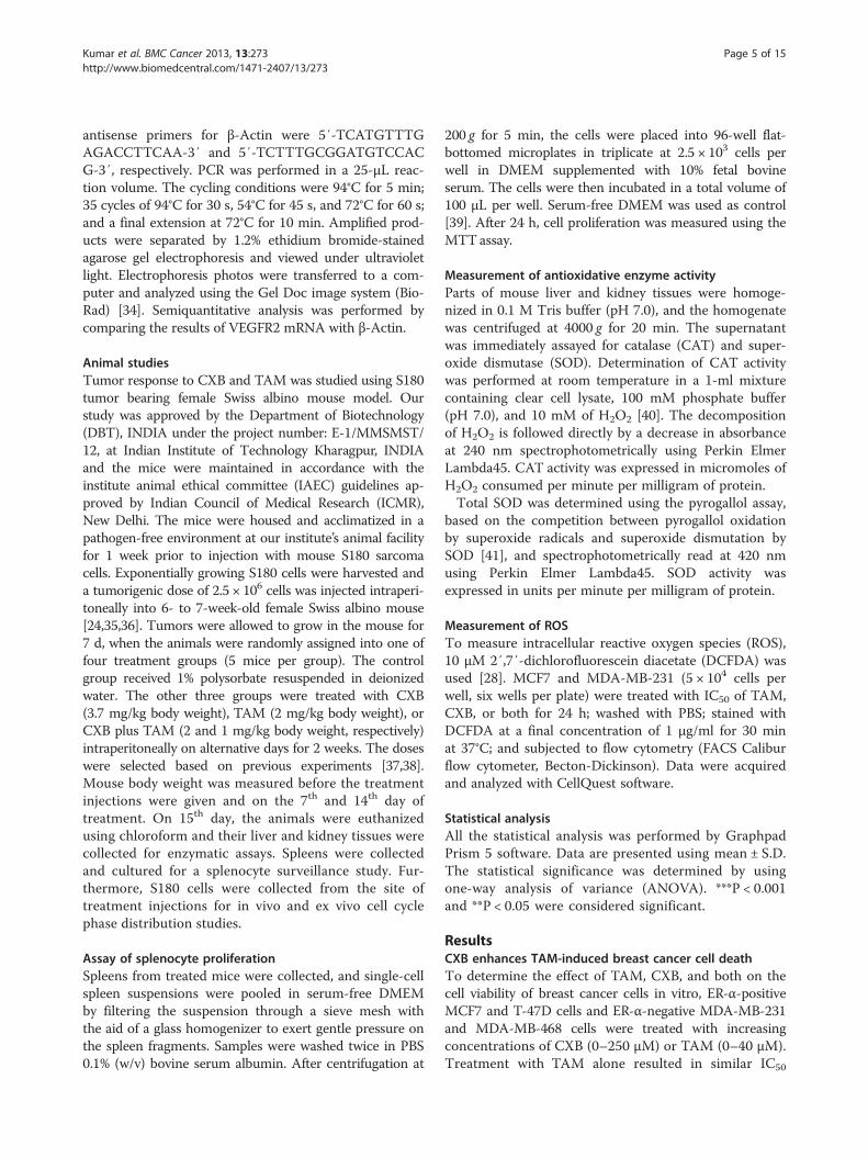
values for the MCF7, T-47D, MDA-MB-231, and MDA-MB-468 cell lines (9.06 ± 0.29, 8.99 ± 0.55, 13.05 ± 0.91,and 11.56 ± 0.65 μM, respectively) (Figure 1A). Treat-ment with CXB alone also resulted in IC50 values thatwere similar in these four cell lines (113.3 ± 0.760, 109.3 ±0.782, 109.8 ± 0.963, and 121.7 ± 0.240, respectively)(Figure 1B). Combination treatment (0–5 μM TAM inthe presence of 30 μM CXB) resulted in a leftward shiftof the concentration-response curve such that the IC50
values were reduced to 2.76 ± 0.10, 1.82 ± 0.13, 2.05 ± 0.13,and 2.86 ± 0.12 μM, respectively (Figure 1C), indicatingthat treatment with both agents was more cytotoxic thaneither one alone. The treatment regimens resulted in littletoxicity in NIH/3T3 and HaCaT cell lines, demonstratingthat TAM and CXB are non toxic to normal cell lines.Based on the results we have chosen respective IC50’s ofdrugs for further treatments throughout the study.
CXB enhances TAM-induced apoptosis and growthinhibitionThe effects of TAM and CXB on the cell cycles ofMCF7 and MDA-MB-231 cells were then analyzed.
Figure 1 TAM combined with CXB additively inhibits survival of breasT-47D, MDA-MB-468, NIH/3T3 and HaCaT cells treated with (A) TAM, (B) CXmeans ± SE of three independent experiments p < 0.05. (D) Representativecells and their cell-cycle distribution after 48 h of treatment, as determinedplus CXB; UT, untreated.
MCF7 cells (IC50 values: 114 μM CXB, 9 μM TAM)treated with TAM or CXB had an increased percentageof apoptotic cells (i.e., cells in the sub-G1 phase) com-pared with untreated cells (Figure 1D, top row). Simi-larly, MDA-MB-231 cells (IC50 values: 110 μM CXB,13 μM TAM) had an increased percentage of apoptoticcells compared with untreated cells (Figure 1D, bottomrow). The low-dose combination (30 μM CXB plus2 μM TAM) resulted in an even greater percentage ofapoptotic cells than the higher doses of either drug alonedid. These data are consistent with the results from theMTT assay. Taken together, these results indicate anadditive mechanism of TAM and CXB in inducing celldeath through apoptosis.
Effect of TAM and CXB on migration and invasion ofbreast cancer cellsTo ascertain the inhibitory effect of TAM and CXB onbreast cancer metastasis, we used the wound-healingassay to investigate their effects on the migration poten-tial of MCF7 and MDA-MB-231 cells. A wound througha confluent cell monolayer was created with a pipette
t cancer cells. In vitro cell viability assay of MCF7, MDA-MB-231,B, or (C) both (0–5 μM TAM plus 30 μM CXB) for 48 h. Data arehistogram of MCF7 cells (top row) and MDA-MB-231 (bottom row)by flow cytometry followed by staining of cells with PI. T + C, TAM
Kumar et al. BMC Cancer 2013, 13:273 Page 7 of 15http://www.biomedcentral.com/1471-2407/13/273
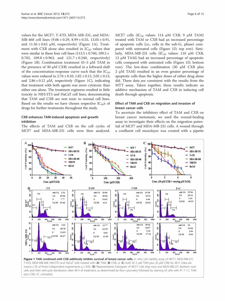
tip, and the migration of cells to fill up the wound wasrecorded by microscopic observation. After 48 h, thewound had almost completely filled in the cleared regionin untreated MCF7 and MDA-MB-231 cells (Figures 2Aand 2B). The migration of MDA-MB-231 cells was re-duced with TAM or CXB with respect to the untreatedcells and greatly reduced when both TAM and CXBwere used. However, TAM and CXB had limited effectsin MCF7 cells, which might be explained by the poor in-vasiveness of this cell line.The ability of TAM and CXB to reduce the invasive-
ness of MDA-MB-231 cells was further investigated bythe Boyden chamber assay. Cells treated with IC50 con-centrations of TAM, CXB, or both for 24 h were platedin the upper chamber, and the number of cells thatmoved to the underside of the coated membrane wascounted 12 h later using a light microscope. The cham-bers were stained with hematoxylin and eosin and ana-lyzed by photography. Again, compared with the resultswith either agent alone, the combination of TAM andCXB greatly inhibited MDA-MB-231 cell invasion(Figure 2C).
Figure 2 Anti-invasive and anti-migratory potential of TAM and CXBeosin-stained cell images migrating into the wounded area in an in vitro w(B) Quantification of wound-healing results. Data are means ± SE of three rphotomicrographs of Boyden chamber assays of MDA-MB-231 cell invasion(D) Top: Gelatinolytic activity of MMP-2 in MCF-7 cells and MDA-MB-231 celevels in gelatin blot. Data are means ± SE of three independent experimen
TAM and CXB inhibit activation of MMP-2 in breast cancercell linesSubstantial levels of MMP secretion have been reportedfor metastatic breast cancer tumors and to be associatedwith the degradation of extraceullular matrix, a crucialstep in metastasis [42]. Zymographic analyses showedthat TAM and CXB additively inhibited MMP-2 activityin both MCF7 and MDA-MB-231 cells (Figure 2D).Thus, apart from its anti-VEGF effect in inhibitingtumor cells, this combination treatment can inhibit themetastasis and spread of breast cancer cells by reducingMMP-2. The addition of CXB enhanced the anti-metastatic potential by more than 2-fold in comparison tocontrol. However, the impact of TAM and CXB on MMP-9 activity is inconclusive because an extremely low level ofMMP-9 was detected in untreated cells (data not shown).
TAM and CXB inhibit in vivo angiogenesis and in vitrotube-like capillary formationThe CAM model was used to investigate the effect of TAMand CXB on angiogenesis in vivo [43]. CAM assay with thePBS group did not show any notable avascular zone around
in MCF7 and MDA-MB-231 cells. (A) Representative hematoxylin- andound healing assay at times 0 h and 48 h. Scale bars, 100 μm.andom widths along the wound. P < 0.05. (C) Representativethrough Matrigel. Cells were stained with hematoxylin- and eosin.lls treated for 48 h. Bottom: Densitometric analysis of MMP-2 proteints. P < 0.05 (t-test).T + C, TAM plus CXB; UT, untreated cells.
Kumar et al. BMC Cancer 2013, 13:273 Page 8 of 15http://www.biomedcentral.com/1471-2407/13/273
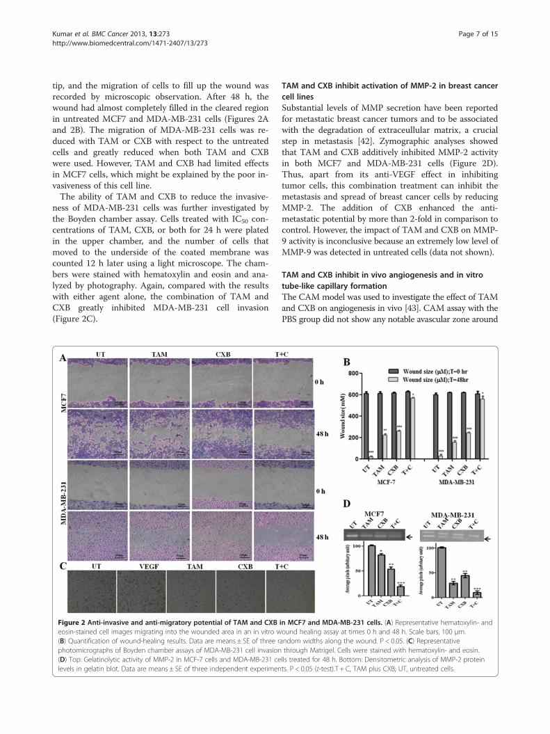
the implanted filter paper (Figures 3A and 3C). In contrast,treatment with TAM, CXB, and both agents togetherinhibited the development of new embryonic capillariesand produced an avascular zone around the implanted filterpapers. The inhibition of angiogenesis was most prominentwhen TAM and CXB were combined.Next we performed tube formation assays with
HUVECs, which are widely used as in vitro assays forangiogenesis. After 24 h, HUVECs treated with PBS onlyrapidly aligned and formed hollow, tube-like structures,whereas HUVECs treated with both TAM and CXB
Figure 3 Anti-angiogenic and anti-tube formation potential of TAM aloaded with serum-free medium alone or supplemented with vascular end(B) Inhibition of capillary-like tube formation in vitro (HUVECs assay). HUVECcoated with 50 μl Matrigel. Then, TAM and/or CXB were added. Cells wereTube formation was observed for 24 h and images were taken (magnificatimeans ± SD of blood vessel count for four independent experiments P < 0.formation assay was counted using light microscopy. Data are presented aanalysis of apoptotic and angiogenic markers in MCF7 and MDA-MB-231 cecontrol for equal loading. Representative blots from three independent exp
showed a significant reduction of tube formation com-pared with TAM or CXB alone (Figures 3B and 3D). Col-lectively, these results suggest that CXB enhances theanti-angiogenic action of TAM by inhibiting HUVEC dif-ferentiation into tube-like structures during angiogenesis.
TAM and CXB inhibit angiogenesis via von Hippel-Lindautumor suppressor protein (VHL)-mediated degradation ofhypoxia-inducible factor 1α (HIF-1α)VHL regulates activated HIF-1α through ubiqitinationby prolyl hydroxylation under normoxia conditions [44].
nd CXB. (A) In vivo CAM assay. CAMs were implanted with spongesothelial growth factor (VEGF), TAM, CXB, or TAM plus CXB.s were seeded (7.5 x 10³ cells/well) into a 96-well tissue culture plateincubated in HUVEC growth medium in a 37°C, 5% CO2 incubator.on of 10×). (C) Number of blood vessels in CAM assay was counted as05. (D) Number of capillary-like structures in capillary-like tubes means ± SD of four independent experiments. (E) Western blottinglls treated with TAM, CXB, or both. β-Actin was used as an invarianteriments are shown. T + C, TAM plus CXB; UT, untreated cells.
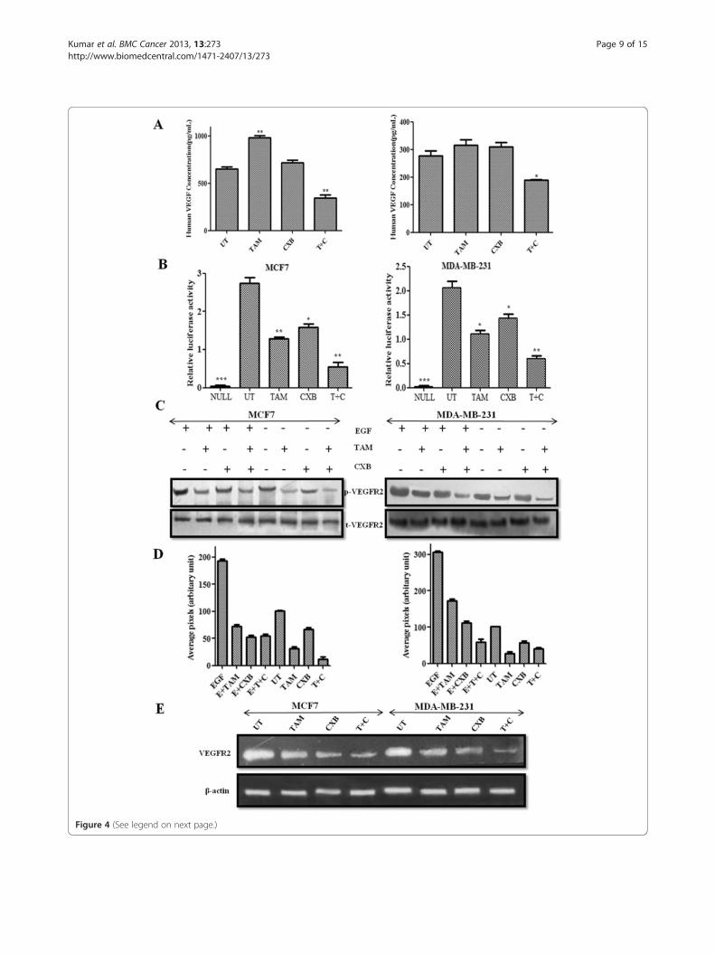
Figure 4 (See legend on next page.)
Kumar et al. BMC Cancer 2013, 13:273 Page 9 of 15http://www.biomedcentral.com/1471-2407/13/273
(See figure on previous page.)Figure 4 TAM- and CXB- inhibit overexpressed VEGFR2 induced angiogenesis in MCF7 cells (left) and MDA-MB-231 cells (right).(A) Cells were treated with TAM, CXB, or both and incubated in serum-free conditioned medium for 24 h. VEGF levels were determined by ELISA.(B) Cells (5 × 10⁵/ml) were transfected with VEGFR-luciferase plasmid, incubated for 24 h, and treated with TAM, CXB, or both for 4 h. Whole-cellextracts were then prepared and analyzed for luciferase activity. Absolute values are normalized to untreated cells without VEGFR2. Data aremeans ± SD of three independent experiments. (C) Western blot analysis for VEGFR2 and phosphorylated VEGFR2. (D) Densitometric analysis ofphosphorylated VEGFR2 protein levels. Data are means ± SD of three independent experiments. P < 0.05 (t-test). (E) The level of VEGFR2 mRNA inMCF7 and MDA-MB-231 cells examined by RT-PCR analysis following TAM, CXB and T + C treatment for 24 h. Data are means ± SD of threeindependent experiments using different cell preparations. *P < 0.05 vs. untreated cells. EGF, epidermal growth factor; T + C, TAM plus CXB; UT,untreated cells.
Kumar et al. BMC Cancer 2013, 13:273 Page 10 of 15http://www.biomedcentral.com/1471-2407/13/273
In reduced oxygen conditions, HIF-1α binds to hypoxia-responsive elements which, in turn, stimulate the tran-scriptional coactivators CREB-binding protein and inducestranscription of various target genes involved in tumor in-vasion, cell survival, and angiogenesis. Apart from its rolein angiogenesis, HIF-1α promotes invasion by regulatingthe expression of COX-2, MMP-2, and other cytokinesand growth factors [45]. Our western blotting results dem-onstrated that the combination of TAM and CXB modu-lated VHL expression in MCF7 and MDA-MB-231 cells,thus regulating HIF-1α, which in turn binds to CREB-binding protein, thereby altering the expression of thedownstream effector molecules involved in metastasis andangiogenesis (e.g., MMP-2, COX-2 and VEGF) (Figure 3E).These features have rendered HIF-1α as an attractive tar-get for our study in inhibiting angiogenesis.
TAM plus CXB lowers VEGF production in breast cancercellsWe investigated the role of TAM and CXB in the inhib-ition of secretory VEGF, a pro-angiogenic factor respon-sible for the migration and invasion of breast cancercells. VEGF secretion in serum-free culture conditionedmedium was assessed in MCF7 and MDA-MB-231 cellsby ELISA 24 h post-treatment. In both cell lines, TAMalone considerably upregulated VEGF secretion and thecombination of CXB and TAM notably decreased VEGFsecretion compared with no treatment (Figure 4A). Pre-cisely, in control cells VEGF levels were found to beapproximately 600 and 280 pg/mL in MCF7 and MDA-MB-231 cells, respectively whereas CXB treatment alonedoes not showed any significant change in the secretedVEGF levels in both cell lines. However, induced VEGFwas suppressed in combination treatment to 400 pg/mLin MCF7 and 190 pg/mL in MDA-MB-231 in compari-son to TAM alone treated MCF7 (1000 pg/mL) andMDA-MB-231 (320 pg/mL).
TAM plus CXB inhibits VEGF-mediated stimulation ofVEGFR2 promoter activityTo further confirm the role of enhanced activity inducedby treatment with TAM and CXB in the transcriptionalregulation of the VEGFR2 gene, cells were transientlytransfected with a chimeric luciferase gene fused with the
5′ region of the VEGFR2 promoter (Tischer et al., 1991),and the activity of the promoter was assayed in the pres-ence and absence of VEGFR2 gene after treatment withthe IC50 doses for 24 h. Transfection induced VEGFR2promoter activity in both MCF7 and MDA-MB-231 cells.To determine the relative fold change in VEGFR2 pro-moter activity, we normalized with respect to untrans-fected control (null) cells. VEGFR2 transfected untreatedcell (UT) showed an approximately 3- and 2-fold increasein promoter activity as compare to null in MCF7 andMDA-MB-231 cells, respectively. There was an approxi-mately 1.2-fold increase in VEGFR2 promoter activityin TAM-treated and approximately 1.5-fold increase inCXB-treated whereas fold increase was observed <1 inTAM-CXB treated with respect to null in both celllines. Concisely, TAM and CXB was effective in blockingVEGFR2 promoter induced expression in MCF7 andMDA-MB-231 cells (Figure 4B). Taken together, theresults of this experiment demonstrated that the activityof the VEGFR2 promoter is downregulated by CXBunder the influence of TAM in both the cell lines.Besides, it also interferes with the phosphorylation ofVEGFR2 (Figures 4C and 4D). Further, RT-PCR analysiswas also in accordance with the VEGFR2 promoterluciferase activity (Figure 4E).
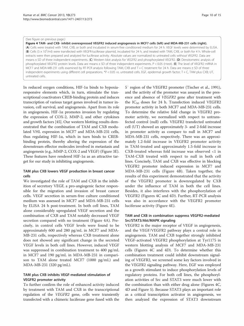
TAM and CXB in combination suppress VEGFR2-mediatedSrc/STAT3/Akt/MAPK signalingVEGFR2 is the major receptor of VEGF in angiogenesis,and the VEGF/VEGFR2 pathway plays a central role inangiogenesis. TAM and CXB together strongly inhibitedVEGF-activated VEGFR2 phosphorylation at Tyr1175 inwestern blotting analysis of MCF7 and MDA-MB-231cells (Figures 4C and 4D). To determine whether thiscombination treatment could inhibit downstream signal-ing of VEGFR2, we screened some key factors involved inthe VEGFR2 signaling pathway. Here, EGF was employedas a growth stimulant to induce phosphorylation levels ofregulatory proteins. For both cell lines, the phosphoryl-ation activities of Src and STAT3 were much lower withthe combination than with either drug alone (Figures 4C,4D and Figure 5). Because STAT3 plays an important roleas a critical transcription activator in angiogenesis, wethen analyzed the expression of STAT3 downstream
Kumar et al. BMC Cancer 2013, 13:273 Page 11 of 15http://www.biomedcentral.com/1471-2407/13/273
genes. Results showed that compared with TAM or CXBalone, TAM-CXB together inhibited the expression ofanti-apoptotic Bcl-2 protein and increased the levels ofpro-apoptotic Bax and Bak proteins (Figure 3E). STAT3 isalso involved in the inhibition of apoptosis in endothelialcells. We found that various death substrates, such as poly(ADP-ribose) polymerase (PARP) (Figure 3E) and othermolecules at conserved aspartic acid residues (data notshown), were more strongly activated by TAM-CXB incombination than by either drug alone in MCF7 andMDA-MB-231 cells. Taken together, these western blot-ting analysis results suggest that the combination ofTAM-CXB blocks the VEGF-induced Src/STAT3 signal-ing pathway. Further, our western blotting analysis provedthe involvement of VEGFR2 signaling in the inhibition ofAKT and MAPK and the phosphorylation of the down-stream protein Bad (Figure 5). Bad plays important rolesin tumor cell function, angiogenesis, and tumor growth.
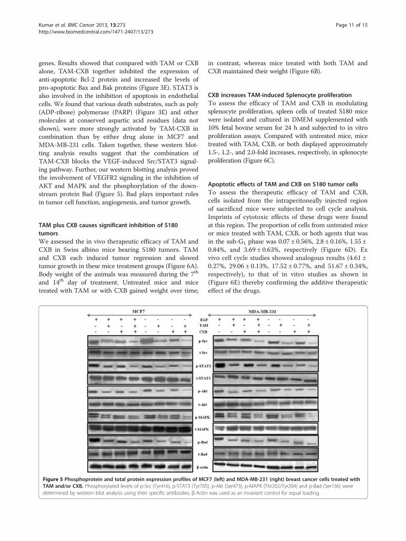
TAM plus CXB causes significant inhibition of S180tumorsWe assessed the in vivo therapeutic efficacy of TAM andCXB in Swiss albino mice bearing S180 tumors. TAMand CXB each induced tumor regression and slowedtumor growth in these mice treatment groups (Figure 6A).Body weight of the animals was measured during the 7th
and 14th day of treatment. Untreated mice and micetreated with TAM or with CXB gained weight over time;
Figure 5 Phosphoprotein and total protein expression profiles of MCTAM and/or CXB. Phosphorylated levels of p-Src (Tyr416), p-STAT3 (Tyr705determined by western blot analysis using their specific antibodies. β-Actin
in contrast, whereas mice treated with both TAM andCXB maintained their weight (Figure 6B).
CXB increases TAM-induced Splenocyte proliferationTo assess the efficacy of TAM and CXB in modulatingsplenocyte proliferation, spleen cells of treated S180 micewere isolated and cultured in DMEM supplemented with10% fetal bovine serum for 24 h and subjected to in vitroproliferation assays. Compared with untreated mice, micetreated with TAM, CXB, or both displayed approximately1.5-, 1.2-, and 2.0-fold increases, respectively, in splenocyteproliferation (Figure 6C).
Apoptotic effects of TAM and CXB on S180 tumor cellsTo assess the therapeutic efficacy of TAM and CXB,cells isolated from the intraperitoneally injected regionof sacrificed mice were subjected to cell cycle analysis.Imprints of cytotoxic effects of these drugs were foundat this region. The proportion of cells from untreated miceor mice treated with TAM, CXB, or both agents that wasin the sub-G1 phase was 0.07 ± 0.56%, 2.8 ± 0.16%, 1.55 ±0.84%, and 3.69 ± 0.63%, respectively (Figure 6D). Exvivo cell cycle studies showed analogous results (4.61 ±0.27%, 29.06 ± 0.13%, 17.52 ± 0.77%, and 51.67 ± 0.34%,respectively), to that of in vitro studies as shown in(Figure 6E) thereby confirming the additive therapeuticeffect of the drugs.
F7 (left) and MDA-MB-231 (right) breast cancer cells treated with), p-Akt (Ser473), p-MAPK (Thr202/Tyr204) and p-Bad (Ser136) werewas used as an invariant control for equal loading.
Figure 6 Antitumor activity of TAM and CXB in Swiss albino mice bearing S180 tumors. Mice were intraperitoneally injected with TAM(2 mg/kg body weight), CXB (3.7 mg/kg body weight), or both (2 and 1 mg/kg body weight, respectively) on alternative days after tumor cellimplantation and continued for 2 weeks. (A) Mice images bearing S180 tumors with different treated groups at the time of sacrifice, (B) Animalbody weight on 7th and 14th day of treatment. Data are means ± SD of three independent experiments. (C) MTT assay of proliferation ofsplenocytes from mice. Data are means ± SD of three independent experiments. p < 0.05 compared with untreated mice. (D) Cell-cycle phasedistribution study of S180 cancer cells isolated from the intraperitoneal region of treated animals exposed to TAM and CXB for 48 h followed byPI staining. (E) Cell-cycle phase distribution analysis of ex vivo grown S180 cells exposed to TAM and CXB for 48 h followed by PI staining.Enzyme activity assays of catalase (F) and superoxide dismutase (G) from liver and kidney tissue homogenates of S180 tumor-bearing Swissalbino mice after drug treatment. Data are means ± SD of three independent experiments. (H) Intracellular ROS accumulation in MCF-7 cells (top)and MDA-MB-231 cells (bottom) treated with TAM, CXB, or both for 24 h was assessed by DCFDA staining and performed flow cytometry. T + C,TAM plus CXB; UT, untreated cells.
Kumar et al. BMC Cancer 2013, 13:273 Page 12 of 15http://www.biomedcentral.com/1471-2407/13/273
TAM and CXB additively decrease CAT and SOD activityCAT and SOD assays were performed to assess the roleof reactive oxygen species in VEGF induction [46]. Theactivities of the antioxidant enzymes CAT and SOD inthe liver and kidney of S180 tumor-bearing mice wereassayed. For both TAM- and CXB-treated mice, the levelsof CAT activity in liver tissue or in kidney tissue were sig-nificantly lower than those of untreated mice (Figure 6F).In addition, for mice treated with both TAM and CXB,
CAT activity in liver or kidney tissue was significantlylower than that in mice treated with TAM or CXBalone. Similar results were observed with SOD activity(Figure 6G).
Role of ROS in the combined effect of TAM and CXBTo establish whether treatment with TAM and CXB for24 h induces ROS-dependent apoptosis, we investigatedwhether they increase ROS generation in MCF7 and
Kumar et al. BMC Cancer 2013, 13:273 Page 13 of 15http://www.biomedcentral.com/1471-2407/13/273
MDA-MB-231 cells by measuring the intracellular levelsof H2O2 using DCFDA staining. Flow cytometricanalysis revealed that for both cell lines, TAM resultedin higher generation of ROS than CXB (Figure 6H). Inaddition, treatment with both agents increased ROS pro-duction by over 50% as compared with the control cells,which was associated with enhanced apoptosis.
DiscussionTAM has been described as ‘the most important drugdeveloped in the history of breast cancer’ [47]. Theintroduction of TAM heralded a new approach to thetreatment of breast cancer. Initial clinical studies ofTAM displayed its antiangiogenic and VEGF reducingability in various tumor models [5,48-51]. Despite itsmeritorious stand in the treatment of breast cancer,prolonged administration of TAM causes intracellularVEGF levels to rise in patients, an undesirable responseleading to enhanced metastasis and angiogenesis andresulting in inferior outcomes [14,52]. In addition, auto-crine VEGF/VEGFR2 loop activation confers resistanceto TAM in breast cancer cells [8]. In this perspective, wemade an attempt to decrease intracellular VEGF levelsby reducing the TAM dose in ER-positive and ER-negative breast cancer cells. For accomplishing the abovegoal we employed combination therapy by decreasingTAM dose and choose CXB, a selective COX-2 inhibitoras an adjuvant agent [53] that induces apoptosis throughinhibiting angiogenesis by suppressing VEGF expressionin gastric and breast cancers [20,54]. From the above re-port, in the current study we aimed to determine the ex-pression profile of VEGFR2 and quantify VEGF in bothMCF7 and MDA-MB-231 breast cancer cells treatedwith TAM, CXB or both. In our study, we observed re-duction in VEGF levels in TAM and CXB treated MCF7but no significant change in MDA-MB-231. Interest-ingly, we also found that the activity of VEGFR2 wasinhibited by TAM and CXB in very low concentrationsthan either drug alone.STAT proteins comprise a transcription factor family
that participates in normal cellular events, such as prolif-eration, apoptosis and angiogenesis [55]. An increasingamount of evidence has suggested that STATs, mainlySTAT3, play a critical role in angiogenesis. Indeed,activated STAT3 is a mediator and biomarker of VEGF-induced endothelial activation [56]. The VEGF/VEGFR2-mediated STAT3 signaling pathway is a potential keytarget of anti-angiogenic tumor therapy [57,58]. Here,we elucidated the VEGFR2-activated STAT3 signalingpathway in human breast cancer cells. In our study, theactivity of VEGFR2 was more strongly inhibited, andthus the activation of Src and STAT3 is suppressed bythe combination of CXB and TAM (in very low concen-trations) than either drug alone. The reduction of
STAT3 activation, in turn, inhibited the downstreamgene expression of the anti-apoptotic Bcl-2 protein andincreased the expression levels of the pro-apoptotic Baxand Bak proteins. Furthermore, the core proteins in-volved in apoptosis, including various death substratessuch as PARP, were activated when treated with CXBand TAM in combination, which was consistent withthe results of our apoptosis analysis.VEGFR2 mediates Src regulation of endothelial cell
junctions and vascular permeability [59,60]. Src proteinsappear to be important for multiple aspects of tumorprogression, including proliferation, disruption of cell-cell contacts, migration and invasiveness [61]. TAM andCXB additively reduced tumor migration and invasion;this finding was supported by our wound-healing andBoyden chamber assay results. We also demonstratedthat CXB and TAM in combination interfered with thebinding of VEGF to VEGFR2, thus suppressing the phos-phorylation of Src protein and contributing to anti-metastatic activity leading to decreased MMP expression,as confirmed through the gelatin zymography andwestern blot analyses. Our study also showed that theROS level decreased after co-administration of TAMand CXB confirmed through our FACS and in vivostudies.Moreover, we proved the involvement of VEGFR2 sig-
naling in the inhibition of Akt and MAPK moleculesand in the phosphorylation of downstream proteins suchas Bad and Bax, which play important roles in angiogen-esis and apoptosis [24]. Supporting evidence concerningin vivo anti-angiogenic effects of TAM-CXB additivelycame from our chick embryonic CAM model andHUVEC-based tube formation assay with an in vitromodel. All these results showed that treatment with bothTAM and CXB suppressed the VEGFR2 pathways.To thoroughly understand the extent of VEGF/
VEGFR2 inhibition by TAM and CXB in combination,we performed VEGFR2 overexpression studies throughluciferase assays and quantified the serum VEGF secre-tion levels. Results showed an approximately 3- and 2-fold increase in VEGFR2 promoter activity in transfectedMCF7 and MDA-MB-231 cells, respectively. The ob-served VEGF-mediated up-regulation of VEGFR2 pro-moter activity in MCF7 and MDA-MB-231 cells waseffectively suppressed by TAM and CXB in combinationat very low concentrations (IC50 values) as comparedwith either drug alone. Finally, to validate the extent ofVEGFR2 expression at mRNA levels, we performed RT-PCR studies and came up with similar results as theoverexpression studies.
ConclusionIn summary, our study indicated that the combinationof TAM and CXB at nontoxic levels exerts potent anti-
Kumar et al. BMC Cancer 2013, 13:273 Page 14 of 15http://www.biomedcentral.com/1471-2407/13/273
angiogenic effects by specifically targeting VEGF/VEGFR2autocrine signaling through ROS generation. This additivecombination suggests an effective approach with promis-ing results in anti-metastatic and apoptotic studies. In aline, our preclinical studies suggest that this combinationis a potential candidate treatment against breast tumorsexpressing high levels of VEGF and VEGFR2.
AbbreviationsTAM: Tamoxifen; CXB: Celecoxib; VEGF-A: Vascular endothelial growth factor-A; VEGFR2: Vascular endothelial growth factor receptor 2; COX-2: Cycloxygenase-2; DCFDA: 2′,7′dichlorofluorescein diacetate; ROS: Reactiveoxygen species; CAT: Catalase; SOD: Superoxide dismutase; RT PCR: Reversetranscriptase polymerase reaction; ER: Estrogen receptor; STAT3: Signaltransducer and activator of transcription 3; MAPK: Mitogen-activated proteinkinase; ELISA: Enzyme-linked immunosorbent assay; PARP: Poly(ADP-ribose)polymerase; HUVEC: Human umbilical vein endothelial cell; HIF-1α: Hypoxia-inducible factor 1α; PI: Propidium iodide; DAPI: 4′,6-diamidino-2-phenylindole;MTT: 3(4,5-dimethylthiazol-2-yl)-2,5 diphenyltetrazolium bromide; CBP: CREB-binding protein; VHL: von Hippel-Lindau tumor suppressor protein; MMP-2: Matrix metalloproteinase2; CAM: Chorioallantoic Membrane.
Competing interestsNo competing financial or personal interest in any company or organizationis reported.
Authors’ contributionsBNP, SR, MM conceived the study, designed the experiments, and draftedthe manuscript. BNP and SR carried out the experiments. SD performedanimal studies. KKD performed the RT-PCR studies. AP conducted VEGFquantification studies. AM and MM provided the critical revision of themanuscript. All authors read and approved the final manuscript.
AcknowledgementsB. N. Prashanth Kumar and Shashi Rajput are the recipients of a Researchfellowship from the Council of Scientific and Industrial Research (CSIR), India.This study was supported by grants from the Department of Biotechnology(DBT: http://dbtindia.nic.in/index.asp), Council of Scientific and IndustrialResearch (CSIR: http://csirhrdg.res.in/) and Department of Science andTechnology (DST: http://www.dst.gov.in/), India.
Author details1School of Medical Science and Technology; Indian Institute of TechnologyKharagpur, Kharagpur-721302, West Bengal PIN-721302, India. 2Departmentof Clinical Cancer Prevention, University of Texas MD Anderson CancerCentre, Houston, TX, USA.
Received: 26 March 2013 Accepted: 31 May 2013Published: 3 June 2013
References1. Brauch H, Jordan VC: Targeting of tamoxifen to enhance antitumour
action for the treatment and prevention of breast cancer: the‘personalised’ approach? Eur J Cancer 2009, 45:2274–2283.
2. Cuzick J, Sestak I, Pinder SE, Ellis IO, Forsyth S, Bundred NJ, Forbes JF, BishopH, Fentiman IS, George WD: Effect of tamoxifen and radiotherapy inwomen with locally excised ductal carcinoma in situ: long-term resultsfrom the UK/ANZ DCIS trial. Lancet Oncol 2011, 12:21–29.
3. Delozier T, Spielmann M, Mace-Lesec’h J, Janvier M, Hill C, Asselain B, JulienJP, Weber B, Mauriac L, Petit JC, et al: Tamoxifen adjuvant treatmentduration in early breast cancer: initial results of a randomized studycomparing short-term treatment with long-term treatment. FederationNationale des Centres de Lutte Contre le Cancer Breast Group. J ClinOncol 2000, 18:3507–3512.
4. Rajput S, Mandal M: Antitumor promoting potential of selectedphytochemicals derived from spices: a review. Eur J Cancer Prev 2012,21:205–215.
5. McNamara DA, Harmey J, Wang JH, Kay E, Walsh TN, Bouchier-Hayes DJ:Tamoxifen inhibits endothelial cell proliferation and attenuates VEGF-
mediated angiogenesis and migration in vivo. Eur J Surg Oncol 2001,27:714–718.
6. Beck B, Driessens G, Goossens S, Youssef KK, Kuchnio A, Caauwe A,Sotiropoulou PA, Loges S, Lapouge G, Candi A, et al: A vascular niche anda VEGF-Nrp1 loop regulate the initiation and stemness of skin tumours.Nature 2011, 478:399–403.
7. Younes MN, Yigitbasi OG, Park YW, Kim SJ, Jasser SA, Hawthorne VS, YaziciYD, Mandal M, Bekele BN, Bucana CD, et al: Antivascular therapy of humanfollicular thyroid cancer experimental bone metastasis by blockade ofepidermal growth factor receptor and vascular growth factor receptorphosphorylation. Cancer Res 2005, 65:4716–4727.
8. Aesoy R, Sanchez BC, Norum JH, Lewensohn R, Viktorsson K, Linderholm B:An autocrine VEGF/VEGFR2 and p38 signaling loop confers resistance to4-hydroxytamoxifen in MCF-7 breast cancer cells. Mol Cancer Res 2008,6:1630–1638.
9. Ryden L, Stendahl M, Jonsson H, Emdin S, Bengtsson NO, Landberg G:Tumor-specific VEGF-A and VEGFR2 in postmenopausal breast cancerpatients with long-term follow-up. Implication of a link between VEGFpathway and tamoxifen response. Breast Cancer Res Treat 2005,89:135–143.
10. Garvin S, Nilsson UW, Dabrosin C: Effects of oestradiol and tamoxifen onVEGF, soluble VEGFR-1, and VEGFR-2 in breast cancer and endothelialcells. Br J Cancer 2005, 93:1005–1010.
11. Qu Z, Van Ginkel S, Roy AM, Westbrook L, Nasrin M, Maxuitenko Y, Frost AR,Carey D, Wang W, Li R, et al: Vascular endothelial growth factor reducestamoxifen efficacy and promotes metastatic colonization anddesmoplasia in breast tumors. Cancer Res 2008, 68:6232–6240.
12. Lee JE, Chung KW, Han W, Kim SW, Kim SW, Shin HJ, Bae JY, Noh DY: Effectof estrogen, tamoxifen and epidermal growth factor on thetranscriptional regulation of vascular endothelial growth factor in breastcancer cells. Anticancer Res 2004, 24:3961–3964.
13. Bogin L, Degani H: Hormonal regulation of VEGF in orthotopic MCF7human breast cancer. Cancer Res 2002, 62:1948–1951.
14. Ruohola JK, Valve EM, Karkkainen MJ, Joukov V, Alitalo K, Harkonen PL:Vascular endothelial growth factors are differentially regulated bysteroid hormones and antiestrogens in breast cancer cells. Mol CellEndocrinol 1999, 149:29–40.
15. Hyder SM, Stancel GM, Chiappetta C, Murthy L, BoettgerTong HL, Makela S:Uterine expression of vascular endothelial growth factor is increased byestradiol and tamoxifen. Cancer Research 1996, 56:3954–3960.
16. Sanchez BC, Sundqvist M, Fohlin H, Spyratos F, Nordenskjold B, Stal O,Linderholm BK: Prolonged tamoxifen treatment increases relapse-freesurvival for patients with primary breast cancer expressing high levels ofVEGF. Eur J Cancer 2010, 46:1580–1587.
17. Wu G, Luo J, Rana JS, Laham R, Sellke FW, Li J: Involvement of COX-2 inVEGF-induced angiogenesis via P38 and JNK pathways in vascularendothelial cells. Cardiovasc Res 2006, 69:512–519.
18. Hoeben A, Landuyt B, Highley MS, Wildiers H, Van Oosterom AT, De BruijnEA: Vascular endothelial growth factor and angiogenesis. Pharmacol Rev2004, 56:549–580.
19. Katharine Kirkpatrick WO: A. Elkak, Stephen Bustin PJ, Margaret Ghilchikand, Mokbel K: The mRNA Expression of Cyclooxygenase-2 (COX-2) andVascular Endothelial Growth Factor (VEGF) in Human Breast Cancer.CURRENT MEDICAL RESEARCH AND OPINION 2002, 18:237–241.
20. Wei D, Wang L, He Y, Xiong HQ, Abbruzzese JL, Xie K: Celecoxib inhibitsvascular endothelial growth factor expression in and reduces angiogenesisand metastasis of human pancreatic cancer via suppression of Sp1transcription factor activity. Cancer Res 2004, 64:2030–2038.
21. Mikula-Pietrasik J, Kuczmarska A, Kucinska M, Murias M, Wierzchowski M,Winckiewicz M, Staniszewski R, Breborowicz A, Ksiazek K: Resveratrol and itssynthetic derivatives exert opposite effects on mesothelial cell-dependent angiogenesis via modulating secretion of VEGF and IL-8/CXCL8. Angiogenesis 2012, 15:361–376.
22. Dash R, Mandal M, Ghosh SK, Kundu SC: Silk sericin protein of tropicaltasar silkworm inhibits UVB-induced apoptosis in human skinkeratinocytes. Mol Cell Biochem 2008, 311:111–119.
23. Sarkar S, Mazumdar A, Dash R, Sarkar D, Fisher PB, Mandal M: ZD6474Enhances Paclitaxel Antiproliferative and Apoptotic Effects in BreastCarcinoma Cells. J Cell Physiol 2011, 226:375–384.
24. Sarkar S, Mazumdar A, Dash R, Sarkar D, Fisher PB, Mandal M: ZD6474, adual tyrosine kinase inhibitor of EGFR and VEGFR-2, inhibits MAPK/ERK
Kumar et al. BMC Cancer 2013, 13:273 Page 15 of 15http://www.biomedcentral.com/1471-2407/13/273
and AKT/PI3-K and induces apoptosis in breast cancer cells. Cancer BiolTher 2010, 9:592–603.
25. Tammali R, Reddy ABM, Srivastava SK, Ramana KV: Inhibition of aldosereductase prevents angiogenesis in vitro and in vivo. Angiogenesis 2011,14:209–221.
26. Mandal M, Myers JN, Lippman SM, Johnson FM, Williams MD, Rayala S,Ohshiro K, Rosenthal DI, Weber RS, Gallick GE, El-Naggar AK: Epithelial tomesenchymal transition in head and neck squamous carcinoma:association of Src activation with E-cadherin down-regulation, vimentinexpression, and aggressive tumor features. Cancer 2008, 112:2088–2100.
27. Venkatesan P, Bhutia SK, Singh AK, Das SK, Dash R, Chaudhury K, Sarkar D,Fisher PB, Mandal M: AEE788 potentiates celecoxib-induced growthinhibition and apoptosis in human colon cancer cells. Life Sci 2012,91:789–799.
28. Wang N, Wang ZY, Mo SL, Loo TY, Wang DM, Luo HB, Yang DP, Chen YL,Shen JG, Chen JP: Ellagic acid, a phenolic compound, exerts anti-angiogenesis effects via VEGFR-2 signaling pathway in breast cancer.Breast Cancer Res Treat 2012, 134:943–955.
29. Santhekadur PK, Gredler R, Chen D, Siddiq A, Shen XN, Das SK, Emdad L,Fisher PB, Sarkar D: Late SV40 factor (LSF) enhances angiogenesis bytranscriptionally up-regulating matrix metalloproteinase-9 (MMP-9). J BiolChem 2012, 287:3425–3432.
30. Koskimaki JE, Lee E, Chen W, Rivera CG, Rosca EV, Pandey NB, Popel AS:Synergy between a collagen IV mimetic peptide and a somatotropin-domain derived peptide as angiogenesis and lymphangiogenesisinhibitors. Angiogenesis 2013, 16:159–170.
31. Chintharlapalli S, Papineni S, Ramaiah SK, Safe S: Betulinic acid inhibitsprostate cancer growth through inhibition of specificity proteintranscription factors. Cancer Res 2007, 67:2816–2823.
32. Shirakawa T, Gotoh A, Zhang Z, Kao C, Chung LW, Gardner TA:Development of human chorionic gonadotropin subunit-beta promoter-based toxic gene therapy for testicular cancer. Urology 2004, 63:613–618.
33. Domingues I, Rino J, Demmers JA, de Lanerolle P, Santos SC: VEGFR2translocates to the nucleus to regulate its own transcription. PLoS One2011, 6:e25668.
34. Levine JJ, Stimson-Crider KM, Vertino PM: Effects of methylation onexpression of TMS1/ASC in human breast cancer cells. Oncogene 2003,22:3475–3488.
35. Venkatesan P, Puvvada N, Dash R, Kumar BNP, Sarkar D, Azab B, Pathak A,Kundu SC, Fisher PB, Mandal M: The potential of celecoxib-loadedhydroxyapatite-chitosan nanocomposite for the treatment of coloncancer. Biomaterials 2011, 32:3794–3806.
36. Das S, Dey KK, Dey G, Pal I, Majumder A: MaitiChoudhury S, kundu SC.Mandal M: Antineoplastic and apoptotic potential of traditional medicinesthymoquinone and diosgenin in squamous cell carcinoma. PLoS One 2012,7:e46641.
37. Yoshida S, Amano H, Hayashi I, Kitasato H, Kamata M, Inukai M, YoshimuraH, Majima M: COX-2/VEGF-dependent facilitation of tumor-associatedangiogenesis and tumor growth in vivo. Lab Invest 2003, 83:1385–1394.
38. Kang HF, Wang XJ, Liu XX, Dai ZJ, Xue FJ: Xue XH: [Chemopreventiveeffect of tamoxifen combined with celecoxib on DMBA-induced breastcancer in rats]. Ai Zheng 2006, 25:1346–1350.
39. Majdalawieh AF, Hmaidan R, Carr RI: Nigella sativa modulates splenocyteproliferation, Th1/Th2 cytokine profile, macrophage function and NKanti-tumor activity. J Ethnopharmacol 2010, 131:268–275.
40. Dhaunsi GS, Yousif MH, Akhtar S, Chappell MC, Diz DI, Benter IF:Angiotensin-(1–7) prevents diabetes-induced attenuation in PPAR-gamma and catalase activities. Eur J Pharmacol 2010, 638:108–114.
41. Marklund S, Marklund G: Involvement of the superoxide anion radical inthe autoxidation of pyrogallol and a convenient assay for superoxidedismutase. Eur J Biochem 1974, 47:469–474.
42. Deryugina EI, Quigley JP: Matrix metalloproteinases and tumor metastasis.Cancer Metastasis Rev 2006, 25:9–34.
43. Emdad L, Lee SG, Su ZZ, Jeon HY, Boukerche H, Sarkar D, Fisher PB:Astrocyte elevated gene-1 (AEG-1) functions as an oncogene andregulates angiogenesis. Proc Natl Acad Sci U S A 2009, 106:21300–21305.
44. Semenza GL: HIF-1 and tumor progression: pathophysiology andtherapeutics. Trends Mol Med 2002, 8:S62–67.
45. Newcomb EW, Ali MA, Schnee T, Lan L, Lukyanov Y, Fowkes M, Miller DC,Zagzag D: Flavopiridol downregulates hypoxia-mediated hypoxia-inducible factor-1alpha expression in human glioma cells by a
proteasome-independent pathway: implications for in vivo therapy.Neuro Oncol 2005, 7:225–235.
46. Kumar B, Gupta SK, Nag TC, Srivastava S, Saxena R: Green tea preventshyperglycemia-induced retinal oxidative stress and inflammation instreptozotocin-induced diabetic rats. Ophthalmic Res 2012, 47:103–108.
47. Zheng J: Yao Z: [Effect of tamoxifen on apoptosis and drug resistance ofbreast cancer cells in vitro]. Zhonghua Zhong Liu Za Zhi 2000, 22:55–57.
48. Rajput S, Kumar BN, Sarkar S, Das S, Azab B, Santhekadur PK, Das SK, EmdadL, Sarkar D, Fisher PB, Mandal M: Targeted Apoptotic Effects ofThymoquinone and Tamoxifen on XIAP Mediated Akt Regulation inBreast Cancer. Plos One 2013, 8:e61342.
49. Marson LP, Kurian KM, Miller WR, Dixon JM: The effect of tamoxifen onbreast tumour vascularity. Breast Cancer Res Treat 2001, 66:9–15.
50. Blackwell KL, Haroon ZA, Shan S, Saito W, Broadwater G, Greenberg CS,Dewhirst MW: Tamoxifen inhibits angiogenesis in estrogen receptor-negative animal models. Clin Cancer Res 2000, 6:4359–4364.
51. Garvin S, Dabrosin C: Tamoxifen inhibits secretion of vascular endothelialgrowth factor in breast cancer in vivo. Cancer Res 2003, 63:8742–8748.
52. Adams J, Carder PJ, Downey S, Forbes MA, MacLennan K, Allgar V, KaufmanS, Hallam S, Bicknell R, Walker JJ, et al: Vascular endothelial growth factor(VEGF) in breast cancer: comparison of plasma, serum, and tissue VEGFand microvessel density and effects of tamoxifen. Cancer Res 2000,60:2898–2905.
53. Jeon YW, Suh YJ: Synergistic apoptotic effect of celecoxib and luteolin onbreast cancer cells. Oncol Rep 2013, 29:819–825.
54. Xu T, Wang NS, Fu LL, Ye CY, Yu SQ, Mei CL: Celecoxib inhibits growth ofhuman autosomal dominant polycystic kidney cyst-lining epithelial cellsthrough the VEGF/Raf/MAPK/ERK signaling pathway. Mol Biol Rep 2012,39:7743–7753.
55. Zhang X, Song Y, Wu Y, Dong Y, Lai L, Zhang J, Lu B, Dai F, He L, Liu M, YiZ: Indirubin inhibits tumor growth by antitumor angiogenesis viablocking VEGFR2-mediated JAK/STAT3 signaling in endothelial cell.Int J Cancer 2011, 129:2502–2511.
56. Lu W, Chen H, Ye F, Wang F, Xie X: VEGF induces phosphorylation ofSTAT3 through binding VEGFR2 in ovarian carcinoma cells in vitro (vol 4,pg 363, 2006). Eur J Gynaecol Oncol 2006, 27:544–544.
57. Chen SH, Murphy DA, Lassoued W, Thurston G, Feldman MD, Lee WM:Activated STAT3 is a mediator and biomarker of VEGF endothelialactivation. Cancer Biol Ther 2008, 7:1994–2003.
58. Bartoli M, Platt D, Lemtalsi T, Gu X, Brooks SE, Marrero MB, Caldwell RB:VEGF differentially activates STAT3 in microvascular endothelial cells.FASEB J 2003, 17:1562–1564.
59. Sun Z, Li X, Massena S, Kutschera S, Padhan N, Gualandi L, Sundvold-Gjerstad V, Gustafsson K, Choy WW, Zang G, et al: VEGFR2 induces c-Srcsignaling and vascular permeability in vivo via the adaptor protein TSAd.J Exp Med 2012, 209:1363–1377.
60. Meyer RD, Sacks DB, Rahimi N: IQGAP1-dependent signaling pathwayregulates endothelial cell proliferation and angiogenesis. PLoS One 2008,3:3848.
61. Pohorelic B, Singh R, Parkin S, Koro K, Yang AD, Egan C, Magliocco A: Roleof Src in breast cancer cell migration and invasion in a breast cell/bone-derived cell microenvironment. Breast Cancer Res Treat 2012, 133:201–214.
doi:10.1186/1471-2407-13-273Cite this article as: Kumar et al.: Celecoxib alleviates tamoxifen-instigated angiogenic effects by ROS-dependent VEGF/VEGFR2autocrine signaling. BMC Cancer 2013 13:273.