causes of amblyopiarepository-tnmgrmu.ac.in/2372/1/2203001ramasubramanianr.pdf · • the incidence...
TRANSCRIPT
AN ANALYTICAL STUDY OF 50 CASES ON CAUSES OF AMBLYOPIA
Dissertation Submitted to
THE TAMILNADU Dr. M.G.R. MEDICAL UNIVERSITY
CHENNAI.
M.S. DEGREE EXAMINATION
BRANCH III – OPHTHALMOLOGY
March – 2007

CERTIFICATE
This is to certify that Dr. R..Ramasubramanian, MS. Post Graduate Student in
Ophthalmology, Regional Institute of Ophthalmology, Government Ophthalmic Hospital,
Madras Medical College, carried out this dissertation titled An Analytical Study of
Causes of Amblyopia by herself under my guidance and direct supervision during the
period of May 2004 to March 2007. This dissertation is submitted to Tamil Nadu
Dr. M.G.R. Medical University, Chennai in partial fulfillment of the award of MS Degree
(Ophthalmology).
Prof. Dr. P. Sudhakar, MS, DO., Prof. Dr.V. Velayutham, MS, DO.,
Chief Dept. of Squint and Director and Superintendent
Neuro ophthalmology Regional Institute of Ophthalmology,
Regional Institute of Ophthalmology, Government Ophthalmic Hospital,
Government Ophthalmic Hospital, Egmore, Chennai.
Egmore, Chennai.
Prof. Dr. Kalavathy Ponniraivan, BSc,MD.,
Dean
Madras Medical College,
Government General Hospital,
Chennai.
Date :
Place :

ACKNOWLEDGEMENT
I express my sincere thanks to Prof. Kalavathy Ponniraivan, M.D., Dean, Madras
Medical College, Chennai for permitting me to conduct this study.
I express my profound gratitude to Prof. V. Velayutham, M.S.D.O., Director and
Superintendent, Regional Institute of Ophthalmology and Government Ophthalmic
Hospital, Chennai for having assigned me this very interesting topic, for providing me all
the necessary facilities and guidance to enable me to complete my study.
I am very grateful to my Unit Chiefs Prof. P. Sudhakar M.S, D.O., Prof. Jaya
Suganthi M.S, D.O., Prof. K. Namitha Bhuvaneswari M.S., D.O, for their guidance
encouragement and valuable help rendered at various stages of this study.
I am deeply indebted to my Assistant Professors Dr. B. Zaibunisha M.S., D.O.,
Dr. Rajini M.S, DO., for their guidance and constructive criticism that has been of
immense help to me to complete the study.
I am grateful to Mr. Anbarasu our orthoptist for his immense help in assessment
of patients in my study.
I would be failing my duty if I were not to express my gratitude to all my patients
and their parents for their sincere Co-operation in completion of this study.

Contents
Page No.
PART – I
1. Introduction 1
2. Classification of Amblyopia 3
3. Abnormal Contour interactions 8
4. Pharmacological Effects on vision in Amblyopic eyes 9
5. Binocular Development and Amblyopia 12
6. Parallel Visual Pathway 14
7. Role of Monocular Deprivation 17
8. Visual Acuity Assessment 20
9. Vision Tests in Infancy 23
10. Factors in Maturation of Vision 26
11. Testing of Hyper Acuity 30
12. Type of Fixation 37

Contents
Page No.
13. Treatment of Amblyopia 38
PART II
14. Aims and Objectives 43
15. Methods and Materials 44
16. Observation and Results 49
17. Discussion 55
18. Conclusion 57
19. • Proforma
• Key to Master Chart
• Master Chart
• Bibliography
• Surgeries Performed

PART - I

INTRODUCTION
It is a condition with unilateral (or) bilateral decrease of visual function
caused by form vision deprivation and (or) abnormal visual interaction, that cannot
be explained by a disorder of ocular media (or) visual path ways itself. In
appropriate cases it is reversible by therapeutic measures. It is caused by abnormal
visual experience during early childhood, the critical period of development.

PREVALENCE:
It affects approximately 1-4% of the general population.
A study by Goel et al found that incidence of <1% in school children, the
incidence was higher in rural school 0.7% than urban school 0.5% at primary
level, probably because of lack of awareness among the rural population, about
regular eye check up and the use of spectacles.
• The incidence of amblyopia in speciality clinics of strabismus and
amblyopia ranges from 3-4% and 30-35%.
• It is a fairly common disease affecting 1% and 2% of the population in
most developed countries.
• 2% of the population in most developed countries.
• 4.7% of patients with anisometropia had amblyopia (Deroirs from
Netherlands) children with low birth weight less than <2,500 gms have
twice the risk of getting amblyopia.
• Heredity also plays a role. In children born to amblyopic or strabismic
arents the risk of amblyopia is 6 times higher.

CLASSIFICATION OF AMBLYOPIA:
1) Strabismic
2) Anisometropic (Unilateral (or) Asymmetrical)
a) Aniso Hyperopic
b) Anisomyopic
3) Form Vision Deprivation Amblyopia (Unilateral or Bilateral)
a) Stimulus Deprivation amblyopia or pupil, media opacities (cornea, vitreous
(or) lens) Unilateral occlusion or penalisation.
b) Ametropic Amblyopia: Uncorrected bilateral high refractive error
i) Hyperopia
ii) Myopia
iii) Astigmatism (Meridional Amblyopia)
4) Nystagmus related amblyopia
ORGANIC AMBLYOPIA:
a) Subclinical macular damage
b) Malorientation of cones
c) cone deficiency syndrome

CASE OF ANISOMYOPIC AMBLYOPIA
A CASE OF CONGENITAL CATARACT CAUSING VISION
DEPRIVATION AMBLYOPIA

A CASE OF STRABIS MIC AMBLYOPIA


CLINICAL FEATURES:
1) Decreased visual acuity (eg) Snellen’s
2) Decreased grating acuity (eg) Tellers
3) Decreased Vernier acuity
4) Decreased (or) lost stereo acuity
5) Decreased contrast sensitivity
6) Decreased brightness perception
7) Increased perception and reaction time Naso temporal asymmetries in
resolution of visual gratings.
8) Motility defects in pursuit, saccades and fixation.
VISUAL ACUITY DEFECTS IN DIFFERENT TYPES OF AMBLOYPIA:
Recognition acuity is more affected than detection acuity recognition acuity
is determined by Tellers, VER. Detection acuity is measured by catford Drum (or)
Bailey Hall cereal test).
Anisometropic and strabismic amblyopia behave diffently snellen’s or
Recognition acuity is more affected in strabismic amblyopic or mixed
amblyopic compared to anisometropic amblyopia.
Both snellens and grating acuity is affected equally in anisometropia
amblyopic, whereas in strabismic ambloypes the grating acuity is affected to
half of the extent of the snellen’s acuity. Thus strabismic amblyopia is under
estimated on grating test.
For diagnosis of amblyopia any diminution of vision, difference between two
eyes or in case of both eyes being affected, difference from the age related
norm is taken to indicate amblyopia. Clinically a two line or snellen’s chart
(one octave difference) is considered significant.
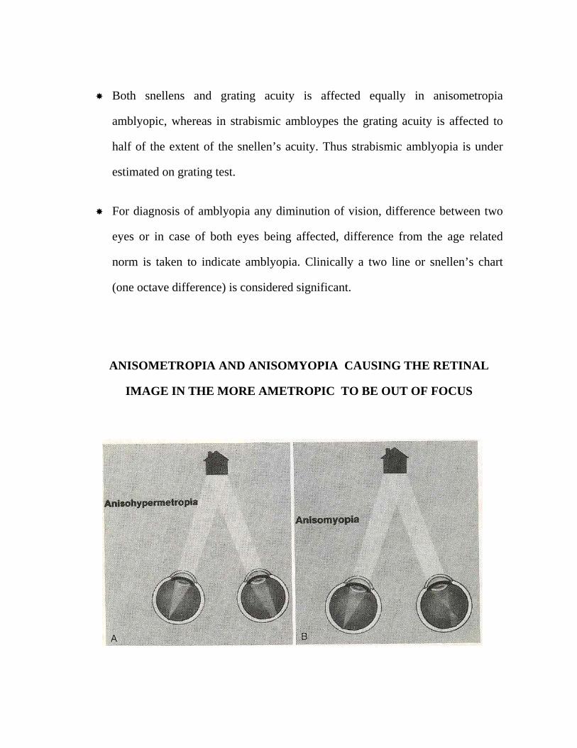
ANISOMETROPIA AND ANISOMYOPIA CAUSING THE RETINAL
IMAGE IN THE MORE AMETROPIC TO BE OUT OF FOCUS

ONLY DIFFUSE AND REDUCED AMOUNTS OF LIGHT ENTER THE
EYE THROUGH THE CATARACTOUS LENS OR BOTH THE LENSES
BLURRED RETINAL IMAGES IN BOTH THE EYES IN UNCORRECTED
HIGH HYPERMETROPIA

TYPES OF HYPER ACUITY:
The visual apparatus is capable of making much finer spatial
discriminations than the resolving capability of the retina may suggest (by
Snellen’s or grating acuity). this is called as Hyper acuity. Two common types of
Hyper acuity are vernier acuity and stero acuity. Vernier acuity includes a variety
of tasks that involve sensing the direction (or) spatial offset of a line or a point of
reference. Vernier acuity can have an accuracy of 3-6 seconds of an arc or better.
This processing is technically of sub pixel resolutions and is done at higher
cortical areas. These are not easily influenced by retinal image motion or optical
blur, implying there less likelihood of deterioration by uncorrected refractive
errors or light media opacities. The later aspect has been used in predicting visual
potential in the presence of a cataract.
Another well recognised feature of strabismic amblyopia is that vision is
not degraded by neutral density filters, it may even show some improvement.
However in anisometropic amblyopia an equal deterioration was seen. Other
organic retinal pathologies causing diminution of vision are susceptible to
deterioration by natural density filters. This distinguish functional amblyopia from
these conditions.

ABNORMAL CONTOUR INTERACTIONS.
(CROWDING PHENOMENON)
Is seen in the form of degradation of visual acuity for objects placed in a
row or line (Linear acuity), compared to the acuity of the same object viewed
separately (single letter acuity). This phenomenon has been described as the
crowding phenomenon).
Crowding phenomenon is present to some extent even in normal subjects
(critical area of separation 1.9 to 3.8 min of arc) In amblyopia it is more
pronounced similar to critical area of separation of peripheral retina in normal
subjects (=8.4 to 23.3 min of arc). The crowding phenomenon has also been
attributed to the poor visual acuity that is there in amblyopia.
The single letter visual acuity improves more rapidly during the course of
treatment. Finally both the single letter and linear acuity should approach other, if
it is not there is always a risk of recurrence of amblyopia.

PHARMACOLOGICAL EFFECTS ON VISION IN
AMBLYOPIC EYES
P. Central norepinephrine System
Pottigrew and kasamatasu and kasomattu used the activation of central nor
epinephrine system for the purpose of enhancing neuronal plasticity and kasamattu
reported that catecholamine depletion prevented the ocular dominance shift seen in
kittens after monocular occlusion.
NERVE GROWTH FACTOR:
Maffee and cocoorkers injected nerve growth factor into rats and found that
exogenous NGF prevents the effect of deprivation, that is shrink age of cells in the
lateral Geniculate Neurons and the function and anatomic organisation of the
visual cortex. They speculated that loss of competition for the deprived eye is
explained by the lack of neurotrophic factor. The monocular portion of the visual
cortex of the deprived rats treated with nerve growth factor did not differ form the
fellow eye.
LEVODOPA:
Administered as long as 1 week appears to provide positive results in the
short term. The eye effects were explained in terms of the general role of
dopamine both in the retina and in the visual pathway. Modest degrees of

improvement of visual acuity (2.7 lines) and contrast sensitivity (72%) have been
reported which by were still significant 1 month after cessation of therapy.
Legnoise and coworkers found an improvement which lasted 6 weeks after
cessation of treatment with some side effects as nausea, head ache and mood
changes.
Campos and coworkers reported a more permanent effect on visual acuity
of the amblyopic and sound eye that lasted as long as 4 months.
CYTIDINE – 5 BIPHOSPHO CHOLINE (CITICOLINE)
Drug that increases the level of consciousness in patients who had trauma
and parkinsonism. CITICOLINE improves membrane Adenosine triphosphatase
(ATP ase) activity and modulates the turn over of catecholamines and serotonin.
The age of the patients may be from 20-40 years, and thus beyond the age at
which improvement can usually be expected because on improvement was found
in both the eyes diplopia never occurred after treatment. Porciatti and coworkers
showed citicoline in adult patients improves not only visual acuity but also
contrast sensitivity and VER’s. CAMPOS and coworkers evaluated the effect of
citicoline in children with amblyopia. It was found that after 1 year follow up
visuals acuity has improved more in patients treated with a combination of
citicoline and part time occlusion that in those treated only with citicoline or with
part time occlusion. It is noteworthy that no systemic side effects were found with
this type of treatment.

LIGHT SENSE AND DARK ADAPTATION IN AMBLYOPIA
Both form vision and brightness perception in amblyopic are affected. Dark
adaptation curves were essentially normal and even if there is an effect on the light
sense. There is clearly a dissociation between the effect on the light sense and the
acuity. While recovery time after a glare stimulus to fovea is normal, the
perception time and reaction time is 6 times longer.
Pupils are generally normal and briskly reacting though afferent pupillary
defect and raised edge light pupil cycle time has been reported by some workers.
It may be generalised that,
1) In amblyopia, the visual perception of fovea stimulates that of
peripheral retina
2) The amblyopic visual system contains abnormally large receptive fields.
3) Functionally the amblyopic eye is at its best in mesopic and scotopic
condition and worst at photopic condition.

BINOCULAR DEVELOPMENT AND AMBLYOPIA:
Though practical difficulties have limited the data pertaining to visual
development in humans elegant and methodical experiments conducted by
investigators on kitten and baby monkeys have revealed a lot. The conclusion
were mentioned in human is by extrapolation from experimental work.
INTRA UTERINE DEVELOPMENT:
In the human retina most of the ganglion cells are generated between eight
and fifteenth weeks of gestation, when the ganglion cell population reaches a
plateau of 2.2 to 2.5 million. This is maintained till the thirtieth week when it starts
to fall drastically due to rapid cell death for about 6-8 weeks. There after the
process continues at a rapid rate through birth for the first few weeks of infancy.
The ganglion cells at the final count are about 1 million. The loss of more than a
million cells and their axons serve to fine tune the topography and specificity of
the reticulo geniculate body in human is generated between gestational 8-11 weeks
and by 10th week the first retinal ganglion cells start invading the lateral geniculate
body. The geniculate lamina emerge between 22-25 weeks. From an initial
intermingling of inputs from each eye, segregation of & afferents occurs by
pruning. The striate cortex cells appear by the age of 10-25 weeks of human
foetus. The geniculate afferents begin to innervate the striate cortex by 26th week.
This has been demonstrated by injection of anatomic tracers. Initially geniculate
afferents representing each eye overlap extensively in layer 4C. The segregation of

inputs into ocular dominance columns occurs during the last weeks of pregnancy
and is almost complete at birth. Until shortly before birth there is a sort of loose
wiring which is extensive and expensive, providing enough of material for fine
tuning, which occurs by a method of making new usable synapses and breaking
old (unused) synapses, synapto genesis.
In the retina two streams of ganglion cells Alpha (A cells) and Beta (B)
cells are present. The A cells communicate via the magno cellular pathways, of
LGB (Large cell Layers 1&2) to the cells of alpha of visual cortex area 17. The B
cells of Retina communicate via the parvo cellular layer of LGB (Small layer 36)
to 4C, Beta cells of the visual cortex, area 17.

PARALLEL VISUAL PATHWAY
Retinal glinglion cells LGB Cortex Other
Alpha Lamina V1 V2
Thick stripes
Magno cellular V3, V5, M+, MST, V4
Beta Parvo cellular Inter strips blob thin stripes.
Note:
V1 striate cortex primary visual area (A17) V2- V3- V4 area in area 18, 19 and
more anterior temporo parietal cortex.
The modular organisation of columns in the cortex is tuned to a variety of
stimulus specifies such as accentuation, binocular disparity, motion perception etc.
Each has discrete inputs and outputs. The Basic applied aspect of all of the
development process is that the visual development is occurring by changes in the
connections over a period of 0-5 years or more upto 9 years, during which is
plastic until it reaches visual maturity later all these inter connections over a
period of 0-5 yeras or more 9 years, during which is plastic until it reaches visual
maturity. Later all these inter connections are difficult to be changed. The two
patho mechanisms of different types of amblyopia are,
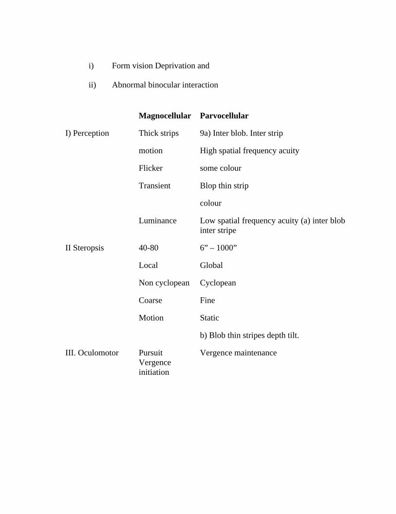
i) Form vision Deprivation and
ii) Abnormal binocular interaction
Magnocellular Parvocellular
I) Perception Thick strips
motion
Flicker
Transient
Luminance
9a) Inter blob. Inter strip
High spatial frequency acuity
some colour
Blop thin strip
colour
Low spatial frequency acuity (a) inter blob inter stripe
II Steropsis 40-80
Local
Non cyclopean
Coarse
Motion
6” – 1000”
Global
Cyclopean
Fine
Static
b) Blob thin stripes depth tilt.
III. Oculomotor Pursuit Vergence initiation
Vergence maintenance

THE ROLE OF VISUAL STIMULUS:
Visual experience loss of significant role to play in the afore mentioned
synaptogenesis. In monkeys born by caesarian section with closed eyes. Precisely
oriented simple and complex cells similar to the adult animals are seen, even with
the orderly sequence of ocular dominance and orientation columns. Those are
maintained in utero by spontaneous action potentials discharged by mammalian
retinal ganglion cells. Abolition of these action potentials discharged by
tetrodotoxin, prevent the normal prenetal segregation of retino geniculate axions
into appropriate laminae of LGB. The same is observed after intra ocular
administration of tetrodotoxin. If a newborn monkey is reared in dark (or) both
eyes sutured, cells in the striate cortex develop bizarre receptive field properties,
losing sharp orientation, tuning and normal bin ocular responses. After a
prolonged period of deprivation, if the monkey is reintroduced into normal visual
environment (lids are opened) the animal is profoundly blind with the minimal
potential for recovery. These observations stress the role of visual stimulus for
normal binocular development and are corroborated by good visual recovery only
if early surgery and rehabilitation is done for bilateral or unilateral cataracts.

THE ROLE OF MONOCULAR DEPRIVATION:
The experiments by Hubie and Weisel has established the role of cortical
competition in binocular visual development. To start with at birth all cortical cells
have potential connection with both the eyes. If both eyes are functioning equally,
the cortical cells driven by both the eyes re equal. About 10% of the cortical cells
are driven by right eye and equal percentage by left eye. The rest 80% of the cells
are driven binocularly C the central 20% of the equally by both eyes and the rest
have a predominance of one eye or the other. If by any chance one of the eye is
not functioning properly, the cortical cells of one eye is stolen or usurped by the
other. This process of competition is easily reversible in the initial period of
plasticity. Monocular deprivation produces a radical attenuation in the ocular
dominance columns in striate cortex in favour the normal eye. It is believed that
the two eyes compete for synaptic contacts in layer 4c of visual cortex. monocular
eye lid closure imposes a severe handicap in this contest. as a consequence of the
deprived eye loses many of the connections already found at birth. The ocular
dominance columns of the deprived eye shrink and those of the favoured eye
swell. a similar changes is observed in the laminae of the LGB., although, there is
no competition at this level. the LGB cells of deprived eye are smaller as they are
required to sustain a lesser arbor of axons in layer 4c of cortex.

IMPORTANT CONCLUSIONS:
Some important conclusions from these studies are
i) The loss of binocularly innervated striate neurons is not specific for
ambyopia, it occurs after any brief disruption of binocular input in early
life. this has been correlated to stereopsis.
ii) The decrease of cells responding to the stimulation of the amblyopic eye is
highly specific for amblyopia, regardless of the etiology and correlated
quantitatively with the decrease of visual activity.
iii) Even one week of disruption is sufficient to cause amblyopia in the
sensitive period. occlusion amblyopia develops rapidly in children upto 4
years but can occur later also.
iv) Recovery of neurons connected with the amblyopic eye doesnot occur on
treatment, but improvement of binocular cells appears to be permanent at
least in monkeys, after binocular vision early in life. infantile esotropia of
early onset doesnot allow good stereopsis.
v) In anisometropic and vision deprivation amblyopia both monocularly and
binocularly innervated portions shrink, but in strabismic amblyopia only
the binocularly innervated part shrinks in LGB. In the cortex, the same is
observed for vision deprivation amblyopia, but need to be confirmed for
anisometropic amblyopia.

vi) Nerve growth factor (neurotropin) seems to prevent shrinkage in LGB in
rats in strabismic and vision deprivation amblyopia yet to be confirmed in
primates.
THE APPLIED ASPECT IN CLINICAL PRACTICE:
The same process occurs during occlusion therapy. In the initial period of
therapy, the vision improves in the amblyopic eye, by the take over of the other
selected cells of the other eye and is reflected in the drop in the visual acuity of the
normal eye. However gradually the earlier “unselected” cells are recruited for the
amblyopic eye. This all the gain of the amblyopic eye is not due to loss of good
eye. But loss is possible if the normal eye is not given chance intermittently a
breather. Then it results in occlusion amblyopia which is a type of vision
deprivation amblyopia and has grave prognosis.
THE CRITICAL PERIOD
The critical period corresponds to the fine phase, when the wiring is still
malleable. It should be clear that amblyopia is likely to be corrected, occlusion
amblyopia is also possible. It has been observed to differ for different types of
amblyopia. For vision deprivation amblyopia the upper limit is 6 years and for
anisometropic amblyopia it is 8 years. The latter cases do respond even in the
teenage, whereas the strabismic amblyopes do not respond after 12 years.

VISUAL ACUITY ASSESSMENT
TYPES OF VISUAL ACUITY
1. Minimum visible (which is actually a function of brightness and is about 1
second of arc).
2. Minimum separable (which hyper acuity, a function of higher cortical centres
and is about 16 – 20 seconds of arc more, than the maximum potential of
foveal cones therefore called as hyper acuity vernier and stereo acuity are
examples).
3. Minimum resolvable and minimum recognisable (which is what is understood
by visual acuity and is limited by foveal configuration). the finest acuity can be
of 30 seconds of arc.
TESTS FOR VISUAL ACUITY
The tests for visual acuity can be grouped into – three types
a. Detection acuity test:
They assess the ability to detect the smallest stimulus (without recognising
correctly) eg
1. Catford drum

2. STYCAR graded balls test
3. Schwarting’s metronome
4. Boecks candy beads
5. Dot visual acuity
b. Recognition acuity test:
They assess the ability to recognise the stimulus (or) distinguish it from other
competing stimuli.
a. direct identification test
• Sjogren hand test
• Landolt C test
• Snellen’s E
• Arrows
b. Letter identification chart
• Snellen’s chart
• Sheridan letter test (STYCAR)
• Lippmann’s HOTV test

c. Picture identification charts / miniature toys
• Beale collin’s picture charts
• Light house test
• Allen’s picture cards
• Domino cards test
• Miniature toy test of sheridan
d. Picture identification on behavioural pattern
• Bailey hall cereal test
• Cardiff acuity cards
• OKNOVIS based on arresto – visuography.
3. resolution acuity test:
• Opto kinetic drum
• Preferential looking tests – forced choice or operant type teller acuity cards.
• Visual evoked responses.

VISION TESTS IN INFANCY:
CATFORD DRUM TEST:
It was devised CATFORD and OLIVER useful in infants and pre school
children. It is based on observation of pendular eye movement (oscillatory eye
movement not an opto kinetic nystagmus). That is elicited as the child follows an
oscillating drum with dots. The dots are displayed in various sizes (15mm to
0.5mm) and the test distance is 60cm (2 feet). The dots represent 20/600 to 20/20
vision. The smallest dot that evokes the pendular eye movement determines the
visual acuity. Unfortunately it is known to over estimate vision by double or
quadruple times and is unreliable for amblyopic screening.
CARDIFF ACUITY CARDS
They are based o the principle of preferential fixation on cards which have
a picture optotype and a blank located vertically. The picture upto types are of the
same size, but have been especially drawn with two dark lines with a white space
of varying width, in between, such that the picture is visible only at a particular
distance or closer. These are known as vanishing optotypes as they vanish at a
farther distance. They have been developed at cardiff UK. The child can identify
the picture by verbalising, pointing or fixation preference. They are more likely to
be in Recognition acuity group. The visual acuity is described in Snellen’s
notation.

VISUAL EVOKED RESPONSE (VER)
VER records the change in the cortical electrical pattern detected by surface
electrodes monitoring the occipital cortex following light stimulation of the retina.
The stimuli may be a pattern checker board or stripes on an unpatterned flash. The
size of grating or checks is to 30,15 minutes of arc. Layer check sixes than 60
minutes (10) are fallacious for visual acuity. The P100 amplitude and latency is
noted. If the Latency is not between 100-145 M sec, it cannot be relied upon and
should indicate poor vision. The VER can be recorded in two modes: Transient
and steady state.
TRANSIENT VER. Where abrupt unique alternation of stimulus is at a
relatively low rate so that each stimulus generates a separate VER output.
STEADY STATE VER.
Where rapid rate of stimulation causes blending of output into a continuos
wave.
Visual acuity of 6/12, 6/6 have been estimated at 6 month of age by VER.
These estimates of visual acuity have not been very reliable and are lighter than by
other methods. They may reflect the electro physiological changes but not truly
indicate the visual characteristics which is a perceptual phenomenon. They are
however useful in giving an objective record of the underlying visual pathway and
to exclude organic pathology.

INDIRECT ASSESSMENT OF VISUAL ACUITY:
i) Reflex responses: The pupil reacts to light after 29 week of gestation. The
baby begins to turn its head to diffuse light after 32nd – 36th week of
gestation.
ii) Fixation: The fixation reflex is a prerequisite for normal visual
development and is present at birth. Even in preterm babies after 33 weeks
of gestation. But it may not be well established. 75% of infants fixate by
two weeks and 100% by two months. New borns show fixation preference
for moving stimuli, blinking light, patterned stimulus, stimuli with high
contrast stimuli with especially red green and human face. Near objects are
preferred. The fixation may not be steady and is interrupted with refixation.
The spans of fixation lasts from seconds to minutes.
Follow Movements
Following horizontally moving targets has been seen in full term new borns
and well developed by first month. Vertical tracking usually is elicited by 4-8
weeks. The movements may have jerky interruption and corrective movements are
slow. The range of following is 450 at birth, 900 by 4 weeks and 1800by 3 months.

FACTORS IN MATURATION OF VISION:
i) Though rods and cones are distinguished even 15 weeks prior to birth they
do not attain adult like dimensions until 14 months after birth.
ii) Myelination of visual pathways is completed by one month, but the amount
of myelin increase in subsequent months upto 2 years.
iii) Cortical neuronal dendritic growth and synapse formation at 25 weeks of
gestational age and is very active in the first two years.
These factors correlate with vision as well as other functions like
i) Accommodation which is minimal at 1 month of age, but well developed
by 4 months of age.
ii) Fusion, starts of 2 months of age and fusional convergence develops by 6
months.
iii) Stereopsis is present at 1-2 months. Well developed by 4 months, though
adult levels may be seen at 5-7 years of age.
B) Vision testing in 1-2 yeras.
Boeck candy test, worth’s ivory ball test, sheridan’s ball test.
C) Visual acuity in 2-3 years.

1) Miniature toys test: Pairs of miniature toys are used. The child is asked
to name or pick the paid from an assortment. Test distance is 10 feet.
Central steady fixation: Usually means good potential for vision. In new
borns atleast 6/60. Later this should indicate 6/9 – 6/6 vision.
A preference of fixation with one eye over the other indicates poor vision in
the non preferred eye. Resentment of the closure in one eye indicates poor vision
in the other eye.
Induced Prism test:
Assessment of visual potential on the preference of fixation of one eye,
over the other is simple in the presence of a squint. In the absence of squint a
deviation can be induced by introducing a 10-15 pd prism base out, the child is
forced to choose to fixate with one eye or the other.
FIXATION AND VISION:
Fixation pattern Visual Acuity
Gross Eccentric fixation Less than CF at 1 metre
Unsteady central fixation Less than 6/60
Steady central fixation but not maintained 6/60 – 6/30
Steady central fixation can maintain but prefers other eye.
6/24 – 6/18
Central steady fixation free alternation or cross fixation
6/9 – 6/6

AGE OF CHILD AND VISUAL ACUITY
Age in months Visual Acuity
1 6/120
2 6/60
12-18 6/48 – 6/12
18-24 6/24-6/7.5
24-30 6/15-6/7.5
30-36 6/12-6/6
Coin test:
Coins of different sizes at different distance are shown. The child is asked
to distinguish between the two faces of the coin.
Dot Visual Acuity Test:
In a darkened room the child is shows an illuminated box, with presented
black dots of different diameters, one at time, successively smaller dots are shown.
The smallest dot identified correctly twice is taken as acuity threshold.

VISION TESTS IN 3-5 YEARS:
i) Tumbling E test:
The test is preferred vision test for mass screening in pre school children. It
consists of different sizes at E in one of the four positions right, left, up or down.
After familiarising the child, he indicates the direction in which ‘E’ is oriented.
Which he does by hand or orally. It is done at 6 metre distance and each eye is test
separately. Single letter acuity is supposed to be better in amblyopes in
comparison to line acuity on chart due to crowding phenomenon ‘E’ charts with
surrounds have been suggested to offset this disadvantage.
ii) Landolts C with is used in a similar manner as also Sjogren’s hands and
arrows.
iii) Sheridan letter test. The sheridan test uses 5 letters H.O.T.V. and in the fine
letter test, A and U are added in 7 letter set. C & L are also added in 9 letter
set. Testing distance is 10 feet (3 meters). The child is expected to name the
letters (or) indicate similar letter on the card in hand.
iv) Lippmanns HOTV test: The method is simpler version of Sheridan’s test
using only 4 letters H, O, T.V at a distance at 3 meters. The method is the
same as above.

TESTING OF HYPER ACUITY:
VERNIER ACUITY TESTING:
Vernier acuity tests are of interest particularly in amblyope, whereas grating
acuity may be fallaciously normal. In has been used in the acuity card format for
infants and small children using the AFC pattern.
Steroacuity:
Has been seen to be developed and testable on the PLT by 6 months. It has
been used as a screening test for binocular vision anomalies in pre school children
but with difficulty.
Steroacuity test:
The real depth tests are not used, most clinical tests are based on
haploscopic principle using two dimensional or vectographic pictures. Some
elments of the two pictures have a disparity which is fused to create a 3D image.
This can be test on
i) TNO test with red green goggles
ii) Synoptophore; with stereopsis slides
iii) Randot Stereo test and titmus stereotest with polaroid spectacles.

iv) Lang acuity test using no glasses.
v) Special 3D pictures.
SYNOPTOPHORE TESTING
This is used to test the binocular single vision and angle of Deviation
THE TITMUS STEREO TEST
RANDOM DOT E TEST SET

RANDOT E - TEST
A THE TNO TEST . B. THE RANDOM DOT STEREOGRAM
The last two examples in which the dissociation is not achieved by glasses,
which may not liked by children.

The Langs test is based on the principle of panography when two images
are printed on the same card each interrupting the other with regular linear
interruptions. A prismatic film laminated over the picture ensures that one image
is visible to the right eye only and the other to the left eye only. The two when
fused in spite of the disparity create a 3D vision.
RANDOT STEREO TEST:
This is the most popular clinical and has replaced the earlier popular titmus
fly test. It uses Julesz’s Random dot back ground to mask the monocular cues
which are there with the animal test and wirts circle test. Geometic figures like
square, circle, triangle, star etc. are also presented. The latter type figures, though
a better test are usually not appreciated by small children. The test requires
polaroid glasses to be worn by the patient. It is used at a distance of 40cm and thus
test near binocular vision, so that myopes upto 3 diopteres can be missed in a
screening test. The wirt’s circle 1-40 test the streo acuity from 400 arc second to
20 arc seconds.
TNO test:
This test is also based on the random dot back ground but uses red green
glasses for dissociation of the two images. It tests steroacuity from 480 are
seconds to 15 are seconds.

FRISBY TEST:
The Frisby Stereotest consists of three Perspex plates of differing thickness
6mm, 3mm and 15mm on one face of each plate are found squares. Three of
which are filled with a random pattern of blue triangles of various sizes and fourth
of which has a central, circular area that is not patterned. On the opposite side of
the plate coincident with this area is a circular pattern of similar blue triangles.
The plate is held in a white board and when viewed directly, the squares are all
filled with random patterns although in one square a binocular viewer will see a
circle standing up from the plate C crossed disparity or lying below the rest of the
design (uncrossed disparity) depending on which side of the plate and distance
from the subject different stereo acuities can be assessed. For 30cm viewing
distance, the 6mm, 3mm and 1.5mm represent 600, 300 and 150 are seconds of
stereo acuity respectively. This assumes the inter pupillary distance of 60mm, but
not significant change is caused by different IPD.
DISTANCE STEREOPSIS TEST
Stereo acuity should be tested for distance also. A projection vectographic
or oculus distance stereo tests can be used to test stereopsis for distance. A
diministed stereopsis for distance may be on early sign of decompensating
exophoria.

NORMAL STEREO ACUITY:
Though adult individual are capable of appreciating stereopsis with
disparties as fine as 15-20 arc seconds. The adult norm is 40 arc second. For
children 3-5 year old the norm is 70 arc seconds, and for 5-7 years it is 50 arc
seconds. Children above 8 year have the adult no norm.
GROSS STEREOPSIS:
In the absence of fine stereotests a gross estimation of stereopsis can be
made by a bed side test. Two pencil test was popularised by Lang. A pencil is held
in the examiners hand horizontally and the child is asked to touch the tip with the
tip of another pencil rapidly come from one side. Care should be taken to avoid
giving the end on view of the pencil, as that can be accomplished even
monocularly, therefore horizontal pencils are better as they do not allow an end on
view. Always compare the binocular task. The test is a gross stereopsis test of
about 400 arc seconds disparity. A rough estimation of visual acuity has been
made on the basis of stereo acuity.

LANG’S TWO PENCIL TEST

A pencil is slide in the examiner hand and the child is asked to touch the tip
with the tip of another pencil rapidly from one side
TYPE OF FIXATION
Site and type of fixation whether it is steady or unsteady and whether it is
foveal (within 20) parafoveal (20-50), para macular (50-100), paracecal or
peripheral (or) temporal. A steady central fixation is a good prognostic sign. An
unsteady but central foveal fixation indicates a possibility of good vision with
conventional occlusion. But a steady peripheral or para macular or paracaceal
fixation generally indicates a poor prognosis. Eccentric fixation is generally
unilateral with the other eye showing central fixation. But some times one may
encountaer bilateral eccentric fixation. This is usually vertical ie the fixating points
are above or below the foveal in both the eyes. It indicates a macular pathology
which may be missed clinically. Electro physiological tests:- EOG and ERG done
is such cases confirm and prognosticate such cases.
VARIOUS TYPES OF NONFOVEOLAR FIXATION
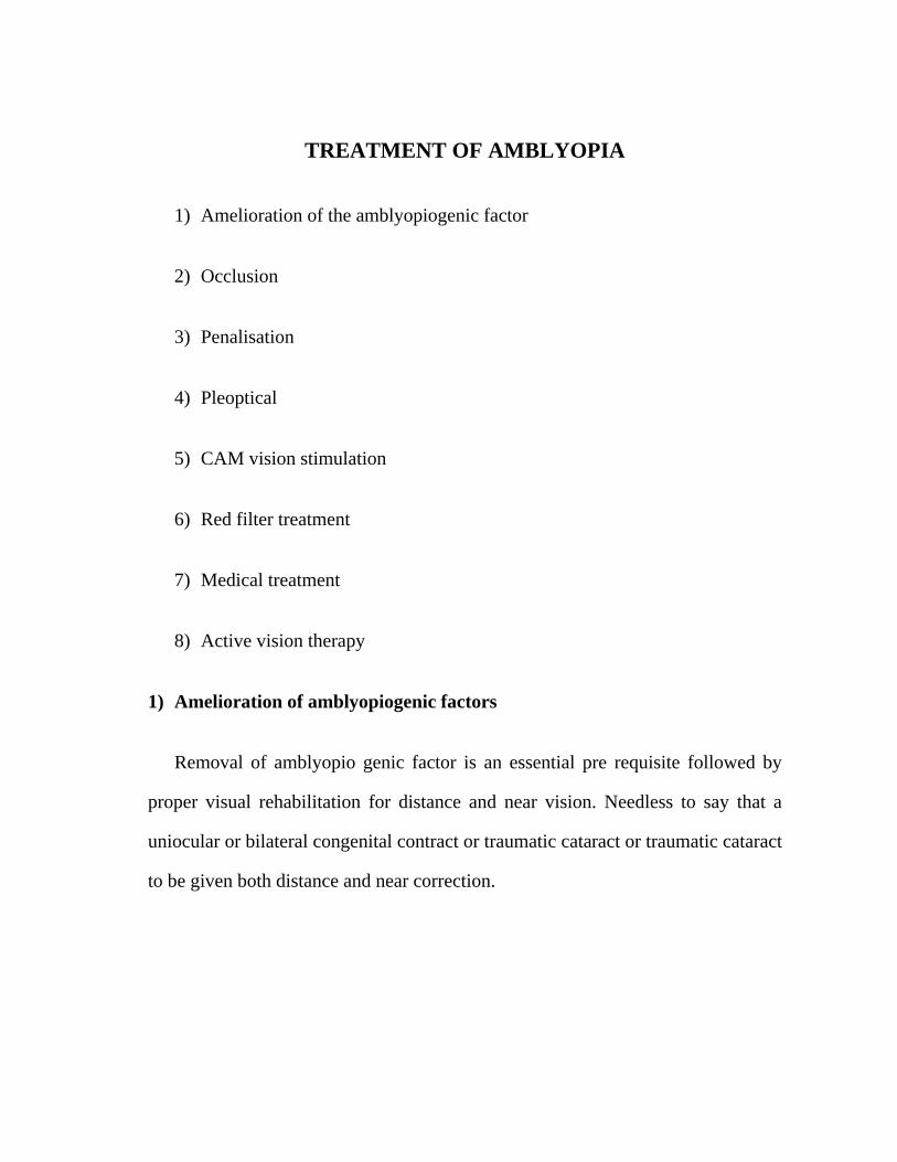
TREATMENT OF AMBLYOPIA
1) Amelioration of the amblyopiogenic factor
2) Occlusion
3) Penalisation
4) Pleoptical
5) CAM vision stimulation
6) Red filter treatment
7) Medical treatment
8) Active vision therapy
1) Amelioration of amblyopiogenic factors
Removal of amblyopio genic factor is an essential pre requisite followed by
proper visual rehabilitation for distance and near vision. Needless to say that a
uniocular or bilateral congenital contract or traumatic cataract or traumatic cataract
to be given both distance and near correction.

2) Occlusion:
Since 1743, when de Buffon first described occlusion has remained the sheet
anchor therapy for amblyopia. In this therapy the amblyopic eye is given a
preferential chance of development, as the dominant eye is totally withheld from
binocular participation. A properly done occlusion with good compliance ensures
an almost 100% success rate, especially if treated upto 7 years of age. The success
rate recedes with increasing age.
TYPES OF OCCLUSION:
a) Total and b) Partial
Total Occlusion:
Which completely obscures both light and form vision and is the type
usually advocated for moderate to severe amblyopia.
i) Direct skin patch A cotton eye pad patched to the eye with the help of a
micropore plaster.
ii) Spectacle patch.
iii) Doyne’s occluder: A black rubber occuder which sticks on the back of the
spectacle glass by suction.
iv) Pirate patch
v) Contact lenses

PARTIAL OCCLUSION
This degrades the vision of the normal eye so that the amblyopic eye has an
advantage. this is a milder form of penalisaiton and is used for milder amblyopia
or in recovered cases for maintenance of binocular vision. It also requires
correction of factors like squint anisometropia or anisokeinia suitably. The
advantage over total occlusion of this modality is that it offers binocular
stimulation. Layers of transparent scratch tape or colourless nail varnish can be
applied on the back surface of the dominant eye.
Another way of differentiation of period of occlusion,
a) Full time occlusion: All waking hours virtually 24 hours.
b) Part time occlusion this is for gradual duration, different waking hours of the
day on the basis of the age of the child,
Duration of occlusion
Full time occlusion is advocated by
It is advised for 24 hours (full day) for,
2 days for 2 years old
3 days for 3 years old
4 days for 4 years old

5 days for 5 years old
6 days for 6 years old
This is alternated with one day of occluding the amblyopic eye when the
dominant eye is opened occlusion of the dominant eye is called conventional
occlusion and occlusion of the amblyopic age is called inverse occlusion. The
follow up period for occlusion is 3 months. There is a risk of occlusion amblyopia
in dominant eye during occlusion, to avoid this the child has to be reviewed every
15 days.
PENALISATION
Penalisation has the advantage of being cosmetically acceptable, but it
doesnot inhibit the abnormal binocular interaction which is the essential cause of
amblyopic. Its indications are limited (eg) moderate amblyopia, in uncooperative
patient, anisometropic amblyopia and as maintenance therapy. The major
disadvantages of penalisation include active inhibition is not eliminated, risk of
occlusion amblyopia persists and also the cost of drugs.
CAM Stimulator:
Campbell and coworkers proposed a new treatment for amblyopia. The
discs are made with light and dark bars of various widths and rotated at the rate of
one rotation per minute to provide different orientation to stimulate a variety of
brain cells. Seven discs of various spatial frequencies are used. Patients views

them for seven minutes. This is based on the principle that the visual areas of brain
respond to stimuli of grating of a specific size at a certain orientation. This
modality is not used now a day.
Pharmacological Therapy for Amblyopia:
L - Dopa:
Various studies indicate the improvement in vision of amblyopia after L-Dopa
therapy.
Gottlob et al shows that L-Dopa improve contrast sensitivity and reduces
binocular suppression in the affected eyes of adult human anisometropic and
strabismic amblyopes. Gott lob et al further investigated the effect of Levo Dopa
with week daily administration of using a cross over double masked design. Visual
acuity improved in 70% of patient after one week of administration of Levo-Dopa
and the improvement of visual acuity and visual fields persisted after completion
of Levo Dopa administration.
Citicholine (Cytidine 5-Diphospho Choline)
Citicholine administration in adult volunteers (1 gram / day 1 / for 15 days)
of strabismus. Amblyopic demonstrated improvement lasting over 6 months in
visual acuity of both amblyopic and dominant eyes. Even contrast sensitivity and
VEP were significantly improved.

PART - II

PART – TWO
AIMS AND OBJECTIVES
To find out the relative proportions of different types of ambloypia in a
referral centre.
To study the characteristics associated with different types of amblyopia
in our population the extent of visual impairment produced by different
types of amblyopia at presentation.

METHODS AND MATERIALS
Over a period of two years 2004 October to 2006 September, 1800 patients
were see in strabismus and Paediatric Ophthalmology clinic at RIOGDH, Chennai.
This is a prospective study of 50 cases of amblyopia.
INCLUSION CRITERIA:
1) Patients aged between 6 months and 60 years of age.
2) No previous history of strabismus surgery.
3) All patients with corrected or uncorrected refractive errors
4) All patients referred from Institute of child health for ophthal evaluation
to squint clinic.
DEFINITION CRITERIA:
1) A difference in the best corrected visual acuity between the two eye in
the Snellen’s line equivalent measure on the Teller’s acuity in children
less than 4 years in absence of any organic lesion that could result in
decrease of vision.
2) A BCVA of less than 6/12 bilaterally on the Snellen’s chart / equivalent
measure on the teller’s acuity chart in children less than 4 years in the
absence of any organic lesion that could result in decrease of vision.
The assessment included, history of
1) Age of onset as noticed by patients or gaurdians.
2) H/O Low birth weight
3) Family history of strabismus
4) Patient coming from Rural or Urban areas.
General and systemic examination were done to rule out any associate problems.
Ocular examination included.
1) Unaided visual acuity
2) Best corrected spectacle visual acuity
(With the help of snellen’s chart in patients above 4 years and Teller’s acuity
chart in children less than 4 years of age.
3) In infants fixation and preferential looking patterns were noted.
4) Refraction was made under appropriate cycloplegics according to the age of
the patient, assessment of ocular alignment and motility and associated
strabismus.
5) Slit lamp examination is done to rule out any anterior segment pathology.

6) A detailed fundus examination is done to rule out any posterior segment
pathology.
7) Assessment of binocular status of the eye is done with the help of Worth Four
dot test.
WORTH FOUR DOT TEST
The images of two eyes are dissociated using red green glasses.

The patients with the normal binocular single vision sees two green dots,
one red dot and one plus.
STANDARD DEFINITION CRITERIA FOR DIFFERENT TYPES OF
AMBLYOPIA
1) STRABISMIC AMBLYOPIA:
Defined as amblyopia in the presence of Heterophoria for distance and near
fixation. Patients with strabismus along with refractive errors of more than 1 D
spherical equivalent in one or both eyes or eyes with regular astigmatism, 1.5 D
cylinder astigmatism in any meridian were included.
2) ANISOMETROPIC AMBLYOPIA:
This included patients who had amblyopia in the presence of anisometropia.
That was 1-D or greater in spherical equivalent or a1.5D or greater difference in
astigmatism in both the eyes that persisted for at least 4 weeks after spectacle
correction in the absence of any measurable heterotropia at distance or near.
3) SENSORY DEPRIVATION AMBLYOPIA:
This group included patients with known documented cause of sensory
deprivation with no primary heterotropia or refractive errors, that could be
causally related to the amblyopia.

4) AMETROPIC AMBLYOPIA
Patients with refractive errors more than 1D spherical equivalent in both the
eyes or one eye and no associated strabismus or any other ocular pathology were
classified under this category. Patients with significant anisometropia (as defined
above) along with high refractive errors in both the eyes were excluded from this
category and were classified under strabismic amblyopia.
5) MERIDIONAL AMBLYOPIA:
Patients with regular astigmatism 1.5D of astigmatism in any meridian or
those with irregular astigmatism or both eyes, resulting in a decrease in vision in
one or both the eyes and no associated strabismus were classified as having
meridional amblyopia. Patients with significant anisometropia (as defined above)
along with the difference of 1.5D or greater astigmatism between the two eyes
were excluded from this group and included under anisometropic amblyopia.
Patients with heterophoria for distance and near with regular astignatism more
than 1.5D in any meridian or irregular astigmatism were included under
strabismic amblyopia.

OBSERVATION AND RESULTS
AGE AND SEX DISTRIBUTION
Age Group No. of patients
< 2 years 5
2 – 5 years 8
5 – 10 years 23
10 – 20 10
> 20 years 4
SEX DISTRIBUTION
Sex No. of Patients
Male 30
Female 20
Average age of presentation of amblyopia in our study is 10 years.

RURAL AND URBAN PROFILE
NO. OF PATIENTS
Rural 35
Urban 15
35
15
05
10152025303540
Rural Urban

INICIDENCE OF DIFFERENT TYPES OF REFRACTIVE ERRORS IN
AMBLYOPIA
NO. OF PATIENTS
Hypermetropia 28 56%
Myopia 17 34%
Astigmatism 5 10%
2817
5
Hypermetropia Myopia Astigmatism

INCIDENCE OF DIFFERENT TYPES OF STRABISMUS
NO. OF PATIENTS
Exotropia 24 48%
Estropia 22 44%
Hypertropia 5 10%
24 22
5
05
1015202530
Exotropia Estropia Hypertropia
Exotropia Estropia Hypertropia

PERCENTAGES OF VARIOUS TYPES OF AMBLYOPIA
Type of Amblyopia
No. of Patients Percentage
Strabismic 38 76%
Ametropic 4 8%
Anisometropic 3 6%
Meridional 1 2%
Vision Deprivation 4 8%
COMPARISON OF TYPES OF FIXATION IN STRABISMIC
AMBLYOPIA AND MERIDIONAL, ANISOMETROPIC AND
AMETROPIC AMBLYOPIA
Strabismic Ametropic, Anisometropic
and Meriditional
Eccentric 8 (21.05%) 3 (37.5%)
Perifoveal 6 (15.7%) 3 (37.5%)
Parafoveal 18 (47.36) 2 (25%)
Peripheral 6 (15.7%) 0

PERCENTAGE OF PATIENTS HAVING LOW VISION <6/60
No. of Patients
Strabismic 17 54%
Anisometropic, Ametropic, Meridional
4 33%
- 4 patients in our study had Nystagmus 8%.
- 6 patients had family history of strabismus 5 patients had history of low birth
weight
- One patient had Duane’s Retraction Syndrome
- One patient was diagnosed to have chronic progressive external
ophthalmoplegia

DISCUSSION
Amblyopia is one of the most common causes of visual impairment in both
children and adults. It affects 4-5% of general population and its incidence in
paediatric ophthalmology and squint clinic is 30-35%.
AGE PROFILE
The average age of presentation is 10 year of age which is very late to start
treatment. This is due to lack of knowledge in the general population and the
ignorance of yearly eye check up in children, this can be brought down by better
awareness among the general population and screening programmes.
ADULT AMBLYOPIA:
The percentage of patient presenting after 10 year is 28% (14 patients). The
relative proportions of different types of amblyopia were strabismic 76%,
Ametropic 8% visual deprivation amblyopia 8%, meridional 2%, and
anisometropic amblyopia 6%. This proportions were different from General
population since many of patients were referred from secondary referral centres
with strabismus. And because of the inclusion creteria used here the relative
proportions of different types of amblyopias is different and the selection bias as it
is a hospital based study and is not representative of the population.

Fixation Pattern:
Patients stratismic amblyopia had low percentage of perifoveal fixation
15.7% as compared to other types of amblyopias 37.5% clumped together.
Type of Amblyopia Our study n=50
Study Conducted by Menon Vimla,
Chaudhuri Zia, et al at AIIMS
Strabismic 76% (38) 37.38
Anisometropic 6% (3) 31.1
Ametropic 8% (4) 18.31
Meridional 2% (1) 5.56
Vision Deprivation 8% (4) 7.63
In the incidence of refractive errors hypermetropia has highest incidence of
56% followed by myopia and Astigmatism.
21 patients had a visual acuity of 6/60 and below and this accounts to 42%.
Rural, urban profile:
This study showed a relatively higher percentage of rural population 70%
compared to urban population 30%. This is because of the lack of awareness
among the rural population.

CONCLUSION
The prevalence of amblyopia in our squint and neruophthalmology clinic was
30-35%. Amblyopia is a very common cause of impaired vision in child hood.
The incidence of anisometropic and ametropic amblyopia is compared to be
less in our population compared to western countries.
94% of the patient in our study had squint.
Hypermetropia is the most common refractive error associated with the
amblyopia followed by myopia and astigmatism .
Esotropia is the most common form of strabismus associated with the
amblyopia.
10% of patients with amblyopia had low birth weight.
12% of patients had family history of strabismus.
The relative proportion of rural population compared to urban population is
higher.
8% of patients with amblyopia in our study had Nystagmus.
42% of patients in our study had low vision C 6/60.
Earlier diagnosis and health education and screening programmes in rural
areas can reduce the prevalence of amblypias.

PROFORMA
EVALUATION OF AMBLYOPIA
NAME AGE SEX
Address:
Presenting Complaints:
Past History
Birth History
Family History
Treatment History

EXAMINATION
General
Ocular Examination
Vision
Extra Ocular Movements
Anterior Segment
Evaluation of Squint
Cover test
Prism Bar Cover test
Worth Four Dot test
Pattern of Deviation

RETINOSCOPY
Subjective
Type of Amblyopia:

KEY TO MASTER CHART
S.No. – Serial number
M.R.D. No. Medical Records Department Number
Age – Age in years
Sex – M – Male
F – Female
R – Rural
U – Urban
RE – Refractive Error
H – Hypermetropia
M- Myopia
A – Astigmatism
V/A Visual Acuity
FIX – Fixation
U/B Unilateral / Bilateral

Laterality of AMB – Type of Amblyopia
T. AMB – Type - Type of Amblyopia
STR - STRABISMIC
ANT - ANISOMETROPIC
AME - AMETROPIC
VD - VISNN DEPRIVATION
M - MERIDIONAL
FIX - Type of Fixation
E – Eccentric
PEF - Perifoveal
PAF - Parafoveal
PER - Peripheral
NYS - NYSTAGMUS
No. 1 Absent

No. 2 Present
TYPS Type of Squint
No.1 ESO - ESOTROPIA
No.2 EXO - EXOTROPIA
No.3 HYP - HYPERTROPIA
No.4 HYPO - HYPOTROPIA
LBW - Low Birth Weight
No (1) Absent
No (2) Present
FHOS - Family history of squint
No. 1 absent
No. 2 Present
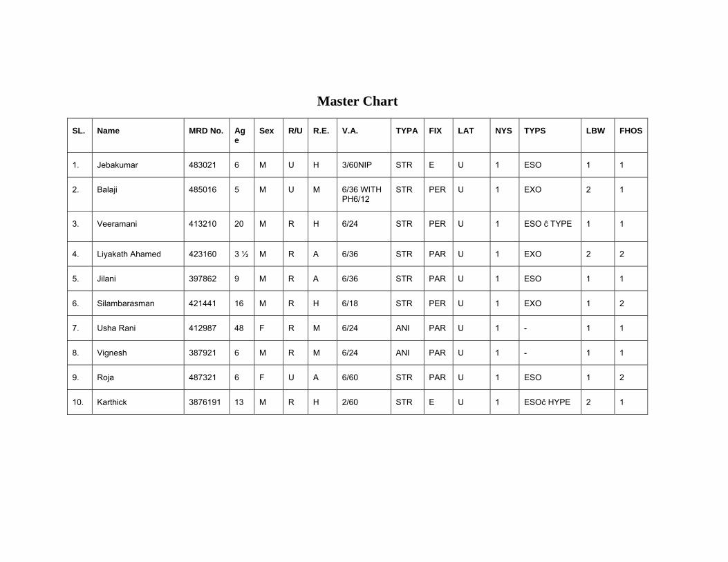
Master Chart
SL. Name MRD No. Age
Sex R/U R.E. V.A. TYPA FIX LAT NYS TYPS LBW FHOS
1. Jebakumar 483021 6 M U H 3/60NIP STR E U 1 ESO 1 1
2. Balaji 485016 5 M U M 6/36 WITH PH6/12
STR PER U 1 EXO 2 1
3. Veeramani 413210 20 M R H 6/24 STR PER U 1 ESO ĉ TYPE 1 1
4. Liyakath Ahamed 423160 3 ½ M R A 6/36 STR PAR U 1 EXO 2 2
5. Jilani 397862 9 M R A 6/36 STR PAR U 1 ESO 1 1
6. Silambarasman 421441 16 M R H 6/18 STR PER U 1 EXO 1 2
7. Usha Rani 412987 48 F R M 6/24 ANI PAR U 1 - 1 1
8. Vignesh 387921 6 M R M 6/24 ANI PAR U 1 - 1 1
9. Roja 487321 6 F U A 6/60 STR PAR U 1 ESO 1 2
10. Karthick 3876191 13 M R H 2/60 STR E U 1 ESOĉ HYPE 2 1

Master Chart
SL. Name MRD No. Age Sex R/U R.E. V.A. TYPA FIX LAT NYS TYPS LBW FHOS
11. Mohammed Hajudeen 371622 4 M R H 6/24 STR PAR U 1 ESO 1 2
12. Kokila 483217 11 F R M 6/24 AME PER B 1 EXO 1 1
13. Vikram 421721 1 M R H <6/60 STR E U 2 ESO 1 1
14. Rahman 4036121 8 M R H 1/60 V.D E U 1 ESOĉ HYPO 1 1
15. Nithya 4031261 3 F U H 6/36 STR PAR U 1 ESO 1 2
16. Sivakumar 391621 8 M U H 6/12 STR PER U 1 EXO 1 2
17. Manikam 391323 10 M R H 6/60 STR E U 1 ESO 1 1
18. Sudha 391474 7 F U H 6/60 STR PL U 1 ESO 1 1
19. Arumugam 487152 7 M R A 3/60 AME PL U 1 ESO 1 1
20. Thenmozhi 497122 11 F U M 6/24 STR PAR U 1 EXD ĉ HYPER 1 1

Master Chart
SL. Name MRD No. Age Sex R/U R.E. V.A. TYPA FIX LAT NYS TYPS LBW FHOS
21. Sumathy 491221 8 F R M 4/60 STR PL U 2 EXO 2 1
22. Arumugam 392125 64 M R M 6/60 STR PL B 1 EXO 1 1
23. Jenifer 392100 7 F R M 6/36 STR PAR U 1 EXO ĉ HYPER 1 1
24. Jagadeesh 397166 3 M R H 6/36 STR PAR U 1 EXO 1 1
25. Chitrangi 391800 9 F R H 6/24 STR PAR U 1 EXO 1 2
26. Karthick 391722 9 M R H 2/60 STR PL U 1 EXO ĉ HYPER 1 1
27. Prakash 399199 35 M R H 6/24 STR PAR U 1 EXO 1 1
28. Joseph 392720 47 M U H 6/36 STR PAR U 1 ESO 1 1
29. Balasubramanian 396726 4 M R H 1/60 STR E U 1 ESO 2 1
30. Vigneshkumar 394220 8 M R M 6/18 STR PAR U 1 ESO ĉ HYPER 1 1

Master Chart
SL. Name MRD No. Age Sex R/U R.E. V.A. TYPA FIX LAT NYS TYPS LBW FHOS
31. Gowri 392222 8 F R H 6/24 STR PAR U 1 ESO 1 1
32. Naramadha 391233 2 F R H <6/60 STR E U 2 ESO 1 2
33. Rajkumar Pande 391166 11 M R H 6/60 V.D E U 1 EXO 1 1
34. Jerald 392112 12 M U A 6/18 M PER U 1 - 1 1
35. Kousalya 391231 6 F R H 6/36 V.D PER B 1 EXO 1 1
36. Dhanush 391125 6 M U H 6/18 STR FOV U 1 ESO 1 2
37. Ajithkumar 461205 8 M R A 6/18 STR PER U 1 ESO 1 1
38. Selvi Soundarya 401075 12 F U M 6/18 STR PER U 1 EXO 1 1
39. Mohan 401023 7 M R M 6/24 STR PAR U 1 EXO 1 2
40. Tejaswari 401069 7 F U M 6/24 ANI PAR U 1 EXO 2 1

Master Chart
SL. Name MRD No. Age Sex R/U R.E. V.A. TYPA FIX LAT NYS TYPS LBW FHOS
41. Vanisha 411233 9 F R H 6/24 STR PER U 1 ESO 1 2
42. Janardhan 400111 5 M R A 6/36 STR PAR U 1 EXO ĉ HYPER
2 1
43. Kameswaran 411332 3 ½ M U M 6/60 ANI PL U 1 ESO 1 1
44. Sakthivel 401723 4 M U M 6/24 AME PAR U 1 EXO 1 1
45. Manikandan 401222 11 M R M 6/36 STR PAR P 1 ESO ĉ HYPER
1 2
46. Elumalai 401976 12 F R M 6/24 STR PAR U 1 EXO 1 1
47. Bagyalakshmi 400111 5 F R H 1/60 VD E U 1 EXO 1 1
48. Kamalesh 401224 6/12 M U M < 6/60 ANI E U 2 ESO 1 1
49. Bharathi 422105 7 F U M 6/24 AME PAR B 1 - 1 1
50. Nagaraj 411122 28 M R H 5/60 STR E U 2 EXO 1 1

BIBLIOGRAPHY
1. Gunter Von Noorden Emilio C. Campus Prevalence, Social and Psychosocial
factor 1974, 246-249.
2. Keech RV, Kictschke PJ, upper age limit for the development of amblyopia J.
Paediatric ophthal strab 1995; 32: 89-93.
3. Quah BL, Tay MT, Chew ST, Lee LK. A study of Amblyopia in 18-19 year
old males. Singapore Medical J 1991; 32: 89-93.
4. Harwerth RS, smith EL, Duncan GC, Crawford ML, Von Noorden GK.
Multiple sensitive periods in the development of primate visual system.
Science 1986: 232: 235-238.
5. Dandona R, Dandona L, Srinivas M, Sahare P, Naraisaih S, Munoz SR et al.
Refractive errors in Children in a rural population invest Oph. Vis Sci 2002;
43: 615-22.
6. Thompson JR, Woodruff G, Hiscox FA, Thompson JR, Smith LK, Factors
affecting the outcome of children treated for amblyopia. Public Health 1996;
105: 455-62.
7. Williams C, Harrad RA, Harvey I, Sparrow JM, the ALSPAC study team.
Screening for amblyopia in pre school children; results of a population – based
randomised controlled trial, ALSPAC study team, Avon Longitudinal study of
Pregnancy and childhood. Ophthal Epidemiol. 2001; 8:279-95.
8. Von Noorden GK, classification of Amblyopia. AMJ Ophthalmology 1967;
63: 238-41.

9. Weakly DR. The association between Anisometropia, Amblyopia and
binocularity in the absence of strabismus. Trans AM Oph SOC 1999; 97: 987-
1021.
10. Gottlab I. The detection, prevention and rehabilition of amblyopia. Curr opin
ophthal 1999:10:300-4.
11. Murthy GV, Gupta SK, Bachani D, Jose R, John N. Current estimates of
blindness in India. Br. J. Ophthalmol. 2005, 89: 257-60.
12. Negrel AD, Maul E, Pokharel GP, Zhau J, Ellwein LB, Refractive Error study
in children, sampling and measurement methods, for a multi country survey.
Am J ophthalmol 2000: 129:421-6.
13. Khan SA, Shamanna B, Nuthethi R, Perceived barriers to the provision of low
vision services among ophthalmologists in India. Indian J Ophthalmol 2005;
53:69-75.
14. Lim HC, Quah BL, Balakrishnan V, Lim HC, Tay V, Emmanuel SC, vision
screening of 4 year old children in Singapore. Singapore med J 2004; 41:271-8.
15. Dandone R, Dandon L childhood Blindness in India; A population based
perspective Br. J. Ophthalmol . 2003; 87: 263-5.
16. Pareja RA, martinez RA, Abreu Reyes JA, Scrrano GM. A study of the visual
acuity and amblyopia in infants aged 3 to 5 years from El Hierro island. Arch
SOC ESP oftalmol 2000; 75:397-402.
17. Schalji Deffos NE, de Graaf ME, Traffers WF, Engel J, cats BP. Long term
follow up of premature infants; detection of strabismus, amblyopia and
refractive errors. Br J. Phthalmol 2000; 84:963-7.

18. Brown SA, with LM, FuCL, Dimitrop Taylor HR, MC Carty CA. Prevalence
of amblyopia and associated refractive errors in an adult population in Victoria,
Australia. Ophthal Epidemiol 2000:7:249-58.
19. Paediatric eye disease investigator group. The clinical profile of moderate
Amblyopia in children younger than 7 years. Arch ophthal 2002; 120:281-7.
20. Arden GB, Barnard WM, Mushin AS; visual evoked responses in amblyopia.
Br. . Ophthalmol 58: 183; 1974.
21. Arden GB, Wooding SL; pattern ERG in amblyopia Invest ophthalmol vis sci
26: 88; 1985.
22. Hoff Van Duin J van; Early and permanent effects of monocular deprivation on
pattern discrimination and visio motor behaviour in cats. Brain Res 111:261,
1976.
23. Irvine SR; Amblyopia ex anopsia. Trans Am Ophthalmolo SOC 46:527, 1948.
24. Noorden GK von. Treatment of amblyopia and surgical outcome (letter). J.
Paediatric Ophthalmol Strabismus, 35:5, 1998.
25. Noorden GK Von, Frank JW; Relationship between amlyopia and the angle of
strabismus. Am orthopt J 26:31, 1976.
26. Martens TG, Remarks on fixation disparity. Am orthopt J 20:68, 1970.
27. Thorn F, Gwaigda J, Cruz AAV, et al. The development of eye alignment
convergence and sensory binocularity in young infants. Invest ophthalmol vis
sci 35:544, 1994

28. Wedner SH, ROSS DA, Balera R, Kajil, Foster A. Prevalence of eye diseases
in primary school children in a rural area of Tanzania. Br. J. Ophthalmol.
2000-84, 1291-7.
29. Quah BL, Tay MT, chew ST, Leeck. A study of amblyopia in 18-19 year old
males. Singapore med J 1991; 32:126-9.
30. Kalkiyaviv, Naduvilath TJ, Bansal AK, Dandona L. Visual impairment in
School Children in South India. Indian J. Ophthalmol 1997:45: 129-34.
31. Altebo K, Mitchell P, Cumming R, Smith, Jolly N, Sparkes R. Prevalence and
causes of amblyopia in adult population. Ophthalmology 1998, 105; 154-9.

LIST OF OPERATIONS PERFORMED
S.No Name Age / Sex
OP / IP No. Date Eye Surgery
1 Vijayalakshmi 64/F 435225 20/07/2005 LE ECCE with PCIOL
2 Arumugam 54/M 435296 27/07/2005 RE ECCE with PCIOL
3 Geetha 28/F 431784 29/07/2005 LE Incision and curettage
4 Venkatesan 34/M 431286 04/08/2005 RE Excision
5 Visalakshi 68/F 432252 06/08/2005 RE DCT
6 Nagamma 64/F 433358 08/08/2005 RE ECCE with PCIOL
7 Dhanalakshmi 52/F 434552 10/08/2005 RE I & D
8 Padmavathi 55/F 434689 02/09/2005 LE Suturing of Corneal tear
9 Mohan 28/M 435002 09/09/2005 RE Suturing of Lid tear
10 Munusamy 56/M 435100 16/09/2005 LE ECCE with PCIOL
11 Virudammal 64/M 481154 23/09/2005 RE ECCE with PCIOL
12 Prasath 18/M 481166 30/09/2005 RE I & C
13 Vasantha 58/F 481177 02/10/2005 LE DCT.
14 Kandasamy 68/M 471128 09/10/2005 LE Evisceration
15 Maniammal 64/F 472228 16/10/2005 RE Suturing of Corneal tear
16 Vedha 72/F 472215 23/09/2006 LE DCT
17 Pappammal 50/F 472318 30/07/2006 LE ECCE with PCIOL
18 Chinnappan 65/M 474812 04/08/2006 RE Suturing of Corneal tear
19 Mahendran 60/M 474915 11/08/2006 RE SICS with PCIOL
20 Manikkam 72/M 484501 18/08/2006 RE SICS with PCIOL