causes and patterns of harbor seal (phoca vitulina) pup...
TRANSCRIPT
Causes and Patterns of Harbor Seal (Phoca vitulina) Pup Mortality
at Smith Island, Washington, 2004-2009
by
Corina L. Leahy
A Thesis
Submitted in partial fulfillment
of the requirements for the degree
Master of Environmental Studies
The Evergreen State College
July 2010

2010 by Corina L. Leahy. All rights reserved.
This Thesis for the Master of Environmental Study Degree
by
Corina L. Leahy
has been approved for
The Evergreen State College
by
________________________
Gerardo Chin Leo
Member of the Faculty
________________________
John Calambokidis
Research Biologist
Cascadia Research Collective
________________________
Martha Henderson
Member of the Faculty
________________________
Date
ABSTRACT
Causes and Patterns of Harbor Seal (Phoca vitulina)
Pup Mortality at Smith Island, Washington, 2004-2009
Corina L. Leahy
Harbor seals (Phoca vitulina) are the most common and widely distributed
pinniped in Washington State waters. Their abundance and proximity to land
allow many opportunities for examination and necropsy once stranded. Serving as
sentinels of marine ecosystem health, stranded animals are useful in detecting
environmental contaminant levels and disease in populations. From 2004 to 2009,
mortality rates and causes of death of harbor seal (Phoca vitulina) pups at Smith
Island, a haulout site in North Puget Sound, Washington State, were examined. A
total of 16 surveys of this site were conducted during pupping seasons (June
through August). Two hundred twelve dead pups were counted, of these 54 were
collected for necropsy. Minimum neonatal mortality ranged from 3% to 27%.
Neonatal mortality was highest in 2005; half of the total number of dead pups
found over the entire study period were collected that year. Infection was the
leading primary cause of death in most years. In 2005, 43% of the pups died from
an infectious process. In 2006, 2008, and 2009, infection was again the leading
cause of death, claiming a total 47% of pups necropsied during those years. The
second leading cause of death was malnutrition; other causes of death included
prematurity and dystocia. Antibiotic resistant bacteria were isolated from 17 of
the 54 pups necropsied. Antibiotic resistant bacterial infections were most
prevalent in 2005 and 2009. Bacteria presenting with antibiotic resistance
included Enterococcus, E. coli, and Actinomyces; some of these isolates were
found to be resistant to all eight routine antibiotics. As antibiotic resistance
becomes more prevalent in marine mammal populations, there could be
significant implications for marine ecosystem health. Long term data collection
from this site may provide invaluable insights into the potential impacts of
contaminants, pathogen introduction, and other perturbations on population
recruitment, health and status.
TABLE OF CONTENTS
List of Figures………………………………………………………………….…v
List of Tables…………………………………………………………………….vi
Acknowledgements……………………………………………………………..vii
Abbreviations…………………………………………………………………..viii
Introduction……………………………………………………………………....1
Background and Management Implications…………………………...1
Research Questions, Hypothesis, and Approach………………………2
Ecology and Biology of Harbor Seals (Phoca vitulina)………………...3
Methods…………………………………………………………………………...7
Study Site Background…………………………………………………..7
Permits and Survey Date Selection……………………………………..7
Survey Procedures……………………………………………………….9
Data and Sample Collection……………………………………………..9
Necropsy and Sample Collection………………………………………10
Development of Methods and Study Design…………………………..12
Results…………………………………………………………………………...13
General Findings………………………………………………………..13
Causes of Mortality……………………………………………………..15
Pup size in Relation to Cause of Mortality……………………………17
Percent Mortality……………………………………………………….17
Other Significant Findings……………………………………………..17
Discussion……………………………………………………………………….22
Standard Length and Prematurity…………………………………….22
Causes of Mortality……………………………………………………..22
Pup Size in Relation to Cause of Mortality…………………………...23
Percent Mortality……………………………………………………….23
Other Significant Findings……………………………………………..23
Study Limitations……………………………………………………….24
Conclusion & Suggestions for Further Research…………………………….25
Literature Cited………………………………………………………………...27
LIST OF FIGURES
Figure 1. Harbor seals hauled out at Smith Island, Washington.
(© Cascadia Research Collective)………………………………………………...5
Figure 2. Map of harbor seal haulout sites within inland Washington waters
(taken from Steiger et al., 1989)…………………………………………………. 8
Figure 3. Graphical representation of total dead pups found for all survey dates
for all years (grouped by week)………………………………………………….14
LIST OF TABLES
Table 1. Survey dates during each year of study period……………………….…9
Table 2. Routinely sampled tissues and corresponding preservation type………11
Table 3. Number of pups found and necropsied by survey date……………...…14
Table 4. Number of female and male pups found per year……………………...15
Table 5. Primary and contributing causes of mortality by year…………………16
Table 6. Annual seal counts, calculated birth rates, & minimum percent
mortality………………………………………………………………………….19
Table 7. Pups with antibiotic resistant Enterococcus sp. isolates…………….…20
Table 8. Pups with antibiotic resistant E. coli (non-hemolytic) isolates……..….20
Table 9. Pups with antibiotic resistant E. coli (hemolytic) isolates………….….21
Table 10. Pups with antibiotic resistant Actinomyces sp. isolates…………...….21

ACKNOWLEDGEMENTS
This thesis would not have been possible without the support of many
people. I would like to thank my thesis readers, Gerardo Chin Leo, John
Calambokidis, and Martha Henderson for their guidance, thoughtful review, and
encouragement throughout this process. I also thank Jessie Huggins, Dr. Steven
Raverty and the staff at Cascadia Research Collective and Washington
Department of Fish & Wildlife for their assistance with data collection, seal
counts, and advice. Finally, I would like to thank my parents. Thank you for
appreciating the value of science and education.
ABBREVIATIONS
ARB-Antibiotic Resistant Bacteria
CRC-Cascadia Research Collective
MMPA- Marine Mammal Protection Act
NMFS-National Marine Fisheries Service
NOAA-National Oceanic and Atmospheric Administration
SBT-Sternal Blubber Thickness
USFWS-United States Fish and Wildlife Service
WDFW-Washington Department of Fish and Wildlife
INTRODUCTION
Background and Significance
Marine mammals are an important component of marine ecosystems. They
serve as effective sentinels of ecosystem health because of their longevity and
extensive fat stores where toxins and contaminants can accumulate (Bossart,
2006; Wells et al., 2004). Harbor seals are a particularly good population to study
given that they spend part of their lives in coastal environments and on land, thus
making them more accessible for research than many other marine mammals.
Unlike other marine mammals, they do not migrate and will remain in one
geographic region throughout their life span. This study seeks to understand the
factors associated with harbor seal pup mortality in the Puget Sound.
An understanding of causes of mortality in this local marine mammal
population can provide essential information for marine mammal management
and ecosystem conservation. Monitoring the health of local seal populations is a
useful tool for examining the health of the entire Puget Sound ecosystem. Some
pathogens that may exist in seal populations have the potential to threaten the
health of other marine mammals, such as the endangered orca, or terrestrial
animals and scavengers, like the bald eagle. In some instances, seals can even
serve as reservoirs of potentially zoonotic pathogens, thus posing a possible health
risk to humans. Conversely, in many cases, seals and other marine animals are
exposed to pathogens from anthropogenic sources such as agricultural and urban
run-off (Bogomolni et al., 2008; Kreuder et al., 2003; Miller et al., 2002). The
ability to quickly discern subtle changes in seal population health can lead to early
detection of potentially devastating environmental disturbances caused by human
activity.
While substantial research has been conducted on marine mammals,
relatively little is known about the causes of mortality in natural populations. This
is due to a variety of limiting factors. Financial cost, man-power, time, and stress
to animals can all prohibit or restrict long-term marine mammal population
studies. One way to overcome some of these obstacles is by analyzing data from
stranded animals. By examining stranded or dead animals, a great deal about
mortality, disease, and pathogens in populations can be learned. Stranded animals
can be sampled for tissue contaminant levels and can be used to detect pathogens
in host populations.
Use of stranded animals is, however, limited. While stranding data may
not reflect mortality causes and trends in the entire population, it may provide
clues as to what contributing factors or environmental disturbances are significant
(Aguilar & Borrell, 1994). This may be particularly true in cases of unusual
mortality events or mass strandings. Knowledge of the trends associated with the
causes of harbor seal mortality can help determine the relative contribution of
disease, malnutrition, or other factors while establishing a baseline of mortalities
that can be considered normal for the population. Deviations from these
established trends can indicate changes in the environment associated with such
disturbances as global climate change, foreign-host pathogen introduction, or
anthropogenic disturbances. The ability to distinguish between normal trends and
unusual mortality events is essential in marine mammal management and
protection. Thus, identification of major causes of mortality can help design
effective policies for the management and protection of marine mammals.
Research Questions, Hypothesis & Approach
This thesis compares rates and causes of mortality in neonatal harbor seals
at Smith Island, Washington over a period of five years. Periodic surveys of the
haul-out site were conducted during pupping season from 2004 through 2009.
During these surveys, dead pups were collected and necropsies performed when
appropriate. I analyzed data collected by Cascadia Research Collective (CRC),
from 2004, 2005, 2006, 2008, and 2009 (no surveys were conducted in 2007). I
assisted with haulout surveys, necropsies, and data collection in 2009. I also
reviewed and analyzed the stranding reports and pathology reports for all years of
this study.
During my initial review of this data I noticed that an unusually high
number of dead pups were recovered in 2005; more pups were found in that year
than in any other year. High numbers of dead pups were found consistently
throughout the 2005 pupping season. Determining the potential causes of this
marked increase in pup mortality motivated this study. This thesis seeks to answer
the following questions:
1) Do primary causes of mortality vary significantly between years?
2) Is there a relationship between pup size (measured by weight, length, and
sternal blubber thickness) and cause of mortality?
3) Are there any pathogens or conditions that are consistently prevalent in
this population?
My hypotheses are that primary causes of mortality will vary significantly
between years; that there is a relationship between cause of mortality and pup
size; and that there are pathogens that consistently affect this population.
Ecology and Biology of Harbor Seals (Phoca vitulina)
Distinguishing Characteristics
Harbor seals are the most common and widely distributed pinniped (fin-
footed marine mammals) in Washington waters. They are easily distinguishable
from other seals by their round, dog-like faces and short snouts. As true (earless)
seals, they have no external ear flaps. Their bodies and flippers are short. Their
pelage (coat) patterns are variable, most harbor seals exhibit a lightly colored base
with dark spots; some individuals will exhibit a reverse pattern of white spots
over a mostly black or dark brown coat. Seals with intermediate coloration are
common as well (WDFW, 2009).
Harbor seals are recognizable on land as they tend to resemble bananas
when hauled out, elevating their head and rear flippers (NMFS, 2009). Their hind
flippers lack flexibility resulting in undulating or scooting movements while on
shore. Harbor seals are small in comparison to other seals. Average length and
weight can vary between populations. In the Pacific Northwest, adult harbor seals
range from 1.2 to 1.9 m in length with an average weight of 80kg. Females are
usually smaller than males. Pups typically weigh 7 to 8 kg at birth (WDFW, 2009;
NMFS, 2009).
Distribution, Movements, & Population Patterns
Harbor seals occur over a latitudinal range from about 30°N to 80°N in the
eastern Atlantic region and about 28°N to 62°N in the eastern Pacific region.
They have the widest distribution and occur in more different habitats than any
other pinniped (Burns, 2008). While total global population estimates vary,
eastern Pacific harbor seal populations are fairly abundant. In waters from Alaska
to California, the total population is estimated to be near 350,000 individuals
(Carretta et al., 2007). In Washington state, harbor seals are abundant and by
some reports, near carrying capacity. In 1999, it was determined that the inland
Washington stock totaled an estimated 14,612 seals. At that time, the total Coastal
Washington/Oregon population was estimated to be at 24,732 seals (Jeffries et al.,
2003).
Harbor seals are generally non-migratory, staying in the same area
throughout the year to feed and breed. Local movements within a region can be
associated with such factors as weather, season, tides, food availability, and
reproduction (Bigg, 1981). Harbor seals have also displayed strong fidelity for
particular haulout sites (Pitcher & McAllister, 1981).
For management purposes within Washington State, two distinct stock
populations are recognized. The first, Washington inland stock, includes those
seals found in all inland waters of the state (including Puget Sound, Hood Canal,
and the Strait of Juan de Fuca out to Cape Flattery). The second consists of seals
found along the Washington/Oregon coastal regions (Boveng, 1988). This thesis
will focus on one site in the inland Washington region.
The inland waters region of Washington is of particular interest as the
health of the Puget Sound has drastically declined. High levels of environmental
contaminants have been found in the resident orca population, shellfish are
frequently not safe to eat due to toxin levels, and storm water runoff are just a few
of the threats to the health of this region. Monitoring seal populations within this
region can provide valuable insight into the state of the Sound.
Foraging, Breeding Habitat & Haulouts
Harbor seals are generalists and will typically forage on easily available
and abundant foods (Burns, 2008). Their diet may vary with seasonal availability
of prey but primarily consists of several species of fish and cephalopods. Harbor
seals generally feed in shallow waters close to shore and as mentioned, may
exhibit strong site fidelity.
Harbor seals breed in both coastal and insular waters. Seals give birth in
rookeries on shore. During breeding season, herds of seals can be found at these
sites, hauled out in large groups with no apparent social structure.
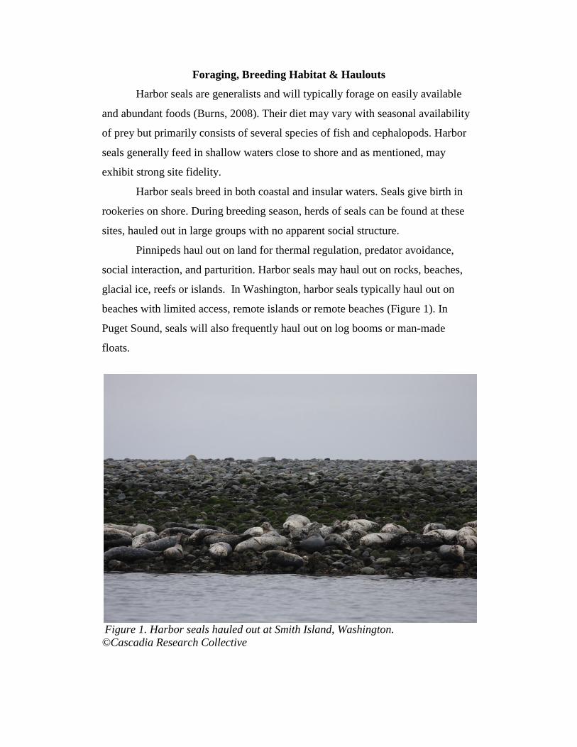
Pinnipeds haul out on land for thermal regulation, predator avoidance,
social interaction, and parturition. Harbor seals may haul out on rocks, beaches,
glacial ice, reefs or islands. In Washington, harbor seals typically haul out on
beaches with limited access, remote islands or remote beaches (Figure 1). In
Puget Sound, seals will also frequently haul out on log booms or man-made
floats.
Figure 1. Harbor seals hauled out at Smith Island, Washington.
©Cascadia Research Collective
Reproduction & Mortality
Female harbor seals reach sexual maturity at ages of 3 to 4 years; physical
maturity is reached at the age of 6 to 7 years. Males reach sexual maturity at 4 to
5 years and physical maturity at 7 to 9 years (Burns, 2008). The maximum
lifespan of a harbor seal is between 30-35 years, although individuals rarely live
this long in the wild. Females tend to live longer than males yet mortality for both
sexes is highest during the first few months after birth (Riedman, 1990).
Individuals are reproductively active throughout their lives with females typically
giving birth to one pup per year, although twinning has been observed (Burns,
2008). The gestation period is approximately 10.5 months.
In most regions, Washington included, pups are born on land. Pupping
season varies throughout populations. Even within Washington, pupping season
varies by location, but tends to occur fairly consistently at each site across
seasons. In inland Washington waters, pupping season starts in late June and lasts
through early September (WDFW, 2009). Pups are nursed for approximately 4 to
6 weeks and can triple their weight by the time they are weaned. These fat
reserves are useful as the pups learn to forage on their own.
Several factors can adversely affect survival, often with varying effects on
different age classes. In young or first time mothers, the risk of abortion or
stillbirth is higher. As these females are typically smaller, they may in turn give
birth to smaller offspring thus increasing vulnerability to injury or hypothermia
(Geraci & Lounsbery, 2008). Starvation or malnutrition can also lead to death,
particularly in dependent young pups, immunocompromised individuals, or older
animals. Trauma may lead to mortality in seal populations, especially at crowded
haulout sites where the density of animals can increase the chances of accidental
trauma, particularly to small pups. Pathogens are another significant source of
mortality. Parasitic, bacterial, viral, and fungal infections can all contribute to seal
death. Seal pups are also more likely to fall victim to predation, as they are often
left alone and vulnerable on shore.
In Washington State, transient orcas, eagles, gulls, and coyotes all prey on
harbor seals (Lambourn, et al., 2010; Steiger et al., 1989). A number of
anthropogenic factors can also affect harbor seal survival. Environmental
contaminants (Calambokidis et al., 1985; Ross et al., 1993), pollution and debris,
and fisheries interactions can all pose threats to harbor seal health.
METHODS
Study Site Background
Smith Island was chosen as the study site because it is subject to relatively
low levels of human disturbance. This is due to the fact that the island is part of
the San Juan Islands National Wildlife Refuge; access to the island is restricted,
requiring a federal permit from the United States Fish and Wildlife Service
(USFWS). Smith Island is a small, rocky island located within the eastern Strait
of Juan de Fuca (48°19’N, 122°50’W) (Figure 2). It is connected to the even
smaller Minor Island, by a spit, which is visible during low tide. The rocky
substrate is an ideal haulout site for seals as the pups are well camouflaged on the
beach, easily blending in with the rocks. The site is also a nesting habitat for gulls
and bald eagles. For this study, both Smith and Minor Islands were surveyed. For
simplicity, the study site will collectively be referred to as Smith Island.
Permits and Survey Date Selection
Survey permits were obtained from USFWS. Annual survey dates were
chosen to coincide with the peak of pupping season at Smith Island (late June
through early August) and precede molting. Attempts were made to schedule
multiple survey dates each year, approximately two weeks apart. Dates were
selected during low tide and were subject to personnel and vessel availability as
well as weather. Due to these constraints, no surveys were conducted in 2007 and
only one survey was conducted in 2008. Subsequently, this study includes data
collected from surveys conducted in 2004, 2005, 2006, 2008, and 2009, (Table 1).
Although surveys were conducted prior to 2004, the sampling effort varied
greatly. Thus, surveys prior to 2004 are not included in this study.
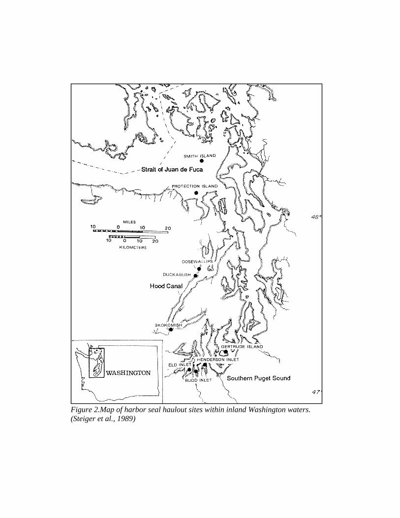
Figure 2.Map of harbor seal haulout sites within inland Washington waters.
(Steiger et al., 1989)
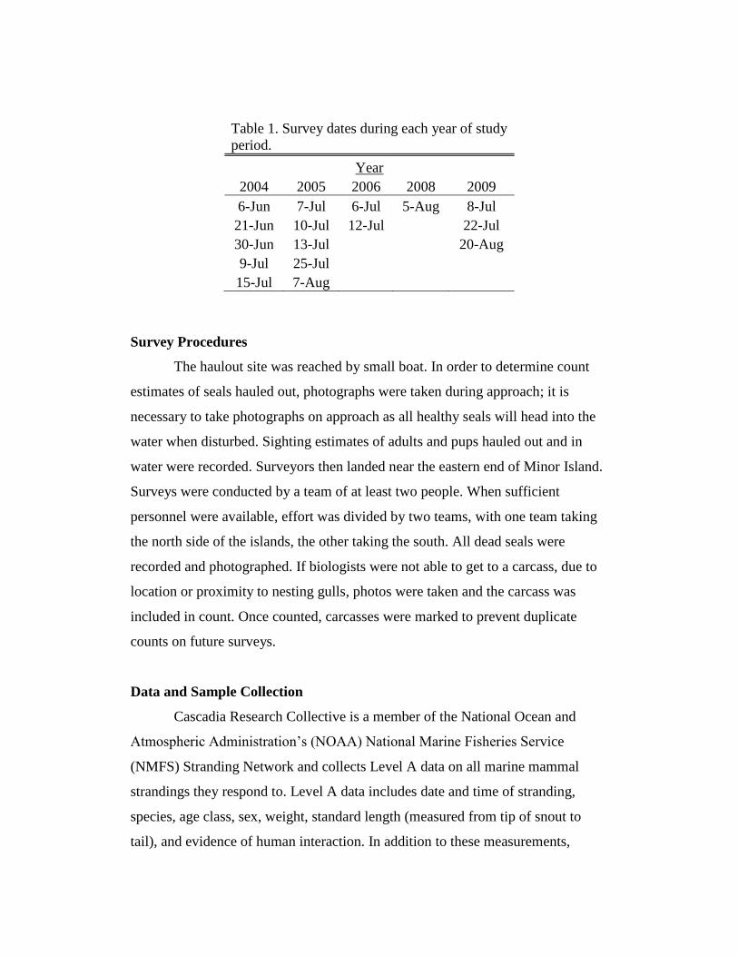
Table 1. Survey dates during each year of study
period.
Year
2004 2005 2006 2008 2009
6-Jun 7-Jul 6-Jul 5-Aug 8-Jul
21-Jun 10-Jul 12-Jul
22-Jul
30-Jun 13-Jul
20-Aug
9-Jul 25-Jul
15-Jul 7-Aug
Survey Procedures
The haulout site was reached by small boat. In order to determine count
estimates of seals hauled out, photographs were taken during approach; it is
necessary to take photographs on approach as all healthy seals will head into the
water when disturbed. Sighting estimates of adults and pups hauled out and in
water were recorded. Surveyors then landed near the eastern end of Minor Island.
Surveys were conducted by a team of at least two people. When sufficient
personnel were available, effort was divided by two teams, with one team taking
the north side of the islands, the other taking the south. All dead seals were
recorded and photographed. If biologists were not able to get to a carcass, due to
location or proximity to nesting gulls, photos were taken and the carcass was
included in count. Once counted, carcasses were marked to prevent duplicate
counts on future surveys.
Data and Sample Collection
Cascadia Research Collective is a member of the National Ocean and
Atmospheric Administration’s (NOAA) National Marine Fisheries Service
(NMFS) Stranding Network and collects Level A data on all marine mammal
strandings they respond to. Level A data includes date and time of stranding,
species, age class, sex, weight, standard length (measured from tip of snout to
tail), and evidence of human interaction. In addition to these measurements,
blubber thickness and axillary girth was measured for all carcasses found when
feasible. Blubber thickness was measured ventrally, at the sternum. Axillary girth
was measured around the animal at the axilla of the front flippers.
Only relatively fresh, minimally scavenged carcasses were collected for
complete necropsy. NMFS utilizes a number system to code decomposition levels
of marine mammal carcasses, described as follows: Code 1: live animal; Code 2:
fresh dead; Code 3: moderate decomposition; Code 4: advanced decomposition,
and Code 5: mummified or skeletal remains. Carcasses collected for necropsy
were typically Code 2.
Necropsy and sample collection
Whole carcasses collected for necropsy were taken back to the WDFW
game farm in Lakewood for complete exam and necropsy per established
protocols (Pugliares, et al., 2007) by CRC or WDFW staff. A detailed external
exam was conducted on all pups prior to necropsy. Any external findings were
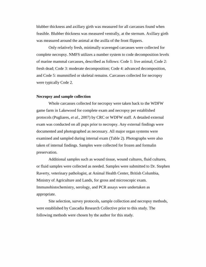
documented and photographed as necessary. All major organ systems were
examined and sampled during internal exam (Table 2). Photographs were also
taken of internal findings. Samples were collected for frozen and formalin
preservation.
Additional samples such as wound tissue, wound cultures, fluid cultures,
or fluid samples were collected as needed. Samples were submitted to Dr. Stephen
Raverty, veterinary pathologist, at Animal Health Center, British Columbia,
Ministry of Agriculture and Lands, for gross and microscopic exam.
Immunohistochemistry, serology, and PCR assays were undertaken as
appropriate.
Site selection, survey protocols, sample collection and necropsy methods,
were established by Cascadia Research Collective prior to this study. The
following methods were chosen by the author for this study.
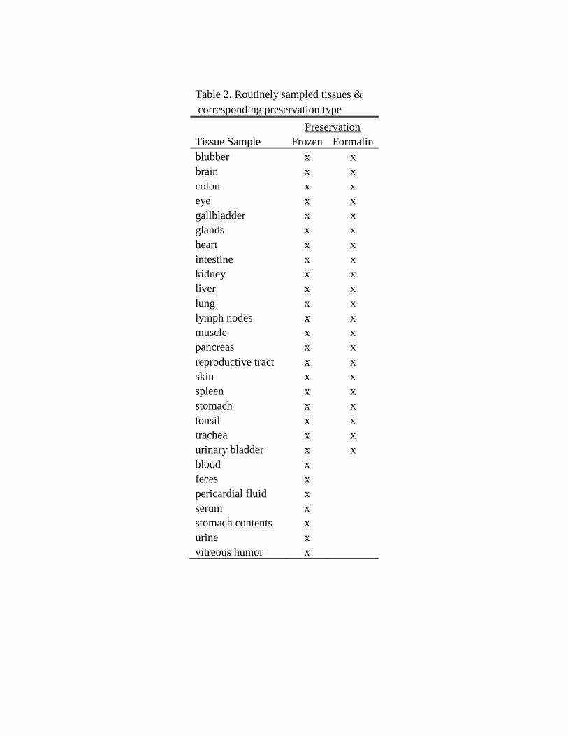
Table 2. Routinely sampled tissues &
corresponding preservation type
Preservation
Tissue Sample Frozen Formalin
blubber x x
brain x x
colon x x
eye x x
gallbladder x x
glands x x
heart x x
intestine x x
kidney x x
liver x x
lung x x
lymph nodes x x
muscle x x
pancreas x x
reproductive tract x x
skin x x
spleen x x
stomach x x
tonsil x x
trachea x x
urinary bladder x x
blood x
feces x pericardial fluid x serum x stomach contents x urine x vitreous humor x

Development of Methods and Study Design
Measurement Selection
Length, sternal blubber thickness, axillary girth, and weight were recorded
for many pups in this study. However, due to scavenging, it was often not
possible to record accurate axillary girth. Blubber thickness, weight and length
were chosen for comparison as they were consistently measured on most pups.
Calculating Percent Mortality
Annual minimum mortality rates were calculated using the total number of
dead pups found as a percentage of the pups born (Calambokidis et al., 1985;
Lambourn, et al., 2010). The total number of pups born was calculated using the
highest number of pups seen at one time, plus the total number of dead pups
found. Total pup count estimates were determined from examination of aerial and
vessel-based survey photos provided by CRC and WDFW.
Total count estimates were conducted both by CRC and WDFW. For
CRC estimates, photos were taken from boat before landing on the island for
surveys. Data from WDFW estimates was collected via aerial surveys. These
surveys were typically conducted in August when seal congregations are highest
as it is the end of the pupping season and beginning of the adult molting season.
Total WDFW counts appear to be more accurate as all parts of the island could be
seen at the same time; CRC staff could only photograph whatever side of the
island they were approaching from. Due to the higher count reliability and
consistency in survey dates, WDFW photos were chosen to use for seal count
estimates.
Determining Cause of Mortality
Primary cause of mortality is defined as the condition most likely to have
caused the animal’s death based on all information provided (Colegrove et al.,
2005).
In order to determine the cause of mortality for necropsied pups, the following
were examined:
initial stranding data and photographs
necropsy photos and notes
histopathology reports
The most significant factor in determining cause of mortality was the total results
of the pathology report. Additional information from initial stranding response
forms and necropsy notes was used as needed for clarification.
RESULTS
General Findings
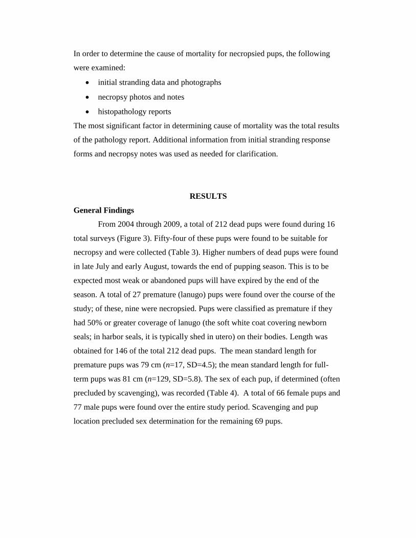
From 2004 through 2009, a total of 212 dead pups were found during 16
total surveys (Figure 3). Fifty-four of these pups were found to be suitable for
necropsy and were collected (Table 3). Higher numbers of dead pups were found
in late July and early August, towards the end of pupping season. This is to be
expected most weak or abandoned pups will have expired by the end of the
season. A total of 27 premature (lanugo) pups were found over the course of the
study; of these, nine were necropsied. Pups were classified as premature if they
had 50% or greater coverage of lanugo (the soft white coat covering newborn
seals; in harbor seals, it is typically shed in utero) on their bodies. Length was
obtained for 146 of the total 212 dead pups. The mean standard length for
premature pups was 79 cm (n=17, SD=4.5); the mean standard length for full-
term pups was 81 cm (n=129, SD=5.8). The sex of each pup, if determined (often
precluded by scavenging), was recorded (Table 4). A total of 66 female pups and
77 male pups were found over the entire study period. Scavenging and pup
location precluded sex determination for the remaining 69 pups.
Figure 3.Total dead pups found for all survey date for all years, (grouped by
week).
Table 3. Number of pups found &
necropsied by survey date.
Number of Pups
Date Found Necropsied
6-Jun-04 0 0
21-Jun-04 1 0
30-Jun-04 1 0
9-Jul-04 4 2
15-Jul-04 1 0
5-Jul-05 17 10
10-Jul-05 15 5
13-Jul-05 13 2
25-Jul-05 24 12
7-Aug-05 37 6
6-Jul-06 2 0
12-Jul-06 17 3
5-Aug-08 30 6
8-Jul-09 12 4
22-Jul-09 11 4
20-Aug-09 27 0
Total 212 54
02468
10121416182022242628303234363840
19-Jun 26-Jun 3-Jul 10-Jul 17-Jul 24-Jul 31-Jul 7-Aug 14-Aug 21-Aug
Nu
mb
er o
f S
eals
Survey Date
Total Dead Seal Pups Found per Survey Date
2004
2005
2006
2008
2009
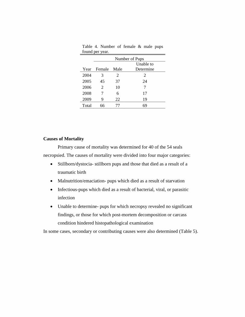
Table 4. Number of female & male pups
found per year.
Number of Pups
Year Female Male
Unable to
Determine
2004 3 2 2
2005 45 37 24
2006 2 10 7
2008 7 6 17
2009 9 22 19
Total 66 77 69
Causes of Mortality
Primary cause of mortality was determined for 40 of the 54 seals
necropsied. The causes of mortality were divided into four major categories:
Stillborn/dystocia- stillborn pups and those that died as a result of a
traumatic birth
Malnutrition/emaciation- pups which died as a result of starvation
Infectious-pups which died as a result of bacterial, viral, or parasitic
infection
Unable to determine- pups for which necropsy revealed no significant
findings, or those for which post-mortem decomposition or carcass
condition hindered histopathological examination
In some cases, secondary or contributing causes were also determined (Table 5).
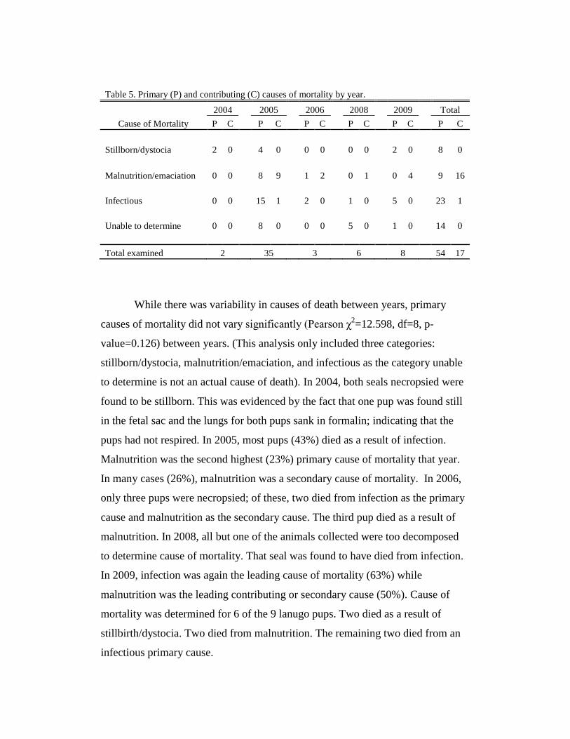
Table 5. Primary (P) and contributing (C) causes of mortality by year.
2004
2005
2006
2008
2009
Total
Cause of Mortality P C P C P C P C P C P C
Stillborn/dystocia 2 0
4 0
0 0
0 0
2 0
8 0
Malnutrition/emaciation 0 0
8 9
1 2
0 1
0 4
9 16
Infectious 0 0
15 1
2 0
1 0
5 0
23 1
Unable to determine 0 0
8 0
0 0
5 0
1 0
14 0
Total examined 2 35 3 6 8 54 17
While there was variability in causes of death between years, primary
causes of mortality did not vary significantly (Pearson χ2=12.598, df=8, p-
value=0.126) between years. (This analysis only included three categories:
stillborn/dystocia, malnutrition/emaciation, and infectious as the category unable
to determine is not an actual cause of death). In 2004, both seals necropsied were
found to be stillborn. This was evidenced by the fact that one pup was found still
in the fetal sac and the lungs for both pups sank in formalin; indicating that the
pups had not respired. In 2005, most pups (43%) died as a result of infection.
Malnutrition was the second highest (23%) primary cause of mortality that year.
In many cases (26%), malnutrition was a secondary cause of mortality. In 2006,
only three pups were necropsied; of these, two died from infection as the primary
cause and malnutrition as the secondary cause. The third pup died as a result of
malnutrition. In 2008, all but one of the animals collected were too decomposed
to determine cause of mortality. That seal was found to have died from infection.
In 2009, infection was again the leading cause of mortality (63%) while
malnutrition was the leading contributing or secondary cause (50%). Cause of
mortality was determined for 6 of the 9 lanugo pups. Two died as a result of
stillbirth/dystocia. Two died from malnutrition. The remaining two died from an
infectious primary cause.
Pup Size (Measured by Length, Weight, & Sternal Blubber Thickness (SBT))
In Relation to Cause of Mortality
Pup length did not vary significantly by cause of mortality (ANOVA,
p=0.285). Pup weight did vary significantly with cause of mortality (ANOVA,
p<0.001). Sternal blubber thickness also varied significantly (ANOVA, p=0.001).
This is not surprising as one of the causes of mortality was
malnutrition/emaciation; these pups would have had lower weights and blubber
thickness.
Percent Mortality
Estimated minimum percent mortality for pups (calculated as described in
methods) was determined for 2004 through 2008 (Table 6). Seal count survey
data from 2009 were not yet available.
These mortality rates are only minimum estimates as some carcasses were
likely scavenged or washed away with the tide. The highest rate of mortality
(27%) occurred in 2005. This was markedly higher than in all other years, where
mortality ranged from only 3% to 16%. While there did not appear to be a great
increase in the total number of all seals that year, surveys did show a higher
number of pups in 2005 than in other study years. Calculated birth rate was also
significantly higher (46%) that year; birth rates in the remaining survey years
ranged from 18% to 22%.
Other Significant Findings
Antibiotic Resistant Bacteria
Antibiotic resistant bacteria (ARB) isolates were found in 30% (n=16) of
the pups necropsied. Bacterium exhibiting antibiotic resistance included
Enterococcus sp. (Table 7), E.coli (hemolytic and non-hemolytic) (Tables 8 & 9),
and Actinomyces sp. (Table10). Several isolates were resistant to multiple
antibiotics. This is an unexpected finding in a wild population and is most likely
caused by fresh water run-off (Bogomolni et al., 2008; Stoddard et al., 2005). As
newborns with underdeveloped immune systems, these pups may have been
inherently more susceptible to bacterial infections and it is not known what effects
ARB may have on the population as a whole.
Phocid herpes virus (PhHV-1)
Four of the pups collected for necropsy tested positive for phocine herpes
virus (PhHV-1). Three of the pups were found in 2005, one was found in 2009.
In all cases, the cause of mortality was infection. All of these pups exhibited
simultaneous bacterial infections. In such cases, the bacteria could be the primary
pathogen; a latent PhHV-1 infection occurring as a result of an already stressed
immune system (Gulland et al., 1997).
Streptococcus canis
In 2005, two seals were found with Streptococcus canis infections.
Streptococcus canis is an opportunistic pathogen in canids; exposure typically
involves close contact with an infected individual, so this is an unusual finding in
harbour seals. In both cases, this isolate was considered significant and likely
represented an environmental source of infection via the umbilicus. Both seals
presenting with Streptococcus canis died as a result of infection; one from
omphalophlebitis (infection of the umbilical vein), and one from omphalitis
(infection of the umbilicus) with subsequent peritonitis. Salmonella typhimurium
was also found in one of these seals.
Salmonella typhimurium
Salmonella typhimurium was isolated from four seals in 2005. In all four
cases, this finding was significant as this bacteria contributed to mortality. This is
also an unusual finding. This disproportionate number of pups found with
Salmonella typhimurium is concerning as it may have represented some sort of
environmental exposure such as untreated sewage runoff, farm runoff or exposure
from other wildlife. A source has yet to be determined. Salmonella typhimurium
was not seen in other years of this study. As with most bacterial and viral
pathogens, suppressed immunity caused by malnutrition or stress may have led to
an increased susceptibility to infection.
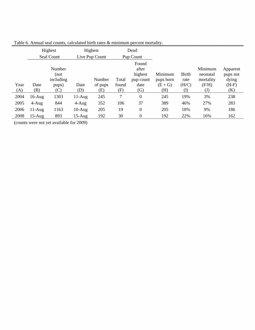
Table 6. Annual seal counts, calculated birth rates & minimum percent mortality.
Highest Highest Dead
Seal Count Live Pup Count Pup Count
Year
(A)
Date
(B)
Number
(not
including
pups)
(C)
Date
(D)
Number
of pups
(E)
Total
found
(F)
Found
after
highest
pup count
date
(G)
Minimum
pups born
(E + G)
(H)
Birth
rate
(H/C)
(I)
Minimum
neonatal
mortality
(F/H)
(J)
Apparent
pups not
dying
(H-F)
(K)
2004 16-Aug 1303 11-Aug 245 7 0 245 19% 3% 238
2005 4-Aug 844 4-Aug 352 106 37 389 46% 27% 283
2006 11-Aug 1163 10-Aug 205 19 0 205 18% 9% 186
2008 15-Aug 893 15-Aug 192 30 0 192 22% 16% 162
(counts were not yet available for 2009)
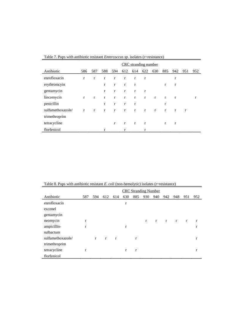
Table 7. Pups with antibiotic resistant Entercoccus sp. isolates (r=resistance)
CRC stranding number
Antibiotic 586 587 588 594 612 614 622 630 885 942 951 952
enrofloxacin r r r r r r r
r
erythromcyin
r r r r
r r
gentamycin
r r r r r
lincomycin r r r r r r r r r r
r
penicillin
r r r r
r
sulfamethoxazole/ r r r r r r r r r r r
trimethroprim
tetracycline
r r r r
r r
florfenicol r r r
Table 8. Pups with antibiotic resistant E. coli (non-hemolytic) isolates (r=resistance)
CRC Stranding Number
Antibiotic 587 594 612 614 630 885 930 940 942 948 951 952
enrofloxacin
r
excenel
gentamycin
neomycin r
r r r r r r
ampicillin- r
r
r
sulbactum
sulfamethoxazole/
r r r
r
r
trimethroprim
tetracycline r
r r
r
florfenicol
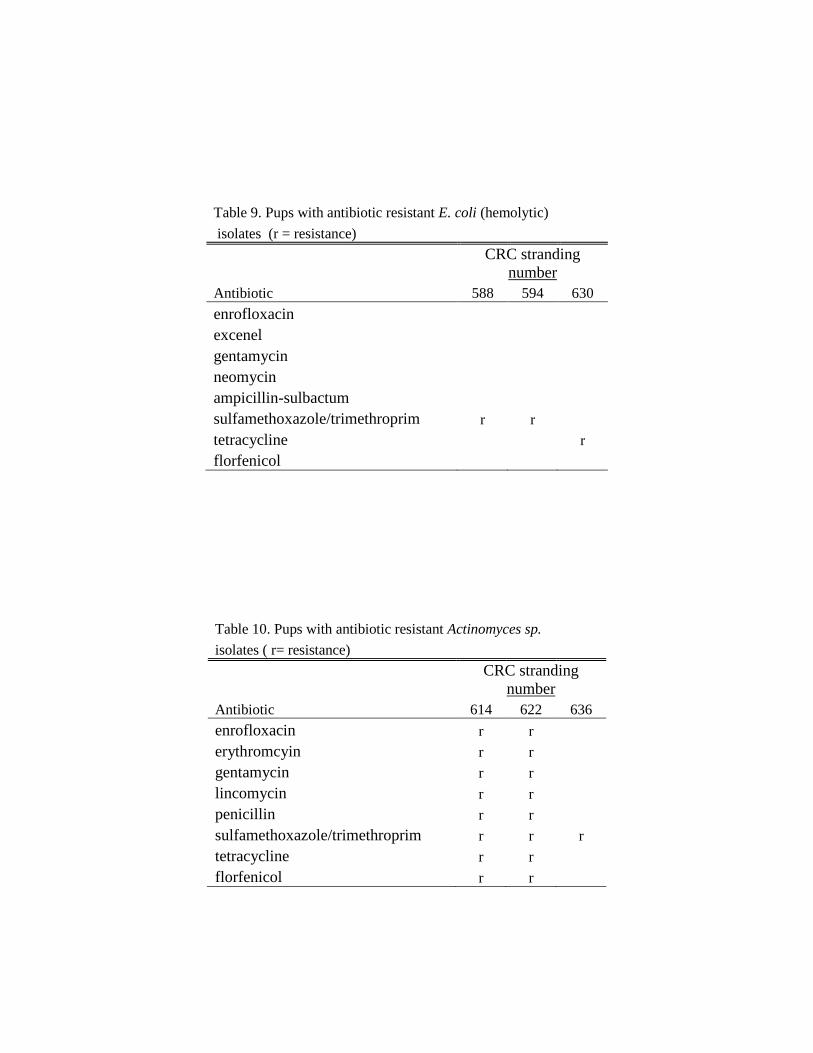
Table 9. Pups with antibiotic resistant E. coli (hemolytic)
isolates (r = resistance)
CRC stranding
number
Antibiotic 588 594 630
enrofloxacin excenel gentamycin neomycin ampicillin-sulbactum sulfamethoxazole/trimethroprim r r
tetracycline
r
florfenicol
Table 10. Pups with antibiotic resistant Actinomyces sp.
isolates ( r= resistance)
CRC stranding
number
Antibiotic 614 622 636
enrofloxacin r r
erythromcyin r r
gentamycin r r
lincomycin r r
penicillin r r
sulfamethoxazole/trimethroprim r r r
tetracycline r r
florfenicol r r
DISCUSSION
Standard Length and Prematurity
The total number (n=27) of premature pups found during this study
appears to be lower than previously recorded at this site (Steiger et al., 1989). The
mean standard length (79cm) for premature pups appears to be slightly higher
than previously determined (69cm) while the length (81cm) of term pups is quite
similar to that found previously (84cm). This could be explained by multiple
variables. The previous study examined multiple sites in one year; pups from
other sites could have influenced the standard length calculations. Also, in at least
two cases, relatively large lanugo pups were found in my study. In such a small
sample size, this can influence the mean standard length. However, both studies
did report a difference in length between premature and term pups as would be
expected.
Causes of Mortality
The primary and secondary causes of mortality found are consistent with
those previously reported at Smith Island. Prematurity, stillbirth/dystocia, and
malnutrition were prevalent in this study as well as previous studies. However,
more pups appear to have succumbed to infection over this study period than
previously observed (Steiger, et al., 1989).
We were only able to necropsy more than 5 pups in three of the study
years (2005, 2008, and 2009). The highest numbers of pups were collected and
necropsied in 2005 and 2009; infection was the leading cause of mortality in both
of these years. Based on these findings, it is likely that infection could be a
prominent cause of death in most years.
Only four categories were used for cause of death in this study. I chose
these categories to simplify the analysis. Categories could be broken down further
to suit the purposes of further studies. For example, infection could be broken
down into viral, bacterial, and parasitic sources. Other classification schemes
(Bogomolni et al., 2010) exist but it seems that more general classifications, such
as those used in this thesis help simplify comparisons between studies,
particularly when examining mortality across a variety of marine mammals.
Pup Size In Relation to Cause of Mortality
The relationship between pup size and cause of mortality is to be
expected. As sternal blubber thickness and weight are indicators of pup health, it
is not surprising that pups with lower weights and inadequate blubber thickness
succumbed to emaciation. What is more difficult to discern is when emaciation is
the primary cause of death rather than a contributing factor. One of the most
difficult tasks of this study was determining this. In some cases where
malnutrition and infection contributed to mortality, it is difficult to say which
came first. Malnutrition can lead to weakened immunity which can in turn lead to
infection. Conversely, animals weakened by infection can become anorexic, thus
succumbing to malnutrition. As newborns, pups are under stress and have
relatively low immunity regardless. It is easy to assume that one single factor is
responsible for pup mortality; but in essence, all life events cumulatively lead to
mortality.
Percent Mortality
The significant increase in neonatal mortality in 2005 appears to correlate
with a dramatic increase in birth rate. This is an interesting finding and suggests
that the increase in pup mortality that year was likely a function of the higher
number of pups born that year. It is likely that pup mortality at Smith Island is
highly variable and dependent on a number of factors, such as prey resources and
maternal age at pupping. Previous work (Calambokidis et al., 1985) has found
smaller size and higher mortality in pups born to young primiparous females.
Other Significant Findings
While some level of antibiotic resistance is expected, this level of multi-
antibiotic resistant bacteria in a wild population is unusual and concerning.
Alarmingly, antibiotic resistance has also been found in other marine mammals
and seabirds along the Northeastern United States (Rose et al., 2009). The source
of this resistance is not clear but Enterococcus and E. coli are pathogens of human
concern as well. Antibiotic resistance in seals is likely contributed to the
prevalence of antibiotic use in humans and agricultural animals. As antibiotic
resistance becomes more prevalent in marine mammal populations, there could be
significant implications for marine ecosystem health.
Studies have demonstrated that PhHV-1 appears to be endemic in Pacific
harbor seal populations but that fatal infections usually only occur in neonates
(Goldstein et al., 2003; Gulland et al., 1997; Harder et al., 1997). It is unknown
what percentage of the population carries PhHV-1. Infected seal pups from my
study were also included in a recent review (Himworth et al., 2010) of all PhHV-1
cases presenting in British Columbia, Canada and Washington state; most of these
seals presented with other simultaneous infections. This is true of the PhHV-1
seals in my study. This makes it difficult to distinguish what role PhHV-1 may
have played in the mortality of these seals. PhHV-1 could have predisposed these
seals to other virulent infections; conversely, these infections could have
weakened pup immunity and subsequently increased the pathogenicity of PhHV-
1. Regardless, PhHV-1 appears to be at least a contributing factor in the loss of
these pups.
It is unclear what may have contributed to the prevalence of Streptococcus
canis and Salmonella typhimurium in this population. Streptococcus canis might
be expected in a coastal population where dogs and other canids could come in
contact with hauled out seals; this is not the case in an isolated location such as
Smith Island. Salmonella typhimurium is another unusual finding. The most likely
source of these bacteria is coastal or untreated sewage runoff. No point source
was ever determined and Salmonella typhimurium has not been isolated in this
population during any year other than 2005.
Study Limitations
When interpreting the results of this study, it is important to keep in mind
a number of limitations. This study examined one age class at one over several
years. Causes of death may vary by age class or site. All surveys in this study
occurred during pupping season; causes of mortality in pups may vary as they
grow and are no longer subject to such a densely packed haulout environment. A
certain sampling bias also exists as only fresh carcasses were collected. A number
of carcasses were lost to decomposition and scavenging or were washed out with
the tide. Also, as only dead animals were sampled, the effect some of these
pathogens may have on live animals is unknown.
The results of this study indicate the annual variability in percent pup
mortality, causes of pup mortality, and birth rate in one population of harbor seals
within Washington inland waters. Care should be used in extrapolating these
results to other populations within the region or other geographical areas.
CONCLUSION & SUGGESTIONS FOR FURTHER RESEARCH
This study has found that at Smith Island, primary causes of death in
harbor seal pups did not vary significantly between years. The findings did
demonstrate a relationship between pup size and cause of mortality. Infectious
disease, malnutrition, stillbirth, and prematurity were all common causes of death
in pups at this site. Common pathogens, such as Enterococcus and E. coli were
found in this population as well as some more unusual findings such as PhHV-1,
Streptococcus canis, Salmonella typhimurium, and high levels of ARB.
As this study only examined one age class, future studies should include
multiple age-classes and multiple sites if possible. WDFW and CRC have
conducted studies at other sites; continuing this work is essential. Future work
should also focus on comparing studies between regions throughout the United
States, as well as comparisons between other marine mammal species.
Standardizing the way data is managed within the various stranding networks
would help facilitate this.
This thesis demonstrates the wealth of information that can be learned
about a population through the use of stranding data. Perhaps the most important
finding of this study is the detection of common pathogens and overall patterns in
mortality. Long-term population monitoring is important to help understand
population dynamics and support critical management decisions. Continuing the
current work of CRC, WDFW, and other agencies is integral to our understanding
of local marine mammal populations. When we understand what is “normal” in a
population, we are better equipped to quickly identify population shifts and
disturbances. Studies such as this also provide a window into the health of the
entire ecosystem. As we become more aware of anthropogenic sources of
degradation in the marine environment, we must be able to quantify the effects on
both animals and the ecosystem as a whole. Long-term data collection from this
site may provide invaluable insights into the potential impacts of contaminants,
pathogen introduction, and other perturbations on population recruitment, health
and status. As seals are sentinels of environmental health, monitoring their health
is a tool for monitoring the health of the Puget Sound and all its inhabitants.
As part of this ecosystem, our well-being is dependent on its
sustainability. We must therefore actively participate in the monitoring and
conservation of its resources. Thus, a critical complement to long-term population
studies such as this would be a comprehensive social analysis on the
anthropogenic disturbances to Puget Sound ecosystem health. Assessing current
perceptions on the health of Puget Sound and how the public relates to the marine
ecosystem is vital to public education and the eventual minimalization of
anthropogenic effects.
Increased prevalence of antibiotic resistant bacteria, emerging pathogens,
and environmental contaminants affect the health of the entire ecosystem.
Monitoring water quality, mitigating urban and agricultural run-off, and proper
use of anti-microbial therapy are all essential to maintaining a healthy marine
ecosystem. If such measures are not taken, the environmental effects will be even
more severe than they are now, potentially lethal for many species. A combination
of multiple long-term studies of several marine species, public education, and
effective environmental policy are needed if we are to conserve and sustain our
marine resources.
LITERATURE CITED
Aguilar A & Borrell A (1994). “Abnormally high polychlorinated biphenyl levels
in striped dolphins (Stenella coeruleoalba)”. Journal of Cetacean Research
and Management 2:17-26.
Bigg MA (1981).Harbour seal, Phoca vitulina, Linnaeus, 1758 and Phoca largha,
Pallas, 1811. Pp 1-27, In: Ridgeway, SH and Harrison, R J (eds). Handbook of
Marine Mammals. Vol 2: Seals. Academic Press, New York.
Bogomolni AL, Pugliares KR, Sharp SM, Patchett K, Harry CT, LaRocque JM,
Touhey KM, Moore M (2010). “Mortality trends of stranded marine mammals
on Cape Cod and southeastern Massachusetts, USA, 2000-2006”. Diseases of
Aquatic Organisms 88:143-155.
Bossart, GD (2006). “Marine Mammals as Sentinel Species for Oceans and
Human Health”. Oceanography 19 (2):134-137.
Boveng P (1988). “Status of the Pacific harbor seal population on the U.S. west
coast”. Admin Rep. LJ-88-06. Southwest Fisheries Science Center, National
Marine Fisheries Service, P.O. Box 271, La Jolla, CA. 43pp.
Burns JJ (2008). Harbor Seal and Spotted Seal. Pp. 533-542. In: Perrin WF,
Wursig B, Teewissen JGM (Eds). Encyclopedia of Marine Mammals. 2nd
edition. Academic Press, Burlington.
Calambokidis J, Gentry RL (1985). “Mortality of Northern Fur Seal Pups in
Relation to Growth and Birth Rates”. Journal of Wildlife Diseases 21(3): 327-
330.
Calambokidis J, Speich SM, Peard J, Steiger GH, Cubbage JC (1985). “Biology
of Puget Sound Marine Mammals and Marine Birds: Population Health and
Evidence of Pollution Effects”. NOAA Technical Memorandum NOS OMA
18.
Carretta JV, Forney KA, Muto MM, Barlow J, Baker J, Hanson B, & Lowry S
(2007). “U.S. Pacific Marine Mammal Stock Assessments: 2006.” U.S.
Department of Commerce, NOAA Technical Memorandum, NMFS-SWFSC-
398.
Colegrove KM, Grieg DJ, Gulland FMD (2005). “Causes of live strandings of
Northern elephant seals (Mirounga angustirostris) and Pacific harbor seals
(Phoca vitulina) along the central California coast, 1992-2001”. Aquatic
Mammals 31:1-10.
Dierauf LA & Gulland FMD (2001). CRC Handbook of Marine Mammal
Medicine. 2nd
Edition. CRC Press, Boca Raton.
Geraci JR & Lounsbery VJ (1999). Health. Pp546-553. In: Perrin WF, Wursig B,
Thewissen JGM (eds). Encyclopedia of Marine Mammals. 2nd
edition.
Academic Press, Burlington.
Goldstein T, Gulland FM, Aldridge BM, Harvey JT, Allen SG (2004). “The
transmission of phocine herpesvirus-1 in rehabilitating and free-ranging Pacific
harbor seals (Phoca vitulina) in California”. Veterinary Microbiology 103:131-
141.
Gulland FMD, Hall AJ (2005). “The Role of Infectious Disease in Influencing
Status and Trends”. Pp 47-61. In: Reynolds JE, Perrin WF, Reeves RR,
Montgomery S, & Ragen TJ (eds). Marine Mammal Research: Conservation
Beyond Crisis. The Johns Hopkins University Press, Baltimore.
Gulland FMD, Lowenstine LJ, Lapointe JM, Spraker T, King DP (1997).
“Herpesvirus infection in stranded Pacific harbor seals of coastal California”.
Journal of Wildlife Diseases 33:450-458.
Harder TC,Vos HW, de Swart RL, Osterhaus ADME (1997). “Age-related
disease in recurrent outbreaks of phocid herpes type-1 infections in a seal
rehabilitation center: Evaluation of diagnostic methods”. Vet Rec 140:500-503.
Himworth CG, Haulena M, Lambourn DM, Gaydos JK, Huggins J, Zaremba J,
Calambokidis J, Ford J, Ross P, Raverty S (2010). “Pathology and
epidemiology of phocid herpesvirus-1 infections in wild and rehabilitating
harbor seals (Phoca vitulina) in the Northeastern Pacific”. In press.
Jeffries S, Huber H, Calambokidis J, & Laake J (2003). “Trends and status of
harbor seals in Washington state: 1978-1999”. Journal of Wildlife
Management 67:208-219.
Kreuder C, Miller MA, Jessup DA, Lowenstine LJ, Harris MD, Ames JA,
Carpenter TE, Conrad PA, & Mazet JA (2003). “Patterns of Mortality in
Southern Sea Otters (Enhydra lutris nereis) from 1998-2001”. Journal of
Wildlife Diseases 39(3):495-509.
Lambourn DM, Jeffries SJ, & Huber HR (2010). “Observations of Harbor Seals
in Southern Puget Sound during 2009”. Washington Department of Fish and
Wildlife Contract Report for NOAA Purchase Order AB133F09SE2836F.
Lockwood SK, Chovan JL, & Gaydos JK (2006). “Aerobic Bacterial Isolations
from harbor seals (Phoca vitulina) stranded in Washington: 1992-2003”.
Journal of Zoo and Wildlife Medicine 37(3):281-291.
Miller MA, Gardner IA, Kreudner C, Paradies DM, Worcester KR, Jessup DA,
Dodd E, Harris MD, Ames JA, Packham AE, Conrad PA (2002). “Coastal
freshwater runoff is a risk factor for Toxoplasma gondii infection of southern
sea otters (Enhydra lutris nereis)”. International Journal for Parasitology
32:997-1006.
National Marine Fisheries Service. (2009).
http://www.nmfs.noaa.gov/pr/species/mammals/pinnipeds/harborseal.html
Pitcher KW & McAllister DC (1981). “Movements and haul out behavior of
radio-tagged harbor seals, Phoca vitulina”. The Canadian Field Naturalist.
95:292-297.
Pugliares K, Herzig S, Bogolmoni A, Harry C, Touhey K, Moore M (2007).
“Marine mammal necropsy: an introductory guide for stranding responders and
field biologists”. Technical Document 2007-06. Woods Hole Oceanographic
Institution, Woods Hole, MA.
Riedman M (1990). The Pinnipeds: Seals, Sea Lions, and Walruses. University of
California Press, Berkeley.
Rose JM, Gast RJ, Bogolmoni A, Ellis JC, Lenell BJ, Touhey K, Moore M
(2009). “Occurrence and patterns of antibiotic resistance in vertebrates off the
Northeastern United States Coast”. FEMS Microbiological Ecology 67:421-
431.
Ross PS, Pohadjak B, Bowen WD, Addison RF (1993). “Immune function in free-
ranging harbor seal (Phoca vitulina) mothers and their pups during lactation”.
Journal of Wildlife Diseases 29(1):21-259.
Steiger GH, Calambokidis, J, Cubbage, JC, Skilling, DE, Smith, AW, & Gribble
DH (1989). “Mortality of harbor seal pups at different sites in the inland waters
of Washington”. Journal of Wildlife Diseases 25 (3):319-328.
Stoddard R, Gulland FMD, Atwill ER, Lawrence J, Jang S, & Conrad PA (2005).
“Salmonella and Campylobacter spp in northern elephant seals, California”.
Emerging Infectious Disease 11:1967-1969.
Washington Department of fish and Wildlife. (2009).
http://wdfw.wa.gov/wildwatch/sealcam/seal_info.html
Wells RS, Rhinehart HL, Hansen LJ, Sweeney JC, Townsend FI, Stone R, Casper
DR, Scott MD, Hohn AA, Rowles TK (2004). “Bottlenose dolphins as marine
ecosystem sentinels: developing a health monitoring system”. EcoHealth
1:246-254.
