case study: seizure disorder presenting as panic disorder with agoraphobia
TRANSCRIPT
Case Study: Seizure Disorder Presenting asPanic Disorder With Agoraphobia
DOUGLAS O . LEE, M.D., SANDRA L. HELMERS, M.D. , RONALD J. STEINGARD, M.D.,
AND DAVID RAY DEMASO, M.D.
ABSTRACT
Seizure disorders can produce anxiety that is almost indistinguishable from psychiatric disorders. There are few reports
of adolescents with seizure disorders that produce fear.The first case of an adolescent female who presented with panic
disorder and agoraphobia which was a consequence of seizure activity is reported. Careful diagnostic evaluation and
correlation with video electroencephalography were important in distinguishing seizure activity from panic disorder. J.
Am. Acad. Child Adolesc. Psychiatry, 1997. 36(9): 1295-1298. Key Words: seizure. panic. agoraphobia. temporal lobe.
More than 100 years ago, Jackson (1880-1881) firstdescribed the clinical relationship between fear and seizures. Subsequently, authors have noted the frequentassociation of fear with seizure auras (Theodore et al.,1983) as well as ictal phases (Mclachlan and Blume,1980). In fact, up to 21% of adult patients with seizuresexperience panic attacks (Pariente et al., 1991) compared with 3.8% of the general population (Katerndahland Realini, 1993).
Complicating the clinical assessment, panic attackshave been often mistaken for seizures (Hirsch et al.,1990) or relapse of seizures (Genton et al., 1995).Likewise, seizures have also been mistaken for panicattacks (Alemayehu et al., 1993; Devinsky et al., 1989;Laidlaw and Zaw, 1993; Weilburg et al., 1987). In addition, the relationship between panic disorders and seizures has received no significant focus in adolescents.The following unique case illustrates the importantclinical dilemma of distinguishing between panic withagoraphobia and seizure disorders.
Acupud February 21, 1997,Dr. Lee is with th« Department ofPsychiatry, Emory University School of
M~dicin~, Atlanta. Dr. Helmers is with the Department ofNeurologyand Drs.St~ingardandD~Masoa" with the Department ofPsychiatry, Harvard MedicalSchool, Boston.
Reprin: "qums to Dr. Lee, Emory Univmity School ofMedicine, Departmrnt ofChi/J and Adolescent Psychiatry, 1365B Clifton Road, Suit« 6100,Atlanta, GA 30322.
0890-8567/97/3609-1295/$0.300/0<01997 by the American Academy ofChild and Adolescent Psychiatry.
CASE STUDY
A 17-year-old right-handed female was referred forevaluation of panic disorder that aroused suspicions ofcomplex partial seizure (CPS). At age 9 years, thepatient suffered from multiple sudden episodes consisting of intense fear, palpitations, tremulousness, dizziness, and chills lasting up to 20 minutes. Nausea, a"funny feeling" in her stomach, chest tightness, bitemporal headache, experiences of "seeing" herself, hyperacusis, occasional staring, photophobia, stiffening, andsudden limpness occasionally followed, ending infatigue.
Other than an infant history of a complex febrilegeneralized tonic seizure followed by Todd's hemiparesison the left, the pediatrician's examination and a noncontrast cranial computed tomographic scan showed noabnormalities. The patient was given a diagnosis ofmigraine headache and treated with cyproheptadine,but her symptoms did not resolve. At the same time, aneurological consultation yielded normal neurologicalexamination results but an EEG revealed "left temporalsharp wave activiry. " The initial impression was ofeither migraine headache or possible CPS. She wastreated for presumed migraine headaches with carbamazepine, 500 mg/day, which resulted in reduction ofher episodes. However, even after 9 months of carbamazepine treatment, fear, tremulousness, palpitations,dizziness, and chills persisted. The clinical impression ofthese symptoms at this time was of either a migrainousaura or anxiety disorder.
J. AM . ACAD. CHILD ADOLE SC. PSYCHIATRY, 36:9, SEPTEMB ER 199 7 1295
LEE ET AL.
When the patient was 12 years old, a psychiatric consultation found that her presentation fulfilled DSM-IIIR (American Psychiatric Association, 1987) criteria forpanic disorder with agoraphobia. Individual trials ofalprazolam 1.5 mg/day, amitriptyline 25 mg/day, andpropranolol (dose unspecified) were ineffective inreducing symptoms. Another consultation suggestedthat episodes were related to social pressures. A 12month course of psychotherapy and biofeedback resulted in no change.
Shortly before the patient reached age 14 years, asecond neurological evaluation yielded a normal neurological examination as well as a normal gadoliniumenhanced magnetic resonance imaging (MRI) scan ofthe brain. The clinical impression continued to be consistent with panic disorder. However, a video EEG wasrecommended to rule out the possibility of seizure.
Almost a year later, after unsuccessful trials of Huox
etine 20 mg/day and clonazepam 1.0 mg/day, thepatient was referred for a comprehensive neurologicalevaluation. While her neurological examination continued to be normal , her ambulatory EEG was nowremarkable for a right frontotemporal focus. Long-termvideo EEG monitoring with sphenoidal leads detectedeven more epileptiform activity, and of 27 panic episodes , 63% were associated with a rhythmic right frontotemporal discharge. Quantitative EEG revealedepileptiform activity originating from the right hemisphere. Two MRI scans were consistent with rightmesial temporal sclerosis (MTS), interictal single photon emission computed tomography showed slighthypoperfusion in the right temporal lobe, and postepisode serum prolactin level was elevated (65 ng/dL). Hermental status examinations were only remarkable formild baseline anxiety and concern about being in public while having subsequent panic symptoms. There wasno evidence of mood, anxiety, or thought disorders.The final impression was of CPS disorder with anxietysymptoms related to this medical condition.
The patient's seizures continued despite therapeutictrials of carbamazepine, phenytoin, valproate, clonazepam, felbamate, and gabapenrin, Her intractable seizures contributed to academic failure, a skiing accidentresulting in a fractured pelvis , compromised socialfunctioning, and agoraphobia. Consequently, surgicaltreatment with electocorticography was pursued.Electrocorticography showed a very active spike focus inthe anterior and basomedial right temporal lobe. A 7-cm
resection of the right temporal lobe was performed, andpostresection electrocorticography showed no spike.Neuropathology was consistent with MTS. Eighteenmonths after her right temporal lobectomy, the patientremained seizure-free and without panic or agoraphobia.
DISCUSSION
This is the first report of an adolescent with a seizuredisorder presenting as panic disorder with agoraphobia.This patient fulfilled DSM-III-R criteria (AmericanPsychiatric Association, 1987) for panic disorder withagoraphobia, and the presentation was so convincingthat experienced clinicians diagnosed panic disorder inthis adolescent.
Symptoms common to both panic disorder and someCPS contribute to the challenge of making an accuratediagnosis (Laidlaw and Zaw, 1993). CPS can producefear as reflected by clinical observation as well as in electrical stimulation studies (Gloor et al., 1982) . In addition , epileptiform activity (Devinsky et al., 1989;Weilburg et al., 1993) and electrical stimulation studiesof the temporal lobe (Gloor et al., 1982) have generatedpanic and associated somatic signs and symptomsincluding dizziness, nausea, tachycardia, chest pain,hot/cold sensations, and depersonalization.
Unanticipated findings complicated the initial diagnostic impression. Several episodes lasted more than 10minutes, a duration consistent with panic disorder andless suggestive of typical CPS. Usually, CPS haveshorter durations of 2 to 3 minutes, although in rareinstances, fear associated with complex partial statusepilepticus can last for hours (McLachlan and Blume,1980). Another confounding finding was the absenceof EEG changes in many episodes , thereby suggestingpanic disorder. Because the location of MTS is deep,surface EEGs may be normal while subdural electrodesmay be positive (Devinsky et al., 1989). While sphenoidalleads provided more information than surface leads,the intensity of this deep seizure focus was only appreciated after electocorticography, Similarly, initial negative imaging results did not suggest MTS. Even thoughMRI has been very useful in identifying MTS, severalfactors may have influenced its sensitivity: the orientation of the images, diagnostic criteria that are used ininterpreting the images, appreciation of T 1- or T y
weighted signal abnormalities, and the clinician's familiarity with the neuroanatomy (jackson, 1995) .
1296 ] . AM . ACAD. C H ILD AD O LES C. PSYCHIATRY, 36:9, SEPTEMBER 1997
Furthermore, the development of agoraphobia as abyproduct of seizures was unexpected. Agoraphobia isoften associated with panic disorder, but it is not specifically associatedwith seizures, although the cases of a fewadults with panic and agoraphobia produced by seizureshave been reported (Handal et al., 1995; Weilburg et al.,1993).The mechanism by which agoraphobia developedin this patient is not clearly understood, but because thispatient's paroxysmal seizures so closely resembled panicattacks, it is conceivable that anticipatory anxiety,phobic avoidance, and agoraphobia arose as a responseto the intense anxiety generated by the seizure.
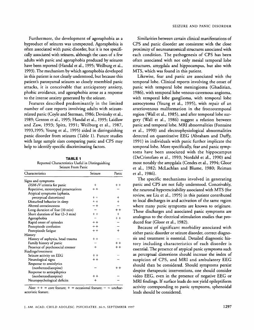
Features described predominantly in the limitednumber of case reports involving adults with seizurerelated panic (Coyle and Sterman, 1986; Devinsky et al.,1989; Genton et al., 1995; Handal et al., 1995; Laidlawand Zaw, 1993; Spitz , 1991; Weilburg et al., 1987,1993,1995; Young et al., 1995) aided in distinguishingpanic disorder from seizures (Table 1). Future studieswith large sample sizes comparing panic and CPS mayhelp to identify specific discriminating factors.
TABLE 1Reported Characteristics Useful in Distinguishing
Seizure From Panic
Character istics Seizure Pan ic
Signs and symptomsDSM-IV criteria for pani c + + +Repetitive. stereotyped presentations + +Atyp ical symptoms (aphasia,
perceptual distortions) ++ +Disturbed behavior in sleep ++ +Altered consciousness ++Long duration of fear (10 min) + +Short duration of fear (2- 3 min) + + +Agoraphobia + +Rapid on set of episodes + +Postepisode confusion + +Posrepisode fatigue + + +
HistoryHistory of asphyxia. head trauma ++Family histor y of panic + +Presence of psychosocial stressor + + +
Findings/rreatmenrSeizure activity on EEG ++Neurological signs ++Response to anxiolyti cs
(nonbenzodiazepine) + +Response to anriepileprics
(nonbenzodiazepine) + +Neuropsychological deficits +
Note: + + = core feature; + = occasional feature ; - = uncharacteristic feature .
SEIZURE AND PANIC DISORDER
Similarities berween certain clinical manifestations ofCPS and panic disorder are consistent with the closeproximity of neuroanatomical structures associated witheach condition. The pathogenesis of CPS has beenoften associated with not only mesial temporal lobestructures, amygdala and hippocampus, but also withMTS, which was found in this patient.
Likewise, fear and panic are associated with thetemporal lobe. Clinical reports involving the onset ofpanic with temporal lobe meningioma (Ghadirian,1986), with temporal lobe venous-cavernous angioma,with temporal lobe ganglioma, with temporal lobeastrocytoma (Young et al., 1995), with repair of anarteriovenous malformation in the frontotemporalregion (Wall et al., 1985) , and after temporal lobe surgery (Wall et al., 1986) suggest a relation berweenpanic and temporal lobe. MRI abnormalities (Fontaineet al., 1990) and electrophysiological abnormalitiesdetected on quantitative EEG (Abraham and Duffy,1991) in individuals with panic further implicate thetemporal lobe. More specifically, fear and panic symptoms have been associated with the hippocampus(DeCristofaro er al., 1993; Nordahl et al., 1990) andmost notably the amygdala (Cendes er al., 1994; Glooret al., 1982; McLachlan and Blume, 1980; Reimaner al., 1986).
The specific mechanisms involved in generatingpanic and CPS are not fully understood. Conceivably,the neuronal hyperexcitability associated with MTS (forreview, see Liu et al., 1995) in this patient contributedto local discharges in and activation of the same regionwhere many panic symptoms are known to originate.These discharges and associated panic symptoms areanalogous to the elecrrical stimulation studies that produced fear (Gloor et al., 1982).
Because of significant morbidity associated witheither panic disorder or seizure disorder, correct diagnosis and treatment is essential. Detailed diagnostic history including characteristics of each disorder isessential.The presence of atypical panic symptoms suchas perceptual distortions should increase the index ofsuspicion of CPS, and MRI and ambulatory EEGshould then be considered. Should symptoms persistdespite therapeutic interventions, one should considervideo EEG, even in the presence of negative EEG orMRI findings. If surface leads do not yield epileptiformactivity corresponding to panic symptoms, sphenoidalleads should be considered.
]. AM . ACAD. CHILD ADOLESC. PSY CHIATRY, 36:9, SEPTEMBER 1997 1297
LEE ET AL.
REFERENCESAbraham HD. Duffy FH (1991). Computed EEG abnormalities in panic
disorder with and without premorbid drug abuse . Bioi Psychiatry29:687-690
A1emayehu S, Bergey GK. Krumholz A et al. (1993), Panic attacks as ictalmanifestations of right parietal lobe lesions. Epilepsia34(suppI6) :58
American Psychiatric Association (1987). Diagnosticand Statistical Manual ofMental Disorders, 3rd edition-revised (DSM -IlI-R). Washington. DC:American Psychiatric Association
Cendes F. Andermann F,Gloor I' et al. (1994) . Relationship between atrophyof the amygdala and ictal fear in temporal lobe epilepsy. Brain117:739-746
Coyle PK. Sterman AB (1986) . Focal neurologic symptoms in pan ic atta cks.Am] Psychiatry 143:648-649
DeCristofaro MTR. Sessarego A. Pupi A. Biond i F. Faravelli C (1993) . Brainperfusion abnormalities in drug-naive, lacrate-sen sirive pan ic patients: aSPECT study. Bioi Psychiatry 33:505-512
Devinsky O. Susumu S. Theodore WH. Porter RJ (1989) . Fear episodes dueto limbic seizures with normal ictal scalp EEG : a subdural electrographicstudy.] Clin Psychiatry 50:28-30
Fontaine R. Breton G. Dery R, Fontaine S. Elie R (1990) . Temporal lobeabnormalities in panic d isorder: an MRI study. Bioi Psychiatry27 :304-310
Genton P. Bartolomei F. Guerrini R (1995) , Panic attacks mistaken forrelapse of epilepsy. Epilepiia 36:48-51
Ghadirian AM (1986), Anxiety attacks in a pat ient with right temporal lobemen ingioma.] C/in Psychiatry 47:270-271
Gloor P,Olivier A. Quesney LF.Andermann F, Horowitz S (1982). The roleof the limb ic system in experiential phenomena of temporal lobe epilepsy. Ann Neural 12:129-144
Handal NM. Masand P, Weilburg JB (1995) . Panic disorder and complexpartial seizures. Psychosomatics36:498-502
Hirsch E. Peretti S, Boulay C. Sellal F. Maron B (1990 ), Panic attacks misdiagnosed as partial epileptic seizures. Epilepsia 31 :636
Jackson GD (1995) . The diagnosis of hippocampal sclerosis: other tech niques . Magn ResonImaging 13:1081-1093
Jackson JH (1880-1881) . On right- or left-sided spasms at the onset of epileptic paroxysms, and on crude sensation warnings and elaborate mentalstates . Brain 22:534-549
Katerndahl DA. Realini [P (1993) . Lifetime prevalence of panic states. Am]Psychiatry 150:246-249
Laidlaw JD . Zaw KM (1993). Epilepsy mistaken for panic attacks in an adolescent girl. BM] 306:709-710
Liu A. Mikat i M. Holmes GL (1995) . Mesial temporal sclerosis: pathogenesis and significance. PediatrNeural 12:5-16
Mclachlan RS, Blume WT (1980) . Isolated fear in complex partial statusepilept icus. Ann Neural 8:639-641
Nordahl TE. Semple WE , Gross M et aI . (1990), Ce rebral glucose metabolicdifferences in patients with panic disorder. N~uropsychopharmacology
3:261-272Pariente PD. Lepine Jp, Lellouch J (1991). Lifetime histoty of panic attacks
and epilepsy : an association from a general population survey. ] ClinPsychiatry 52:88-89
Reiman EM, Raichle ME . Robins E et al. (1986) . The application of positron em ission tomography to the study of panic disorder. Am] Psychiatry143:469-477
Spitz MC (1991) . Panic disorder in seizure patients: a diagnostic pitfall.Epilepsia 32:33- 38
Theodore WH. Porter RJ, Penry JK (1983) . Complex partial seizures: clin ical characteristics and differential diagnosis. N~rology 33:1115-1121
Wall M. Mielke D, Luther JS (1986) , Panic attacks and psychomotor seizuresfollowing right temple lobectomy.] C/in Psychiatry 47:219
Wall M. Tuchman M. Mielke D (1985), Panic attacks and temporal lobe seizures associated with a right temporal lobe arteriovenous malformation:case report.] C/in Psychiatry 46:143-145
Weilburg JB. Bear DM. Sachs G (1987). Three patients with concomitantpanic attacks and seizure disorder: possible clues to the neurology of anxiety. Am] Psychiatry 144:1053-1056
Weilburg JB, Schachter S. Sachs GS et al. (1993 ). Focal paroxysmal EEGchanges during atypical pani c attacks. ] N~uropsychiatry Clin Neurosci5:50-55
Weilburg JB , Schachter S. Worth J et al. (1995) . EEG abnormalities inpatients with atypical panic attacks.] C/in Psychiatry 56:358-362
Young GB , Chandarana PC. Blume WT, McLachlan RS. Munoz DG.Girvin Jp (1995) . Mesial temporal lobe seizures presenting as anxietydisorders.] Neuropsychiatry C/in Neurosci 7:352- 357
1298 J. AM. ACAD. CHILD ADOLES C. PSYCHIATRY, 36 :9 . SEPTEMBER 199 7