case report spontaneous intramural duodenal hematoma with...
TRANSCRIPT

Case Report
Spontaneous Intramural Duodenal Hematoma With Concomitant Acute Pancreatitis
Deshpande A A¹, Takalkar Y P¹, Shukla K¹, Kaddu D¹, Thakur B A¹, Hira P¹
Introduction: Intramural duodenal hematoma (IDH) is a rare condition. There have been very few reported cases of IDH in
association with Acute Pancreatitis (AP). It is usually associated with trauma, intervention or coagulopathy. We are reporting herewith a case of a 42 year old male, who presented with symptoms of AP and gastric outlet obstruction. He was diagnosed to have duodenal hematoma with AP based on imaging findings. In this case, IDH was spontaneous without any precipitating factors. Patient responded well to conservative management.
Keywords: Intramural duodenal hematoma, Acute Pancreatitis, spontaneous.
International Journal of Surgical Cases 2015 July-Sep: 1(1):Page 18-20
Abstract
1Departments of Surgery & *Radiology, Seth G.S.Medical College & K.E.M. Hospital, Parel, Mumbai
400012.
Address of Correspondence
Dr. Aparna Deshpande
Department of Surgery, Seth G. S. Medical College & K.E.M. Hospital, Acharya Donde Marg, Parel,
Mumbai 400012.
Email: [email protected]
Copyright © 2015 by International Journal of Surgical CasesInternational Journal of Surgical Cases | eISSN 2321-3817 | Available on www.surgicalcasesjournal.com/ |
This is an Open Access article distributed under the terms of the Creative Commons Attribution Non-Commercial License (http://creativecommons.org/licenses/by-nc/3.0) which permits unrestricted non-commercial use, distribution, and reproduction in any medium, provided the original work is properly cited.
18
Author’s Photo Gallery
IntroductionIntramural duodenal hematoma (IDH) is a rare condition characterized by hematoma formation within the duodenal wall. It was first reported at autopsy in 1838 by MacLauchlan.1 It occurs mostly in children, following blunt abdominal trauma.[2] In adults, 30% cases are due to nontraumatic causes such as anticoagulation therapy, hemophilia and other coagulation disorders.[3] IDH is rarely associated with pancreatic conditions like acute or chronic pancreatitis, pancreatic tumor or pseudocyst.[4]We present a rare case of IDH associated with Acute Pancreatitis. (AP).
Case ReportA 42 year oldchronic alcoholic male presented with acute epigastric pain and non-bilious vomiting for 3-4 days.There was no history of abdominal trauma or any
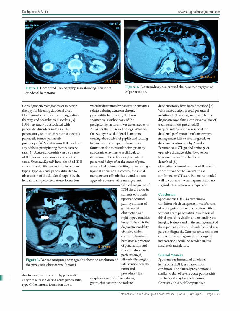
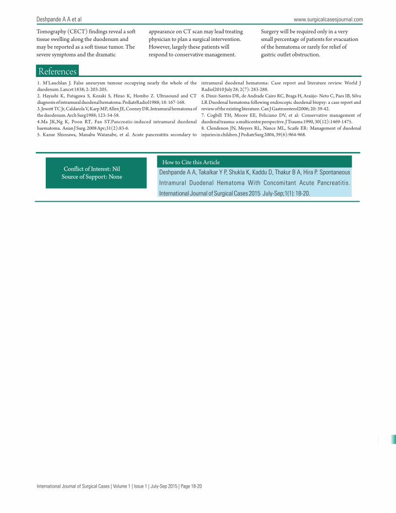
intervention. Patient did not have any hematological disorder and was not on anticoagulants. Pulse and blood pressure were not recordable at presentation. Glasgow coma score was 15. Abdomen was distended, tender and guarded with a diffuse lump of around 10×10 cm in epigastrium and right hypochondrium. Hemoglobin was 8.2 gm%, total leucocyte count was 12200/cmm, serum lipase was 2685 IU. Patient was suspected to have severe AP based on clinical features. Abdominal Computed Tomography (CT) Scan performed after resuscitation revealed a large intramural duodenal hematoma seen as a large hyperdense mass along the second and third part of duodenum.(Fig.1) Mild pancreatic enlargement with adjacent fat stranding was noted. (Fig.2). Patient was resuscitated and managed in intensive care unit with monitoring of parameters, fluid, electrolytes and arterial blood gases.
Duodenoscopy revealed an intraluminal bulge with edematous mucosa in the second part of duodenum beyond which the scope could not be negotiated. Mucosal biopsy revealed inflammatory cells. Initially patient was kept on total parenteral nutrition and later on enteral nutrition through nasojejunal tube. There was good response to conservative management and marked clinical recovery in 6-7 days. A repeat CT scan after 15 days revealed complete resolution of pancreatitis as well as the duodenal mass lesion. (Fig. 3).
DiscussionIntramural duodenal hematoma is a rare condition in which there is formation of hematoma within the wall of duodenum. In adults, traumatic IDH occurs either after blunt abdominal trauma or following any intervention like endoscopic biopsy, Endoscopic Retrograde
Author’s Photo Gallery
Dr. Shukla KDr. Deshpande A A Dr. Takalkar Y P Dr. Thakur B A Dr. Hira P
Access this article online
Website:www.surgicalcasesjournal.com
DOI:
Cholangiopancreatography, or injection therapy for bleeding duodenal ulcer. Nontraumatic causes are anticoagulation therapy, and coagulation disorders.[3] IDH may rarely be associated with pancreatic disorders such as acute pancreatitis, acute on chronic pancreatitis, pancreatic tumor, pancreatic pseudocyst.[4] Spontaneous IDH without any of these precipitating factors is very rare.[5] Acute pancreatitis can be a cause of IDH as well as a complication of the same. ShiozawaK,et al5 have classified IDH concomitant with pancreatitis into three types; type A- acute pancreatitis due to obstruction of the duodenal papilla by the hematoma, type B- hematoma formation
due to vascular disruption by pancreatic enzymes released during acute pancreatitis, type C- hematoma formation due to
vascular disruption by pancreatic enzymes released during acute on chronic pancreatitis.In our case, IDH was spontaneous without any of the precipitating factors. It was associated with AP as per the CT scan findings. Whether this was type A: duodenal hematoma causing obstruction of papilla and leading to pancreatitis or type B : hematoma formation due to vascular disruption by pancreatic enzymes; was difficult to determine. This is because, the patient presented 3 days after the onset of pain, already had bilious vomiting as well as high lipase at admission .However, the initial management of both these conditions is aggressive conservative management.
Clinical suspicion of IDH should arise in patients with acute upper abdominal pain, symptoms of gastric outlet obstruction and right hypochondriac lump. CTscan is the diagnostic modality ofchoice which confirms duodenal hematoma, presence of pancreatitis and rules out duodenal perforation.[6]Historically, surgical intervention was the norm and procedures like
simple evacuation of hematoma, gastrojejunostomy or duodeno-
duodenostomy have been described.[7] With introduction of total parenteral nutrition, ICU management and better diagnostic modalities, conservative line of treatment is now preferred.[8]Surgical intervention is reserved for duodenal perforation or if conservative management fails to resolve gastric or duodenal obstruction by 2 weeks. Percutaneous CT guided drainage or operative drainage either by open or laparoscopic method has been described.[8]Our patient showed features of IDH with concomitant Acute Pancreatitis as confirmed on CT scan. Patient responded well to conservative management and no surgical intervention was required.
ConclusionSpontaneous IDH is a rare clinical condition which can present with features of acute gastric outlet obstruction with or without acute pancreatitis. Awareness of this diagnosis is vital in understanding the imaging features and in the management of these patients. CT scan should be used as a guide in diagnosis. Current consensus is for conservative management and surgical intervention should be avoided unless absolutely mandatory.
Clinical MessageSpontaneous Intramural duodenal hematoma (IDH) is a rare clinical condition. The clinical presentation is similar to that of severe acute pancreatitis and hence it may be misdiagnosed. Contrast enhanced Computerised
www.surgicalcasesjournal.comDeshpande A A et al
19
International Journal of Surgical Cases | Volume 1 | Issue 1 | July-Sep 2015 | Page 18-20
Figure 1. Computed Tomography scan showing intramural duodenal hematoma.
Figure 2. Fat stranding seen around the pancreas suggestive of pancreatitis.
Figure 3. Repeat computed tomography showing resolution of the preexisting hematoma (arrow)
International Journal of Surgical Cases | Volume 1 | Issue 1 | July-Sep 2015 | Page 18-20
20
www.surgicalcasesjournal.comDeshpande A A et al
Tomography (CECT) findings reveal a soft tissue swelling along the duodenum and may be reported as a soft tissue tumor. The severe symptoms and the dramatic
appearance on CT scan may lead treating physician to plan a surgical intervention. However, largely these patients will respond to conservative management.
Surgery will be required only in a very small percentage of patients for evacuation of the hematoma or rarely for relief of gastric outlet obstruction.
References1. M'Lauchlan J. False aneurysm tumour occupying nearly the whole of the duodenum. Lancet 1838; 2: 203-205.2. Hayashi K, Futagawa S, Kozaki S, Hirao K, Hombo Z. Ultrasound and CT diagnosis of intramural duodenal hematoma. PediatrRadiol1988; 18: 167-168.3. Jewe� TC Jr, Caldarola V, Karp MP, Allen JE, Cooney DR.Intramural hematoma of the duodenum. Arch Surg1988; 123: 54-58.4.Ma JK,Ng K, Poon RT, Fan ST.Pancreatic-induced intramural duodenal haematoma. Asian J Surg. 2008 Apr;31(2):83-6. 5. Kazue Shiozawa, Manabu Watanabe, et al. Acute pancreatitis secondary to
intramural duodenal hematoma: Case report and literature review. World J Radiol2010 July 28; 2(7): 283-288.6. Diniz-Santos DR, de Andrade Cairo RC, Braga H, Araújo- Neto C, Paes IB, Silva LR Duodenal hematoma following endoscopic duodenal biopsy: a case report and review of the existing literature. Can J Gastroenterol2006; 20: 39-42.7. Cogbill TH, Moore EE, Feliciano DV, et al: Conservative management of duodenal trauma: a multicentre perspective. J Trauma 1990, 30(12):1469-1475.8. Clendenon JN, Meyers RL, Nance ML, Scaife ER: Management of duodenal injuries in children. J PediatrSurg 2004, 39(6):964-968.
Conflict of Interest: Nil Source of Support: None
How to Cite this Article
Deshpande A A, Takalkar Y P, Shukla K, Kaddu D, Thakur B A, Hira P. Spontaneous
Intramural Duodenal Hematoma With Concomitant Acute Pancreatitis.
International Journal of Surgical Cases 2015 July-Sep;1(1): 18-20.