case report serous tubal intraepithelial carcinoma:...
TRANSCRIPT

Case ReportSerous Tubal Intraepithelial Carcinoma: An Incidental Findingat the Time of Prophylactic Bilateral Salpingo-Oophorectomy
Monique Hiersoux Vaughan,1 Susan C. Modesitt,2 Yunchuan Mo,3 and Elisa R. Trowbridge4
1Department of Obstetrics and Gynecology, University of Virginia Health System, Charlottesville, VA, USA2Thornton Gynecologic Oncology Division, Department of Obstetrics and Gynecology, University of Virginia Health System,Charlottesville, VA, USA3Department of Pathology, University of Virginia Health System, Charlottesville, VA, USA4Division of Female Pelvic Medicine and Reconstructive Surgery, Department of Obstetrics and Gynecology,University of Virginia Health System, Charlottesville, VA, USA
Correspondence should be addressed to Elisa R. Trowbridge; [email protected]
Received 23 November 2014; Revised 7 February 2015; Accepted 7 February 2015
Academic Editor: Yoshio Yoshida
Copyright © 2015 Monique Hiersoux Vaughan et al. This is an open access article distributed under the Creative CommonsAttribution License, which permits unrestricted use, distribution, and reproduction in any medium, provided the original work isproperly cited.
Background. Serous tubal intraepithelial carcinoma (STIC) is a precursor lesion for high-grade pelvic serous carcinoma. Theincidence of STIC is estimated to occur in 0.6% to 6% of women who are BRCA positive or have a strong family history of breast orovarian cancer. Case. A 56-year-old woman underwent robotic-assisted sacrocolpopexy, rectocele repair, and concurrent bilateralsalpingo-oophorectomy for recurrent stage 3 pelvic organ prolapse and reported family history of ovarian cancer. Histopathologicexamination of her left fallopian tube revealed STIC. Conclusion. We report this rare occurrence of STIC in a patient undergoingsurgery primarily for pelvic organ prolapse and having a family history of ovarian cancer. Possible management options includeobservation with annual physical exam and CA-125, surgical staging, or empiric chemotherapy. However, due to the lack ofconsensus regarding management options, referral to a gynecologic oncologist is recommended.
1. Introduction
Serous tubal intraepithelial carcinoma (STIC) is a rare patho-logic finding at the time of benign gynecologic surgery.It arises in the distal fimbriated end of the fallopian tubeand likely represents a precursor lesion to high-grade pelvicserous carcinoma. In this case report, we describe a patientwho was unexpectedly found to have STIC at the time ofbenign gynecologic surgery. We discuss the diagnosis, impli-cations, management options, and prevention strategies forSTIC.
2. Case Presentation
In March 2013, a 56-year-old G4P4 presented with recurrentstage 3 pelvic organ prolapse (apical and posterior) after totalvaginal hysterectomy, transobturator TVT sling, and anterior
colporrhaphy with Gynecare Prolift (Ethicon, Somerville,New Jersey) mesh in 2007. On presentation, the patientcomplained of a one-year history of vaginal bulge, obstructeddefecation, and symptoms of urge incontinence. She wasotherwise healthy with body mass index of 20 kg/m2 buthad a 15-pack-year smoking history. On pelvic exam, shehad a normal bimanual exam with no tenderness or adnexalmasses. Her pelvic organ prolapse quantification system(POP–Q) exam revealed apical prolapse with the vaginal cufflocated 3 cm above the hymen and posterior vaginal walldefect to 3 cm below the hymen. Additionally, the patientreported a family history of ovarian cancer in amaternal auntin her 30s as well as possible ovarian cancer in a maternalcousin in her 30s, both still living. These family membershad not undergone genetic testing. She had two healthyreproductive-age daughters. A robotic-assisted laparoscopicsacrocolpopexy, rectocele repair, and risk-reducing bilateral
Hindawi Publishing CorporationCase Reports in Obstetrics and GynecologyVolume 2015, Article ID 760429, 4 pageshttp://dx.doi.org/10.1155/2015/760429
2 Case Reports in Obstetrics and Gynecology
salpingo-oophorectomy (RRSO) were recommended. Due toher strong family history of ovarian cancer, she was offeredreferral to the High Risk Breast and Ovarian Cancer Clinicpreoperatively, but she declined.
At the time of laparoscopy, her abdominal and pelvicsurveys were normal with atrophic appearing ovaries. Herbilateral fallopian tubes and ovaries were sent for histopatho-logic examination. Given her family history of ovarian carci-noma, the protocol for sectioning and extensively examiningthe fimbria (SEE-FIM) was utilized in this case (please see adescription of the SEE-FIMprotocol in the discussion below).Both ovaries were also serially sectioned along the longitu-dinal axis and submitted in their entirety. No abnormalitieswere noted in either of the salpingo-oophorectomy speci-mens at the time of gross examination.
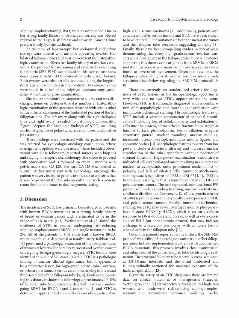
She had an uneventful postoperative course and was dis-charged home on postoperative day number 2. Histopatho-logic examination of the specimen returned with serous tubalintraepithelial carcinoma of the fimbriated portion of the leftfallopian tube. The left ovary along with the right fallopiantube and right ovary revealed no pathologic abnormality.Figure 1 depicts the histologic findings of STIC, includingnuclear atypia, loss of polarity, increasedmitoses, and positivep53 staining.
These findings were discussed with the patient and shewas referred for gynecologic oncology consultation, wheremanagement options were discussed. These included obser-vation with close follow-up, repeated surgery with biopsiesand staging, or empiric chemotherapy. She chose to proceedwith observation and is followed up every 6 months withpelvic exam and CA-125. Her last CA-125 was normal at5 u/mL. At her initial visit with gynecologic oncology, thepatient was very fearful of genetic testing due to concerns thatit was “experimental.” She subsequently met with a geneticcounselor but continues to decline genetic testing.
3. Discussion
The incidence of STIC has primarily been studied in patientswith known BRCA mutations or a strong family historyof breast or ovarian cancer and is estimated to be in therange of 0.6% to 6% [1–3]. Wethington et al. [2] found theincidence of STIC in women undergoing risk-reducingsalpingo-oophorectomy (RRSO) at a single institution to be2%. All of the patients in that study had a known BRCAmutation or high-risk personal or family history. Rabban et al.[4] performed a pathologic evaluation of the fallopian tubesof women at low risk for hereditary breast and ovarian cancerundergoing benign gynecologic surgery. STIC lesions wereidentified in 4 out of 522 cases (0.76%). STIC is a pathologicfinding of unclear clinical significance, but it appears tobe a precursor lesion for high-grade pelvic (tubal, ovarian,or primary peritoneal) serous carcinoma arising in the distalfimbriated end of the fallopian tube [5, 6]. Evidence support-ing this theory includes the following: approximately 10–15%of fallopian tube STIC cases are detected in women under-going RRSO for BRCA 1 and 2 mutations [1] and STIC isdetected in approximately 50–60% of cases of sporadic pelvic
high-grade serous carcinoma [7]. Additionally, patients withconcurrent pelvic serous tumors and STIC have been shownto have identical TP53mutations in both themetastatic tumorand the fallopian tube precursor, suggesting clonality [8].Finally, there have been compelling studies in recent yearsdemonstrating that many high-grade serous “ovarian” can-cers actually originate in the fallopian tube mucosa. Evidencesupporting this theory came originally fromRRSOs in BRCAmutation carriers, where many occult ovarian cancers werefound to have tubal involvement. Given this new data, thefallopian tubes of high-risk women are now more closelyscrutinized (see below regarding the SEE-FIM protocol) [9,10].
There are currently no standardized criteria for diag-nosis of STIC lesions, as the histopathologic spectrum isvery wide and no two STICs appear exactly the same.However, STIC is traditionally diagnosed with a combina-tion of histopathologic and morphologic evaluation withimmunohistochemical staining. Histopathologic features ofSTIC include a variable combination of epithelial stratifi-cation (including loss of cellular polarity and exfoliation ofcells into the lumen), intraepithelial fracture lines, irregularluminal surface, pleomorphism, loss of ciliation, irregularchromatin pattern, nuclear rounding, nuclear molding,increased nuclear-to-cytoplasmic ratio, mitotic figures, andapoptotic bodies [11]. Morphologic features evident from lowpower include architectural disarray and increased nuclearstratification of the tubal epithelium without evidence ofstromal invasion. High-power examination demonstratesindividual cells with enlarged nuclei resulting in an increasednuclear to cytoplasmic ratio, prominent nucleoli, loss ofpolarity, and lack of ciliated cells. Immunohistochemicalstaining usually is positive for TP53 andKi-67 [2, 4]. TP53 is atumor suppressor gene that is typically mutated in STIC andpelvic serous tumors. The overexpressed, nonfunctional P53protein accumulates, leading to strong, nuclear reactivity in aconfluent distribution. Conversely, Ki-67 is a protein markerof cellular proliferation and is typically overexpressed in STICand pelvic serous tumors. Finally, immunohistochemicalstaining for STIC may reveal overexpression of phosphory-lated histone H2AX (𝛾-H2AX), which is an early cellularresponse to DNA double stand breaks, as well as overexpres-sion of Bcl-2 (an antiapoptotic protein) that may indicatea change to a secretory phenotype, with complete loss ofciliated cells in the fallopian tube [12].
Given this patient’s reported family history, the SEE-FIMprotocol was utilized for histologic examination of her fallop-ian tubes. Initially implemented in patients with documentedBRCA mutations, this protocol involves close examinationand submission of the entire fallopian tube for histologic eval-uation.The proximal fallopian tube is serially cross-sectionedat 2.0-3.0mm intervals, and the distal fimbriated endis longitudinally sectioned for maximal exposure of thefimbrial epithelium [13].
Given the rarity of an STIC diagnosis, there are limiteddata on clinical outcomes or management strategies.Wethington et al. [2] retrospectively evaluated 593 high-riskwomen who underwent risk-reducing salpingo-oopho-rectomy and concomitant peritoneal washings. Twelve
Case Reports in Obstetrics and Gynecology 3
(a) (b)
(c) (d)
Figure 1: (a) H&E stain of the distal fimbriated end of the left fallopian tube. At low magnification, the area of STIC demonstrates increasedepithelial thickness and nuclear stratification compared to areas of normal tubal epithelium. (b) Area of STIC with nuclear stratification andmoderate variability in size and shape. Also note the absence of ciliated cells within the lesional area. (c) At highmagnification, nuclear atypiais clearly visible, including hyperchromatism, prominent nucleoli, loss of polarity, and increasedmitotic figures. (d) Strong and diffuse nuclearTP53 staining of STIC.
patients (2%) were diagnosed with STIC, with one patienthaving positive peritoneal cytology. Subsequently, seven ofthose patients had completion surgical staging and none wasfound to have any further malignant pathology. Additionally,those patients were followed up with individualizedcombinations of CA-125 testing, imaging, and pelvic examfor roughly 28 months, and no recurrences were diagnosed.Although this study had a small sample size, the resultssuggest that the yield of surgical staging is low, and short-term clinical outcomes are favorable [2]. There is nocurrent consensus among gynecologic oncologists regardingappropriate management for incidental findings of STIC.Proposed management strategies of STIC include closesurveillance, surgical staging, or empiric adjuvant chemo-therapy. Close surveillance could be performed with annualreview of systems, pelvic exam, CA-125, and/or imaging.As demonstrated by Wethington et al. [2], the value ofsurgical staging may be low, but performing a pelvic washingat the time of RRSO may be beneficial in evaluating forspread of disease. As positive peritoneal washings indicatethe presence of circulating premalignant cells in theperitoneal cavity, it may be reasonable to offer patients withpositive peritoneal cytology empiric chemotherapy [2]. Forwomen with only STIC in the absence of positive washingsor evidence of malignant spread, empiric adjuvantchemotherapy similar to what would be recommended by
National Comprehensive Cancer Center Network (NCCN)guidelines for stage I ovarian or fallopian tube cancer (3–6cycles of paclitaxel/carboplatin chemotherapy) could begiven [14]. However, given the possibility of adverse effects,a risk-benefit ratio should be performed between thephysician and patient prior to initiating chemotherapy. Morestudies are needed assessing effective management strategiesbut will be limited by low numbers.
It is also important to describe prevention strategies forSTIC and invasive pelvic high-grade serous carcinoma. Givenan estimated 80–90% of BRCA-related “ovarian” cancersoriginating in the fallopian tube, ACOG and the NCCNrecommend that all women known to have a BRCA 1 or 2mutation (or other hereditary cancers associatedwith ovariancancer, like Lynch syndrome) undergo prophylactic RRSOby the age of 40 or at completion of childbearing, as thereare extensive limitations to current ovarian cancer screeningstrategies [14, 15]. Of note, approximately 80% of ovariancancer is not hereditary and thus there has been a strongmovement toward the consideration of bilateral salpingec-tomy in all patients undergoing benign gynecologic surgery,whether BRCA status is known or not, in an attempt tofurther reduce ovarian cancer risk for all women. Other riskreducing measures include the use of oral contraceptive pills,which can reduce ovarian cancer risk by 50% if taken for 5years [14].
4 Case Reports in Obstetrics and Gynecology
Particularly challenging in this case was the patient’srefusal to undergo genetic testing in light of her significantfamily history of ovarian cancer and personal STIC history.The knowledge of her BRCA status could affect whichscreening options she is offered for breast cancer surveillanceand could also impact her family members. If she werepositive for a BRCA germline mutation, immediate BRCAtesting could be implemented in her familymembers to eitherconfirm their high-risk status or clear them of risk. If herdaughters were found to have amutation, it would be impera-tive to discuss heightened cancer screening for breast/ovariancancer as well as both surgical and nonsurgical preventionmeasures. For now, she continues to be evaluated every sixmonths with pelvic exam and CA-125 level.
All gynecologic surgeons, including those practicingfemale pelvic medicine and reconstructive surgery, shouldhave an understanding of the risk of fallopian tube and ovar-ian cancers. Surgeons should consider removal of fallopiantubes on all patients undergoing surgery for pelvic organprolapse, as approximately 80% of “ovarian” cancers arisein women without known familial etiology. However, givenfallopian tubes and ovaries that can be technically challengingto remove during vaginal surgery, urogynecologists should atleast consider bilateral salpingectomy or risk-reducing BSOin patients who have a strong family history of breast/ovariancancer or those that are known BRCA mutation carrier. Inpatients undergoing robotic surgery, where it is less tech-nically challenging to remove fallopian tubes, considerationshould be made for bilateral salpingectomy on all patients.
Conflict of Interests
The authors declare that there is no conflict of interestsregarding the publication of this paper.
References
[1] A. Finch, P. Shaw, B. Rosen, J. Murphy, S. A. Narod, and T.J. Colgan, “Clinical and pathologic findings of prophylacticsalpingo-oophorectomies in 159 BRCA1 and BRCA2 carriers,”Gynecologic Oncology, vol. 100, no. 1, pp. 58–64, 2006.
[2] S. L. Wethington, K. J. Park, R. A. Soslow et al., “Clinical out-come of isolated Serous tubal intraepithelial carcinomas(STIC),” International Journal of Gynecological Cancer, vol. 23,no. 9, pp. 1603–1611, 2013.
[3] R.Manchanda, A. Abdelraheim,M. Johnson et al., “Outcome ofrisk-reducing salpingo-oophorectomy in BRCA carriers andwomen of unknown mutation status,” BJOG: An InternationalJournal of Obstetrics and Gynaecology, vol. 118, no. 7, pp. 814–824, 2011.
[4] J. T. Rabban, K. Garg, B. Crawford, L.-M. Chen, and C. J.Zaloudek, “Early detection of high-grade tubal serous carci-noma in women at low risk for hereditary breast and ovariancancer syndrome by systematic examination of fallopian tubesincidentally removed during benign surgery,”American Journalof Surgical Pathology, vol. 38, no. 6, pp. 729–742, 2014.
[5] H. Ishikawa, T. Kiyokawa, E. Utsuno, K.Matsushita, F. Nomura,and M. Shozu, “Serous tubal intraepithelial carcinoma in aJapanese woman with a deleterious BRCA1 mutation,” JapaneseJournal of Clinical Oncology, vol. 44, no. 6, pp. 597–601, 2014.
[6] S. Nasser, R. Arsenic, P. Lohneis, P. Kosian, and J. Sehouli, “Acase of primary peritoneal carcinoma: evidence for a precursorin the fallopian tube,”Anticancer Research, vol. 34, no. 1, pp. 407–412, 2014.
[7] M. J. Callahan, C. P. Crum, F. Medeiros et al., “Primary fal-lopian tubemalignancies in BRCA-positive women undergoingsurgery for ovarian cancer risk reduction,” Journal of ClinicalOncology, vol. 25, no. 25, pp. 3985–3990, 2007.
[8] D.W.Kindelberger, Y. Lee, A.Miron et al., “Intraepithelial carci-noma of the fimbria and pelvic serous carcinoma: evidence for acausal relationship,”TheAmerican Journal of Surgical Pathology,vol. 31, no. 2, pp. 161–169, 2007.
[9] S. C. Modesitt, “What is new in gynecologic oncology?Thought-provoking articles from the past year,” Obstetrics andGynecology, vol. 121, no. 4, pp. 872–873, 2013.
[10] C. P. Crum, F. D. McKeon, and W. Xian, “The oviduct andovarian cancer: causality, clinical implications, and ‘targetedprevention’,” Clinical Obstetrics and Gynecology, vol. 55, no. 1,pp. 24–35, 2012.
[11] R. Vang, I.-M. Shih, and R. J. Kurman, “Fallopian tube pre-cursors of ovarian low- and high-grade serous neoplasms,”Histopathology, vol. 62, no. 1, pp. 44–58, 2013.
[12] G. Chene, A. Cayre, I. Raoelfils, N. Lagarde, J. Dauplat, and F.Penault-Llorca, “Morphological and immunohistochemicalpattern of tubo-ovarian dysplasia and serous tubal intraepithe-lial carcinoma,” European Journal of Obstetrics & Gynecologyand Reproductive Biology, vol. 183, pp. 89–95, 2014.
[13] F. Medeiros, M. G. Muto, Y. Lee et al., “The tubal fimbria is apreferred site for early adenocarcinoma in women with familialovarian cancer syndrome,” The American Journal of SurgicalPathology, vol. 30, no. 2, pp. 230–236, 2006.
[14] National Comprehensive Cancer Network, Genetic/FamilialHigh-Risk Assessment: Breast and Ovarian (Version 1.2014),2014, http://www.nccn.org/professionals/physician gls/pdf/genetics screening.pdf.
[15] American College of Obstetricians and Gynecologists, ACOGCommittee on Practice Bulletins, ACOGCommittee on Genet-ics, and Society of Gynecologic Oncologists, “ACOG PracticeBulletin No. 103: hereditary breast and ovarian cancer syn-drome,” Obstetrics & Gynecology, vol. 113, no. 4, pp. 957–966,2009.

Submit your manuscripts athttp://www.hindawi.com
Stem CellsInternational
Hindawi Publishing Corporationhttp://www.hindawi.com Volume 2014
Hindawi Publishing Corporationhttp://www.hindawi.com Volume 2014
MEDIATORSINFLAMMATION
of
Hindawi Publishing Corporationhttp://www.hindawi.com Volume 2014
Behavioural Neurology
EndocrinologyInternational Journal of
Hindawi Publishing Corporationhttp://www.hindawi.com Volume 2014
Hindawi Publishing Corporationhttp://www.hindawi.com Volume 2014
Disease Markers
Hindawi Publishing Corporationhttp://www.hindawi.com Volume 2014
BioMed Research International
OncologyJournal of
Hindawi Publishing Corporationhttp://www.hindawi.com Volume 2014
Hindawi Publishing Corporationhttp://www.hindawi.com Volume 2014
Oxidative Medicine and Cellular Longevity
Hindawi Publishing Corporationhttp://www.hindawi.com Volume 2014
PPAR Research
The Scientific World JournalHindawi Publishing Corporation http://www.hindawi.com Volume 2014
Immunology ResearchHindawi Publishing Corporationhttp://www.hindawi.com Volume 2014
Journal of
ObesityJournal of
Hindawi Publishing Corporationhttp://www.hindawi.com Volume 2014
Hindawi Publishing Corporationhttp://www.hindawi.com Volume 2014
Computational and Mathematical Methods in Medicine
OphthalmologyJournal of
Hindawi Publishing Corporationhttp://www.hindawi.com Volume 2014
Diabetes ResearchJournal of
Hindawi Publishing Corporationhttp://www.hindawi.com Volume 2014
Hindawi Publishing Corporationhttp://www.hindawi.com Volume 2014
Research and TreatmentAIDS
Hindawi Publishing Corporationhttp://www.hindawi.com Volume 2014
Gastroenterology Research and Practice
Hindawi Publishing Corporationhttp://www.hindawi.com Volume 2014
Parkinson’s Disease
Evidence-Based Complementary and Alternative Medicine
Volume 2014Hindawi Publishing Corporationhttp://www.hindawi.com