case report osteitis fibrosa cystica mistaken for ... · 112 clinical and experimental...
TRANSCRIPT
110
Copyright © 2013 by Korean Society of Otorhinolaryngology-Head and Neck Surgery.This is an open-access article distributed under the terms of the Creative Commons Attribution Non-Commercial License (http://creativecommons.org/licenses/by-nc/3.0) which permits unrestricted non-commercial use, distribution, and reproduction in any medium, provided the original work is properly cited.
Osteitis Fibrosa Cystica Mistaken for Malignant Disease
Ja Hyun Lee1·Sung Min Chung2·Han Su Kim2
1Department of Otorhinolaryngology, National Health Insurance Service Ilsan Hospital, Goyang; 2Department of Otolaryngology and Head & Neck Surgery, Ewha Womans University School of Medicine, Seoul, Korea
Case Report
INTRODUCTION
Primary hyperparathyroidism results from excessive secretion of parathyroid hormone (PTH). It can be caused by a solitary ade-noma in 80% patients, parathyroid hyperplasia in 15%, multi-ple adenomas in 5%, and parathyroid carcinoma in less than 5% of patients [1]. Osteitis fibrosa cystica is a skeletal disease related with long lasting, end-stage hyperparathyroidism and is some-times referred to as a “brown tumor” because of its reddish col-or [2]. However, it is not a true neoplasm but rather a reactive osteolytic lesion of bone and may mimic other diseases such as giant cell tumors, multiple bone metastasis or multiple myeloma [3,4]. Because it is rare, it may not be initially considered in the differential diagnosis of bony tumors. Here we report a patient with hyperparathyroism caused by parathyroid hyperplasia who
had osteitis fibrosa cystica mistaken for a malignancy.
CASE REPORT
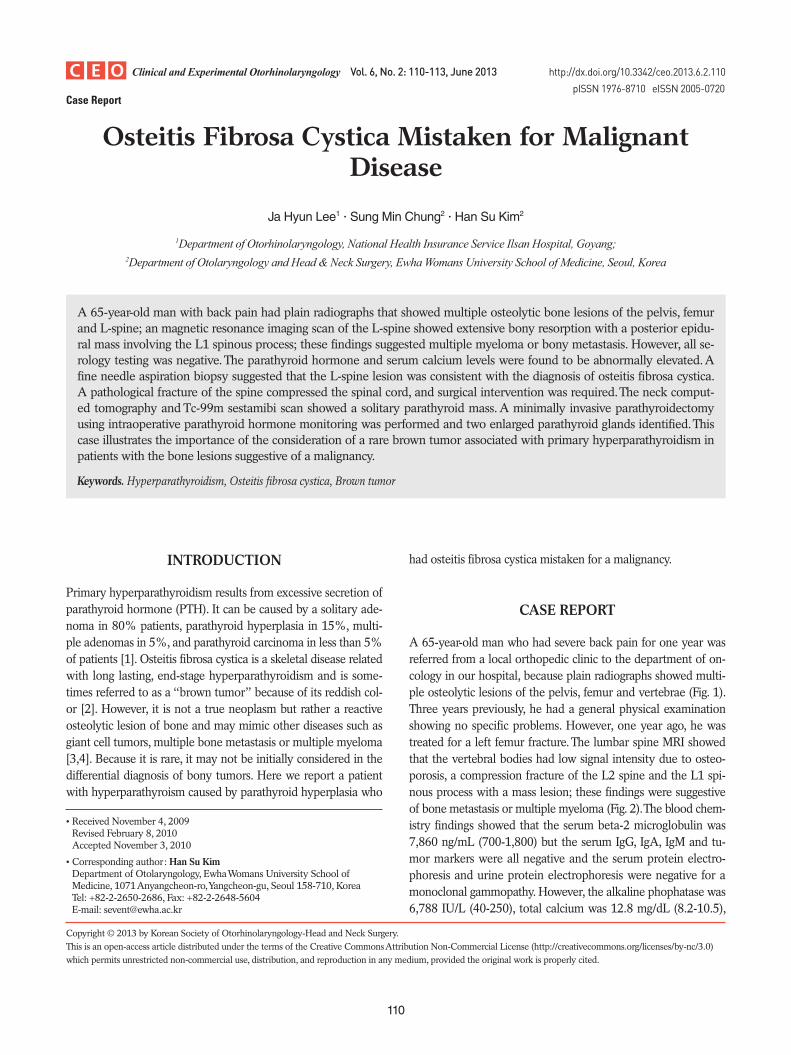
A 65-year-old man who had severe back pain for one year was referred from a local orthopedic clinic to the department of on-cology in our hospital, because plain radiographs showed multi-ple osteolytic lesions of the pelvis, femur and vertebrae (Fig. 1). Three years previously, he had a general physical examination showing no specific problems. However, one year ago, he was treated for a left femur fracture. The lumbar spine MRI showed that the vertebral bodies had low signal intensity due to osteo-porosis, a compression fracture of the L2 spine and the L1 spi-nous process with a mass lesion; these findings were suggestive of bone metastasis or multiple myeloma (Fig. 2). The blood chem-istry findings showed that the serum beta-2 microglobulin was 7,860 ng/mL (700-1,800) but the serum IgG, IgA, IgM and tu-mor markers were all negative and the serum protein electro-phoresis and urine protein electrophoresis were negative for a monoclonal gammopathy. However, the alkaline phophatase was 6,788 IU/L (40-250), total calcium was 12.8 mg/dL (8.2-10.5),
A 65-year-old man with back pain had plain radiographs that showed multiple osteolytic bone lesions of the pelvis, femur and L-spine; an magnetic resonance imaging scan of the L-spine showed extensive bony resorption with a posterior epidu-ral mass involving the L1 spinous process; these findings suggested multiple myeloma or bony metastasis. However, all se-rology testing was negative. The parathyroid hormone and serum calcium levels were found to be abnormally elevated. A fine needle aspiration biopsy suggested that the L-spine lesion was consistent with the diagnosis of osteitis fibrosa cystica. A pathological fracture of the spine compressed the spinal cord, and surgical intervention was required. The neck comput-ed tomography and Tc-99m sestamibi scan showed a solitary parathyroid mass. A minimally invasive parathyroidectomy using intraoperative parathyroid hormone monitoring was performed and two enlarged parathyroid glands identified. This case illustrates the importance of the consideration of a rare brown tumor associated with primary hyperparathyroidism in patients with the bone lesions suggestive of a malignancy.
Keywords. Hyperparathyroidism, Osteitis fibrosa cystica, Brown tumor
• Received November 4, 2009 Revised February 8, 2010 Accepted November 3, 2010
• Corresponding author: Han Su KimDepartment of Otolaryngology, Ewha Womans University School of Medicine, 1071 Anyangcheon-ro, Yangcheon-gu, Seoul 158-710, Korea Tel: +82-2-2650-2686, Fax: +82-2-2648-5604 E-mail: [email protected]
Clinical and Experimental Otorhinolaryngology Vol. 6, No. 2: 110-113, June 2013 http://dx.doi.org/10.3342/ceo.2013.6.2.110pISSN 1976-8710 eISSN 2005-0720
Lee JH et al.: Primary Hyperparathyroidism led to Bony Lesion 111
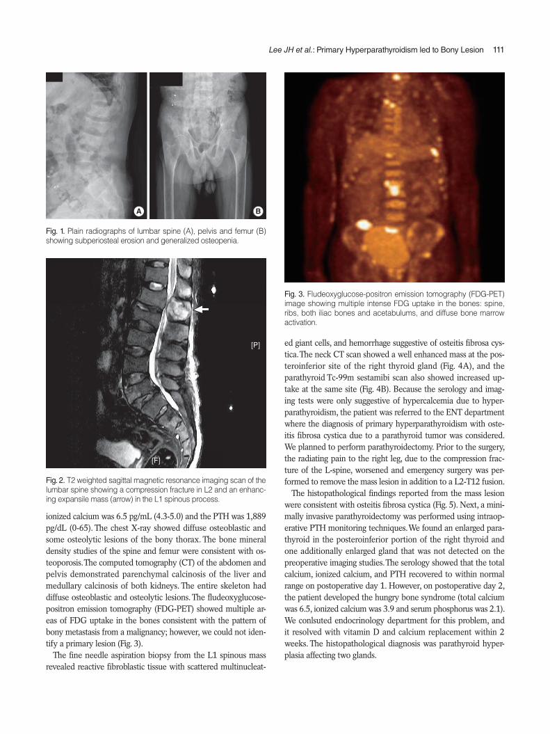
ionized calcium was 6.5 pg/mL (4.3-5.0) and the PTH was 1,889 pg/dL (0-65). The chest X-ray showed diffuse osteoblastic and some osteolytic lesions of the bony thorax. The bone mineral density studies of the spine and femur were consistent with os-teoporosis. The computed tomography (CT) of the abdomen and pelvis demonstrated parenchymal calcinosis of the liver and medullary calcinosis of both kidneys. The entire skeleton had diffuse osteoblastic and osteolytic lesions. The fludeoxyglucose-positron emission tomography (FDG-PET) showed multiple ar-eas of FDG uptake in the bones consistent with the pattern of bony metastasis from a malignancy; however, we could not iden-tify a primary lesion (Fig. 3). The fine needle aspiration biopsy from the L1 spinous mass revealed reactive fibroblastic tissue with scattered multinucleat-
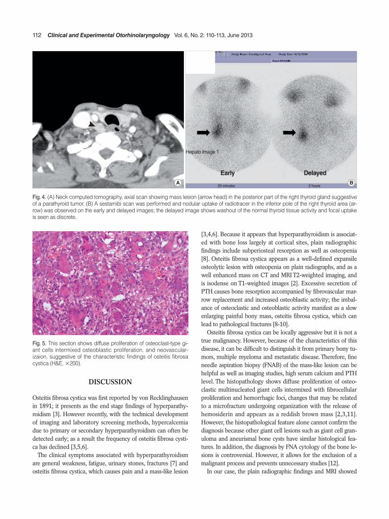
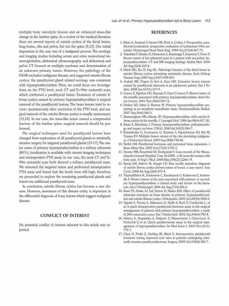
ed giant cells, and hemorrhage suggestive of osteitis fibrosa cys-tica. The neck CT scan showed a well enhanced mass at the pos-teroinferior site of the right thyroid gland (Fig. 4A), and the parathyroid Tc-99m sestamibi scan also showed increased up-take at the same site (Fig. 4B). Because the serology and imag-ing tests were only suggestive of hypercalcemia due to hyper-parathyroidism, the patient was referred to the ENT department where the diagnosis of primary hyperparathyroidism with oste-itis fibrosa cystica due to a parathyroid tumor was considered. We planned to perform parathyroidectomy. Prior to the surgery, the radiating pain to the right leg, due to the compression frac-ture of the L-spine, worsened and emergency surgery was per-formed to remove the mass lesion in addition to a L2-T12 fusion. The histopathological findings reported from the mass lesion were consistent with osteitis fibrosa cystica (Fig. 5). Next, a mini-mally invasive parathyroidectomy was performed using intraop-erative PTH monitoring techniques. We found an enlarged para-thyroid in the posteroinferior portion of the right thyroid and one additionally enlarged gland that was not detected on the preoperative imaging studies. The serology showed that the total calcium, ionized calcium, and PTH recovered to within normal range on postoperative day 1. However, on postoperative day 2, the patient developed the hungry bone syndrome (total calcium was 6.5, ionized calcium was 3.9 and serum phosphorus was 2.1). We conlsuted endocrinology department for this problem, and it resolved with vitamin D and calcium replacement within 2 weeks. The histopathological diagnosis was parathyroid hyper-plasia affecting two glands.
A B
Fig. 1. Plain radiographs of lumbar spine (A), pelvis and femur (B) showing subperiosteal erosion and generalized osteopenia.
Fig. 3. Fludeoxyglucose-positron emission tomography (FDG-PET) image showing multiple intense FDG uptake in the bones: spine, ribs, both iliac bones and acetabulums, and diffuse bone marrow activation.
Fig. 2. T2 weighted sagittal magnetic resonance imaging scan of the lumbar spine showing a compression fracture in L2 and an enhanc-ing expansile mass (arrow) in the L1 spinous process.
[P]
[F]
112 Clinical and Experimental Otorhinolaryngology Vol. 6, No. 2: 110-113, June 2013
DISCUSSION
Osteitis fibrosa cystica was first reported by von Recklinghausen in 1891; it presents as the end stage findings of hyperparathy-roidism [3]. However recently, with the technical development of imaging and laboratory screening methods, hypercalcemia due to primary or secondary hyperparathyroidism can often be detected early; as a result the frequency of osteitis fibrosa cysti-ca has declined [3,5,6]. The clinical symptoms associated with hyperparathyroidism are general weakness, fatigue, urinary stones, fractures [7] and osteitis fibrosa cystica, which causes pain and a mass-like lesion
[3,4,6]. Because it appears that hyperparathyroidism is associat-ed with bone loss largely at cortical sites, plain radiographic findings include subperiosteal resorption as well as osteopenia [8]. Osteitis fibrosa cystica appears as a well-defined expansile osteolytic lesion with osteopenia on plain radiographs, and as a well enhanced mass on CT and MRI T2-weighted imaging, and is isodense on T1-weighted images [2]. Excessive secretion of PTH causes bone resorption accompanied by fibrovascular mar-row replacement and increased osteoblastic activity; the imbal-ance of osteoclastic and osteoblastic activity manifest as a slow enlarging painful bony mass, osteitis fibrosa cystica, which can lead to pathological fractures [8-10]. Osteitis fibrosa cystica can be locally aggressive but it is not a true malignancy. However, because of the characteristics of this disease, it can be difficult to distinguish it from primary bony tu-mors, multiple myeloma and metastatic disease. Therefore, fine needle aspiration biopsy (FNAB) of the mass-like lesion can be helpful as well as imaging studies, high serum calcium and PTH level. The histopathology shows diffuse proliferation of osteo-clastic multinucleated giant cells intermixed with fibrocellular proliferation and hemorrhagic foci, changes that may be related to a microfracture undergoing organization with the release of hemosiderin and appears as a reddish brown mass [2,3,11]. However, the histopathological feature alone cannot confirm the diagnosis because other giant cell lesions such as giant cell gran-uloma and aneurismal bone cysts have similar histological fea-tures. In addition, the diagnosis by FNA cytology of the bone le-sions is controversial. However, it allows for the exclusion of a malignant process and prevents unnecessary studies [12]. In our case, the plain radiographic findings and MRI showed
Fig. 5. This section shows diffuse proliferation of osteoclast-type gi-ant cells intermixed osteoblastic proliferation, and neovascular-izaion, suggestive of the characteristic findings of osteitis fibrosa cystica (H&E, ×200).
Fig. 4. (A) Neck computed tomography, axial scan showing mass lesion (arrow head) in the posterior part of the right thyroid gland suggestive of a parathyroid tumor. (B) A sestamibi scan was performed and nodular uptake of radiotracer in the inferior pole of the right thyroid area (ar-row) was observed on the early and delayed images; the delayed image shows washout of the normal thyroid tissue activity and focal uptake is seen as discrete.
Hepato image 1
Early Delayed
20 minutes 2 hoursA B
Lee JH et al.: Primary Hyperparathyroidism led to Bony Lesion 113
multiple bony osteolytic lesions and an enhanced mass-like change in the lumbar spine. In a review of the medical literature there are several reports of osteitis cystica of the facial bones, long bones, ribs and pelvis, but not the spine [5,13]. Our initial impression in the case was of a malignant process. The serology and imaging studies including serum and urine monoclonal im-munoglobulins, abdominal ultrasonography and abdominal and pelvic CT focused on multiple myeloma and determination of an unknown primary tumor. However, the pathology of the FANB excluded malignant disease, and suggested osteitis fibrosa cystica; the parathyroid gland related serology was consistent with hyperparathyroidism. Then, we could focus our investiga-tions on the PTH level, neck CT and Tc-99m sestamibi scan, which confirmed a parathyroid lesion. Treatment of osteitis fi-brosa cystica caused by primary hyperparathyroidism is surgical removal of the parathyroid lesions. The bone lesions tend to re-cover spontaneously after correction of the PTH level, and sur-gical removal of the osteitis fibrosa cystica is usually unnecessary [13,14]. In our case, the mass-like lesion caused a compression fracture of the lumbar spine, surgical removal should be per-formed. The surgical techniques used for parathyroid lesions have changed from exploration of all parathyroid glands to minimally invasive surgery for targeted parathyroid glands [15-17]. The ma-jor cause of primary hyperparathyroidism is a solitary adenoma (80%); localization is available with current imaging techniques and intraoperative PTH assay. In our case, the neck CT and Tc-99m sestamibi scan both showed a solitary parathyroid mass. We removed the targeted lesion and performed intraoperative PTH assay and found that the levels were still high; therefore, we proceeded to explore the remaining parathyroid glands and found one additional parathyroid mass. In conclusion, osteitis fibrosa cystica has become a rare dis-ease. However, awareness of this disease entity is important in the differential diagnosis of bony lesions which suggest malignant disease.
CONFLICT OF INTEREST
No potential conflict of interest relevant to this article was re-ported.
REFERENCES
1. Khan A, Samtani S, Varma VM, Frost A, Cohen J. Preoperative para-thyroid localization: prospective evaluation of technetium 99m ses-tamibi. Otolaryngol Head Neck Surg. 1994 Oct;111(4):467-72.
2. Takeshita T, Tanaka H, Harasawa A, Kaminaga T, Imamura T, Furui S. Brown tumor of the sphenoid sinus in a patient with secondary hy-perparathyroidism: CT and MR imaging findings. Radiat Med. 2004 Jul-Aug;22(4):265-8.
3. Hsieh MC, Ko JY, Eng HL. Pathologic fracture of the distal femur in osteitis fibrosa cystica simulating metastatic disease. Arch Orthop Trauma Surg. 2004 Sep;124(7):498-501.
4. Atabek ME, Pirgon O, Sert A, Esen HH. Extensive brown tumors caused by parathyroid adenoma in an adolescent patient. Eur J Pe-diatr. 2008 Jan;167(1):117-9.
5. Guney E, Yigitbasi OG, Bayram F, Ozer V, Canoz O. Brown tumor of the maxilla associated with primary hyperparathyroidism. Auris Na-sus Larynx. 2001 Nov;28(4):369-72.
6. Dinkar AD, Sahai S, Sharma M. Primary hyperparathyroidism pre-senting as an exophytic mandibular mass. Dentomaxillofac Radiol. 2007 Sep;36(6):360-3.
7. Shanmugham MS, Alhady SF. Hyperparathyroidism with osteitis fi-brosa cystica in the maxilla. J Laryngol Otol. 1984 Apr;98(4):417-20.
8. Khan A, Bilezikian J. Primary hyperparathyroidism: pathophysiolo-gy and impact on bone. CMAJ. 2000 Jul;163(2):184-7.
9. Kalambokis G, Economou G, Kamina S, Papachristou DJ, Bai M, Tsianos EV. Multiple brown tumors of the ribs simulating malignan-cy. J Endocrinol Invest. 2005 Sep;28(8):738-40.
10. Parfitt AM. Parathyroid hormone and periosteal bone expansion. J Bone Miner Res. 2002 Oct;17(10):1741-3.
11. Demay MB, Rosenthal DI, Deshpande V. Case records of the Massa-chusetts General Hospital: Case 16-2008 - a 46-year-old woman with bone pain. N Engl J Med. 2008 May;358(21):2266-74.
12. Kemp AM, Bukvic M, Sturgis CD. Fine needle aspiration diagnosis of osteitis fibrosa cystica (brown tumor of bone): a case report. Acta Cytol. 2008 Jul-Aug;52(4):471-4.
13. Triantafillidou K, Zouloumis L, Karakinaris G, Kalimeras E, Iordani-dis F. Brown tumors of the jaws associated with primary or second-ary hyperparathyroidism: a clinical study and review of the litera-ture. Am J Otolaryngol. 2006 Jul-Aug;27(4):281-6.
14. Knee TS, Drake AJ 3rd, Turton D, Shakir KM. Effect of parathyroid adenoma resection on bone density in primary hyperparathyroid-ism and osteitis fibrosa cystica. Orthopedics. 2001 Oct;24(10):1000-2.
15. Vignali E, Picone A, Materazzi G, Steffe S, Berti P, Cianferotti L, et al. A quick intraoperative parathyroid hormone assay in the surgical management of patients with primary hyperparathyroidism: a study of 206 consecutive cases. Eur J Endocrinol. 2002 Jun;146(6):783-8.
16. Halevy A, Stepansky A, Halpern Z, Wassermann I, Chen-Levy Z, Pytlovich S, et al. Quick parathormone assay in the surgical man-agement of hyperparathyroidism. Isr Med Assoc J. 2003 Nov;5(11): 775-7.
17. Chen H, Pruhs Z, Starling JR, Mack E. Intraoperative parathyroid hormone testing improves cure rates in patients undergoing mini-mally invasive parathyroidectomy. Surgery. 2005 Oct;138(4):583-7.