case report mucoepidermoid carcinoma of the palatine...
TRANSCRIPT
Case ReportMucoepidermoid Carcinoma of the Palatine Tonsil
Lucas Novaes Teixeira, Victor Angelo Martins Montalli, Luiz Carlos Santana Teixeira,Fabrício Passador-Santos, Andresa Borges Soares, and Vera Cavalcanti de Araújo
Department of Oral Pathology, Sao Leopoldo Mandic Institute and Research Center, Rua Jose Rocha Junqueira 13, Ponte Preta,13045-755 Campinas, SP, Brazil
Correspondence should be addressed to Lucas Novaes Teixeira; [email protected]
Received 4 May 2015; Accepted 30 September 2015
Academic Editor: Ossama W. Tawfik
Copyright © 2015 Lucas Novaes Teixeira et al. This is an open access article distributed under the Creative Commons AttributionLicense, which permits unrestricted use, distribution, and reproduction in any medium, provided the original work is properlycited.
Mucoepidermoid carcinoma (MEC) is the most common primary salivary gland malignancy in both adults and children. It has aslight female predilection and usually presents as a painless, rubber-like or soft mass, which may be fixed or mobile. Histologically,MEC is comprised of a mixture of cell types including mucous, epidermoid, and intermediate cells that can be arranged in solidnests or cystic structures. In the oral cavity, it most frequently occurs at the palate or buccal mucosa. The present paper aimed todescribe an unusual case of MEC arising in the palatine tonsil.
1. Introduction
Many different malignant neoplasms may arise from thepalatine tonsils, with the most common histological typebeing the squamous cell carcinoma (SCC), which accountsfor up to 85% of all cases [1–3]. Malignant lymphoprolifer-ative diseases are the second most frequent malignancy ofthe palatine tonsil, with the diffuse large B-cell lymphoma(DLBCL) comprising approximately 30% of all lymphomas[4]. Metastatic deposits of lung [5] and gastric carcinomas[6], as well as melanoma [7], renal carcinoma [8], andadenocarcinoma of the colon [9], have also been describedin the palatine tonsils.
Minor salivary gland tumors exhibit diverse histopatho-logical features as well as a varied clinical behavior [10].They may be derived from any of the minor salivary glandsdistributed throughout the oral cavity [11]. Interestingly,despite the presence of minor salivary glands in the palatinetonsils, the development of malignant salivary tumors here isunusual. Indeed, a scarce number of case reports have beendocumented in the scientific literature [12–14]. The presentpaper, therefore, aimed to report a case of MEC arising in thepalatine tonsil.
2. Case History
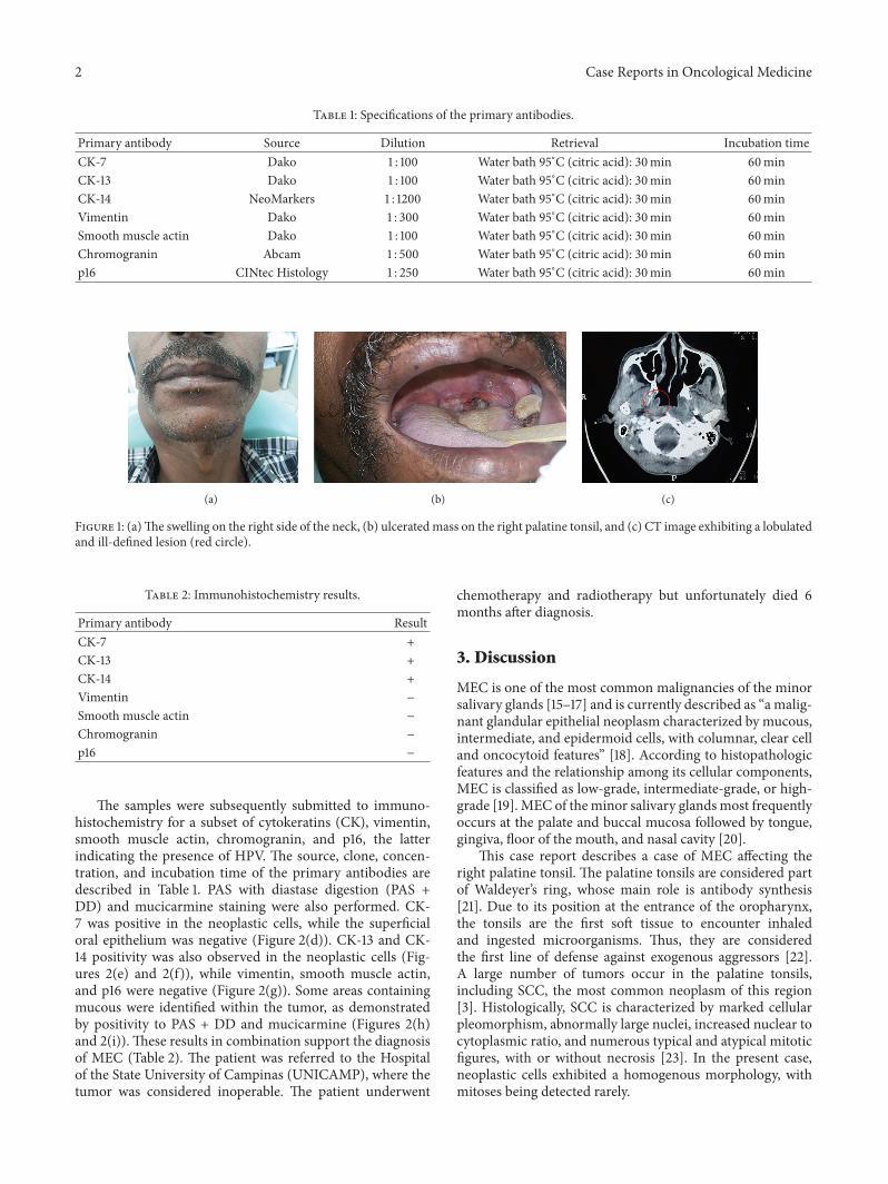
A 47-year-old man reported experiencing dysphagia and asore throat for 4 months. The patient was both a heavysmoker (40 cigarettes daily) and an alcoholic. His medicalhistory was significant for Type 2 Diabetes Mellitus. Thepatient presented with a swelling on the right side of the neck(Figure 1(a)). Intraoral examination revealed an ulceratedmass on the right palatine tonsil (Figure 1(b)). Axial com-puted tomography (CT) revealed a solid lesionwith lobulatedand ill-defined margins (Figure 1(c)). An incisional biopsywas performed and the specimen was fixed in 10% bufferedformalin.
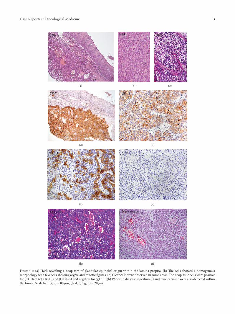
Paraffin sections were prepared for light microscopyusing routine procedures. The sections were stained withhematoxylin and eosin (H&E). Microscopic examinationrevealed a fragment of oral mucosa covered with a nonker-atinized stratified squamous epithelium. A neoplasm ofglandular epithelial origin was identified in the lamina pro-pria (Figure 2(a)). The tumor cells were arranged in sheets,exhibiting a uniform morphology with few cells showingatypia and mitotic figures (Figure 2(b)). Clear cells wereobserved in some areas of the tumor (Figure 2(c)).
Hindawi Publishing CorporationCase Reports in Oncological MedicineVolume 2015, Article ID 827560, 6 pageshttp://dx.doi.org/10.1155/2015/827560
2 Case Reports in Oncological Medicine
Table 1: Specifications of the primary antibodies.
Primary antibody Source Dilution Retrieval Incubation timeCK-7 Dako 1 : 100 Water bath 95∘C (citric acid): 30min 60minCK-13 Dako 1 : 100 Water bath 95∘C (citric acid): 30min 60minCK-14 NeoMarkers 1 : 1200 Water bath 95∘C (citric acid): 30min 60minVimentin Dako 1 : 300 Water bath 95∘C (citric acid): 30min 60minSmooth muscle actin Dako 1 : 100 Water bath 95∘C (citric acid): 30min 60minChromogranin Abcam 1 : 500 Water bath 95∘C (citric acid): 30min 60minp16 CINtec Histology 1 : 250 Water bath 95∘C (citric acid): 30min 60min
(a) (b) (c)
Figure 1: (a)The swelling on the right side of the neck, (b) ulceratedmass on the right palatine tonsil, and (c) CT image exhibiting a lobulatedand ill-defined lesion (red circle).
Table 2: Immunohistochemistry results.
Primary antibody ResultCK-7 +CK-13 +CK-14 +Vimentin −
Smooth muscle actin −
Chromogranin −
p16 −
The samples were subsequently submitted to immuno-histochemistry for a subset of cytokeratins (CK), vimentin,smooth muscle actin, chromogranin, and p16, the latterindicating the presence of HPV. The source, clone, concen-tration, and incubation time of the primary antibodies aredescribed in Table 1. PAS with diastase digestion (PAS +DD) and mucicarmine staining were also performed. CK-7 was positive in the neoplastic cells, while the superficialoral epithelium was negative (Figure 2(d)). CK-13 and CK-14 positivity was also observed in the neoplastic cells (Fig-ures 2(e) and 2(f)), while vimentin, smooth muscle actin,and p16 were negative (Figure 2(g)). Some areas containingmucous were identified within the tumor, as demonstratedby positivity to PAS + DD and mucicarmine (Figures 2(h)and 2(i)). These results in combination support the diagnosisof MEC (Table 2). The patient was referred to the Hospitalof the State University of Campinas (UNICAMP), where thetumor was considered inoperable. The patient underwent
chemotherapy and radiotherapy but unfortunately died 6months after diagnosis.
3. Discussion
MEC is one of the most common malignancies of the minorsalivary glands [15–17] and is currently described as “a malig-nant glandular epithelial neoplasm characterized by mucous,intermediate, and epidermoid cells, with columnar, clear celland oncocytoid features” [18]. According to histopathologicfeatures and the relationship among its cellular components,MEC is classified as low-grade, intermediate-grade, or high-grade [19]. MEC of theminor salivary glandsmost frequentlyoccurs at the palate and buccal mucosa followed by tongue,gingiva, floor of the mouth, and nasal cavity [20].
This case report describes a case of MEC affecting theright palatine tonsil. The palatine tonsils are considered partof Waldeyer’s ring, whose main role is antibody synthesis[21]. Due to its position at the entrance of the oropharynx,the tonsils are the first soft tissue to encounter inhaledand ingested microorganisms. Thus, they are consideredthe first line of defense against exogenous aggressors [22].A large number of tumors occur in the palatine tonsils,including SCC, the most common neoplasm of this region[3]. Histologically, SCC is characterized by marked cellularpleomorphism, abnormally large nuclei, increased nuclear tocytoplasmic ratio, and numerous typical and atypical mitoticfigures, with or without necrosis [23]. In the present case,neoplastic cells exhibited a homogenous morphology, withmitoses being detected rarely.
Case Reports in Oncological Medicine 3
H&E
(a)
H&E
(b)
H&E
(c)
CK-7
(d)
CK-13
(e)
CK-14
(f)
p16
(g)
PAS + DD
(h)
Mucicarmine
(i)
Figure 2: (a) H&E revealing a neoplasm of glandular epithelial origin within the lamina propria. (b) The cells showed a homogenousmorphology with few cells showing atypia and mitotic figures. (c) Clear cells were observed in some areas. The neoplastic cells were positivefor (d) CK-7, (e) CK-13, and (f) CK-14 and negative for (g) p16. (h) PAS with diastase digestion (i) andmucicarmine were also detected withinthe tumor. Scale bar: (a, c) = 80 𝜇m; (b, d, e, f, g, h) = 20 𝜇m.
4 Case Reports in Oncological Medicine
The primary risk factors traditionally associated withthe development of oral SCC development are smokingand alcohol consumption [24–26]. However, cases of SCCarising from the tonsil area may also be associated withHPV infection [27]. Indeed, approximately 51% of thesecarcinomas contain HPV DNA [28], with the most preva-lent HPV subtype identified being HPV-16, detected in84% HPV DNA positive tumors [28]. The chance of HPVinfection associated with the development of the carcinomadescribed in the present case report should be discarded,since immunostaining for p16, which is indicative of thepresence of HPV, was negative.
Adenocarcinomas account for less than 1% of all malig-nancies of the palatine tonsils [3], with only a few studieshaving described the presence of such tumors at this site.The most frequent histological subtypes are represented byadenoid cystic carcinoma [13, 14] and polymorphous low-grade adenocarcinoma [12]. Regarding MEC, only two casesarising in the palatine tonsils have been described in theliterature [29, 30].
Approximately 70% of all cases of MEC are comprisedby low-grade and intermediate-grade tumors [31, 32]. Thus,routine diagnosis of MEC is generally based on conventionalH&E staining looking to identify the presence of cysticstructures andmucous cells [20]. High-gradeMEC, however,can resemble SCC, as observed in the present case report.In this context, immunohistochemical analysis represents auseful diagnostic tool for salivary gland tumors [33]. Theuse of this technique to improve understanding of salivarygland tumors was initiated in the 1980s with studies ofintermediate keratin filaments, vimentin, and desmin [34–39]. Keratins are a group of intermediate filaments restrictedto the epithelium, which are preserved in both malignanttransformation and metastasis [40, 41]. During the devel-opment of normal human minor salivary glands, one mayobserve the expression of different CK subtypes [42]. CK-7 was the first CK identified in salivary glands, with itsexpression remaining in adult salivary glands [38, 42, 43]. Inthe present case report, immunohistochemistry revealed thepresence of large areas showing positivity toCK-7, confirmingits glandular origin. Interestingly, Regauer et al. detected asubset of CK-7 positive carcinomas in the Waldeyer’s ringarea [44]. They suggested that these neoplasms are betterclassified as basaloid SCC. Although CK-7 was reported inthis case report, the morphologic characteristics of basaloidSCC were not encountered, namely, lobules of basaloid cellswith central areas of comedonecrosis [45].
Immunohistochemistry for the present case indicatedthat, besides CK-7, the neoplastic cells were also positive forCK-13 and CK-14, as described previously [33, 46]. Someauthors have suggested that positivity for CK-14 may be adistinctive marker in the diagnosis of oral SCC [47, 48].However, one should note that this CK can also be detectedin MEC [33, 46]. Indeed, the basal cells of the epithelium ofthe oral mucosa, as well as the basal cells of the excretoryduct in normal salivary tissues, are positive for CK-14 [47–49]. Thus, positive immunostaining for CK-14 is most likelyrelated to the excretory duct origin of MEC [50]. Moreover,the neoplastic cells exhibited CK-7 positivity, which excludes
the chance of SCC.The expression of CK-13 may be detectedinMEC, since luminal cells of excretory ducts are positive forthis CK,whichmay also be used as amarker in the differentialdiagnosis of other salivary gland tumors [33]. In additionto the CKs, staining with PAS + DD and mucicarmine canalso be useful markers to discriminate SCC from MEC [51].Positive areas of PAS andmucicarmine detected in some areasof the tumor corroborated the diagnosis ofMEC described inthe present case report.
The standard approach for the treatment of MEC istotal excision of the tumor [52]. However, minor salivarygland carcinomas of the oropharynx are often challengingto manage [53], with radical tonsillectomy and ipsilateralneck dissection having been used as a treatment option[30, 54]. In the present case, due to the advanced stageof the tumor at presentation, the treatment of choice waschemo- and radiotherapy. Some studies have advised the useof radiotherapy for both high-grade MEC and patients withunclear surgical margins [55–57]. On the other hand, thedata regarding chemotherapy in MEC is scarce, with thistreatment generally being adopted in palliative managementof inoperable tumors [58]. Despite the use of these twomodalities in the treatment of the present case, the patientunfortunately died.
In conclusion, this case report highlights the possibility ofsalivary gland tumor affecting the oropharynx. Thus, duringthe differential diagnosis of a tumor mass at this specificsite, whether asymptomatic or not, salivary gland tumorsshould not be ignored, since early diagnosis and appropriatemanagement are a determining factor in the prognosis of thepatient.
Conflict of Interests
The authors declare no conflict of interests regarding thepublication of this paper.
Acknowledgments
The authors would like to thank Jeruza Pinheiro da SilveiraBossonaro and Nadir Freitas for their helpful technicalassistance.
References
[1] M. E. Guay and P. Lavertu, “Tonsillar carcinoma,” EuropeanArchives of Oto-Rhino-Laryngology, vol. 252, no. 5, pp. 259–264,1995.
[2] G. S. Mizono, R. F. Diaz, K. K. Fu, and R. Boles, “Carcinoma ofthe tonsillar region,” Laryngoscope, vol. 96, no. 3, pp. 240–244,1986.
[3] S. M. Golas, “Trends in palatine tonsillar cancer incidence andmortality rates in the United States,” Community Dentistry andOral Epidemiology, vol. 35, no. 2, pp. 98–108, 2007.
[4] A. Lopez-Guillermo, L. Colomo, M. Jimenez et al., “Diffuselarge B-cell lymphoma: clinical and biological characterizationand outcome according to the nodal or extranodal primaryoriginc,” Journal of Clinical Oncology, vol. 23, no. 12, pp. 2797–2804, 2005.
Case Reports in Oncological Medicine 5
[5] W. Hong, X. Wang, X.-M. Yu, B. Chen, G.-J. Ding, and Y.-P. Zhang, “Palatine tonsillar metastasis of lung cancer duringchemotherapy,” International Journal of Clinical and Experimen-tal Pathology, vol. 5, no. 5, pp. 468–471, 2012.
[6] E. Yamaguchi,M.Uchida, Y.Makino et al., “Tonsillarmetastasisof gastric cancer,”Clinical Journal of Gastroenterology, vol. 3, no.6, pp. 289–295, 2010.
[7] R. Cecchi, M. Pavesi, P. Calamandrei, V. Rapicano, and C.De Gaudio, “Tonsil metastasis from cutaneous melanoma:first clinical sign of recurrence after complete lymph nodedissection,” Journal of Cutaneous Medicine and Surgery, vol. 14,no. 1, pp. 43–45, 2010.
[8] M. Massaccesi, A. G. Morganti, G. Serafini et al., “Late tonsilmetastases from renal cell cancer: a case report,” Tumori, vol.95, no. 4, pp. 521–524, 2009.
[9] L.-M. Sheng, L.-Z. Zhang, H.-M. Xu, and Y. Zhu, “Ascendingcolon adenocarcinoma with tonsillar metastasis: a case reportand review of the literature,”World Journal of Gastroenterology,vol. 14, no. 46, pp. 7138–7140, 2008.
[10] A. Dalgic, O. Karakoc, U. Aydin et al., “Minor salivary glandneoplasms,” Journal of Craniofacial Surgery, vol. 25, no. 3, pp.e289–e291, 2014.
[11] K. Balogh and L. Pantanowitz, “Mouth, nose, and paranasalsinuses,” in Histology for Pathologists, S. E. Mills, Ed., pp. 403–430, Lippincott Williams & Wilkins, Philadelphia, Pa, USA,2007.
[12] C. B. Pittman and R. P. Zitsch III, “Polymorphous low-gradeadenocarcinoma of the tonsil: report of a case and review of theliterature,” American Journal of Otolaryngology: Head and NeckMedicine and Surgery, vol. 23, no. 5, pp. 297–299, 2002.
[13] N. Azarpira, M. J. Ashraf, and M. Shishegar, “Fine-needleaspiration biopsy of adenoid cystic carcinoma of the palatinetonsil,” Indian Journal of Pathology andMicrobiology, vol. 54, no.2, pp. 424–425, 2011.
[14] E. Azizli,M. Akpinar, F. Gunver, andO. Yigit, “Bilateral tonsillaradenoid cystic carcinoma,” Journal of Craniofacial Surgery, vol.22, no. 6, pp. 2408–2409, 2011.
[15] F. A. de Oliveira, E. C. B. Duarte, C. T. Taveira et al., “Salivarygland tumor: a review of 599 cases in a Brazilian population,”Head and Neck Pathology, vol. 3, no. 4, pp. 271–275, 2009.
[16] X.-D.Wang, L.-J. Meng, T.-T. Hou, and S.-H. Huang, “Tumoursof the salivary glands in northeastern China: a retrospectivestudy of 2508 patients,” British Journal of Oral andMaxillofacialSurgery, vol. 53, no. 2, pp. 132–137, 2015.
[17] R. H. Spiro, “Management of malignant tumors of the salivaryglands,” Oncology, vol. 12, no. 5, pp. 671–680, 1998.
[18] R. K. Goode and A. K. El-Naggar, “Mucoepidermoid carci-noma,” inWorld Health Organization Classification of Tumours.Pathology and Genetics of Head and Neck Tumours, L. Barnes,J. W. Eveson, P. Reichart, and D. Sidransky, Eds., pp. 219–220,IARC Press, Lyon, France, 2005.
[19] A. Coca-Pelaz, J. P. Rodrigo, A. Triantafyllou et al., “Salivarymucoepidermoid carcinoma revisited,” European Archives ofOto-Rhino-Laryngology, vol. 272, no. 4, pp. 799–819, 2015.
[20] M. A. Luna, “Salivary mucoepidermoid carcinoma: revisited,”Advances in Anatomic Pathology, vol. 13, no. 6, pp. 293–307,2006.
[21] S. Masieri, D. Trabattoni, C. Incorvaia et al., “A role forWaldeyer’s ring in immunological response to allergens,” Cur-rent Medical Research and Opinion, vol. 30, no. 2, pp. 203–205,2014.
[22] P. Hellings, M. Jorissen, and J. L. Ceuppens, “The Waldeyer’sring,” Acta Oto-Rhino-Laryngologica Belgica, vol. 54, no. 3, pp.237–241, 2000.
[23] A. Cardesa, N. Gale, A. Nadal, and N. Zidal, “Squamouscell carcinoma,” in World Health Organization Classification ofTumours. Pathology and Genetics of Head and Neck Tumours,L. Barnes, J. W. Eveson, P. Reichart, and D. Sidransky, Eds., pp.118–121, IARC Press, Lyon, France, 2005.
[24] C. Scully and J. V. Bagan, “Oral squamous cell carcinoma:overview of current understanding of aetiopathogenesis andclinical implications,” Oral Diseases, vol. 15, no. 6, pp. 388–399,2009.
[25] A. G. Zygogianni, G. Kyrgias, P. Karakitsos et al., “Oral squa-mous cell cancer: early detection and the role of alcohol andsmoking,” Head & Neck Oncology, vol. 6, no. 3, pp. 1–12, 2011.
[26] A. H. Madani, M. Dikshit, D. Bhaduri, T. Aghamolaei, S. H.Moosavy, and A. Azarpaykan, “Interaction of alcohol use andspecific types of smoking on the development of oral cancer,”International Journal of High Risk Behaviors and Addiction, vol.3, no. 1, pp. 1–4, 2014.
[27] T. Ramqvist, N. Grun, and T. Dalianis, “Human papillomavirusand tonsillar and base of tongue cancer,” Viruses, vol. 7, no. 3,pp. 1332–1343, 2015.
[28] S. Syrjanen, “HPV infections and tonsillar carcinoma,” Journalof Clinical Pathology, vol. 57, no. 5, pp. 449–455, 2004.
[29] L. Jing-Xian, “Clinico-pathologic studies on 143 cases oftonsillar malignancies with special reference to lymphomas,”Zhonghua Zhong Liu Za Zhi, vol. 14, no. 6, pp. 433–436, 1992.
[30] S. J. Jarvis, V. Giangrande, and P. A. Brennan, “Mucoepidermoidcarcinoma of the tonsil: a very rare presentation,” Acta Otorhi-nolaryngologica Italica, vol. 33, no. 4, pp. 286–288, 2013.
[31] S. A. Miguens Jr., A. C. Uchoa Vasconcelos, M. A. Figueiredo,L. Soares Yurgel, F. Salum, andK. Cherubini, “Mucoepidermoidcarcinoma: a retrospective study,” Minerva Stomatologica, vol.59, no. 6, pp. 325–332, 2010.
[32] K. Yamazaki, H. Ohta, R. Shodo, H. Matsuyama, and S.Takahashi, “Clinicopathological features of mucoepidermoidcarcinoma,” The Journal of Laryngology and Otology, vol. 128,no. 1, pp. 91–95, 2014.
[33] V. C. de Araujo, S. O. M. de Sousa, Y. R. Carvalho, and N. S. deAraujo, “Application of immunohistochemistry to the diagnosisof salivary gland tumors,” Applied Immunohistochemistry &Molecular Morphology, vol. 8, no. 3, pp. 195–202, 2000.
[34] J. Caselitz and T. Loning, “Specific demonstration of actinand keratin filaments in pleomorphic adenomas by means ofimmunoelectron microscopy,” Virchows Archiv A PathologicalAnatomy and Histology, vol. 393, no. 2, pp. 153–158, 1981.
[35] J. Caselitz, M. Osborn, G. Seifert, and K. Weber, “Intermediate-sized filament proteins (prekeratin, vimentin, desmin) in thenormal parotid gland and parotid gland tumours,” VirchowsArchiv A, vol. 393, no. 3, pp. 273–286, 1981.
[36] J. Caselitz, M. Osborn, J. Wustrow, G. Seifert, and K. Weber,“The expression of different intermediate-sized filaments inhuman salivary glands and their tumours,” Pathology Researchand Practice, vol. 175, no. 2-3, pp. 266–278, 1982.
[37] R. M. Palmer, “The identification of myoepithelial cells inhuman salivary glands. A review and comparison of lightmicroscopical methods,” Journal of Oral Pathology & Medicine,vol. 15, no. 4, pp. 221–229, 1986.
[38] H. Gustafsson, U. Kjorell, A. Eriksson, I. Virtanen, and L.-E. Thornell, “Distribution of intermediate filament proteins in
6 Case Reports in Oncological Medicine
developing and adult salivary glands in man,” Anatomy andEmbryology, vol. 178, no. 3, pp. 243–257, 1988.
[39] H. Gustafsson, I. Virtanen, and L.-E. Thornell, “Glial fibrillaryacidic protein and desmin in salivary neoplasms,” VirchowsArchiv B, vol. 57, no. 1, pp. 303–313, 1989.
[40] P. Chu, E. Wu, and L. M. Weiss, “Cytokeratin 7 and cytokeratin20 expression in epithelial neoplasms: a survey of 435 cases,”Modern Pathology, vol. 13, no. 9, pp. 962–972, 2000.
[41] G. A. Stopyra, M. J. Warhol, and H. A. B. Multhaupt, “Cytok-eratin 20 immunoreactivity in renal oncocytomas,” Journal ofHistochemistry&Cytochemistry, vol. 49, no. 7, pp. 919–920, 2001.
[42] M. D. Martins, V. Cavalcanti de Araujo, R. Raitz, and N. Soaresde Araujo, “Expression of cytoskeletal proteins in developinghuman minor salivary glands,” European Journal of Oral Sci-ences, vol. 110, no. 4, pp. 316–321, 2002.
[43] P. G. Chu and L.M.Weiss, “Keratin expression in human tissuesand neoplasms,” Histopathology, vol. 40, no. 5, pp. 403–439,2002.
[44] S. Regauer, A. Beham, and S. Mannweiler, “CK7 expression incarcinomas of the Waldeyer’s ring area,” Human Pathology, vol.31, no. 9, pp. 1096–1101, 2000.
[45] A. Cardesa, N. Zidar, and C. Ereno, “Basaloid squamous cellcarcinoma,” in World Health Organization Classification ofTumours. Pathology and Genetics of Head and Neck Tumours,L. Barnes, J. W. Eveson, P. Reichart, and D. Sidransky, Eds., pp.124–125, IARC Press, Lyon, France, 2005.
[46] E. J. D. da Silveira, S. S. L. Veras Barros, R. F. B. de Amorim, L.M. G. Queiroz, R. D. A. Freitas, and L. B. de Souza, “Cytokeratinprofile in mucoepidermoid carcinoma is not related to its his-tological grading of malignancy,” Experimental and MolecularPathology, vol. 81, no. 1, pp. 72–76, 2006.
[47] V. C. de Araujo and S. O. M. de Sousa, “Expression of differentkeratins in salivary gland tumours,” European Journal of CancerPart B: Oral Oncology, vol. 32, no. 1, pp. 14–18, 1996.
[48] A. P. V. Sobral, S. V. L. Loducca, L. P. Kowalski et al., “Immuno-histochemical distinction of high-grade mucoepidermoid car-cinoma and epidermoid carcinoma of the parotid region,” OralOncology, vol. 38, no. 5, pp. 437–440, 2002.
[49] R. B. Presland and B. A. Dale, “Epithelial structural proteins ofthe skin and oral cavity: function in health and disease,”CriticalReviews in Oral Biology & Medicine, vol. 11, no. 4, pp. 383–408,2000.
[50] R. S. Azevedo, O. P. de Almeida, L. P. Kowalski, and F. R. Pires,“Comparative cytokeratin expression in the different cell typesof salivary gland mucoepidermoid carcinoma,” Head and NeckPathology, vol. 2, no. 4, pp. 257–264, 2008.
[51] G. L. Ellis and P. L. Auclair, Atlas of Tumor Pathology: Tumorsof the Salivary Glands, Armed Forces Institute of Pathology,Washington, DC, USA, 1996.
[52] H. Ozawa, T. Tomita, K. Sakamoto et al., “Mucoepidermoidcarcinoma of the head and neck: clinical analysis of 43 patients,”Japanese Journal of Clinical Oncology, vol. 38, no. 6, pp. 414–418,2008.
[53] N. G. Iyer, L. Kim, I. J. Nixon et al., “Factors predicting outcomein malignant minor salivary gland tumors of the oropharynx,”Archives of Otolaryngology - Head andNeck Surgery, vol. 136, no.12, pp. 1240–1247, 2010.
[54] V. Vander Poorten, J. Hunt, P. J. Bradley et al., “Recent trends inthe management of minor salivary gland carcinoma,”Head andNeck, vol. 36, no. 3, pp. 444–455, 2014.
[55] Y. Hosokawa, H. Shirato, K. Kagei et al., “Role of radiotherapyfor mucoepidermoid carcinoma of salivary gland,” Oral Oncol-ogy, vol. 35, no. 1, pp. 105–111, 1999.
[56] M. S. Brandwein, K. Ivanov, D. I. Wallace et al., “Mucoepider-moid carcinoma: a clinicopathologic study of 80 patients withspecial reference to histological grading,”The American Journalof Surgical Pathology, vol. 25, no. 7, pp. 835–845, 2001.
[57] K. Triantafillidou, J. Dimitrakopoulos, F. Iordanidis, and D.Koufogiannis, “Mucoepidermoid carcinoma of minor salivaryglands: a clinical study of 16 cases and review of the literature,”Oral Diseases, vol. 12, no. 4, pp. 364–370, 2006.
[58] S. A. Laurie and L. Licitra, “Systemic therapy in the palliativemanagement of advanced salivary gland cancers,” Journal ofClinical Oncology, vol. 24, no. 17, pp. 2673–2678, 2006.

Submit your manuscripts athttp://www.hindawi.com
Stem CellsInternational
Hindawi Publishing Corporationhttp://www.hindawi.com Volume 2014
Hindawi Publishing Corporationhttp://www.hindawi.com Volume 2014
MEDIATORSINFLAMMATION
of
Hindawi Publishing Corporationhttp://www.hindawi.com Volume 2014
Behavioural Neurology
EndocrinologyInternational Journal of
Hindawi Publishing Corporationhttp://www.hindawi.com Volume 2014
Hindawi Publishing Corporationhttp://www.hindawi.com Volume 2014
Disease Markers
Hindawi Publishing Corporationhttp://www.hindawi.com Volume 2014
BioMed Research International
OncologyJournal of
Hindawi Publishing Corporationhttp://www.hindawi.com Volume 2014
Hindawi Publishing Corporationhttp://www.hindawi.com Volume 2014
Oxidative Medicine and Cellular Longevity
Hindawi Publishing Corporationhttp://www.hindawi.com Volume 2014
PPAR Research
The Scientific World JournalHindawi Publishing Corporation http://www.hindawi.com Volume 2014
Immunology ResearchHindawi Publishing Corporationhttp://www.hindawi.com Volume 2014
Journal of
ObesityJournal of
Hindawi Publishing Corporationhttp://www.hindawi.com Volume 2014
Hindawi Publishing Corporationhttp://www.hindawi.com Volume 2014
Computational and Mathematical Methods in Medicine
OphthalmologyJournal of
Hindawi Publishing Corporationhttp://www.hindawi.com Volume 2014
Diabetes ResearchJournal of
Hindawi Publishing Corporationhttp://www.hindawi.com Volume 2014
Hindawi Publishing Corporationhttp://www.hindawi.com Volume 2014
Research and TreatmentAIDS
Hindawi Publishing Corporationhttp://www.hindawi.com Volume 2014
Gastroenterology Research and Practice
Hindawi Publishing Corporationhttp://www.hindawi.com Volume 2014
Parkinson’s Disease
Evidence-Based Complementary and Alternative Medicine
Volume 2014Hindawi Publishing Corporationhttp://www.hindawi.com