case report - hindawi publishing corporationdownloads.hindawi.com/journals/criu/2011/514373.pdf ·...
TRANSCRIPT
Hindawi Publishing CorporationCase Reports in UrologyVolume 2011, Article ID 514373, 5 pagesdoi:10.1155/2011/514373
Case Report
The Operative Challenges of Advanced Renal Cell Carcinoma withVena Cava Involvement: A Report of Three Cases
Muftau Jimoh Bioku, Abdulwaid Niran Saliu, Stephen Odunayo Ikuerowo,Olufunmilade Omisanjo, and Julius Olusanmi Esho
Urology Unit, Department of Surgery, Lagos State University Teaching Hospital, Ikeja, Lagos, Nigeria
Correspondence should be addressed to Muftau Jimoh Bioku, [email protected]
Received 21 August 2011; Accepted 15 September 2011
Academic Editors: T. Cai and H. Zakhour
Copyright © 2011 Muftau Jimoh Bioku et al. This is an open access article distributed under the Creative Commons AttributionLicense, which permits unrestricted use, distribution, and reproduction in any medium, provided the original work is properlycited.
Surgical resection remains an important component in the care of advanced renal cell carcinoma (RCC). Some of the patientsso managed had relief of symptoms and improved quality of life. However, palliative nephrectomies in late cases with venacava involvement are not without challenges. An important factor to be considered for successful surgery is adequate vena cavamanagement. We report in this paper three patients who had metastatic RCC. For over three decades now, researchers in Lagos hadrecorded the abysmal prognosis of advanced cases of RCC. Yet, late presentation and diagnosis still persisted in our environment.There is therefore the need to repackage our strategies aimed at early detection of this pathology and thus improved postoperativeoutcome.
1. Introduction
Renal cell carcinoma was first reported by Paul Grawitz in1883. It was named after him as Grawitz tumuor, or hyper-nephroma, according to his belief that the tumor originatedin adrenal rests at the upper pole of the kidney. Later, theorigin of this tumor in renal tubular cells was documented[1].
Accounting for 2% of all adult malignancies [2], RCChas tendency to spread into the renal vein and the IVC. Latepresentation is the common initial diagnosis in our part ofthe world [3], making operative treatment more challenging.
In this report, we describe three cases of metastatic RCC.Each had radical nephrectomy and vena caval management.The perioperative and postoperative challenges peculiar toeach case were elucidated.
2. Case 1
2.1. Clinical Presentation. A 65-year-old female was referredwith seven-month history of right flank pain and weight loss.She did not have haematuria.
On examination, she was wasted. She had no pedal ede-ma or supraclavicular lymphadenopathy. A mass was pal-pable in the right lumbar region extending to the righthypochondrium and crossing the midline. The mass had anirregular surface and was tender and ballotable.
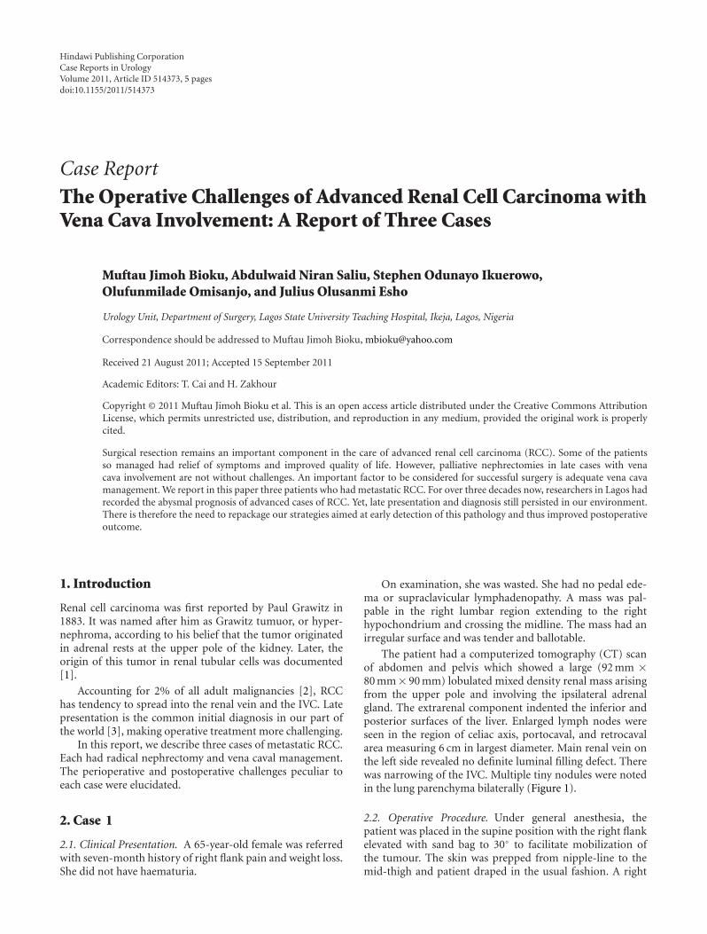
The patient had a computerized tomography (CT) scanof abdomen and pelvis which showed a large (92 mm ×80 mm× 90 mm) lobulated mixed density renal mass arisingfrom the upper pole and involving the ipsilateral adrenalgland. The extrarenal component indented the inferior andposterior surfaces of the liver. Enlarged lymph nodes wereseen in the region of celiac axis, portocaval, and retrocavalarea measuring 6 cm in largest diameter. Main renal vein onthe left side revealed no definite luminal filling defect. Therewas narrowing of the IVC. Multiple tiny nodules were notedin the lung parenchyma bilaterally (Figure 1).
2.2. Operative Procedure. Under general anesthesia, thepatient was placed in the supine position with the right flankelevated with sand bag to 30◦ to facilitate mobilization ofthe tumour. The skin was prepped from nipple-line to themid-thigh and patient draped in the usual fashion. A right
2 Case Reports in Urology
Figure 1
transverse upper abdominal incision was made from tipof 12th rib crossing the midline 2 cm above the level ofthe umbilicus and extending to lateral border of the leftrectus abdominis muscle. The peritoneal cavity was enteredand entire abdomen explored. A firm, irregular brownish-yellow mass measuring 19 cm × 10 cm was found. This hadalmost completely replaced the right kidney. tumor hadinfiltrated the Gerota’s fascia and was adherent to the inferiorsurface of the liver. The right renal vein and adjacent IVCwall were infiltrated. There was perihilar and para-aorticlymphadenopathy. However, there was no palpable livermetastasis.
The colon and duodenum were free.Initial renal pedicle control was not possible. Thus, the
tumor was mobilized inferiorly and laterally. The superiorpole of the tumour was separated from the inferior hepaticsurface with careful blunt dissection. The distorted renalvessels were carefully dissected, and divided in betweenSatinsky vascular clamps. Partial longitudinal resection of thetumor-infiltrated IVC wall was done. This was accompaniedby transient hypotension which was controlled by thepreinformed anaesthetists.
The ureter was then ligated and divided far beyondthe level of tumour involvement. The tumour, the adrenalgland, regional lymph nodes, and resected wall of IVC wereremoved in one block.
The renal fossa and inferior hepatic surface were packedas the latter continued to ooze despite efforts at meticuloushemostasis. The pack was removed forty-eight hours postop-eratively by the bed side.
Patient had hypovolemic shock with acute renal failuretwelve hours after-surgery. This was successfully managed inconjunction with nephrologists.
She also had right facial palsy and bilateral pitting pedaledema up to midleg. She was discharged to the clinic ten dayslater.
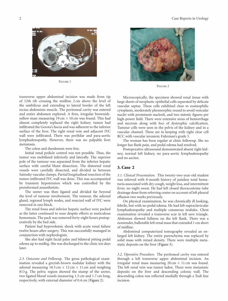
2.3. Outcome and Followup. The gross pathological exam-ination revealed a greyish-brown nodular kidney with theadrenal measuring 18.5 cm × 12 cm × 11 cm and weighing813 g. The pelvic region showed the stump of the ureter,two ligated blood vessels measuring 1.5 cm and 1.7 cm long,respectively, with external diameter of 0.4 cm (Figure 2).
Figure 2
Microscopically, the specimen showed renal tissue withlarge sheets of neoplastic epithelial cells separated by delicatevascular septae. These cells exhibited clear to eosinophiliccytoplasm, moderately pleomorphic round to avoid vesicularnuclei with prominent nucleoli, and two mitotic figures perhigh-power field. There were extensive areas of hemorrhageand necrosis along with foci of dystrophic calcification.Tumour cells were seen in the pelvis of the kidney and in avascular channel. These are in keeping with right clear cellRCC with vascular invasion; Fuhrman’s grade 3.
The woman has been regular at clinic followup. She nolonger has flank pain, and pedal edema had resolved.
Postoperative ultrasound demonstrated absent right kid-ney, normal left kidney, no para-aortic lymphadenopathyand no ascites.
3. Case 2
3.1. Clinical Presentation. This twenty-two-year-old studentwas referred with 8-month history of painless total hema-turia associated with dry cough, weight loss, and intermittentfever, no night sweat. He had left closed thoracostomy tubedrainage done from referring center on account of left pleuraleffusion two weeks previously.
On physical examination, he was chronically ill looking,febrile, but with no pedal edema. He had left supraclavicularlymphadenopathy and multiple cutaneous nodules. Chestexamination revealed a transverse scar in left save triangle.Abdomen showed fullness on the left flank. There was anontender, ballotable left renal mass that extended 1 cm shortof midline.
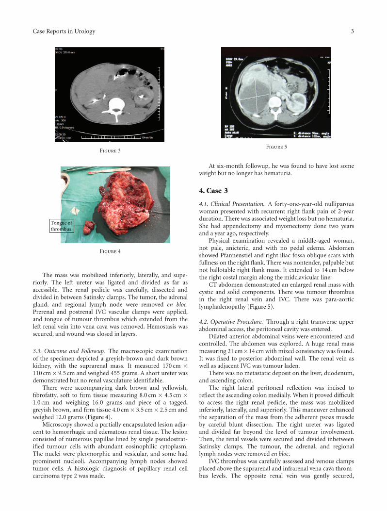
Abdominal computerized tomography revealed an en-larged left kidney. The entire parenchyma was replaced bysolid mass with mixed density. There were multiple meta-static deposits on the liver (Figure 3).
3.2. Operative Procedure. The peritoneal cavity was enteredthrough a left transverse upper abdominal incision. Anirregular renal mass measuring 20 cm × 11 cm was found.The left renal vein was tumor laden. There were metastaticdeposits on the liver and descending colonic wall. Thedescending colon was reflected medially through a Todt lineincision.
Case Reports in Urology 3
Figure 3
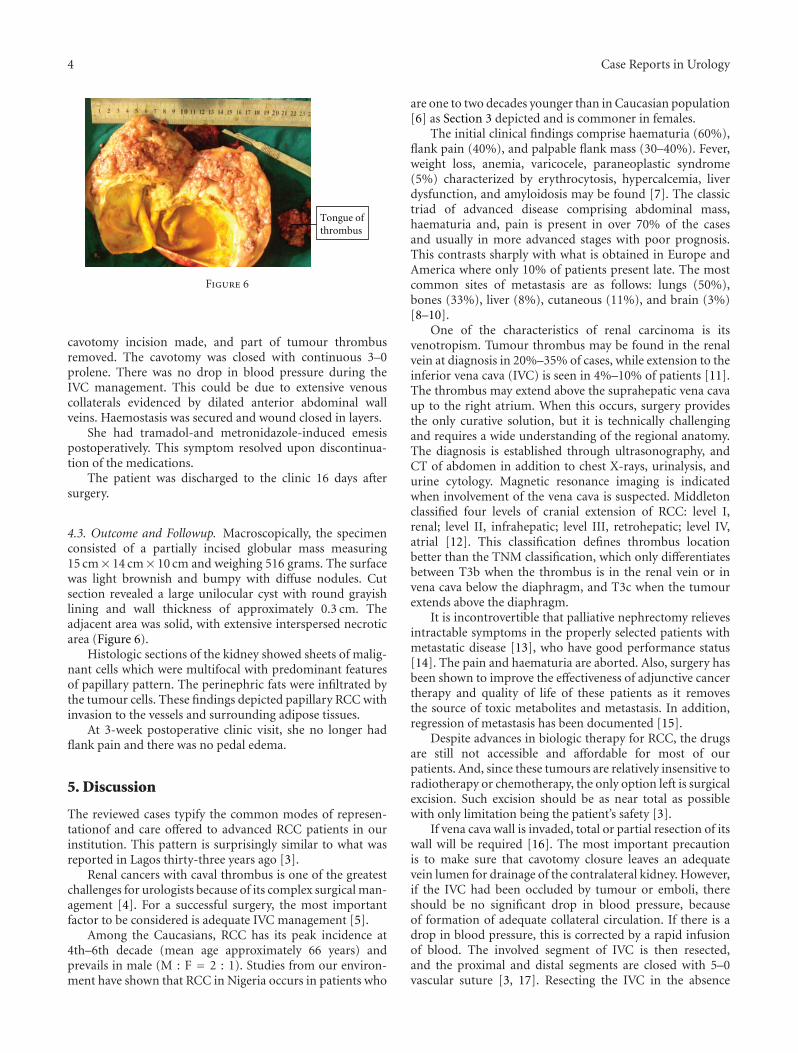
Tongue of thrombus
Figure 4
The mass was mobilized inferiorly, laterally, and supe-riorly. The left ureter was ligated and divided as far asaccessible. The renal pedicle was carefully, dissected anddivided in between Satinsky clamps. The tumor, the adrenalgland, and regional lymph node were removed en bloc.Prerenal and postrenal IVC vascular clamps were applied,and tongue of tumour thrombus which extended from theleft renal vein into vena cava was removed. Hemostasis wassecured, and wound was closed in layers.
3.3. Outcome and Followup. The macroscopic examinationof the specimen depicted a greyish-brown and dark brownkidney, with the suprarenal mass. It measured 170 cm ×110 cm× 9.5 cm and weighed 455 grams. A short ureter wasdemonstrated but no renal vasculature identifiable.
There were accompanying dark brown and yellowish,fibrofatty, soft to firm tissue measuring 8.0 cm × 4.5 cm ×1.0 cm and weighing 16.0 grams and piece of a tagged,greyish brown, and firm tissue 4.0 cm× 3.5 cm× 2.5 cm andweighed 12.0 grams (Figure 4).
Microscopy showed a partially encapsulated lesion adja-cent to hemorrhagic and edematous renal tissue. The lesionconsisted of numerous papillae lined by single pseudostrat-ified tumour cells with abundant eosinophilic cytoplasm.The nuclei were pleomorphic and vesicular, and some hadprominent nucleoli. Accompanying lymph nodes showedtumor cells. A histologic diagnosis of papillary renal cellcarcinoma type 2 was made.
Figure 5
At six-month followup, he was found to have lost someweight but no longer has hematuria.
4. Case 3
4.1. Clinical Presentation. A forty-one-year-old nulliparouswoman presented with recurrent right flank pain of 2-yearduration. There was associated weight loss but no hematuria.She had appendectomy and myomectomy done two yearsand a year ago, respectively.
Physical examination revealed a middle-aged woman,not pale, anicteric, and with no pedal edema. Abdomenshowed Pfannenstiel and right iliac fossa oblique scars withfullness on the right flank. There was nontender, palpable butnot ballotable right flank mass. It extended to 14 cm belowthe right costal margin along the midclavicular line.
CT abdomen demonstrated an enlarged renal mass withcystic and solid components. There was tumour thrombusin the right renal vein and IVC. There was para-aorticlymphadenopathy (Figure 5).
4.2. Operative Procedure. Through a right transverse upperabdominal access, the peritoneal cavity was entered.
Dilated anterior abdominal veins were encountered andcontrolled. The abdomen was explored. A huge renal massmeasuring 21 cm×14 cm with mixed consistency was found.It was fixed to posterior abdominal wall. The renal vein aswell as adjacent IVC was tumour laden.
There was no metastatic deposit on the liver, duodenum,and ascending colon.
The right lateral peritoneal reflection was incised toreflect the ascending colon medially. When it proved difficultto access the right renal pedicle, the mass was mobilizedinferiorly, laterally, and superiorly. This maneuver enhancedthe separation of the mass from the adherent psoas muscleby careful blunt dissection. The right ureter was ligatedand divided far beyond the level of tumour involvement.Then, the renal vessels were secured and divided inbetweenSatinsky clamps. The tumour, the adrenal, and regionallymph nodes were removed en bloc.
IVC thrombus was carefully assessed and venous clampsplaced above the suprarenal and infrarenal vena cava throm-bus levels. The opposite renal vein was gently secured,
4 Case Reports in Urology
Tongue of thrombus
Figure 6
cavotomy incision made, and part of tumour thrombusremoved. The cavotomy was closed with continuous 3–0prolene. There was no drop in blood pressure during theIVC management. This could be due to extensive venouscollaterals evidenced by dilated anterior abdominal wallveins. Haemostasis was secured and wound closed in layers.
She had tramadol-and metronidazole-induced emesispostoperatively. This symptom resolved upon discontinua-tion of the medications.
The patient was discharged to the clinic 16 days aftersurgery.
4.3. Outcome and Followup. Macroscopically, the specimenconsisted of a partially incised globular mass measuring15 cm× 14 cm× 10 cm and weighing 516 grams. The surfacewas light brownish and bumpy with diffuse nodules. Cutsection revealed a large unilocular cyst with round grayishlining and wall thickness of approximately 0.3 cm. Theadjacent area was solid, with extensive interspersed necroticarea (Figure 6).
Histologic sections of the kidney showed sheets of malig-nant cells which were multifocal with predominant featuresof papillary pattern. The perinephric fats were infiltrated bythe tumour cells. These findings depicted papillary RCC withinvasion to the vessels and surrounding adipose tissues.
At 3-week postoperative clinic visit, she no longer hadflank pain and there was no pedal edema.
5. Discussion
The reviewed cases typify the common modes of represen-tationof and care offered to advanced RCC patients in ourinstitution. This pattern is surprisingly similar to what wasreported in Lagos thirty-three years ago [3].
Renal cancers with caval thrombus is one of the greatestchallenges for urologists because of its complex surgical man-agement [4]. For a successful surgery, the most importantfactor to be considered is adequate IVC management [5].
Among the Caucasians, RCC has its peak incidence at4th–6th decade (mean age approximately 66 years) andprevails in male (M : F = 2 : 1). Studies from our environ-ment have shown that RCC in Nigeria occurs in patients who
are one to two decades younger than in Caucasian population[6] as Section 3 depicted and is commoner in females.
The initial clinical findings comprise haematuria (60%),flank pain (40%), and palpable flank mass (30–40%). Fever,weight loss, anemia, varicocele, paraneoplastic syndrome(5%) characterized by erythrocytosis, hypercalcemia, liverdysfunction, and amyloidosis may be found [7]. The classictriad of advanced disease comprising abdominal mass,haematuria and, pain is present in over 70% of the casesand usually in more advanced stages with poor prognosis.This contrasts sharply with what is obtained in Europe andAmerica where only 10% of patients present late. The mostcommon sites of metastasis are as follows: lungs (50%),bones (33%), liver (8%), cutaneous (11%), and brain (3%)[8–10].
One of the characteristics of renal carcinoma is itsvenotropism. Tumour thrombus may be found in the renalvein at diagnosis in 20%–35% of cases, while extension to theinferior vena cava (IVC) is seen in 4%–10% of patients [11].The thrombus may extend above the suprahepatic vena cavaup to the right atrium. When this occurs, surgery providesthe only curative solution, but it is technically challengingand requires a wide understanding of the regional anatomy.The diagnosis is established through ultrasonography, andCT of abdomen in addition to chest X-rays, urinalysis, andurine cytology. Magnetic resonance imaging is indicatedwhen involvement of the vena cava is suspected. Middletonclassified four levels of cranial extension of RCC: level I,renal; level II, infrahepatic; level III, retrohepatic; level IV,atrial [12]. This classification defines thrombus locationbetter than the TNM classification, which only differentiatesbetween T3b when the thrombus is in the renal vein or invena cava below the diaphragm, and T3c when the tumourextends above the diaphragm.
It is incontrovertible that palliative nephrectomy relievesintractable symptoms in the properly selected patients withmetastatic disease [13], who have good performance status[14]. The pain and haematuria are aborted. Also, surgery hasbeen shown to improve the effectiveness of adjunctive cancertherapy and quality of life of these patients as it removesthe source of toxic metabolites and metastasis. In addition,regression of metastasis has been documented [15].
Despite advances in biologic therapy for RCC, the drugsare still not accessible and affordable for most of ourpatients. And, since these tumours are relatively insensitive toradiotherapy or chemotherapy, the only option left is surgicalexcision. Such excision should be as near total as possiblewith only limitation being the patient’s safety [3].
If vena cava wall is invaded, total or partial resection of itswall will be required [16]. The most important precautionis to make sure that cavotomy closure leaves an adequatevein lumen for drainage of the contralateral kidney. However,if the IVC had been occluded by tumour or emboli, thereshould be no significant drop in blood pressure, becauseof formation of adequate collateral circulation. If there is adrop in blood pressure, this is corrected by a rapid infusionof blood. The involved segment of IVC is then resected,and the proximal and distal segments are closed with 5–0vascular suture [3, 17]. Resecting the IVC in the absence
Case Reports in Urology 5
of collaterals can be associated with severe edema of lowerextremities.
Usually, IVC obstruction by tumour thrombus inducesvenous collateralization. The extent and distribution of thesecollaterals depend on the obstruction location, obstructionlength, acute or chronic, and whether or not obstructivelesion involves tributaries of IVC [18].
The dominant collateral systems are arranged into twogroups—the superficial and the deep collaterals. The deepgroup includes azygous-hemiazygous, vertebral venous ple-xus, gonadal, ureteral, and portal (inferior mesenteric vein)while the superficial system consists of lateral thoracic,internal thoracic, and portal (paraumbilical veins).
In the lower-level IVC occlusion, azygous-hemiazygouspathway, is of great importance. Also, abdominal wallcollaterals, as observed in Section 4, may occur. Midlevelobstruction induces collateralization from portal system,perinephric and capsular drainage into the azygous-hemi-azygous system. However, in the upper-level obstruction,communication between the IVC and superior vena cavadevelops from the portal system both deep and superficial.Besides, the vertebral plexus becomes widely dilated [19].
Surgical approach for patients with IVC tumour throm-bus depends on the level of cranial extension of RCC asdefined by Neves and Zincke. A chevron incision with orwithout a midline abdominal cephalad T extension andliver mobilisation when the thrombus is infradiaphragmaticis favoured. However, if the thrombus extends above thediaphragm, a thoracoabdominal access or midline sterno-tomy combined with an abdominal incision may be required.
Cardiopulmonary bypass has been utilised as adjunct toremove cavoatrial tumour thrombus with or without hypo-thermic circulatory arrest. The advantages of this includecareful, controlled dissection in essentially bloodless surgicalfield [20]. However, nonavailability of heart-lung machine isa limiting factor in most centres of the third world.
6. Conclusion
Inspite of operability and resectability challenges posed byadvanced RCC, surgical resection remains an integral partof its care. Worrisome, however, is the persistence of latepresentation and diagnosis of the disease in our environ-ment. For over three decades now, researchers in Lagoshad reechoed the abysmal prognosis of advanced cases ofRCC. There is therefore the need to repackage our strategiesgeared at early detection of this pathology and thus improvedpostoperative survival.
References
[1] A. C. Novick and S. C. Campbell, “Renal tumors,” in Camp-bell’s Urology. 8, M. F. Campbell, P. C. Walsh, A. B. Retik etal., Eds., vol. 4, pp. 2672–2731, WB Saunders, New York, NY,USA, 2002.
[2] S. H. Landis, T. Murray, S. Bolden, and P. A. Wingo, “Cancerstatistics, 1999,” Ca: A Cancer Journal for Clinicians, vol. 49,no. 1, pp. 8–31, 1999.
[3] J. O. Esho, “Radical surgery for renal cell carcinoma,” EuropeanUrology, no. 4, pp. 338–341, 1978.
[4] Z. Kirkali and H. Van Poppel, “A critical analysis of surgery forkidney cancer with vena cava invasion,” European Urology, vol.52, no. 3, pp. 658–662, 2007.
[5] D. I. Swierzewski, M. J. Swierzewski, and J. A. Libertino,“Radical nephrectomy in patients with renal cell carcinomawith venous, vena caval, and atrial extension,” AmericanJournal of Surgery, vol. 168, no. 2, pp. 205–209, 1994.
[6] M. O. Odunbanjo, A. O. Oluwasola, and S. O. Ikuerowo,“Histopathological pathern of renal cell carcinoma in Ibadan,”African Journal of Medicine & Medical Sciences, vol. 39, pp.317–321, 2010.
[7] M. W. Linehan, C. Cordon-Cardo, and W. Isaacs, “Cancersofgenitourinary system,” in Cancer: Principles & Practice ofOncology, V. T. DeVita, S. Hellman, and S. A. Rosenberg,Eds., pp. 1253–1270, JB Lippincott, Philadelphia, Pa, USA, 5thedition, 1997.
[8] G. M. Bordin and S. Weitzner, “Cutaneous metastases as amanifestation of internal carcinoma: diagnostic and prognos-tic significance,” American Surgeon, vol. 38, no. 11, pp. 629–634, 1972.
[9] D. L. Wahner-Roedler and T. J. Sebo, “Renal cell carcinoma:diagnosis based on metastatic manifestations,” Mayo ClinicProceedings, vol. 72, no. 10, pp. 935–941, 1997.
[10] F. V. Alonso, F. J. Vicente de Prados, J. M. Cozar Olmo et al.,“Renal cell carcinoma with vena cava involvement: update andreview of our series,” Actas Urologicas Espanolas, vol. 33, no. 5,pp. 569–574, 2009.
[11] R. J. Neves and H. Zincke, “Surgical treatment of renal cancerwith vena cava extension,” British Journal of Urology, vol. 59,no. 5, pp. 390–395, 1987.
[12] A. W. Middleton, “Indications for and results of nephrectomyfor metastatic renal cell carcinoma,” Urologic Clinics of NorthAmerica, vol. 7, no. 3, pp. 711–717, 1980.
[13] J. Lokich, “Spontaneous regression of metastatic renal cancer:case report and literature review,” American Journal of ClinicalOncology, vol. 20, no. 4, pp. 416–418, 1997.
[14] L. Borje, C. C. Nigel, C. H. Damian et al., “EUA guidelines onrenal cell carcinoma: the 2010 update,” European Urology, vol.58, pp. 398–406, 2010.
[15] F. J. Vicente Prados, M. Tallada Bunuel, J. Pastor et al., “Renaladenocarcinoma with vena cava invasion: current status of itsdiagnosis and treatment using total segmentary cavectomy,”Archivos Espanoles de Urologıa, vol. 51, no. 1, pp. 35–41, 1998.
[16] J. O. Esho and A. Owoseni, “Vena cava resection with renal cellcarcinoma,” European Urology, vol. 3, no. 2, pp. 111–115, 1977.
[17] A. Rodriguez and W. J. Sexton, “Management of locally ad-vanced renal cell carcinoma,” Cancer Control, vol. 13, no. 3,pp. 199–210, 2006.
[18] R. M. Golub, R. E. Parsons, B. Sigel, and U. Anne, “Barnes: areview of venous collaterals in inferior vena cava obstruction,”Clinical Anatomy, vol. 5, pp. 441–451, 1992.
[19] R. M. Filler and E. A. Edwards, “Collaterals of the lowerinferior vena cava in man revealed by venography,” Archivesof Surgery, vol. 84, pp. 10–16, 1962.
[20] A. C. Novick, “Renal malignancy,” in Operative Urology at theCleveland Clinic, A. C. Novick and J. S. Jones, Eds., pp. 31–49,Humana Press, 2006.

Submit your manuscripts athttp://www.hindawi.com
Stem CellsInternational
Hindawi Publishing Corporationhttp://www.hindawi.com Volume 2014
Hindawi Publishing Corporationhttp://www.hindawi.com Volume 2014
MEDIATORSINFLAMMATION
of
Hindawi Publishing Corporationhttp://www.hindawi.com Volume 2014
Behavioural Neurology
EndocrinologyInternational Journal of
Hindawi Publishing Corporationhttp://www.hindawi.com Volume 2014
Hindawi Publishing Corporationhttp://www.hindawi.com Volume 2014
Disease Markers
Hindawi Publishing Corporationhttp://www.hindawi.com Volume 2014
BioMed Research International
OncologyJournal of
Hindawi Publishing Corporationhttp://www.hindawi.com Volume 2014
Hindawi Publishing Corporationhttp://www.hindawi.com Volume 2014
Oxidative Medicine and Cellular Longevity
Hindawi Publishing Corporationhttp://www.hindawi.com Volume 2014
PPAR Research
The Scientific World JournalHindawi Publishing Corporation http://www.hindawi.com Volume 2014
Immunology ResearchHindawi Publishing Corporationhttp://www.hindawi.com Volume 2014
Journal of
ObesityJournal of
Hindawi Publishing Corporationhttp://www.hindawi.com Volume 2014
Hindawi Publishing Corporationhttp://www.hindawi.com Volume 2014
Computational and Mathematical Methods in Medicine
OphthalmologyJournal of
Hindawi Publishing Corporationhttp://www.hindawi.com Volume 2014
Diabetes ResearchJournal of
Hindawi Publishing Corporationhttp://www.hindawi.com Volume 2014
Hindawi Publishing Corporationhttp://www.hindawi.com Volume 2014
Research and TreatmentAIDS
Hindawi Publishing Corporationhttp://www.hindawi.com Volume 2014
Gastroenterology Research and Practice
Hindawi Publishing Corporationhttp://www.hindawi.com Volume 2014
Parkinson’s Disease
Evidence-Based Complementary and Alternative Medicine
Volume 2014Hindawi Publishing Corporationhttp://www.hindawi.com