case report canine choroid plexus tumor with intracranial dissemination...
TRANSCRIPT
Hindawi Publishing CorporationCase Reports in Veterinary MedicineVolume 2013, Article ID 759054, 4 pageshttp://dx.doi.org/10.1155/2013/759054
Case ReportCanine Choroid Plexus Tumor with Intracranial DisseminationPresenting as Multiple Cystic Lesions
Trisha J. Oura,1 Peter J. Early,2 Samuel H. Jennings,2 Melissa J. Lewis,2
Jeremy R. Tobias,2 and Donald E. Thrall3
1 Tufts Veterinary Emergency Treatment and Specialties, 525 South Street, Walpole, MA 02081, USA2North Carolina State University Veterinary Teaching Hospital, 1052 William Moore Drive, Raleigh, NC 27607, USA3Associate Dean for Research, Ross University School of Veterinary Medicine, P.O. Box 334, Basseterre, Saint Kitts, USA
Correspondence should be addressed to Peter J. Early; [email protected]
Received 9 April 2013; Accepted 9 May 2013
Academic Editors: S. C. Rahal, M. Santos, R. M. Santos, and S. Stuen
Copyright © 2013 Trisha J. Oura et al. This is an open access article distributed under the Creative Commons Attribution License,which permits unrestricted use, distribution, and reproduction in any medium, provided the original work is properly cited.
A Miniature Pinscher developed acute blindness and behavioral changes. On magnetic resonance imaging (MRI), therewere multiple small intra-axial cystic lesions, and primary differential diagnoses included primary or metastatic neoplasiaand neurocysticercosis. These cystic lesions were subsequently diagnosed histopathologically as disseminated choroid plexuscarcinoma. This is only the second documented description of this diagnosis in a dog, but both patients had very similar MRIfindings. This patient adds to the literature about the MRI characteristics of choroid plexus tumors and indicates that choroidplexus tumor should be considered as a possible cause of small multifocal intra-axial cystic brain lesions in dogs, regardless ofwhether a primary intraventricular lesion is visible.
1. Introduction
Choroid plexus tumors arise from the epithelium of thechoroid plexus and account for approximately 10% of allprimary intracranial tumors in dogs [1].These predominantlybenign tumors are found most commonly in the lateral,third, or fourth ventricles. Dogs are typically middle-aged atthe time of presentation, and males and Golden Retrieversmay be overrepresented [1]. Clinical signs, which can includeataxia, circling, blindness, and behavior changes, are oftendue to hydrocephalus as ventricular lesions may cause cere-brospinal fluid (CSF) accumulation due to obstruction and/oroverproduction [1, 2].
Definitive diagnosis of choroid plexus tumors is byhistopathologic evaluation; however, there are often distinc-tive findings on magnetic resonance imaging (MRI), suchas well-differentiated, irregularly margined, intraventricularmasses. These masses are typically hyperintense on T2 andproton density (PD) sequences with strong, homogeneouscontrast enhancement [3]. Perilesional edemamay be presentor absent, and primary choroid plexus tumors may containfoci of hemorrhage or mineralization [3].
Both choroid plexus papillomas and carcinomas canmetastasize, often to the spinal subarachnoid space, creatingthe so-called drop metastases. Rarely, tumor disseminationandmeningeal carcinomatosis of choroid plexus tumors havebeen documented, with spread of neoplastic cells within theleptomeninges [4, 5]. Although documented histologically[5], there has been only one prior report of the MRI findingsof a dog with choroid plexus carcinoma and secondaryleptomeningeal metastases [4, 5]. The purpose of this reportis to add information from one additional patient withthis interesting and unusual manifestation of choroid plexustumor.
2. Case Presentation
A 9-year-old neutered male Miniature Pinscher developedacute-onset blindness and possible seizure activity. Whenexamined, the patient was anxious, vocalizing, and exhibit-ing compulsive behaviors. Both pupils were mydriatic withabsent menace response, absent dazzle, and absent direct andindirect papillary light reflexes. Mild to moderate cervicalpain was elicited on palpation and during range of motion.
2 Case Reports in Veterinary Medicine
(a) (b)
(c) (d)
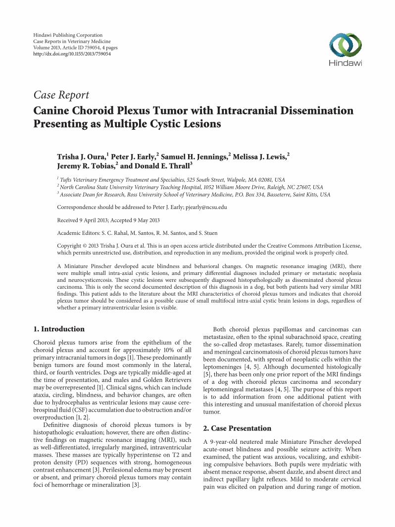
Figure 1: Transverse magnetic resonance images (MRIs) at the level of the mesencephalic aqueduct. (a) T2W image (TE/TR, 104/4440ms)with several round lesions within the caudal thalamus that are isoattenuating to cerebrospinal fluid within the lateral ventricles. (b) Theselesions null on FLAIR sequences (TR/TE, 78/9000ms) with mild perilesional hyperintensities representing edema. (c) The lesions areisoattenuating to CSF on T1W images (TE/TR, 12/53ms) and have mild contrast enhancement of the lesion wall (d).
The neuroanatomic localization was multifocal involvingthe prosencephalon and brainstem and or possible cervicalinvolvement.
Magnetic resonance imaging of the brain was performedusing a 1.5 Tmagnet (Symphony 1.5 T; SiemensMedical Solu-tions USA, Inc., Malvern, PA, USA). Caudal to the intertha-lamic adhesion, there were numerous small, ∼1.0–10.0mmdiameter, intra-axial, thin-walled, T2 hyperintense (TE/TR,104/4440ms), T1-hypointense (TE/TR, 12/53ms) noduleswithin the thalamus, colliculi, and hippocampus. The T2-signal of these nodules was nulled in FLAIR images (TR/TE,78/9000ms) indicating a cystic nature, and the wall of thenodules enhanced following intravenous administration ofgadoversetamide contrast medium (Optimark; MallinkrodtInc., St. Louis, MO, USA). Based on the FLAIR sequences,there was also mild perilesional edema (Figure 1). Therewas mild obstructive hydrocephalus due to mesencephalicaqueduct compression by the more caudal nodules, mildtranstentorial herniation causing compression of the rostral
aspect of the cerebellum, and syrinx formation in the cranialaspect of the cervical spinal cord. Additional smaller noduleswere present in the ventral aspect of the cerebellum. Basedon the MRI findings, differential diagnoses for the cysticlesions included atypical cystic metastatic neoplasia or neuralcysticercosis, though the latter was considered much lesslikely as the animalwas not known to have lived in or travelledto an area endemic for cysticercosis.
Differential diagnoses for the cystic lesions includedatypical cystic metastatic neoplasia or neural cysticercosis.Obstructive hydrocephalus and compression of the lateralgeniculate nuclei by the cystic lesions were thought to bepossible causes of the blindness.
On CSF analysis, there was a mild mononuclear pleocy-tosis (21 cells/uL) and an elevated total protein (54.5mg/dL).No etiologic agents or neoplastic cells were identified.
The patient was discharged with prednisone 5mg tabletgiven orally once every 24 hours, omeprazole 10mg capsulegiven orally once every 24 hours, aspirin 5mg capsule given
Case Reports in Veterinary Medicine 3
orally once every 24 hours, enalapril 3.75mg tablet orallygiven every 12 hours, albendazole 227mg oral suspensiongiven orally every 12 hours for 3 days, and praziquantel/pyrantel pamoate/febantel 54.4mg tablet given orally once.
Within four days of discharge, the patient had animproved mental status with decreased compulsive behavior.However, within one week the patient exhibited generalizedseizure activity, and anticonvulsant therapy was initiated.Approximately three weeks after discharge, the patient devel-oped cluster seizures and was euthanized.
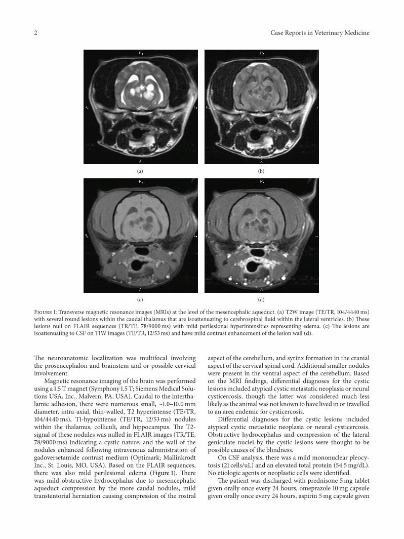
At postmortem examination, the cystic lesions and mildhydrocephalus were confirmed. There were also severaladditional pinpoint to 0.1 cm cystic foci within the cerebralleptomeninges (Figure 2). On histopathology, approximately20% of the left thalamus was replaced by scattered, variablysized, well-demarcated, expansile, unencapsulated, cysticintraventricular, and intraparenchymal neoplasms. Most ofthe clear cystic cavities were often lined by a relativelyuniformpopulation of cuboidal to columnar cells arranged ina single layer with a few, simple to complex, intraluminal pap-illary projections supported by thin fibrovascular cores.Thesecells had well-defined cell borders with small to moderateamount of eosinophilic cytoplasmwith an oval, basal nucleuswith finely stippled chromatin and an indistinct nucleolus.The cells exhibited minimal anisocytosis and anisokaryosis.Occasionally, however, the cysts and papillary projectionswere lined by up to eight layers of jumbled epithelial cells withmore variably sized, shaped, and located nuclei with moreopen chromatin. There was rare single cell necrosis withinthe neoplastic population. The mitotic rate varied regionallyfrom zero to seven mitotic figures in 10 high magnification(400x) fields.The cysts occasionally contained small amountsof laminated, mineralized material. The adjacent neuropilwas compressed and vacuolated with mild gliosis and, rarely,a few perivascular lymphocytes and plasma cells. Similarcystic neoplasms were present within sections of brainstembilaterally adjacent to the fourth ventricle, presumed to bein the lateral apertures, and throughout the cerebral andcerebellar meninges. The final diagnosis was choroid plexuscarcinoma with multifocal intracranial metastasis and mild,secondary hydrocephalus.
3. Discussion
Presently there is only one other description of theMRI find-ings associated with intracranial dissemination of a choroidplexus tumor in a dog. Given the rarity of this condition,it is important that additional patients be described as theyare discovered. Similar clinical finding between both dogsincluded blindness and behavioral changes. On MRI, bothpatients hadmild hydrocephalus and similar sized intra-axialcystic lesions which, in our patient, were located primarilyin the caudal thalamus, colliculi and hippocampus, while inthe previously reported patient, additional lesions were alsofound throughout the temporal and frontal lobes [4]. Thecystic lesions in our patient were also associated with mildperilesional edema and mild contrast enhancement of thecyst walls which was not observed previously. This difference
Figure 2: Cross-section of the dog’s brain at the level of the mes-encephalic aqueduct. Several 2-3mm, round to oval, cystic lesionsmultifocally expand and replace the dorsal thalamic parenchymaand compress the mesencephalic aqueduct. Histopathologically,these corresponded to choroid plexus tumors.
may be due to the fact that the previous patient receivedglucocorticoids prior to MRI acquisition [4]. The primarytumor could not be identified in either patient.
Meningeal carcinomatosis is uncommon in animals buthas been documented with metastatic mammary carcinomaand carcinoma without an identified primary tumor [6,7]. Specifically, intraparenchymal dissemination of choroidplexus tumors is documented rarely [4, 5]. Intracranialdissemination of choroid plexus tumors is also uncommonin people, with few reports of neoplasia involving the lep-tomeninges or seeding of the subarachnoid space [8–10]. Onehuman patient had multiple, nonenhancing, cystic subarach-noid lesions of the cerebellum, brainstem, and frontal lobe onMRI, an appearance very similar to our findings [9].
4. Conclusion
Multiple intra-axial cystic lesions are an unusual manifesta-tion of choroid plexus tumors in both people and animals.More commonly, choroid plexus tumors are typically solitary,lobulated, strongly contrast-enhancing lesions within theventricular system [3]. This case report of a canine patientadds to the available literature regarding choroid plexustumors and indicates that disseminated choroid plexus tumorshould be considered as a differential diagnosis for multiplesmall intra-axial cysts in dogs, regardless of whether aprimary intraventricular tumor is visible.
References
[1] D. R. Westworth, P. J. Dickinson, W. Vernau et al., “Choroidplexus tumors in 56 dogs (1985–2007),” Journal of VeterinaryInternal Medicine, vol. 22, no. 5, pp. 1157–1165, 2008.
[2] F. A. Zaki and L. A. Nafe, “Choroid plexus tumors in the dog,”Journal of the American VeterinaryMedical Association, vol. 176,no. 4, pp. 328–330, 1980.
4 Case Reports in Veterinary Medicine
[3] S. L. Kraft, P. R. Gavin, C. DeHaan, M. Moore, L. R. Wendling,and C.W. Leathers, “Retrospective review of 50 canine intracra-nial tumors evaluated by magnetic resonance imaging,” Journalof Veterinary Internal Medicine, vol. 11, no. 4, pp. 218–225, 1997.
[4] D. Lipsitz, R. E. Levitski, and A. E. Chauvet, “Magnetic reso-nance imaging of a choroid plexus carcinoma and meningealcarcinomatosis in a dog,” Veterinary Radiology and Ultrasound,vol. 40, no. 3, pp. 246–250, 1999.
[5] A. K. Patnaik, R. A. Erlandson, and P. H. Lieberman, “Choroidplexus carcinoma with meningeal carcinomatosis in a dog,”Veterinary Pathology, vol. 17, no. 3, pp. 381–385, 1980.
[6] M. Pumarola and M. Balasch, “Meningeal carcinomatosis in adog,” Veterinary Record, vol. 138, no. 21, pp. 523–524, 1996.
[7] I. Mateo, V. Lorenzo, A. Munoz, and J. Molın, “Meningealcarcinomatosis in a dog: magnetic resonance imaging featuresand pathological correlation,” Journal of Small Animal Practice,vol. 51, no. 1, pp. 43–48, 2010.
[8] R. Leblanc, S. Bekhor, D. Melanson, and S. Carpenter, “Diffusecraniospinal seeding from a benign fourth ventricle choroidplexus papilloma. Case report,” Journal of Neurosurgery, vol. 88,no. 4, pp. 757–760, 1998.
[9] T. McCall, M. Binning, D. T. Blumenthal, and R. L. Jensen,“Variations of disseminated choroid plexus papilloma: 2 casereports and a review of the literature,” Surgical Neurology, vol.66, no. 1, pp. 62–67, 2006.
[10] F. Doglietto, L. Lauretti, T. Tartaglione, M. Gessi, E. Fernandez,and G. Maira, “Diffuse craniospinal choroid plexus papillomawith involvement of both cerebellopontine angles,” Neurology,vol. 65, no. 6, p. 842, 2005.

Submit your manuscripts athttp://www.hindawi.com
Veterinary MedicineJournal of
Hindawi Publishing Corporationhttp://www.hindawi.com Volume 2014
Veterinary Medicine International
Hindawi Publishing Corporationhttp://www.hindawi.com Volume 2014
Hindawi Publishing Corporationhttp://www.hindawi.com Volume 2014
International Journal of
Microbiology
Hindawi Publishing Corporationhttp://www.hindawi.com Volume 2014
AnimalsJournal of
EcologyInternational Journal of
Hindawi Publishing Corporationhttp://www.hindawi.com Volume 2014
PsycheHindawi Publishing Corporationhttp://www.hindawi.com Volume 2014
Evolutionary BiologyInternational Journal of
Hindawi Publishing Corporationhttp://www.hindawi.com Volume 2014
Hindawi Publishing Corporationhttp://www.hindawi.com
Applied &EnvironmentalSoil Science
Volume 2014
Biotechnology Research International
Hindawi Publishing Corporationhttp://www.hindawi.com Volume 2014
Agronomy
Hindawi Publishing Corporationhttp://www.hindawi.com Volume 2014
International Journal of
Hindawi Publishing Corporationhttp://www.hindawi.com Volume 2014
Journal of Parasitology Research
Hindawi Publishing Corporation http://www.hindawi.com
International Journal of
Volume 2014
Zoology
GenomicsInternational Journal of
Hindawi Publishing Corporationhttp://www.hindawi.com Volume 2014
InsectsJournal of
Hindawi Publishing Corporationhttp://www.hindawi.com Volume 2014
The Scientific World JournalHindawi Publishing Corporation http://www.hindawi.com Volume 2014
Hindawi Publishing Corporationhttp://www.hindawi.com Volume 2014
VirusesJournal of
ScientificaHindawi Publishing Corporationhttp://www.hindawi.com Volume 2014
Cell BiologyInternational Journal of
Hindawi Publishing Corporationhttp://www.hindawi.com Volume 2014
Hindawi Publishing Corporationhttp://www.hindawi.com Volume 2014
Case Reports in Veterinary Medicine