case report a pilocytic astrocytoma mimicking a clinoidal...
TRANSCRIPT
Case ReportA Pilocytic Astrocytoma Mimicking a Clinoidal Meningioma
Christopher S. Hong,1 Norman L. Lehman,2 and Eric Sauvageau1
1 Department of Neurological Surgery, The Ohio State University Wexner Medical Center, 410 W. 10th Avenue,N1022 Doan Hall, Columbus, OH 43210, USA
2Department of Pathology, The Ohio State University Wexner Medical Center, 333 W. 10th Avenue,4169 Graves Hall, Columbus, OH 43210, USA
Correspondence should be addressed to Eric Sauvageau; [email protected]
Received 25 December 2013; Accepted 25 February 2014; Published 13 March 2014
Academic Editors: C. Chaskis, T. Krings, M. Leonardi, and A. Spalice
Copyright © 2014 Christopher S. Hong et al. This is an open access article distributed under the Creative Commons AttributionLicense, which permits unrestricted use, distribution, and reproduction in any medium, provided the original work is properlycited.
Pilocytic astrocytomas andmeningiomas are benign, primary brain tumors thatmay involve the optic tract. Classically, the presenceof a dural “tail” sign may differentiate a meningioma from other intracranial lesions. In this report, we describe a mass with thetypical appearance of a clinoidal meningioma on magnetic resonance imaging (MRI) but postoperatively diagnosed as a pilocyticastrocytoma. This case illustrates the rare occurrence of a pilocytic astrocytoma mimicking a meningioma on MRI.
1. Background
Pilocytic astrocytomas are benign, World Health Organiza-tion (WHO) grade I gliomas that most frequently arise inadolescents and young adults and usually involve the cere-bellum, hypothalamus, and optic pathways. Radiographically,they are relatively well demarcated and strongly enhanceon MRI as they are well vascularized and often exhibitglomeruloid vascular proliferation. Although rare, pilocyticastrocytomas can originate from the optic pathway, anterioror posterior to the optic chiasm. These lesions have a strongassociation with neurofibromatosis type 1 (NF1) and areestimated to occur in 15% of all patients with this inheritedsyndrome [1].
In contrast, meningiomas are benign growths of arach-noid cap cells, usually found at sites of dural reflectionswithin the skull, and classically exhibit a homogenouslyenhancing thickening of the dural margin with peripheraltapering on MRI, known as the “tail” sign [2]. Because ofthese characteristic features, meningiomas are usually readilyidentified based on clinical and radiographic findings alone,often precluding a need for tissue biopsy prior to treatment.
In this case report, we describe a young patient whoseclinical and radiographic presentation strongly supported adiagnosis of a meningioma but upon neurosurgical interven-tion was found to have a pilocytic astrocytoma. We describe
the clinical course of our patient and highlight the unusualfeatures that were subsequently found.
2. Case Report
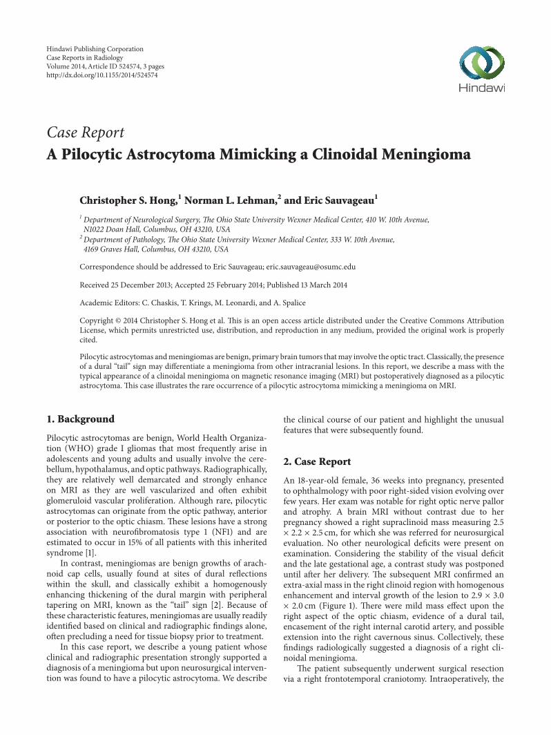
An 18-year-old female, 36 weeks into pregnancy, presentedto ophthalmology with poor right-sided vision evolving overfew years. Her exam was notable for right optic nerve pallorand atrophy. A brain MRI without contrast due to herpregnancy showed a right supraclinoid mass measuring 2.5× 2.2 × 2.5 cm, for which she was referred for neurosurgicalevaluation. No other neurological deficits were present onexamination. Considering the stability of the visual deficitand the late gestational age, a contrast study was postponeduntil after her delivery. The subsequent MRI confirmed anextra-axial mass in the right clinoid region with homogenousenhancement and interval growth of the lesion to 2.9 × 3.0× 2.0 cm (Figure 1). There were mild mass effect upon theright aspect of the optic chiasm, evidence of a dural tail,encasement of the right internal carotid artery, and possibleextension into the right cavernous sinus. Collectively, thesefindings radiologically suggested a diagnosis of a right cli-noidal meningioma.
The patient subsequently underwent surgical resectionvia a right frontotemporal craniotomy. Intraoperatively, the
Hindawi Publishing CorporationCase Reports in RadiologyVolume 2014, Article ID 524574, 3 pageshttp://dx.doi.org/10.1155/2014/524574
2 Case Reports in Radiology
(a) (b) (c)
Figure 1: (a) Axial, (b) coronal, and (c) sagittal T1-weighted MRI showing a homogenously enhancing lesion, measuring 2.9 × 3.0 × 2.0 cmin the right clinoidal region. There is probable extension into the right optic canal, sphenoid sinus, right temporal fossa, and possibly rightcavernous sinus, suggestive of a right clinoidal meningioma.
(a) (b)
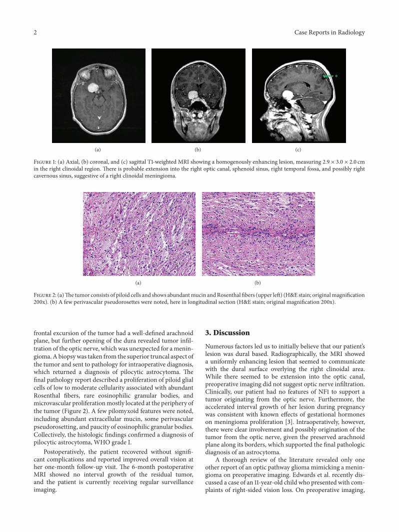
Figure 2: (a)The tumor consists of piloid cells and shows abundantmucin andRosenthal fibers (upper left) (H&E stain; originalmagnification200x). (b) A few perivascular pseudorosettes were noted, here in longitudinal section (H&E stain; original magnification 200x).
frontal excursion of the tumor had a well-defined arachnoidplane, but further opening of the dura revealed tumor infil-tration of the optic nerve, whichwas unexpected for amenin-gioma.A biopsywas taken from the superior truncal aspect ofthe tumor and sent to pathology for intraoperative diagnosis,which returned a diagnosis of pilocytic astrocytoma. Thefinal pathology report described a proliferation of piloid glialcells of low to moderate cellularity associated with abundantRosenthal fibers, rare eosinophilic granular bodies, andmicrovascular proliferationmostly located at the periphery ofthe tumor (Figure 2). A few pilomyxoid features were noted,including abundant extracellular mucin, some perivascularpseudorosetting, and paucity of eosinophilic granular bodies.Collectively, the histologic findings confirmed a diagnosis ofpilocytic astrocytoma, WHO grade I.
Postoperatively, the patient recovered without signifi-cant complications and reported improved overall vision ather one-month follow-up visit. The 6-month postoperativeMRI showed no interval growth of the residual tumor,and the patient is currently receiving regular surveillanceimaging.
3. Discussion
Numerous factors led us to initially believe that our patient’slesion was dural based. Radiographically, the MRI showeda uniformly enhancing lesion that seemed to communicatewith the dural surface overlying the right clinoidal area.While there seemed to be extension into the optic canal,preoperative imaging did not suggest optic nerve infiltration.Clinically, our patient had no features of NF1 to support atumor originating from the optic nerve. Furthermore, theaccelerated interval growth of her lesion during pregnancywas consistent with known effects of gestational hormoneson meningioma proliferation [3]. Intraoperatively, however,there were clear involvement and possibly origination of thetumor from the optic nerve, given the preserved arachnoidplane along its borders, which supported the final pathologicdiagnosis of an astrocytoma.
A thorough review of the literature revealed only oneother report of an optic pathway gliomamimicking a menin-gioma on preoperative imaging. Edwards et al. recently dis-cussed a case of an 11-year-old child who presented with com-plaints of right-sided vision loss. On preoperative imaging,
Case Reports in Radiology 3
there were an enlarged right optic nerve and a homogenouslyenhancing, dural-based lesion that extended into the righttemporal lobe [4]. During surgery, however, it was clear thatthe tumor involved white matter tracts, most likely derivedfrom the right temporal lobe. Notably, while hyperintensityon T2-weighted MRI suggested infiltration into the optictracts, this could not be visualized, intraoperatively. The finalhistologic diagnosis was a pilomyxoid astrocytoma.
Pilomyxoid astrocytomas were first described in 1999 andsubsequently were recognized by theWHO in 2007 as amoreaggressive, histological variant of pilocytic astrocytomas. OnMRI, these tumors usually enhance homogenously withinthe chiasmatic-hypothalamic region. Unlike pilocytic astro-cytomas, they exhibit T2 signal extension into surroundingdeep white and grey matter, in contrast to our patient’s radio-graphic presentation. Interestingly however, the pathology ofour biopsy sample did report limited pilomyxoid features.Generally, less than 5% of pilocytic astrocytomas progresstowards malignancy [5]. Still, our patient will continue to beclosely monitored for signs of disease progression.
4. Conclusion
In this case report, we describe the clinical course of apatient who presented with clinical and radiographic signs,all suggestive of clinoidal meningioma. However, the grossappearance and histology of the lesion demonstrated apilocytic astrocytoma. Therefore, physicians should considera pilocytic astrocytoma in their differential diagnosis ofany dural-based, enhancing lesion within the periclinoidalregion.
Conflict of Interests
The authors declare that there is no conflict of interestsregarding the publication of this paper.
References
[1] R. Listernick, J. Charrow, M. Greenwald, andM.Mets, “Naturalhistory of optic pathway tumors in children with neurofibro-matosis type 1: a longitudinal study,” Journal of Pediatrics, vol.125, no. 1, pp. 63–66, 1994.
[2] S. T. Qi, Y. Lui, J. Pan et al., “A radiopathological classificationof dural tail sign of meningiomas,” Journal of Neurosurgery, vol.117, no. 4, pp. 645–653, 2012.
[3] E. A. Lusis, B.W. Scheithauer, A. T. Yachnis et al., “Meningiomasin pregnancy: a clinicopathological study of 17 cases,” Neuro-surgery, vol. 71, no. 5, pp. 951–961, 2012.
[4] J. R. Edwards, C. G. Kulwin, S. E. Martin et al., “Temporaland optic pathway pilomyxoid astrocytoma mimicking dural-based lesion: case report and review of the literature,” PediatricNeurosurgery, vol. 48, no. 4, pp. 253–257, 2012.
[5] P. B. Dirks, V. Jay, L. E. Becker et al., “Development ofanaplastic changes in low-grade astrocytomas of childhood,”Neurosurgery, vol. 34, no. 1, pp. 68–78, 1994.

Submit your manuscripts athttp://www.hindawi.com
Stem CellsInternational
Hindawi Publishing Corporationhttp://www.hindawi.com Volume 2014
Hindawi Publishing Corporationhttp://www.hindawi.com Volume 2014
MEDIATORSINFLAMMATION
of
Hindawi Publishing Corporationhttp://www.hindawi.com Volume 2014
Behavioural Neurology
EndocrinologyInternational Journal of
Hindawi Publishing Corporationhttp://www.hindawi.com Volume 2014
Hindawi Publishing Corporationhttp://www.hindawi.com Volume 2014
Disease Markers
Hindawi Publishing Corporationhttp://www.hindawi.com Volume 2014
BioMed Research International
OncologyJournal of
Hindawi Publishing Corporationhttp://www.hindawi.com Volume 2014
Hindawi Publishing Corporationhttp://www.hindawi.com Volume 2014
Oxidative Medicine and Cellular Longevity
Hindawi Publishing Corporationhttp://www.hindawi.com Volume 2014
PPAR Research
The Scientific World JournalHindawi Publishing Corporation http://www.hindawi.com Volume 2014
Immunology ResearchHindawi Publishing Corporationhttp://www.hindawi.com Volume 2014
Journal of
ObesityJournal of
Hindawi Publishing Corporationhttp://www.hindawi.com Volume 2014
Hindawi Publishing Corporationhttp://www.hindawi.com Volume 2014
Computational and Mathematical Methods in Medicine
OphthalmologyJournal of
Hindawi Publishing Corporationhttp://www.hindawi.com Volume 2014
Diabetes ResearchJournal of
Hindawi Publishing Corporationhttp://www.hindawi.com Volume 2014
Hindawi Publishing Corporationhttp://www.hindawi.com Volume 2014
Research and TreatmentAIDS
Hindawi Publishing Corporationhttp://www.hindawi.com Volume 2014
Gastroenterology Research and Practice
Hindawi Publishing Corporationhttp://www.hindawi.com Volume 2014
Parkinson’s Disease
Evidence-Based Complementary and Alternative Medicine
Volume 2014Hindawi Publishing Corporationhttp://www.hindawi.com