cardiopulmonary exercise testing in heart...
TRANSCRIPT
J A C C : H E A R T F A I L U R E V O L . 4 , N O . 8 , 2 0 1 6
ª 2 0 1 6 B Y T H E AM E R I C A N C O L L E G E O F C A R D I O L O G Y F O UN DA T I O N I S S N 2 2 1 3 - 1 7 7 9 / $ 3 6 . 0 0
P U B L I S H E D B Y E L S E V I E R h t t p : / / d x . d o i . o r g / 1 0 . 1 0 1 6 / j . j c h f . 2 0 1 6 . 0 3 . 0 2 2
MINI-FOCUS ISSUE: EXERCISE AND HEART FAILURE
STATE-OF-THE-ART PAPER
Cardiopulmonary Exercise Testingin Heart Failure
Rajeev Malhotra, MD,a Kristian Bakken, NP,b Emilia D’Elia, MD, PHD,c,d Gregory D. Lewis, MDa,bABSTRACT
Fro
Ma
MedU
(N
Am
rec
oth
Ma
Exercise intolerance, indicated by dyspnea and fatigue during exertion, is a cardinal manifestation of heart failure (HF).
Cardiopulmonary exercise testing (CPET) precisely defines maximum exercise capacity through measurement of peak
oxygen uptake (VO2). Peak VO2 values have a critical role in informing patient selection for advanced HF interventions
such as heart transplantation and ventricular assist devices. Oxygen uptake and ventilatory patterns obtained during the
submaximal portion of CPET are also valuable to recognize because of their ease of ascertainment during low-level
exercise, relevance to ability to perform activities of daily living, independence from volitional effort, and strong
relationship to prognosis in HF. The ability of peak VO2 and other CPET variables to be measured reproducibly and to
accurately reflect HF severity is increasingly recognized and endorsed by scientific statements. Integration of CPET with
invasive hemodynamic monitoring and cardiac imaging during exercise provides comprehensive characterization of
multisystem reserve capacity that can inform prognosis and the need for cardiac interventions. Here, we review both
practical aspects of conducting CPETs in patients with HF for clinical and research purposes as well as interpretation of
gas exchange patterns across the spectrum of preclinical HF to advanced HF. (J Am Coll Cardiol HF 2016;4:607–16)
© 2016 by the American College of Cardiology Foundation.
I n patients with heart failure (HF), the func-tional reserve capacity of the integrated meta-bolic machinery required to perform exercise
is impaired at multiple levels. Starting with oxygen(O2) uptake in the lungs, the requisite increase inventilation is challenged by frequently abnormallung mechanics and diffusing capacity. The needfor increased convective O2 transport to skeletalmuscle is limited by prevalent anemia as well asabnormal cardiac output (CO) augmentation arisingfrom chronotropic incompetence, inability toaugment ventricular contractility, and functionalmitral regurgitation. Shortening of diastole duringheart rate (HR) elevation and increased venousreturn can lead to sharp increases in filling pres-sures during exercise; impaired vasoreactivity
m the aCardiology Division, Department of Medicine, Massachusetts G
ssachusetts; bPulmonary and Critical Care Unit of the Department of M
dical School, Boston, Massachusetts; cCardiovascular Department, Hos
niversity of Pavia, Pavia, Italy. Dr.Malhotra is supportedby theK08HL111210
HLBI) and theWild Family Foundation; and also has received consultant fee
erican Heart Association Award 15GPSGC24800006, NHLBI R01 HL131029-0
eived research grant support from Abbott Vascular, Stealth Peptides, Biom
er authors have reported that they have no relationships relevant to the co
nuscript received June 17, 2015; revised manuscript received February 26
further contributes to dynamic ventriculovascularuncoupling.
Upon delivery of O2 to the periphery, diffusive O2
conductance and utilization is limited by reducedcapillary density, impaired sympatholysis, decreasedmitochondrial volume, and selective loss of type 1muscle fibers having oxidative fatigue-resistant prop-erties (1). Finally, exaggerated ventilatory responses toexercise signaled through intramuscular afferents(i.e., ergoreflex signaling) are present in HF. It there-fore comes as no surprise that exercise intolerance isthe cardinal manifestation of HF. Careful measure-ment of ventilatory and O2 uptake patterns in HF canquantify disease severity and prognosis while shed-ding light on relative contributions of organ systems toexercise intolerance.
eneral Hospital, Harvard Medical School, Boston,
edicine, Massachusetts General Hospital, Harvard
pital Papa Giovanni XXIII, Bergamo Italy; and the
grant of theNationalHeart, Lung, andBlood Institute
s fromMallinckrodt, Inc. Dr. Lewis is supported by the
1, and the Hassenfeld Clinical Scholar Award; and has
arin, Pfizer, Shape Medical Systems, and Novartis. All
ntents of this paper to disclose.
, 2016, accepted March 2, 2016.

TABLE 1 Clinical Indications fo
Clinical Scenario
Unexplained or multifactorialdyspnea/exercise intolerance
Established advanced cardiacor pulmonary disease
Valvular or congenitalheart disease
Initiation of an intervention(clinical trial)
ABBR EV I A T I ON S
AND ACRONYMS
CaO2 = arterial concentration
of oxygen
CvO2 = mixed venous
concentration of oxygen
CavO2 = (CaO2 - CvO2)
difference
CPET = cardiopulmonary
exercise testing
EOV = exercise oscillatory
ventilation
MRT = mean response time
OUES = oxygen uptake
efficiency slope
PAP = pulmonary artery
pressure
PAWP = pulmonary artery
wedge pressure
RER = respiratory exchange
ratio
VCO2 = carbon dioxide output
VE = ventilation
VO2 = oxygen uptake
Malhotra et al. J A C C : H E A R T F A I L U R E V O L . 4 , N O . 8 , 2 0 1 6
Cardiopulmonary Exercise Testing A U G U S T 2 0 1 6 : 6 0 7 – 1 6
608
Cardiopulmonary exercise testing (CPET)provides breath-by-breath gas exchangemeasures of 3 variables: O2 uptake (VO2), car-bon dioxide output (VCO2), and ventilation(VE). These 3 measures are used to derivevarious other gas exchange patterns thatreflect organ-specific maladaptive responsesto exercise, particularly when CPET is coupledwith standard exercise variables (HR, bloodpressure, electrocardiogram), cardiac imag-ing, and invasive hemodynamic measure-ments during exercise.
Recent consensus statements and guide-line documents have provided an overallsummary of the utility of CPET (2–4). Anapproach to using CPET in patients with HF isprovided in the Online Appendix. Here, weprovide an overview on interpretation ofCPET with a specific focus on the HF popu-lation. Table 1 summarizes the current clin-ical indications for performing CPET. OnlineTable 1 describes gas exchange patternseasily and reproducibly derived from non-invasive CPET, their physiologic relevance,
and their clinical significance in HF.
O2 UPTAKE VARIABLES
PEAK VO2. Measured VO2 during a maximalsymptom-limited CPET is the most objective methodto assess functional capacity and consists of thefollowing components (2):
Peak VO2 ¼ HRMAX � SVMAX � ðCaO2-CvO2ÞMAX
where SV is stroke volume, and (CaO2 – CvO2) is the netoxygen extraction of the peripheral tissues and isdependent on the hemoglobin concentration(Figure 1). Peak VO2 is an important predictor of prog-nosis in HF patients (2). Mancini and colleagues (5)conducted a landmark study in 114 ambulatory pa-tients with HF and reduced ejection fraction (HFrEF)
r Cardiopulmonary Exercise Testing
Objective
To define the organ system(s) limiting gas exchange
To grade severity of disease, prognosticate, and prioritizepatients for heart transplantation and mechanicalcirculatory support
To determine whether to intervene, particularly withcardiac surgical interventions, and to estimateperioperative risk
To precisely evaluate the functional response to anintervention (i.e., change in peak oxygen uptake witha novel treatment)
that established a peak VO2 cutoff of#14 ml/kg/min asa criterion for which 1-year survival was significantlylower than that achieved through transplantation (i.e.,70%). In contrast, individuals with a peak VO2 >14 ml/kg/min had 6% 1-year mortality, suggesting thattransplantation could be safely deferred in this sub-group of symptomatic HF patients. There was no dif-ference in resting left ventricular ejection fraction orcardiac index between the groups. Multivariate anal-ysis identified peak VO2 as the best predictor of sur-vival in this HF population. Recent studies havedemonstrated that peak VO2 potently risk stratifies thecontemporary HF (HFrEF and HF and preserved ejec-tion fraction [HFpEF]) populations: Weber classes A,B, C, and D corresponding to peak VO2 >20, 16 to 20, 10to 16, and <10 ml/kg/min was associated with 3-yeartransplant and mechanical circulatory support-freesurvival of 97%, 93%, 83%, and 64%, respectively(Central Illustration) (6). In the HF-ACTION (HeartFailure: A Controlled Trial Investigating Outcomes ofExercise Training) trial, of multiple CPET variablesthat were assessed, peak VO2, percent predicted peakVO2, and exercise duration had the strongest ability topredict mortality in HFrEF (7). Peak VO2 retains itsprognostic significance in HFrEF patients on beta-blockers (8,9) and when natriuretic peptides andother clinical variables are considered (10). Peak VO2 isalso an important predictor of mortality in HF patientswith preserved left ventricular ejection fraction(HFpEF) (11,12).
Peak VO2 is influenced by noncardiac factors suchas age, gender, and muscle mass (13); therefore, it isappropriate to interpret peak VO2 normalized to age,gender, and weight-based normative values (14).The Wasserman-Hansen percent-predicted equationoffers optimal HF prognostication among peak VO2
percent-predicted equations, with a peak VO2 <47%of predicted serving as an optimal cutpoint fordetermining mortality risk in HF (15). Obesity reducesVO2 in ml/kg/min out of proportion to HF severity andhas led to examination of peak VO2 corrected for leanbody mass (LBM), where LBM was defined as: actualbody weight � (1 � % body fat/100) in ml/min/kg ofLBM. When corrected for LBM, a peak VO2 #19 ml/kg/min outperformed standard peak VO2 #14 ml/kg/minin predicting transplant-free survival within a HFrEFpopulation with a 37% prevalence of obesity (asdefined by body mass index [BMI] >30 kg/m2) (16).SUBMAXIMAL O2 UPTAKE MEASUREMENTS. Inter-estingly, among patients with HF, submaximal exer-cise gas exchange variables have emerged that rival oreven exceed the prognostic utility of peak VO2 (17–19).Submaximal CPET variables (Online Table 1) areparticularly attractive to study based on ease of
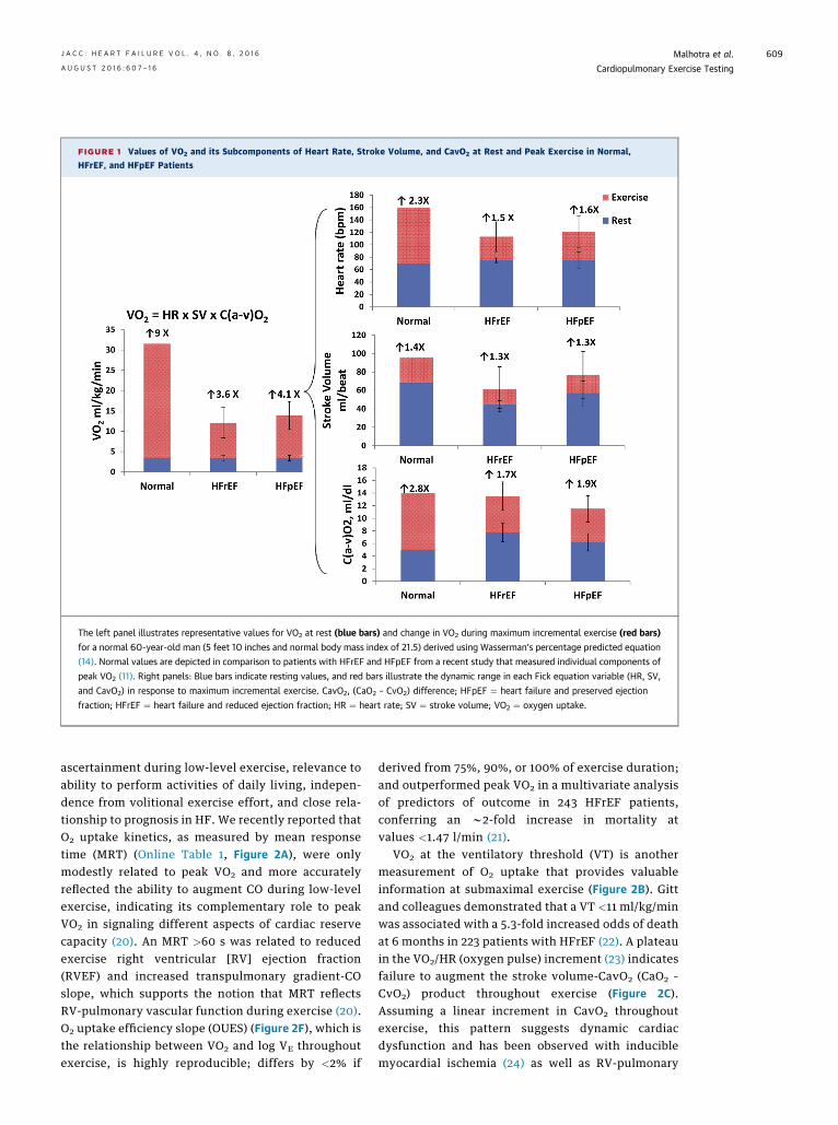
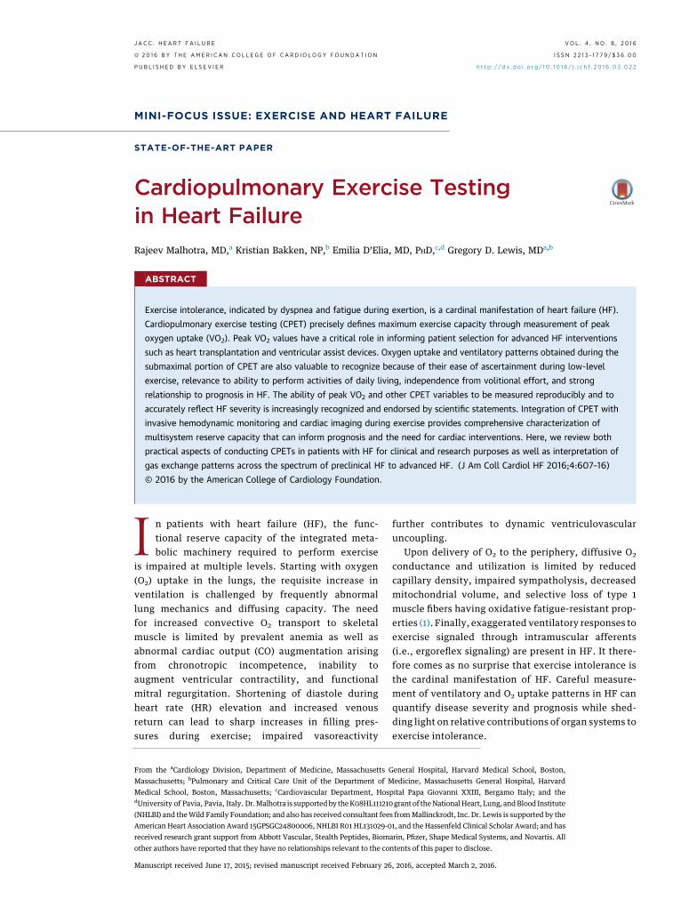
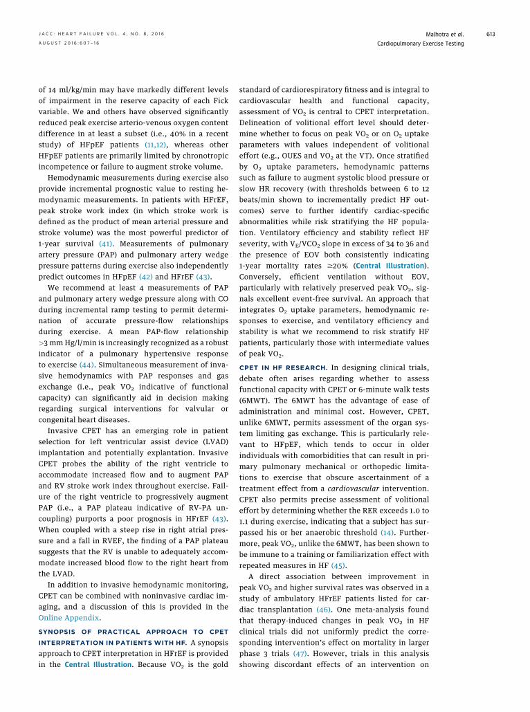
FIGURE 1 Values of VO2 and its Subcomponents of Heart Rate, Stroke Volume, and CavO2 at Rest and Peak Exercise in Normal,
HFrEF, and HFpEF Patients
The left panel illustrates representative values for VO2 at rest (blue bars) and change in VO2 during maximum incremental exercise (red bars)
for a normal 60-year-old man (5 feet 10 inches and normal body mass index of 21.5) derived using Wasserman’s percentage predicted equation
(14). Normal values are depicted in comparison to patients with HFrEF and HFpEF from a recent study that measured individual components of
peak VO2 (11). Right panels: Blue bars indicate resting values, and red bars illustrate the dynamic range in each Fick equation variable (HR, SV,
and CavO2) in response to maximum incremental exercise. CavO2, (CaO2 – CvO2) difference; HFpEF ¼ heart failure and preserved ejection
fraction; HFrEF ¼ heart failure and reduced ejection fraction; HR ¼ heart rate; SV ¼ stroke volume; VO2 ¼ oxygen uptake.
J A C C : H E A R T F A I L U R E V O L . 4 , N O . 8 , 2 0 1 6 Malhotra et al.A U G U S T 2 0 1 6 : 6 0 7 – 1 6 Cardiopulmonary Exercise Testing
609
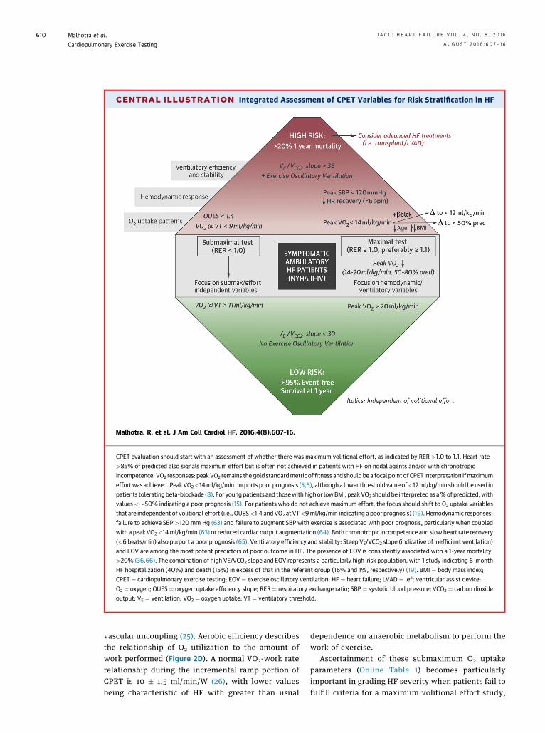
ascertainment during low-level exercise, relevance toability to perform activities of daily living, indepen-dence from volitional exercise effort, and close rela-tionship to prognosis in HF. We recently reported thatO2 uptake kinetics, as measured by mean responsetime (MRT) (Online Table 1, Figure 2A), were onlymodestly related to peak VO2 and more accuratelyreflected the ability to augment CO during low-levelexercise, indicating its complementary role to peakVO2 in signaling different aspects of cardiac reservecapacity (20). An MRT >60 s was related to reducedexercise right ventricular [RV] ejection fraction(RVEF) and increased transpulmonary gradient-COslope, which supports the notion that MRT reflectsRV-pulmonary vascular function during exercise (20).O2 uptake efficiency slope (OUES) (Figure 2F), which isthe relationship between VO2 and log VE throughoutexercise, is highly reproducible; differs by <2% if
derived from 75%, 90%, or 100% of exercise duration;and outperformed peak VO2 in a multivariate analysisof predictors of outcome in 243 HFrEF patients,conferring an w2-fold increase in mortality atvalues <1.47 l/min (21).
VO2 at the ventilatory threshold (VT) is anothermeasurement of O2 uptake that provides valuableinformation at submaximal exercise (Figure 2B). Gittand colleagues demonstrated that a VT <11 ml/kg/minwas associated with a 5.3-fold increased odds of deathat 6 months in 223 patients with HFrEF (22). A plateauin the VO2/HR (oxygen pulse) increment (23) indicatesfailure to augment the stroke volume-CavO2 (CaO2 –
CvO2) product throughout exercise (Figure 2C).Assuming a linear increment in CavO2 throughoutexercise, this pattern suggests dynamic cardiacdysfunction and has been observed with induciblemyocardial ischemia (24) as well as RV-pulmonary
CENTRAL ILLUSTRATION Integrated Assessment of CPET Variables for Risk Stratification in HF
Malhotra, R. et al. J Am Coll Cardiol HF. 2016;4(8):607–16.
CPET evaluation should start with an assessment of whether there was maximum volitional effort, as indicated by RER >1.0 to 1.1. Heart rate
>85% of predicted also signals maximum effort but is often not achieved in patients with HF on nodal agents and/or with chronotropic
incompetence. VO2 responses: peakVO2 remains the gold standardmetric offitness and should be a focal point of CPET interpretation ifmaximum
effortwas achieved. Peak VO2<14ml/kg/min purports poor prognosis (5,6), although a lower threshold value of<12ml/kg/min should be used in
patients tolerating beta-blockade (8). For young patients and thosewith highor lowBMI, peakVO2 should be interpreted as a%ofpredicted, with
values<w50% indicating a poor prognosis (15). For patients who do not achieve maximum effort, the focus should shift to O2 uptake variables
that are independent of volitional effort (i.e., OUES<1.4 and VO2 at VT<9ml/kg/min indicating a poor prognosis) (19). Hemodynamic responses:
failure to achieve SBP >120 mm Hg (63) and failure to augment SBP with exercise is associated with poor prognosis, particularly when coupled
with a peak VO2<14ml/kg/min (63) or reduced cardiac output augmentation (64). Both chronotropic incompetence and slow heart rate recovery
(<6 beats/min) also purport a poor prognosis (65). Ventilatory efficiency and stability: Steep VE/VCO2 slope (indicative of inefficient ventilation)
and EOV are among the most potent predictors of poor outcome in HF. The presence of EOV is consistently associated with a 1-year mortality
>20% (36,66). The combination of high VE/VCO2 slope and EOV represents a particularly high-risk population, with 1 study indicating 6-month
HF hospitalization (40%) and death (15%) in excess of that in the referent group (16% and 1%, respectively) (19). BMI ¼ body mass index;
CPET ¼ cardiopulmonary exercise testing; EOV ¼ exercise oscillatory ventilation; HF ¼ heart failure; LVAD ¼ left ventricular assist device;
O2 ¼ oxygen; OUES ¼ oxygen uptake efficiency slope; RER ¼ respiratory exchange ratio; SBP ¼ systolic blood pressure; VCO2 ¼ carbon dioxide
output; VE ¼ ventilation; VO2 ¼ oxygen uptake; VT ¼ ventilatory threshold.
Malhotra et al. J A C C : H E A R T F A I L U R E V O L . 4 , N O . 8 , 2 0 1 6
Cardiopulmonary Exercise Testing A U G U S T 2 0 1 6 : 6 0 7 – 1 6
610
vascular uncoupling (25). Aerobic efficiency describesthe relationship of O2 utilization to the amount ofwork performed (Figure 2D). A normal VO2-work raterelationship during the incremental ramp portion ofCPET is 10 � 1.5 ml/min/W (26), with lower valuesbeing characteristic of HF with greater than usual
dependence on anaerobic metabolism to perform thework of exercise.
Ascertainment of these submaximum O2 uptakeparameters (Online Table 1) becomes particularlyimportant in grading HF severity when patients fail tofulfill criteria for a maximum volitional effort study,
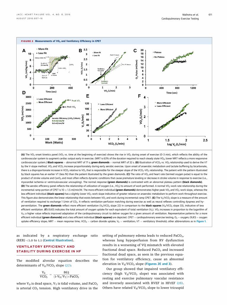
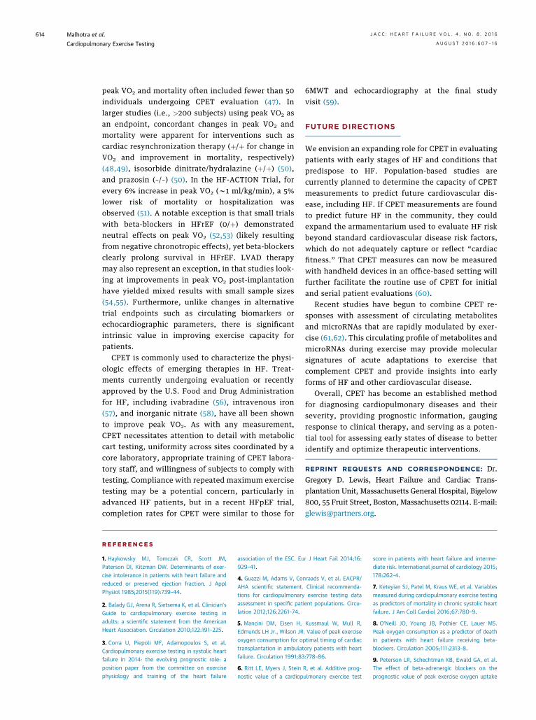
FIGURE 2 Measurements of VO2 and Ventilatory Efficiency in CPET
(A) The VO2 onset kinetics panel (VO2 vs. time at the beginning of exercise) shows the rise in VO2 during onset of exercise (0-3 min), which reflects the ability of the
cardiovascular system to augment cardiac output early in exercise. (MRT is 63% of the duration required to reach steady state VO2; lower MRT reflects a more responsive
cardiovascular system.) Black squares ¼ abnormal MRT of 71 s; green diamonds ¼ normal MRT of 32 s. (B) Illustration of VCO2 vs. VO2 relationship used to derive the VT
by the V-slope method. VO2 and VCO2 increase proportionately during early aerobic exercise. Upon onset of anaerobic metabolism and lactate buffering by bicarbonate,
there is a disproportionate increase in VCO2 relative to VO2 that is responsible for the steeper slope of the VCO2-VO2 relationship. The patient with the pattern illustrated
by black squares has an earlier VT (less fit) than the patient illustrated by the green diamonds. (C) The ratio of VO2 and heart rate (termed oxygen pulse) is equal to the
product of stroke volume and CavO2 and most often reflects dynamic conditions that cause premature leveling or decrease in stroke volume in response to exercise (i.e.,
myocardial ischemia or ventriculovascular uncoupling). The normal response (green diamonds) is contrasted with an abnormal plateau pattern (black diamonds).
(D) The aerobic efficiency panel reflects the relationship of utilization of oxygen (i.e., VO2) to amount of work performed. A normal VO2-work rate relationship during the
incremental ramp portion of CPET is 10 � 1.5 ml/min/W. The more efficient individual (green diamonds) demonstrates higher peak VO2 and VO2-work slope, whereas the
less efficient individual (black squares) has a slightly lower VO2-work slope indicative of greater reliance on anaerobic metabolism to perform work throughout exercise.
This figure also demonstrates the linear relationship that exists between VO2 and work during incremental ramp CPET. (E) The VE/VCO2 slope is a measure of the amount
of ventilation required to exchange 1 l/min of CO2. It reflects ventilation-perfusion matching during exercise as well as neural reflexes controlling dyspnea and hy-
perventilation. The green diamonds reflect more efficient ventilation (VE/VCO2 slope 22) in comparison to the black squares (VE/VCO2 slope 33), indicative of less
efficient ventilation. (F) OUES indicates the total amount of oxygen uptake for each equivalent of total ventilation (VE). VO2 increases in proportion to the logarithm of
VE; a higher value reflects improved adaptation of the cardiopulmonary circuit to deliver oxygen for a given amount of ventilation. Representative patterns for a more
efficient individual (green diamonds) and a less efficient individual (black squares) are depicted. CPET ¼ cardiopulmonary exercise testing; O2 ¼ oxygen; OUES ¼ oxygen
uptake efficiency slope; MRT ¼ mean response time; VCO2 ¼ carbon dioxide uptake; VE ¼ ventilation; VT ¼ ventilatory threshold; other abbreviations as in Figure 1.
J A C C : H E A R T F A I L U R E V O L . 4 , N O . 8 , 2 0 1 6 Malhotra et al.A U G U S T 2 0 1 6 : 6 0 7 – 1 6 Cardiopulmonary Exercise Testing
611
as indicated by a respiratory exchange ratio(RER) <1.0 to 1.1 (Central Illustration).
VENTILATORY EFFICIENCY AND
STABILITY DURING EXERCISE IN HF
The modified alveolar equation describes thedeterminants of VE/VCO2 slope (27):
VE
VCO2¼ 863
ð1-VD=VTÞ�PaCO2
where VD is dead space, VT is tidal volume, and PaCO2
is arterial CO2 tension. High ventilatory drive in the
setting of pulmonary edema leads to reduced PaCO2,whereas lung hypoperfusion from RV dysfunctionresults in a worsening of VQ mismatch with elevatedfractional dead space. Reduced PaCO2 and increasedfractional dead space, as seen in the previous equa-tion for ventilatory efficiency, cause an abnormalelevation in VE/VCO2 slope (Figures 2E and 3).
Our group showed that impaired ventilatory effi-ciency (high VE/VCO2 slope) was associated withresting and exercise pulmonary vascular resistanceand inversely associated with RVEF in HFrEF (28).Others have related VE/VCO2 slope to lower tricuspid
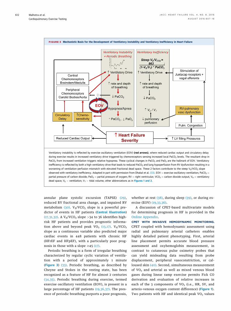
FIGURE 3 Mechanistic Basis for the Development of Ventilatory Instability and Ventilatory Inefficiency in Heart Failure
Ventilatory instability is reflected by exercise oscillatory ventilation (EOV) (red arrows), where reduced cardiac output and circulatory delay
during exercise results in increased ventilatory drive triggered by chemoreceptors sensing increased local PaCO2 levels. The resultant drop in
PaCO2 from increased ventilation triggers relative hypopnea. These cyclical changes in PaCO2 and PaO2 are the hallmark of EOV. Ventilatory
inefficiency is reflected by both a high ventilatory drive that leads to reduced PaCO2 and lung hypoperfusion from RV dysfunction resulting in a
worsening of ventilation-perfusion mismatch with elevated fractional dead space. These 2 factors contribute to the steep VE/VCO2 slope
observed with ventilatory inefficiency. Adapted in part with permission from Dhakal et al. (33). EOV ¼ exercise oscillatory ventilation; PaCO2 ¼partial pressure of carbon dioxide; PaO2 ¼ partial pressure of oxygen; RV ¼ right ventricular; VCO2 ¼ carbon dioxide output; VD ¼ ventilatory
dead space; VE ¼ ventilation; VT ¼ tidal volume; other abbreviations as in Figures 1 and 2.
Malhotra et al. J A C C : H E A R T F A I L U R E V O L . 4 , N O . 8 , 2 0 1 6
Cardiopulmonary Exercise Testing A U G U S T 2 0 1 6 : 6 0 7 – 1 6
612
annular plane systolic excursion (TAPSE) (29),reduced RV fractional area change, and impaired RVmetabolism (30). VE/VCO2 slope is a powerful pre-dictor of events in HF patients (Central Illustration)(17,31,32). A VE/VCO2 slope >34 to 36 identifies high-risk HF patients and provides prognostic informa-tion above and beyond peak VO2 (13,17). VE/VCO2
slope as a continuous variable also predicted majorcardiac events in 448 patients with chronic HF(HFrEF and HFpEF), with a particularly poor prog-nosis in those with a slope $45 (17).
Periodic breathing is a form of irregular breathingcharacterized by regular cyclic variation of ventila-tion with a period of approximately 1 minute(Figure 3) (33). Periodic breathing, as described byCheyne and Stokes in the resting state, has beenrecognized as a feature of HF for almost 2 centuries(34,35). Periodic breathing during exercise, termedexercise oscillatory ventilation (EOV), is present in alarge percentage of HF patients (19,36,37). The pres-ence of periodic breathing purports a poor prognosis,
whether at rest (38), during sleep (39), or during ex-ercise (EOV) (19,33,36).
A discussion of CPET-based multivariate modelsfor determining prognosis in HF is provided in theOnline Appendix.CPET WITH INVASIVE HEMODYNAMIC MONITORING.
CPET coupled with hemodynamic assessment usingradial and pulmonary arterial catheters enableshighly detailed patient phenotyping. First, arterialline placement permits accurate blood pressureassessment and oxyhemoglobin measurement, incontrast to cutaneous pulse oximetry probes thatcan yield misleading data resulting from probedisplacement, peripheral vasoconstriction, or cal-loused skin (40). Second, simultaneous measurementof VO2 and arterial as well as mixed venous bloodgases during linear ramp exercise permits Fick COderivation and evaluation of relative increases ineach of the 3 components of VO2 (i.e., HR, SV, andarterio-venous oxygen content difference) (Figure 1).Two patients with HF and identical peak VO2 values
J A C C : H E A R T F A I L U R E V O L . 4 , N O . 8 , 2 0 1 6 Malhotra et al.A U G U S T 2 0 1 6 : 6 0 7 – 1 6 Cardiopulmonary Exercise Testing
613
of 14 ml/kg/min may have markedly different levelsof impairment in the reserve capacity of each Fickvariable. We and others have observed significantlyreduced peak exercise arterio-venous oxygen contentdifference in at least a subset (i.e., 40% in a recentstudy) of HFpEF patients (11,12), whereas otherHFpEF patients are primarily limited by chronotropicincompetence or failure to augment stroke volume.
Hemodynamic measurements during exercise alsoprovide incremental prognostic value to resting he-modynamic measurements. In patients with HFrEF,peak stroke work index (in which stroke work isdefined as the product of mean arterial pressure andstroke volume) was the most powerful predictor of1-year survival (41). Measurements of pulmonaryartery pressure (PAP) and pulmonary artery wedgepressure patterns during exercise also independentlypredict outcomes in HFpEF (42) and HFrEF (43).
We recommend at least 4 measurements of PAPand pulmonary artery wedge pressure along with COduring incremental ramp testing to permit determi-nation of accurate pressure-flow relationshipsduring exercise. A mean PAP-flow relationship>3 mm Hg/l/min is increasingly recognized as a robustindicator of a pulmonary hypertensive responseto exercise (44). Simultaneous measurement of inva-sive hemodynamics with PAP responses and gasexchange (i.e., peak VO2 indicative of functionalcapacity) can significantly aid in decision makingregarding surgical interventions for valvular orcongenital heart diseases.
Invasive CPET has an emerging role in patientselection for left ventricular assist device (LVAD)implantation and potentially explantation. InvasiveCPET probes the ability of the right ventricle toaccommodate increased flow and to augment PAPand RV stroke work index throughout exercise. Fail-ure of the right ventricle to progressively augmentPAP (i.e., a PAP plateau indicative of RV-PA un-coupling) purports a poor prognosis in HFrEF (43).When coupled with a steep rise in right atrial pres-sure and a fall in RVEF, the finding of a PAP plateausuggests that the RV is unable to adequately accom-modate increased blood flow to the right heart fromthe LVAD.
In addition to invasive hemodynamic monitoring,CPET can be combined with noninvasive cardiac im-aging, and a discussion of this is provided in theOnline Appendix.
SYNOPSIS OF PRACTICAL APPROACH TO CPET
INTERPRETATION IN PATIENTS WITH HF. A synopsisapproach to CPET interpretation in HFrEF is providedin the Central Illustration. Because VO2 is the gold
standard of cardiorespiratory fitness and is integral tocardiovascular health and functional capacity,assessment of VO2 is central to CPET interpretation.Delineation of volitional effort level should deter-mine whether to focus on peak VO2 or on O2 uptakeparameters with values independent of volitionaleffort (e.g., OUES and VO2 at the VT). Once stratifiedby O2 uptake parameters, hemodynamic patternssuch as failure to augment systolic blood pressure orslow HR recovery (with thresholds between 6 to 12beats/min shown to incrementally predict HF out-comes) serve to further identify cardiac-specificabnormalities while risk stratifying the HF popula-tion. Ventilatory efficiency and stability reflect HFseverity, with VE/VCO2 slope in excess of 34 to 36 andthe presence of EOV both consistently indicating1-year mortality rates $20% (Central Illustration).Conversely, efficient ventilation without EOV,particularly with relatively preserved peak VO2, sig-nals excellent event-free survival. An approach thatintegrates O2 uptake parameters, hemodynamic re-sponses to exercise, and ventilatory efficiency andstability is what we recommend to risk stratify HFpatients, particularly those with intermediate valuesof peak VO2.
CPET IN HF RESEARCH. In designing clinical trials,debate often arises regarding whether to assessfunctional capacity with CPET or 6-minute walk tests(6MWT). The 6MWT has the advantage of ease ofadministration and minimal cost. However, CPET,unlike 6MWT, permits assessment of the organ sys-tem limiting gas exchange. This is particularly rele-vant to HFpEF, which tends to occur in olderindividuals with comorbidities that can result in pri-mary pulmonary mechanical or orthopedic limita-tions to exercise that obscure ascertainment of atreatment effect from a cardiovascular intervention.CPET also permits precise assessment of volitionaleffort by determining whether the RER exceeds 1.0 to1.1 during exercise, indicating that a subject has sur-passed his or her anaerobic threshold (14). Further-more, peak VO2, unlike the 6MWT, has been shown tobe immune to a training or familiarization effect withrepeated measures in HF (45).
A direct association between improvement inpeak VO2 and higher survival rates was observed in astudy of ambulatory HFrEF patients listed for car-diac transplantation (46). One meta-analysis foundthat therapy-induced changes in peak VO2 in HFclinical trials did not uniformly predict the corre-sponding intervention’s effect on mortality in largerphase 3 trials (47). However, trials in this analysisshowing discordant effects of an intervention on
Malhotra et al. J A C C : H E A R T F A I L U R E V O L . 4 , N O . 8 , 2 0 1 6
Cardiopulmonary Exercise Testing A U G U S T 2 0 1 6 : 6 0 7 – 1 6
614
peak VO2 and mortality often included fewer than 50individuals undergoing CPET evaluation (47). Inlarger studies (i.e., >200 subjects) using peak VO2 asan endpoint, concordant changes in peak VO2 andmortality were apparent for interventions such ascardiac resynchronization therapy (þ/þ for change inVO2 and improvement in mortality, respectively)(48,49), isosorbide dinitrate/hydralazine (þ/þ) (50),and prazosin (–/–) (50). In the HF-ACTION Trial, forevery 6% increase in peak VO2 (w1 ml/kg/min), a 5%lower risk of mortality or hospitalization wasobserved (51). A notable exception is that small trialswith beta-blockers in HFrEF (0/þ) demonstratedneutral effects on peak VO2 (52,53) (likely resultingfrom negative chronotropic effects), yet beta-blockersclearly prolong survival in HFrEF. LVAD therapymay also represent an exception, in that studies look-ing at improvements in peak VO2 post-implantationhave yielded mixed results with small sample sizes(54,55). Furthermore, unlike changes in alternativetrial endpoints such as circulating biomarkers orechocardiographic parameters, there is significantintrinsic value in improving exercise capacity forpatients.
CPET is commonly used to characterize the physi-ologic effects of emerging therapies in HF. Treat-ments currently undergoing evaluation or recentlyapproved by the U.S. Food and Drug Administrationfor HF, including ivabradine (56), intravenous iron(57), and inorganic nitrate (58), have all been shownto improve peak VO2. As with any measurement,CPET necessitates attention to detail with metaboliccart testing, uniformity across sites coordinated by acore laboratory, appropriate training of CPET labora-tory staff, and willingness of subjects to comply withtesting. Compliance with repeated maximum exercisetesting may be a potential concern, particularly inadvanced HF patients, but in a recent HFpEF trial,completion rates for CPET were similar to those for
6MWT and echocardiography at the final studyvisit (59).
FUTURE DIRECTIONS
We envision an expanding role for CPET in evaluatingpatients with early stages of HF and conditions thatpredispose to HF. Population-based studies arecurrently planned to determine the capacity of CPETmeasurements to predict future cardiovascular dis-ease, including HF. If CPET measurements are foundto predict future HF in the community, they couldexpand the armamentarium used to evaluate HF riskbeyond standard cardiovascular disease risk factors,which do not adequately capture or reflect “cardiacfitness.” That CPET measures can now be measuredwith handheld devices in an office-based setting willfurther facilitate the routine use of CPET for initialand serial patient evaluations (60).
Recent studies have begun to combine CPET re-sponses with assessment of circulating metabolitesand microRNAs that are rapidly modulated by exer-cise (61,62). This circulating profile of metabolites andmicroRNAs during exercise may provide molecularsignatures of acute adaptations to exercise thatcomplement CPET and provide insights into earlyforms of HF and other cardiovascular disease.
Overall, CPET has become an established methodfor diagnosing cardiopulmonary diseases and theirseverity, providing prognostic information, gaugingresponse to clinical therapy, and serving as a poten-tial tool for assessing early states of disease to betteridentify and optimize therapeutic interventions.
REPRINT REQUESTS AND CORRESPONDENCE: Dr.Gregory D. Lewis, Heart Failure and Cardiac Trans-plantation Unit, Massachusetts General Hospital, Bigelow800, 55 Fruit Street, Boston, Massachusetts 02114. E-mail:[email protected].
RE F E RENCE S
1. Haykowsky MJ, Tomczak CR, Scott JM,Paterson DI, Kitzman DW. Determinants of exer-cise intolerance in patients with heart failure andreduced or preserved ejection fraction. J ApplPhysiol 1985;2015(119):739–44.
2. Balady GJ, Arena R, Sietsema K, et al. Clinician’sGuide to cardiopulmonary exercise testing inadults: a scientific statement from the AmericanHeart Association. Circulation 2010;122:191–225.
3. Corra U, Piepoli MF, Adamopoulos S, et al.Cardiopulmonary exercise testing in systolic heartfailure in 2014: the evolving prognostic role: aposition paper from the committee on exercisephysiology and training of the heart failure
association of the ESC. Eur J Heart Fail 2014;16:929–41.
4. Guazzi M, Adams V, Conraads V, et al. EACPR/AHA scientific statement. Clinical recommenda-tions for cardiopulmonary exercise testing dataassessment in specific patient populations. Circu-lation 2012;126:2261–74.
5. Mancini DM, Eisen H, Kussmaul W, Mull R,Edmunds LH Jr., Wilson JR. Value of peak exerciseoxygen consumption for optimal timing of cardiactransplantation in ambulatory patients with heartfailure. Circulation 1991;83:778–86.
6. Ritt LE, Myers J, Stein R, et al. Additive prog-nostic value of a cardiopulmonary exercise test
score in patients with heart failure and interme-diate risk. International journal of cardiology 2015;178:262–4.
7. Keteyian SJ, Patel M, Kraus WE, et al. Variablesmeasured during cardiopulmonary exercise testingas predictors of mortality in chronic systolic heartfailure. J Am Coll Cardiol 2016;67:780–9.
8. O’Neill JO, Young JB, Pothier CE, Lauer MS.Peak oxygen consumption as a predictor of deathin patients with heart failure receiving beta-blockers. Circulation 2005;111:2313–8.
9. Peterson LR, Schechtman KB, Ewald GA, et al.The effect of beta-adrenergic blockers on theprognostic value of peak exercise oxygen uptake

J A C C : H E A R T F A I L U R E V O L . 4 , N O . 8 , 2 0 1 6 Malhotra et al.A U G U S T 2 0 1 6 : 6 0 7 – 1 6 Cardiopulmonary Exercise Testing
615
in patients with heart failure. J Heart LungTransplant 2003;22:70–7.
10. Chyu J, Fonarow GC, Tseng CH, Horwich TB.Four-variable risk model in men and women withheart failure. Circ Heart Fail 2014;7:88–95.
11. Dhakal BP, Malhotra R, Murphy RM, et al.Mechanisms of exercise intolerance in heart failurewith preserved ejection fraction: the role ofabnormal peripheral oxygen extraction. Circ HeartFail 2015;8:286–94.
12. Haykowsky MJ, Brubaker PH, John JM,Stewart KP, Morgan TM, Kitzman DW. De-terminants of exercise intolerance in elderly heartfailure patients with preserved ejection fraction.J Am Coll Cardiol 2011;58:265–74.
13. Wasserman K. Cardiopulmonary ExerciseTesting and Cardiovascular Health. Armonk, NY:Futura Pub. Co., 2002.
14. Hansen JE, Sue DY, Wasserman K. Predictedvalues for clinical exercise testing. Am Rev RespDis 1984;129:S49–55.
15. Arena R, Myers J, Abella J, et al. Determiningthe preferred percent-predicted equation for peakoxygen consumption in patients with heart failure.Circ Heart Fail 2009;2:113–20.
16. Osman AF, Mehra MR, Lavie CJ, Nunez E,Milani RV. The incremental prognostic importanceof body fat adjusted peak oxygen consumption inchronic heart failure. J Am Coll Cardiol 2000;36:2126–31.
17. Arena R, Myers J, Abella J, et al. Developmentof a ventilatory classification system in patientswith heart failure. Circulation 2007;115:2410–7.
18. Brunner-La Rocca HP, Weilenmann D,Schalcher C, et al. Prognostic significance of oxy-gen uptake kinetics during low level exercise inpatients with heart failure. Am J Cardiol 1999;84:741–4.
19. Sun XG, Hansen JE, Beshai JF, Wasserman K.Oscillatory breathing and exercise gas exchangeabnormalities prognosticate early mortality andmorbidity in heart failure. J Am Coll Cardiol 2010;55:1814–23.
20. Chatterjee NA, Murphy RM, Malhotra R, et al.Prolonged mean VO2 response time in systolicheart failure: an indicator of impaired rightventricular-pulmonary vascular function. CircHeart Fail 2013;6:499–507.
21. Hollenberg M, Tager IB. Oxygen uptake effi-ciency slope: an index of exercise performance andcardiopulmonary reserve requiring only submaxi-mal exercise. J Am Coll Cardiol 2000;36:194–201.
22. Gitt AK, Wasserman K, Kilkowski C, et al. Ex-ercise anaerobic threshold and ventilatory effi-ciency identify heart failure patients for high riskof early death. Circulation 2002;106:3079–84.
23. Lim JG, McAveney TJ, Fleg JL, et al. Oxygenpulse during exercise is related to resting systolicand diastolic left ventricular function in olderpersons with mild hypertension. Am Heart J 2005;150:941–6.
24. Chaudhry S, Arena R, Wasserman K, et al. Ex-ercise-induced myocardial ischemia detected bycardiopulmonary exercise testing. Am J Cardiol2009;103:615–9.
25. Guazzi M, Naeije R, Arena R, et al. Echocardi-ography of right ventriculoarterial coupling com-bined with cardiopulmonary exercise testing topredict outcome in heart failure. Chest 2015;148:226–34.
26. Tanabe Y, Nakagawa I, Ito E, Suzuki K. He-modynamic basis of the reduced oxygen uptakerelative to work rate during incremental exercisein patients with chronic heart failure. Int J Cardiol2002;83:57–62.
27. Johnson RL Jr. Gas exchange efficiency incongestive heart failure. Circulation 2000;101:2774–6.
28. Lewis GD, Shah RV, Pappagianopolas PP,Systrom DM, Semigran MJ. Determinants ofventilatory efficiency in heart failure: the role ofright ventricular performance and pulmonaryvascular tone. Circ Heart Fail 2008;1:227–33.
29. Methvin AB, Owens AT, Emmi AG, et al.Ventilatory inefficiency reflects right ventriculardysfunction in systolic heart failure. Chest 2011;139:617–25.
30. Ukkonen H, Burwash IG, Dafoe W, et al. Isventilatory efficiency (VE/VCO(2) slope) associ-ated with right ventricular oxidative metabolism inpatients with congestive heart failure? Eur J HeartFail 2008;10:1117–22.
31. Jaussaud J, Aimable L, Douard H. The time fora new strong functional parameter in heart failure:the VE/VCO2 slope. Int J Cardiol 2011;147:189–90.
32. Cornelis J, Taeymans J, Hens W, Beckers P,Vrints C, Vissers D. Prognostic respiratory param-eters in heart failure patients with and withoutexercise oscillatory ventilation—a systematic re-view and descriptive meta-analysis. Int J Cardiol2015;182:476–86.
33. Dhakal BP, Murphy RM, Lewis GD. Exerciseoscillatory ventilation in heart failure. TrendsCardiovasc Med 2012;22:185–91.
34. Cheyne J. A case of apoplexy in which thefleshy part of the heart was converted in fat.Dublin Hosp Rep 1818;2:216–9.
35. Stokes W. The Disease of the Heart and Aorta.Dublin: Hodges and Smith, 1854.
36. Corra U, Pistono M, Mezzani A, et al. Sleep andexertional periodic breathing in chronic heartfailure: prognostic importance and interdepen-dence. Circulation 2006;113:44–50.
37. Murphy RM, Shah RV, Malhotra R, et al.Exercise oscillatory ventilation in systolic heartfailure: an indicator of impaired hemodynamicresponse to exercise. Circulation 2011;124:1442–51.
38. Ponikowski P, Chua TP, Anker SD, et al.Peripheral chemoreceptor hypersensitivity: anominous sign in patients with chronic heart failure.Circulation 2001;104:544–9.
39. Nilsson KR, Duscha BD, Hranitzky PM,Kraus WE. Chronic heart failure and exerciseintolerance: the hemodynamic paradox. Curr Car-diol Rev 2008;4:92–100.
40. Ries AL, Fedullo PF, Clausen JL. Rapidchanges in arterial blood gas levels after exercisein pulmonary patients. Chest 1983;83:454–6.
41. Metra M, Faggiano P, D’Aloia A, et al. Use ofcardiopulmonary exercise testing with hemody-namic monitoring in the prognostic assessment ofambulatory patients with chronic heart failure.J Am Coll Cardiol 1999;33:943–50.
42. Dorfs S, Zeh W, Hochholzer W, et al. Pulmo-nary capillary wedge pressure during exercise andlong-term mortality in patients with suspectedheart failure with preserved ejection fraction. EurHeart J 2014;35:3103–12.
43. Lewis GD, Murphy RM, Shah RV, et al. Pul-monary vascular response patterns during exercisein left ventricular systolic dysfunction predictexercise capacity and outcomes. Circ Heart Fail2011;4:276–85.
44. Lewis GD, Bossone E, Naeije R, et al. Pulmo-nary vascular hemodynamic response to exercisein cardiopulmonary diseases. Circulation 2013;128:1470–9.
45. Bensimhon DR, Leifer ES, Ellis SJ, et al.Reproducibility of peak oxygen uptake and othercardiopulmonary exercise testing parameters inpatients with heart failure (from the Heart Failureand A Controlled Trial Investigating Outcomes ofexercise traiNing). Am J Cardiol 2008;102:712–7.
46. Stevenson LW, Steimle AE, Fonarow G, et al.Improvement in exercise capacity of candidatesawaiting heart transplantation. J Am Coll Cardiol1995;25:163–70.
47. Wessler BS, Kramer DG, Kelly JL, et al. Drugand device effects on peak oxygen consumption,6-minute walk distance, and natriuretic peptidesas predictors of therapeutic effects on mortality inpatients with heart failure and reduced ejectionfraction. Circ Heart Fail 2011;4:578–88.
48. Young JB, Abraham WT, Smith AL, et al.Combined cardiac resynchronization and implant-able cardioversion defibrillation in advancedchronic heart failure: the MIRACLE ICD Trial. JAMA2003;289:2685–94.
49. Higgins SL, Hummel JD, Niazi IK, et al. Cardiacresynchronization therapy for the treatment ofheart failure in patients with intraventricular con-duction delay and malignant ventricular tachyar-rhythmias. J Am Coll Cardiol 2003;42:1454–9.
50. Ziesche S, Cobb FR, Cohn JN, Johnson G,Tristani F. Hydralazine and isosorbide dinitratecombination improves exercise tolerance in heartfailure. Results from V-HeFT I and V-HeFT II. TheV-HeFT VA Cooperative Studies Group. Circulation1993;87:VI56–64.
51. Swank AM, Horton J, Fleg JL, et al. Modestincrease in peak VO2 is related to better clinicaloutcomes in chronic heart failure patients: resultsfrom heart failure and a controlled trial to inves-tigate outcomes of exercise training. Circ HeartFail 2012;5:579–85.
52. Krum H, Sackner-Bernstein JD, Goldsmith RL,et al. Double-blind, placebo-controlled study ofthe long-term efficacy of carvedilol in patientswith severe chronic heart failure. Circulation 1995;92:1499–506.
53. Gullestad L, Manhenke C, Aarsland T, et al.Effect of metoprolol CR/XL on exercise tolerancein chronic heart failure—a substudy to the MERIT-HF trial. Eur J Heart Fail 2001;3:463–8.
Malhotra et al. J A C C : H E A R T F A I L U R E V O L . 4 , N O . 8 , 2 0 1 6
Cardiopulmonary Exercise Testing A U G U S T 2 0 1 6 : 6 0 7 – 1 6
616
54. Dunlay SM, Allison TG, Pereira NL. Changes incardiopulmonary exercise testing parametersfollowing continuous flow left ventricular assistdevice implantation and heart transplantation.J Card Fail 2014;20:548–54.
55. Maybaum S, Mancini D, Xydas S, et al.Cardiac improvement during mechanical circula-tory support: a prospective multicenter study ofthe LVAD Working Group. Circulation 2007;115:2497–505.
56. Sarullo FM, Fazio G, Puccio D, et al. Impact of“off-label” use of ivabradine on exercise capacity,gas exchange, functional class, quality of life, andneurohormonal modulation in patients withischemic chronic heart failure. J Cardiovasc Phar-macol Ther 2010;15:349–55.
57. Okonko DO, Grzeslo A, Witkowski T, et al. Ef-fect of intravenous iron sucrose on exercisetolerance in anemic and nonanemic patients withsymptomatic chronic heart failure and iron defi-ciency FERRIC-HF: a randomized, controlled,observer-blinded trial. J Am Coll Cardiol 2008;51:103–12.
58. Zamani P, Rawat D, Shiva-Kumar P, et al.Effect of inorganic nitrate on exercise capacity inheart failure with preserved ejection fraction. Cir-culation 2015;131:371–80.
59. Redfield MM, Chen HH, Borlaug BA, et al. Ef-fect of phosphodiesterase-5 inhibition on exercisecapacity and clinical status in heart failure withpreserved ejection fraction: a randomized clinicaltrial. JAMA 2013;309:1268–77.
60. Woods PR, Bailey KR, Wood CM, Johnson BD.Submaximal exercise gas exchange is an importantprognostic tool to predict adverse outcomes inheart failure. Eur J Heart Fail 2011;13:303–10.
61. Lewis GD, Farrell L, Wood MJ, et al. Metabolicsignatures of exercise in human plasma. Sci TranslMed 2010;2:33ra37.
62. Baggish AL, Hale A, Weiner RB, et al. Dynamicregulation of circulating microRNA during acuteexhaustive exercise and sustained aerobic exercisetraining. J Physiol 2011;589:3983–94.
63. Osada N, Chaitman BR, Miller LW, et al. Car-diopulmonary exercise testing identifies low riskpatients with heart failure and severely impaired
exercise capacity considered for heart trans-plantation. J Am Coll Cardiol 1998;31:577–82.
64. Mancini D, LeJemtel T, Aaronson K. PeakVO(2): a simple yet enduring standard. Circulation2000;101:1080–2.
65. Myers J, Arena R, Dewey F, et al. A cardio-pulmonary exercise testing score for predicting out-comes in patientswith heart failure. AmHeart J 2008;156:1177–83.
66. Guazzi M, Raimondo R, Vicenzi M, et al.Exercise oscillatory ventilation may predict suddencardiac death in heart failure patients. J Am CollCardiol 2007;50:299–308.
KEY WORDS cardiopulmonary exercisetesting, exercise physiology, heart failure,oxygen uptake, ventilatory efficiency
APPENDIX For supplemental material,references, and a table, please see theonline version of this article.